original article anatomical descriptionof the proximal ... · femur head necrosis. original...
TRANSCRIPT
21
All the authors declare that there is no potential conflict of interest referring to this article.
1 - Hospital Regional do Gama (HRG) of the Federal District - Distrito Federal (DF), Brazil.2 - Hospital das Clínicas da Universidade Federal do Goiás (HC-UFG) – Goiânia, GO, Brazil.
Study conducted at the Department of Orthopedics and Traumatology of Hospital Regional do Gama. (SOT-HRG) - Gama- (DF).Mailing address: Rua Fortaleza N 355, Setor Alto da Glória, Goiânia, Goiás. – CEP 74 815- 710. Brazil. E-mail: [email protected]
anatomiCal deSCription of the proXimal thirdof the medial femoral CirCumfleX artery.
a CadaveriC Study
anderson Freitas1, helder noGueira aires1, saulo teixeira Pansiere1, dioGo de macedo souto1, mônica meireles costa2
Article received on 5/20/10 and approved on 6/10/10.
INTRODUCTION
The increase in high-energy trauma has been causing the inciden-ce of pelvic girdle fractures to grow.1 Such trauma-related condi-tions can be managed with orthopedic surgical treatment, and for their execution require the Kocher-Langenbeck (KL) approach.2,3
The KL approach allows optimum exposure for some types of acetabular fracture, especially fractures that affect posterior com-ponents of this region; however, it involves risks of neurovascular injuries,2 one of which is the lesion of the medial femoral circu-mflex artery.4,5 In the need for an approach to the quadratus fe-moris muscle mass, there is the possibility of injuring the proximal third of the MFCA, thus causing insufficient vascularization of the femoral head and consequently a picture of avascular necrosis.5-7
Due to the importance of the MFCA’s role in supplying blood to the femoral head and the poor literary description of its ana-tomy, this study is aimed at providing an anatomical description of the proximal third of the MFCA and at suggesting data to readers to achieve greater safety in the performance of Kocher--Langenbeck approaches, targeting the preservation of the vascular supply of the femoral head and neck.
MATERIAL AND METHODS
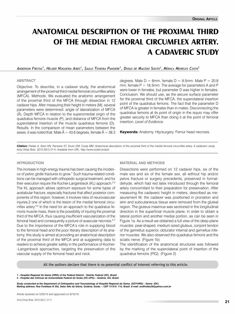
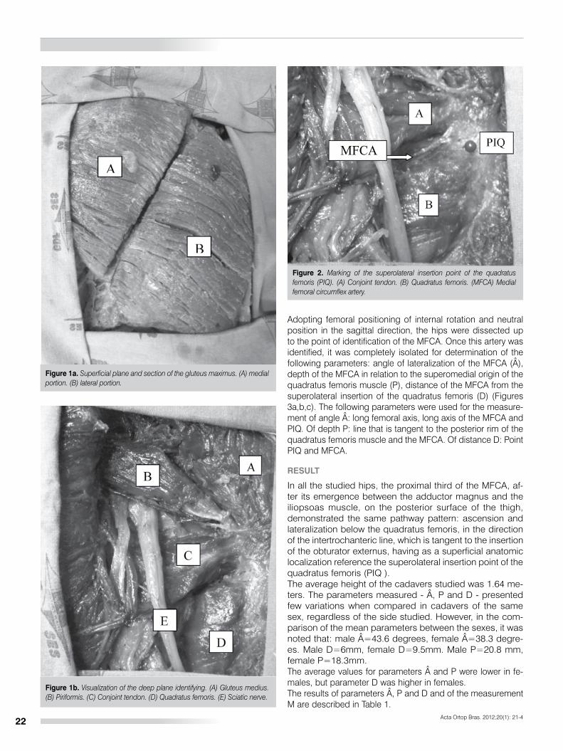
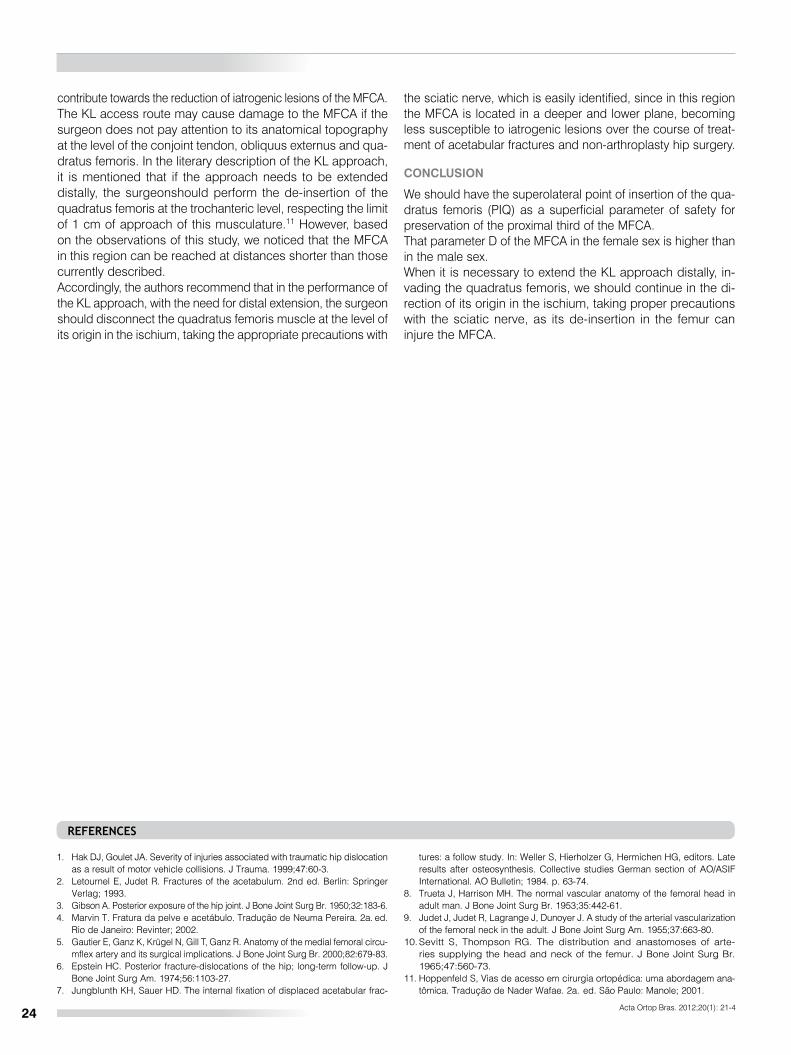
Dissections were performed on 12 cadaver hips, six of the male sex and six of the female sex, all without hip and/or pelvis fracture or surgery precedents, preserved in formal-dehyde, which had red latex introduced through the femoral artery concomitant to their preparation for preservation. After measuring the cadaveric height in meters, described as me-asurement M, the cadaver was positioned in pronation and skin and subcutaneous tissue were removed from the gluteal region. The gluteus maximus was sectioned in the longitudinal direction in the superficial muscle plane, in order to obtain a lateral portion and another medial portion, as can be seen inFigure 1a. As a result we obtained a full view of the deep plane muscles: pear-shaped, medium sized gluteus, conjoint tendon of the gemellus superior, obturator internal and gemellus infe-rior muscles. We also observed the quadratus femoris and the sciatic nerve. (Figure 1b)The identification of the anatomical structures was followed by the marking of the superolateral point of insertion of the quadratus femoris (PIQ). (Figure 2)
Citation: Freitas A, Aires HN, Pansiere ST, Souto DM, Costa MM. Anatomical description of the proximal third of the medial femoral circumflex artery. A cadaveric study. Acta Ortop Bras. 2012;20(1):21-4. Available from URL: http://www.scielo.br/aob.
Acta Ortop Bras. 2012;20(1): 21-4
ABSTRACT
Objective: To describe, in a cadaver study, the anatomical arrangement of the proximal third medial femoral circumflex artery (MFCA). Methods: We evaluated the anatomic arrangement of the proximal third of the MFCA through dissection in 12 cadaver hips. After measuring their height in meters (M), several parameters were determined: angle of lateralization of MFCA (Â), Depth MFCA in relation to the superomedial origin of the quadratus femoris muscle (P), and distance of MFCA from the superolateral insertion of the muscle quadratus femoris (D). Results. In the comparison of mean parameters between the sexes, it was noted that: Male  = 43.6 degrees, female  = 38.3
degrees. Male D = 6mm, female D = 9.5mm. Male P = 20.8 mm, female P = 18.3mm. The average for parameters A and P were lower in females, but parameter D was higher in females. Conclusion. We should use, as the secure surface parameter for the proximal third of the MFCA, the superolateral insertion point of the quadratus femoris. The fact that the parameter D of MFCA is greater in females than in males. Disconnecting the quadratus femoris at its point of origin in the isquio may offer greater security to MFCA than doing it at the point of femoral insertion. Level of Evidence.
Keywords: Anatomy. Hip/surgery. Femur head necrosis.
Original article
22
Adopting femoral positioning of internal rotation and neutral position in the sagittal direction, the hips were dissected up to the point of identification of the MFCA. Once this artery was identified, it was completely isolated for determination of the following parameters: angle of lateralization of the MFCA (Â), depth of the MFCA in relation to the superomedial origin of the quadratus femoris muscle (P), distance of the MFCA from the superolateral insertion of the quadratus femoris (D) (Figures 3a,b,c). The following parameters were used for the measure-ment of angle Â: long femoral axis, long axis of the MFCA and PIQ. Of depth P: line that is tangent to the posterior rim of the quadratus femoris muscle and the MFCA. Of distance D: Point PIQ and MFCA.
RESULT
In all the studied hips, the proximal third of the MFCA, af-ter its emergence between the adductor magnus and the iliopsoas muscle, on the posterior surface of the thigh, demonstrated the same pathway pattern: ascension and lateralization below the quadratus femoris, in the direction of the intertrochanteric line, which is tangent to the insertion of the obturator externus, having as a superficial anatomic localization reference the superolateral insertion point of the quadratus femoris (PIQ ).The average height of the cadavers studied was 1.64 me-ters. The parameters measured - Â, P and D - presented few variations when compared in cadavers of the same sex, regardless of the side studied. However, in the com-parison of the mean parameters between the sexes, it was noted that: male Â=43.6 degrees, female Â=38.3 degre-es. Male D=6mm, female D=9.5mm. Male P=20.8 mm, female P=18.3mm.The average values for parameters  and P were lower in fe-males, but parameter D was higher in females.The results of parameters Â, P and D and of the measurement M are described in Table 1.
Figure 1a. Superficial plane and section of the gluteus maximus. (A) medial portion. (B) lateral portion.
Figure 1b. Visualization of the deep plane identifying. (A) Gluteus medius. (B) Piriformis. (C) Conjoint tendon. (D) Quadratus femoris. (E) Sciatic nerve.
Figure 2. Marking of the superolateral insertion point of the quadratus femoris (PIQ). (A) Conjoint tendon. (B) Quadratus femoris. (MFCA) Medial femoral circumflex artery.
Acta Ortop Bras. 2012;20(1): 21-4
MFCA

23
Figure 3a. Method used to measure angle Â. PIQ, Superolateral insertion point of the quadratus femoris, B) Conjoint tendon, C) Quadratus femoris, D) Long axis of the MFCA, E) Long axis of the femur.
Figure 3b. Method used to measure depth P. (A) Conjoint tendon. (B) Quadratus femoris, C) Line that runs tangent to the posterior rim of the quadratus femoris. (MFCA) Medial femoral circumflex artery.
Figure 3c. Method used to measure distance D. (A) Conjoint tendon. (B) Quadratus femoris. (MFCA) Medical femoral circumflex artery. (PIQ) superolateral insertion point of the quadratus femoris.
DISCUSSION
Trueta et al, through a tissue slide, after angiographic prepara-tion, of a cadaver bone, described the intraosseous vascular mesh of the femoral head, thus justifying the importance of the lateral epiphyseal retinacular branches originating from the MFCA for vascularization of the femoral head.8
Judet at al9, in a study that closely resembles the one previously described, ratifies the importance of the MFCA for the supply of blood to the femoral head, and indicates the possibility of injury to the MFCA in extensive distal capsulotomies, thus giving
Table 1. Values of the parameters: M – Height in meters of the cadaver. Â- Angle of lateralization of the MFCA; P- Depth of the MFCA in relation to the superomedial origin of the quadratus femoris; D- Distance of the MFCA from the superolateral insertion of the quadratus femoris.
Pelvis Sex Height(Meters)
Parameter A(Degrees)
Parameter D(Mm)
Parameter P(Mm)
Side Side Side
Right Left Right Left Right Left
1 Fem 1.64 39 40 13 10 18 19
2 Fem 1.56 38 37 9 10 16 18
3 Fem 1.60 37 39 7 8 19 20
4 Male 1.70 45 45 9 9 20 21
5 Male 1.68 40 5 5 21 22
6 Male 1.66 44 44 4 4 20 21
rise to concerns regarding iatrogenic lesionsin femoral head vascularization.Sevitt et al.10, using the angiographic and histological techni-que, and associating 11 different types of lesion, which can affect bones, or the superior retinaculum, or the inferior retina-culum, or ligaments, confirms the vascular importance of the medial circumflex artery, yet does not mention the possibility of injury to the MFCA in the advent of the treatment.Gautier et al.5, in an excellent cadaveric study, describes the anatomy of the medial circumflex artery, determining anas-tomoses, the protection mechanism of the MFCA, in cases of hip dislocation, by the obturator externus, and proposes a modification in the surgical approach of the KL type, at the level of the external hip rotators. This author determines the preservation of the obturator externus tendon and suggests a tenotomy safety zone of the conjoint tendon, located 1.5 cm from the intertrochanteric line, a measurement that is confirme-dly safe as shown by the data obtained in this trial. However, in our study we observed that anatomically the obturator exter-nus is located in a deep plane, below the superolateral inser-tion of the quadratus femoris, and that it determines a repair point at a more superficial level in the KL approach, which may
Acta Ortop Bras. 2012;20(1): 21-4
MFCA
MFCA
24
contribute towards the reduction of iatrogenic lesions of the MFCA.The KL access route may cause damage to the MFCA if the surgeon does not pay attention to its anatomical topography at the level of the conjoint tendon, obliquus externus and qua-dratus femoris. In the literary description of the KL approach, it is mentioned that if the approach needs to be extended distally, the surgeonshould perform the de-insertion of the quadratus femoris at the trochanteric level, respecting the limit of 1 cm of approach of this musculature.11 However, based on the observations of this study, we noticed that the MFCA in this region can be reached at distances shorter than those currently described. Accordingly, the authors recommend that in the performance of the KL approach, with the need for distal extension, the surgeon should disconnect the quadratus femoris muscle at the level of its origin in the ischium, taking the appropriate precautions with
the sciatic nerve, which is easily identified, since in this region the MFCA is located in a deeper and lower plane, becoming less susceptible to iatrogenic lesions over the course of treat-ment of acetabular fractures and non-arthroplasty hip surgery.
CONCLUSION
We should have the superolateral point of insertion of the qua-dratus femoris (PIQ) as a superficial parameter of safety for preservation of the proximal third of the MFCA.That parameter D of the MFCA in the female sex is higher than in the male sex. When it is necessary to extend the KL approach distally, in-vading the quadratus femoris, we should continue in the di-rection of its origin in the ischium, taking proper precautions with the sciatic nerve, as its de-insertion in the femur can injure the MFCA.
REFERENCES
Acta Ortop Bras. 2012;20(1): 21-4
1. Hak DJ, Goulet JA. Severity of injuries associated with traumatic hip dislocation as a result of motor vehicle collisions. J Trauma. 1999;47:60-3.
2. Letournel E, Judet R. Fractures of the acetabulum. 2nd ed. Berlin: Springer Verlag; 1993.
3. Gibson A. Posterior exposure of the hip joint. J Bone Joint Surg Br. 1950;32:183-6. 4. Marvin T. Fratura da pelve e acetábulo. Tradução de Neuma Pereira. 2a. ed.
Rio de Janeiro: Revinter; 2002. 5. Gautier E, Ganz K, Krügel N, Gill T, Ganz R. Anatomy of the medial femoral circu-
mflex artery and its surgical implications. J Bone Joint Surg Br. 2000;82:679-83.6. Epstein HC. Posterior fracture-dislocations of the hip; long-term follow-up. J
Bone Joint Surg Am. 1974;56:1103-27. 7. Jungblunth KH, Sauer HD. The internal fixation of displaced acetabular frac-
tures: a follow study. In: Weller S, Hierholzer G, Hermichen HG, editors. Late results after osteosynthesis. Collective studies German section of AO/ASIF International. AO Bulletin; 1984. p. 63-74.
8. Trueta J, Harrison MH. The normal vascular anatomy of the femoral head in adult man. J Bone Joint Surg Br. 1953;35:442-61.
9. Judet J, Judet R, Lagrange J, Dunoyer J. A study of the arterial vascularization of the femoral neck in the adult. J Bone Joint Surg Am. 1955;37:663-80.
10. Sevitt S, Thompson RG. The distribution and anastomoses of arte-ries supplying the head and neck of the femur. J Bone Joint Surg Br. 1965;47:560-73.
11. Hoppenfeld S, Vias de acesso em cirurgia ortopédica: uma abordagem ana-tômica. Tradução de Nader Wafae. 2a. ed. São Paulo: Manole; 2001.