original article biological characteristics of …original article biological characteristics of...
TRANSCRIPT
Int J Clin Exp Med 2015;8(9):14718-14726www.ijcem.com /ISSN:1940-5901/IJCEM0007292
Original ArticleBiological characteristics of tracheal smooth muscle cells regulated by NK-1R in asthmatic rat with airway remodeling
Bing Wei1, Yali Liu1, Xiaozhe Yue1, Yinping Li1, Yunxiao Shang2
1Department of Pediatrics, General Hospital of Shenyang Military Area Command, Shenyang 110015, China; 2Department of Paediatrics, Shengjing Hospital of China Medical University, Shengyang 110004, China
Received February 24, 2015; Accepted July 28, 2015; Epub September 15, 2015; Published September 30, 2015
Abstract: This study aims to investigate the biological characteristic changes of infant rat tracheal smooth muscle cells in asthma airway remodeling and the impact of NK-1R on the mechanism. Ovalbumin (OVA) was used to excited juvenile SD rats by 8 w. Immunofluorescence, MTT assay, transwell chambers, real time quantitative PCR, Western blot and other methods were used to observe the proliferation, migration, synthesis and secretion changes of infant airway remodeling in rat tracheal smooth muscle cell and the Neurokinin 1 receptor (NK-1R) expression. 1. NK-1R mRNA, protein expression of airway smooth muscle cell (ASMC) of each asthma group were higher than that of the control group, especially the asthma 8 w group had highest expression (P<0.01). 2. The average A value of 8 w asthma group measured by MTT method were significantly higher than that of the control group (P<0.05), WIN62577 10-8 mol/L group had the strongest inhibition of ASMC proliferation (P<0.01). 3. The number of cell mi-gration in the asthma group significantly increased than that in the control group. The number of migrating cells in the NK-1R antagonist group significantly reduced compared with the asthma 8 w group (P<0.05). 4. The average gray value of type III collagen in each asthma group were higher than that of the control group, and the asthma 8 w group had the highest (P<0.01). After NK-1R blocking, the average gray value of type III collagen was significantly lower (P<0.05). ASMC proliferation, migration, synthesis and secretion function increased in the airway remodeling group, and NK-1R played an important role.
Keywords: Airway remodeling, smooth muscle cells, NK-1R, proliferation, migration
Introduction
Bronchus asthma is a chronic respiratory dis-ease seriously impacted human health, its prevalence and mortality are on the rise in recent years. Airway remodeling is the main reason for inducing airway hyper responsive-ness and chronic asthma. Therefore, the mech-anisms and treatment of airway remodeling have become the focus of the current domestic and foreign research [1-3]. Studies have shown that Substance P (SP) can promote the prolif-eration and migration of fibroblasts, endotheli-al cells, epidermal stem cells [4-6]. SP plays a role through Neurokinin 1 receptor (NK-1R). Our previous studies also showed that NK-1R pro-moted airway remodeling [7], and changes in airway smooth muscle cells was the basis of airway remodeling [8]. But the role of NK-1R in
the proliferation, migration synthesis and secre-tion of airway smooth muscle cells (ASMC) is still rarely reported. In this study, SD rat trache-al smooth muscle cells were primary cultured and to given NK-1R antagonist intervention. Immunofluorescence, MTT assay, transwell chambers, real time quantitative PCR and Western blot were used to explore the impact of NK-1R on the biological behavior changes such as proliferation, migration, synthesis and secre-tion of asthma airway remodeling in rat tracheal smooth muscle cells.
Materials and methods
Experimental animals
Juvenile female SD rats with SPF level, weighed 60-80 g were purchased from China Medical University Experimental Animal Center. This
Effect of NK-1R on tracheal smooth muscle cells in airway remodeling induced by asthma
14719 Int J Clin Exp Med 2015;8(9):14718-14726
study was carried out in strict accordance with the recommendations in the Guide for the Care and Use of Laboratory Animals of the National Institutes of Health. The animal use protocol has been reviewed and approved by the Institutional Animal Care and Use Committee (IACUC) of Shenyang Military Area Command.
Preparation of airway remodeling model
1 ml antigen sensitization was used to perform intraperitoneal injection on the first and eighth days respectively (including 100 mg egg pro-tein, 100 mg aluminum hydroxide, 5×10⁹ unit inactivated pertussis). The control group was injected with an equal volume of saline. From the 15th days, the rats were placed in a sealed container (20 cm × 20 cm × 20 cm) with 1% ovalbumin (OVA) ultrasonic inhalation, 30 min for each time, and 3 times a week, respectively excited by 2 w, 4 w, 6 w and 8 w. Normal control group was treated with saline instead of ovalbu-min, and excited by 2 w.
ASMC primary culture
After routine disinfection and anesthesia, the rats were sacrificed by collecting blood from ventriculus sinister. Antiseptically isolated rat trachea, carefully peeled off the outer mem-brane, removed the inner membrane. Cut the tracheal smooth muscle, the tissues were cut into pieces, added 0.1% trypsin, shook at 37°C
water and bathed for 10 min, 1000 rpm/min centrifuged for 5 min, the supernatant were discarded. Added 0.1% IV collagenase, shook at 37°C and water bathed for 30 min, 1000 rpm/min centrifuged for 5 min, the supernatant was discarded. Then added 10% fetal bovine serum (FBS) in DMEM to terminate the reac-tion, gently pipetted, filtered cells by 100 mesh sieve, inoculated to 50 ml culture flask, 37°C incubated and cultured, purified ASMC based on the characteristics of rapid adherent fibro-blasts. After the cells covering the bottom, cell passage was performed by 1:2, and collected the 3rd generation cell for experiment using.
Immunofluorescence staining
Cells in the logarithmic growth phase were digested to prepare single cell suspension, after appropriate adjusting the density, seeded on coverslips on six-well plates. Cultured in the incubator at 37°C with CO2 for overnight, fixed with 4% paraformaldehyde when the cells adhered and stretched. 3 M H2O2 were used to treat cells under at room temperature for 10 min, cells were treated by 3 g/L Triton100 at 37°C for 10 min, and blocked by 3% goat serum at room temperature for 15 min. Rabbit anti-rat NK-1R antibody (1:100) (Santa Cruz, USA) was added. Set the cells in a humid chamber at 4°C for overnight. FITC-labeled goat anti-rabbit IgG (Boster Biological Engineering Co., Ltd., Wuhan, China) treated at 37°C for 30 min. DAPI 5 μg/ml was used at room temperature for 5 min. Mounted by 50% glycerol. Washed with PBS between steps. PBS instead of the first anti-body was used for negative control in the experiment.
MTT assay
ASMC proliferation was measured. The 3th-5th generations ASMC of the asthma 2 w, 4 w, 6 w, 8 w group and the 2 w control group were col-lected. When ASMC grew to 80% confluence,
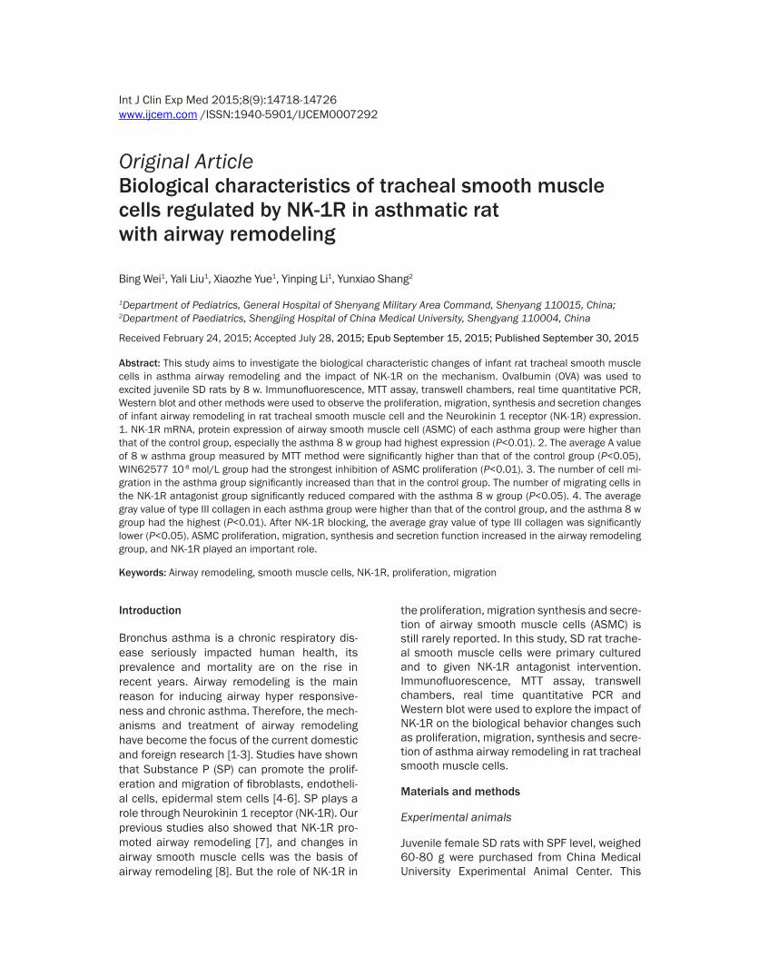
Figure 1. NK-1R immunofluorescence staining in ASMC. A. Control group; B. Asthma 2 w group; C. Asthma 4 w group; D. Asthma 6 w group; E. Asthma 8 w group.
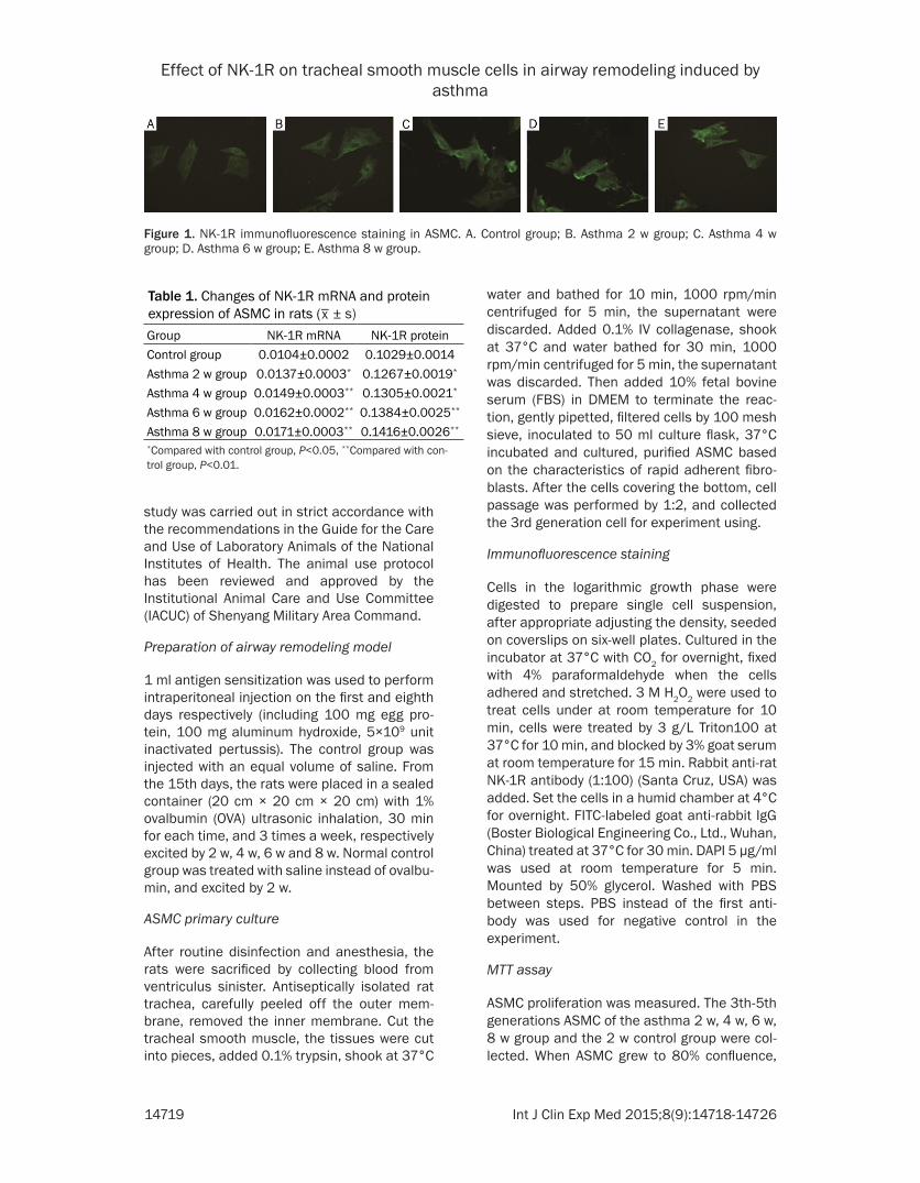
Table 1. Changes of NK-1R mRNA and protein expression of ASMC in rats (
_x ± s)
Group NK-1R mRNA NK-1R proteinControl group 0.0104±0.0002 0.1029±0.0014Asthma 2 w group 0.0137±0.0003* 0.1267±0.0019*
Asthma 4 w group 0.0149±0.0003** 0.1305±0.0021*
Asthma 6 w group 0.0162±0.0002** 0.1384±0.0025**
Asthma 8 w group 0.0171±0.0003** 0.1416±0.0026**
*Compared with control group, P<0.05, **Compared with con-trol group, P<0.01.
Effect of NK-1R on tracheal smooth muscle cells in airway remodeling induced by asthma
14720 Int J Clin Exp Med 2015;8(9):14718-14726
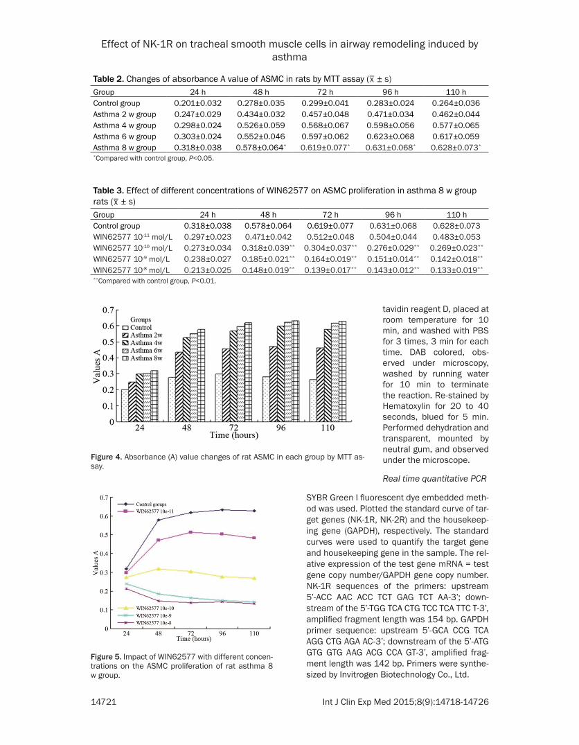
0.25% trypsin was used for digestion, seeded in 96-well plates according to 1×10⁴ cells/well, cultured for 24 h, 48 h, 72 h, 96 h, 110 h by DMEM containing 10% FBS. The asthma 8 w group were randomly divided into the following five groups: 1) control group: without any inter-vention agents; 2) WIN62577 10-11 mol/L group; 3) WIN62577 10-10 mol/L group; 4) WIN62577 10-9 mol/L group; 5) WIN62577 10-8 mol/L group. Five repeated wells were set for each group. At the end of ASMC culture, 20 μl MTT (5 mg/ml) was added to each well, cul-tured for 4 h and discarded culture medium, added 150 μl DMSO. Absorbance (A) value of each well was measured at 490 nm by ELISA.
ASMC migration testing
ASMC migration was detected by transwell chamber. The 3th-5th generations ASMC of the asthma 2 w, 4 w, 6 w, 8 w group and the 2 w control group were collected. When ASMC grew to 80% confluence, 0.25% trypsin was used for digestion, the cells were counted and the den-sity was adjusted to 1×10⁵/ml with 10% FBS-DMEM, 100 μl cell suspension was added to the each group of the upper chamber, the low- er chamber was filled with 600 μl 10% FBS-
DMEM, placed the small chamber in 37°C incu-bator, removed 24 h later, carefully removed the membrane, gently wiped up the non-mem-brane worn cells on the upper side of micropo-rous membrane. ASMC under the membrane were fixed by 4% paraformaldehyde for 20 min, stained by hematoxylin for 10 min, performed transplant by xylene for 1 min, mounted with neutral gum, counted five vision cells under an inverted microscope (400×) and averaged, repeated for three times. After migration exper-iment of the NK-1 receptor antagonist group, cells on the upper chamber of the asthma 8 w group were pretreated by NK-1 receptor antagonist WIN62577 (final concentration 10-8 mol/L) for 30 min.
Immunochemical method
Slice were dewaxed by benzene, then removed benzene by gradient alcohol, washed by dis-tilled water for 5 min, washed with PBS for 3 times, 3 min for each time. Sections were immersed in containers with antigen retrieval solution (sodium citrate buffer PH 6.0, 0.01 M), heated in microwave by high fire for 10 min, opened to place for 5 min, reheated for 5 min, and let cool at room temperature. Dropped 3% hydrogen peroxide reagent A and placed in wet box at room temperature for 10 min, washed with PBS for 3 times, 3 min for each time. Removed the PBS, dropped normal goat serum blocking solution reagent B and incubated at room temperature for 10 min. Added primary antibody (rabbit anti-rat collagen III) with ratio of 1:100 dilution, placed at 4°C for overnight. Dropped the secondary antibody reagent C, placed at room temperature for 10 min, washed with PBS for 3 times, 3 min for each time. Then dropped horseradish peroxidase labeled strep-
Figure 2. NK-1R mRNA and protein expression changes in ASMC of rat groups. A. NK-1R mRNA expression; B. NK-1R protein expression.
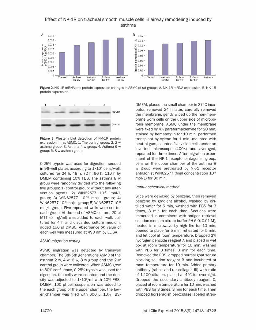
Figure 3. Western blot detection of NK-1R protein expression in rat ASMC. 1. The control group; 2. 2 w asthma group; 3. Asthma 4 w group; 4. Asthma 6 w group; 5. 8 w asthma group.
Effect of NK-1R on tracheal smooth muscle cells in airway remodeling induced by asthma
14721 Int J Clin Exp Med 2015;8(9):14718-14726
SYBR Green I fluorescent dye embedded meth-od was used. Plotted the standard curve of tar-get genes (NK-1R, NK-2R) and the housekeep-ing gene (GAPDH), respectively. The standard curves were used to quantify the target gene and housekeeping gene in the sample. The rel-ative expression of the test gene mRNA = test gene copy number/GAPDH gene copy number. NK-1R sequences of the primers: upstream 5’-ACC AAC ACC TCT GAG TCT AA-3’; down-stream of the 5’-TGG TCA CTG TCC TCA TTC T-3’, amplified fragment length was 154 bp. GAPDH primer sequence: upstream 5’-GCA CCG TCA AGG CTG AGA AC-3’; downstream of the 5’-ATG GTG GTG AAG ACG CCA GT-3’, amplified frag-ment length was 142 bp. Primers were synthe-sized by Invitrogen Biotechnology Co., Ltd.
Table 2. Changes of absorbance A value of ASMC in rats by MTT assay (_x ± s)
Group 24 h 48 h 72 h 96 h 110 hControl group 0.201±0.032 0.278±0.035 0.299±0.041 0.283±0.024 0.264±0.036Asthma 2 w group 0.247±0.029 0.434±0.032 0.457±0.048 0.471±0.034 0.462±0.044Asthma 4 w group 0.298±0.024 0.526±0.059 0.568±0.067 0.598±0.056 0.577±0.065Asthma 6 w group 0.303±0.024 0.552±0.046 0.597±0.062 0.623±0.068 0.617±0.059Asthma 8 w group 0.318±0.038 0.578±0.064* 0.619±0.077* 0.631±0.068* 0.628±0.073*
*Compared with control group, P<0.05.
Table 3. Effect of different concentrations of WIN62577 on ASMC proliferation in asthma 8 w group rats (
_x ± s)
Group 24 h 48 h 72 h 96 h 110 hControl group 0.318±0.038 0.578±0.064 0.619±0.077 0.631±0.068 0.628±0.073WIN62577 10-11 mol/L 0.297±0.023 0.471±0.042 0.512±0.048 0.504±0.044 0.483±0.053WIN62577 10-10 mol/L 0.273±0.034 0.318±0.039** 0.304±0.037** 0.276±0.029** 0.269±0.023**
WIN62577 10-9 mol/L 0.238±0.027 0.185±0.021** 0.164±0.019** 0.151±0.014** 0.142±0.018**
WIN62577 10-8 mol/L 0.213±0.025 0.148±0.019** 0.139±0.017** 0.143±0.012** 0.133±0.019**
**Compared with control group, P<0.01.
Figure 4. Absorbance (A) value changes of rat ASMC in each group by MTT as-say.
tavidin reagent D, placed at room temperature for 10 min, and washed with PBS for 3 times, 3 min for each time. DAB colored, obs- erved under microscopy, washed by running water for 10 min to terminate the reaction. Re-stained by Hematoxylin for 20 to 40 seconds, blued for 5 min. Performed dehydration and transparent, mounted by neutral gum, and observed under the microscope.
Real time quantitative PCR
Figure 5. Impact of WIN62577 with different concen-trations on the ASMC proliferation of rat asthma 8 w group.
Effect of NK-1R on tracheal smooth muscle cells in airway remodeling induced by asthma
14722 Int J Clin Exp Med 2015;8(9):14718-14726
Western blot analysis
BCA method was used for protein quantitation, 10% SDS2 polyacrylamide gel electrophoresis was performed with constant voltage com-prised of stocking gel for 80 V and removing gel for 120 V. 280 mA constant current was used to transfer for 50 min. NK-1R antibody (rabbit anti-rat) was diluted by 1:400, secondary anti-body (alkaline phosphatase-labeled goat anti-rabbit) was diluted by 1:2000, chromogenic enzyme method was used. FlourChem V2.0 using gel image analysis software (America) was used, recorded the gray value of each pro-tein electrophoresis bands, and performed quantitative analysis. Protein content = gray value of sample protein/gray value of β-actin in the same sample.
Statistical analysis
SPSS13.0 statistical software was used. The statistical data was described as mean ± stan-dard deviation (
_x
± s). The comparisons bet- ween the two groups were performed using t test, and P<0.05 was considered to be statisti-cally significant.
Results
NK-1R positioning expression
Observed under the fluorescence microscope, NK-1R mainly distributed in the cytoplasm and membrane with green fluorescence. The green fluorescence intensity of asthma 2 w, 4 w, 6 w, 8 w group was higher than that of the control group (Figure 1).
Expression of NK-1R protein and mRNA
Protein and mRNA expression of NK-1R in asth-ma ASMC groups were higher than that of the control group, and the asthma 8 w group had the highest expression (P<0.01, Table 1; Figures 2, 3).
ASMC proliferation activity
The results showed that the proliferation of rat ASMC in the asthma groups significantly faster compared with the control group. MTT mea-sured results showed that the average A value of cells in the asthma groups at each time point were higher than that of the control group, and the A value in the 8 w group was the highest, the difference was significant (P<0.05, Tables 2, 3). WIN62577 inhibited the ASMC prolifera-tion in a dose-dependent manner, WIN62577 10-8 mol/L group had the strongest inhibition (Tables 2, 3; Figures 4, 5).
ASMC migration
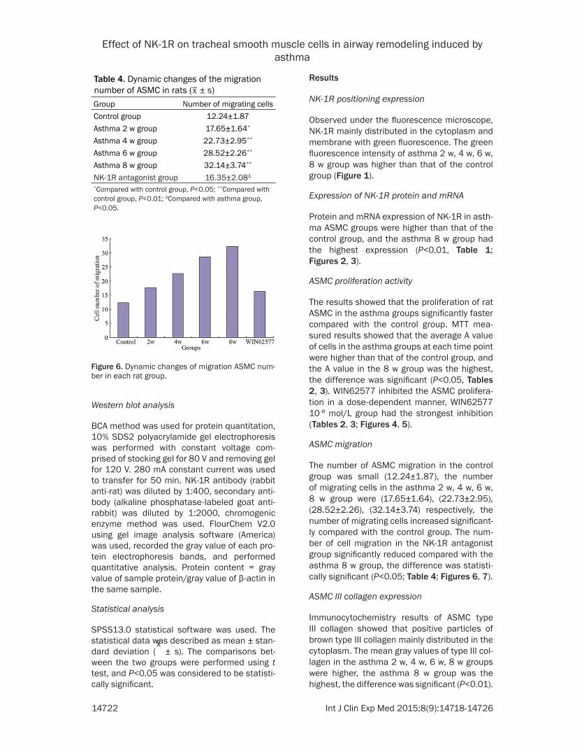
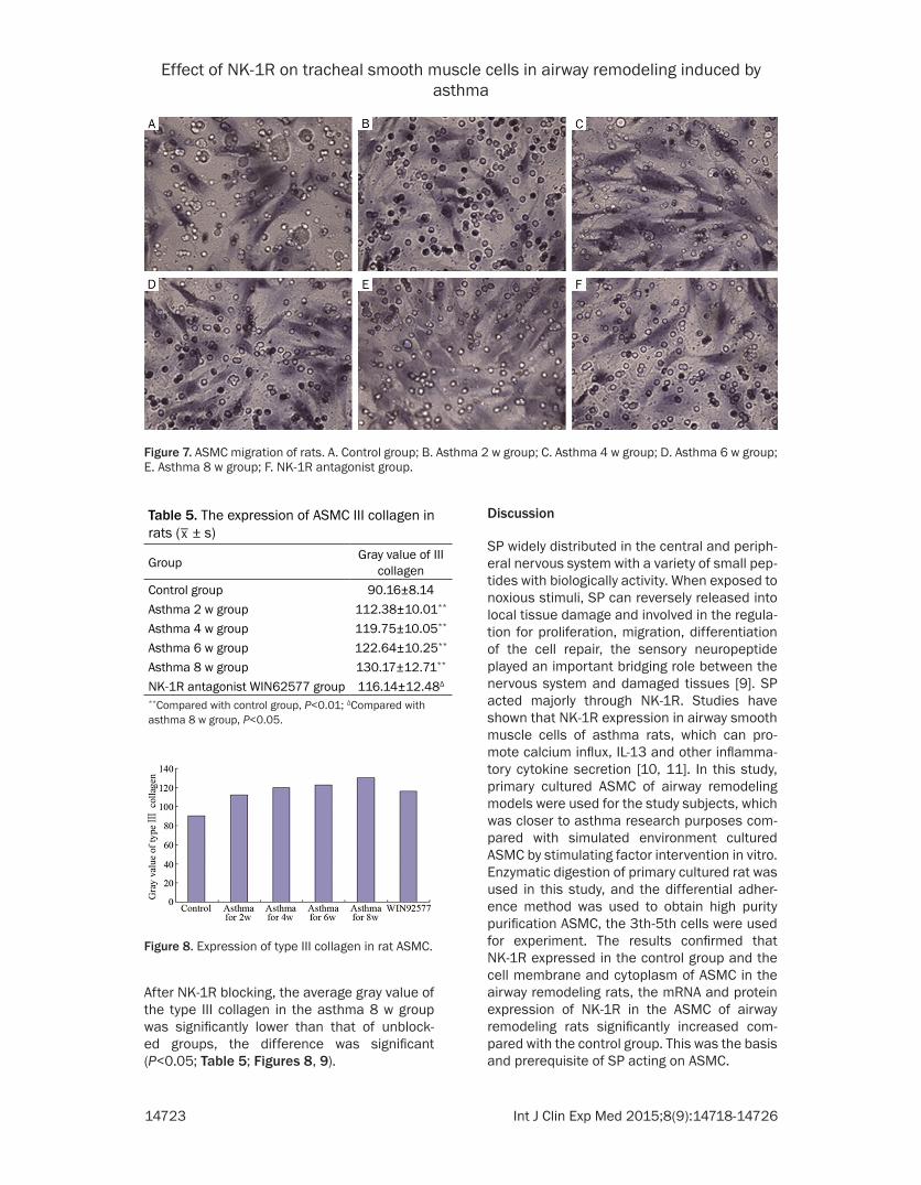
The number of ASMC migration in the control group was small (12.24±1.87), the number of migrating cells in the asthma 2 w, 4 w, 6 w, 8 w group were (17.65±1.64), (22.73±2.95), (28.52±2.26), (32.14±3.74) respectively, the number of migrating cells increased significant-ly compared with the control group. The num-ber of cell migration in the NK-1R antagonist group significantly reduced compared with the asthma 8 w group, the difference was statisti-cally significant (P<0.05; Table 4; Figures 6, 7).
ASMC III collagen expression
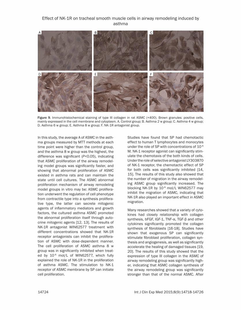
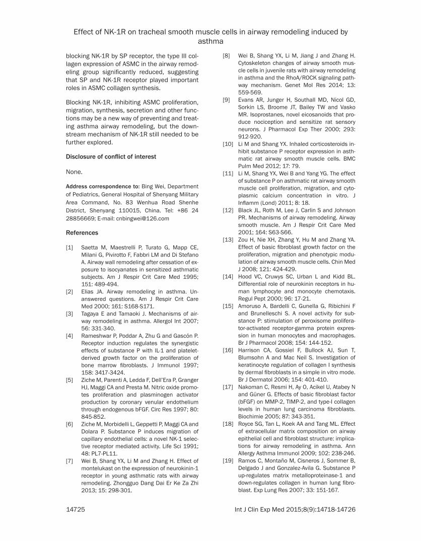
Immunocytochemistry results of ASMC type III collagen showed that positive particles of brown type III collagen mainly distributed in the cytoplasm. The mean gray values of type III col-lagen in the asthma 2 w, 4 w, 6 w, 8 w groups were higher, the asthma 8 w group was the highest, the difference was significant (P<0.01).
Table 4. Dynamic changes of the migration number of ASMC in rats (
_x ± s)
Group Number of migrating cellsControl group 12.24±1.87Asthma 2 w group 17.65±1.64*
Asthma 4 w group 22.73±2.95**
Asthma 6 w group 28.52±2.26**
Asthma 8 w group 32.14±3.74**
NK-1R antagonist group 16.35±2.08Δ
*Compared with control group, P<0.05; **Compared with control group, P<0.01; ΔCompared with asthma group, P<0.05.
Figure 6. Dynamic changes of migration ASMC num-ber in each rat group.
Effect of NK-1R on tracheal smooth muscle cells in airway remodeling induced by asthma
14723 Int J Clin Exp Med 2015;8(9):14718-14726
After NK-1R blocking, the average gray value of the type III collagen in the asthma 8 w group was significantly lower than that of unblock- ed groups, the difference was significant (P<0.05; Table 5; Figures 8, 9).
Discussion
SP widely distributed in the central and periph-eral nervous system with a variety of small pep-tides with biologically activity. When exposed to noxious stimuli, SP can reversely released into local tissue damage and involved in the regula-tion for proliferation, migration, differentiation of the cell repair, the sensory neuropeptide played an important bridging role between the nervous system and damaged tissues [9]. SP acted majorly through NK-1R. Studies have shown that NK-1R expression in airway smooth muscle cells of asthma rats, which can pro-mote calcium influx, IL-13 and other inflamma-tory cytokine secretion [10, 11]. In this study, primary cultured ASMC of airway remodeling models were used for the study subjects, which was closer to asthma research purposes com-pared with simulated environment cultured ASMC by stimulating factor intervention in vitro. Enzymatic digestion of primary cultured rat was used in this study, and the differential adher-ence method was used to obtain high purity purification ASMC, the 3th-5th cells were used for experiment. The results confirmed that NK-1R expressed in the control group and the cell membrane and cytoplasm of ASMC in the airway remodeling rats, the mRNA and protein expression of NK-1R in the ASMC of airway remodeling rats significantly increased com-pared with the control group. This was the basis and prerequisite of SP acting on ASMC.
Figure 7. ASMC migration of rats. A. Control group; B. Asthma 2 w group; C. Asthma 4 w group; D. Asthma 6 w group; E. Asthma 8 w group; F. NK-1R antagonist group.
Table 5. The expression of ASMC III collagen in rats (
_x ± s)
Group Gray value of III collagen
Control group 90.16±8.14Asthma 2 w group 112.38±10.01**
Asthma 4 w group 119.75±10.05**
Asthma 6 w group 122.64±10.25**
Asthma 8 w group 130.17±12.71**
NK-1R antagonist WIN62577 group 116.14±12.48Δ
**Compared with control group, P<0.01; ΔCompared with asthma 8 w group, P<0.05.
Figure 8. Expression of type III collagen in rat ASMC.
Effect of NK-1R on tracheal smooth muscle cells in airway remodeling induced by asthma
14724 Int J Clin Exp Med 2015;8(9):14718-14726
In this study, the average A of ASMC in the asth-ma groups measured by MTT methods at each time point were higher than the control group, and the asthma 8 w group was the highest, the difference was significant (P<0.05), indicating that ASMC proliferation of the airway remodel-ing model groups was significantly faster, and showing that abnormal proliferation of ASMC existed in asthma rats and can maintain the state until cell cultures. The ASMC abnormal proliferation mechanism of airway remodeling model groups in vitro may be: ASMC prolifera-tion underwent the regulation of cell phenotype from contractile type into a synthesis prolifera-tive type, the latter can secrete mitogenic agents of inflammatory mediators and growth factors, the cultured asthma ASMC promoted the abnormal proliferation itself through auto-crine mitogenic agents [12, 13]. The results of NK-1R antagonist WIN62577 treatment with different concentrations showed that NK-1R receptor antagonists can inhibit the prolifera-tion of ASMC with dose-dependent manner. The cell proliferation of ASMC asthma 8 w group was in significantly inhibited when treat-ed by 10-8 mol/L of WIN62577, which fully explained the role of NK-1R in the proliferation of asthma ASMC. The stimulation to NK-1 receptor of ASMC membrane by SP can initiate cell proliferation.
Studies have found that SP had chemotactic effect to human T lymphocytes and monocytes under the role of SP with concentrations of 10-6 M. NK-1 receptor agonist can significantly stim-ulate the chemotaxis of the both kinds of cells. Under the role of selective antagonist LY303870 of NK-1 receptor, the chemotactic effect of SP for both cells was significantly inhibited [14, 15]. The results of this study also showed that the number of migration in the airway remodel-ing ASMC group significantly increased. The blocking NK-1R by 10-8 mol/L WIN62577 may inhibit the migration of ASMC, indicating that NK-1R also played an important effect in ASMC migration.
Many researches showed that a variety of cyto-kines had closely relationship with collagen synthesis, bFGF, IGF-1, TNF-α, TGF-β and other cytokines significantly promoted the collagen synthesis of fibroblasts [16-18]. Studies have shown that exogenous SP can significantly stimulate fibroblast proliferation, collagen syn-thesis and angiogenesis, as well as significantly accelerate the healing of damaged tissues [19, 20]. The results of this study showed that the expression of type III collagen in the ASMC of airway remodeling group was significantly high-er, indicating that ASMC collagen synthesis of the airway remodeling group was significantly stronger than that of the normal ASMC. After
Figure 9. Immunohistochemical staining of type III collagen in rat ASMC (×400). Brown granules: positive cells, mainly expressed in the cell membrane and cytoplasm. A. Control group; B. Asthma 2 w group; C. Asthma 4 w group; D. Asthma 6 w group; E. Asthma 8 w group; F. NK-1R antagonist group.
Effect of NK-1R on tracheal smooth muscle cells in airway remodeling induced by asthma
14725 Int J Clin Exp Med 2015;8(9):14718-14726
blocking NK-1R by SP receptor, the type III col-lagen expression of ASMC in the airway remod-eling group significantly reduced, suggesting that SP and NK-1R receptor played important roles in ASMC collagen synthesis.
Blocking NK-1R, inhibiting ASMC proliferation, migration, synthesis, secretion and other func-tions may be a new way of preventing and treat-ing asthma airway remodeling, but the down-stream mechanism of NK-1R still needed to be further explored.
Disclosure of conflict of interest
None.
Address correspondence to: Bing Wei, Department of Pediatrics, General Hospital of Shenyang Military Area Command, No. 83 Wenhua Road Shenhe District, Shenyang 110015, China. Tel: +86 24 28856669; E-mail: [email protected]
References
[1] Saetta M, Maestrelli P, Turato G, Mapp CE, Milani G, Pivirotto F, Fabbri LM and Di Stefano A. Airway wall remodeling after cessation of ex-posure to isocyanates in sensitized asthmatic subjects. Am J Respir Crit Care Med 1995; 151: 489-494.
[2] Elias JA. Airway remodeling in asthma. Un- answered questions. Am J Respir Crit Care Med 2000; 161: S168-S171.
[3] Tagaya E and Tamaoki J. Mechanisms of air-way remodeling in asthma. Allergol Int 2007; 56: 331-340.
[4] Rameshwar P, Poddar A, Zhu G and Gascón P. Receptor induction regulates the synergistic effects of substance P with IL-1 and platelet-derived growth factor on the proliferation of bone marrow fibroblasts. J Immunol 1997; 158: 3417-3424.
[5] Ziche M, Parenti A, Ledda F, Dell’Era P, Granger HJ, Maggi CA and Presta M. Nitric oxide promo-tes proliferation and plasminogen activator production by coronary venular endothelium through endogenous bFGF. Circ Res 1997; 80: 845-852.
[6] Ziche M, Morbidelli L, Geppetti P, Maggi CA and Dolara P. Substance P induces migration of capillary endothelial cells: a novel NK-1 selec-tive receptor mediated activity. Life Sci 1991; 48: PL7-PL11.
[7] Wei B, Shang YX, Li M and Zhang H. Effect of montelukast on the expression of neurokinin-1 receptor in young asthmatic rats with airway remodeling. Zhongguo Dang Dai Er Ke Za Zhi 2013; 15: 298-301.
[8] Wei B, Shang YX, Li M, Jiang J and Zhang H. Cytoskeleton changes of airway smooth mus-cle cells in juvenile rats with airway remodeling in asthma and the RhoA/ROCK signaling path-way mechanism. Genet Mol Res 2014; 13: 559-569.
[9] Evans AR, Junger H, Southall MD, Nicol GD, Sorkin LS, Broome JT, Bailey TW and Vasko MR. Isoprostanes, novel eicosanoids that pro-duce nociception and sensitize rat sensory neurons. J Pharmacol Exp Ther 2000; 293: 912-920.
[10] Li M and Shang YX. Inhaled corticosteroids in-hibit substance P receptor expression in asth-matic rat airway smooth muscle cells. BMC Pulm Med 2012; 17: 79.
[11] Li M, Shang YX, Wei B and Yang YG. The effect of substance P on asthmatic rat airway smooth muscle cell proliferation, migration, and cyto-plasmic calcium concentration in vitro. J Inflamm (Lond) 2011; 8: 18.
[12] Black JL, Roth M, Lee J, Carlin S and Johnson PR. Mechanisms of airway remodeling. Airway smooth muscle. Am J Respir Crit Care Med 2001; 164: S63-S66.
[13] Zou H, Nie XH, Zhang Y, Hu M and Zhang YA. Effect of basic fibroblast growth factor on the proliferation, migration and phenotypic modu-lation of airway smooth muscle cells. Chin Med J 2008; 121: 424-429.
[14] Hood VC, Cruwys SC, Urban L and Kidd BL. Differential role of neurokinin receptors in hu-man lymphocyte and monocyte chemotaxis. Regul Pept 2000; 96: 17-21.
[15] Amoruso A, Bardelli C, Gunella G, Ribichini F and Brunelleschi S. A novel activity for sub-stance P: stimulation of peroxisome prolifera-tor-activated receptor-gamma protein expres-sion in human monocytes and macrophages. Br J Pharmacol 2008; 154: 144-152.
[16] Harrison CA, Gossiel F, Bullock AJ, Sun T, Blumsohn A and Mac Neil S. Investigation of keratinocyte regulation of collagen I synthesis by dermal fibroblasts in a simple in vitro mode. Br J Dermatol 2006; 154: 401-410.
[17] Nakoman C, Resmi H, Ay O, Acikel U, Atabey N and Güner G. Effects of basic fibroblast factor (bFGF) on MMP-2, TIMP-2, and type-I collagen levels in human lung carcinoma fibroblasts. Biochimie 2005; 87: 343-351.
[18] Royce SG, Tan L, Koek AA and Tang ML. Effect of extracellular matrix composition on airway epithelial cell and fibroblast structure: implica-tions for airway remodeling in asthma. Ann Allergy Asthma Immunol 2009; 102: 238-246.
[19] Ramos C, Montaño M, Cisneros J, Sommer B, Delgado J and Gonzalez-Avila G. Substance P up-regulates matrix metalloproteinase-1 and down-regulates collagen in human lung fibro-blast. Exp Lung Res 2007; 33: 151-167.
Effect of NK-1R on tracheal smooth muscle cells in airway remodeling induced by asthma
14726 Int J Clin Exp Med 2015;8(9):14718-14726
[20] Burssens P, Steyaert A, Forsyth R, van Ovost EJ, Depaepe Y and Verdonk R. Exogenously ad-ministered substance P and nertral endopepti-dase inhibitors stimulate fibroblast proliferati-
on angiogenesis and collagen organization during Achilles tendon healing. Foof Ankle Int 2005; 26: 832-839.