original article detection of factor xiii-a is a valuable ...emy of sciences and lajos szodoray...
TRANSCRIPT
ORIGINAL ARTICLE
Detection of factor XIII-A is a valuable tool for distinguishing
dendritic cells and tissue macrophages in granuloma
2 annulare and necrobiosis lipoidica
D. T€or€ocsik,1 H. B�ardos,2 Zs. Hataly�ak,2 B. Dezs}o,3 G. Losonczy,4 L. Paragh,1 Z. P�eter,1 M. Bal�azs,2
E. Remenyik1 R. �Ad�any2,*
1Department of Dermatology, Faculty of Medicine, Medical and Health Science Centre, University of Debrecen, Debrecen, Hungary2Department of Preventive Medicine, Faculty of Public Health, Medical and Health Science Centre, University of Debrecen, Debrecen,
Hungary3Department of Pathology, Faculty of Medicine, Medical and Health Science Centre, University of Debrecen, Debrecen, Hungary4Department of Ophthalmology, Faculty of Medicine, Medical and Health Science Centre, University of Debrecen, Debrecen, Hungary
*Correspondence: R. �Ad�any. E-mail: [email protected]
Abstract
Background Factor XIII subunit A (FXIII-A) is used as a diagnostic marker in a wide range of dermatological diseases
ranging from inflammatory lesions to malignancies, although neither the cell types responsible for its expression nor the
mechanism(s) resulting in its local accumulation in pathological conditions have been characterized.
Objective In this study, we aimed to gain information on the cells showing an immunohistochemical reaction for FXIII-
A and answer the question whether macrophages and/or dendritic cells are labelled for FXIII-A.
Methods We carried out our studies on samples of granuloma annulare (GA) and necrobiosis lipoidica (NL), the prime
examples for granulomatous skin lesions with a non-infectious background in which extracellular matrix remodelling is a
key feature without any sign of malignant transformation. We used markers for macrophages and dendritic cells in com-
bination with the detection of FXIII-A in double labelling immunohistochemical reactions.
Results We demonstrated that FXIII-A positivity clearly distinguishes macrophages (CD163+/FXIII-A+) from dendritic
cells (CD11c+/FXIII-A�) not only in the normal dermis as previously described by Zaba et al. (J Clin Invest 2007; 117:
2517–2525) but also in the pathological conditions of GA and NL. Detecting the expression of DC-SIGN/CD209 and
mannose receptor molecules on FXIII-A+ macrophages we confirmed that FXIII-A is expressed in the alternatively acti-
vated macrophages. However, while DC-SIGN/CD209 was invariably expressed on FXIII-A+ cells both in normal and
pathological conditions of GA/NL (98.7% vs. 93.5/96%), mannose receptor was only partially coexpressed with FXIII-A
(94.8% vs. 74.7/52.2%), suggesting that FXIII-A+macrophages do not represent a homogenous population.
Conclusions Factor XIII (FXIII)-A selectively marks macrophages and distinguishes them from dendritic cells. The pres-
ence of FXIII-A is not a disease-specific marker but indicates a possible common mechanism of macrophage activation
in various dermatological diseases.
Received: 19 February 2013; Accepted: 18 September 2013
Conflicts of interest
The authors state no conflict of interest.
Funding sources
This work was supported in part by National Research Funds OTKA K81847 R.A., and PD101557 (D.T.) and by the
T�AMOP 4.2.2.A-11/1/KONV-2012-0031 projects. D.T. is a recipient of Janos Bolyai Fellowships of the Hungarian Acad-
emy of Sciences and Lajos Szodoray Prize of the University of Debrecen and holds a BMC grant from South Korea.
Introduction
Factor XIII (FXIII) involved in blood coagulation consists of two
globular A subunits (FXIII-A) exerting a transglutaminase activity
upon its activation, and two inhibitory B subunits (FXIII-B). It is
well known that the activated form of FXIII-A has an important
role in the final step of blood coagulation (fibrin stabilization);
however, its role is not restricted to the area of haemostasis.1,2 In
addition to megakaryocytes and platelets, monocytes and macro-
phages can also synthesize FXIII-A but are unable to secrete it.
FXIII-A can be detected from the very early stage of monocyte
differentiation (monoblasts in the bone marrow) via circulating
blood monocytes to the macrophages of connective tissue and
1
2
3
4
5
6
7
8
9
10
11
12
13
14
15
16
17
18
19
20
21
22
23
24
25
26
27
28
29
30
31
32
33
34
35
36
37
38
39
40
41
42
43
44
45
46
47
48
49
50
51
52
© 2013 European Academy of Dermatology and VenereologyJEADV 2013
DOI: 10.1111/jdv.12290 JEADV
J D V 1 2 2 9 0 Dispatch: 9.10.13 Journal: JDV CE: Banu Priya
Journal Name Manuscript No. No. of pages: 10 PE: Sudhakar S
serous cavities. Based on these findings, FXIII-A is interpreted as
a marker protein of the cell lines in which it acts as an intracellu-
lar transglutaminase with roles in various intracellular (intracyto-
plasmic and intranuclear) processes.3–9
The immunohistochemical detection of FXIII-A is widely
used in dermatological pathology diagnosis,10–20 although
neither the FXIII-A-positive cell types nor the possible mecha-
nisms behind their abundant accumulation in certain lesions
have been convincingly interpreted. Historically, in addition to
monocyte-derived tissue histiocytes, dendritic cells, the key anti-
gen-presenting cells of the immune system, were also considered
to be positive for FXIII-A.10,15,18–20 It became widely accepted
that ‘FXIII-A+ cells in human skin represent a specific popula-
tion of bone marrow-derived dermal dendritic cells, distinct
from Langerhans cells, that share some features common to
mononuclear phagocytes (monocyte/macrophages)’.21
Concerning the application of immunohistochemical detec-
tion of FXIII-A in dermatohistopathology, we face the following
severe inconsistencies:
1 The presence of FXIII-A in monocyte-derived macrophages
is not ubiquitous. While under pathological conditions
observed in various tumours a high number of FXIII-A-posi-
tive monocyte-derived macrophages can be found,22–24 in
granulomas of sarcoidosis and tuberculosis the majority of
macrophages are negative for FXIII-A.25,26 To explain this
phenomenon, our previous studies have confirmed that the
presence of FXIII-A in macrophages indicates that they are
alternatively activated through a pathway induced by the
cytokines interleukin 4 (IL-4) and interleukin 13 (IL-13),
whereas its absence is a trait of classically activated macro-
phages, in which interferon gamma (IFc) is the triggering
cytokine molecule.26 Dissecting the two pathways of macro-
phage activation upon FXIII-A positivity suggests that FXIII-
A-positive macrophages are important players in conditions
where extracellular remodelling is the key feature (as in
tumour tissues), but not in the host vs. pathogen responses
as it is in tuberculosis granulomas.
2 A recent study that aimed to distinguish macrophages from
dendritic cells based on the expression of various markers
and functional characteristics of these cell types in the normal
human dermis illustrated that, while dermal macrophages are
CD163+ FXIII-A+, dendritic cells are CD11c+ CD1c+, and
these markers define discrete dermal cell populations with no
overlap.27 However, this type of classification has been chal-
lenged in pathological conditions only to a limited extent.28
Our study was designed to determine whether the distinct cell
populations of macrophages and dendritic cells can be clearly
identified under pathological conditions upon FXIII-A positivity
and whether the accumulation of FXIII-A+ cells is related to the
matrix remodelling processes, a characteristic feature of the
alternatively activated macrophages. For our studies, we chose to
use samples of granuloma annulare (GA) and necrobiosis lipoi-
dica (NL), the prime examples for dermatological diseases with a
large accumulation of macrophages, in which extracellular
matrix remodelling is a key feature without malignant transfor-
mation and with a non-infectious background.29,30
Here, we demonstrate that the immunohistochemical detec-
tion of FXIII-A is a suitable marker to differentiate macrophages
from dendritic cells also in pathological conditions that are char-
acterized with extracellular matrix remodelling. The character-
ization of FXIII-A+ cells confirmed our previous findings that
FXIII-A macrophages express other markers of alternative acti-
vation, as well. In the interpretation of the development and
progression of GA and NL lesions, the impressive histological
appearance of foci consisting exclusively of dendritic cells sur-
rounded by tissue compartments extremely rich in FXIII-A+
macrophages may open new avenues for research, as well.
Materials and methods
Skin samples
Skin samples were obtained from four normal volunteers who
underwent plastic surgery, nine GA patients and six NL patients
in the Department of Dermatology, University of Debrecen,
Medical and Health Sciences Center (UDMHS). Histological
diagnosis was made in the Department of Pathology, UDMHS.
The acquisition of all skin specimens was reviewed and
approved by the Regional and Institutional Ethics Committee,
UDMHS. Informed consent was obtained, and the study was
performed in accordance with the Principles of the Declaration
of Helsinki.
Double immunofluorescence (DIF) studies of
macrophages and dendritic cells
Frozen sections were fixed in acetone for 10 min and incubated
in 5% normal goat serum diluted in Serum-Free Protein Block
(SFPB) (DAKO, Glostrup, Denmark). Factor XIII-A was
detected by rabbit affinity purified anti-human FXIII-A antibody
(diluted 1 : 100, Acris Antibodies, San Diego, CA, USA) for 2 h
at room temperature. This procedure was followed by visualiza-
tion using DyLight 488 goat anti-rabbit IgG antibody for 45 min
(diluted 1 : 40, Vector Labs, Burlingame, CA, USA). For coex-
pression, the detection of FXIII-A was sequentially combined
with different reference markers using mouse monoclonal anti-
human antibodies against CD antigens (see Table S1). Following
a 10-min incubation with normal horse serum containing SFPB,
the second primaries’ specific binding was visualized by DyLight
549 horse anti-mouse IgG antibody (diluted 1 : 40, Vector
Labs). Slides were washed in PBS and mounted with Vectashield
Mounting Medium with DAPI (Vector Labs) to counterstain
nuclei. For negative controls, the appropriate non-immune con-
trol sera [rabbit IgG from Vector Labs, mouse IgG1 or mouse
IgG2b from BD Pharmingen (Heidelberg, Germany)] were used
in place of primary antibodies followed by the same procedure
© 2013 European Academy of Dermatology and VenereologyJEADV 2013
2 T€or€ocsik et al.
1
2
3
4
5
6
7
8
9
10
11
12
13
14
15
16
17
18
19
20
21
22
23
24
25
26
27
28
29
30
31
32
33
34
35
36
37
38
39
40
41
42
43
44
45
46
47
48
49
50
51
52
as above. In general, no staining was observed; however, in some
cases, non-specific background fluorescence was observed in the
epidermis (not shown).
Images were acquired with an Axioplan microscope (Carl Ze-
iss, Oberkochen, Germany) equipped with selective filters and
connected to a CCD IMAC camera (Sony, Tokyo, Japan) and
ISIS fluorescent imaging system (MetaSystems, Altlussheim,
Germany).
To show unambiguous colocalization, some tissue samples
that were double labelled for FXIII-A and CD11c were also
imaged with a confocal laser-scanning microscope (LSM 700,
Carl Zeiss) equipped with solid-state lasers.
Analysis of coexpression of FXIII-A with CD antigens
On DIF samples, the degree of colocalization of FXIII-A with
CD antigens was analysed semiquantitatively by counting 100–
100 FXIII-A+ cells in association with other markers. Each sam-
ple was then evaluated separately, with the average percentages
of cells coexpressing FXIII-A and CD antigens (Table 1 and
Table S2).
Results
Normal and pathological skin samples harbouring
granuloma annulare and NL contain distinct FXIII-A+ and
CD11c+ cell populations
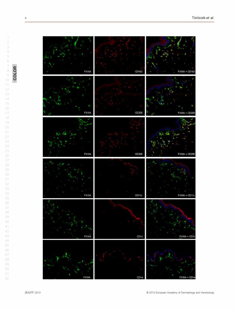
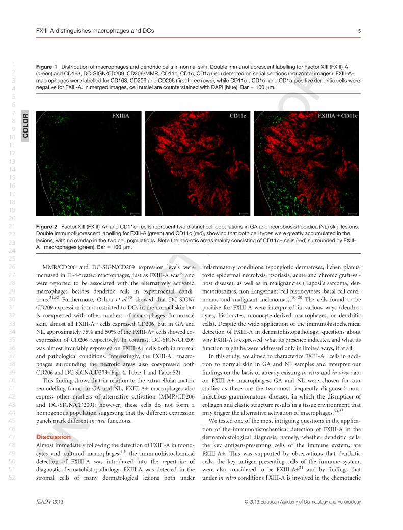
In normal skin, FXIII-A+ cells were found scattered in the papil-
lary and reticular dermis, whereas CD11c+ cells showing no col-
ocalization with FXIII-A+ cells were found in low numbers and
mainly localized beneath the epidermis. Confirming previous
data by Zaba et al.,27 FXIII-A+ cells were also positive for the
macrophage specific protein CD163, and coexpressed mannose
receptor (MMR/CD206) and DC-SIGN/CD209 markers. On the
other hand, besides the CD11c+ cells, the few cells found to
express CD1a or CD1c, markers of different dendritic cell sub-
sets, were also negative for FXIII-A (Fig. 1).
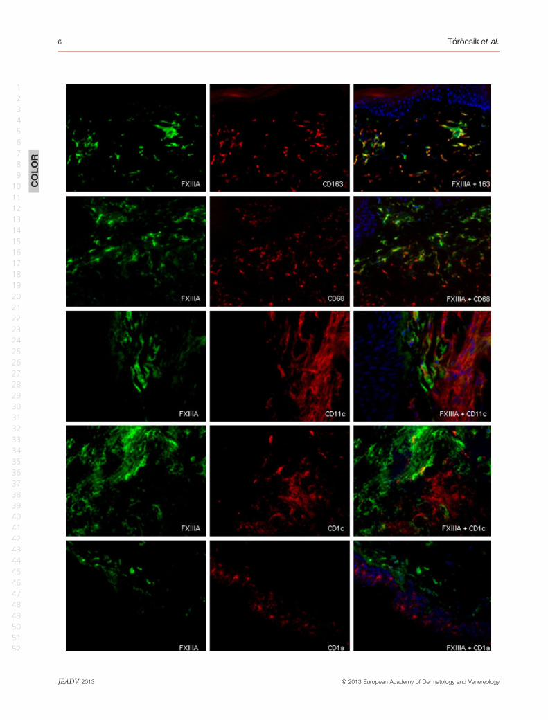
In samples of GA and NL, FXIII-A+ cells were much more
abundant, and the density of CD11c+ dendritic cells was also
elevated. The FXIII-A+ and CD11c+ cells showed two distinct
cell populations in both skin lesions, as was observed in normal
samples. Interestingly, the necrotic areas of the skin samples
mainly consisted of CD11c+ cells, which were surrounded by
FXIII-A+ macrophages (Fig. 2).
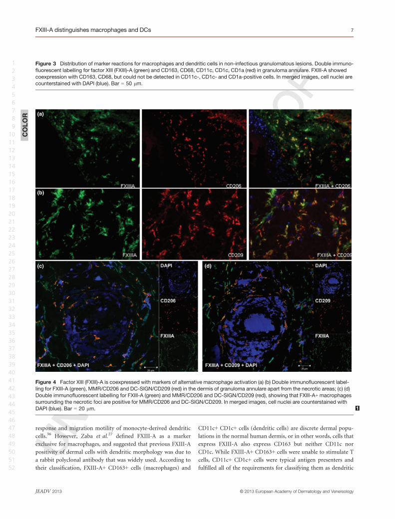
Regarding CD68, which is expressed in a large proportion of
macrophages, a high number of CD68+ cells were present, but
found to only partially coexpress FXIII-A. Compared with nor-
mal skin tissues, GA and NL samples exhibited more cells identi-
fied as CD1c and CD1a (Fig. 3). Very few cells were stained for
the mature DC markers DC-LAMP/CD208 and/or CD83. In all
cases studied, CD1c+, CD1a+, DC-LAMP/CD208+ and CD83+
cells were always negative for FXIII-A (not shown).
These results indicate that immunohistochemical profiling
to separate macrophages from dendritic cells with the use of
the above markers for normal skin described by Zaba et al.27
can also be applied to pathological samples including GA
and NL.
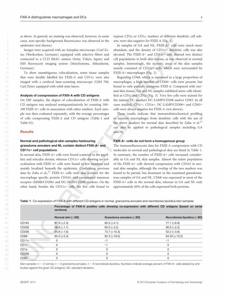
FXIII-A+ cells do not form a homogenous group
The immunofluorescent data for FXIII-A coexpression with CD
molecules in normal and pathological skin are listed in Table 1.
In summary, the number of FXIII-A+ cells increased consider-
ably in GA and NL skin samples. Almost the entire population
of the FXIII-A+ cells showed coexpression with CD163 in nor-
mal skin samples, although the overlap of the two markers was
found to be partial, but dominant in the examined granuloma-
tous samples of GA and NL. CD68 was expressed in most of the
FXIII-A+ cells in the normal skin, whereas in GA and NL only
approximately 60% of the cells expressed both proteins.
Table 1 Co-expression of FXIII-A with different CD antigens in normal, granuloma annulare and necrobiosis lipoidica skin samples
Percentage of FXIII-A positive cells showing co-expression with different CD antigens (based on serial
sections)
Normal skin (�SD) Granuloma annulare (�SD) Necrobiosis lipoidica (�SD)
CD163 97.8 (�2.3) 85.5 (�4.1) 77.1 (�6.9)
CD209 98.8 (�1.1) 93.5 (�3.2) 96.0 (�2.2)
CD206 94.8 (�1.6) 74.7 (�10.3) 52.2 (�3.9)
CD68 85.5 (�5.3) 62.3 (�18.5) 64.33 (�10.2)
CD11c 0 <1 <1
CD1c 0 <1 0
CD1a 0 0 0
CD208 0 0 0
CD83 0 0 0
Skin samples: n = 4 normal, n = 9 granuloma annulare, n = 6 necrobiosis lipoidica. Numbers indicate average percent of FXIII-A+ cells labeled by anti-
bodies against the given CD antigens; SD, standard deviation.
© 2013 European Academy of Dermatology and VenereologyJEADV 2013
FXIII-A distinguishes macrophages and DCs 3
1
2
3
4
5
6
7
8
9
10
11
12
13
14
15
16
17
18
19
20
21
22
23
24
25
26
27
28
29
30
31
32
33
34
35
36
37
38
39
40
41
42
43
44
45
46
47
48
49
50
51
52
COLOR
© 2013 European Academy of Dermatology and VenereologyJEADV 2013
4 T€or€ocsik et al.
1
2
3
4
5
6
7
8
9
10
11
12
13
14
15
16
17
18
19
20
21
22
23
24
25
26
27
28
29
30
31
32
33
34
35
36
37
38
39
40
41
42
43
44
45
46
47
48
49
50
51
52
MMR/CD206 and DC-SIGN/CD209 expression levels were
increased in IL-4-treated macrophages, just as FXIII-A was26 and
were reported to be associated with the alternatively activated
macrophages besides dendritic cells in experimental condi-
tions.31,32 Furthermore, Ochoa et al.33 showed that DC-SIGN/
CD209 expression is not restricted to DCs in the normal skin but
is coexpressed with other markers of macrophages. In normal
skin, almost all FXIII-A+ cells expressed CD206, but in GA and
NL, approximately 75% and 50% of the FXIII-A+ cells showed co-
expression of CD206 respectively. In contrast, DC-SIGN/CD209
was almost invariably expressed on FXIII-A+ cells both in normal
and pathological conditions. Interestingly, the FXIII-A+ macro-
phages surrounding the necrotic areas also coexpressed both
CD206 and DC-SIGN/CD209 (Fig. 4, Table 1 and Table S2).
This finding shows that in relation to the extracellular matrix
remodelling found in GA and NL, FXIII-A+ macrophages also
express other markers of alternative activation (MMR/CD206
and DC-SIGN/CD209); however, these cells do not form a
homogenous population suggesting that the different expression
panels mark different in vivo functions.
Discussion
Almost immediately following the detection of FXIII-A in mono-
cytes and cultured macrophages,4,5 the immunohistochemical
detection of FXIII-A was introduced into the repertoire of
diagnostic dermatohistopathology. FXIII-A was detected in the
stromal cells of many dermatological lesions both under
inflammatory conditions (spongiotic dermatoses, lichen planus,
toxic epidermal necrolysis, psoriasis, acute and chronic graft-vs.-
host disease), as well as in malignancies (Kaposi’s sarcoma, der-
matofibromas, non-Langerhans cell histiocytoses, basal cell carci-
nomas and malignant melanomas).10–20 The cells found to be
positive for FXIII-A were interpreted in various ways (dendro-
cytes, histiocytes, monocyte-derived macrophages, or dendritic
cells). Despite the wide application of the immunohistochemical
detection of FXIII-A in dermatohistopathology, questions about
why FXIII-A is expressed, what its presence indicates, and what its
function might be were addressed only in limited ways, if at all.
In this study, we aimed to characterize FXIII-A+ cells in addi-
tion to normal skin in GA and NL samples and interpret our
findings on the basis of already existing in vitro and in vivo data
on FXIII-A+ macrophages. GA and NL were chosen for our
studies as these are the two most frequently diagnosed non-
infectious granulomatous diseases, in which the disruption of
collagen and elastic structure results in a tissue environment that
may trigger the alternative activation of macrophages.34,35
We tested one of the most intriguing questions in the applica-
tion of the immunohistochemical detection of FXIII-A in the
dermatohistological diagnosis, namely, whether dendritic cells,
the key antigen-presenting cells of the immune system, are
FXIII-A+. This was supported by observations that dendritic
cells, the key antigen-presenting cells of the immune system,
were also considered to be FXIII-A+21 and by findings that
under in vitro conditions FXIII-A is involved in the chemotactic
Figure 2 Factor XIII (FXIII)-A+ and CD11c+ cells represent two distinct cell populations in GA and necrobiosis lipoidica (NL) skin lesions.
Double immunofluorescent labelling for FXIII-A (green) and CD11c (red), showing that both cell types were greatly accumulated in the
lesions, with no overlap in the two cell populations. Note the necrotic areas mainly consisting of CD11c+ cells (red) surrounded by FXIII-
A+macrophages (green). Bar = 100 lm.
Figure 1 Distribution of macrophages and dendritic cells in normal skin. Double immunofluorescent labelling for Factor XIII (FXIII)-A
(green) and CD163, DC-SIGN/CD209, CD206/MMR, CD11c, CD1c, CD1a (red) detected on serial sections (horizontal images). FXIII-A+
macrophages were labelled for CD163, CD209 and CD206 (first three rows), while CD11c-, CD1c- and CD1a-positive dendritic cells were
negative for FXIII-A. In merged images, cell nuclei are counterstained with DAPI (blue). Bar = 100 lm.
COLOR
© 2013 European Academy of Dermatology and VenereologyJEADV 2013
FXIII-A distinguishes macrophages and DCs 5
1
2
3
4
5
6
7
8
9
10
11
12
13
14
15
16
17
18
19
20
21
22
23
24
25
26
27
28
29
30
31
32
33
34
35
36
37
38
39
40
41
42
43
44
45
46
47
48
49
50
51
52
COLOR
© 2013 European Academy of Dermatology and VenereologyJEADV 2013
6 T€or€ocsik et al.
1
2
3
4
5
6
7
8
9
10
11
12
13
14
15
16
17
18
19
20
21
22
23
24
25
26
27
28
29
30
31
32
33
34
35
36
37
38
39
40
41
42
43
44
45
46
47
48
49
50
51
52
response and migration motility of monocyte-derived dendritic
cells.36 However, Zaba et al.27 defined FXIII-A as a marker
exclusive for macrophages, and suggested that previous FXIII-A
positivity of dermal cells with dendritic morphology was due to
a rabbit polyclonal antibody that was widely used. According to
their classification, FXIII-A+ CD163+ cells (macrophages) and
CD11c+ CD1c+ cells (dendritic cells) are discrete dermal popu-
lations in the normal human dermis, or in other words, cells that
express FXIII-A also express CD163 but neither CD11c nor
CD1c. While FXIII-A+ CD163+ cells were unable to stimulate T
cells, CD11c+ CD1c+ cells were typical antigen presenters and
fulfilled all of the requirements for classifying them as dendritic
(a)
(b)
(c) (d)
Figure 4 Factor XIII (FXIII)-A is coexpressed with markers of alternative macrophage activation (a) (b) Double immunofluorescent label-
ling for FXIII-A (green), MMR/CD206 and DC-SIGN/CD209 (red) in the dermis of granuloma annulare apart from the necrotic areas; (c) (d)
Double immunofluorescent labelling for FXIII-A (green) and MMR/CD206 and DC-SIGN/CD209 (red), showing that FXIII-A+macrophages
surrounding the necrotic foci are positive for MMR/CD206 and DC-SIGN/CD209. In merged images, cell nuclei are counterstained with
DAPI (blue). Bar = 20 lm. 1
Figure 3 Distribution of marker reactions for macrophages and dendritic cells in non-infectious granulomatous lesions. Double immuno-
fluorescent labelling for factor XIII (FXIII)-A (green) and CD163, CD68, CD11c, CD1c, CD1a (red) in granuloma annulare. FXIII-A showed
coexpression with CD163, CD68, but could not be detected in CD11c-, CD1c- and CD1a-positive cells. In merged images, cell nuclei are
counterstained with DAPI (blue). Bar = 50 lm.
COLOR
© 2013 European Academy of Dermatology and VenereologyJEADV 2013
FXIII-A distinguishes macrophages and DCs 7
1
2
3
4
5
6
7
8
9
10
11
12
13
14
15
16
17
18
19
20
21
22
23
24
25
26
27
28
29
30
31
32
33
34
35
36
37
38
39
40
41
42
43
44
45
46
47
48
49
50
51
52
cells.37 Regarding the interpretation of the in vitro findings on
the role of FXIII-A in dendritic cell motility,36 the method used
for the differentiation of monocytes into dendritic cells under
in vitro conditions notably represents a combined stimulus of
IL-4 and GM-CSF.38 We have previously demonstrated that IL-4
is a very potent inducer of FXIII-A expression,26 therefore the
detection of FXIII-A in DCs differentiated in vitro can be consid-
ered as a direct effect of the cytokine instead of being a marker
for DC differentiation.
In this study, we provide evidence that using the marker reac-
tions mentioned above, the two cell populations characterized
by Zaba et al.27 can be clearly distinguished also in pathological
samples of GA and NL in which FXIII-A+ macrophages and
CD11c+ dendritic cells also show distinct population.
Dissecting the cells expressing FXIII-A, we found that CD163
was present in almost the whole FXIII-A+ cell population in
both the normal and the pathological samples. On the other
hand CD68, another widely used macrophage marker, was found
to overlap with FXIII-A only partially in the normal skin, and to
a lesser extent in GA and NL samples. These findings are consis-
tent with previous observations that besides macrophages, other
cell types such as dendritic cells, granulocytes and also non-mye-
loid cells express CD68 under certain conditions, therefore it
cannot be considered as a selective macrophage marker.39
MMR/CD206 and DC-SIGN/DC209 are markers of alterna-
tive macrophage activation; however, using these markers alone
is not sufficient to detect macrophages as they are also expressed
in dendritic cells. Examining their expression in combination
with FXIII-A, in normal skin, FXIII+ cells expressed MMR/
CD206 and DC-SIGN/DC209. These data agree with our previ-
ous findings that FXIII-A-positive macrophages are alternatively
activated in the normal human dermis. Examining the macro-
phages in the GA and NL samples, the coexpression of FXIII-A
with DC-SIGN/DC209 was practically complete, but only par-
tially so for MMR/DC206; nevertheless, MMR/DC206 could be
observed in the majority of FXIII-A+ cells. Interestingly, in the
FXIII-A+ macrophages with a pivotal role in the granuloma for-
mation, detected around the necrotic areas mainly consisted of
CD11c+ cells in the skin samples, both DC-SIGN/DC209 as well
as MMR/DC206 was positive. Comparing GA and NL samples
with the used marker reactions, they exerted similar macrophage
populations, suggesting similar macrophage activation mecha-
nisms in the background of the two diseases, in which matrix
remodelling might be a key player. These findings and further
immunohistochemical analyses of FXIII-A+ macrophages in GA
and NL samples revealed that these cells do not represent a
homogenous cell population. The existence of different cell clus-
ters suggests that certain in vivo stimuli during the activation of
macrophages under pathological conditions can influence their
marker characteristics. On the other hand, the high number of
CD11c+ DCs found in the lesions of both GA and NL suggest
that despite previous histological studies where macrophages
were in the focus30 to explain the underlying pathomechanisms,
DCs should be taken into account as well. Based on the role of
DCs in antigen presentation their accumulation likely suggests
the presence of the primary antigen(s) within the lesions that
trigger(s) the granuloma formation. Further analysis of the dif-
ferent macrophage subsets as well as a more complex analysis of
the dendritic cells, the stimuli behind their accumulation and
their interaction with the FXIII-A+ macrophages surrounding
the necrotic foci could lead to a better understanding of their
aetiology and may even provide possible therapeutic targets not
only in GA and NL but also in other diseases with granuloma
formation.
To draw definitive conclusions on the role of intracellular
FXIII-A in dermal macrophages on the basis of its immunohis-
tological detection is most likely not possible, but it is still inter-
esting to speculate on the possible functions. As FXIII-A lacks a
signal peptide and as there is no proof for its secretion, its extra-
cellular activity is limited to conditions where FXIII-A is released
from destroyed macrophages.40,41 Based on the findings that
FXIII-A is localized mainly around the cytoplasmic vacuoles and
pseudopodia and on the fact that cytoskeletal proteins such as
actin, myosin and vinculin are known substrates for activated
FXIII-A, FXIII-A might play important roles in certain intracel-
lular processes in which cytoskeletal remodelling is a key fea-
ture.42–45 Our previous studies showed that monocytes/
macrophages from FXIII-A-deficient patients exerted an
impaired capacity of Fcc and complement receptor-mediated
phagocytosis46 suggests that FXIII-A is essential in the tissue
remodelling activity of macrophages both in GA and NL. The
translocation of FXIII-A with a maintained transglutaminase
activity to the nuclei of macrophages differentiating from mono-
cytes has also been demonstrated, and its possible involvement
in gene expression regulation was suggested.47 Comparing gene
expression profiles of normal and FXIII-A-deficient cultured
human macrophages by microarray revealed that FXIII-A has an
important role(s) in mediating gene expression changes in mac-
rophages during alternative activation. Clustering the genes that
were differentially regulated in the absence of the functionally
active FXIII-A protein showed that FXIII-A might influence
immune functions, extracellular matrix remodelling and wound
healing at the level of gene expression regulation.48 To support
these speculations on the role of FXIII-A in the pathogenesis of
GA and NL, the most important tool would be the dermatologi-
cal follow-up of FXIII-A-deficient patients, but the register49
does not contain any information about these patients’ dermato-
logical diseases.
Our findings clearly demonstrate that the classification of
macrophages and dendritic cells based on certain markers previ-
ously described by Zaba et al.27 is useful not only under normal
skin conditions but also under pathological conditions repre-
sented by GA and NL. With this work we put forward the need
for revisiting the ‘traditionally’ used marker reactions to identify
© 2013 European Academy of Dermatology and VenereologyJEADV 2013
8 T€or€ocsik et al.
1
2
3
4
5
6
7
8
9
10
11
12
13
14
15
16
17
18
19
20
21
22
23
24
25
26
27
28
29
30
31
32
33
34
35
36
37
38
39
40
41
42
43
44
45
46
47
48
49
50
51
52
macrophages and suggest the use of CD163 and CD11c to distin-
guish the populations of macrophages and dendritic cells under
different pathological conditions. We also showed that identify-
ing macrophages upon CD68 positivity might be misleading in
the dermatopathological diagnosis, as in the examined granu-
lomatous lesions a large population of FXIII-A macrophages
were found to be negative for CD68 but not for CD163. Further-
more, we provide evidence that FXIII-A is not a disease-specific
marker but indicates a possible common mechanism of macro-
phage activation in various dermatological diseases, which may
contribute to a better understanding of disease development and
progression.
Acknowledgements
We thank Imre Veres for histological verification of the diagno-
ses and �Agnes Bana for technical support.
References1 Lorand L, Losowsky MS, Miloszewski KJM. Human factor XIII: fibrin-
stabilizing factor. Progr Haemost Thromb 1980; 5: 245–290.
2 Muszbek L, �Ad�any R, Mikkola H. Novel aspects of blood coagulation fac-
tor XIII I. Structure, distribution, activation, function. Crit Rev Clin Lab
Sci 1996; 33: 357–421.
3 Muszbek L, �Ad�any R, Szegedi G et al. Factor XIII of blood coagulation in
human monocytes. Thrombos Res 1985; 37: 401–410.
4 �Ad�any R, Belkin A, Vasilevskaya T et al. Identification of blood coagula-
tion factor XIII in human peritoneal macrophages. Eur J Cell Biol 1985;
38: 171–173.
5 Henriksson P, Becker S, Lynch G et al. Identification of intracellular fac-
tor XIII in human monocytes and macrophages. J Clin Invest 1985; 76:
528–534.
6 �Ad�any R, Kiss A, Muszbek L. Factor XIII: a marker of mono- and mega-
kariocytopoesis. Br J Haematol 1987; 67: 167–172.
7 �Ad�any R. Intracellular factor XIII: cellular distribution of factor XIII sub-
unit a in humans. Semin Thromb Hamost 1996; 22: 399–408.
8 �Ad�any R, B�ardos H. Factor XIII subunit A as an intracellular transgluta-
minase. Cell Mol Life Sci 2003; 60: 1049–1060.
9 Inbal A, Muszbek L, Lubetsky A et al. Platelets but not monocytes con-
tribute to the plasma levels of factor XIII subunit A in patients undergo-
ing autologous peripheral blood stem cell transplantation. Blood Coagul
Fibrinolysis 2004; 15: 249–253.
10 Nickoloff BJ, Griffiths CEM. Factor XIIIa-expressing dermal dendrocytes in
AIDS-associated cutaneous Kaposi’s sarcoma. Science 1989; 143: 1736–1737.
11 Denton KJ, Cotton DW, Wright A et al. Factor XIIIa in nodular malig-
nant melanoma and Spitz naevi. Br J Dermatol 1990; 12: 783–786.
12 Altman DA, Nickoloff BJ. Differential expression of Factor XIIIa and CD34
in cutaneous mesenchymal tumors. J Cutan Pathol 1993; 20: 154–158.
13 Regezi JA, Daniels TE, Saeb F et al. Increased submucosal Factor XIIIa
positive dendrocytes in oral lichen planus. J Oral Pathol Lab Med 1994;
13: 114.
14 Misery L, Boucheron S, Claudy AL. Factor XIIIa expression in juvenile
xanthogranuloma. Acta Derma Venereol 1995; 74(Suppl): S43–S44.
15 Fivenson DP, Nickoloff BJ. Distinctive dendritic cell subsets expressing
factor XIIIa, CD1a, CD1b and CD1c in mycosis fungoides and psoriasis.
J Cutan Pathol 1995; 22: 223–228.
16 Goldblum JR, Tuthill RJ. CD34 and factor-XIIIa immunoreactivity in
dermatofibrosarcoma protuberans and dermatofibroma. Am J Dermato-
pathol 1997; 19: 147–153.
17 Moretto JC, Soslow R, Smoller BR. Atypical cells in radiation dermatitis
express Factor XIIIa. Am J Dermatopathol 1998; 20: 370–372.
18 Fullen DR, Headington JT. Factor XIIIa-positive dermal dendritic cells
and HLA-DR expression in radial versus vertical growth-phase melano-
mas. J Cutan Pathol 1998; 25: 553–558.
19 Jardim Criado RF, Sotto MN et al. . Dermal dendrocytes FXIIIA+ phago-
cytizing extruded mast cell granules in drug-induced acute urticaria. J Eur
Acad Dermatol Venereol 2013; 27: 105–112. 1
20 Deguchi M, Aiba S, Ohtani H et al. Comparison of the distribution and
numbers of antigenpresenting cells among T-lymphocyte-mediated der-
matoses: CD1a+, factor XIIIa+, and CD68+ cells in eczematous dermati-
tis, psoriasis, lichen planus and graft-versus-host disease. Arch Dermatol
Res 2002; 294: 297–302.
21 Cerio R, Griffiths CE, Cooper KD et al. Characterization of factor XIIIa
positive dermal dendritic cells in normal and inflamed skin. Br J Dermatol
1989; 121: 421–431.
22 �Ad�any R, Muszbek L. Cells containing factor XIII subunit a in benign and
malignant soft tissue tumours. Histopathology 1987; 11: 1341–1343.
23 Bardos H, Molnar P, Csecsei G et al. Fibrin deposition in primary and met-
astatic human brain tumours. Blood Coagul Fibrinolysis 1996; 7: 536–548.
24 Bardos H, Juhasz A, Repassy G et al. Fibrin deposition in squamous cell
carcinomas of the larynx and hypopharynx. Thromb Haemost 1998; 80:
767–772.
25 Probst-Cousin S, Poremba C, Rickert CH et al. Factor XIIIa expression in
granulomatous lesions due to sarcoidosis or mycobacterial infection.
Pathol Res Pract 1997; 193: 741–745.
26 T€or€ocsik D, B�ardos H, Nagy L et al. Identification of factor XIII-A as a
marker of alternative macrophage activation. Cell Mol Life Sci 2005; 62:
2132–2139.
27 Zaba LC, Fuentes-Duculan J, Steinman RM et al. Normal human dermis
contains distinct populations of CD11c+BDCA-1+ dendritic cells and
CD163+FXIII-A+ macrophages. J Clin Invest 2007; 117: 2517–2525.
28 Fuentes-Duculan J, Su�arez-Fari~nas M, Zaba LC et al. A subpopulation of
CD163-positive macrophages is classically activated in psoriasis. J Invest
Dermatol 2010; 130: 2412–2422.
29 Stefanaki K, Tsivitanidou-Kakourou T, Stefanaki C et al. Histological and
immunohistochemical study of granuloma annulare and subcutaneous
granuloma annulare in children. J Cutan Pathol 2007; 34: 392–396.
30 M€uller CS, Hiatt KM, Vogt T et al. Expression of CD163 in granuloma-
tous dermatitis is not the tool that makes the difference. J Eur Acad Der-
matol Venereol 2012; 26: 793–795.
31 Stein M, Keshav S, Harris N et al. Interleukin 4 potently enhances murine
macrophage mannose receptor activity: a marker of alternative immuno-
logic macrophage activation. J Exp Med 1992; 176: 287–292.
32 Puig-Kr€oger A, Serrano-G�omez D, Caparr�os E et al. Regulated expression
of the pathogen receptor dendritic cell-specific intercellular adhesion
molecule 3 (ICAM-3)-grabbing nonintegrin in THP-1 human leukemic
cells, monocytes, and macrophages. J Biol Chem 2004; 279: 25680–25688.
33 Ochoa MT, Loncaric A, Krutzik SR et al. “Dermal dendritic cells” com-
prise two distinct populations: CD1+ dendritic cells and CD209+ macro-
phages. J Invest Dermatol 2008; 128: 2225–2231.
34 Martinez FO, Helming L, Gordon S. Alternative activation of macrophag-
es: an immunologic functional perspective. Annu Rev Immunol 2009; 27:
451–483.
35 Mantovani A, Sozzani S, Locati M et al. Macrophage polarization:
tumor-associated macrophages as a paradigm for polarized M2 mononu-
clear phagocytes. Trends Immunol 2002; 23: 549–555.
36 Jayo A, Conde I, Lastres P et al. Possible role for cellular FXIII in
monocyte-derived dendritic cell motility. Eur J Cell Biol 2009; 8: 423–431.
37 Zaba LC, Krueger JG, Lowes MA. Resident and “inflammatory” dendritic
cells in human skin. J Invest Dermatol 2009; 129: 302–308.
38 Sallusto F, Lanzavecchia A. Efficient presentation of soluble antigen by
cultured human dendritic cells is maintained by granulocyte/macrophage
colony-stimulating factor plus interleukin 4 and downregulated by tumor
necrosis factor alpha. J Exp Med 1994; 179: 1109–1118.
39 Gottfried E, Kunz-Schughart LA, Weber A et al. Expression of CD68 in
non-myeloid cell types. Scand J Immunol 2008; 67: 453–463.
© 2013 European Academy of Dermatology and VenereologyJEADV 2013
FXIII-A distinguishes macrophages and DCs 9
1
2
3
4
5
6
7
8
9
10
11
12
13
14
15
16
17
18
19
20
21
22
23
24
25
26
27
28
29
30
31
32
33
34
35
36
37
38
39
40
41
42
43
44
45
46
47
48
49
50
51
52
40 Kaetsu H, Hashiguchi T, Foster D et al. Expression and release of the a
and b subunits for human coagulation factor XIII in baby hamster kidney
(BHK) cells. J Biochem 1996; 119: 961–969.
41 Katona E, Nagy B, Kappelmayer J et al. Factor XIII in bronchoalveolar
lavage fluid from children with chronic bronchoalveolar inflammation.
J Thromb Haemost 2005; 3: 1407–1413.
42 Cohen I, Blankenberg TA, Border D et al. Factor XIIIa-catalyzed cross-
linking of platelet and muscle actin: regulation by nucleotides. Biochim
Biophys Acta 1980; 628: 365–375.
43 Cohen I, Young-Bandala L, Blankenberg TA et al. Fibrinoligase-catalyzed
crosslinking of myosin from platelet and skeletal muscle. Arch Biochem
Biophys 1979; 192: 100–111.
44 Asijee GM, Muszbek L, Kappelmayer J et al. Platelet vinculin: a substrate
of activated factor XIII. Biochim Biophys Acta 1988; 954: 303–308.
45 Muszbek L, Bereczky Z, Bagoly Z et al. Factor XIII: a coagulation factor
with multiple plasmatic and cellular functions. Physiol Rev 2011; 91: 931–
972.
46 S�arv�ary A, Sz}ucs S, Balogh I et al. Possible role of factor XIII subunit A in
Fcy and complement receptor-mediated phagocytosis. Cell Immunol
2004; 228: 81–90.
47 �Ad�any R, B�ardos H, Antal M et al. Factor XIII of blood coagulation as a
nuclear crosslinking enzyme. Thromb Haemost 2001; 85: 845–851.
48 T€or€ocsik D, Szeles L, Paragh G Jr et al. Factor XIII-A is involved in the
regulation of gene expression in alternatively activated human macro-
phages. Thromb Haemost 2010; 104: 709–717.
49 Seitz R, Duckert F, Lopaciuk S et al. ETRO Working Party on Factor
XIII questionnaire on congenital factor XIII deficiency in Europe: sta-
tus and perspectives study group. Semin Thromb Hemost 1996; 22: 415–
418.
Supporting information
Additional Supporting Information may be found in the online
version of this article:
Table S1. Antibodies used in the study.
Table S2. Co-expression of FXIII-A in association with CD
antigens was analyzed semi-quantitatively by counting 100-
100 FXIII-A+ cells on DIF samples from nine patients with
granuloma annulare (GA1-GA9), six patients with necrobiosis
lipoidica (NL1-NL6) and four samples from normal skin
(NORM1-NORM4).
© 2013 European Academy of Dermatology and VenereologyJEADV 2013
10 T€or€ocsik et al.
1
2
3
4
5
6
7
8
9
10
11
12
13
14
15
16
17
18
19
20
21
22
23
24
25
26
27
28
29
30
31
32
33
34
35
36
37
38
39
40
41
42
43
44
45
46
47
48
49
50
51
52
Author Query Form
Journal: JDVArticle: 12290
Dear Author,
During the copy-editing of your paper, the following queries arose. Please respond to these by marking up your proofs
with the necessary changes/additions. Please write your answers on the query sheet if there is insufficient space on the
page proofs. Please write clearly and follow the conventions shown on the attached corrections sheet. If returning the
proof by fax do not write too close to the paper’s edge. Please remember that illegible mark-ups may delay publica-
tion.Many thanks for your assistance.
Query reference Query Remarks
1 AUTHOR: If there are fewer than 7 authors for all References, please supply all of
their names. If there are 7 or more authors, please supply the first 3 author names
then et al. Please check and update all such references found in the list.
2 AUTHOR: Please note that no additional changes will be accepted after your correc-
tions are submitted.

USING e-ANNOTATION TOOLS FOR ELECTRONIC PROOF CORRECTION
Required software to e-Annotate PDFs: Adobe Acrobat Professional or Adobe Reader (version 8.0 or
above). (Note that this document uses screenshots from Adobe Reader X)
The latest version of Acrobat Reader can be downloaded for free at: http://get.adobe.com/reader/
Once you have Acrobat Reader open on your computer, click on the Comment tab at the right of the toolbar:
1. Replace (Ins) Tool Î for replacing text.
Strikes a line through text and opens up a text
box where replacement text can be entered.
How to use it
‚ Highlight a word or sentence.
‚ Click on the Replace (Ins) icon in the Annotations
section.
‚ Type the replacement text into the blue box that
appears.
This will open up a panel down the right side of the document. The majority of
tools you will use for annotating your proof will be in the Annotations section,
rkevwtgf"qrrqukvg0"YgÓxg"rkemgf"qwv"uqog"qh"vjgug"vqqnu"dgnqy<
2. Strikethrough (Del) Tool Î for deleting text.
Strikes a red line through text that is to be
deleted.
How to use it
‚ Highlight a word or sentence.
‚ Click on the Strikethrough (Del) icon in the
Annotations section.
3. Add note to text Tool Î for highlighting a section
to be changed to bold or italic.
Highlights text in yellow and opens up a text
box where comments can be entered.
How to use it
‚ Highlight the relevant section of text.
‚ Click on the Add note to text icon in the
Annotations section.
‚ Type instruction on what should be changed
regarding the text into the yellow box that
appears.
4. Add sticky note Tool Î for making notes at
specific points in the text.
Marks a point in the proof where a comment
needs to be highlighted.
How to use it
‚ Click on the Add sticky note icon in the
Annotations section.
‚ Click at the point in the proof where the comment
should be inserted.
‚ Type the comment into the yellow box that
appears.

USING e-ANNOTATION TOOLS FOR ELECTRONIC PROOF CORRECTION
For further information on how to annotate proofs, click on the Help menu to reveal a list of further options:
5. Attach File Tool Î for inserting large amounts of
text or replacement figures.
Inserts an icon linking to the attached file in the
appropriate pace in the text.
How to use it
‚ Click on the Attach File icon in the Annotations
section.
‚ Enkem"qp"vjg"rtqqh"vq"yjgtg"{qwÓf"nkmg"vjg"cvvcejgf"file to be linked.
‚ Select the file to be attached from your computer
or network.
‚ Select the colour and type of icon that will appear
in the proof. Click OK.
6. Add stamp Tool Î for approving a proof if no
corrections are required.
Inserts a selected stamp onto an appropriate
place in the proof.
How to use it
‚ Click on the Add stamp icon in the Annotations
section.
‚ Select the stamp you want to use. (The Approved
stamp is usually available directly in the menu that
appears).
‚ Enkem"qp"vjg"rtqqh"yjgtg"{qwÓf"nkmg"vjg"uvcor"vq"appear. (Where a proof is to be approved as it is,
this would normally be on the first page).
7. Drawing Markups Tools Î for drawing shapes, lines and freeform
annotations on proofs and commenting on these marks.
Allows shapes, lines and freeform annotations to be drawn on proofs and for
comment to be made on these marks..
How to use it
‚ Click on one of the shapes in the Drawing
Markups section.
‚ Click on the proof at the relevant point and
draw the selected shape with the cursor.
‚ To add a comment to the drawn shape,
move the cursor over the shape until an
arrowhead appears.
‚ Double click on the shape and type any
text in the red box that appears.