original article experimental study on the toxicity of ... · keywords: povidone-iodine, rabbits,...
TRANSCRIPT
Int J Clin Exp Med 2015;8(9):14863-14870www.ijcem.com /ISSN:1940-5901/IJCEM0012105
Original Article Experimental study on the toxicity of povidone-iodine solution in brain tissues of rabbits
Shu-Hua Li1, Yu Wang2, Hai-Bin Gao2,3, Kun Zhao1, Yu-Chen Hou1, Wei Sun2,3
1College of Rehabilitation, Capital Medical University, China; 2Department of Neurosurgery, Beijing Boai Hospital, China Rehabilitation Research Center, China; 3Institute of Nerve Injury and Repair, Beijing Institute for Brain Disorders, China
Received June 29, 2015; Accepted September 10, 2015; Epub September 15, 2015; Published September 30, 2015
Abstract: Objective: To determine whether Povidone-iodine was toxic to brain tissues by rinsing the cerebral cortex of New Zealand rabbits with Povidone-iodine Solution of different concentrations. Methods: 12 New Zealand rabbits were randomly divided into 4 groups (Group A, B, C and D, 3 rabbits each group). In each group, the left cerebral cortex of rabbits was rinsed with physiological saline after the craniotomy; in Group A and B, the right cerebral cortex of rabbits was also locally rinsed with Povidone-iodine Solution (0.01%), in Group C and D, the right cerebral cortex of rabbits was also locally rinsed with Povidone-iodine Solution (0.05%). In Group A and C, the rabbits were sacrificed at D3 after the operation, and the brain was taken out; and in Group B and D, the rabbits were sacrificed at D7 after the operation, and the brain was taken out. Under the optical and electron microscope, the change in micro-structure of brain tissues was observed in each group. Results: In each group, there was no epilepsy or paralysis during and after the operation. At the treatment side of physiological saline, there was no significant cell damage in the local brain tissues. At the treatment side of Povidone-iodine Solution, there was no cell apoptosis or degeneration in the local brain tissues. Conclusion: The Povidone-iodine Solution (0.05% and 0.01%) was toxic to brain tissues, with a more obvious damage of brain tissues for the former concentration. The histological sign was more serious at D7 than that at D3.
Keywords: Povidone-iodine, rabbits, brain tissues, toxicity
Introduction
Preface
Povidone-iodine is a complex of variable form after the loose binding of element iodine with polyvinyl pyrrolidone, where the element iodine is slowly released for continuous disinfection. As verified by the in vitro test, Povidone-iodine is a good broad-spectrum antibacterial agent against gonite, fungi, protozoon, virus, actino-mycete and rickettsia [1].
As stated in the package insert, Povidone-iodine can disinfect the wound and infected site. The brain tissues of open cerebral trauma and even operation incision are also a wound, and the brain abscess and ventriculitis are also an infection. In terms of literal meaning, Povidone-iodine should also be applicable to disinfect such wound. However, in our clinical practice, after the disinfection of the local brain tissues with Povidone-iodine, there is an occa-
sional epilepsy or even epileptic state. Is such occasional adverse reaction related to the tox-icity of Povidone-iodine?
At our study, the clinical disinfection course was simulated by rinsing the local brain tissues of rabbits with Povidone-iodine Solution, and then whether Povidone-iodine Solution was toxic to brain tissues was judged by observing the change in nervous symptoms and brain tissues of rabbits under the optical and electron mic- roscope.
Materials and methods
At our study was approved by the Animal Ex- periment Ethics Committee of Capital Medical University, and completed in the Animal La- boratory of College of Rehabilitation, Capital Medical University.
Twelve New Zealand rabbits (3~4 months old, weight 2.0~2.5 kg) were randomly divided into
Cerebral toxicity of povidone-iodine rabbits
14864 Int J Clin Exp Med 2015;8(9):14863-14870
four groups (Group A, B, C and D, three rabbits each group). The rabbits were raised in our ani-mal room of quiet state, good ventilation and clean environment (about 20°C and RH 40%~70%). The rabbits were provided with suf-ficient food and water every day.
About 5~10 min after the injection of 10% chlo-ral hydrate (1.5 ml/kg) via the vein in ear edge, the rabbits became anesthetic. At a pronate posture, the scalp was retained, and the opera-tion area was disinfected in a routine way. The head of rabbits was cut vertically at a length of about 5 cm from the midmost site, and each layer of scalp was incised to expose the skull at both sides. Under the microscope, the skull was perforated near the left coronal suture, and the skull was clamped off at 1.6 cm × 0.8 cm away from the left/right of sagittal suture and before/after the coronal suture by prevent-ing the damage of brain tissues. Under the sur-gical microscope, the pachymeninx and arach-noid were opened at 0.6 × 0.6 cm away from the excision area of skull at both sides. In each group, the left cerebral cortex was locally rinsed with physiological saline; in Group A and B, the right cerebral cortex of rabbits was locally rinsed with Povidone-iodine Solution (0.01%); and in Group C and D, the right cerebral cortex of rabbits was locally rinsed with Povidone-iodine Solution (0.05%). 5 min later, the cere-bral cortex was rinsed with physiological saline, and the Povidone-iodine Solution was eliminat-ed. The epicranial aponeurosis, hypodermal tis-sues and each dermal layer were stitched up sequentially.
Observation and determination of test indices
Every day after the operation, the fodder/water was fed; at 8:00 and 16:00 of every day, the anal temperature was measured once, and the change of nervous symptoms (limb convulsion and paralysis) was observed for 15 min respec-tively. In Group A and C, the rabbits were sacri-ficed at D3 after the craniotomy, and the brain was taken out; and in Group B and D, the rab-bits were sacrificed at D7 after the craniotomy, and the brain was taken out. The brain tissues of one rabbit from each group were randomly selected for electron microscopy (a total of 8 specimens for electron microscopy at the left and right test areas), and those of remaining eight rabbits were selected for optical micros-
copy (a total of 16 specimens for optical micros-copy at the left and right test areas). The change in micro-structure of brain tissues was com-pared in each group.
Obtaining of specimens
Anesthetize the rabbits with chloral hydrate, fix at a supine posture, incise the chest at the mid-most to enter the thoracic cavity and expose the heart, insert the lavage syringe needle from left ventricle into ascending aorta, lavage sequentially with the physiological saline and 10% neutral formalin solution, rapidly make the craniotomy, and take out the whole brain tis-sues rinsed with physiological saline or Povi- done-iodine Solution.
Preparation of specimens for optical micros-copy
Put the brain tissues into 10% neutral formalin solution, fix for 24 h, embed with paraffin in a routine way, cut into 4 μm sections, bake at 60°C until the dissolving of paraffin, dewax with xylene, rinse with distilled water and etha-nol (100%, 95%, 80% and 75%) respectively, make a HE-staining; dehydrate in a routine way until the transparent solution, seal with neutral resin, and air dry.
Preparation of specimens for transmission electron microscopy
Put the brain tissues into 2.5% glutaraldehyde, fix for 2 h in 4°C refrigerator, rinse with H3PO4 buffer solution for 3 times, fix with 1% osmic acid, put into 1% uranium acetate, make a staining for 2 h in 37°C oven at a dark place, dehydrate gradiently with acetone (70%, 80%, 90% and 100%) respectively, soak up with anhydrous acetone, embed with embedding agent, cut into semi-thin section for staining via Celestine and Methylene blue, and cut into ultra-thin sections for staining via uranium ace-tate and lead citrate.
Test results
Body temperature and nervous symptoms
The normal body temperature is 38.5°C ~39.5°C for rabbits. In Group D, the body tem-perature was 40.7°C at D3 after the operation in one rabbit (whose brain tissues were taken
Cerebral toxicity of povidone-iodine rabbits
14865 Int J Clin Exp Med 2015;8(9):14863-14870
out for optical microscopy) which dropped to 39.0°C 16 h later without any special treat-ment, and the body temperature rose to below 39.8°C at D2~D3 after the operation and dropped to normal range at D4 after the opera-tion in the remaining two rabbits which did not receive any special treatment. In Group B, the body temperature dropped to normal range at D4 after the operation in all rabbits.
At D1 and D2 after the operation, the activity and water drinking of all rabbits decreased, and the food intake decreased by about 1/3~1/2; and at D3 after the operation, the food intake basically resumed to normal level. During the observation period, there was no limb convul-sion or paralysis.
Optical microscopy
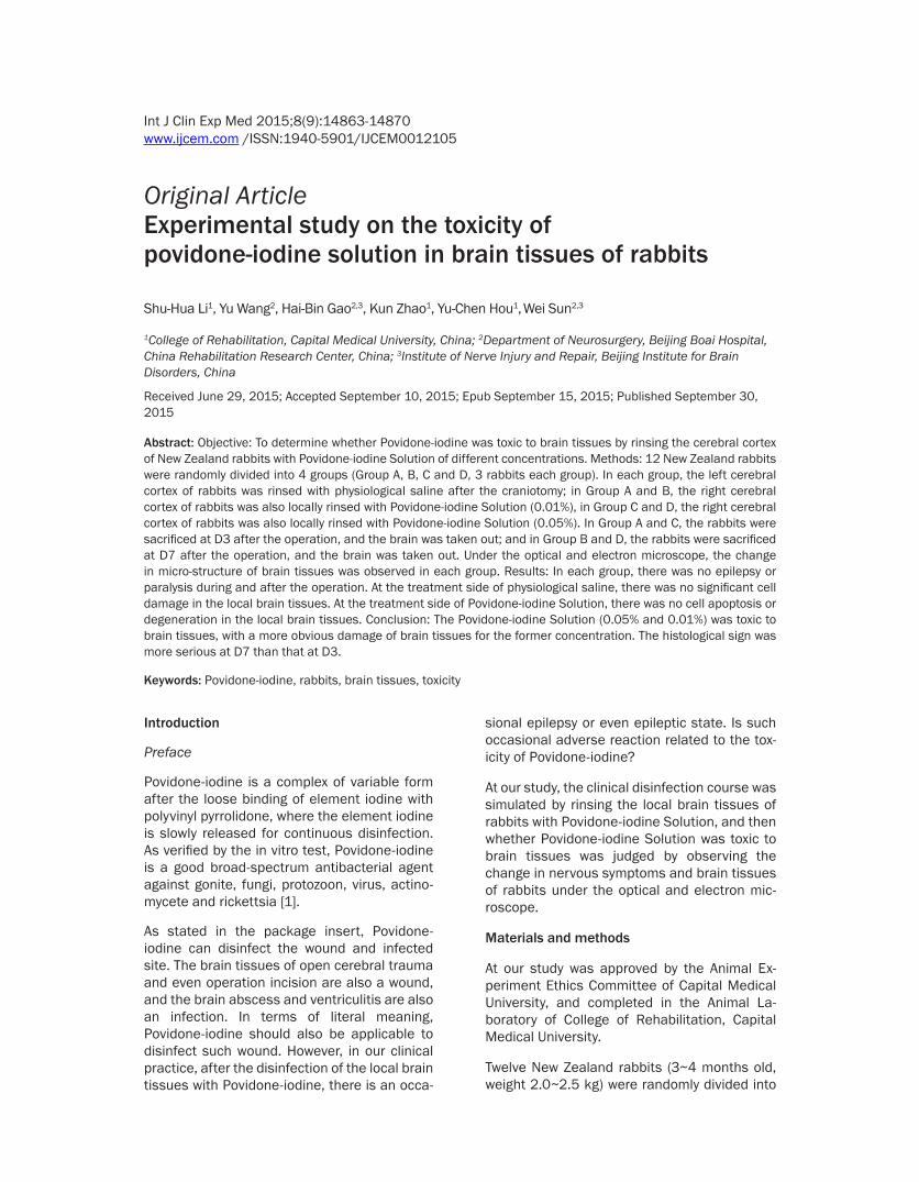
HE staining of local brain tissues rinsed with physiological saline at D3 and D7 after the operation in all eight rabbits: Tidy dense array of nerve cells, normal polarity, intact shape, abundant cytoplasm, plump nucleus, and clear nucleolus (Figure 1).
HE staining of local brain tissues rinsed with Povidone-iodine Solution (0.01%) at D3 after the operation in two rabbits of Group A: Dis- orderly lamination of nerve cells, deficiency of some nerve cells, and loose irregular array of remaining nerve cells (Figure 2).
HE staining of local brain tissues rinsed with Povidone-iodine Solution (0.01%) at D7 after
Figure 1. HE staining of local brain tissues rinsed with physiological saline after the operation in the 3rd day and the 7th day rabbit of different proportions contrast. A. HE staining of local brain tissues rinsed with physiological saline at D3 after the operation in the 2nd rabbit of Group A (× 100): Clear lamination of nerve cells, tidy dense array of nerve cells. B. HE staining of local brain tissues rinsed with physiological saline at D3 after the operation in the 2nd rabbit of Group A (× 400): Intact shape of nerve cells, abundant cytoplasm, plump nucleus, clear nucleolus. C. HE staining of local brain tissues rinsed with physiological saline at D7 after the operation in the 2nd rabbit of Group B (× 100): Clear lamination of nerve cells, tidy dense array of nerve cells. D. HE staining of local brain tissues rinsed with physiological saline at D7 after the operation in the 2nd rabbit of Group B (× 400): Intact shape of nerve cells, abundant cytoplasm, plump nucleus, clear nucleolus.

Cerebral toxicity of povidone-iodine rabbits
14866 Int J Clin Exp Med 2015;8(9):14863-14870
the operation in two rabbits of Group B and those rinsed with Povidone-iodine Solution (0.05%) at D3 after the operation in two rabbits of Group C: Disorderly lamination of nerve cells, deficiency of some nerve cells, apoptosis of very few cells under the high-power microscope (i.e. diminution of cyton, dense staining of cyto-plasm, and pyknosis of nucleus), degeneration of some cells (i.e. empty staining area around the cells, unclear display of cytomembrane/karyotheca, slight staining of cytoplasm/nucle-us and unclear display of nucleolus) (Figure 3).
HE staining of local brain tissues rinsed with Povidone-iodine Solution (0.05%) at D7 after the operation in two rabbits of Group D: Complete depolarity of nerve cells, apoptosis of numerous cells under the high-power micro-scope (Figure 4).
Transmission electron microscopy
Electron microscopy of local brain tissues rin- sed with physiological saline at D3 and D7 after the operation in four rabbits: Abundant cyto-plasm, slight edema of very few mitochondria,
Figure 2. HE staining of local brain tissues rinsed with Povidone-iodine Solution (0.01%) after the operation in the 3rd day rabbit of different proportions contrast. A. HE staining of local brain tissues rinsed with Povidone-iodine Solution (0.01%) at D3 after the operation in the 3rd rabbit of Group A (× 100): Disorderly lamination of nerve cells, deficiency of some nerve cells. B. HE staining of local brain tissues rinsed with Povidone-iodine Solution (0.01%) at D3 after the operation in the 3rd rabbit of Group A (× 400): Count decrease of nerve cells, apoptosis of some nerve cells.
Figure 3. HE staining of local brain tissues rinsed with Povidone-iodine Solution (0.05%) after the operation in the 3rd day rabbit of different proportions contrast. A. HE staining of local brain tissues rinsed with Povidone-iodine Solution (0.05%) at D3 after the operation in the 2nd rabbit of Group C (× 100): Disorderly lamination of nerve cells, deficiency of some nerve cells. B. HE staining of local brain tissues rinsed with Povidone-iodine Solution (0.05%) at D3 after the operation in the 2nd rabbit of Group C (× 400): Count decrease of nerve cells, apoptosis of some nerve cells, degeneration of some nerve cells.

Cerebral toxicity of povidone-iodine rabbits
14867 Int J Clin Exp Med 2015;8(9):14863-14870
regular nucleus, intact neural medullary sheath (Figure 5).
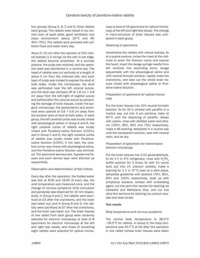
Electron microscopy of local brain tissues rinsed with Povidone-iodine Solution (0.01%) at D3 after the operation in Group A, those rinsed with Povidone-iodine Solution (0.01%) at D7 after the operation in Group B and those rinsed with Povidone-iodine Solution (0.05%) at D3 after the operation in Group C: Obvious edema in cytoplasm/mitochondrion of nerve cells, shallow stroma, short mitochondrial crista, pyk-nosis of nucleus, condensation of nuclear chro-
matin, dispersion of some laminae in medullary sheath (Figure 6).
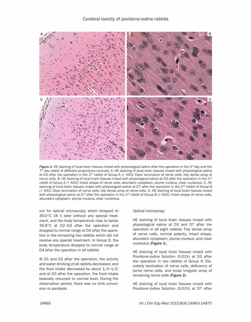
Electron microscopy of local brain tissues rinsed with Povidone-iodine Solution (0.05%) at D7 after the operation in Group D: Significant decrease in the count of organelle in the cyto-plasm of nerve cells (e.g. mitochondrion and endoplasmic reticulum), many vacuole in cyto-plasm, serious pyknosis of nucleus, condensa-tion of many chromatins in nucleus, dispersion and serious fracture of medullary sheath (Figure 7).
Figure 4. HE staining of local brain tissues rinsed with Povidone-iodine Solution (0.05%) after the operation in the 7th day rabbit of different proportions contrast. A. HE staining of local brain tissues rinsed with Povidone-iodine Solution (0.05%) at D7 after the operation in the 2nd rabbit of Group D (× 100): Disorderly lamination of nerve cells, deficiency of some nerve cells. B. HE staining of local brain tissues rinsed with Povidone-iodine Solution (0.05%) at D7 after the operation in the 2nd rabbit of Group D (× 400): Count decrease of nerve cells, apoptosis of numerous nerve cells.
Figure 5. Transmission electron microscopy of local brain tissues different magnification rinsed with physiological saline after the operation in 7th day rabbit of different proportions contrast. A. Transmission electron microscopy (bar = 5 um) of local brain tissues rinsed with physiological saline at D7 after the operation in Group B: Regular nucleus of nerve cells, clear nucleolus. B. Transmission electron microscopy (bar = 1 um) of local brain tissues rinsed with physiological saline at D7 after the operation in Group B: Slight swelling of very few mitochondrion in nerve cells. C. Transmission electron microscopy (bar = 1 um) of local brain tissues rinsed with physiological saline at D7 after the operation in Group B: Intact medullary sheath.
Cerebral toxicity of povidone-iodine rabbits
14868 Int J Clin Exp Med 2015;8(9):14863-14870
Discussion
Toxic effect of Povidone-iodine
In the relevant Chinese and English literatures, no unanimous conclusion was reached on whether Povidone-iodine was toxic to the body and tissues.
The safety of Povidone-iodine for body was reported in some studies. In the study of Zeng
Ling et al, the toxic symptoms and death did not appear in the mice within 14 d after the gastric lavage of Povidone-iodine Solution (5000 mg/kg), indicating a LD50 > 5000 mg/kg and no acute toxicity. In the sub-acute toxicity study of Li Li et al, the various indices were not signifi-cantly different between each test group and control group (e.g. weight, blood routine exami-nation, biochemical examination and visceral index) after the 30 d continuous gastric lavage of Povidone-iodine Solution (250, 500 and
Figure 6. Transmission electron microscopy of local brain tissues different magnification rinsed with Povidone-iodine Solution (0.01%) after the operation in 7th day rabbit of different proportions contrast. A. Transmission electron mi-croscopy (bar = 2 um) of local brain tissues rinsed with Povidone-iodine Solution (0.01%) at D7 after the operation in Group B. Pyknosis of nucleus, condensation of chromatin. B: Transmission electron microscopy (bar = 1 um) of local brain tissues rinsed with Povidone-iodine Solution (0.01%) at D7 after the operation in Group B: Obvious swelling of mitochondrion in nerve cells. C. Transmission electron microscopy (bar = 1 um) of local brain tissues rinsed with Povidone-iodine Solution (0.01%) at D7 after the operation in Group B: Dispersion of medullary sheath.
Figure 7. Transmission electron microscopy of local brain tissues different magnification rinsed with Povidone-iodine Solution (0.05%) after the operation in 7th day rabbit of different proportions contrast. A. Transmission electron mi-croscopy (bar = 5 um) of local brain tissues rinsed with Povidone-iodine Solution (0.05%) at D7 after the operation in Group D: Count decrease of organelle in the cytoplasm of nerve cells, vacuole in cytoplasm, pyknosis of nucleus, condensation of chromatin. B. Transmission electron microscopy (bar = 2 um) of local brain tissues rinsed with Povidone-iodine Solution (0.05%) at D7 after the operation in Group D: Decrease in the count of organelle in nerve cells (e.g. mitochondrion and endoplasmic reticulum), vacuole in cytoplasm. C. Transmission electron microscopy (bar = 1 um) of local brain tissues rinsed with Povidone-iodine Solution (0.05%) at D7 after the operation in Group D: Disintegration of medullary sheath.

Cerebral toxicity of povidone-iodine rabbits
14869 Int J Clin Exp Med 2015;8(9):14863-14870
1000 mg/kg). In these two studies, since the toxic effect of Povidone-iodine was assessed through its gastric lavage in mice or rats, the study results were influenced by the degree of its gastrointestinal absorption (i.e. if Povidone-iodine was very little or even not absorbed by the intestines, the observation indices would be seriously influenced). In the study of Müller G [2], Povidone-iodine Solution of different con-centrations was added into ovarian cell of Chinese hamsters (CHO-K1 cells) to cultivate for 4 h, and the damage conditions of chromo-some were assessed through the comet elec-trophoresis and chromosome aberration test: Povidone-iodine Solution (≤ 2.5%) was not toxic to the chromosome of ovarian cell of Chinese hamsters. In the randomized control study of Chang FY et al [3] on 244 patients with unsta-ble vertebral degeneration and necessary for lumbosacral posterolateral fusion, compared with that after the lavage of physiological saline before the vertebral fusion, the infection rate was lower but other indices (i.e. vertebral fusion, wound heal, pain improvement, func-tional score and walking ability) were not signi- ficantly different after the local lavage of Povidone-iodine Solution (0.35%) before the vertebral fusion, indicating that Povidone-io- dine Solution (0.35%) was safe for body, which was not observed in a histological way. In the study of Zeng Ling et al, 30 s after the dropping of Povidone-iodine Solution (0.5%), the eyes of rabbits were rinsed with physiological saline, there was no cornea/iris damage or conjuncti-va edema within the observation time of 1 h, 24 h, 48 h, 72 h, 7 d, 14 d and 21 d, indicating that Povidone-iodine Solution (0.5%) did not irritate the eyes of rabbits, which did not con-sist with the results of other studies.
However, some studies showed that Povidone-iodine was toxic. In the study of Balin AK et al [4], Povidone-iodine Solution of different con-centrations was added into the culture medium to cultivate the skin fibroblast of adults and the lung fibroblast of infants, Povidone-iodine So- lution (0.01% and 0.025%) could prolong the multiplication time of fibrocyte but Povidone-iodine Solution (0.1% and 1%) could stop the mitosis of fibrocyte for 0.1%, indicating that Povidone-iodine was toxic at a correlation with the concentration. In the study of Morizono T [5], the vestibular window of chinchilla was rinsed with Povidone-iodine Solution of differ-ent concentrations, and Povidone-iodine Solu-
tion (0.25%) could significantly change the threshold of complex action potential of cochle-ar auditory nerve of chinchilla, indicating that Povidone-iodine Solution could be absorbed into internal ear via vestibular window to influ-ence the function of cochlear auditory sensor. In the study of Naor J et al [6], at D1 and D7 after the injection of Povidone-iodine Solution into ocular antechamber, the relevant examina-tions were made (i.e. micro-structure observa-tion of cornea, ultrasonic thickness measure-ment of cornea, measurement of intraocular pressure and pathological examination), Povi- done-iodine Solution (1%) could cause an ede- ma and opacity in cornea (with a deficiency of endothelial cells under the microscope) and an obvious increase in intraocular pressure, but Povidone-iodine Solution (0.01% and 0.1%) did not influence the cornea, indicating that the toxicity of Povidone-iodine Solution depended on its concentration. In the study of Robertson P et al [7] on a burn infant of 22 months old, at D29 after the local rinsing with Povidone-iodine (once every 2 d), there was a thyroid crisis (with a significant rise of iodine ion and thyroid hor-mone in blood) which was gradually improved after its withdrawal and the symptomatic treat-ment with Propranolol + Carbimazole (po), indi-cating that Povidone-iodine Solution could be absorbed into blood via the local burned skin.
Neurotoxicity of Povidone-iodine
In the study of Doan L et al [8], Povidone-iodine Solution (≥ 0.2%) could significantly reduce the survival rate of human SH-SY5Y neuroblastoma cells and mice RSC96 Schwann’s cells. In the study of Akcay E et al [9], 16 rats were random-ly divided into two groups, the vertebral pla- te and endorachis were incised, the gelatin sponge soaked with Povidone-iodine Solution (0.1%) was put at the subdural site in test group, the dry gelatin sponge was put at the subdural site, the incision was stitched up, the local spinal cord tissues were taken 3 weeks later for pathological examination: both groups were significantly different in the degeneration of spinal cord neuron (P = 0.038), the degener-ation of neuraxon (P = 0.038) and the demyelin-ation of neuron (P = 0.005), indicating that Povidone-iodine Solution was toxic to the ner- ves of spinal cord. In the relevant Chinese and English literatures on the toxicity of Povidone-iodine, there were no studies on whether Po- vidone-iodine was toxic to the living brain tis-

Cerebral toxicity of povidone-iodine rabbits
14870 Int J Clin Exp Med 2015;8(9):14863-14870
sues. However, in the study of Strohecker J et al [10], the epidural site of lumbar vertebra in rab-bits was rinsed with Povidone-iodine Solution, the incision was stitched up and then opened 1 month later, and the local spinal cord was taken for electron microscopy, which showed no fibril-lation of spinal cord or adhesion of arachnoid.
At our study, the clinical disinfection course was simulated by rinsing the local brain tissues of rabbits with Povidone-iodine Solution, and then whether Povidone-iodine Solution was toxic to brain tissues was observed, which filled the blank of studies on whether Povidone-iodine was toxic to the living brain tissues. If Povidone-iodine Solution (0.01% and 0.05%, i.e. lower than the common clinical dose of 0.5%) was verified as toxic to the brain tissues of rabbits, Povidone-iodine Solution of higher concentration would theoretically also be toxic to the living brain tissues of rabbits. In the study of Berkelman RL [11], Povidone-iodine Solution (0.01%) was well antibacterial, at a disinfection rate of up to 100% for Staphylococcus aureus and mycobacteria. There were no studies on the disinfection effect of Povidone-iodine at a lower concentration.
At our study, after the local brain tissues of rab-bits were rinsed with Povidone-iodine Solution (0.05% and 0.01%), there was a damage of nerve cells but no nerve irritation/deficiency of rabbits under the optical and electron micro-scope, because the important functional areas of rabbits were not rinsed with Povidone-iodine during the operation and the observation time was short after the operation. At our study, Povidone-iodine Solution was toxic to brain tis-sues, whose correlation to iodine element, polyvinyl pyrrolidone or their combined action should be determined through the further studies.
Conclusion
Povidone-iodine Solution (0.05% and 0.01%) was toxic to brain tissues, with a more obvious damage of brain tissues for the former concen-tration. The histological sign was more serious at D7 than that at D3.
Disclosure of conflict of interest
None.
Address correspondence to: Wei Sun, Head of Research Group, Institute of Nerve Injury and Repair, Beijing Institute for Brain Disorders; Head of Department of Neurosurgery, Beijing Boai Hospital, China Rehabilitation Research Center, Beijing 100068, China. Tel: +8601087569052; Fax: +860- 1067563322; E-mail: [email protected]
References
[1] Özkiriş M, Kapusuz Z, Saydam L. Ototoxicity of different concentrations povidone-iodine solu-tion applied to the middle ear cavity of rats. Indian J Otolaryngol Head Neck Surg 2013; 65: 168-172.
[2] Müller G, Hai DN, Kramer A. Lack of in vitro Genotoxicity of Povidone-Iodine in Solution, in Ointment or in a Liposomal Formulation (Re- pithel). Dermatology 2006; 212: 94-97.
[3] Chang FY, Chang MC, Wang ST, Yu WK, Liu CL, Chen TH. Can povidone-iodine solution be used safely in a spinal surgery? Eur Spine J 2006; 15: 1005-1014.
[4] Balin AK, Pratt L. Dilute povidone-iodine solu-tions inhibit human skin fibroblast growth. Dermatol Surg 2002; 28: 210-214.
[5] Morizono T, Sikora MA. The ototoxicity of topi-cally applied povidone-iodine preparations. A Arch Otolaryngol 1982; 108: 210-213.
[6] Naor J, Savion N, Blumenthal M, Assia EI. Corneal endothelial cytotoxicity of diluted povi-done--iodine. J Cataract Refract Surg 2001; 27: 941-947.
[7] Robertson P, Fraser J, Sheild J, Weir P. Thyrotoxicosis related to iodine toxicity in a paediatric burn patient. Intensive Care Med 2002; 28: 1369.
[8] Doan L, Piskoun B, Rosenberg AD, Blanck TJ, Phillips MS, Xu F. In Vitro Antiseptic Effects on Viability of Neuronal and Schwann Cells. Reg Anesth Pain Med 2012; 37: 131-138.
[9] Akcay E, Ersahin Y, Ozer F, Duransoy YK, Camlar M, Atci I, Yagci A, Ozer O. Neurotoxic effect of povidone-iodine on the rat spine us-ing a laminectomy-durotomy model. Childs Nerv Syst 2012; 28: 2071-2075.
[10] Strohecker J, Lametschwandtner A, Piotrowski WP. Scanning electron microscopic findings on the rabbit meninges after local lavage with Polyvinylpyrrolidon-Jod-Komplex. Acta Neuro- chir (Wien) 1985; 77: 72-74.
[11] Berkelman RL, Holland BW, Anderson RL. Increased bactericidal activity of dilute prepa-rations of povidone-iodine solutions. J Clin Microbiol 1982; 15: 635-639.