original article human b-cell killing by autoreactive ... · human b-cell killing by autoreactive...
TRANSCRIPT
Human b-Cell Killing by Autoreactive Preproinsulin-Specific CD8 T Cells Is Predominantly Granule-MediatedWith the Potency Dependent Upon T-CellReceptor AvidityRobin R. Knight,
1Deborah Kronenberg,
1,2Min Zhao,
3Guo Cai Huang,
3Martin Eichmann,
1
Anna Bulek,4Linda Wooldridge,
4David K. Cole,
4Andrew K. Sewell,
4Mark Peakman,
1,2
and Ania Skowera1,2
The end-stage immunopathology of type 1 diabetes resulting inb-cell destruction appears to be strongly dominated by cytotoxicCD8 T lymphocytes (CD8 T cells). However, the mechanismof cytotoxicity used by autoreactive CD8 T cells in the humansetting remains unknown. Using type 1 diabetes patient–derivedpreproinsulin-specific CD8 T-cell clones recognizing eitheran HLA-A2 (A*0201) or HLA-A24 (A*2402)-restricted epitope(peptide of preproinsulin [PPI]15–24, ALWGPDPAAA; or PPI3–11,LWMRLLPLL), we assessed the use of conventional mediators ofcytotoxicity in the destruction of human b-cells in vitro comparedwith virus-specific cytotoxic CD8 T-cell clones. We show thatPPI-specific CD8 T-cell clones are mainly reliant upon cytotoxicdegranulation for inducing b-cell death. Furthermore, we findthat in comparison with virus-specific CD8 T cells, there are dif-ferences in the killing potency of PPI-specific CD8 T cells that arenot due to cell-intrinsic differences, but rather are mediated bydifferences in strength of signaling by peptide–HLA ligands. Thestudy highlights the regulation of b-cell killing as a potential pointfor therapeutic control, including the possibility of blockingautoreactive CD8 T-cell function without impacting upon generalimmune competence. Diabetes 62:205–213, 2013
The impaired glucose homeostasis that is charac-teristic of type 1 diabetes results from the se-lective destruction of insulin producing b-cellswithin pancreatic islets. Cytotoxic CD8 T lym-
phocytes (CD8 T cells) have been implicated as majormediators of b-cell damage on the basis of multiple strandsof evidence including CD8 T-cell dominance of the isletinfiltrate (1,2); single nucleotide polymorphism studies (3)showing that HLA class I genes predispose to type 1 di-abetes; and numerous studies in animal models that sup-port a CD8-centric view of b-cell killing (4,5), including,more recently, direct visualization (6). Evidence that CD8
T cells recognizing b-cell autoantigens have a direct role inb-cell death in human disease was obtained recently fol-lowing the establishment of CD8 T-cell clones specific foran epitope from the signal peptide of preproinsulin (PPI)from the blood of a newly diagnosed type 1 diabetes pa-tient (7). These CD8 T cells recognize residues 15–24 ofPPI presented by HLA-A*0201 and selectively kill HLA-A2+ human b-cells in vitro (7). Using both enzyme-linkedimmunospot assays and PPI15–24 tetramer staining, it wassubsequently shown that CD8 T cells recognizing PPI15–24are enriched in the peripheral blood of patients near todisease onset (7,8). More recently, in situ PPI15–24 tetramerstaining has shown that cells of this specificity are presentin insulitic lesions characteristic of the disease (9).
CD8 T cells deliver cytolytic effector functions againsttarget cells via contact-dependent mechanisms that use twodiscrete pathways: cytotoxic degranulation or interactionwith tumor necrosis factor (TNF) family-related deathreceptors. Cytotoxic degranulation involves the release ofperforin, which facilitates entry of coreleased granzymeswith serine protease activity into cells, resulting in rapidcell death (10). Fas ligand (FasL) is the best-characterizedTNF family-related death receptor, binding to Fas ex-pressed on the target cell surface and initiating a series ofintracellular pathways resulting in apoptosis (11). Althoughcytotoxic degranulation is recognized as the main, verypotent lytic process used in the clearance of bacterial andviral pathogens, as well as being implicated in the killing oftumor cells (10,12), it is also known that this pathway canact in concert with FasL for pathogen removal (13).
Given the persuasive evidence that CD8 T cells con-tribute to b-cell death, the identity of the cytotoxic mech-anisms that are used in this selective, destructive processrepresent an important knowledge gap. Studies that havepreviously attempted to address this question have usedthe nonobese diabetic (NOD) mouse model or human isletspulsed to present viral peptide to cognate antiviral CD8 T-cell clones (14). The results of these studies highlight theimportance of granzyme and perforin in the murine modelof type 1 diabetes and in virus-specific CD8 T-cell killingof virus peptide-pulsed islets, but raise the importantquestion of the role of these mediators in the human au-toimmune setting (15), in which CD8 T-cell biology may bedifferent (16).
Novel insights into this process in a relevant humanmodel could potentially enable the development of im-mune-based therapeutic approaches that specifically tar-get pathways used by b-cell–specific CD8 T cells, but leave
From the 1Department of Immunobiology, King’s College London, London,United Kingdom; the 2National Institute for Health Research comprehensiveBiomedical Research Centre, Guy’s and St. Thomas’ National Health ServiceFoundation Trust and King’s College London, London, United Kingdom;3Diabetes and Nutritional Science, King’s College London, London, UnitedKingdom; and the 4Institute of Infection and Immunity, Cardiff UniversitySchool of Medicine, Cardiff, United Kingdom.
Corresponding author: Mark Peakman, [email protected] 11 March 2012 and accepted 7 July 2012.DOI: 10.2337/db12-0315This article contains Supplementary Data online at http://diabetes
.diabetesjournals.org/lookup/suppl/doi:10.2337/db12-0315/-/DC1.M.P. and A.S. contributed equally to this work.� 2013 by the American Diabetes Association. Readers may use this article as
long as the work is properly cited, the use is educational and not for profit,and the work is not altered. See http://creativecommons.org/licenses/by-nc-nd/3.0/ for details.
diabetes.diabetesjournals.org DIABETES, VOL. 62, JANUARY 2013 205
ORIGINAL ARTICLE
antiviral and antitumor killing intact (16). The currentstudy was therefore designed to elucidate the mechanism(s) of CD8 T-cell–mediated b-cell death in human type 1diabetes using autoreactive CD8 T-cell clones isolatedfrom patients with new-onset disease. We obtained CD8 Tcells from two donors with specificity for different PPIepitopes, restricted by two different HLA-A alleles, as wellas virus-specific CD8 T cells as a well-established refer-ence point. The roles of TNF-related death receptorligands, together with degranulation, were assessed incoculture experiments using HLA-matched human isletcells and antigen-pulsed target cell lines in the presenceand absence of relevant cytotoxic pathway inhibitors. Inaddition, we examined whether any differences in themechanism and potency of killing by autoreactive andantiviral CD8 T cells could be attributable to cell-intrinsic
factors or extrinsic effects such as the strength of peptide–HLA/T-cell receptor (TCR) interaction.
RESEARCH DESIGN AND METHODS
Generation and maintenance of CD8 T-cell clones and cell lines. PPIsignal peptide (SP) epitope-specific CD8 T-cell clones were isolated frompatients with new-onset type 1 diabetes as previously described (7,17). TheHLA-A2–restricted A2-PPI15–24 CD8 T-cell clone recognizes residues 15–24 ofPPI SP (ALWGPDPAAA), and the HLA-A24–restricted A24-PPI3–11 CD8 T-cellclone recognizes a novel naturally b-cell–processed and HLA-A24–presentedepitope of PPI SP represented by amino acids 3–11 (LWMRLLPLL) (17). ViralCD8 T-cell clones specific for cytomegalovirus (CMV) antigens and restrictedby either HLA-A2 or HLA-A24 peptide were isolated alongside using a similarapproach. The HLA-A2–restricted CD8 T-cell clone A2-CMVpp65495–503 isspecific for residues 495–503 of CMV protein pp65 (NLVPMVATV). The HLA-A24–restricted CD8 T-cell clone A24-CMV-IE1248–257 recognizes an epitopefrom CMV 1E-1 (248–257; AYAQKIFKIL). Target cell lines C1R and K562
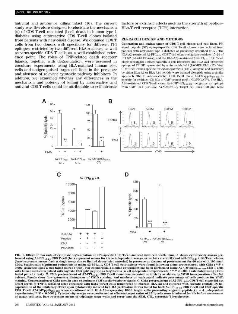
FIG. 1. Effect of blockade of cytotoxic degranulation on PPI-specific CD8 T-cell–induced islet cell death. Panel A shows cytotoxicity assays per-formed using A2-PPI15–24 CD8 T-cell (bars represent means for three independent assays; error bars are SEM) and A24-PPI3–11 CD8 T-cell clones(bars represent means from a single assay due to limited donor islet material) in presence or absence of pretreatment for 60 min with 100 nmolCMA. Statistically significant reductions in mean A2-PPI15–24 CD8 T-cell cytotoxicity were found following clone pretreatment with CMA (**P =0.002; assigned using a two-tailed paired t test). For comparison, a similar experiment has been performed using A2-CMVpp65495–503 CD8 T cellswith human islet cells pulsed with cognate CMVpp65 peptide as target cells (n = 3 independent experiments; ***P < 0.0001 calculated using a two-tailed paired t test). B: CMA pretreatment of A2-PPI15–24 CD8 T-cell clone demonstrated no toxicity as shown by ViViD incorporation after 5-hculture. Panels show flow cytometry histograms of ViViD staining, and numbers on each panel indicate percentage of cells positive for ViViDstaining. Concentration of CMA used in each experiment (nM) is shown above panels. C: CMA pretreatment of A2-PPI15–24 CD8 T-cell clone did notaffect levels of TNF-a released after coculture with K562 target cells transfected to express HLA-A2 and cultured with cognate peptide. D: Re-capitulation of the inhibitory effect upon cytotoxicity induced by CMA pretreatment was found for both A2-PPI15–24 CD8 T-cell and CMV-specificCD8 T-cell A2-CMVpp65495–503 when cocultured with HLA-A2–expressing K562 target cells presenting cognate peptide (n = 4 independentexperiments; ***P < 0.0001). Cytotoxicity assays were performed at effector/target ratios of 25:1; cells were incubated for 4 h before assessmentof target cell lysis. Bars represent means of triplicate assay wells and error bars the SEM. CTL, cytotoxic T lymphocyte.
b-CELL KILLING BY CTLs
206 DIABETES, VOL. 62, JANUARY 2013 diabetes.diabetesjournals.org
transfected with HLA-A*0201 were cultured in RPMI 1640 (Gibco) supple-mented with 10% FCS (Gibco) containing 100 IU/ml penicillin and 100 mg/mLstreptomycin and pulsed with peptide (10 mg/mL) for 1 h (37°C) followed bywashing.Islet-cell cytotoxicity assays. Human islet isolations were performed aspreviously described (18) using pancreata retrieved with the consent ofdonors’ relatives and permission of the Ethical Review Committee of King’sCollege Hospital. Islet cells in monolayer cultures were cultured for 16–24 h inmedium containing 16 mmol glucose and cytokines interleukin (IL)-1b (50 IU/mL; Strathmann Biotec), TNF-a (2500 IU/mL; Mitenyi Biotec), interferon(IFN)-g (500 IU/mL; Miltenyi Biotec), and IFN-a (1000 IU/mL; Roche Labora-tories) to increase class I expression and provide a greater dynamic range foranalyzing the effect of inhibitory reagents on pathways of cytotoxicity (19).Cytotoxicity was analyzed as described (7,20) and specific cytotoxicity cal-culated using the formula: percent specific release = (experimental 2 spon-taneous release) 3 100/(maximum 2 spontaneous release). In a modifiedcytotoxicity assay conducted over 24 h to examine the role of Fas–FasLinteractions, RNA was isolated from islet cell/CD8 T-cell clone coculturesusing the RNeasy Mini Kit (Qiagen) and cDNA generated (High-Capacity cDNAReverse-Transcription Kit; Applied Biosystems) before quantitative PCR wasperformed on an Applied Biosystems 7900HT Fast Real-Time PCR System withTaqMan Gene Expression Master Mix and TaqMan Gene Expression Assaysfor Insulin (Hs02741908-m1) and GAPDH (4326317E; Applied Biosystems).Cytotoxicity was measured as the percentage loss of insulin mRNA expressionin test cocultures compared with islets cultured with an irrelevant T-cell clone.Inhibitory reagents. To inhibit perforin activity, CD8 T cells were cultured for1 h prior to assay in medium containing concanamycin A (CMA, 100 nmol;Sigma-Aldrich) or diluent (DMSO). Blocking antibodies (or isotype-matchedirrelevant controls) were present throughout assays for FasL (5 mg/mL cloneNOK-1; BioLegend), TNF-related apoptosis-inducing ligand (TRAIL; 5 mg/mLclone RIK-1; BioLegend), and TNF-a (12.5 mg/mL; provided by Dr. Tim Bourne,UCB Celltech).Carboxyfluorescein succinimidyl ester–based cytotoxicity assay. Pe-ripheral blood mononuclear cells (PBMCs) (106) from HLA-A2+ healthy do-nors were incubated with high or low concentrations of carboxyfluoresceinsuccinimidyl ester (CFSE; 1.25 and 0.1 mmol, respectively) for 15 min at roomtemperature (RT). Labeling was quenched using 1 mL FCS (Invitrogen) andCFSE-low–labeled PBMCs were peptide-pulsed, whereas CFSE-high–labeledcells were pulsed with peptide diluent. PBMCs were then washed thoroughly,combined at a 1:1 ratio, and cocultured with T-cell clones for 4 or 8 h beforestaining with anti-CD14 antibody (BD Biosciences). Assessment of CD14+
CFSELOW and CFSEHIGH cell percentages was performed by flow cytometryand analyzed using FlowJo software (Tree Star, Ashland, OR).Assessing intracellular protein and CD107a surface expression. Anti-CD107a (fluorescein isothiocyanate; clone H4A3; BD Biosciences) was addedto 5 3 105 cloned CD8 T cells in FACS tubes containing X-Vivo 15/5% ABserum/IL-7 (10 ng/mL), IL-15 (0.1 ng/mL), and 2.5% Cellkine (ZeptoMetrixCorporation). Islet or nonislet target cell preparations were added to tubes (53 105) for 1 h at 37°C (5% CO2) before 0.7 mg/mL monensin (GolgiStop; BDBiosciences) and 1 mg/mL brefeldin A (GolgiPlug; BD Biosciences) wereadded to cocultures. After 4 h incubation, cells were washed twice andresuspended in PBS containing the amine reactive dye Violet Viable Dye(ViViD; Invitrogen/Molecular Probes) for exclusion of dead cells. Cells wereincubated at 4°C, washed in PBS containing 2% FCS, and stained with anti-CD8antibody (BD Pharmingen). After 25 min at 4°C, cells were washed andresuspended in 300 mL BD-Cytofix/Cytoperm solution (BD Biosciences) andincubated at RT for 15 min. Cells were washed and resuspended in 500 mL BD-Fixation/Permeabilization solution for 10 min (RT) before being centrifuged at4003 g for 5 min and stained with antibodies for granzyme B (clone GB11; BDBiosciences) and TNF-a (clone MAb11; BD Pharmingen). Cells were in-cubated with antibody for 25 min at 4°C, washed, and analyzed by flowcytometry.Measuring cytokines in coculture supernatant. Cloned CD8 T cells (2 3104) were seeded in 96-well plates containing 2 3 104 target cells and culturedfor 18 h (37°C/5% CO2) in medium containing X-Vivo 15/5% AB serum/IL-7 (10ng/mL), IL-15 (0.1 ng/mL), and 2.5% Cellkine and cytokines measured byLuminex technology (Milliplex Mag Kit; Millipore).Surface plasmon resonance analysis. Binding analysis was performed usinga BIAcore T100 equipped with a CM5 sensor chip (BIAcore) as reportedpreviously (21).
RESULTS
PPI-specific CD8 T-cell clones kill human isletsprimarily through cytotoxic degranulation. For thesestudies, we focused on two CD8 T-cell clones that
recognize PPI and kill b-cells: those specific for PPI15–24(ALWGPDPAAA) presented by HLA-A2 (A2-PPI15–24 CD8 Tcells) (7) and for PPI3–11 (LWMRLLPLL; A24-PPI3–11 CD8 Tcells) (17). In a replication of our original report (7), A2-PPI15–24 CD8 T-cell clone 3F2 kills human HLA-matchedislets (different donor than that used in the original stud-ies) (Supplementary Fig. 1A). As a comparator, we useda CD8 T-cell clone recognizing the dominant pp65 epitopeof CMV (pp65495–503; NLVPMVATV) presented by HLA-A2that does not kill human islets in the absence of pulsingwith cognate peptide (Supplementary Fig. 1A). During theinteraction with HLA-A2+ human islets, the A2-PPI15–24CD8 T-cell clones secrete IFN-g and macrophage in-flammatory protein-1b (Supplementary Fig. 1B and C) andare also known to secrete TNF-a (7). It is noteworthy thatunder conditions using islets prepulsed with cognatepeptide, levels of killing and cytokine secretion are gen-erally higher with the antiviral CD8 T cell (see below).Similar results for human islet killing were obtained usingthe A24-PPI3–11 CD8 T cell (17) and an HLA-A24–restrictedCMV-specific comparator clone (A24-CMV-IE1248–257;AYAQKIFKIL) (22,23).
We next examined the effects of degranulation in-hibition on this killing process. CMA degrades perforinwithin cytotoxic granules of CD8 T cells and has littleimpact upon the degranulation process (24). Pretreatmentof CD8 T-cell clones with CMA 60 min before human isletcoculture resulted in significant reduction in islet killing byA2-PPI15–24 CD8 T cells (P = 0.002; Fig. 1A). In comple-mentary studies, a similar reduction in A24-PPI3–11 CD8T-cell cytotoxicity against A24+ human islet cells was alsoobserved (Fig. 1A). As a comparison, CMA pretreatment ofA2-CMVpp65495–503CD8 T-cell clones also inhibited killingof pp65495–503 peptide-pulsed HLA-A2+ human islet cells(P , 0.0001; Fig. 1A). The observed reduction in cytotox-icity was not due to toxic effects of CMA, as demonstratedby assessing viability of A2-PPI15–24 CD8 T-cell clone (Fig.1B), nor did it impede release of TNF-a (Fig. 1C). Thesedata implied usage of perforin for the targeted cellularentry of granule contents as a major mechanism ofautoreactive CD8 T-cell killing of islet cells.
These effects could be recapitulated using the K562myeloid cell line transfected with HLA-A2 (A*0201) (K562-A2) (7). Pretreatment of A2-PPI15–24 CD8 T cells with CMAreduced killing of K562-A2 cells pulsed with cognate PPI15–24peptide (P , 0.0001; Fig. 1D). A similar reliance uponperforin was observed for A2-CMVpp65495–503 CD8 T cellswhen cocultured with target K562-A2 cells pulsed withCMVpp65495–503 (P , 0.0001; Fig. 1D). As further evidencethat granule content release is an important sequel of ac-tivation of these CD8 T cells, we showed a modest re-duction in intracellular granzyme B following engagementwith human islets (example shown for A2-PPI15–24 CD8 Tcell) (Supplementary Fig. 1D). Taken together, theseresults indicate that autoreactive CD8 T cells specific forPPI targets presented by human islet cells release cyto-toxic granule contents as their predominant mechanism oftarget cell killing.The role of Fas–FasL interactions. Using flow cytom-etry, we observed that all PPI- and CMV-specific CD8 Tcells have intracellular stores of TRAIL and FasL (Sup-plementary Fig. 2A). We have shown that pretreatment ofhuman islets with proinflammatory cytokines increasessurface expression of HLA class I and enhances theirkilling by A2-PPI15–24 CD8 T cells (19); phenotypic analysisof cytokine pretreated islets from three donors revealed
R.R. KNIGHT AND ASSOCIATES
diabetes.diabetesjournals.org DIABETES, VOL. 62, JANUARY 2013 207
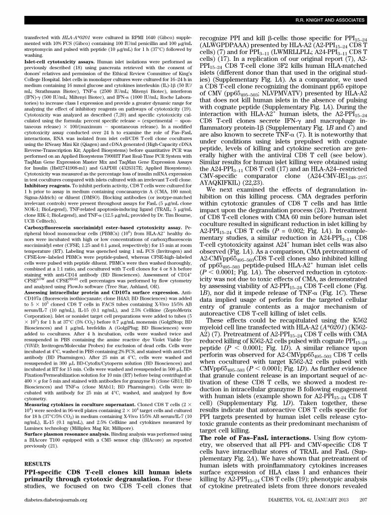
FIG. 2. Assessing Fas-induced cell death of peptide-pulsed monocytes and human islet target cells by PPI-specific CD8 T-cell clone. Panel A showsmean levels of Fas (CD95) expression on islets from three donors using flow cytometry analysis. Fas expression increases when islet cells arepretreated with proinflammatory cytokines (*P = 0.013). B and C: To assess potential FasL usage in killing, PBMCs were labeled with two differentCFSE concentrations and pulsed with wild-type peptide (CFSE
LOW) or DMSO diluent (CFSE
HIGH) and cocultured together with CD8 T-cell clone in
the presence or absence of anti-FasL antibody. The Fas-expressing CD14+population was assessed for CFSE loss. A2-PPI15–24 CD8 T cell showed
reduced cytotoxicity of Fas+peptide-pulsed monocytes after 8 h with FasL blockade. Example staining is shown in panel B and accumulated data
from three independent experiments in panel C (8-h time point; **P = 0.0095). D: A2-PPI15–24 and A24-PPI3–11 CD8 T cell showed no inhibition ofcytotoxicity when cultured with islet targets in the presence of monoclonal anti-FasL antibody (5 mg/mL) or isotype control. Cytotoxicity assayswere performed for 4 h at an effector/target ratio of 25:1. Bars represent means of triplicate assay wells and error bars SEMs. E: Blocking FasL onA2-PPI15–24 CD8 T cells in coculture with islet cells does not preserve b-cell function, as measured by insulin mRNA levels. Bars represent meaninsulin mRNA levels within islet coculture conditions, relative to insulin mRNA in cocultures with control clone A2-CMVpp65495–503. CD8 T-cellclones were incubated with islet cells at a ratio of 25:1 for 24 h to maximize visualization of the involvement by FasL on b-cell killing. BlockingFasL antibody remained present within cultures throughout assays. CTL, cytotoxic T lymphocyte.
b-CELL KILLING BY CTLs
208 DIABETES, VOL. 62, JANUARY 2013 diabetes.diabetesjournals.org
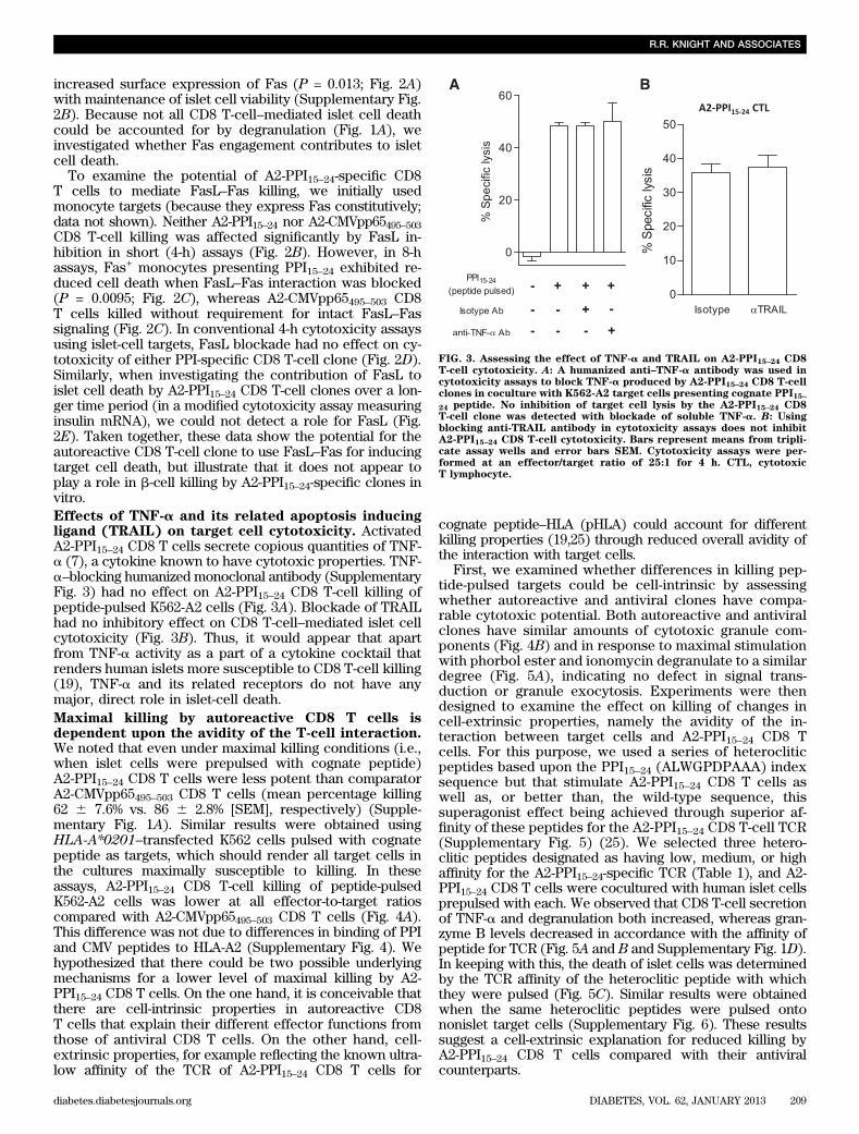
increased surface expression of Fas (P = 0.013; Fig. 2A)with maintenance of islet cell viability (Supplementary Fig.2B). Because not all CD8 T-cell–mediated islet cell deathcould be accounted for by degranulation (Fig. 1A), weinvestigated whether Fas engagement contributes to isletcell death.
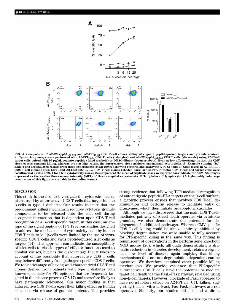
To examine the potential of A2-PPI15–24-specific CD8T cells to mediate FasL–Fas killing, we initially usedmonocyte targets (because they express Fas constitutively;data not shown). Neither A2-PPI15–24 nor A2-CMVpp65495–503CD8 T-cell killing was affected significantly by FasL in-hibition in short (4-h) assays (Fig. 2B). However, in 8-hassays, Fas+ monocytes presenting PPI15–24 exhibited re-duced cell death when FasL–Fas interaction was blocked(P = 0.0095; Fig. 2C), whereas A2-CMVpp65495–503 CD8T cells killed without requirement for intact FasL–Fassignaling (Fig. 2C). In conventional 4-h cytotoxicity assaysusing islet-cell targets, FasL blockade had no effect on cy-totoxicity of either PPI-specific CD8 T-cell clone (Fig. 2D).Similarly, when investigating the contribution of FasL toislet cell death by A2-PPI15–24 CD8 T-cell clones over a lon-ger time period (in a modified cytotoxicity assay measuringinsulin mRNA), we could not detect a role for FasL (Fig.2E). Taken together, these data show the potential for theautoreactive CD8 T-cell clone to use FasL–Fas for inducingtarget cell death, but illustrate that it does not appear toplay a role in b-cell killing by A2-PPI15–24-specific clones invitro.Effects of TNF-a and its related apoptosis inducingligand (TRAIL) on target cell cytotoxicity. ActivatedA2-PPI15–24 CD8 T cells secrete copious quantities of TNF-a (7), a cytokine known to have cytotoxic properties. TNF-a–blocking humanized monoclonal antibody (SupplementaryFig. 3) had no effect on A2-PPI15–24 CD8 T-cell killing ofpeptide-pulsed K562-A2 cells (Fig. 3A). Blockade of TRAILhad no inhibitory effect on CD8 T-cell–mediated islet cellcytotoxicity (Fig. 3B). Thus, it would appear that apartfrom TNF-a activity as a part of a cytokine cocktail thatrenders human islets more susceptible to CD8 T-cell killing(19), TNF-a and its related receptors do not have anymajor, direct role in islet-cell death.Maximal killing by autoreactive CD8 T cells isdependent upon the avidity of the T-cell interaction.We noted that even under maximal killing conditions (i.e.,when islet cells were prepulsed with cognate peptide)A2-PPI15–24 CD8 T cells were less potent than comparatorA2-CMVpp65495–503 CD8 T cells (mean percentage killing62 6 7.6% vs. 86 6 2.8% [SEM], respectively) (Supple-mentary Fig. 1A). Similar results were obtained usingHLA-A*0201–transfected K562 cells pulsed with cognatepeptide as targets, which should render all target cells inthe cultures maximally susceptible to killing. In theseassays, A2-PPI15–24 CD8 T-cell killing of peptide-pulsedK562-A2 cells was lower at all effector-to-target ratioscompared with A2-CMVpp65495–503 CD8 T cells (Fig. 4A).This difference was not due to differences in binding of PPIand CMV peptides to HLA-A2 (Supplementary Fig. 4). Wehypothesized that there could be two possible underlyingmechanisms for a lower level of maximal killing by A2-PPI15–24 CD8 T cells. On the one hand, it is conceivable thatthere are cell-intrinsic properties in autoreactive CD8T cells that explain their different effector functions fromthose of antiviral CD8 T cells. On the other hand, cell-extrinsic properties, for example reflecting the known ultra-low affinity of the TCR of A2-PPI15–24 CD8 T cells for
cognate peptide–HLA (pHLA) could account for differentkilling properties (19,25) through reduced overall avidity ofthe interaction with target cells.
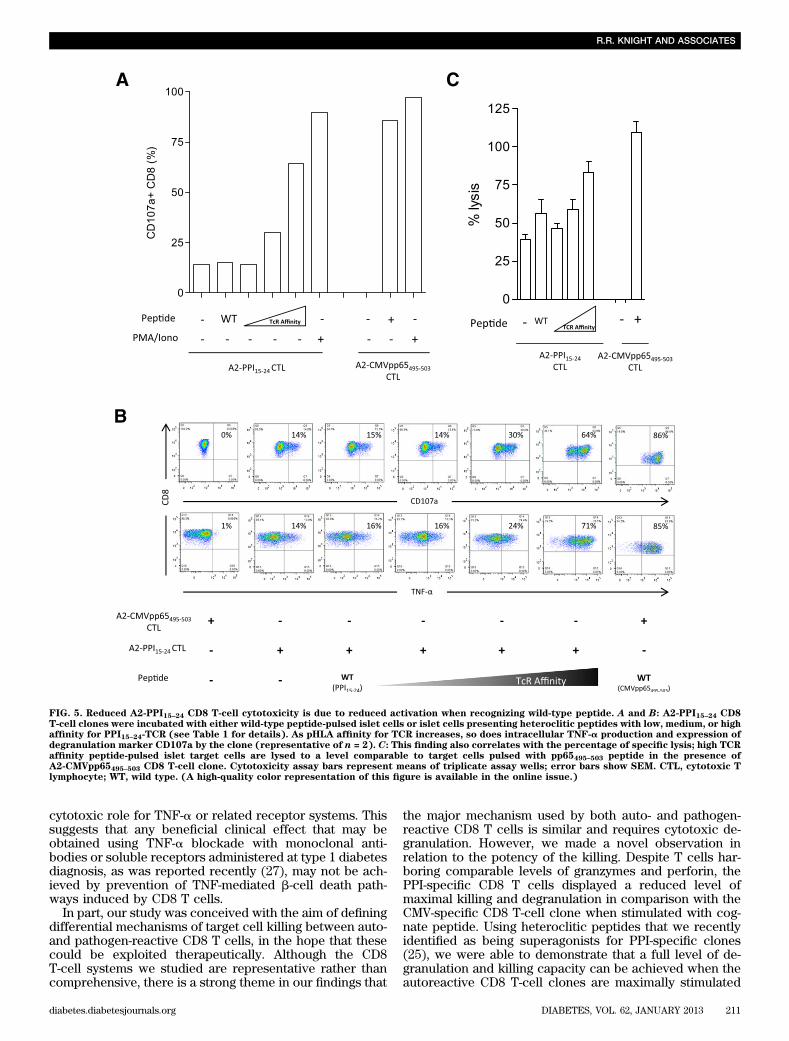
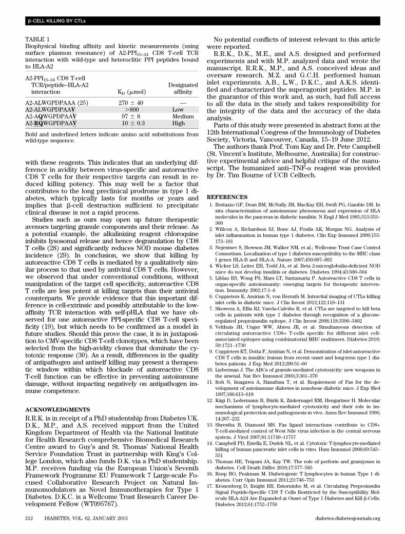
First, we examined whether differences in killing pep-tide-pulsed targets could be cell-intrinsic by assessingwhether autoreactive and antiviral clones have compa-rable cytotoxic potential. Both autoreactive and antiviralclones have similar amounts of cytotoxic granule com-ponents (Fig. 4B) and in response to maximal stimulationwith phorbol ester and ionomycin degranulate to a similardegree (Fig. 5A), indicating no defect in signal trans-duction or granule exocytosis. Experiments were thendesigned to examine the effect on killing of changes incell-extrinsic properties, namely the avidity of the in-teraction between target cells and A2-PPI15–24 CD8 Tcells. For this purpose, we used a series of heterocliticpeptides based upon the PPI15–24 (ALWGPDPAAA) indexsequence but that stimulate A2-PPI15–24 CD8 T cells aswell as, or better than, the wild-type sequence, thissuperagonist effect being achieved through superior af-finity of these peptides for the A2-PPI15–24 CD8 T-cell TCR(Supplementary Fig. 5) (25). We selected three hetero-clitic peptides designated as having low, medium, or highaffinity for the A2-PPI15–24-specific TCR (Table 1), and A2-PPI15–24 CD8 T cells were cocultured with human islet cellsprepulsed with each. We observed that CD8 T-cell secretionof TNF-a and degranulation both increased, whereas gran-zyme B levels decreased in accordance with the affinity ofpeptide for TCR (Fig. 5A and B and Supplementary Fig. 1D).In keeping with this, the death of islet cells was determinedby the TCR affinity of the heteroclitic peptide with whichthey were pulsed (Fig. 5C). Similar results were obtainedwhen the same heteroclitic peptides were pulsed ontononislet target cells (Supplementary Fig. 6). These resultssuggest a cell-extrinsic explanation for reduced killing byA2-PPI15–24 CD8 T cells compared with their antiviralcounterparts.
FIG. 3. Assessing the effect of TNF-a and TRAIL on A2-PPI15–24 CD8T-cell cytotoxicity. A: A humanized anti–TNF-a antibody was used incytotoxicity assays to block TNF-a produced by A2-PPI15–24 CD8 T-cellclones in coculture with K562-A2 target cells presenting cognate PPI15–24 peptide. No inhibition of target cell lysis by the A2-PPI15–24 CD8T-cell clone was detected with blockade of soluble TNF-a. B: Usingblocking anti-TRAIL antibody in cytotoxicity assays does not inhibitA2-PPI15–24 CD8 T-cell cytotoxicity. Bars represent means from tripli-cate assay wells and error bars SEM. Cytotoxicity assays were per-formed at an effector/target ratio of 25:1 for 4 h. CTL, cytotoxicT lymphocyte.
R.R. KNIGHT AND ASSOCIATES
diabetes.diabetesjournals.org DIABETES, VOL. 62, JANUARY 2013 209
DISCUSSION
This study is the first to investigate the cytotoxic mecha-nisms used by autoreactive CD8 T cells that target humanb-cells in type 1 diabetes. Our results indicate that thepredominant killing mechanism requires cytotoxic granulecomponents to be released onto the islet cell duringa cognate interaction that is dependent upon CD8 T-cellrecognition of a b-cell specific target; in this case, an epi-tope of the signal peptide of PPI. Previous studies designedto address the mechanisms of cytotoxicity used by humanCD8 T cells to kill b-cells were limited by the use of virus-specific CD8 T cells and virus peptide-pulsed islet cells astargets (14). This approach can indicate the susceptibilityof islet cells to classic types of effector functions used tocombat viruses, but has the disadvantage that it takes noaccount of the possibility that autoreactive CD8 T cellsmay behave differently from pathogen-specific CD8 T cells.We took advantage of having well-characterized CD8 T-cellclones derived from patients with type 1 diabetes withknown specificity for PPI epitopes that are frequently tar-geted in the disease process (7,8,17) and therefore likely tohave pathogenic relevance. Our major finding is thatautoreactive CD8 T cells exert their killing effect on humanislet cells via release of granule contents. This provides
strong evidence that following TCR-mediated recognitionof autoantigenic peptide–HLA targets on the b-cell surface,a cytolytic process ensues that involves CD8 T-cell de-granulation and perforin release to facilitate entry ofgranzymes, which then initiate proapoptotic cascades.
Although we have discovered that the main CD8 T-cell–mediated pathway of b-cell death operates via cytotoxicgranules, we also demonstrate the potential for de-ployment of additional pathways. Whereas CMV-specificCD8 T-cell killing could be almost entirely inhibited byblocking degranulation, we were unable to fully accountfor PPI-specific killing in the same way. This finding isreminiscent of observations in the perforin gene knockoutNOD mouse (26), which, although demonstrating a dra-matic reduction in diabetes development, retains a signifi-cant low level of disease, again implying that killingmechanisms that are not degranulation-dependent can beoperative. We therefore examined other possible killingmechanisms. We provide evidence that PPI-specificautoreactive CD8 T cells have the potential to mediatetarget cell death via the FasL–Fas pathway, revealed usingnon–b-cell targets. However, blockade of FasL appeared tohave no inhibitory effect on A2-PPI15–24 CTL killing, sug-gesting that, in vitro at least, Fas–FasL pathways are notoperative. Similarly, our studies did not find a direct
FIG. 4. Comparison of A2-CMVpp65495–503 and A2-PPI15–24 CD8 T-cell clones killing of cognate peptide-pulsed targets and granule content.A: Cytotoxicity assays were performed with A2-PPI15–24 CD8 T cells (triangles) and A2-CMVpp65495–503 CD8 T cells (diamonds) using K562-A2target cells pulsed with 10 mg/mL cognate peptide (filled symbols) or DMSO diluent (open symbols). Even at low effector/target ratios, the CMVclone causes maximal killing, whereas even at high ratios, the autoreactive clone achieves submaximal cytotoxicity. B: Example staining (leftpanel) and accumulated results from three experiments (right panel) showing perforin and granzyme A (GzA) and B (GzB) levels in A2-PPI15–24CD8 T-cell clones (open bars) and A2-CMVpp65495–503 CD8 T-cell clones (shaded bars) are shown. Effector CD8 T-cell and target cells werecocultured at a ratio of 25:1 for 4 h in cytotoxicity assays. Bars represent the mean of triplicate assay wells; error bars indicate the SEM. Staining isexpressed as the median fluorescence intensity (MFI) of three compiled experiments. CTL, cytotoxic T lymphocyte. (A high-quality color rep-resentation of this figure is available in the online issue.)
b-CELL KILLING BY CTLs
210 DIABETES, VOL. 62, JANUARY 2013 diabetes.diabetesjournals.org
cytotoxic role for TNF-a or related receptor systems. Thissuggests that any beneficial clinical effect that may beobtained using TNF-a blockade with monoclonal anti-bodies or soluble receptors administered at type 1 diabetesdiagnosis, as was reported recently (27), may not be ach-ieved by prevention of TNF-mediated b-cell death path-ways induced by CD8 T cells.
In part, our study was conceived with the aim of definingdifferential mechanisms of target cell killing between auto-and pathogen-reactive CD8 T cells, in the hope that thesecould be exploited therapeutically. Although the CD8T-cell systems we studied are representative rather thancomprehensive, there is a strong theme in our findings that
the major mechanism used by both auto- and pathogen-reactive CD8 T cells is similar and requires cytotoxic de-granulation. However, we made a novel observation inrelation to the potency of the killing. Despite T cells har-boring comparable levels of granzymes and perforin, thePPI-specific CD8 T cells displayed a reduced level ofmaximal killing and degranulation in comparison with theCMV-specific CD8 T-cell clone when stimulated with cog-nate peptide. Using heteroclitic peptides that we recentlyidentified as being superagonists for PPI-specific clones(25), we were able to demonstrate that a full level of de-granulation and killing capacity can be achieved when theautoreactive CD8 T-cell clones are maximally stimulated
FIG. 5. Reduced A2-PPI15–24 CD8 T-cell cytotoxicity is due to reduced activation when recognizing wild-type peptide. A and B: A2-PPI15–24 CD8T-cell clones were incubated with either wild-type peptide-pulsed islet cells or islet cells presenting heteroclitic peptides with low, medium, or highaffinity for PPI15–24-TCR (see Table 1 for details). As pHLA affinity for TCR increases, so does intracellular TNF-a production and expression ofdegranulation marker CD107a by the clone (representative of n = 2). C: This finding also correlates with the percentage of specific lysis; high TCRaffinity peptide-pulsed islet target cells are lysed to a level comparable to target cells pulsed with pp65495–503 peptide in the presence ofA2-CMVpp65495–503 CD8 T-cell clone. Cytotoxicity assay bars represent means of triplicate assay wells; error bars show SEM. CTL, cytotoxic Tlymphocyte; WT, wild type. (A high-quality color representation of this figure is available in the online issue.)
R.R. KNIGHT AND ASSOCIATES
diabetes.diabetesjournals.org DIABETES, VOL. 62, JANUARY 2013 211
with these reagents. This indicates that an underlying dif-ference in avidity between virus-specific and autoreactiveCD8 T cells for their respective targets can result in re-duced killing potency. This may well be a factor thatcontributes to the long preclinical prodrome in type 1 di-abetes, which typically lasts for months or years andimplies that b-cell destruction sufficient to precipitateclinical disease is not a rapid process.
Studies such as ours may open up future therapeuticavenues targeting granule components and their release. Asa potential example, the alkalinizing reagent chloroquineinhibits lysosomal release and hence degranulation by CD8T cells (28) and significantly reduces NOD mouse diabetesincidence (29). In conclusion, we show that killing byautoreactive CD8 T cells is mediated by a qualitatively sim-ilar process to that used by antiviral CD8 T cells. However,we observed that under conventional conditions, withoutmanipulation of the target cell specificity, autoreactive CD8T cells are less potent at killing targets than their antiviralcounterparts. We provide evidence that this important dif-ference is cell-extrinsic and possibly attributable to the low-affinity TCR interaction with self-pHLA that we have ob-served for one autoreactive PPI-specific CD8 T-cell speci-ficity (19), but which needs to be confirmed as a model infuture studies. Should this prove the case, it is in juxtaposi-tion to CMV-specific CD8 T-cell clonotypes, which have beenselected from the high-avidity clones that dominate the cy-totoxic response (30). As a result, differences in the qualityof antipathogen and antiself killing may present a therapeu-tic window within which blockade of autoreactive CD8T-cell function can be effective in preventing autoimmunedamage, without impacting negatively on antipathogen im-mune competence.
ACKNOWLEDGMENTS
R.R.K. is in receipt of a PhD studentship from Diabetes UK.D.K., M.P., and A.S. received support from the UnitedKingdom Department of Health via the National Institutefor Health Research comprehensive Biomedical ResearchCentre award to Guy’s and St. Thomas’ National HealthService Foundation Trust in partnership with King’s Col-lege London, which also funds D.K. via a PhD studentship.M.P. receives funding via the European Union’s SeventhFramework Programme EU Framework 7 Large-scale Fo-cused Collaborative Research Project on Natural Im-munomodulators as Novel Immunotherapies for Type 1Diabetes. D.K.C. is a Wellcome Trust Research Career De-velopment Fellow (WT095767).
No potential conflicts of interest relevant to this articlewere reported.
R.R.K., D.K., M.E., and A.S. designed and performedexperiments and with M.P. analyzed data and wrote themanuscript. R.R.K., M.P., and A.S. conceived ideas andoversaw research. M.Z. and G.C.H. performed humanislet experiments. A.B., L.W., D.K.C., and A.K.S. identi-fied and characterized the superagonist peptides. M.P. isthe guarantor of this work and, as such, had full accessto all the data in the study and takes responsibility forthe integrity of the data and the accuracy of the dataanalysis.
Parts of this study were presented in abstract form at the12th International Congress of the Immunology of DiabetesSociety, Victoria, Vancouver, Canada, 15–19 June 2012.
The authors thank Prof. Tom Kay and Dr. Pete Campbell(St. Vincent’s Institute, Melbourne, Australia) for construc-tive experimental advice and helpful critique of the manu-script. The humanized anti–TNF-a reagent was providedby Dr. Tim Bourne of UCB Celltech.
REFERENCES
1. Bottazzo GF, Dean BM, McNally JM, MacKay EH, Swift PG, Gamble DR. Insitu characterization of autoimmune phenomena and expression of HLAmolecules in the pancreas in diabetic insulitis. N Engl J Med 1985;313:353–360
2. Willcox A, Richardson SJ, Bone AJ, Foulis AK, Morgan NG. Analysis ofislet inflammation in human type 1 diabetes. Clin Exp Immunol 2009;155:173–181
3. Nejentsev S, Howson JM, Walker NM, et al.; Wellcome Trust Case ControlConsortium. Localization of type 1 diabetes susceptibility to the MHC classI genes HLA-B and HLA-A. Nature 2007;450:887–892
4. Wicker LS, Leiter EH, Todd JA, et al. Beta 2-microglobulin-deficient NODmice do not develop insulitis or diabetes. Diabetes 1994;43:500–504
5. Liblau RS, Wong FS, Mars LT, Santamaria P. Autoreactive CD8 T cells inorgan-specific autoimmunity: emerging targets for therapeutic interven-tion. Immunity 2002;17:1–6
6. Coppieters K, Amirian N, von Herrath M. Intravital imaging of CTLs killingislet cells in diabetic mice. J Clin Invest 2012;122:119–131
7. Skowera A, Ellis RJ, Varela-Calviño R, et al. CTLs are targeted to kill betacells in patients with type 1 diabetes through recognition of a glucose-regulated preproinsulin epitope. J Clin Invest 2008;118:3390–3402
8. Velthuis JH, Unger WW, Abreu JR, et al. Simultaneous detection ofcirculating autoreactive CD8+ T-cells specific for different islet cell-associated epitopes using combinatorial MHC multimers. Diabetes 2010;59:1721–1730
9. Coppieters KT, Dotta F, Amirian N, et al. Demonstration of islet-autoreactiveCD8 T cells in insulitic lesions from recent onset and long-term type 1 dia-betes patients. J Exp Med 2012;209:51–60
10. Lieberman J. The ABCs of granule-mediated cytotoxicity: new weapons inthe arsenal. Nat Rev Immunol 2003;3:361–370
11. Itoh N, Imagawa A, Hanafusa T, et al. Requirement of Fas for the de-velopment of autoimmune diabetes in nonobese diabetic mice. J Exp Med1997;186:613–618
12. Kägi D, Ledermann B, Bürki K, Zinkernagel RM, Hengartner H. Molecularmechanisms of lymphocyte-mediated cytotoxicity and their role in im-munological protection and pathogenesis in vivo. Annu Rev Immunol 1996;14:207–232
13. Shrestha B, Diamond MS. Fas ligand interactions contribute to CD8+T-cell-mediated control of West Nile virus infection in the central nervoussystem. J Virol 2007;81:11749–11757
14. Campbell PD, Estella E, Dudek NL, et al. Cytotoxic T-lymphocyte-mediatedkilling of human pancreatic islet cells in vitro. Hum Immunol 2008;69:543–551
15. Thomas HE, Trapani JA, Kay TW. The role of perforin and granzymes indiabetes. Cell Death Differ 2010;17:577–585
16. Roep BO, Peakman M. Diabetogenic T lymphocytes in human Type 1 di-abetes. Curr Opin Immunol 2011;23:746–753
17. Kronenberg D, Knight RR, Estorninho M, et al. Circulating PreproinsulinSignal Peptide-Specific CD8 T Cells Restricted by the Susceptibility Mol-ecule HLA-A24 Are Expanded at Onset of Type 1 Diabetes and Kill b-Cells.Diabetes 2012;61:1752–1759
TABLE 1Biophysical binding affinity and kinetic measurements (usingsurface plasmon resonance) of A2-PPI15–24 CD8 T-cell TCRinteraction with wild-type and heteroclitic PPI peptides boundto HLA-A2
A2-PPI15–24 CD8 T-cellTCR/peptide–HLA-A2interaction KD (mmol)
Designatedaffinity
A2-ALWGPDPAAA (25) 270 6 40 —
A2-ALWGPDPAAV .800 LowA2-AQWGPDPAAV 97 6 8 MediumA2-RQWGPDPAAV 10 6 0.3 High
Bold and underlined letters indicate amino acid substitutions fromwild-type sequence.
b-CELL KILLING BY CTLs
212 DIABETES, VOL. 62, JANUARY 2013 diabetes.diabetesjournals.org
18. Huang GC, Zhao M, Jones P, et al. The development of new density gra-dient media for purifying human islets and islet-quality assessments.Transplantation 2004;77:143–145
19. Bulek AM, Cole DK, Skowera A, et al. Structural basis for the killing ofhuman beta cells by CD8(+) T cells in type 1 diabetes. Nat Immunol 2012;13:283–289
20. Blomberg K, Hautala R, Lövgren J, Mukkala VM, Lindqvist C, Akerman K.Time-resolved fluorometric assay for natural killer activity using targetcells labelled with a fluorescence enhancing ligand. J Immunol Methods1996;193:199–206
21. Cole DK, Dunn SM, Sami M, Boulter JM, Jakobsen BK, Sewell AK. Tcell receptor engagement of peptide-major histocompatibility com-plex class I does not modify CD8 binding. Mol Immunol 2008;45:2700–2709
22. Kuzushima K, Hayashi N, Kimura H, Tsurumi T. Efficient identification ofHLA-A*2402-restricted cytomegalovirus-specific CD8(+) T-cell epitopes bya computer algorithm and an enzyme-linked immunospot assay. Blood2001;98:1872–1881
23. Burrows SR, Elkington RA, Miles JJ, et al. Promiscuous CTL recognitionof viral epitopes on multiple human leukocyte antigens: biological vali-dation of the proposed HLA A24 supertype. J Immunol 2003;171:1407–1412
24. Kataoka T, Takaku K, Magae J, et al. Acidification is essential for main-taining the structure and function of lytic granules of CTL. Effect of con-canamycin A, an inhibitor of vacuolar type H(+)-ATPase, on CTL-mediatedcytotoxicity. J Immunol 1994;153:3938–3947
25. Wooldridge L, Ekeruche-Makinde J, van den Berg HA, et al. A single au-toimmune T cell receptor recognizes more than a million different pep-tides. J Biol Chem 2012;287:1168–1177
26. Kägi D, Odermatt B, Seiler P, Zinkernagel RM, Mak TW, Hengartner H.Reduced incidence and delayed onset of diabetes in perforin-deficientnonobese diabetic mice. J Exp Med 1997;186:989–997
27. Mastrandrea L, Yu J, Behrens T, et al. Etanercept treatment in childrenwith new-onset type 1 diabetes: pilot randomized, placebo-controlled,double-blind study. Diabetes Care 2009;32:1244–1249
28. Klempner MS, Styrt B. Alkalinizing the intralysosomal pH inhibits de-granulation of human neutrophils. J Clin Invest 1983;72:1793–1800
29. Zhang Y, Lee AS, Shameli A, et al. TLR9 blockade inhibits activation ofdiabetogenic CD8+ T cells and delays autoimmune diabetes. J Immunol2010;184:5645–5653
30. Day EK, Carmichael AJ, ten Berge IJ, Waller EC, Sissons JG, Wills MR.Rapid CD8+ T cell repertoire focusing and selection of high-affinity clonesinto memory following primary infection with a persistent human virus:human cytomegalovirus. J Immunol 2007;179:3203–3213
R.R. KNIGHT AND ASSOCIATES
diabetes.diabetesjournals.org DIABETES, VOL. 62, JANUARY 2013 213