original article immunosuppressive effects of...
TRANSCRIPT
Immunosuppressive Effects of Streptozotocin-InducedDiabetes Result in Absolute Lymphopenia and aRelative Increase of T Regulatory CellsYannick D. Muller,
1,2Déla Golshayan,
3Driss Ehirchiou,
1Jean Christophe Wyss,
3
Laurianne Giovannoni,2Raphael Meier,
2Véronique Serre-Beinier,
2Gisella Puga Yung,
1
Philippe Morel,2Leo H. Bühler,
2and Jörg D. Seebach
1
OBJECTIVE—Streptozotocin (STZ) is the most widely used di-abetogenic agent in animal models of islet transplantation. However,the immunomodifying effects of STZ and the ensuing hyperglycemiaon lymphocyte subsets, particularly on T regulatory cells (Tregs),remain poorly understood.
RESEARCH DESIGN AND METHODS—This study evaluatedhow STZ-induced diabetes affects adaptive immunity and theconsequences thereof on allograft rejection in murine models ofislet and skin transplantation. The respective toxicity of STZ andhyperglycemia on lymphocyte subsets was tested in vitro. Theeffect of hyperglycemia was assessed independently of STZ invivo by the removal of transplanted syngeneic islets, using aninsulin pump, and with rat insulin promoter diphtheria toxinreceptor transgenic mice.
RESULTS—Early lymphopenia in both blood and spleen wasdemonstrated after STZ administration. Direct toxicity of STZ onlymphocytes, particularly on CD8+ cells and B cells, was shownin vitro. Hyperglycemia also correlated with blood and spleenlymphopenia in vivo but was not lymphotoxic in vitro. Indepen-dently of hyperglycemia, STZ led to a relative increase of Tregsin vivo, with the latter retaining their suppressive capacity invitro. The higher frequency of Tregs was associated with Tregproliferation in the blood, but not in the spleen, and higherblood levels of transforming growth factor-b. Finally, STZ ad-ministration delayed islet and skin allograft rejection comparedwith naive mice.
CONCLUSIONS—These data highlight the direct and indirectimmunosuppressive effects of STZ and acute hyperglycemia,respectively. Thus, these results have important implicationsfor the future development of tolerance-based protocols andtheir translation from the laboratory to the clinic. Diabetes60:2331–2340, 2011
Many animal models of diabetes depend on theadministration of diabetogenic drugs such asstreptozotocin (STZ) or alloxan. These aretoxic glucose analogs that target pancreatic
b-cells via GLUT2 transporter uptake. Because of thesimplicity and reproducibility of diabetes induction withSTZ, a highly stable glucosamine-nitrosourea compound, itrepresents by far the most widely used model (1). Indeed,among 131 analyzed articles published in 2010 on murineislet transplantation, 100 (76.3%) used STZ to induce di-abetes, whereas 21 (16%) were performed in models ofspontaneous diabetes (mostly NOD mice), 3 (2.3%) in-volved alloxan, 5 (3.8%) relied on transplanted islets innondiabetic mice, and 2 (1.5%) used alternative ways fordiabetes induction. Whereas STZ is known to target b-cellsvia the transporter GLUT2, it is not specific, since thekidney and liver are also susceptible to STZ toxicity (1).Moreover, several groups have described immunomodify-ing effects of STZ both in vitro and in vivo, although theconfounding impact of the induced hyperglycemia was notclearly distinguished (2–6). Luo et al. (7) reported thathyperglycemia, potentially via an increased level of corti-costeroids, causes a rapid depletion in thymocytes andsplenic T cells followed by homeostatic T-cell proliferation.However, the exact mechanisms leading to the observedimmunosuppression after STZ administration, especially theeffect on different lymphocyte subpopulations, includingT regulatory cells (Tregs), remain elusive.
One of the major goals in islet transplantation (Tx) re-search is to find strategies to obviate the need for lifelongimmunosuppression that is toxic to the b-cells and detri-mental to the host (8). Indeed, using STZ-induced diabetesmodels, novel immunosuppressive and tolerance inductionprotocols for allogeneic as well as xenogeneic islet Txhave demonstrated long-term graft survival (9,10). How-ever, many costimulation blockade–based Tx toleranceprotocols that are successful in chemically induced di-abetic mice failed to produce long-term graft tolerance inNOD mice or in other Tx models such as skin Tx, with thelatter being considered as one of the most stringent models.The underlying autoimmunity directed against b-cells, themode of Tx, and the skin-specific antigen–presenting cells allhave been put forward to explain the observed resistance toTx tolerance (10,11). Another possible explanation for theseapparent differences is that immunosuppression related toSTZ-induced diabetes could result in an overestimation of theefficacy of tolerance induction. Consistent with this notion,a protocol targeting costimulation that induced tolerance
From the 1Department of Internal Medicine, Division of Clinical Immunology andAllergology, University Hospital and Medical Faculty, Geneva, Switzerland;the 2Department of Surgery, Surgical Research Unit, University HospitalGeneva, Geneva, Switzerland; and the 3Department of Medicine, Transplan-tation Centre and Transplantation Immunopathology Laboratory, Centre Hos-pitalier Universitaire Vaudois (CHUV), and University of Lausanne, Lausanne,Switzerland.
Corresponding author: Yannick D. Muller, [email protected] 8 February 2011 and accepted 24 May 2011.DOI: 10.2337/db11-0159This article contains Supplementary Data online at http://diabetes.
diabetesjournals.org/lookup/suppl/doi:10.2337/db11-0159/-/DC1.L.H.B. and J.D.S. share co-senior authorship in this article.� 2011 by the American Diabetes Association. Readers may use this article as
long as the work is properly cited, the use is educational and not for profit,and the work is not altered. See http://creativecommons.org/licenses/by-nc-nd/3.0/ for details.
diabetes.diabetesjournals.org DIABETES, VOL. 60, SEPTEMBER 2011 2331
ORIGINAL ARTICLE
in STZ-induced diabetic recipients was unable to inducetolerance in nondiabetic recipients (7). Thus, STZ and/orthe ensuing hyperglycemia seem to downregulate theadaptive immune response against islet grafts.
In the past few years, Tregs were reported to play a keyrole in long-term islet graft tolerance (12–15). However,none of these studies addressed the possible effect of STZadministration and the ensuing hyperglycemia on Tregs.Using a rat-to-mouse islet Tx model, we recently reportedan increase of Tregs in lymphoid organs and within graftsof tolerant mice treated with rapamycin and anti-CD154monoclonal antibody (mAb), suggesting a critical role forTregs in the induction phase of tolerance (9,16). The aim ofthe current study was to analyze whether STZ-induceddiabetes leads to changes in Treg numbers and functionand whether this affects subsequent immune responsesusing models of islet and skin Tx.
RESEARCH DESIGN AND METHODS
C57BL/6 (H2b) mice, 6–10 weeks of age, were used as recipients (Centred’Elevage R. Janvier, Le Genest-St-Isle, France). BALB/c (H2d) mice were used asislet donors and BL/6xDBA/2 F1 (H2bxH2d) as skin donors (Janvier). Rat insulinpromoter diphtheria toxin receptor (RIP-DTR) transgenic mice were provided byPedro Herrera (Geneva University Medical School, Geneva, Switzerland), andFoxp3 green fluorescent protein (GFP) knock-in mice were provided by DavidScotts (Center for Vascular and Inflammatory Diseases, University of Marylandat Baltimore, Baltimore, MD). The Foxp3 knock-in mice have the GFP codingsequence inserted in the first exon of the Foxp3 gene and have been backcrossedon a C57BL/6 background. Animals were maintained in conventional housingfacilities. Experiments involving animals were performed in compliance with therelevant laws according to the Geneva Medical Faculty’s Veterinary Authorities.Islet and skin Tx. C57BL/6 mice were rendered diabetic by a single in-traperitoneal injection of STZ (220mg/kg; Sigma-Aldrich, St. Louis,MO) andweretransplanted 72 h later. Blood glucose levels were monitored three times weeklyusing a blood glucose meter (Precision Q.I.D, MediSense; Abbott Laboratories,Bedford, MA). Only mice with blood glucose levels.17 mmol/L were used for Tx.BALB/c pancreatic islets were isolated as previously described (9). A total of 400islet equivalents were transplanted under the left kidney capsule. Blood glucoselevels of,11 mmol/L on 2 consecutive days defined successful islet function. Forskin Tx, full-thickness tail skin was grafted onto the flank of the recipients. Graftsites were protected under sterile gauze and plaster until day 10, observed dailyafterward, and considered rejected when no viable skin remained.
Experimental design. STZ was solubilized in citrate buffer and used in vitro ata concentration of 4.4 mmol/L. RIP-DTR transgenic mice were injected with 126ng i.p. diphtheria toxin (DT) on days 0, 3, and 4 as previously described (17).Insulin pumps (Linbit; LinShin Canada, Inc., Toronto, Ontario, Canada) wereimplanted subcutaneously in STZ-induced diabetic mice 3 days after STZ ad-ministration and allowed a sustained release of insulin (0.1 units/day) for .30days. Anti-CD154 mAb (MR1, hamster antimouse CD154 mAb [CD40 L]; BioExpress, West Lebanon, NH) diluted in PBS (Sigma-Aldrich) was administeredintraperitoneally at 0.5 mg per mouse on days 0, 2, and 4 after Tx. The fol-lowing experimental groups were performed:
1) STZ-induced diabetic C57BL/6 mice transplanted with syngeneic islets onday 3 post-STZ administration (+/2) nephrectomy on day 60 (n = 7)
2) DT-induced diabetic RIP-DTR mice (n = 4)3) RIP-DTR control mice without DT treatment (n = 4)4) STZ-induced diabetic C57BL/6 mice treated with an insulin pump starting
on day 3 (n = 4)5) Nondiabetic C57BL/6 mice transplanted with allogeneic BALB/c islets
(n = 3)6) STZ-induced diabetic C57BL/6 mice transplanted with allogeneic BALB/c
islets (n = 3)7) STZ-induced diabetic C57BL/6 mice treated with an insulin pump (day 3)
and grafted with BL/6xDBA/2 F1 skin (day 3) (n = 7)8) Nondiabetic C57BL/6 mice grafted with BL/6xDBA/2 F1 skin (day 3) (n = 8)9) MR1-treated STZ-induced diabetic C57BL/6 mice treated with an insulin pump
(day 3) and MR1 and grafted with BL/6xDBA/2 F1 skin (day 3) (n = 7)10) MR1-treated nondiabetic C57BL/6 mice grafted with BL/6xDBA/2 F1 skin
(day 3) (n = 11)
Blood leukocytes analysis. Blood was harvested from the tail vein intoheparinized tubes. White blood cell (WBC) counts and differentials were an-alyzed using a pocH-100i hematology analyzer (Sysmex, Mundelein, IL), whichanalyzed percent ratio of small WBCs (SCR, lymphocytes), middleWBCs (MCR,monocytes), and large WBCs (LCR, granulocytes). Alternatively, absolutenumbers of WBCs were counted in a Neubauer chamber after red blood lysisusing Zapoglobulin (Beckam Coulter, Fullerton, CA).Flow cytometry. Blood samples were analyzed on days 3, 7, and 12 after STZadministration, and splenocytes were harvested at day 3 or 13 after STZ ad-ministration. Cells were stained with anti-CD4 fluorescein isothiocyanate(FITC) or allophycocyanin (APC) (RM4–5; eBioscience, San Diego, CA), anti-CD25 APC (PC61.5; eBioscience), anti-Foxp3 phycoerythrin (PE) (FJK16s;eBioscience), anti-CD3 FITC (eBioscience), and anti-CD8 PE (eBioscience). Allsamples were stained, fixed, and permeabilized according to the eBioscience’sinstructions described in the mouse regulatory T-cell staining kit. Isotypecontrol antibodies were purchased from Becton Dickinson (Franklin Lakes,NJ) (rat IgG2a FITC, rat IgG2a PE, rat IgG1 APC, and rat IgG2b APC mAb).
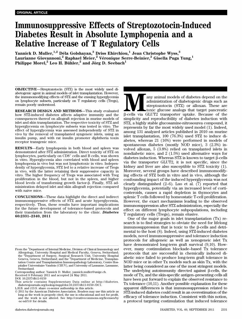
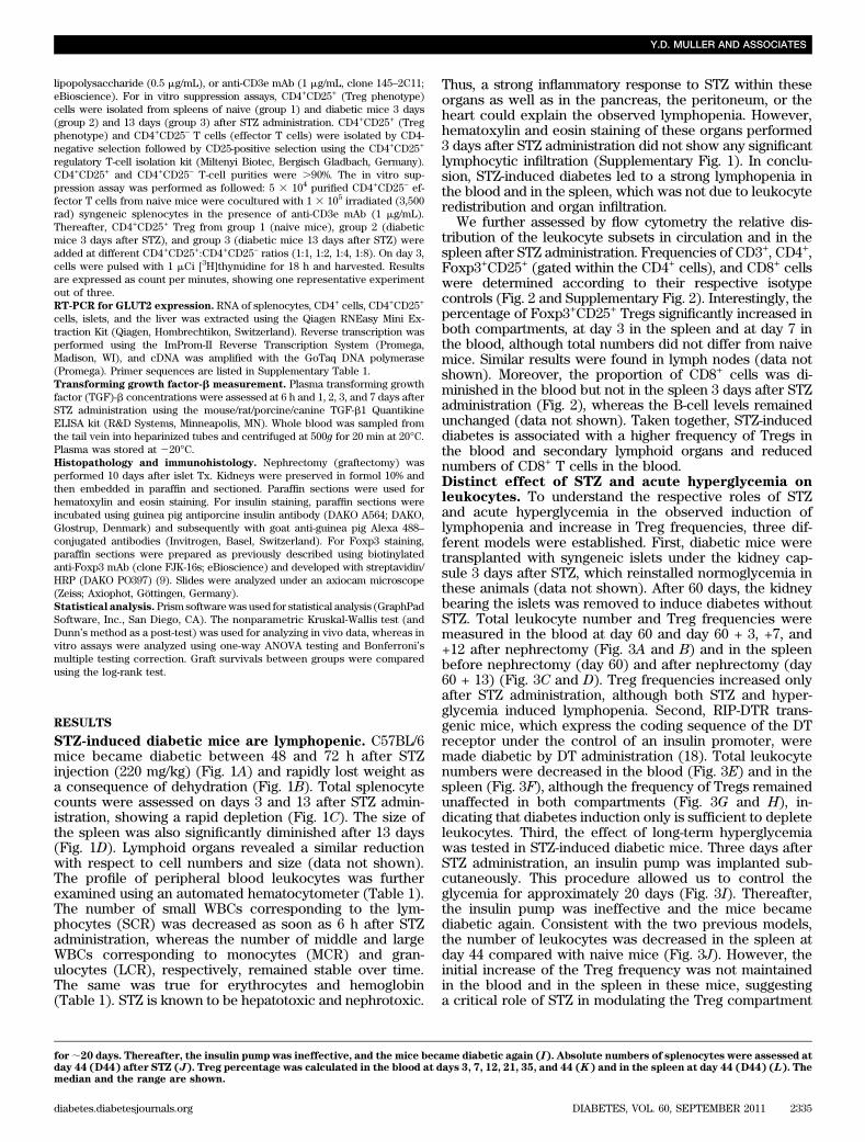
FIG. 1. Diabetes induction with STZ diminishes the absolute numbers of splenocytes. A: Blood glucose levels (mmol/L).○, STZ-induced diabetic mice;□, naive mice. B: Weight (g) of C57BL/6 mice after STZ administration. ○, STZ-induced diabetic mice; □, naive mice. C: Absolute numbers ofsplenocytes 3 (D3) and 13 (D13) days after STZ compared with naive mice (n = 5). The median and the range are shown and were analyzed witha nonparametric Kruskal-Wallis test. D: Three spleens of STZ-induced diabetic mice at day 13 compared with naive mice are shown. A P valueinferior to 0.05 was considered statistically significant (*P < 0.05, **P < 0.01).
STREPTOZOTOCIN AND ISLET TRANSPLANTATION
2332 DIABETES, VOL. 60, SEPTEMBER 2011 diabetes.diabetesjournals.org
Proliferating cells were stained with PE Cy7 anti-CD4, PerCP anti-CD8, APCanti-CD25, and then fixed and permeabilized with Cytofix/Cytoperm and Perm/Wash buffers (eBioscience) and stained with Ki67/isotype control (BectonDickinson, Franklin Lakes, NJ ) and anti-Foxp3 (eBioscience). For determi-nation of cell death/apoptosis, Annexin V and 7AAD were purchased from
Becton Dickinson. Data were collected on a FACScanto (Becton Dickinson).All data were analyzed using Flowjo software (version 8.7.3; TreeStar, Inc.,Ashland, OR).In vitro proliferation and suppression assays. For proliferation assays,0.2 3 106 splenocytes were stimulated with concanavalin A (3 mg/mL),
TABLE 1Blood repartition after STZ administration
Peripheralblood WBC (103/mL) SCR (%) MCR (%) LCR (%) RBC (106/mL)
Hemoglobin(g/dL) n
Naive 18.9 6 (10.5/23.6) 93.2 6 (89.5/96.6) 2.8 6 (2.1/5.4) 3.7 6 (0.9/6) 11.43 6 (10.4/12.4) 15.9 6 (14.1/17) 11Six h 11.7 6 (10.4/13.7) 80.9 6 (73.2/89.5) 10 6 (7.4/16.7) 8.7 6 (3.1/10.1) 11.5 6 (10.2/12.7) 15.6 6 (14.7/16.8) 6Day 2 7.9 6 (6.8/10) 83.9 6 (81.4/86.8) 6.4 6 (4.3/7.8) 9.7 6 (7.6/11.9) 10 6 (9.5/10.8) 14 6 (13.6/15) 6Day 5 7.6 6 (5.1/10.6) 84.2 6 (71.3/88.3) 6.9 6 (5.3/12.5) 8.9 6 (6.4/16.2) 9.9 6 (9.3/10.6) 14 6 (12.5/14.1) 5Day 7 9.6 6 (5.5/11.2) 80.2 6 (70.3/88.5) 8.3 6 (5.5/11.8) 11.1 6 (4.9/17.9) 9.8 6 (9/10.7) 13.7 6 (12.5/14.3) 6Day 12 8.2 6 (7.1/10.3) 88 6 (83.8/91.9) 5.1 6 (3.2/8.4) 6.9 6 (4.9/11.9) 10.1 6 (9.2/11.8) 13.9 6 (12.8/16) 4
Absolute numbers of WBCs and erythrocytes (RBC); percent ratio of SCR (lymphocytes), MCR (monocytes), and LCR (granulocytes) WBCs;and hemoglobin were assessed at 6 h and 2, 5, 7, and 12 days.
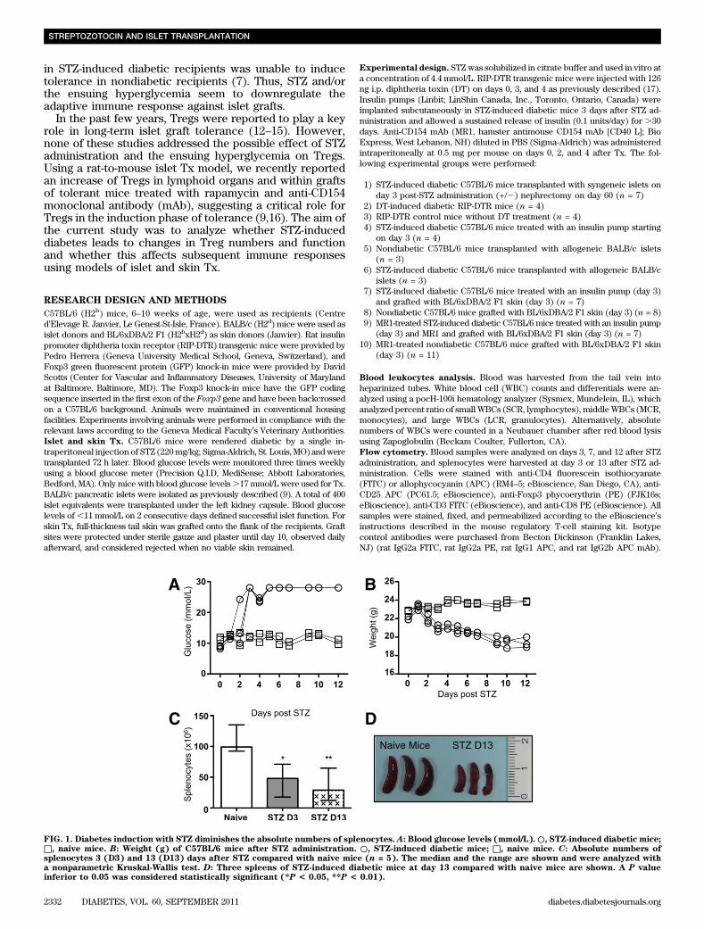
FIG. 2. Flow cytometry analysis of T cells in STZ-induced diabetic mice. CD3+, CD4
+, CD25
+Foxp3
+(gated in the CD4
+population), and CD8
+cell
percentages were analyzed in the blood at day 3 (D3), 7 (D7), and 12 (D12) and in the spleen at days 3 (D3) and 13 (D13). Blood from 15 naive and8 STZ-induced diabetic mice was analyzed. Spleens of eight naive and five STZ-induced diabetic mice were analyzed. Median and the range areshown. A nonparametric Kruskal-Wallis test was performed. *P < 0.05, **P < 0.01, ***P < 0.001.
Y.D. MULLER AND ASSOCIATES
diabetes.diabetesjournals.org DIABETES, VOL. 60, SEPTEMBER 2011 2333
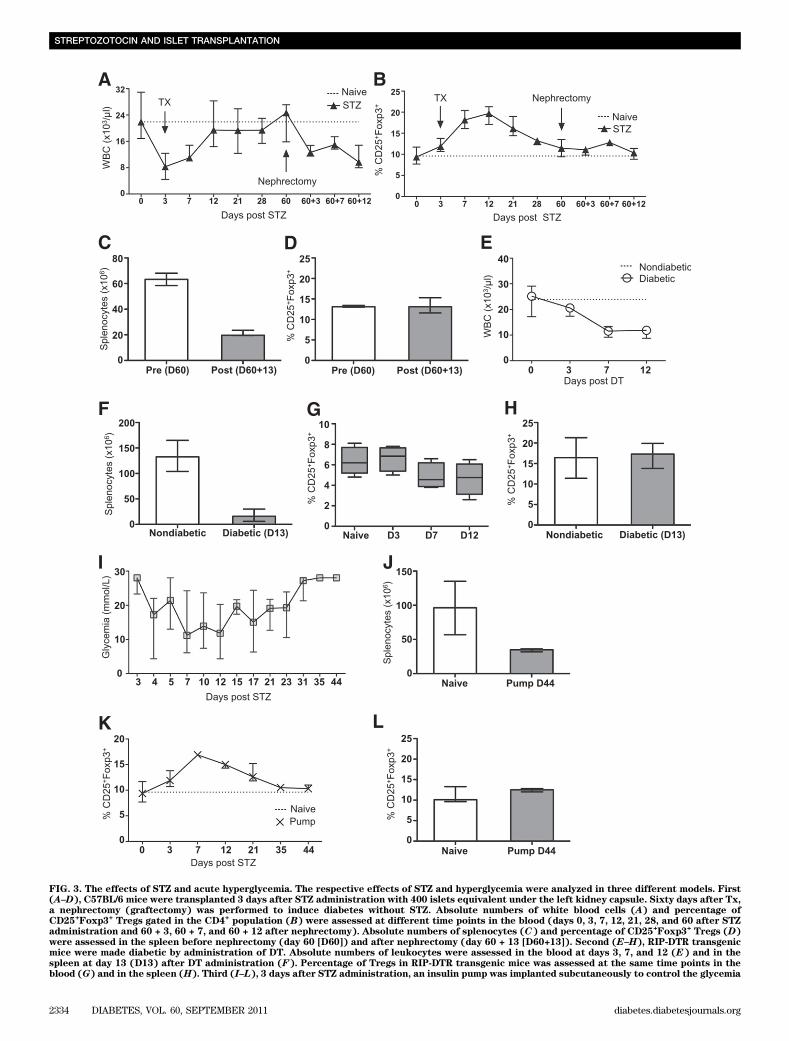
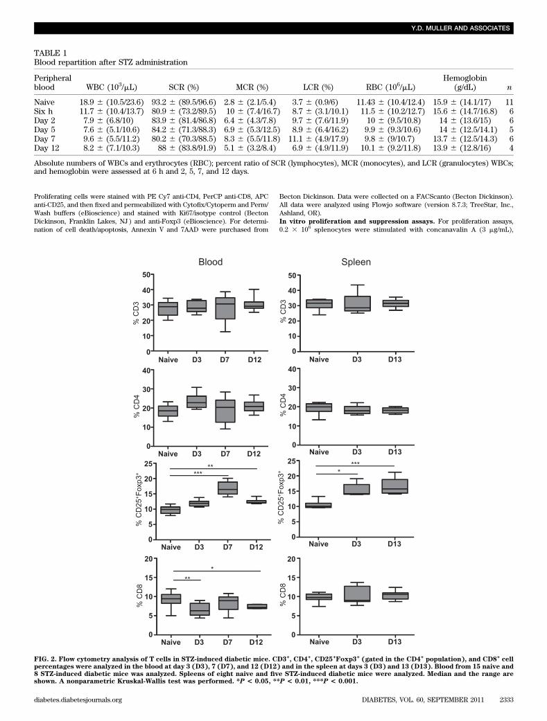
FIG. 3. The effects of STZ and acute hyperglycemia. The respective effects of STZ and hyperglycemia were analyzed in three different models. First(A–D), C57BL/6 mice were transplanted 3 days after STZ administration with 400 islets equivalent under the left kidney capsule. Sixty days after Tx,a nephrectomy (graftectomy) was performed to induce diabetes without STZ. Absolute numbers of white blood cells (A) and percentage ofCD25
+Foxp3
+Tregs gated in the CD4
+population (B) were assessed at different time points in the blood (days 0, 3, 7, 12, 21, 28, and 60 after STZ
administration and 60 + 3, 60 + 7, and 60 + 12 after nephrectomy). Absolute numbers of splenocytes (C) and percentage of CD25+Foxp3
+Tregs (D)
were assessed in the spleen before nephrectomy (day 60 [D60]) and after nephrectomy (day 60 + 13 [D60+13]). Second (E–H), RIP-DTR transgenicmice were made diabetic by administration of DT. Absolute numbers of leukocytes were assessed in the blood at days 3, 7, and 12 (E) and in thespleen at day 13 (D13) after DT administration (F). Percentage of Tregs in RIP-DTR transgenic mice was assessed at the same time points in theblood (G) and in the spleen (H). Third (I–L), 3 days after STZ administration, an insulin pump was implanted subcutaneously to control the glycemia
STREPTOZOTOCIN AND ISLET TRANSPLANTATION
2334 DIABETES, VOL. 60, SEPTEMBER 2011 diabetes.diabetesjournals.org
lipopolysaccharide (0.5 mg/mL), or anti-CD3e mAb (1 mg/mL, clone 145–2C11;eBioscience). For in vitro suppression assays, CD4+CD25+ (Treg phenotype)cells were isolated from spleens of naive (group 1) and diabetic mice 3 days(group 2) and 13 days (group 3) after STZ administration. CD4+CD25+ (Tregphenotype) and CD4+CD25– T cells (effector T cells) were isolated by CD4-negative selection followed by CD25-positive selection using the CD4+CD25+
regulatory T-cell isolation kit (Miltenyi Biotec, Bergisch Gladbach, Germany).CD4+CD25+ and CD4+CD25– T-cell purities were .90%. The in vitro sup-pression assay was performed as followed: 5 3 104 purified CD4+CD25– ef-fector T cells from naive mice were cocultured with 1 3 105 irradiated (3,500rad) syngeneic splenocytes in the presence of anti-CD3e mAb (1 mg/mL).Thereafter, CD4+CD25+ Treg from group 1 (naive mice), group 2 (diabeticmice 3 days after STZ), and group 3 (diabetic mice 13 days after STZ) wereadded at different CD4+CD25+:CD4+CD25– ratios (1:1, 1:2, 1:4, 1:8). On day 3,cells were pulsed with 1 mCi [3H]thymidine for 18 h and harvested. Resultsare expressed as count per minutes, showing one representative experimentout of three.RT-PCR for GLUT2 expression. RNA of splenocytes, CD4+ cells, CD4+CD25+
cells, islets, and the liver was extracted using the Qiagen RNEasy Mini Ex-traction Kit (Qiagen, Hombrechtikon, Switzerland). Reverse transcription wasperformed using the ImProm-II Reverse Transcription System (Promega,Madison, WI), and cDNA was amplified with the GoTaq DNA polymerase(Promega). Primer sequences are listed in Supplementary Table 1.Transforming growth factor-b measurement. Plasma transforming growthfactor (TGF)-b concentrations were assessed at 6 h and 1, 2, 3, and 7 days afterSTZ administration using the mouse/rat/porcine/canine TGF-b1 QuantikineELISA kit (R&D Systems, Minneapolis, MN). Whole blood was sampled fromthe tail vein into heparinized tubes and centrifuged at 500g for 20 min at 20°C.Plasma was stored at 220°C.Histopathology and immunohistology. Nephrectomy (graftectomy) wasperformed 10 days after islet Tx. Kidneys were preserved in formol 10% andthen embedded in paraffin and sectioned. Paraffin sections were used forhematoxylin and eosin staining. For insulin staining, paraffin sections wereincubated using guinea pig antiporcine insulin antibody (DAKO A564; DAKO,Glostrup, Denmark) and subsequently with goat anti-guinea pig Alexa 488–conjugated antibodies (Invitrogen, Basel, Switzerland). For Foxp3 staining,paraffin sections were prepared as previously described using biotinylatedanti-Foxp3 mAb (clone FJK-16s; eBioscience) and developed with streptavidin/HRP (DAKO PO397) (9). Slides were analyzed under an axiocam microscope(Zeiss; Axiophot, Göttingen, Germany).Statistical analysis. Prism softwarewas used for statistical analysis (GraphPadSoftware, Inc., San Diego, CA). The nonparametric Kruskal-Wallis test (andDunn’s method as a post-test) was used for analyzing in vivo data, whereas invitro assays were analyzed using one-way ANOVA testing and Bonferroni’smultiple testing correction. Graft survivals between groups were comparedusing the log-rank test.
RESULTS
STZ-induced diabetic mice are lymphopenic. C57BL/6mice became diabetic between 48 and 72 h after STZinjection (220 mg/kg) (Fig. 1A) and rapidly lost weight asa consequence of dehydration (Fig. 1B). Total splenocytecounts were assessed on days 3 and 13 after STZ admin-istration, showing a rapid depletion (Fig. 1C). The size ofthe spleen was also significantly diminished after 13 days(Fig. 1D). Lymphoid organs revealed a similar reductionwith respect to cell numbers and size (data not shown).The profile of peripheral blood leukocytes was furtherexamined using an automated hematocytometer (Table 1).The number of small WBCs corresponding to the lym-phocytes (SCR) was decreased as soon as 6 h after STZadministration, whereas the number of middle and largeWBCs corresponding to monocytes (MCR) and gran-ulocytes (LCR), respectively, remained stable over time.The same was true for erythrocytes and hemoglobin(Table 1). STZ is known to be hepatotoxic and nephrotoxic.
Thus, a strong inflammatory response to STZ within theseorgans as well as in the pancreas, the peritoneum, or theheart could explain the observed lymphopenia. However,hematoxylin and eosin staining of these organs performed3 days after STZ administration did not show any significantlymphocytic infiltration (Supplementary Fig. 1). In conclu-sion, STZ-induced diabetes led to a strong lymphopenia inthe blood and in the spleen, which was not due to leukocyteredistribution and organ infiltration.
We further assessed by flow cytometry the relative dis-tribution of the leukocyte subsets in circulation and in thespleen after STZ administration. Frequencies of CD3+, CD4+,Foxp3+CD25+ (gated within the CD4+ cells), and CD8+ cellswere determined according to their respective isotypecontrols (Fig. 2 and Supplementary Fig. 2). Interestingly, thepercentage of Foxp3+CD25+ Tregs significantly increased inboth compartments, at day 3 in the spleen and at day 7 inthe blood, although total numbers did not differ from naivemice. Similar results were found in lymph nodes (data notshown). Moreover, the proportion of CD8+ cells was di-minished in the blood but not in the spleen 3 days after STZadministration (Fig. 2), whereas the B-cell levels remainedunchanged (data not shown). Taken together, STZ-induceddiabetes is associated with a higher frequency of Tregs inthe blood and secondary lymphoid organs and reducednumbers of CD8+ T cells in the blood.Distinct effect of STZ and acute hyperglycemia onleukocytes. To understand the respective roles of STZand acute hyperglycemia in the observed induction oflymphopenia and increase in Treg frequencies, three dif-ferent models were established. First, diabetic mice weretransplanted with syngeneic islets under the kidney cap-sule 3 days after STZ, which reinstalled normoglycemia inthese animals (data not shown). After 60 days, the kidneybearing the islets was removed to induce diabetes withoutSTZ. Total leukocyte number and Treg frequencies weremeasured in the blood at day 60 and day 60 + 3, +7, and+12 after nephrectomy (Fig. 3A and B) and in the spleenbefore nephrectomy (day 60) and after nephrectomy (day60 + 13) (Fig. 3C and D). Treg frequencies increased onlyafter STZ administration, although both STZ and hyper-glycemia induced lymphopenia. Second, RIP-DTR trans-genic mice, which express the coding sequence of the DTreceptor under the control of an insulin promoter, weremade diabetic by DT administration (18). Total leukocytenumbers were decreased in the blood (Fig. 3E) and in thespleen (Fig. 3F), although the frequency of Tregs remainedunaffected in both compartments (Fig. 3G and H), in-dicating that diabetes induction only is sufficient to depleteleukocytes. Third, the effect of long-term hyperglycemiawas tested in STZ-induced diabetic mice. Three days afterSTZ administration, an insulin pump was implanted sub-cutaneously. This procedure allowed us to control theglycemia for approximately 20 days (Fig. 3I). Thereafter,the insulin pump was ineffective and the mice becamediabetic again. Consistent with the two previous models,the number of leukocytes was decreased in the spleen atday 44 compared with naive mice (Fig. 3J). However, theinitial increase of the Treg frequency was not maintainedin the blood and in the spleen in these mice, suggestinga critical role of STZ in modulating the Treg compartment
for;20 days. Thereafter, the insulin pump was ineffective, and the mice became diabetic again (I). Absolute numbers of splenocytes were assessed atday 44 (D44) after STZ (J). Treg percentage was calculated in the blood at days 3, 7, 12, 21, 35, and 44 (K) and in the spleen at day 44 (D44) (L). Themedian and the range are shown.
Y.D. MULLER AND ASSOCIATES
diabetes.diabetesjournals.org DIABETES, VOL. 60, SEPTEMBER 2011 2335
(Fig. 3K and L). Thus, the increase in Treg percentagecorrelated with STZ administration, whereas the hyper-glycemia alone also induced leukopenia.STZ is toxic for leukocytes in vitro. The effect of STZwas further tested on splenocytes in vitro. As a positivecontrol, we first exposed C57BL/6 islets with 4.4 mmol/L STZin 10% FCS medium or citrate buffer control medium for1 h. This concentration was based on previous work eval-uating the effect of STZ on islets in vitro (18). Thereafter,islets were washed and maintained in culture for 24 hin 10% FCS medium. STZ conditioned islets showed atypical necrotic aspect with loss of the integrity of thecollagen capsule. In contrast, the morphology of isletscultured for 1 h in citrate buffer remained intact after 24 h(Supplementary Fig. 3A). These finding were confirmed by7AAD staining of disrupted islet cells (Supplementary Fig.3B). Splenocytes were harvested from Foxp3 knock-in miceand cultured for 1 h in STZ or citrate buffer–conditionedmedium. Thereafter, the cells were washed and analyzedfor 7AAD 0, 2, and 5 h after STZ/citrate exposition. STZ-conditioned splenocytes first shifted toward apoptosis at 2 hand then mainly toward death at 5 h (Fig. 4A). These resultswere confirmed by trypan blue staining (data not shown).Percentage of 7AAD+ cells in CD4+, Foxp3+, CD8+, and Bcells were further analyzed at time point 2 h after STZ orcitrate exposition to determine which cell subtypes weremore sensitive to STZ. The ratio of 7AAD+ CD8 and B cellsafter STZ exposure compared with the citrate buffer controlwas 4.5 and 4.1. In contrast, this ratio was only 1.3 and 1.2for CD4+ and Treg, respectively (Fig. 4B). Moreover, CD8and B cells stained positive for Annexin V 2 h after expo-sure to STZ, suggesting that cell death was mediatedthrough apoptotic pathways (Fig. 4C). Functional tests werealso performed. The proliferation of splenocytes stimulatedwith concanavalin A, lipopolysaccharide, or anti-CD3e mAbwas abolished if exposed for 1 h to STZ (Fig. 4D). The sus-ceptibility of Treg to STZ was further analyzed in vitro.Purified CD4+CD25+ T cells were incubated for 1 h withSTZ or citrate buffer. Thereafter, an in vitro suppressionassay was performed. CD4+CD25+ T cells still suppressed,although not completely, anti–CD3e-stimulated proliferationof CD4+CD25– T cells (Fig. 4E). Furthermore, we analyzedGLUT2 mRNA in the subsets of lymphocytes, since it isone of the main pathways for uptake of STZ (1). Neithersplenocytes nor purified CD4+CD25– nor CD4+CD25+ cellswere positive for GLUT2 mRNA (Fig. 4F).
To test whether high levels of glycemia directly affecttheir viability and proliferation, splenocytes harvestedfrom naive mice were incubated with increasing amountsof glucose (11, 22, and 33 mmol/L) for 24 h. Hyperglycemiafailed to induce significant toxicity, as shown by 7AADstaining (Supplementary Fig. 3C). Moreover, high levels ofglucose in the medium did not affect the proliferation ofsplenocytes (Supplementary Fig. 3D). Altogether, theseresults showed a high susceptibility of CD8 and B cells toSTZ, whereas Tregs were partially resistant. These effectswere not mediated via the GLUT2 transporter, suggestingthat STZ enters into lymphocytes by other pathways. Fi-nally, the observed lymphopenia in response to hypergly-cemia in vivo was not explained by a direct lymphotoxiceffect of glucose in vitro.Tregs of STZ-induced diabetic mice are suppressivein vitro and proliferate in vivo. The suppressive func-tion of CD4+CD25+ (Treg phenotype) purified from naive(group 1) or diabetic mice 3 (group 2) and 13 (group 3)days after STZ administration was then tested in vitro. The
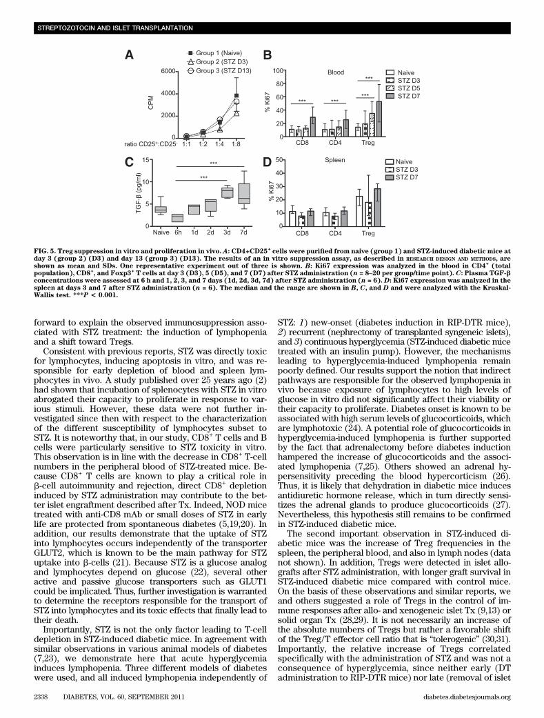
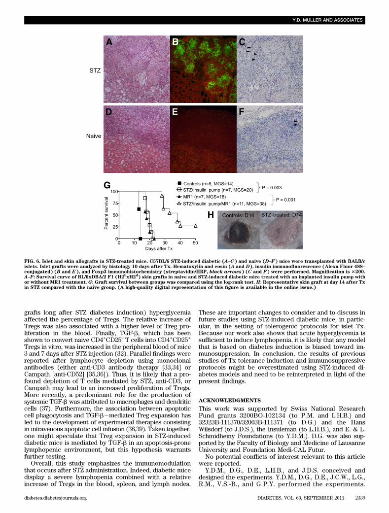
purity of CD4+CD25+ T cells from STZ-induced diabeticmice was similar to those isolated from naive mice (data notshown). No difference was found with respect to the in vitrosuppressive capacity of CD4+CD25+ Tregs, neither at day 3nor at day 13 after STZ administration in comparison withTregs of naive mice. These results suggest that Tregs in di-abetic mice retain their suppressive function in vivo (Fig. 5A).The proliferation rate of Tregs was further analyzed in theblood and in the spleen. Ki67-positive cells were gated inCD8+, total CD4+, and CD4+CD25+Foxp3+ T cells. Treg pro-liferation increased at days 5 and 7 in the blood (Fig. 5B).Moreover, TGF-b measured in the plasma of STZ mice wasalso enhanced at day 7 (Fig. 5C). In the spleen, no differencewas detected in Treg proliferation between days 3 and 7,although their basal proliferation rate was higher than thatof total CD4+ cells (Fig. 5D). In addition, Treg proliferationaccounted for up to 50% of the total CD4+ cell proliferation(data not shown). Thus, these results demonstrate that theproliferative response of Tregs to STZ in vivo also contrib-utes to the increase of their frequency in the blood but not inthe spleen.STZ-induced diabetic mice are immunosuppressed. Wefurther investigated the effect of STZ-induced lymphopeniaand increased percentage of Tregs on allograft rejection.First, STZ-induced diabetic and naive C57BL/6 mice weretransplanted with BALB/c pancreatic islets. Ten days afterTx, the mice were killed and grafts were analyzed by im-munohistology. Islets were completely destroyed in naivemice (Fig. 6D and E), whereas in STZ-induced diabeticmice, the islets were stained positive for insulin (Fig. 6A andB). The median graft survival (MGS) in STZ-induced di-abetic mice was 17.5 days (data not shown). Moreover,higher numbers of Foxp3+ cells were found within the graftof diabetic mice compared with naive mice (Fig. 6C–F).To confirm these results in a more stringent Tx model,STZ-induced diabetic and naive C57BL/6 (H2b) mice weregrafted with BL/6xDBA/2 F1 (H2bxH2d) skin 3 days afterSTZ administration. At the same time, an insulin pump wasimplanted subcutaneously in STZ-induced diabetic mice tocorrect the blood glucose, allowing the mice to survive.Skin grafts were rejected approximately 1 week later inSTZ-induced diabetic mice (MGS 20 days) compared withuntreated mice (MGS, 14 days) (Fig. 6G and H). Theseresults demonstrate that STZ treatment delays allogeneicgraft rejection and that this immunosuppressive effect is notspecific to islet Tx. To further confirm the immunosup-pressive effect of STZ-induced diabetes, BL/6xDBA/2 F1(H2bxH2d) skin grafts were transplanted to naive and STZ-induced diabetic mice (+ insulin pump) treated with anti-CD154 mAb (MR1). MR1 alone had a similar effect than STZwith an MGS of 18 days. MR1 therapy significantly improvedthe skin graft survival in STZ-induced diabetic mice (MGS38 days, P , 0.0001), demonstrating that diabetes inductionwith STZ potentiates the immunosuppressive effect of theMR1 (Fig. 6G). Thus, these results suggest that many resultsobtained in STZ-induced diabetic mice with respect to long-term graft tolerance may be overestimated.
DISCUSSION
The goal of this study was to analyze the effect of STZ onthe immune system and the outcome of allotransplantationwith respect to graft rejection and tolerance. Our resultsdemonstrate that STZ-induced diabetic mice are immuno-suppressed compared with naive mice, leading to prolongedislet and skin allograft survival. Two mechanisms are put
STREPTOZOTOCIN AND ISLET TRANSPLANTATION
2336 DIABETES, VOL. 60, SEPTEMBER 2011 diabetes.diabetesjournals.org
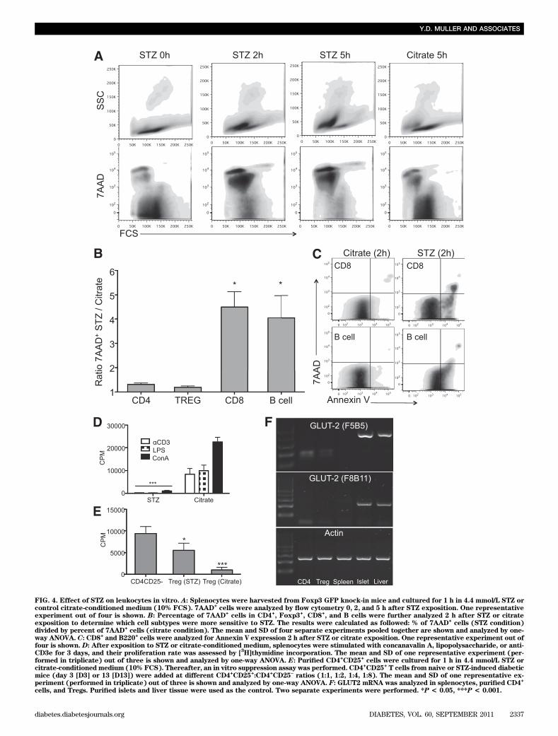
FIG. 4. Effect of STZ on leukocytes in vitro. A: Splenocytes were harvested from Foxp3 GFP knock-in mice and cultured for 1 h in 4.4 mmol/L STZ orcontrol citrate-conditioned medium (10% FCS). 7AAD
+cells were analyzed by flow cytometry 0, 2, and 5 h after STZ exposition. One representative
experiment out of four is shown. B: Percentage of 7AAD+cells in CD4
+, Foxp3
+, CD8
+, and B cells were further analyzed 2 h after STZ or citrate
exposition to determine which cell subtypes were more sensitive to STZ. The results were calculated as followed: % of 7AAD+cells (STZ condition)
divided by percent of 7AAD+cells (citrate condition). The mean and SD of four separate experiments pooled together are shown and analyzed by one-
way ANOVA. C: CD8+ and B220+cells were analyzed for Annexin V expression 2 h after STZ or citrate exposition. One representative experiment out of
four is shown. D: After exposition to STZ or citrate-conditioned medium, splenocytes were stimulated with concanavalin A, lipopolysaccharide, or anti-CD3e for 3 days, and their proliferation rate was assessed by [
3H]thymidine incorporation. The mean and SD of one representative experiment (per-
formed in triplicate) out of three is shown and analyzed by one-way ANOVA. E: Purified CD4+CD25
+cells were cultured for 1 h in 4.4 mmol/L STZ or
citrate-conditioned medium (10% FCS). Thereafter, an in vitro suppression assay was performed. CD4+CD25
+T cells from naive or STZ-induced diabetic
mice (day 3 [D3] or 13 [D13]) were added at different CD4+CD25
+:CD4
+CD25
–ratios (1:1, 1:2, 1:4, 1:8). The mean and SD of one representative ex-
periment (performed in triplicate) out of three is shown and analyzed by one-way ANOVA. F: GLUT2 mRNA was analyzed in splenocytes, purified CD4+
cells, and Tregs. Purified islets and liver tissue were used as the control. Two separate experiments were performed. *P < 0.05, ***P < 0.001.
Y.D. MULLER AND ASSOCIATES
diabetes.diabetesjournals.org DIABETES, VOL. 60, SEPTEMBER 2011 2337
forward to explain the observed immunosuppression asso-ciated with STZ treatment: the induction of lymphopeniaand a shift toward Tregs.
Consistent with previous reports, STZ was directly toxicfor lymphocytes, inducing apoptosis in vitro, and was re-sponsible for early depletion of blood and spleen lym-phocytes in vivo. A study published over 25 years ago (2)had shown that incubation of splenocytes with STZ in vitroabrogated their capacity to proliferate in response to var-ious stimuli. However, these data were not further in-vestigated since then with respect to the characterizationof the different susceptibility of lymphocytes subset toSTZ. It is noteworthy that, in our study, CD8+ T cells and Bcells were particularly sensitive to STZ toxicity in vitro.This observation is in line with the decrease in CD8+ T-cellnumbers in the peripheral blood of STZ-treated mice. Be-cause CD8+ T cells are known to play a critical role inb-cell autoimmunity and rejection, direct CD8+ depletioninduced by STZ administration may contribute to the bet-ter islet engraftment described after Tx. Indeed, NOD micetreated with anti-CD8 mAb or small doses of STZ in earlylife are protected from spontaneous diabetes (5,19,20). Inaddition, our results demonstrate that the uptake of STZinto lymphocytes occurs independently of the transporterGLUT2, which is known to be the main pathway for STZuptake into b-cells (21). Because STZ is a glucose analogand lymphocytes depend on glucose (22), several otheractive and passive glucose transporters such as GLUT1could be implicated. Thus, further investigation is warrantedto determine the receptors responsible for the transport ofSTZ into lymphocytes and its toxic effects that finally lead totheir death.
Importantly, STZ is not the only factor leading to T-celldepletion in STZ-induced diabetic mice. In agreement withsimilar observations in various animal models of diabetes(7,23), we demonstrate here that acute hyperglycemiainduces lymphopenia. Three different models of diabeteswere used, and all induced lymphopenia independently of
STZ: 1) new-onset (diabetes induction in RIP-DTR mice),2) recurrent (nephrectomy of transplanted syngeneic islets),and 3) continuous hyperglycemia (STZ-induced diabetic micetreated with an insulin pump). However, the mechanismsleading to hyperglycemia-induced lymphopenia remainpoorly defined. Our results support the notion that indirectpathways are responsible for the observed lymphopenia invivo because exposure of lymphocytes to high levels ofglucose in vitro did not significantly affect their viability ortheir capacity to proliferate. Diabetes onset is known to beassociated with high serum levels of glucocorticoids, whichare lymphotoxic (24). A potential role of glucocorticoids inhyperglycemia-induced lymphopenia is further supportedby the fact that adrenalectomy before diabetes inductionhampered the increase of glucocorticoids and the associ-ated lymphopenia (7,25). Others showed an adrenal hy-persensitivity preceding the blood hypercorticism (26).Thus, it is likely that dehydration in diabetic mice inducesantidiuretic hormone release, which in turn directly sensi-tizes the adrenal glands to produce glucocorticoids (27).Nevertheless, this hypothesis still remains to be confirmedin STZ-induced diabetic mice.
The second important observation in STZ-induced di-abetic mice was the increase of Treg frequencies in thespleen, the peripheral blood, and also in lymph nodes (datanot shown). In addition, Tregs were detected in islet allo-grafts after STZ administration, with longer graft survival inSTZ-induced diabetic mice compared with control mice.On the basis of these observations and similar reports, weand others suggested a role of Tregs in the control of im-mune responses after allo- and xenogeneic islet Tx (9,13) orsolid organ Tx (28,29). It is not necessarily an increase ofthe absolute numbers of Tregs but rather a favorable shiftof the Treg/T effector cell ratio that is “tolerogenic” (30,31).Importantly, the relative increase of Tregs correlatedspecifically with the administration of STZ and was not aconsequence of hyperglycemia, since neither early (DTadministration to RIP-DTR mice) nor late (removal of islet
FIG. 5. Treg suppression in vitro and proliferation in vivo. A: CD4+CD25+cells were purified from naive (group 1) and STZ-induced diabetic mice at
day 3 (group 2) (D3) and day 13 (group 3) (D13). The results of an in vitro suppression assay, as described in RESEARCH DESIGN AND METHODS, areshown as mean and SDs. One representative experiment out of three is shown. B: Ki67 expression was analyzed in the blood in CD4
+(total
population), CD8+, and Foxp3
+T cells at day 3 (D3), 5 (D5), and 7 (D7) after STZ administration (n = 8–20 per group/time point). C: Plasma TGF-b
concentrations were assessed at 6 h and 1, 2, 3, and 7 days (1d, 2d, 3d, 7d) after STZ administration (n = 6). D: Ki67 expression was analyzed in thespleen at days 3 and 7 after STZ administration (n = 6). The median and the range are shown in B, C, and D and were analyzed with the Kruskal-Wallis test. ***P < 0.001.
STREPTOZOTOCIN AND ISLET TRANSPLANTATION
2338 DIABETES, VOL. 60, SEPTEMBER 2011 diabetes.diabetesjournals.org
grafts long after STZ diabetes induction) hyperglycemiaaffected the percentage of Tregs. The relative increase ofTregs was also associated with a higher level of Treg pro-liferation in the blood. Finally, TGF-b, which has beenshown to convert naive CD4+CD25– T cells into CD4+CD25+
Tregs in vitro, was increased in the peripheral blood of mice3 and 7 days after STZ injection (32). Parallel findings werereported after lymphocyte depletion using monoclonalantibodies (either anti-CD3 antibody therapy [33,34] orCampath [anti-CD52] [35,36]). Thus, it is likely that a pro-found depletion of T cells mediated by STZ, anti-CD3, orCampath may lead to an increased proliferation of Tregs.More recently, a predominant role for the production ofsystemic TGF-b was attributed to macrophages and dendriticcells (37). Furthermore, the association between apoptoticcell phagocytosis and TGF-b2mediated Treg expansion hasled to the development of experimental therapies consistingin intravenous apoptotic cell infusion (38,39). Taken together,one might speculate that Treg expansion in STZ-induceddiabetic mice is mediated by TGF-b in an apoptosis-pronelymphopenic environment, but this hypothesis warrantsfurther testing.
Overall, this study emphasizes the immunomodulationthat occurs after STZ administration. Indeed, diabetic micedisplay a severe lymphopenia combined with a relativeincrease of Tregs in the blood, spleen, and lymph nodes.
These are important changes to consider and to discuss infuture studies using STZ-induced diabetic mice, in partic-ular, in the setting of tolerogenic protocols for islet Tx.Because our work also shows that acute hyperglycemia issufficient to induce lymphopenia, it is likely that any modelthat is based on diabetes induction is biased toward im-munosuppression. In conclusion, the results of previousstudies of Tx tolerance induction and immunosuppressiveprotocols might be overestimated using STZ-induced di-abetes models and need to be reinterpreted in light of thepresent findings.
ACKNOWLEDGMENTS
This work was supported by Swiss National ResearchFund grants 3200BO-102134 (to P.M. and L.H.B.) and32323B-111370/32003B-111371 (to D.G.) and the HansWilsdorf (to J.D.S.), the Insuleman (to L.H.B.), and E. & L.Schmidheiny Foundations (to Y.D.M.). D.G. was also sup-ported by the Faculty of Biology and Medicine of LausanneUniversity and Foundation Medi-CAL Futur.
No potential conflicts of interest relevant to this articlewere reported.
Y.D.M., D.G., D.E., L.H.B., and J.D.S. conceived anddesigned the experiments. Y.D.M., D.G., D.E., J.C.W., L.G.,R.M., V.S.-B., and G.P.Y. performed the experiments.
FIG. 6. Islet and skin allografts in STZ-treated mice. C57BL/6 STZ-induced diabetic (A–C) and naive (D–F) mice were transplanted with BALB/cislets. Islet grafts were analyzed by histology 10 days after Tx. Hematoxylin and eosin (A and D), insulin immunofluorescence (Alexa Fluor 488–conjugated) (B and E), and Foxp3 immunohistochemistry (streptavidin/HRP, black arrows) (C and F) were performed. Magnification is 3200.A–F: Survival curve of BL/6xDBA/2 F1 (H2
bxH2
d) skin grafts in naive and STZ-induced diabetic mice treated with an implanted insulin pump with
or without MR1 treatment. G: Graft survival between groups was compared using the log-rank test. H: Representative skin graft at day 14 after Txin STZ compared with the naive group. (A high-quality digital representation of this figure is available in the online issue.)
Y.D. MULLER AND ASSOCIATES
diabetes.diabetesjournals.org DIABETES, VOL. 60, SEPTEMBER 2011 2339

Y.D.M., D.G., L.H.B., and J.D.S. analyzed the data. D.G., P.M., L.H.B., and J.D.S. contributed reagents/materials/analy-sis tools. Y.D.M., L.H.B., and J.D.S. wrote the manuscript.
We thank Christian Toso and Géraldine Parnaud (fromthe Surgical Research Unit, Department of Surgery, Uni-versity Hospital Geneva, Geneva, Switzerland) and Anne-Laurence Blanc (from Institut Pasteur, Paris, France) forcritical reading of the manuscript; Isabelle Avril (from theDepartment of Genetic Medicine and Development, Uni-versity of Geneva Medical School, Geneva, Switzerland)and Domenico Bosco (from the Surgical Research Unit,Department of Surgery, University Hospital Geneva, Ge-neva, Switzerland) for helpful discussions; and CorinneSinigaglia, Nadine Pernin, David Matthey-Doret, SolangeMasson, and Caroline Rouget (all from the Surgical Re-search Unit, Department of Surgery, University HospitalGeneva, Geneva, Switzerland) for technical assistance.
REFERENCES
1. Lenzen S. The mechanisms of alloxan- and streptozotocin-induced di-abetes. Diabetologia 2008;51:216–226
2. Gaulton GN, Schwartz JL, Eardley DD. Assessment of the diabetogenicdrugs alloxan and streptozotocin as models for the study of immune de-fects in diabetic mice. Diabetologia 1985;28:769–775
3. Koulmanda M, Qipo A, Auchincloss HJ Jr, Smith RN. Effects of streptozotocinon autoimmune diabetes in NOD mice. Clin Exp Immunol 2003;134:210–216
4. Nichols WK, Spellman JB, Vann LL, Daynes RA. Immune responses ofdiabetic animals: direct immunosuppressant effects of streptozotocin inmice. Diabetologia 1979;16:51–57
5. Takayama Y, Ichikawa T, Maki T. Effect of STZ administration on isletisograft and allograft survival in NOD mice. Diabetes 1993;42:324–329
6. Hugues S, Mougneau E, Ferlin W, et al. Tolerance to islet antigens andprevention from diabetes induced by limited apoptosis of pancreatic betacells. Immunity 2002;16:169–181
7. Luo B, Chan WF, Lord SJ, et al. Diabetes induces rapid suppression ofadaptive immunity followed by homeostatic T-cell proliferation. Scand JImmunol 2007;65:22–31
8. Niclauss N, Bosco D, Morel P, Giovannoni L, Berney T, Parnaud G.Rapamycin impairs proliferation of transplanted islet b cells. Trans-plantation 2011;91:714–722
9. Muller YD, Mai G, Morel P, et al. Anti-CD154 mAb and rapamycin induce Tregulatory cell mediated tolerance in rat-to-mouse islet transplantation.PLoS ONE 2010;5:e10352
10. Pearson T, Markees TG, Serreze DV, et al. Islet cell autoimmunity andtransplantation tolerance: two distinct mechanisms? Ann N Y Acad Sci2003;1005:148–156
11. Horner BM, Randolph MA, Huang CA, Butler PE. Skin tolerance: in searchof the Holy Grail. Transpl Int 2008;21:101–112
12. Arefanian H, Tredget EB, Rajotte RV, Gill RG, Korbutt GS, Rayat GR.Short-term administrations of a combination of anti-LFA-1 and anti-CD154monoclonal antibodies induces tolerance to neonatal porcine islet xeno-grafts in mice. Diabetes 2010;59:958–966
13. Webster KE, Walters S, Kohler RE, et al. In vivo expansion of T reg cellswith IL-2-mAb complexes: induction of resistance to EAE and long-termacceptance of islet allografts without immunosuppression. J Exp Med2009;206:751–760
14. Luo X, Pothoven KL, McCarthy D, et al. ECDI-fixed allogeneic splenocytesinduce donor-specific tolerance for long-term survival of islet transplants viatwo distinct mechanisms. Proc Natl Acad Sci U S A 2008;105:14527–14532
15. Tao R, de Zoeten EF, Ozkaynak E, et al. Deacetylase inhibition promotes thegeneration and function of regulatory T cells. Nat Med 2007;13:1299–1307
16. Muller YD, Golshayan D, Ehirchiou D, Wekerle T, Seebach JD, Bühler LH. Tregulatory cells in xenotransplantation. Xenotransplantation 2009;16:121–128
17. Thorel F, Nepote V, Avril I, et al. Conversion of adult pancreatic alpha-cellsto beta-cells after extreme beta-cell loss. Nature 2010;464:1149-1154
18. Strandell E, Eizirik DL, Korsgren O, Sandler S. Functional character-istics of cultured mouse pancreatic islets following exposure to differentstreptozotocin concentrations. Mol Cell Endocrinol 1988;59:83–91
19. Knip M, Siljander H. Autoimmune mechanisms in type 1 diabetes. Auto-immun Rev 2008;7:550–557
20. Thivolet C, Bendelac A, Bedossa P, Bach JF, Carnaud C. CD8+ T cellhoming to the pancreas in the nonobese diabetic mouse is CD4+ T cell-dependent. J Immunol 1991;146:85–88
21. Hosokawa M, Dolci W, Thorens B. Differential sensitivity of GLUT1- andGLUT2-expressing beta cells to streptozotocin. Biochem Biophys ResCommun 2001;289:1114–1117
22. Maciver NJ, Jacobs SR, Wieman HL, Wofford JA, Coloff JL, Rathmell JC.Glucose metabolism in lymphocytes is a regulated process with significanteffects on immune cell function and survival. J Leukoc Biol 2008;84:949–957
23. Otton R, Soriano FG, Verlengia R, Curi R. Diabetes induces apoptosis inlymphocytes. J Endocrinol 2004;182:145–156
24. De Nicola AF, Fridman O, Del Castillo EJ, Foglia VG. Abnormal regulationof adrenal function in rats with streptozotocin diabetes. Horm Metab Res1977;9:469–473
25. Han F, Ozawa H, Matsuda KI, Lu H, De Kloet ER, Kawata M. Changes inthe expression of corticotrophin-releasing hormone, mineralocorticoidreceptor and glucocorticoid receptor mRNAs in the hypothalamic para-ventricular nucleus induced by fornix transection and adrenalectomy.J Neuroendocrinol 2007;19:229–238
26. Revsin Y, van Wijk D, Saravia FE, Oitzl MS, De Nicola AF, de Kloet ER.Adrenal hypersensitivity precedes chronic hypercorticism in streptozotocin-induced diabetes mice. Endocrinology 2008;149:3531–3539
27. Roberts EM, Pope GR, Newson MJ, Lolait SJ, O’Carroll AM. The vasopressinV1b receptor modulates plasma corticosterone responses to dehydration-induced stress. J Neuroendocrinol 2011;23:12–19
28. Golshayan D, Jiang S, Tsang J, Garin MI, Mottet C, Lechler RI. In vitro-expanded donor alloantigen-specific CD4+CD25+ regulatory T cells pro-mote experimental transplantation tolerance. Blood 2007;109:827–835
29. Joffre O, Santolaria T, Calise D, et al. Prevention of acute and chronicallograft rejection with CD4+CD25+Foxp3+ regulatory T lymphocytes. NatMed 2008;14:88–92
30. Mansour H, Homs S, Desvaux D, et al. Intragraft levels of Foxp3 mRNApredict progression in renal transplants with borderline change. J Am SocNephrol 2008;19:2277–2281
31. Kawai T, Cosimi AB, Spitzer TR, et al. HLA-mismatched renal trans-plantation without maintenance immunosuppression. N Engl J Med 2008;358:353–361
32. Chen W, Jin W, Hardegen N, et al. Conversion of peripheral CD4+CD25–naive T cells to CD4+CD25+ regulatory T cells by TGF-beta induction oftranscription factor Foxp3. J Exp Med 2003;198:1875–1886
33. Belghith M, Bluestone JA, Barriot S, Mégret J, Bach JF, Chatenoud L. TGF-beta-dependent mechanisms mediate restoration of self-tolerance inducedby antibodies to CD3 in overt autoimmune diabetes. Nat Med 2003;9:1202–1208
34. Ochi H, Abraham M, Ishikawa H, et al. Oral CD3-specific antibody sup-presses autoimmune encephalomyelitis by inducing CD4+ CD25- LAP+T cells. Nat Med 2006;12:627–635
35. Noris M, Casiraghi F, Todeschini M, et al. Regulatory T cells and T celldepletion: role of immunosuppressive drugs. J Am Soc Nephrol 2007;18:1007–1018
36. Bloom DD, Chang Z, Fechner JH, et al. CD4+ CD25+ FOXP3+ regulatoryT cells increase de novo in kidney transplant patients after immunodepletionwith Campath-1H. Am J Transplant 2008;8:793–802
37. Perruche S, Zhang P, Liu Y, Saas P, Bluestone JA, Chen W. CD3-specificantibody-induced immune tolerance involves transforming growth factor-beta from phagocytes digesting apoptotic T cells. Nat Med 2008;14:528–535
38. Maeda A, Schwarz A, Kernebeck K, et al. Intravenous infusion of syngeneicapoptotic cells by photopheresis induces antigen-specific regulatory T cells.J Immunol 2005;174:5968–5976
39. Kleinclauss F, Perruche S, Masson E, et al. Intravenous apoptotic spleencell infusion induces a TGF-beta-dependent regulatory T-cell expansion.Cell Death Differ 2006;13:41–52
STREPTOZOTOCIN AND ISLET TRANSPLANTATION
2340 DIABETES, VOL. 60, SEPTEMBER 2011 diabetes.diabetesjournals.org