original article isolation of paenibacillus sp. and
TRANSCRIPT

Int J Biochem Mol Biol 2012;3(4):352-364www.ijbmb.org /ISSN:2152-4114/IJBMB1211003
Original ArticleIsolation of Paenibacillus sp. and Variovorax sp. strains from decaying woods and characterization of their potential for cellulose deconstruction
Silvina Ghio1, Gonzalo Sabarís Di Lorenzo2, Verónica Lia2, Paola Talia2, Angel Cataldi2, Daniel Grasso1, Eleonora Campos2
1Instituto de Suelos, CIRN, Inst. Nacional de Tecnología Agropecuaria (INTA); 2Instituto de Biotecnología, CICVyA, Inst. Nacional de Tecnología Agropecuaria (INTA). Dr. N. Repetto y Los Reseros s/n, 1686 Hurlingham, Buenos Aires, Argentina
Received November 28, 2012; Accepted December 3, 2012; Epub December 24, 2012; Published December 30, 2012
Abstract: Prospection of cellulose-degrading bacteria in natural environments allows the identification of novel cellu-lases and hemicellulases that could be useful in second-generation bioethanol production. In this work, cellulolytic bacteria were isolated from decaying native forest soils by enrichment on cellulose as sole carbon source. There was a predominance of Gram positive isolates that belonged to the phyla Proteobacteria and Firmicutes. Many primary isolates with cellulolytic activity were not pure cultures. From these consortia, isolation of pure constitu-ents was attempted in order to test the hypothesis whether microbial consortia are needed for full degradation of complex substrates. Two isolates, CB1-2-A-5 and VG-4-A-2, were obtained as the pure constituents of CB1-2 and VG-4 consortia, respectively. Based on 16S RNA sequence, they could be classified as Variovorax paradoxus and Paenibacillus alvei. Noteworthy, only VG-4 consortium showed measurable xylan degrading capacity and signs of filter paper degradation. However, no xylan or filter paper degrading capacities were observed for the pure cultures isolated from it, suggesting that other members of this consortium were necessary for these hydrolyzing activities. Our results indicated that Paenibacillus sp. and Variovorax sp. as well as VG-4 consortium, might be a useful source of hydrolytic enzymes. Moreover, although Variovorax sp. had been previously identified in metagenomic studies of cellulolytic communities, this is the first report on the isolation and characterization of this microorganism as a cellulolytic genus.
Keywords: Cellulolytic bacteria, variovorax, paenibacillus, soil
Introduction
Lignocellulosic biomass is considered the larg-est known renewable carbon source. This kind of biomass comprises three main types of car-bon based polymers: cellulose, hemicellulose and lignin. The main component is cellulose, a β-(1,4)-linked chain of glucose molecules. Hemicellulose, the second most abundant component, consists of heteropolymers of pen-toses (D-xylose, D-arabinose), hexoses (D-mannose, D-glucose and galactose) and sugar acids. Lignin is composed of three major phenolic components, namely p-coumaryl alco-hol, coniferil alcohol and sinapyl alcohol [1].
Over the last few years, the rising demand of energy all over the world and the expected future shortage of petroleum-based fossil fuels, have led to the development of alternative, sus-tainable and renewable forms of fuels. In this context, lignocellulosic biomass has long been recognized as a potential source of mixed sug-ars for bioethanol production, an emerging sec-ond generation biofuel [2].
In nature, lignocellulose is degraded by hydro-lytic and oxidative enzymes produced mainly by fungi and bacteria that are able to synergisti-cally degrade cellulose, hemicellulose and lig-nin [3]. From a phylogenetic point of view, the

Cellulolytic soil bacteria
353 Int J Biochem Mol Biol 2012;3(4):352-364
bacterial cellulolytic ability has a broad distribu-tion. Aerobic cellulolytic bacteria in soil are rather heterogeneous and belong to various phyla including Firmicutes, Actinobacteria, Bacteroidetes, and Proteobacteria. Among them, Bacillus, Cellulomonas, Streptomyces, Cythophaga, Cellvibrio and Pseudomonas have been characterized [4].
Cellulose-degrading bacteria produce a com-plex combination of enzymes (cellulases, hemi-cellulases and pectinases) that belong to differ-ent sequence-based families of glycoside hydrolases (GH), carbohydrate-binding modules (CBMs), polysaccharide lyases (PL) and carbo-hydrate esterases (CE), among others, which are classified in the Carbohydrate-Active Enzyme database (http://www.cazy.org/) [5]. Cellulases represent the primary family needed to deconstruct lignocellulosic substrates. The major enzymatic activities necessary for cellu-
lose deconstruction are endoglucanases (EC 3.2.1.4), which bind randomly along the cellu-lose molecule and hydrolyze the β-1,4 glyco-sidic linkage generating new chain ends; exo-glucanases or cellobiohydrolases (EC 3.2.1.74) (EC 3.2.1.91), which act processively on the reducing or non reducing ends of the molecule, generating either glucose or cellobiose as major products, and β-glucosidases (EC 3.2.1.21), which convert cellobiose to glucose [2, 6, 7]. Another class of enzymes, which include xylanases and xylosidases, are involved in the breakdown of hemicelluloses mainly by hydrolyzing β-1, 4-xylan into xylose [8].
Due to the recalcitrance of cellulosic sub-strates, enzymes that breakdown lignocellulos-ic biomass are a key factor for bioethanol pro-duction. To assure the sustainability of the process, all the steps need to be optimized and
Table 1. Gram staining, morphology and 16S rRNA gene analysis of primary cellulolytic bacterial iso-latesIsolate Morphology/
Gram reactionGenBank Acce-
sion numberGenus identified byRDPa Top BLASTb Hit GenBank Iden-
tity(%)CB1-2 Rod/Gram
negativeJX992634 Pseudomonas sp. Pseudomonas jessenii strain
AMBI2391100
JX992635 Stenotrophomonas sp. 1 Stenotrophomonas maltophilia strain CQ1
99
JX992636 Paenibacillus sp. Paenibacillus sp. 61724 99JX992637 Stenotrophomonas sp. 1 Stenotrophomonas maltophilia
strain CQ199
CB1-7 Rod/Gram positive
JX992638 Bacillus sp. 1 Bacillus sp. CMB 26 99JX992639 Bacillus sp. 2 Bacillus sp. S3.TSA.017 98JX992640 Bacillus sp. 3 Bacillus sp. A2022 100
CB1-8 Rod/Gram positive
JX992641 Viridibacillus sp. Bacillus arenosi isolate M18-3 99JX992642 Brevundimonas sp. Brevundimonas sp.SOZ3-5041 100JX992643 Viridibacillus sp Bacillus arenosi isolate M18-3 99
CB2-1 Rod/ Gram positive
JX992644 Bacillus sp. 4 Bacillus cereus SH 01 99JX992645 Bacillus sp. 4 Bacillus cereus SH 01 99
CB2-5 Rod/Gram positive
JX992646 Lysinibacillus sp. 1 Lysinibacillus sphaericus strain DE4 99JX992647 Lysinibacillus sp. 1 Lysinibacillus sphaericus strain DE4 99JX992648 Lysinibacillus sp. 1 Lysinibacillus sphaericus strain DE4 99JX992649 Lysinibacillus sp. 1 Lysinibacillus sphaericus strain DE4 99
VG-4 Rod/Gram positive-Gram
negative
JX992651 Stenotrophomonas sp. 2 Xanthomonas sp.X1 100JX992652 Achromobacter sp. Achromobacter xylosoxidans
strainX96100
VG-5 Rod/Gram positive
JX992653 Lysinibacillus sp. 2 Lysinibacillus sp. KB1 99JX992654 Lysinibacillus sp. 2 Lysinibacillus sp. KB1 100JX992655 Lysinibacillus sp. 2 Lysinibacillus sp. KB1 100
aRibosomal Database Project (http://rdp.cme.msu.edu/). bBasic Local Aligment Search Tool (http://www.ncbi.nlm.nih.gov/).

Cellulolytic soil bacteria
354 Int J Biochem Mol Biol 2012;3(4):352-364
costs should be lowered. In order to improve biomass deconstruction, one option is to obtain better and more efficient enzymatic cocktails, a goal that could be reached by searching for complementing activities from within natural biodiversity.
In order to achieve this objective, the prospec-tion of bacterial communities present on bio-mass-rich soils could provide insights into their cellulose enzymatic activities and be an attrac-tive source of new enzymes. Therefore, the aim of this study was to screen, isolate and charac-terize cellulose-degrading bacteria present on decaying forest soils, as well as test their enzy-matic activity on different cellulosic substrates.
Materials and methods
Soil sampling
Soil samples were collected from the surface layer (0-20 cm depth) of forest soils from two different regions of Argentina, Valle Grande in Mendoza (VG)(-34° 48’ 56.03”,-68° 27’ 6.19”) and two areas of Cerro Bayo in Neuquén (CB1 and CB2)(-40° 56’ 22.55”,-71° 23’ 44.76”). Soil samples were analyzed for organic matter content with Walkley and Black semi micro method and pH was determined using a glass electrode in a 1:2.5 soil:water suspension. Samples were stored at 4°C until use.
Enrichment, isolation and gram characteriza-tion of cellulolytic bacteria
Since soil samples were stored at 4°C, they were pre-cultured at 30°C for 7 days before use, to allow the reactivation of microorgan-isms. Five grams of each soil sample were sus-pended on 45 ml of sterile physiological solu-tion, agitated on magnetic shaker for 40 min and then centrifuged at 5000 rpm for 10 min. Five ml of the supernatant were added to 45 ml of minimal medium (MM), according to Hanking and Anagostakis [9], with some modifications: (g/l) K2HPO4, 1.67; KH2PO4, 0.87; NaCl, 0.05; Mg SO4 x 7H2O, 0.1; CaCl2 , 0.04; FeCl3 , 0.004; Na2MoO4 x 2H2O, 0.005; biotin, 0.01; nicotinic acid, 0.02; pantotenic acid, 0.01; NH4Cl, 1 and supplemented with 1% (w/v) carboxymethyl-cellulose (CMC, Sigma, USA) and 0.1 % (w/v) yeast extract (YE) for enrichment in cellulolytic
bacteria. Incubations were carried out in an orbital shaker at 30°C and 200 rpm for 48 hs. After this period, a loopful of each culture was streaked out on agar plates (1.5% (w/v) Bacto agar, Difco) of MM-1%CMC or MM-1%CMC-0.1%YE, as indicated in the text, and grown at 30ºC for 5 days. Cellulolytic bacterial strains were detected by staining of agar plates with 0.1% (w/v) Congo red for 15 min and then dis-tained with 1M NaCl for 15 min, twice [10]. Colonies with clear halo were re-streaked in the same media to confirm the positive phenotype.
In order to obtain pure cultures from mixed bac-terial populations, a loopful of liquid culture was streaked out on MM-1%CMC-0.1%YE and 0.01% (v/v) Trypan Blue (Sigma, USA) and incu-bated 3 days at 30°C. Single colony-forming units that showed clear halo were re-streaked four times. For long term storage, all bacterial isolates were cultured overnight in 2 ml of MM-1%CMC-0.1%YE and stored at - 80°C with 20% glycerol.
Gram staining was performed on fresh suspen-sions of bacterial strains using a commercial kit (Britania, Argentina) and an Olympus optical microscope.
Amplification and Identification of bacterial isolates by 16S rRNA gene analysis
Total bacterial genomic DNA from each primary isolate was extracted from 1 ml of an overnight culture in MM-1%CMC-0.1%YE. Each culture was centrifuged at 7000 rpm 5 min and the cell pellet suspended on 50 µl nuclease free water. Samples were heated at 100°C 10 min and then frozen at -80°C twice, followed by a final heating at 100°C for 10 min, centrifuged at 14.000 rpm for 5 min, and the supernatant was used for polymerase chain reaction (PCR) amplification. The nucleotide sequence of 16S rRNA gene was amplified by PCR using the primers fD1 (5’-CCG AAT TCG TCG ACA ACA GAG TTT GAT CCT GGC TCA- 3’) and rD1 (5’-CCC GGG ATC CAA GCT TAA GGA GGT GAT CCA GCC- 3’) [11] on a final reaction volume of 25 µl. The reaction mixture contained 5 µl of 5X poly-merase buffer, 0.7 µM of each primer, 20 mM of dNTPs and 1,25 U of Go-Taq DNA polymerase (Promega, USA). The reaction was run on a Mastercycler™ Gradient (Eppendorf, Germany),

Cellulolytic soil bacteria
355 Int J Biochem Mol Biol 2012;3(4):352-364
using the following cycle: 95°C 4 min, 30 cycles of 30 seg at 95°C, 30 seg at 53°C and 2 min at 72°C, with a final extension of 7 min at 72°C. Amplification products were resolved by elec-trophoresis on 0.8% agarose gel stained with ethidium bromide. The expected amplicon size was approximately 1.5 Kb.
PCR products were purified with Wizard™ SV Gel and PCR Clean-Up system (Promega, USA) and cloned in pGEM™-T Easy (Promega, USA). Escherichia coli DH5-α-competent cells were transformed and plated on LB agar with IPTG and Xgal. Plasmids from white colonies corre-sponding to each amplification product were extracted by UltraClean™ 6 Minute Mini Plasmid Prep (Mo Bio, USA). Sequencing was performed by the Biotechnology Institute Sequencing Service, INTA Castelar, using an ABI 3130xl Capillary DNA sequencer (Applied Biosystems, USA) with universal T7 or SP6 primers.
For pure isolates, total genomic DNA was extracted using a 10% Chelex-based resine (Bio-Rad, USA). Direct sequencing of amplified 16S rRNA gene product was carried out with fD1 and rD1 primers. Sequences were submit-ted to GenBank and have been provided acces-sion numbers from JX992628 to JX992655 according to Table 1.
Phylogenetic analysis
The 16S rRNA gene sequences of all strains were analyzed by comparison to the sequences available on the Ribosomal Database Project (http://rdp.cme.msu.edu/) and using the BLAST algorithm from NCBI (http://www.ncbi.nlm.nih.gov/). The best hits resulting from this analysis were selected for phylogenetic reconstruction, along with representatives of several reference genera.
Multiple alignment of DNA sequences was accomplished using T-coffee [12], followed by minor manual editing. Phylogenetic trees were obtained under the Maximum Parsimony crite-rion using the software TNT [13]. Heuristic searches were performed starting from 100 Wagner trees with tree bisection-reconnection branch-swapping (TBR). All character changes were given equal weight and gaps were treated as fifth base. Node support values were assessed using 1000 bootstrap replicates. The Archaea Halobacterium salinarum (AB60351) was used as outgroup.
Cellulosic substrates degrading assays
Primary and pure isolates were grown on MM-1%CMC-0.1%YE, for 48 hs at 30°C, until
Table 2. Characterization and cellulolytic potential of primary isolates (A) and primary isolates com-pared with their cellulolytic constituent pure isolates (B)
Isolate Growth on MM-1%CMC
Hydrolytic ca-pacity on CMC
(HC valuea)
CMCase activity (mg reducing sug-
ars/ml hydrolysate)
Growth on MM-0.5%
xylan
Growth on MM-0.5%
Avicel
Growth (G) and/or degradation (D) of
filter paperA CB1-2 + 3.61 (0.64) n/e + + G
CB1-7 + 1.23 (0.10) n/e + + GCB1-8 + 1.21 (0.11) n/e + + GCB2-1 + 1.11 (0.10) n/e + + GCB2-5 + 2.97 (0.9) n/e + + GVG-4 + 3.33 (1.45) n/e + + G + DVG-5 + 3.76 (0.41) n/e + + G
B CB1-2 + 3.37 (0.42) 0.95 + + GCB1-2-A-1 - n/e 0.03 - - NGCB1-2-A-4 + 1.25 (0.16) 0.01 - - NGCB1-2-A-5 + 4.53 (0.12) 1.15 + + NGVG-4 + 3.27 (0.64) 0.16 + + GVG-4-A-1 - n/e 0.03 - - NGVG-4-A-2 + 2.54 (0.24) 0.73 + + NG
ahydrolysis capacity (HC) value was determined by the ratio between the diameter of clear halo and the diameter of the colony. Averages of triplicate experiments with the standard deviation in parentheses were calculated. +: positive; - negative, n/e: not evaluated, NG: no growth observed.
Cellulolytic soil bacteria
356 Int J Biochem Mol Biol 2012;3(4):352-364
an optical density at 600 nm of 0.5 for all cul-tures. Endocellulase capacity was estimated on MM-1%CMC solid medium. From each cul-ture, a 5 µl drop was seeded per triplicate and grown for 5 days at 30°C and then the clear degrading halo by Congo red staining was mea-sured. To determine the hydrolysis capacity (HC) value, the ratio between the diameter of clear halo and the diameter of the colony was calculated [9]. Exocellulase capacity was esti-mated by growth on MM supplemented with 0.5% (w/v) microcrystaline cellulose Avicel (Fluka, Switzerland) at 30°C, 5 days. Hemicellulase capacity was determined on solid MM supplemented with 0.5 % (w/v) Xylan from beechwood (Sigma, USA), with the same experimental procedure used with CMC as sub-strate, using Congo red staining and establish-ing the HC value. Total cellulose capacity was analyzed by culture on liquid MM with a 1 cm x 6 cm filter paper strip for 14 days at 30°C.
CMCase activity in liquid medium was also eval-uated by DNS (3,5-dinitrosalicylic acid) reduc-ing sugar assay adapted to a 96-well micro-plate system using a calibration curve of glucose, on replicate experiments. First, cell free culture supernatants (containing secreted soluble enzymes) were obtained by centrifuga-tion of a 10 ml culture (MM-1%CMC-0.1% YE) at 4000 rpm, 20 min at 4°C and then filtering the
supernatant through 0.2 µm syringe filters. Two ml of the resulting supernatant were fivefold concentrated by lyophilization and subsequent resuspension in 400 µl of sterile mqH2O. Hydrolysis reactions were carried out per dupli-cate in 96-well PCR microplates (adapted from King et al. [14]). Each well contained 90 µl of 2% CMC substrate in 0.1 M sodium acetate buffer pH 5 and 90 µl of the concentrated cell free supernatant. The plate was incubated at 50°C for 3 hs. Then, 50 µl of reaction were mixed with 100 µl of DNS reagent. DNS reac-tion was carried out in a PCR thermocycler by heating at 98°C for 5 min followed by cooling at 4°C for 1 min, and holding at 20°C. One hun-dred microliters of the reaction were added to 100 µl of ultrapure water in flat-bottom micro-plates and absorbencies were measured at 540 nm. Reducing sugar quantities were calcu-lated from the linear regression of the stan-dards. Endoglucanase activity was expressed as mg of reducing sugar per ml of hydrolysate.
Results
Isolation and identification of cellulolytic bacte-ria from decaying forest soils
In order to isolate novel bacteria capable of degrading cellulose, soil samples from forest soils from Valle Grande in Mendoza (VG) and
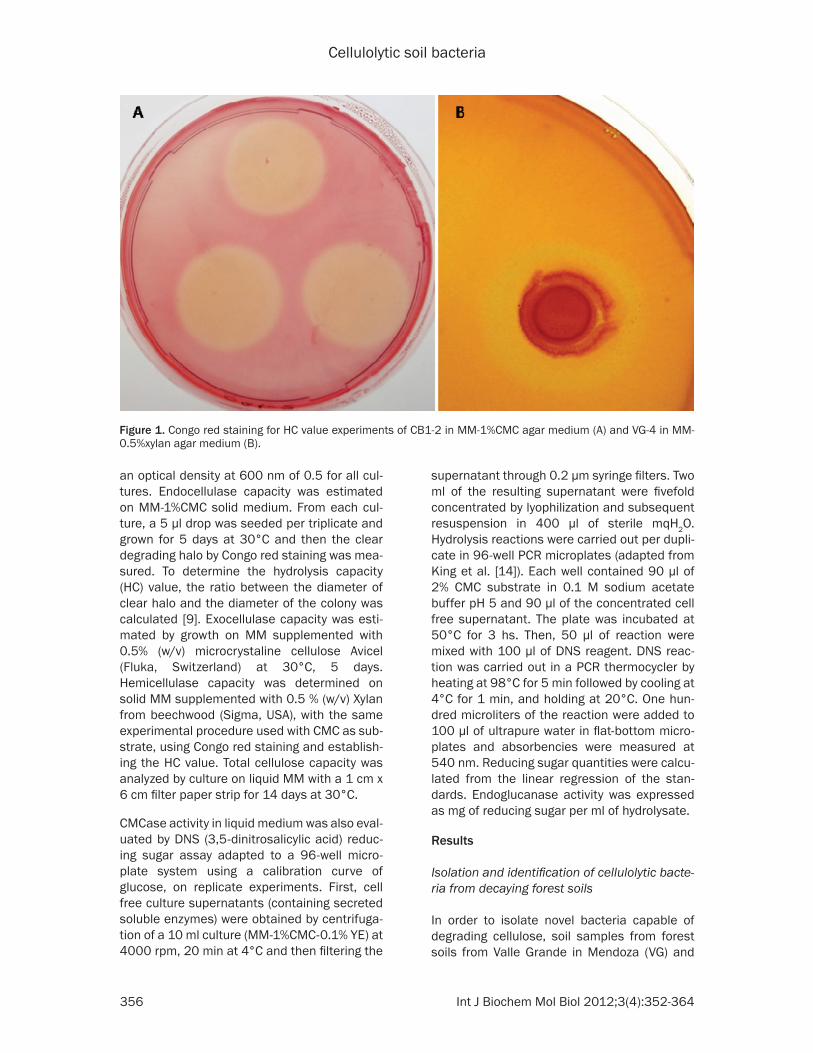
Figure 1. Congo red staining for HC value experiments of CB1-2 in MM-1%CMC agar medium (A) and VG-4 in MM-0.5%xylan agar medium (B).
Cellulolytic soil bacteria
357 Int J Biochem Mol Biol 2012;3(4):352-364
from two areas of Cerro Bayo in Neuquén (CB1 and CB2), Argentina, were studied. These regions are rich in biodiversity and have not been influenced by agronomic exploitation. The pH of the soil samples were 7.7, 4.5 and 5.2 for VG, CB1 and CB2 respectively. The organic mat-ter content was determined in samples without removing lignocellulosic material, and results were 4.24%, 13.69% and 13.96% for VG, CB1 and CB2, respectively. As expected for soils from neighboring areas, CB samples showed similar soil parameters, with their elevated organic matter content probably due to a higher a presence of leaves and wood pieces
The three samples were enriched in cellulose degrading bacteria by growth on minimal media, supplemented with carboxymethyl-cel-lulose and yeast extract (MM-1%CMC-0.1% YE) and bacterial strains were then isolated by suc-cessive streak out, on agar plates of the same media, with and without yeast extract. A pre-liminary screening for cellulolytic bacteria was carried out using Congo red staining. From those colonies which produced a significant and similar clear halo, 7 bacterial isolates were randomly chosen for further characterization: 3 from CB1 (CB1-2, CB1-7 and CB1-8), 2 from CB2 (CB2-1 and CB2-5) and 2 from VG (VG-4 and VG-5).
Isolates were analyzed by Gram staining and optic microscopy, showing a predominance of Gram positive rod-shaped bacteria across the three soil samples. With the aim of identifying the selected bacteria, 16S rRNA was amplified and cloned. Plasmid DNA from several clones of each isolate was purified and inserts were sequenced. Resulting partial sequences corre-sponding to the 5’ end of the gene were ana-lyzed by RDP database and NCBI BLAST (Table 1). Analysis of these sequences indicated that
four primary isolates, CB1-2, CB1-7, CB1-8 and VG-4, corresponded to bacterial consortia since they consisted of more than one bacterial genus (Table 1). In contrast, CB2-1, CB2-5 and VG-5 seemed to correspond to homogeneous isolates, as they showed high sequence similar-ity (more than 99%) to sequences of a single genus (Table 1). However, streak out of these isolates on MM-1%CMC-0.1%YE-0.01%Trypan Blue revealed colonies with different macro-scopical characteristics and some colonies did not show cellulose-degrading capacity, sug-gesting the presence of more than one bacte-rial type, possibly including different species of the same genus. For CB1-7, CB2-1, CB2-5 and VG-5, identification based on 16S rRNA analy-sis was concordant to Gram staining. In con-trast, only Gram negative bacteria were observed in CB1-2 culture, while in CB1-8 only Gram positive bacteria were observed. Analyzing VG-4 isolate, Gram positive and neg-ative bacteria were found to be equally distrib-uted. These results could be attributed to dif-ferent representation of the bacterial constituents in the consortia.
Growth and degrading potential on different cellulosic substrates
Bacterial primary isolates were further charac-terized for their potential to deconstruct cellu-losic substrates. As expected, all isolates were able to grow on solid minimal medium supple-mented with carboxymethyl-cellulose as sole carbon source (MM-1%CMC). In terms of hydro-lysis capacity (HC) on CMC as substrate, VG-5 (Lysinibacillys) and CB1-2 (consortium) showed the highest values (3.76 and 3.61, respective-ly), followed by VG-4 (consortium) (3.33) and CB2-5 (Lysinibacillus) (2.97), suggesting that these isolates have high endocellulase poten-tial (Table 2A). To evaluate hemicellulose
Table 3. 16S rRNA gene analysis of pure bacterial isolatesPure isolate Clear halo on
MM-1%CMC-0.1%YE-0.01%TB
GenBankAccesion number
Genus identified by RDPa
Top BLASTb Hit GenBank Identity (%)
CB1-2-A-1 No JX992628 Stenotrophomonas sp. Stenotrophomonas sp. VTAE 129 100CB1-2-A-4 Yes JX992629 Staphylococcus sp. Staphylococcus warnieri 99CB1-2-A-5 Yes JX992630 Variovorax sp. Variovorax sp. DC2 a-35 100VG-4-A-1 No JX992631 Stenotrophomonas sp. Stenotrophomonas sp. VTAE 129 100VG-4-A-2 Yes JX992632 Paenibacillus sp. Paenibacillus sp.H3029 99VG-4-A-3 Yes JX992633 Paenibacillus sp. Paenibacillus sp. H3029 100aRibosomal Database Project (http://rdp.cme.msu.edu/). bBasic Local Aligment Search Tool (http://www.ncbi.nlm.nih.gov/).

Cellulolytic soil bacteria
358 Int J Biochem Mol Biol 2012;3(4):352-364
degrading potential, growth and HC values on xylan agar medium (MM-0.5% xylan) were ana-lyzed. All isolates except CB2-1 (which showed 99% identity with Bacillus cereus) grew on xylan as sole carbon source. However, only VG-4 (consortium) produced a measurable zone of xylan degradation after Congo red staining, with an HC value of 1.27 (Figure 1). All isolates were also able to grow on Avicel as sole carbon source, indicating capacity to degrade crystalline cellulose. Filter paper degradation was assayed to evaluate total cellulose poten-tial. After a two-week incubation period, all iso-lates presented signs of growth, although only VG-4 showed mild filter paper degradation signs (data not shown).
Identification and characterization of consor-tium constituent bacteria
With the purpose of testing the hypothesis of whether bacterial consortia are necessary for deconstruction of complex substrates or only one of the components is truly necessary and the rest of the bacterial components are merely using the simple sugars generated, we attempt-ed isolation of pure constituents of CB1-2 and VG4 and compared their cellulolytic and/or hemicellulolytic activity with the corresponding consortia from which they were isolated.
To obtain pure bacterial cultures from consortia isolates CB1-2 and VG-4, four rounds of suc-cessive streak out on MM-1%CMC-0.1%YE-0.01%Trypan Blue were done (Figure 2). Two pure colonies of CB1-2 and VG-4 with cellulose
degrading capacity and one colony from each sample without cellulose degrading capacity were selected for further identification by 16S rRNA amplification and sequencing. According to Blast homology search and RDP classifica-tion, colonies without cellulose degrading capacity isolated from both VG-4 and CB1-2 showed high sequences identity (>99%) to Stenotrophomonas sp. (VG-4-A-1 and CB1-2-A-1, respectively). On the other hand, pure isolates with CMCase capacity from CB1-2 and VG-4 also showed sequence identity (> 99%) to Variovorax sp. (CB1-2-A-5), Staphylococcus sp. (CB1-2-A-4) and Paenibacillus sp. (VG-4-A-2 and VG-4-A-3), respectively (Table 3). Phyloge- netic analysis of 16S rRNA strongly supports the placement of CB1-2-A-5 within the Variovorax paradoxus clade, and the inclusion of CB1-2-A-4 within the Staphylococcus group. The position of VG-4-A-2 and VG-4-A-3 as part of the Paenibacillus clade is also highly sup-ported; however the sister-group relationship between these isolates and Paenibacillus alvei shows only moderate bootstrap values (Figure 3).
On the other hand, although they had been identified as part of the initial consortia, pure cultures of Paenibacillus sp. or Pseudomonas sp. could not be isolated from CB1-2. Similarly, pure isolates of Achromobacter sp. were not obtained from VG-4. Moreover, pure isolates with detectable xylan or avicel degrading capacity could not be obtained either from VG-4 or from CB1-2 consortia, respectively.
In order to evaluate the contribution of individu-al components to the consortium in a measur-able way, cellulose degrading potential of CB1-2 and VG-4 consortia was compared to that of the pure strains isolated from them (Table 2B). Using CMC as substrate, CB1-2-A-4 (Staphylococcus) had a HC value (1.25) lower than that of its respective consortium CB1-2 (3.37) while CB1-2-A-5 (Variovorax) had a high-er HC value (4.53). Also, CB1-2-A-1 (Stenotrophomonas), the pure component of the consortia isolated without cellulose-degrading capacity, did not grow on MM-1%CMC (although it grew well when the same culture medium was supplemented with yeast extract). These results suggest that Variovorax could be the component of the consortium mainly responsible for endocellulase capacity and that
Figure 2. Pure constituent CB1-2 colonies with (A) and without (B) cellulose degrading capacity on MM-1%CMC-0.1%YE-0.01%trypan blue agar medium.

Cellulolytic soil bacteria
359 Int J Biochem Mol Biol 2012;3(4):352-364
Stenotrophomonas (CB1-2-A-1) could lack the enzymatic pathways necessary to use CMC as sole carbon source, hence using the sugars
released by other members of the consortium. The reason why HC value of Variovorax was higher than that of the consortium may be
Figure 3. Phylogenetic analysis of 16S rRNA. Strict consensus of four Maximum Parsimony trees. Bootstrap values are given below branches (>50%). Accession numbers are indicated in parenthesis.

Cellulolytic soil bacteria
360 Int J Biochem Mol Biol 2012;3(4):352-364
explained by the fact that, at the same culture OD, less number of bacteria from a single genus, in this case Variovorax sp., would be present in the consortium than in the pure cul-ture. This further supports our hypothesis that Variovorax sp. is the bacterial genus responsi-ble for the measured activity. In the case of VG-4 consortia, it showed a higher HC value (3.27) than the pure cellulolytic component VG-4-A-2 (Paenibacillus) (2.54). Also, VG-4-A-1 (Stenotrophomonas) did not grow on MM-1%CMC.
Xylan and Avicel degrading potential were also evaluated by growth in MM supplemented with 0.5% xylan or 0.5% Avicel, as sole carbon sourc-es. CB1-2-A-1, VG-4-A-1 (Stenotrophomonas) and CB1-2-A-4 (Staphylococcus) did not grow on these substrates while CB1-2-A-5 (Variovorax) and VG-4-A-2 (Paenibacillus) grew well on xylan and Avicel. However, no degrada-tion halo could be observed on MM 0.5% xylan agar plates by Congo red staining.
Evaluating total cellulase capacity, pure iso-lates were not able to use filter paper as carbon source suggesting that complete primary con-sortia were needed to fully hydrolyze complex substrates. The fact that VG-4 consortium pro-duced a clear degradation halo on xylan and could degrade filter paper as substrate, while the pure isolate VG-4-A-2 (Paenibacillus) did not, also reinforces the idea that other not iso-lated members of this consortium could be responsible or necessary for complete degra-dation of these substrates.
CMCase activity in culture supernatants
CMCase activity from free enzymes in culture supernatants was assayed by DNS method to determine the amount of reducing sugars released from CMC (Table 2B) for CB1-2 and VG-4 and their pure bacterial components. Variovorax (CB1-2-A-5) presented the highest endoglucanase activity, which was slightly high-er than that of CB1-2 consortium (1.15 and 0.95 mg of reducing sugars/ml of hydrolysate, respectively). The activity on culture superna-tant of the other members of CB1-2 consor-tium, Staphylococcus (CB1-2-A-1) and Stenotrophomonas (CB1-2-A-4) was almost undetectable (0.03 and 0.01, respectively). This result supports the idea that Variovorax sp. is the constituent of the consortium mainly
responsible for CMC degradation and that this activity could be attributed to free secreted enzymes.
In contrast, while VG-4 (consortium) showed a higher HC value than VG-4-A-2 (Paenibacillus), the latter showed higher endoglucanase activi-ty in DNS assays (0.73 and 0.16 mg of reducing sugars/ml of hydrolysate, respectively). VG-4-A-1 (Stenotrophomonas) presented an almost undetectable activity (0.03), which is in accordance with the results from the other iso-late with similarity to Stenotrophomonas (CB1-2-A-4) obtained in this study. All assays were repeated twice, with independent cultures, and similar results were obtained.
The discrepancy between HC values and DNS assay results from VG-4 and VG4-A-2 (Paenibacillus) could be explained by differenc-es in activity of free secreted enzymes present in the culture supernatant (evaluated by DNS assay) as opposed to whole bacteria activity (determined by HC value measurement).
Discussion
In this study, cellulolytic bacterial strains were isolated from native decaying forest soils from two different regions of Argentina. These areas were chosen because they are pristine forests and have not been influenced by agricultural practices. There was a predominance of Gram positive isolates, with a marked prevalence of bacteria from the order Bacillales. The isolation of Bacillus strains with cellulolytic activity has also been reported in prospection of Brazilian semi-arid caatinga soils [15] and in forest soils [16]. More recently, it was described the isola-tion of Bacillus strains from agricultural soils on CMC agar plates, with high cellulolytic potential [17]. In agreement with our work, different spe-cies of Lysinibacillus have also been isolated and described as a cellulolytic and xylanolytic genus from different sources, such as the gut of an earthworm [18], soil [19] and from forest humus [20]. In this study, the species of Lysinibacillus isolated showed high endocellu-lase capacity on solid medium, using CMC as substrate.
Regarding endoglucanase potential, the HC val-ues of all the isolates in this study were between 1.11 and 4.53. This range was higher than pre-viously reported results from bacteria isolated

Cellulolytic soil bacteria
361 Int J Biochem Mol Biol 2012;3(4):352-364
from forest soils (0.15 to 2.80) and from agri-cultural soils (1.38 to 2.79) although no identifi-cation of bacterial genus was done in those studies [21]. In contrast, HC values were lower than those obtained with mesophilic bacteria isolated from a flower stalks-vegetables waste co-composting system (Bacillus pasteurii, Bacillus cereus, Halobacillus, Aeromicrobium and Brevibacterium) (between 4.24 and 10.36) [22]. Whether this is due to differences in the isolation or enrichment procedures or to the presence of bacterial genus of higher cellulo-lytic potential in compost sources, still needs to be further investigated.
When analyzing the pure isolates with cellulo-lytic potential, the identified genera were not identified as part of the original consortium. In the case of CB1-2 and VG4 consortia, the pure isolates with the highest cellulolytic capacity were identified as Variovorax paradoxus and Paenibacillus alvei, respectively, although none of them had been identified as part of the pri-mary consortium. On the other hand, Achromobacter sp. was identified as part of VG-4 consortium, but it could not be isolated as a pure culture. These discrepancies may be due to a different degree of representation of bacteria in the consortia or could imply that some bacterial genus were not able to use the substrate on their own, but rather needed the presence of other genus, probably because they lacked all required enzymes for decon-struction of the substrates. In this context, while different strains of Pseudomonas sp. were isolated from diverse sources (decompos-ing rice straw, soil, rhizospheric areas) and characterized as cellulolytic [23-25], no isolat-ed Achromobacter sp. strains have been reported so far. However, genome sequencing of A. xylosoxydans A8 [26] has revealed a gene encoding a GH3 beta-glucosidase and also cel-lulolytic strains of Achromobacter were report-ed in switchgrass and soil metagenomic stud-ies [27, 28], although no further experimental evidence of the cellulolytic capacity of this genus was published. On the other hand, the role of Stenotrophomonas in consortia dynam-ics remains elusive.
Analysis of pure constituent bacteria from cel-lulolytic consortium CB1-2 revealed that Variovorax sp. had a high endoglucanase capacity, in agar plates and liquid medium.
Variovorax sp. is a metabolically diverse, aero-bic bacterium that engages in mutually benefi-cial interactions with a variety of bacteria and plants. Whole genome sequencing of V. para-doxus S110 has shown that it presents diverse metabolic capabilities. However, it appears to lack genes involved in the production of com-mon hydrolytic enzymes that macerate plant cell wall polymers. One exception is a gene that encodes a β-glucosidase and seven genes enconding feruloylesterases, although genes involved in the production of pectinases and other cellulases remained unidentified [29]. Therefore, our finding that an isolate from this genus presents a high endoglucanase capacity is noteworthy. Even when Variovorax sp. has been reported before as a cellulolytic genus [28-30], to the best of our knowledge, this is the first report of isolation and evaluation of its cellulolytic potential. Based on phylogenetic analysis, the strain isolated in this study is closely related to Variovorax paradoxus.
In contrast, different species of Paenibacillus have been previously identified for their cellu-lose and xylan degrading activities [31-35]. In this study, the pure isolate identified as cellulo-lytic constituent of VG-4 consortium, shows high homology with Paenibacillus alvei. While VG-4 consortium showed considerable cellu-lose, xylan and filter paper degrading capacity, Paenibacillus sp. only presented CMCase activ-ity and was able to grow on xylan as sole car-bon source. Strains closely related to P. woosongensis islated from sawdust compost showed cellulase, xylanase, mannanase and high β-glucanase activity [36]. In addition it was reported the isolation of a Paenibacillus sp. strain from corn ensilage with high xylanase activity and cloning of the xylanase gene revealed that it encoded a bifunctional xyla-nase-glucanase [37]. According to filter paper degradation capacity, two cellulolytic Paeniba- cillus sp. strains that showed a complete break-down of filter paper were isolated [38]. However, Paenibacillus sp. isolated in our assay did not show detectable xylanase or filter paper degrading capacity. Nevertheless, our finding of this genus in this bacterial prospection pro-vides further evidence of its potential. The fact that it has been ubiquitously isolated from a wide range of sources (soil, insects gut, etc) by diverse strategies and different research groups makes it an interesting candidate to fur-

Cellulolytic soil bacteria
362 Int J Biochem Mol Biol 2012;3(4):352-364
ther study the evolution of the hydrolytic enzymes of this genus.
On the other hand, Stenotrophomonas sp. was found as a component of both VG-4 and CB1-2 consortia, as well as it has been previously identified in other cellulolytic consortia isolated from other sources [28-39]. However, it did not seem to have a crucial role in endoglucanase activity. Although pure cultures of this genus did not show any of the celluloytic or xylanolytic degrading potential assayed in this study, its role in the consortia total cellulase degradation is still not clear and needs to be further explored. By analyzing GH present in Stenotrophomonas, only GH3 β-glucosidase genes have been reported (http://www.cazy.org/) [40]. Therefore, this genus may be impor-tant for degrading residual cellobiose to its fer-mentable components.
The present study supported the concept that forest soils are a promissory source for the iso-lation of novel cellulolytic strains. We have obtained seven primary cellulolytic isolates. Among them, VG-4 consortium had a complete cellulose-degrading potential since it showed high endocellulase, xylanase and filter paper degrading capacities. Also, Variovorax sp. and Paenibacillus sp. were isolated as pure constit-uents of CB1-2 and VG-4 consortia and both showed high endoglucanase potential in solid and liquid medium. To the best of our knowl-edge, this is the first report of isolation and evaluation of the cellulolytic potential of Variovorax sp.
This study describes a preliminary cellulolytic characterization of bacterial isolates which sets the foundations for further evaluation in biotechnological applications. Future studies based on this initial work will be carried out to identify and characterize coding genes for cel-lulases, hemicellulases and other enzymatic activities of interest.
Acknowledgments
This work was financially supported by grant PN141130 from the Instituto Nacional de Tecnología Agropecuaria of Argentina (INTA). S. Ghio has an INTA fellowship. E. Campos, V. Lia and A. Cataldi acknowledge CONICET as career research members.
Address correspondence to: Dr. Eleonora Campos, Inst. de Biotecnología, CICVyA, Inst. Nacional de
Tecnología Agropecuaria (INTA) Buenos Aires, Argentina. Tel: (54 11) 4621-1447 ext 174; Fax: (54 11) 4621-0199; E-mail: [email protected]; or: [email protected]
References
[1] Rubin EM. Genomics of cellulosic biofuels. Na-ture 2002; 454: 841-845.
[2] Li LL, Taghavi S, McCorkle SM, Zhang YB, Ble-witt MG, Brunecky R, Adney WS, Himmel ME, Brumm P, Drinkwater C, Mead DA, Tringe SG, Lelie Dv. Bioprospecting metagenomics of de-caying wood: mining for new glycoside hydro-lases. Biotechnol Biofuels 2011; 4: 23.
[3] Pérez JA, Muñoz-Delgado J, de la RubiaT, Mar-tinez J. Biodegradation and biological treat-ments of cellulose, hemicellulose and lignin: an overview. Inter Microbiol 2002; 5: 53-63.
[4] Lynd LR, Weimer PJ, Van Zyl WH, Pretorius IS. Microbial cellulose utilization: Fundamentals and Biotechnology. Microbiol Mol Biol Rev 2002; 66: 506-577.
[5] Cantarel BL, Coutinho PM, Rancurel C, Ber-nard T, Lombard V, Henrissat B. The Carbohy-drate-Active EnZymes database (CAZy): an ex-pert resource for Glycogenomics. Nucleic Acids Res 2009; 37: D233-238.
[6] Sukharnikov LO, Cantwell BJ, Podar M, Zhulin IB. Cellulases: ambiguous non homologous en-zymes in a genomic perspective. Trends Bio-technol 2011; 29: 473-479.
[7] Liang Y, Yesuf J, Schmitt S, Bender K, Bozzola J. Study of cellulases from a newly isolated thermophilic and cellulolytic Brevibacillus sp. strain JXL. J Ind Microbiol Biotechnol 2009; 36: 961-970.
[8] Bëlien T, Van Campenhout S, Robben J, Volck-aert G. Microbial endoxylanases: effective weapons to breach the plant cell-wall barrier or, rather, triggers of plant defense systems? Mol Plant Microbe Interact 2006; 19: 1072-1081.
[9] Hankin L, Anagnostakis SL. Solid media con-taining carboxymethylcellulose to detect cellu-lase activity of microorganisms. J Gen Microbi-ol 1977; 98: 109-115.
[10] Teather RM, Wood PJ. Use of Congo red-poly-saccharide interactions in enumeration and characterization of cellulolytic bacteria from the bovine rumen. App Environ Microbiol 1982; 43: 777-780.
[11] Weisburg WG, Barns SM, Pelletier DA, Lane DJ. 16S Ribosomal DNA amplification for phyloge-netic study. J Bacteriol 1991; 173: 697-703.
[12] Notredame C, Higgins DG, Heringa J. T-Coffee: A novel method for fast and accurate multiple sequence alignment. J Mol Biol 2000; 302: 205-217.

Cellulolytic soil bacteria
363 Int J Biochem Mol Biol 2012;3(4):352-364
[13] Goloboff PA, Farris JS, Nixon KC. TNT, a free program for phylogenetic analysis. Cladistics 2008; 24: 774-786.
[14] King BC, Donnelly MK, Bergstrom GC, Walker LP, Gibson DM. An optimized microplate assay system for quantitative evaluation of plant cell wall-degrading enzymes activity of fungal cul-ture extracts. Biotechnol Bioeng 2009; 102: 1033-1044.
[15] Soares FL Jr, Melo IS, Dias AC, Andreote FD. Cellulolytic bacteria from soils in harsh envi-ronments. World J Microbiol Biotechnol 2012; 28: 2195-203.
[16] Sudiana IM, Rahayu RD, Imanuddin H, Rah-mansyah M. Cellulolytic bacteria of soil of Gu-nung Halimun National Park. Edisi Khusus. Biodiversitas Taman Nasional Gunung Halim-un. Berita Biologi 2011; 25: 703-709.
[17] Kim JK, Lee SC, Cho YY, Oh HJ, Ko YH. Isolation of cellulolytic Bacillus subtilis strains from agri-cultural environments. ISRN Microbiol 2012; doi:10.5402/2012/650563.
[18] Pavana Jyotsna KP, Vijayalakshmi K, Prasanna ND, Shaheen SK. Isolation and characteriza-tion of cellulose producing Lysinibacillus sphaericus MTCC No 9468 from gut of Eisenia Foetida. The Bioscan 2010; 6: 325-327.
[19] Alves-Prado HF, Pavezzi FC, Leite RS, de Olive-ra VM, Sette LD, Dasilva R. Screening and pro-duction study of microbial xylanase producers from Brazilian Cerrado. Appl Biochem Biotech-nol 2010; 161: 333-346.
[20] Lee CS, Jung Y-T, Park S, Oh T-K, Yoon J-H. Ly-sinibacillus xylanilyticus sp. nov., a xylan de-grading bacterium isolated from forest humus. Int Journ Syst Evol Microb 2010; 60: 281-286.
[21] Hatami S, Alikhani HA, Besharati H, Salehras-tin N, Afrousheh M, Yazdani Jahromi Z. Investi-gation on aerobic cellulolytic bacteria in some of north forest and farming soils. Am-Eu J Agri Environ Sci 2008; 3: 713-716.
[22] Lu WJ, Tang HT, Yang SJ, Wang ZC, Nie YF. Iso-lation and characterization of mesophilic cellu-lose-degrading bacteria from flower stalks-veg-etable waste co-composting system. J Gen Appl Microbiol 2005; 51: 353-360.
[23] Chen H-J, Chang H-J, Fan C, Chen W-H, Lee M-S. Screening, isolation and characterization of biotransformation bacteria from specific soils. International Conference on Environ-ment and Industrial Innovation IPCBEE 2011; 12: 216-20.
[24] Sindhu SS and Dadarwall KR. Chitinolytic and cellulolytic Pseudomonas sp. antagonistic to fungal pathogens enhances nodulation by Me-sorhizobium sp. Cicer in chickpea. Microbiol Res 2011; 156: 353-358.
[25] Sirisena DM and Manamendra TP. Isolation and characterization of cellulolytic bacteria
from decomposing rice straw. J Natn Sci Coun Sri Lanka 1995; 23: 25-30.
[26] Strnad H, Ridl J, Paces J, Kolar M, Vlcek C, Va-clav P. Complete genome sequence of the haloaromatic acid-degrading bacterium Achro-mobacter xylosoxidans A8. J Bacteriol 2011; 193: 791-792.
[27] Yang H, Wu H, Wang H, Cui Z, Li Y. Selection and characteristics of a switchgrass-colonizing microbial community to produce extracellular cellulases and xylanases. Bioresour Technol 2011; 102: 3546-3550.
[28] Talia P, Sede SM, Campos E, Rorig M, Principi D, Tosto D, Hopp HE, Grasso D, Cataldi A. Bio-diversity characterization of cellulolytic bacte-ria present on native Chaco soil by comparison of ribosomal RNA genes. Res Microbiol 2012; 163: 221-232.
[29] Han JI, Choi HK, Lee SW, Orwin PM, Kim J, Laroe SL, Kim TG, O´Neil J, Leadbetter JR, Lee SY, Hur CG, Spain JC, Ovchinnikova G, Goodwin L, Han C. Complete genome sequence of the metabolically versatile plant growth-promoting endophyte Variovorax paradoxus S110. J Bac-teriol 2011; 193: 1183-1190.
[30] Huang S, Sheng P, Zhang H. Isolation and iden-tification of cellulolytic bacteria from the gut of Holotrichia parallela larvae (Coleoptera: Scar-abaeidae). Int J Mol Sci 2012; 13: 2563-2577.
[31] Sánchez MM, Fritze D, Blanco A, Spröer C, Tin-dall BJ, Schumann P, Kroppenstedt RM, Diaz P, Pastor FIJ. Paenibacillus barcinonensis sp. nov., a xylanase-producing bacterium isolated from a rice field in the Ebro River delta. Int J Syst Evol Microbiol 2005; 55: 935–939.
[32] Pason P, Kyu KL, Ratanakhanokchai K. Paeni-bacillus curdlanolyticus strain B-6 xylanolytic-cellulolytic enzyme system that degrades in-soluble polysaccharides. Appl Environ Microbiol 2006; 72: 2483–2490.
[33] Khianngam S, Akaracharanya A, Tanasupawat S, Lee KC, Lee J-S. Paenibacillus thailandensis sp. nov. and Paenibacillus nanensis sp. nov., xylanase-producing bacteria from Thai soils. Int J Syst Evol Microbiol 2009; 59: 564–568.
[34] Khianngam S, Tanasupawat S, Lee J-S, Lee KC, Akaracharanya A. Paenibacillus siamensis sp. nov., Paenibacillus septentrionalis sp. nov., and Paenibacillus montaniterrae sp. nov., xyla-nase-producing bacteria from Thai soils. Int J Syst Evol Microbiol 2009; 59: 130–134.
[35] Adlakha N, Rajagopal R, Kumar S, Reddy VS, Yazdani SS. Synthesis and characterization of chimeric proteins based on cellulase and xyla-nase from an insect gut bacterium. Appl and Environ Microbiol 2011; 77: 4859-4866.
[36] Fathallh Eida M, Nagaoka T, WasakiJ, Kouno K. Isolation and characterization of cellulose-de-composing bacteria inhabiting sawdust and

Cellulolytic soil bacteria
364 Int J Biochem Mol Biol 2012;3(4):352-364
coffee residue composts. Microbes Environ 2012; 27: 226-233.
[37] Shi P, Tian J, Yuan T, Liu X, Huang H, Bai Y, Yang P, Chen X, Wu N, Yao B. Paenibacillus sp. strain E18 bifunctional xylanase-glucanase with a single catalytic domain. Appl Environ Microbiol 2010; 76: 3620-3624.
[38] Maki ML, Broere M, Leung KT, Qin W. Charac-terization of some efficient cellulase producing bacteria isolated from paper mill sludges and organic fertilizers. Int J Biochem Mol Biol 2011; 2: 146-154.
[39] Samira M, Mohammea R, Gholamreza G. Car-boxymethyl-cellulase and filter-paperase activ-ity of new strains isolated from Persian Gulf. Microbiol J 2011; 1: 8-16.
[40] Crossman LC, Gould VC, Dow MJ, Vernikos GS, Okazaki A, Sebaihia M, Saunders D, Arrow-smith C, Carver T, Peters N, Adlem E, Kerhor-nou A, Lord A, Murphy L, Seeger K, Squares R, Rutter S, Quail MA, Rajandream MA, Harris D, Churcher C, Bentley SD, Parkhill J, Thompson NR, Avison MB. The complete genome, com-parative and functional analysis of Stenotroph-omonas maltophilia reveals and organism heavily shielded by drug resistant determinant. Genome Biol 2008; 9: R74.