original article mir-183 inhibits connective tissue growth ... · by inhibiting ctgf mrna and...
TRANSCRIPT
Int J Clin Exp Pathol 2017;10(6):6425-6434www.ijcep.com /ISSN:1936-2625/IJCEP0037242
Original ArticlemiR-183 inhibits connective tissue growth factor (CTGF) production in TGF-β1-treated keloid fibroblasts in vitro
Xue Zhang, Dong Lan, Shuhua Ning, Liwei Ran
Department of Dermatology and Plastic Surgery, Beijing Chaoyang Hospital, Xijing Campus, Capital Medical University, Beijing, China
Received August 4, 2016; Accepted September 27, 2016; Epub June 1, 2017; Published June 15, 2017
Abstract: CTGF plays a critical role in the development and progression of keloid (KL) fibrogenesis. However, the potential mechanisms of ECM deposition induced by overexpressing CTGF in keloid have not been completely elabo-rated. In the present study, we hypothesized that a post-translational mechanism of miRs regulated the expression of CTGF in keloid fibroblasts (KFs). In this study, we collected 22 KL tissues and paired corresponding adjacent normal tissues from clinical patients and measured the expression of CTGF and differential expressed miRs. The re-sults demonstrated that the expression of miR-183 was down-regulated in human KL tissues and KFs. In contrast to that CTGF was overexpressed in human KL tissues and KFs exposure to TGF-β1. We found that miR-183 suppressed CTGF by direct binding to the 3’-UTR of CTGF, with subsequently inhibition of KFs growth and induction of apoptosis in the present of TGF-β1. miR-183 loss-of-function combination with TGF-β1 treatment significantly increased CTGF protein expression and exhibited a significant increase in proliferation and decrease in apoptosis in KFs. However, CTGF knockdown was able to inhibit proliferation and induce apoptosis in KFs when miR-182 was knockdown and exposed to TGF-β1. In conclusion, we defined an antiproliferative role of miR-183 in human KFs, and the underlying mechanism was mediated, at least partially, through the inhibition of CTGF expression.
Keywords: miR-183, keloid fibrogenesis, fibroblasts, TGF-β1, CTGF
Introduction
Keloid is a benign dermal fibroproliferative dis-ease, which is characterized by excessive ex- tracellular matrix (ECM) production after skin injury [1]. Migratory fibroblast beyond the ori- ginal wound border is one of the key features in the initiation and development of keloid [2]. In the process of fibroblasts migration, a varie- ty of factors, including matrix metalloprotein-ases [3], transcription factors [4] and growth factors, have been proposed [5] to explain ke- loid pathogenesis. Though not malignant in nature, treatment of keloid is extremely diffi- cult because keloid is highly recurrent after surgical excision [6]. Therefore, there is an urgent need for a better understanding of ke- loid pathogenesis in order to develop better prevention and treatment approaches in clini-cal trials.
Connective tissue growth factor (CTGF) is a cysteine-rich peptide and can be transcription-
ally activated by TGF-β in various types of fi- brotic disorders [7]. In human lung epithelial cells, TGF-β1-induced epithelial-to-mesenchy-mal transition (EMT) accelerates collagen I de- position, which may be involved in the regula-tion of CTGF signaling pathway [8]. In skin fibro-sis, the levels of CTGF and TGF-β have been observed to be synchronously upregulated [9]. Studies have revealed that TGF-β1 increases CTGF expression markedly in human foreskin fibroblasts [9, 10]. Pharmacological studies have shown that genistein [11] or Simvastatin [12] protects against dermal fibrosis of keloids by inhibiting CTGF mRNA and protein expres-sion in keloid fibroblasts. CTGF plays a major role in the adverse remodeling through the pro-motion of fibroblast proliferation and ECM pro-duction in connective tissues [13]. Emerging evidence shows that CTGF plays a critical role in keloid pathogenesis by promoting collagen synthesis and deposition [9]. Interestingly, the expression of intrinsic CTGF is up-regulated in

miR-183 targets to CTGF in KFs
6426 Int J Clin Exp Pathol 2017;10(6):6425-6434
hypertrophic scar fibroblasts, and CTGF is sig-nificantly higher in response to TGF-β1 stimu- lation than untreated hypertrophic scar fi- broblasts [14]. However, the post-translation- al mechanism of microRNAs (miRs) regulated the expression of CTGF in keloid fibroblasts remains unclear.
Increasing evidence implicates miRs gene ther-apies may inhibit tissue fibrosis in interstitial fibrosis and cardiac hypertrophy [15]. Recent- ly, some miRs have been reported to partici-pate in the initiation and development of ke- loid by accelerating fibrosis and ECM deposi-tion, including miR-196a, miR-29a, miR-200b and miR-21 [2, 16-18]. In the present study, we demonstrated that miR-183 was under-ex- pressed and CTGF was overexpressed in ke- loid tissues compared with corresponding ad- jacent normal tissues. In addition, we reported for the first time that miR-183 inhibited the expression of CTGF in TGF-β1-induced fibrogen-esis in keloid fibroblasts in vitro. Overexpressed miR-183 could inhibit keloid fibroblasts grow- th and induce apoptosis. Therefore, we have reason to believe that miR-183 may be a po- tential therapeutic target to develop novel strategies for keloid fibrogenesis prevention and treatment.
Materials and methods
Patients’ samples
Twenty-two keloid tissues (KL) and paired cor-responding adjacent normal tissues (NC) were collected from patients who had undergone surgical excision at the Department of Der- matology and Plastic Surgery, Beijing Chao- yang Hospital Xijing Campus, Capital Medi- cal University (Beijing, China) between January 2014 and September 2015. All collected tis- sue samples were immediately stored at li- quid nitrogen until use. Human samples were obtained with written informed consent from all patients. The study was approved by the Ethics Committee of the Capital Medical Uni- versity (Beijing, China).
Cell culture
Human keloid fibroblasts (KFs) and normal fi- broblasts (NFs) were isolated from six differ- ent KL tissues and paired corresponding adja-cent normal tissues, respectively, as previously
described [19]. Cells were cultured in Dulbe- cco’s modified Eagle’s medium (DMEM, Gibco Life Technologies) that contained 10% fetal calf serum (Gibco Life Technologies), 10% L-glu- tamine, 0.5% penicillin/streptomycin, 10% non-essential amino acids and 10% pyruvate, in a 5% CO2 atmosphere and incubated at 37°C.
MTT assay
Cell proliferation was monitored by a 3-(4, 5-dimethylthiazol-2-yl)-2, 5-diphenyltetrazolium bromide (MTT) Cell Proliferation/Viability As- say kit (R&D SYSTEMS) in according to the guidelines.
TUNEL staining analysis
The induction of apoptosis was also moni- tored by terminal deoxynucleotidyl transfer- ase-mediated dUTP nick end labeling (TUNEL) method. The TUNEL assay was performed by TUNEL Apoptosis Kit (R&D SYSTEMS) in ac- cording to the guidelines.
Luciferase reporter gene activity assay
The 3’-UTR of CTGF gene containing the puta-tive binding site for miR-183 was obtained by PCR amplification. The fragment was inserted into the firefly luciferase gene of the dual-lucif-erase miRNA target expression vector lucifer-ase reporter vector (pGL3) (Promega, WI, USA). KFs were co-transfected with 0.1 μg of lucifer-ase reporters containing CTGF 3’-UTR and miR-183 inhibitors. We harvested the cell lysates after 24 hours transfection and measured the luciferase activity with a dual luciferase report-er assay kit according to manufacturer’s in- struction (Promega, WI, USA).
Transfection of miR-183 mimics and inhibitors
The FAM modified 2’-OMe-oligonucleotides were chemically synthesized and purified by high-performance liquid chromatography (Ge- nePharma, Shanghai, China). The 2’-OMe-miR- 183 mimics were composed of RNA duplexes with the following sequence: 5’-UAUGGCACC- GUGGUAAGAAUUCACU-3’. The sequences of 2’-OMe-miR-183 inhibitor and 2’-Ome-scram- ble oligonucleotides were as follows: 5’-AGU- GAAUUCUUACCACGGUGCCAUA-3’; and 5’-CG- UAAGUCGAGCCGGUGAAGGCAGU-3’. Cells were transfected using Lipofectamine2000 (Invitro- gen, CA, USA) at a final concentration of 100

miR-183 targets to CTGF in KFs
6427 Int J Clin Exp Pathol 2017;10(6):6425-6434
nM. At 24 h post-transfection, the culture medi-um was changed.
Retrovirus package and transduction
Specific sh-RNAs targeting CTGF were purch- ased from Invitrogen, and the corresponding sequences were cloned into the pSIREN-Ret-
roQ plasmid (Addgene) for retrovirus produc- tion with 293T cells. For transduction, 293T cells were incubated with virus-containing su- pernatant in the presence of 8 mg/ml poly-brene. After 48 hours, infected cells were se- lected for with puromycin (2 mg/ml). Then the clones were picked and cultured for further experiment.
Quantitative RT-PCR for miRNA
The quantitative RT-PCR for miRNA was per-formed using TaqMan MicroRNA Assays (Ap- plied Biosystems). Briefly, 10 ng of total RNA were reverse transcribed using a specific loop- ed RT primer for each miRNA using a corre-sponding TaqMan MicroRNA Reverse Trans- cription kit (Applied Biosystems). U6 was used as an internal control. The cycle threshold va- lue, which was determined using second de- rivative, was used to calculate the normalized expression of the indicated miRNAs using the Q-Gene software [20].
Western blotting
Proteins were extracted by NP-40 buffer (Beyotime Institute of Biotechnology, Haimen, China). Protein samples (50 μg) were separa- ted by 10% sodium dodecyl sulfate-polya- crylimide gel electrophoresis and transferred to polyvinylidene difluoride membranes (EMD Millipore, Billerica, MA, USA). Membranes were blocked with 5% (w/v) non-fat milk powder in Tris-buffered saline and 0.1% (w/v) Tween 20 (TBST), and incubated with the following pri- mary antibodies: CTGF (1:1000), FN (1:1000), type I collagen (1:1000) and β-actin (1:500) all from Santa Cruz Biotechnoogy, Inc. (Dallas, TX, USA), at 4°C overnight. After being wash- ed, the membranes were incubated with HRP-conjugated anti-IgG at room temperature for 2 hour. Signal detection was carried out with an ECL system (Amersham Pharmacia, Pisca- taway, NJ, USA).
Statistical analysis
All values are expressed as the mean ± SEM of at least three independent experiments. Statistical differences between two groups were determined using Student’s t test. The correlation of CTGF level and miR-183 level was analyzed with linear regression analysis. All statistical analyses were performed using
Figure 1. Down-regulated miR-183 in keloid tissues and fibroblasts. miR-183 expression was examined by qRT-PCR and normalized to U6 expression in 22 keloid tissues and paired corresponding adjacent normal tissues (A). Pair-wise comparison of miR-183 expression between keloid tissues and paired corre-sponding adjacent normal tissues, more than 80% (18/22) of KL tissues showed reduction of miR-183 (B). Relative expression of miR-183 in KF and NF was measured by qRT-PCR (C).

miR-183 targets to CTGF in KFs
6428 Int J Clin Exp Pathol 2017;10(6):6425-6434
GraphPad Prism software, version 5.0 (Graph Pad Software, Inc., La Jolla, CA, USA). Groups were compared using one-way analysis of vari-ance, followed by Turkey’s multiple comparison tests as a post hoc test to compare the mean values of each group. P < 0.05 was considered to indicate a statistically significant difference.
Results
Down-regulated miR-183 in keloid tissues and fibroblasts
To explore the miRs expression profiles in KL tissues, we compared miRs expression be-
Figure 2. KFs were transfected with miR-183 mimic (A), miR-183 inhibitor (B) or scrambled oligonucleotide. Non-transfected cells were used as blank control group (Blank). miR-183 expression levels in KFs were measured by qRT-PCR analysis after 24 hours transfection. MTT assays were performed to measure cell viability in the present of miR-183 mimic (C) or miR-183 inhibitor (D). KFs were transfected with miR-183 mimic (E) or miR-183 inhibitor (F) for 3 days, and the cells apoptosis were measured by TUNEL analysis.

miR-183 targets to CTGF in KFs
6429 Int J Clin Exp Pathol 2017;10(6):6425-6434
tween KL tissues and adjacent normal tissues (NC) using by qRT-PCR analysis. A total of 36 miRNAs could bind to the 3’-untranslated regions (3’-UTR) of CTGF and were selected by miRanda (http://www.microrna.org). Among these miRs, miR-183 showed the highest fold change (0.17-fold in KL as compared to NC, data no shown). We then focused on miR-183 in the following analysis. As shown in Figure 1A, the levels of miR-183 in 22 KL tissues were 2-fold lower than that of the adjacent normal tissues. Pair-wise comparison indica- ted that over 80% (18/22) of KL tissues show- ed reduction of miR-183 expression compared with their matching adjacent normal tissues, with only four pairs showing increase in KL tis- sues (Figure 1B). As expected, the expression of miR-183 was also decreased in KF as com-pared to NF group (Figure 1C).
miR-183 regulates proliferation and apoptosis in KF
First, KFs were transfected with the miR-183 mimics or inhibitors to evaluate the express- ion capacity of miR-183 in KFs. qRT-PCR con-firmed the elevated level of miR-183 in KFs transfected with miR-183 mimics and redu- ced level of miR-183 in KFs transfected with miR-183 inhibitors (Figure 2A and 2B). KFs transfected with miR-183 mimics exhibited a significant decrease in proliferation compared with blank control group (Figure 2C). In contrast to that inhibition of miR-183 increased the growth of KFs as compared to control group (Figure 2D). To investigate whether DNA dam-age was involved in apoptosis induced by ab- normal miR-183 expression, and TUNEL assay was performed. KFs transfected with miR-183
mimics significantly increased number of TU- NEL-positive cells compared with blank control group (Figure 2E). However, the decrease of TUNEL-positive cells transfected with miR-183 inhibitors (Figure 2F). Taken together, miR-183 can regulate proliferation and apoptosis in KFs.
miR-183 directly targets to the 3’-UTR of CTGF
To further investigate the post-translational mechanism of miR-183, the miRanda predic-tion algorithms were used in this study, and we found that CTGF contained one putative target of miR-488 in the 3’-UTR of CTGF (Figure 3A). Next, the 3’-UTR of CTGF containing the poten-tial miR-183 binding site was cloned for use in a firefly luciferase reporter assay. The wild type sequence of CTGF or its mutant sequence was transfected into KFs along with the miR-183 inhibitor or scramble control. Compared with the scramble control group, transfected with miR-183 inhibitor increased the relative lucifer-ase activity in the present of CTGF wild-type sequence. However, the mutant reporter plas-mid reversed the miR-183 inhibitor-induced increase in luciferase activity (Figure 3B). These findings suggest that miR-183 suppress-es CTGF by direct binding to the 3’-UTR of CTGF.
miR-183 inhibits TGF-β1-induced CTGF expres-sion in KFs
Western blotting analysis showed an elevated expression of CTGF in KL tissues (Figure 4A) or KFs (Figure 4B and 4C) as compared to the adjacent normal tissues or NFs, respective- ly. These results indicate a possible role of the CTGF in keloid pathogenesis. As shown in Figure 4D, TGF-β1 treatment significantly inhib-
Figure 3. miR-183 directly targets to the 3’-UTR of CTGF. Base pairing between miR-183 and CTGF was predicted by bioinformatics analysis. Schematic representation of the putative miR-183 binding site in the 3’-UTR of CTGF was predicted by miRanda (A). The wild or mutant (Mut) reporter plasmid was cotransfected into KFs with miR-183 inhibitor or scrambled oligonucleotide, and luciferase activity was significantly increased by miR-21 inhibitor (B).

miR-183 targets to CTGF in KFs
6430 Int J Clin Exp Pathol 2017;10(6):6425-6434
ited miR-183 expression in KFs. Simultaneously, TGF-β1 induced significant expression of CTGF in KFs in a dose-dependent manner (Figure 4E and 4F). There was no obvious different of TGF-β1 at concentration of 10 or 20 ng/mL to regulate miR-183 and CTGF expression (Figure 4D-F). As shown in Figure 4G and 4H, 10 ng/mL of TGF-β1 treatment caused a significant increase in cell viability and decrease in apop-tosis of KFs. However, overexpressed miR-183 could inhibit TGF-β1-induced cell growth and reverse TGF-β1-induced the decrease of apop-tosis. To examine whether miR-183 inhibited CTGF expression in the present of TGF-β1, KFs were incubated with TGF-β1 (10 ng/mL) and transfected with miR-183 mimics. Western blo- tting results demonstrated that miR-183 mim-ics markedly inhibited TGF-β1-induced up-re- gulation of CTGF in KFs (Figure 4I). Moreover, we also found that overexpressed miR-183 re-
sulted in the down-regulation of FN and type I collagen in the present of TGF-β1 at the con-centration of 10 ng/mL (Figure 4I).
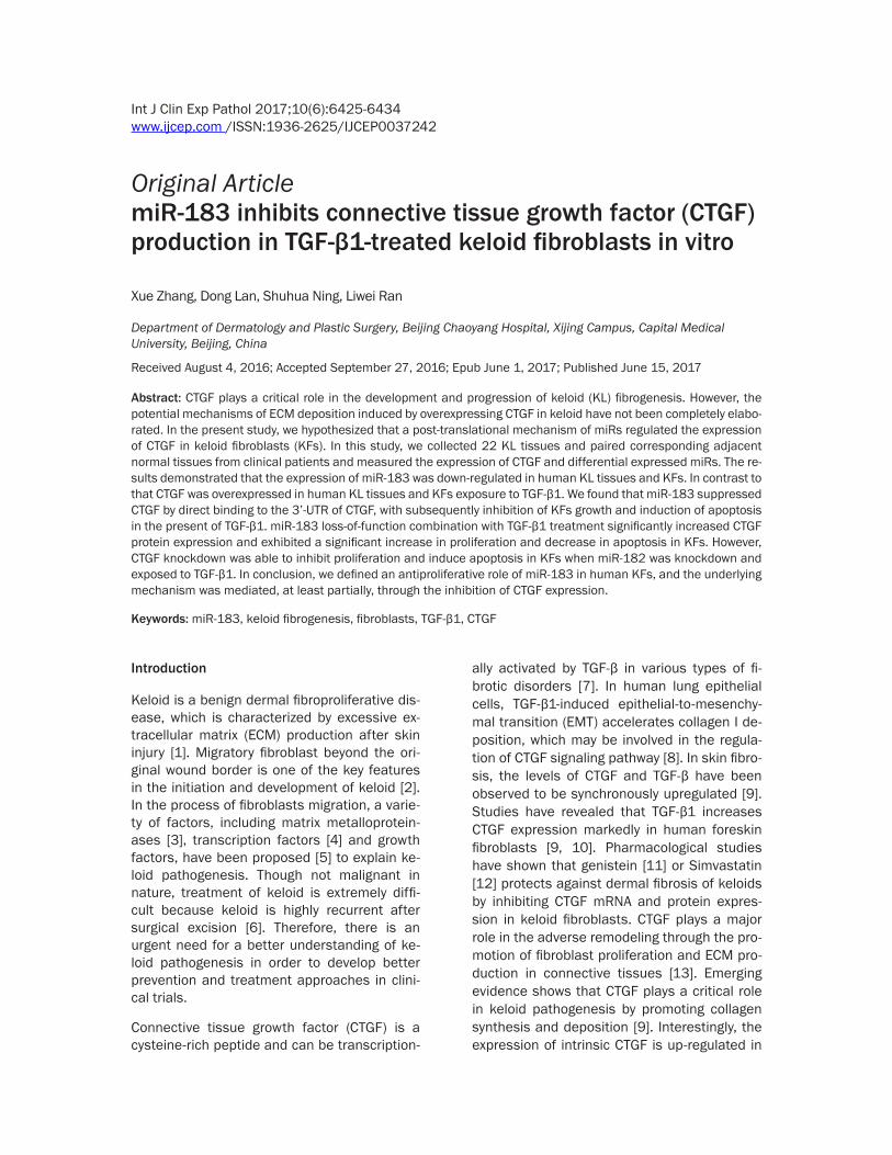
miR-183/CTGF signaling is involved in TGF-β1-induced KFs growth in vitro
Based on these studies, we concluded that miR-183 could inhibit CTGF expression by tar-geting its 3’-UTR, which was further confirmed by linear regression analysis between the miR-183 levels and CTGF protein expression in KL tissues. The linear regression analysis demon-strated that miR-183 levels were significant- ly and negatively correlated with CTGF protein expression in human KL tissues (Figure 5A). In this study, miR-183 loss-of-function combina-tion with TGF-β1 treatment significantly incre- ased CTGF protein expression in KFs as com-pared to TGF-β1 single treatment group (Figure
Figure 4. miR-183 inhibits TGF-β1-induced CTGF expression in KFs. The expression of CTGF in KL tissues was mea-sured by western blotting analysis (A). KFs or NFs were isolated from KL tissues or corresponding adjacent normal, respectively, and the expression of CTGF was measured by western blotting after 3 days culture in vitro (B and C). miR-183 expression levels in KFs were measured by qRT-PCR analysis in the present of TGF-β1 for 3 days (D). The expression of CTGF in KFs was measured by western blotting analysis in the present of TGF-β1 for 3 days (E and F). MTT assays were performed to measure cell viability in the present of TGF-β1 and with or without miR-183 mimics (G), and cells apoptosis were measured by TUNEL analysis (H). The protein expression of CTGF, FN and type I colla-gen was performed by western blotting analysis in KFs in the present of TGF-β1 and with or without miR-183 mimics for 3 days culture in vitro (I).
miR-183 targets to CTGF in KFs
6431 Int J Clin Exp Pathol 2017;10(6):6425-6434
5B). miR-183 loss-of-function exhibited a sig-nificant increase in proliferation (Figure 5C) and decrease in apoptosis (Figure 5D) in KFs in the present of TGF-β1 as compared to TGF-β1 single treatment group. However, CTGF knock-down was able to inhibit proliferation and in- duce apoptosis in KFs when miR-183 was knockdown and exposed to TGF-β1 (Figure 5C and 5D). These results demonstrated that CTGF was critically essential for the function of miR-183 in KFs proliferation and apoptosis in the present of TGF-β1.
Discussion
Here, we define an antiproliferative role of miR-183 in human keloid. We discovered that CTGF is overexpressed in human KL tissues and KFs exposure to TGF-β1, which can be a keloid fibro-genic cell model. In this process, a multitude of
cytokines, growth factors, and proteins are released at the wound-healing site, these com-pounds stimulate profibrotic activity [21]. In- terestingly, miR-183 suppresses CTGF by direct binding to the 3’-UTR of CTGF, with subsequent-ly inhibition of KFs growth and induction of apoptosis in the present of TGF-β1. These data provide new insights into the mechanism underlying KL formation and therapeutic stra- tegies for this disorder.
Overexpressed miR-183 is a frequent event in various cancers, including colorectal cancer [22], breast cancer [23] and non-small cell lung cancer [24]. In contrast to that miR-183 is sig-nificantly downregulated in gastric cancer, and up-regulation of miR-183 significantly inhibits gastric cancer cell proliferation and invasion [25]. However, the role of miR-183 in keloid fibrogenesis remains to be elucidated. Previous
Figure 5. miR-183/CTGF signaling is involved in TGF-β1-induced KFs growth in vitro. Linear regression analysis was performed to analyze the correlation between miR-183 level and CTGF protein expression, n = 22 (A). The protein expression of CTGF was performed by western blotting analysis in KFs in the present of TGF-β1 and with or without miR-183 inhibitors for 3 days culture in vitro (B). MTT assays were performed to measure cell viability in the present of TGF-β1 and with or without miR-183 inhibitors (C), and cells apoptosis were measured by TUNEL analysis for 3 days culture in vitro (D).

miR-183 targets to CTGF in KFs
6432 Int J Clin Exp Pathol 2017;10(6):6425-6434
study indicates that miR-29a is significantly decreased in keloid as compared to healthy fibroblasts, miR-29a loss-of-function up-regu-lates the mRNA and protein expression of type I and type III collagen in the fibroblasts [16]. In our study, we found that miR-183 inhibi- ted TGF-β1-induced type I collagen protein ex- pression. Collagen family is a dominant com- ponent of ECM, increased ECM deposition is the major findings in keloid pathology [2, 26]. Overproduction of collagen and ECM in fibro-blasts is easy to induce fibrotic diseases, in- cluding keloid and hypertrophic scars [27]. Therefore, inhibition of collagen and ECM pro-duction and deposition in fibroblasts is an im- portant goal for pharmaceutical agents used in the treatment of keloid. In the present study, we found that miR-183 might be a new thera-peutic approach for fibrotic disorders.
CTGF is a downstream effector of TGFβ signal-ing and can be secreted in fibroblasts [11]. CTGF is known to elicit fibroblast-specific mito-genesis, chemotaxis and ECM synthesis in vitro [28]. A growing body of evidence suggests that CTGF can mediate TGF-β-induced fibrogenesis, including fibroblast proliferation and the pro-duction of extracellular matrix proteins such as collagen and fibronectin [29]. Elevated levels of CTGF have frequently been associated with fibrotic skin disorders such as scleroderma and keloids [30]. Upregulation of secretory CTGF in keratinocyte-fibroblast coculture contributes to keloid pathogenesis [9]. Gene array profiling has also indicated CTGF may be up-regulated in isolated KF in the present of hydrocortisone [31]. In our study, we demonstrated that the protein expression of CTGF was markedly in- creased in KL tissues and KFs. This observa-tion was consistent with a statistically signifi-cant increase in downstream protein markers, FN and type I collagen, in the present of TGF-β1. Overexpressed miR-183 decreased TGF- β1-induced expression of CTGF, FN and type I collagen in cultured KFs. In addition, we show- ed that CTGF knockdown inhibited KFs grow- th and induced apoptosis in the present of TGF-β1. Noticeably, CTGF knockdown increa- sed number of TUNEL-positive cells in KFs, in- dicating that CTGF knockdown induced DNA damage. Collectively, our evidence indicates that CTGF regulates cellular proliferation and apoptosis behaviors in KFs, which can be re- gulated by miR-183.
To the best of our knowledge, overexpressed miR-183 can inhibit KFs growth and induce apoptosis, and the underlying mechanism is mediated, at least partially, through the sup-pression of CTGF expression. But above all, these findings offer a treatment for abnor- mal wound-healing processes that respond to multiple upstream events, such as TGF-β1 stimulation.
Disclosure of conflict of interest
None.
Address correspondence to: Dr. Xue Zhang, De- partment of Dermatology and Plastic Surgery, Bei- jing Chaoyang Hospital, Xijing Campus, Capital Me- dical University, Beijing 100043, China. Tel: (+86) 10-51718999; E-mail: [email protected]
References
[1] Ishiko T, Naitoh M, Kubota H, Yamawaki S, Ikeda M, Yoshikawa K, Fujita H, Yamaguchi H, Kurahashi Y and Suzuki S. Chondroitinase in-jection improves keloid pathology by reorganiz-ing the extracellular matrix with regenerated elastic fibers. J Dermatol 2013; 40: 380-383.
[2] Kashiyama K, Mitsutake N, Matsuse M, Ogi T, Saenko VA, Ujifuku K, Utani A, Hirano A and Yamashita S. miR-196a downregulation in-creases the expression of type I and III colla-gens in keloid fibroblasts. J Invest Dermatol 2012; 132: 1597-1604.
[3] Lee DE, Trowbridge RM, Ayoub NT and Agrawal DK. High-mobility Group Box Protein-1, Matrix Metalloproteinases, and Vitamin D in Keloids and Hypertrophic Scars. Plast Reconstr Surg Glob Open 2015; 3: e425.
[4] Zhang Z, Nie F, Kang C, Chen B, Qin Z, Ma J, Ma Y and Zhao X. Increased periostin expression affects the proliferation, collagen synthesis, migration and invasion of keloid fibroblasts under hypoxic conditions. Int J Mol Med 2014; 34: 253-261.
[5] Harn HI, Wang YK, Hsu CK, Ho YT, Huang YW, Chiu WT, Lin HH, Cheng CM and Tang MJ. Mechanical coupling of cytoskeletal elasticity and force generation is crucial for understand-ing the migrating nature of keloid fibroblasts. Exp Dermatol 2015; 24: 579-584.
[6] Lee WJ, Ahn HM, Roh H, Na Y, Choi IK, Lee JH, Kim YO, Lew DH and Yun CO. Decorin-ex- pressing adenovirus decreases collagen syn-thesis and upregulates MMP expression in keloid fibroblasts and keloid spheroids. Exp Dermatol 2015; 24: 591-597.
miR-183 targets to CTGF in KFs
6433 Int J Clin Exp Pathol 2017;10(6):6425-6434
[7] Pi L, Robinson PM, Jorgensen M, Oh SH, Brown AR, Weinreb PH, Trinh TL, Yianni P, Liu C, Leask A, Violette SM, Scott EW, Schultz GS and Petersen BE. Connective tissue growth factor and integrin alphavbeta6: a new pair of regula-tors critical for ductular reaction and biliary fi-brosis in mice. Hepatology 2015; 61: 678-691.
[8] Shi L, Dong N, Fang X and Wang X. Regulatory mechanisms of TGF-beta1-induced fibrogene-sis of human alveolar epithelial cells. J Cell Mol Med 2016; 90: 698.e1-5.
[9] Khoo YT, Ong CT, Mukhopadhyay A, Han HC, Do DV, Lim IJ and Phan TT. Upregulation of se-cretory connective tissue growth factor (CTGF) in keratinocyte-fibroblast coculture contributes to keloid pathogenesis. J Cell Physiol 2006; 208: 336-343.
[10] Song R, Li G and Li S. Aspidin PB, a novel natu-ral anti-fibrotic compound, inhibited fibrogene-sis in TGF-beta1-stimulated keloid fibroblasts via PI-3K/Akt and Smad signaling pathways. Chem Biol Interact 2015; 238: 66-73.
[11] Jurzak M, Adamczyk K, Antonczak P, Garn- carczyk A, Kusmierz D and Latocha M. Eva- luation of genistein ability to modulate CTGF mRNA/protein expression, genes expression of TGFbeta isoforms and expression of select-ed genes regulating cell cycle in keloid fibro-blasts in vitro. Acta Pol Pharm 2014; 71: 972-986.
[12] Mun JH, Kim YM, Kim BS, Kim JH, Kim MB and Ko HC. Simvastatin inhibits transform- ing growth factor-beta1-induced expression of type I collagen, CTGF, and alpha-SMA in keloid fibroblasts. Wound Repair Regen 2014; 22: 125-133.
[13] Angelini A, Li Z, Mericskay M and Decaux JF. Regulation of Connective Tissue Growth Factor and Cardiac Fibrosis by an SRF/MicroRNA-133a Axis. PLoS One 2015; 10: e0139858.
[14] Colwell AS, Phan TT, Kong W, Longaker MT and Lorenz PH. Hypertrophic scar fibroblasts have increased connective tissue growth factor ex-pression after transforming growth factor-beta stimulation. Plast Reconstr Surg 2005; 116: 1387-1390; discussion 1391-1382.
[15] Patrick DM, Montgomery RL, Qi X, Obad S, Kauppinen S, Hill JA, van Rooij E and Olson EN. Stress-dependent cardiac remodeling occurs in the absence of microRNA-21 in mice. J Clin Invest 2010; 120: 3912-3916.
[16] Zhang GY, Wu LC, Liao T, Chen GC, Chen YH, Zhao YX, Chen SY, Wang AY, Lin K, Lin DM, Yang JQ, Gao WY and Li QF. A novel regulatory function for miR-29a in keloid fibrogenesis. Clin Exp Dermatol 2016; 41: 341-345.
[17] Liu Y, Wang X, Yang D, Xiao Z and Chen X. MicroRNA-21 affects proliferation and apopto-sis by regulating expression of PTEN in human
keloid fibroblasts. Plast Reconstr Surg 2014; 134: 561e-573e.
[18] Li P, He QY and Luo CQ. Overexpression of miR-200b inhibits the cell proliferation and pro-motes apoptosis of human hypertrophic scar fibroblasts in vitro. J Dermatol 2014; 41: 903-911.
[19] Phan TT, Lim IJ, Aalami O, Lorget F, Khoo A, Tan EK, Mukhopadhyay A and Longaker MT. Smad3 signalling plays an important role in keloid pathogenesis via epithelial-mesenchymal in-teractions. J Pathol 2005; 207: 232-242.
[20] Muller PY, Janovjak H, Miserez AR and Dobbie Z. Processing of gene expression data generated by quantitative real-time RT-PCR. Biotechniques 2002; 32: 1372-1374, 1376, 1378, 1379.
[21] Shi-Wen X, Chen Y, Denton CP, Eastwood M, Renzoni EA, Bou-Gharios G, Pearson JD, Dash- wood M, du Bois RM, Black CM, Leask A and Abraham DJ. Endothelin-1 promotes myofibro-blast induction through the ETA receptor via a rac/phosphoinositide 3-kinase/Akt-depen- dent pathway and is essential for the en-hanced contractile phenotype of fibrotic fibro-blasts. Mol Biol Cell 2004; 15: 2707-2719.
[22] Huangfu L, Liang H, Wang G, Su X, Li L, Du Z, Hu M, Dong Y, Bai X, Liu T, Yang B and Shan H. miR-183 regulates autophagy and apoptosis in colorectal cancer through target-ing of UVRAG. Oncotarget 2016; 7: 4735-4745.
[23] Chang YY, Kuo WH, Hung JH, Lee CY, Lee YH, Chang YC, Lin WC, Shen CY, Huang CS, Hsieh FJ, Lai LC, Tsai MH, Chang KJ and Chuang EY. Deregulated microRNAs in triple-negative breast cancer revealed by deep sequencing. Mol Cancer 2015; 14: 36.
[24] Zhang L, Quan H, Wang S, Li X and Che X. MiR-183 promotes growth of non-small cell lung cancer cells through FoxO1 inhibition. Tumour Biol 2015; 36: 8121-8126.
[25] Xu L, Li Y, Yan D, He J and Liu D. MicroRNA-183 inhibits gastric cancer proliferation and inva-sion via directly targeting Bmi-1. Oncol Lett 2014; 8: 2345-2351.
[26] Al-Attar A, Mess S, Thomassen JM, Kauffman CL and Davison SP. Keloid pathogenesis and treatment. Plast Reconstr Surg 2006; 117: 286-300.
[27] Aoki M, Miyake K, Ogawa R, Dohi T, Akaishi S, Hyakusoku H and Shimada T. siRNA knock-down of tissue inhibitor of metalloproteinase-1 in keloid fibroblasts leads to degradation of collagen type I. J Invest Dermatol 2014; 134: 818-826.
[28] Grotendorst GR. Connective tissue growth fac-tor: a mediator of TGF-beta action on fibro-blasts. Cytokine Growth Factor Rev 1997; 8: 171-179.

miR-183 targets to CTGF in KFs
6434 Int J Clin Exp Pathol 2017;10(6):6425-6434
[29] Lopes LB, Furnish EJ, Komalavilas P, Flynn CR, Ashby P, Hansen A, Ly DP, Yang GP, Longaker MT, Panitch A and Brophy CM. Cell permeant peptide analogues of the small heat shock protein, HSP20, reduce TGF-beta1-in- duced CTGF expression in keloid fibroblasts. J Invest Dermatol 2009; 129: 590-598.
[30] Abraham DJ, Shiwen X, Black CM, Sa S, Xu Y and Leask A. Tumor necrosis factor alpha sup-presses the induction of connective tissue growth factor by transforming growth factor-beta in normal and scleroderma fibroblasts. J Biol Chem 2000; 275: 15220-15225.
[31] Smith JC, Boone BE, Opalenik SR, Williams SM and Russell SB. Gene profiling of keloid fibro-blasts shows altered expression in multiple fi-brosis-associated pathways. J Invest Dermatol 2008; 128: 1298-1310.