original article retinal hemorrhage associated with ... · original article retinal hemorrhage...
TRANSCRIPT

311
Korean J Ophthalmol 2011;25(5):311-316http://dx.doi.org/10.3341/kjo.2011.25.5.311pISSN: 1011-8942 eISSN: 2092-9382
Original Article
Retinal Hemorrhage Associated with Perinatal Distress in Newborns
Youn Joo Choi1, Moon Sun Jung2, So Young Kim2
1Department of Ophthalmology, Soonchunhyang University Hospital, Soonchunhyang University College of Medicine, Seoul, Korea2Department of Ophthalmology, Soonchunhyang University Cheonan Hospital, Soonchunhyang University
College of Medicine, Cheonan, Korea
Purpose: To describe the ophthalmoscopic features and natural history in a case series of eyes that developed in-traocular hemorrhages associated with perinatal distress and to evaluate their clinical courses.
Methods: A retrospective chart review of 289 neonates with a medical history of perinatal distress was conducted. Among these 289 patients (578 eyes), 29 eyes of 17 neonates were found to have had retinal hemorrhages or vit-reous hemorrhages (VH). A comprehensive chart review, including details of fundoscopic findings and perinatal history, was conducted.
Results: Intraocular hemorrhage was present in 5.5% of the patients. Most hemorrhages (82.7%) were intraretinal. In our population, 17% (n = 5) of hemorrhages resolved within two weeks, but 31% (n = 9) did not resolve even af-ter four weeks. Most hemorrhages spontaneously resolved without any specific sequelae; however, one infant’s dense unilateral VH persisted up to three months after birth. When the patient was seen again at 3.5 years of age, she had developed axial myopia and severe amblyopia of the involved eye.
Conclusions: In asphyxiated newborns, the possibility of intraocular hemorrhages should be considered. Long-standing, dense hemorrhages obscuring the macula may lead to severe vision deprivation amblyopia. Therefore, oph-thalmic examination should be considered in neonates with perinatal distress, and close observation is neces-sary for hemorrhages that do not resolve in this amblyogenic age group.
Key Words: Newborn infant, Perinatal distress, Retinal hemorrhage, Vitreous hemorrhage
ⓒ2011 The Korean Ophthalmological SocietyThis is an Open Access article distributed under the terms of the Creative Commons Attribution Non-Commercial License (http://creativecommons.org/licenses/by-nc/3.0/) which permits unrestricted non-commercial use, distribution, and reproduction in any medium, provided the original work is properly cited.
Received: September 28, 2010 Accepted: March 10, 2011
Reprint requests to So Young Kim, MD. Department of Ophthalmology, Soonchunhyang University Cheonan Hospital, #23-20 Bongmyeong- dong, Seonam-gu, Cheonan 330-720, Korea. Tel: 82-41-570-2260, Fax: 82-41-576-2262, E-mail: [email protected]
There are several potential causes of retinal hemorrhages (RH) in neonates. Hemorrhages at birth may occur after trau-matic deliveries; neonatal coagulopathies are associated with sepsis, shaken baby syndrome and intracranial hemorrhage [1]. The reported incidence of neonatal retinal hemorrhages varies widely, from 2.6% to 50%, which is possibly due to different patient demographics, how soon after birth exami-nations are conducted, and different examination techniques [2]. In general, birth-related RH resolves quickly and does not cause subsequent visual or neurological deficits [2].
Despite the number of articles documenting the incidence and duration of RH in healthy newborns, there have been no
prior reports on RH in asphyxiated newborns. Asphyxia or respiratory distress is defined as a condition
of impaired gas exchange that leads to hypoxemia, hyper-capnia and metabolic acidosis [3]. Clinical criteria include the following: abnormalities in electronic fetal monitoring, meconium-stained amniotic fluid, metabolic acidemia, low Apgar scores, and post-asphyxia neurological and/or extra neurological abnormalities [4].
In this report, we discuss the incidence, pattern and dura-tion of RH and vitreous hemorrhages (VH) associated with perinatal distress in newborns and evaluate their clinical significance.
Materials and Methods
We retrospectively reviewed medical records of peri-natally distressed newborns hospitalized at the Neonatal Intensive Care Unit of our hospital between March 2006 and December 2009 who were referred for ophthalmic examina-

Korean J Ophthalmol Vol.25, No.5, 2011
312
A B
Fig. 1. Fundus photographs of retinal hemorrhages in neonates that had a history of birth asphyxia and amniotic fluid aspiration. (A) Participant 15, (B) participant 9.
Table 1. Demographic features of 17 neonates with intraocular hemorrhages
No. of infants 17No. of affected eyes 29Laterality
Bilateral 11 (65)Unilateral 6 (35)
Right 1Left 5Mean gestational age (wk) 39.5 ± 1.53 Mean birth weight (g) 3,186 ± 519.6Gender (male:female) 3:14 (18:82)Mean age at first examination (day) 11.5 ± 1.81 Method of delivery
Spontaneous vaginal 12 (71)Cesarean section delivery 5* (29)
Values are presented as mean ± SD or number (%).*Includes four deliveries by emergency cesarean section.
tion by the pediatrics department. Perinatal distress included the following: birth asphyxia, meconium aspiration, amni-otic fluid aspiration, fetal distress, transient tachypnea of the newborn, and dysphagic choking. The Institutional Review Board at our hospital approved this retrospective chart review. We identified 289 patients. Among these patients, 17 were noted to have RH or VH. Our department performed a detailed chart review, including comprehensive ocular find-ings and perinatal history. All RH or VH cases were diag-nosed by indirect ophthalmoscopy and scleral depression with proparacaine 0.5% topical anesthetic. The shape, loca-tion and other associated features of the hemorrhages were noted for each eye in a drawing of the fundus. Hemorrhages were classified according to their location in three retinal zones [2,5].
The newborns were reexamined every one or two weeks until the hemorrhages resolved completely. In one participant with unilateral VH, the follow-up continued until the child was 3.5 years old.
Results
The mean age at the first funduscopic examination was 11.5 days. Among the 17 infants, 11 (65%) had bilateral hemorrhages. The RH were dot, blot or flame shaped (Fig. 1). Among the 29 eyes with hemorrhages, 82.7% (n = 24) were intraretinal; one neonate had unilateral preretinal hemor-rhages, and another had bilateral subretinal hemorrhages. Two neonates had unilateral VH. Table 1 summarizes the demographic information for those infants found to have RH or VH. The perinatal histories, pattern, distribution, and du-ration of the hemorrhages are shown in Table 2.
The newborns were reexamined every one to two weeks until the hemorrhages were completely resolved. The aver-age duration of hemorrhages after the first examination was 5.1 weeks. Among the 29 eyes, only 17% (n = 5) of hemor-
rhages resolved within two weeks; 31% (n = 9) did not re-solve completely even after four weeks. The one hemorrhage that persisted for up to three months was a case with dense unilateral VH. The posterior pole of the involved eye was al-most completely obscured. B-scan ultrasonography showed a diffuse VH concentrated at the macular area (Fig. 2A). Both retinas were otherwise normal with no sign of infection or other abnormality. Even after complete resolution, the cause of the VH could not be determined. When the patient was seen again at 3.5 years of age, she had developed severe amblyopia. A cycloplegic refraction revealed +1.5 D sph in the right eye and -3.50 D sph = -1.5 D cyl × 180° A in the left eye.
Discussion
RH can occur in healthy newborn infants during delivery.

YJ Choi, et al. Retinal Hemorrhage with Perinatal Distress
313
Table 2. Perinatal history and ocular findings of 17 patents
Participant Perinatal historyGestati-onal age
(wk)
Birth weight
(g)
Mode of delivery
Age at first exam
(wk)
Hemorrhages
Type Location(zone)
Duration(wk)
1 Birth asphyxiaMeconium aspirationHypoxic brain damage
38+4 3,030 C-section for failure to progress
39+3 B) IRH I B) 8
2 PneumoniaTTNGerminal matrix hemorrhage
41 3,600 NSVD 43 L) IRH II L) 8
3 Birth asphyxiaMeconium aspiration
39+6 3,790 NSVD 40+6 B) IRH II B) 4
4 Meconium aspirationTTN
40+2 3,300 NSVD 43+2 R) IRHL) VH
R) IL) VH
B) 4
5 Choking history while breastfeeding at three days after birth
37+3 3,000 NSVD 38+6 B) IRH II B) 4
6 Birth asphyxiaMeconium aspiration
39+3 3,380 C-section for failure to progress
40+2 B) IRH I B) 4
7 Fetal distressBirth asphyxia
40+5 3,770 C-section for failure to progress
41+6 B) IRH II B) 3
8 Birth asphyxia 39+4 3,780 NSVD 42+5 L) IRH II L) 29 Birth asphyxia
Amniotic fluid aspiration39+3 3,410 NSVD 40+2 B) IRH I B) 5
10 Birth asphyxiaMeconium aspirationHypoxic brain damageIntrauterine growth retardation
41+2 2,640 NSVD 43+3 B) IRH I B) 6
11 Birth asphyxiaAmniotic fluid aspiration
39 3,630 NSVD 42+2 L) Pre RH I L) 4
12 TTNPremature rupture of membranes
40+1 2,810 NSVD 41+5 B) IRH II B) 2
13 PneumoniaTTN
40 3,500 Elective C-section 41+1 B) IRH I B) 2
14 Meconium aspiration 39+3 2,700 NSVD 40+4 L) IRH II L) 415 Birth asphyxia
Amniotic fluid aspirationIntrauterine growth retardation
38+3 2,330 NSVD 39+1 B) IRH L R) 4L) 6
16 Fetal distressBirth asphyxiaPrematurityRDS grade IVMeconium aspirationHypoxic brain damageFetal hydropsNeonatal seizureSepsisDIC
36+5 2,100 C-section for fetal distress
39+1 L) VH Obscure macula
L) 12
17 Pneumonia TTN
40+1 3,400 NSVD 41+5 B) Sub RH
I B) 4
IRH = intraretinal hemorrhage; TTN = transient tachypnea of the newborn; NSVD = normal spontaneous vaginal delivery; VH = vitreous hemorrhage; C-section = cesarean section; RH = retinal hemorrhage; RDS = respiratory distress syndrome; DIC = disseminated intravascular coagulopathy; B = both eyes; R = right eye; L = left eye.
The reported incidence varies from 2.6 to 50% [2]. Giles [6] found that the incidence was reduced from 40% at 1 hour post delivery to 20% at 72 hours. Sezen [7] showed the in-cidence was only 2.6% after three to five days. A study by Kim et al. [8] reported the prevalence of neonatal RH in
Korea to be 19.1% at 24 hours after birth. The presence of RH in healthy newborns presumably relates to the birth proc-ess, and spontaneous absorption generally occurs over time.
Among the 289 perinatally distressed newborns in the cur-rent retrospective chart review, 17 neonates were found to
Korean J Ophthalmol Vol.25, No.5, 2011
314
A B
C D
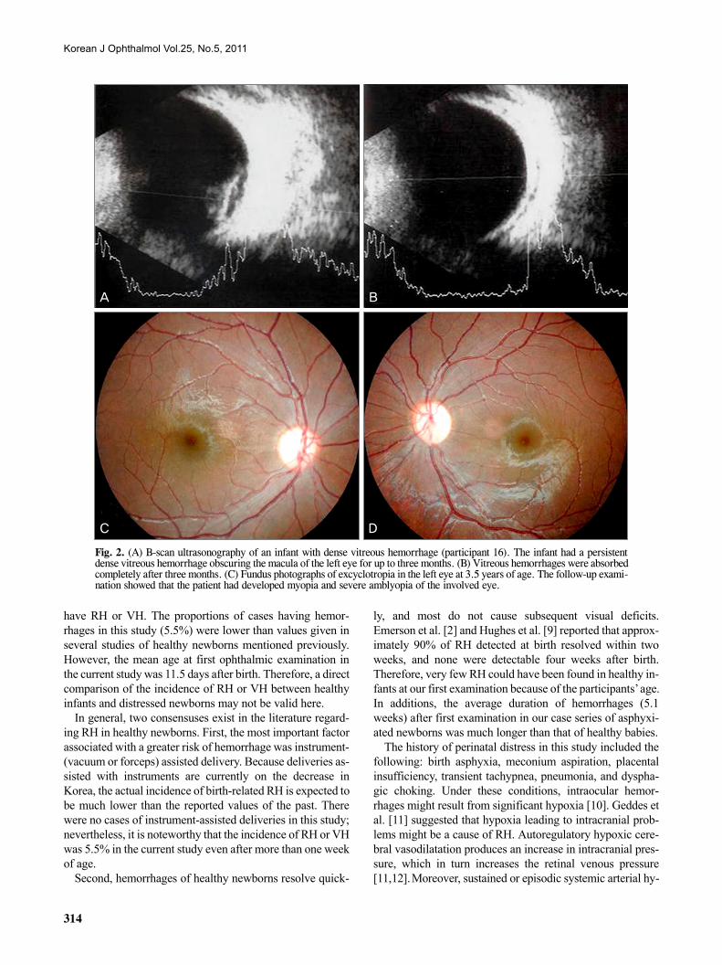
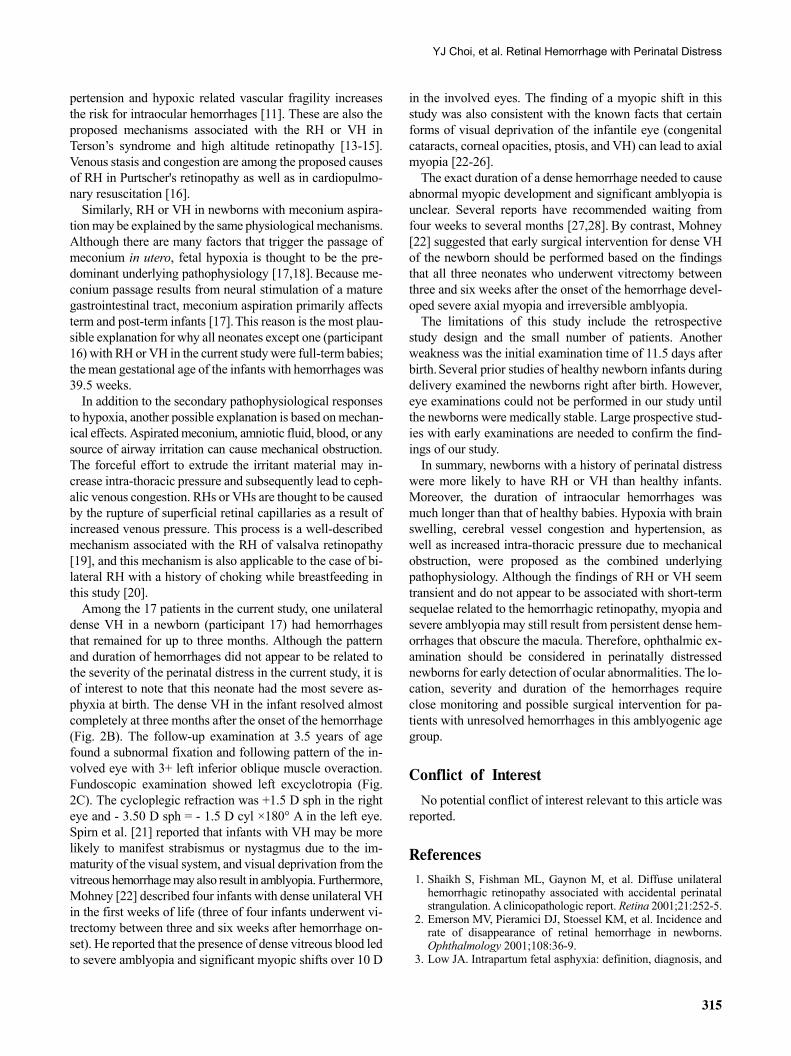
Fig. 2. (A) B-scan ultrasonography of an infant with dense vitreous hemorrhage (participant 16). The infant had a persistent dense vitreous hemorrhage obscuring the macula of the left eye for up to three months. (B) Vitreous hemorrhages were absorbed completely after three months. (C) Fundus photographs of excyclotropia in the left eye at 3.5 years of age. The follow-up exami-nation showed that the patient had developed myopia and severe amblyopia of the involved eye.
have RH or VH. The proportions of cases having hemor-rhages in this study (5.5%) were lower than values given in several studies of healthy newborns mentioned previously. However, the mean age at first ophthalmic examination in the current study was 11.5 days after birth. Therefore, a direct comparison of the incidence of RH or VH between healthy infants and distressed newborns may not be valid here.
In general, two consensuses exist in the literature regard-ing RH in healthy newborns. First, the most important factor associated with a greater risk of hemorrhage was instrument- (vacuum or forceps) assisted delivery. Because deliveries as-sisted with instruments are currently on the decrease in Korea, the actual incidence of birth-related RH is expected to be much lower than the reported values of the past. There were no cases of instrument-assisted deliveries in this study; nevertheless, it is noteworthy that the incidence of RH or VH was 5.5% in the current study even after more than one week of age.
Second, hemorrhages of healthy newborns resolve quick-
ly, and most do not cause subsequent visual deficits. Emerson et al. [2] and Hughes et al. [9] reported that approx-imately 90% of RH detected at birth resolved within two weeks, and none were detectable four weeks after birth. Therefore, very few RH could have been found in healthy in-fants at our first examination because of the participants’ age. In additions, the average duration of hemorrhages (5.1 weeks) after first examination in our case series of asphyxi-ated newborns was much longer than that of healthy babies.
The history of perinatal distress in this study included the following: birth asphyxia, meconium aspiration, placental insufficiency, transient tachypnea, pneumonia, and dyspha-gic choking. Under these conditions, intraocular hemor-rhages might result from significant hypoxia [10]. Geddes et al. [11] suggested that hypoxia leading to intracranial prob-lems might be a cause of RH. Autoregulatory hypoxic cere-bral vasodilatation produces an increase in intracranial pres-sure, which in turn increases the retinal venous pressure [11,12]. Moreover, sustained or episodic systemic arterial hy-
YJ Choi, et al. Retinal Hemorrhage with Perinatal Distress
315
pertension and hypoxic related vascular fragility increases the risk for intraocular hemorrhages [11]. These are also the proposed mechanisms associated with the RH or VH in Terson’s syndrome and high altitude retinopathy [13-15].
Venous stasis and congestion are among the proposed causes of RH in Purtscher's retinopathy as well as in cardiopulmo-nary resuscitation [16].
Similarly, RH or VH in newborns with meconium aspira-tion may be explained by the same physiological mechanisms. Although there are many factors that trigger the passage of meconium in utero, fetal hypoxia is thought to be the pre-dominant underlying pathophysiology [17,18]. Because me-conium passage results from neural stimulation of a mature gastrointestinal tract, meconium aspiration primarily affects term and post-term infants [17]. This reason is the most plau-sible explanation for why all neonates except one (participant 16) with RH or VH in the current study were full-term babies; the mean gestational age of the infants with hemorrhages was 39.5 weeks.
In addition to the secondary pathophysiological responses to hypoxia, another possible explanation is based on mechan-ical effects. Aspirated meconium, amniotic fluid, blood, or any source of airway irritation can cause mechanical obstruction. The forceful effort to extrude the irritant material may in-crease intra-thoracic pressure and subsequently lead to ceph-alic venous congestion. RHs or VHs are thought to be caused by the rupture of superficial retinal capillaries as a result of increased venous pressure. This process is a well-described mechanism associated with the RH of valsalva retinopathy [19], and this mechanism is also applicable to the case of bi-lateral RH with a history of choking while breastfeeding in this study [20].
Among the 17 patients in the current study, one unilateral dense VH in a newborn (participant 17) had hemorrhages that remained for up to three months. Although the pattern and duration of hemorrhages did not appear to be related to the severity of the perinatal distress in the current study, it is of interest to note that this neonate had the most severe as-phyxia at birth. The dense VH in the infant resolved almost completely at three months after the onset of the hemorrhage (Fig. 2B). The follow-up examination at 3.5 years of age found a subnormal fixation and following pattern of the in-volved eye with 3+ left inferior oblique muscle overaction. Fundoscopic examination showed left excyclotropia (Fig. 2C). The cycloplegic refraction was +1.5 D sph in the right eye and - 3.50 D sph = - 1.5 D cyl ×180° A in the left eye. Spirn et al. [21] reported that infants with VH may be more likely to manifest strabismus or nystagmus due to the im-maturity of the visual system, and visual deprivation from the vitreous hemorrhage may also result in amblyopia. Furthermore, Mohney [22] described four infants with dense unilateral VH in the first weeks of life (three of four infants underwent vi-trectomy between three and six weeks after hemorrhage on-set). He reported that the presence of dense vitreous blood led to severe amblyopia and significant myopic shifts over 10 D
in the involved eyes. The finding of a myopic shift in this study was also consistent with the known facts that certain forms of visual deprivation of the infantile eye (congenital cataracts, corneal opacities, ptosis, and VH) can lead to axial myopia [22-26].
The exact duration of a dense hemorrhage needed to cause abnormal myopic development and significant amblyopia is unclear. Several reports have recommended waiting from four weeks to several months [27,28]. By contrast, Mohney [22] suggested that early surgical intervention for dense VH of the newborn should be performed based on the findings that all three neonates who underwent vitrectomy between three and six weeks after the onset of the hemorrhage devel-oped severe axial myopia and irreversible amblyopia.
The limitations of this study include the retrospective study design and the small number of patients. Another weakness was the initial examination time of 11.5 days after birth. Several prior studies of healthy newborn infants during delivery examined the newborns right after birth. However, eye examinations could not be performed in our study until the newborns were medically stable. Large prospective stud-ies with early examinations are needed to confirm the find-ings of our study.
In summary, newborns with a history of perinatal distress were more likely to have RH or VH than healthy infants. Moreover, the duration of intraocular hemorrhages was much longer than that of healthy babies. Hypoxia with brain swelling, cerebral vessel congestion and hypertension, as well as increased intra-thoracic pressure due to mechanical obstruction, were proposed as the combined underlying pathophysiology. Although the findings of RH or VH seem transient and do not appear to be associated with short-term sequelae related to the hemorrhagic retinopathy, myopia and severe amblyopia may still result from persistent dense hem-orrhages that obscure the macula. Therefore, ophthalmic ex-amination should be considered in perinatally distressed newborns for early detection of ocular abnormalities. The lo-cation, severity and duration of the hemorrhages require close monitoring and possible surgical intervention for pa-tients with unresolved hemorrhages in this amblyogenic age group.
Conflict of Interest
No potential conflict of interest relevant to this article was reported.
References 1. Shaikh S, Fishman ML, Gaynon M, et al. Diffuse unilateral
hemorrhagic retinopathy associated with accidental perinatal strangulation. A clinicopathologic report. Retina 2001;21:252-5.
2. Emerson MV, Pieramici DJ, Stoessel KM, et al. Incidence and rate of disappearance of retinal hemorrhage in newborns. Ophthalmology 2001;108:36-9.
3. Low JA. Intrapartum fetal asphyxia: definition, diagnosis, and

Korean J Ophthalmol Vol.25, No.5, 2011
316
classification. Am J Obstet Gynecol 1997;176:957-9. 4. Gonzalez de Dios J. Definition of perinatal asphyxia in medical
literature: the need of a consensus. Rev Neurol 2002;35:628-34. 5. Studies of ocular complications of AIDS Foscarnet-Ganciclovir
Cytomegalovirus Retinitis Trial: 1. Rationale, design, and methods. AIDS Clinical Trials Group (ACTG). Control Clin Trials 1992;13:22-39.
6. Giles CL. Retinal hemorrhages in the newborn. Am J Ophthalmol 1960;49:1005-11.
7. Sezen F. Retinal haemorrhages in newborn infants. Br J Ophthalmol 1971;55:248-53.
8. Kim KH, Rhee MG, Shin TY. Retinal hemorrhages in newborn infants. J Korean Ophthalmol Soc 1980;21:441-4.
9. Hughes LA, May K, Talbot JF, Parsons MA. Incidence, dis-tribution, and duration of birth-related retinal hemorrhages: a prospective study. J AAPOS 2006;10:102-6.
10. Madan A, Hamrik S, Ferriero DM. Central nervous system in-jury and neuroprotection. In: Taeusch HW, Ballard RA, Gleason CA, editors. Avery’s diseases of the newborn. 8th ed. Philadelphia: Elsevier Saunders; 2005. p. 971-83.
11. Geddes JF, Tasker RC, Hackshaw AK, et al. Dural haemorrhage in non-traumatic infant deaths: does it explain the bleeding in 'shaken baby syndrome'? Neuropathol Appl Neurobiol 2003; 29:14-22.
12. Ladjimi A, Zaouali S, Messaoud R, et al. Valsalva retinopathy induced by labour. Eur J Ophthalmol 2002;12:336-8.
13. Smith DC, Kearns TP, Sayre GP. Preretinal and optic nerve-sheath hemorrhage: pathologic and experimental aspects in subarachnoid hemorrhage. Trans Am Acad Ophthalmol Otolaryngol 1957;61:201-11.
14. Gardner H. Correlation between retinal abnormalities and intra-cranial abnormalities in the shaken baby syndrome. Am J Ophthalmol 2003;135:745.
15. Shults WT, Swan KC. High altitude retinopathy in mountain climbers. Arch Ophthalmol 1975;93:404-8.
16. Goetting MG, Sowa B. Retinal hemorrhage after cardiopulmo-nary resuscitation in children: an etiologic reevaluation. Pediatrics 1990;85:585-8.
17. Van Ierland Y, de Beaufort AJ. Why does meconium cause me-conium aspiration syndrome? Current concepts of MAS pathophysiology. Early Hum Dev 2009;85:617-20.
18. Tyler DC, Murphy J, Cheney FW. Mechanical and chemical damage to lung tissue caused by meconium aspiration. Pediatrics 1978;62:454-9.
19. Duane TD. Valsalva hemorrhagic retinopathy. Trans Am Ophthalmol Soc 1972;70:298-313.
20. Barnes PD, Galaznik J, Gardner H, Shuman M. Infant acute life-threatening event: dysphagic choking versus nonaccidental injury. Semin Pediatr Neurol 2010;17:7-11.
21. Spirn MJ, Lynn MJ, Hubbard GB 3rd. Vitreous hemorrhage in children. Ophthalmology 2006;113:848-52.
22. Mohney BG. Axial myopia associated with dense vitreous hemorrhage of the neonate. J AAPOS 2002;6:348-53.
23. Von Noorden GK, Lewis RA. Ocular axial length in unilateral congenital cataracts and blepharoptosis. Invest Ophthalmol Vis Sci 1987;28:750-2.
24. Gee SS, Tabbara KF. Increase in ocular axial length in patients with corneal opacification. Ophthalmology 1988;95:1276-8.
25. Hoyt CS, Stone RD, Fromer C, Billson FA. Monocular axial myopia associated with neonatal eyelid closure in human infants. Am J Ophthalmol 1981;91:197-200.
26. Miller-Meeks MJ, Bennett SR, Keech RV, Blodi CF. Myopia in-duced by vitreous hemorrhage. Am J Ophthalmol 1990;109: 199-203.
27. Ferrone PJ, de Juan E Jr. Vitreous hemorrhage in infants. Arch Ophthalmol 1994;112:1185-9.
28. Isenberg SJ. Acquired disorders of the infant eye, ocular trauma. In: Isenberg SJ, editor. The eye in infancy. St. Louis: Mosby; 1994. p. 487.