original article strongly induced hypertrophic scar in a ... · stenosis (ts) is still a difficult...
TRANSCRIPT
Int J Clin Exp Med 2018;11(4):3679-3685www.ijcem.com /ISSN:1940-5901/IJCEM0064175
Original Article Strongly induced hypertrophic scar in a rat model of tracheal stenosis
Tingbao Hu1,2*, Fanqin Wei1,2,3*, Wei Sun1,2, Hua Zhong1,2, Yi Wei1,2, Jiawei Wei1,2, Dehua Chen1,2, Weiping Wen1,2
1Department of Otorhinolaryngology Head and Neck Surgery, The First Affiliated Hospital of Sun Yat-sen University, Guangzhou, China; 2Institute of Otorhinolaryngology Head and Neck Surgery, Sun Yat-sen University, Guangzhou, China; 3Department of Otorhinolaryngology Head and Neck Surgery, The Sixth Affiliated Hospital of Sun Yat-sen University, Guangzhou, China. *Equal contributors.
Received March 14, 2017; Accepted February 4, 2018; Epub April 15, 2018; Published April 30, 2018
Abstract: Objective: Previous rat models of tracheal stenosis have demonstrated a high mortality rate with little scar formation as well as interference by chemical factors. This study aimed to develop an improved rat model for tracheal stenosis. Methods: Forty-five Wistar albino rats were randomly divided into two control groups and one ex-perimental group with 15 rats in each group. Electrical cauterization was performed anterior of the 1-2 and 4-6 tra-chea rings through the tracheostoma. About 0.5 cm of skin on both sides of the incision in the experimental group was removed and fixed to keep the tracheostoma open. In control group 1, the incisions were closed after electrical cauterization, whereas rats in control group 2 had an open tracheostoma but no electrocautery injury. Results: Fourteen (93.3%) of 15 animals in the experimental group survived. All rats survived in control group 2, but a high rate of mortality (100%) was observed in control group 1. Compared with control group 2, the experimental group showed a significant increase in lamina propria thickness at days 7, 14, and 21, resulting in luminal narrowing with the maximal stenosis degree at day 21 (67.8%). Conclusion: We developed a rat model of tracheal stenosis, result-ing from mucosal injury administered by electrocauterization. The advantages of this model are ease of handling, a high survival rate, and remarkable scar hyperplasia.
Keywords: Animal model, tracheostomy, electrocauterization, tracheal mucosa, upper airway obstruction
Introduction
Postoperative recurrence of cicatricial tracheal stenosis (TS) is still a difficult problem to over-come. Current strategies, such as end-to-end anastomosis, CO2 laser incision, balloon dila-tion, and stent replacement cannot guarantee favorable results and may lead to complica-tions [1-3]. Therefore, various animal models have been developed to study the mechanism of TS and to explore effective treatment meth-ods. Previously, large and medium sized ani-mals such as pigs, dogs, and rabbits were used to demonstrate effects related to airway injury [4-6]. TS is easy to induce in these animal models because of their large airway lumen. However, because relevant drugs and antibod-ies are not readily available, the use of these animal models precludes molecular biology or immunology studies [7]. Although mouse TS
models have been commonly used for genetic and molecular investigation, they are difficult to use because their airway lumen diameter is too small to provide sufficient scar tissue for fur-ther study [8, 9].
Recently, rats have been used as an animal model to study airway stenosis. However, these models do not develop sufficient scar hyperpla-sia in contrast to that seen in the clinic. This is because mild damage to the tracheal mucosa only induces a small quantity of scar tissue, whereas a deep and severe injury induces a large amount of scarring but might lead to a high rate of mortality in rats. To resolve this issue, we developed an improved rat model that produces an adequate cicatrix for the study of TS. This model demonstrated a high proba-bility of survival and might be useful for the evaluation of therapies to prevent or reverse TS.

Hypertrophic scar in rat tracheal stenosis
3680 Int J Clin Exp Med 2018;11(4):3679-3685
Material and methods
Experimental animals
This study was approved by the institutional Animal Studies Committee of Sun Yat-sen Uni- versity (Permit Number: 2012-349), and was conducted in accordance with the Institutional
1.5 mm) was made by partially excising the anterior wall of the third tracheal ring and inter-vals above and below it. Monopolar electrocau-terization (Spring Medical, Wuhan, Hubei, China) was set at 10 W, and the anterior part of the electrosurgical cautery unit was bent at a right angle. Wounds were made on the anterior of the 1-2 and 4-6 tracheal rings by intratrache-
Figure 1. Surgical technique. A. Appearance of the trachea after dissecting strap muscles laterally. B. A tracheostoma was made between the second and fourth rings. C. Wounds were made on the anterior of the 1-2 and 4-6 trachea rings by intratracheal electric coagulation. The anterior part of the electrosur-gical cautery unit was bent at a right angle. D. The trachea wall turned white (arrow), when the intended depth of injury in the lateral surface of cartilage was reached.
Animal Care and Use Com- mittee (IACUC) guidelines. All efforts were made to ensure minimal animal suf-fering during the experi-mental procedures. Forty-five healthy Wistar Albino female rats were purcha- sed from Sun Yat-sen Uni- versity Laboratory Animal Center. Prior to experimen- ts, rats weighed 220-250 g. The rats were randomly divided into one experimen-tal group and two control groups of 15 rats per group. All animals were kept at a room temperature of 22°C and were adapted to diet for one week according to the feeding requirements of specific pathogen free rats.
Surgical procedure
Rats were anesthetized by intraperitoneal injection of 10% chloral hydrate (3 ml/kg). Rats were placed in a supine position, the anteri-or cervical area was sha- ved, and then prepped with povidone-iodine solution. A 1.5-cm vertical incision was made in the midline of the neck between the level of the hyoid bone and sup- rasternal fossa. The under-lying strap muscles were dissected bluntly to expose the trachea below the le- vel of the cricoid cartilage. After making a transverse incision between the sec-ond and third tracheal car-tilage, a tracheostoma (2×
Hypertrophic scar in rat tracheal stenosis
3681 Int J Clin Exp Med 2018;11(4):3679-3685
al electric coagulation. The mucosa and the underlying perichondrium and cartilage were injured at a width of 1.5 mm, using 6-second exposures. When the trachea wall became white, the intended depth of injury in the lateral surface of cartilage was reached. In the experi-mental group, about 0.5 cm of skin on both sides of the incision was removed, then the strap muscles were separated and fixed with 4-0 silk sutures in each side of the skin flap to keep the tracheostoma open (Figure 1A- D). In control group 1, rats received incisions that were closed after electrical cauterization, whereas rats in control group 2 had open tra-cheostoma but no electrocautery injury. To min-imize variations in the intervention, all opera-tions were performed by a single surgeon, who used the instruments in a uniform fashion.
Post-operative care
After surgery, the rats were returned to their cages and supplied with food and water. They were monitored at 20-min intervals until they
tochemical hematoxylin-eosin and Masson’s trichrome stains were applied. Specimens were assessed to determine the lamina propria (LP) thickness, and identify angiogenesis, fibro-blasts, and collagen production. Measurements were taken from the medial aspect of the tracheal cartilage to the epithelial basement membrane at ×40 magnification.
Statistical analysis
To determine differences in the degree of ste-nosis between groups, the Mann-Whitney U- test was performed using SPSS 17.0 software for Windows (Chicago, IL, USA). All values were expressed as the mean ± the standard error of the mean. Statistical significance was assumed at P<0.05.
Results
High survival rate of TS in experimental rats
In the experimental group, 14 (93.3%) of 15 animals survived until sacrifice. Only one rat
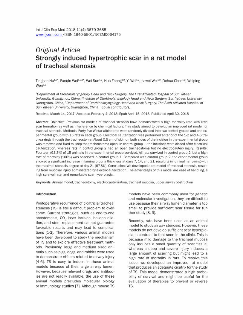
Figure 2. Airway histologic characteristics. (A) Control airway displaying normal epithelium, lamina propria, and cartilage layers (H&E, original magnification ×40). (B-D) Anterior injury model (B, H&E, original magnification ×40; C, Mas-son trichrome stain, original magnification ×100; D, H&E, original magnifica-tion ×40) showing thickened lamina propria (LP), narrowed tracheal lumen (TL), collagen bundle (CB), new hypervascularity (HV), fibrosis of lamina pro-pria (FB), and re-epithelialization (RE).
recovered from conscious-ness and were able to lift their heads. Airways and wounds were checked fre-quently for the first 48 hours. During airway checks, rats were evaluated for respirato-ry distress and obstructing sputum and blood clots we- re suctioned away. The rats were also observed for level of mental state, activity and appetite. The presence or absence of symptoms as well as the weight of the rats were recorded.
Tissue collection and histo-pathological analysis
Animals were euthanized painlessly at days 7, 14, and 21 after surgery for histolo- gic analysis (n=5 for each group at each time point). Tracheal specimens were fixed in 10% buffered forma-lin solution for 24 hours, fol-lowed by dehydration in eth-anol and embedding in par-affin. Cross sections of 0.4-µm thickness were obtained from these samples and his-
Hypertrophic scar in rat tracheal stenosis
3682 Int J Clin Exp Med 2018;11(4):3679-3685
(6.7%) died of an intestinal obstruction at day 11 postoperatively as determined by autopsy. All rats survived in control group 2; however, in control group 1, 4 of 5 rats died within 24 hours after surgery and the others died at post-operative day 5. Autopsy showed that the air-way obstruction that caused death was due to excretions, accompanied by pulmonary edema and bleeding. Because of the high mortality rate in control group 1, the subsequent study of this group was terminated. Animals in control group 2 and the experimental group developed a reduced mental state, decreased activity, dietary reduction after the first day, and an unobstructed stoma. On the second day, there was decreased activity but an improved mental state as the wounds began to scab and the
stoma was partially obstructed. On the third day, the rats were livelier with increased activity and normal appetite, and the stoma was com-pletely closed by normal wound healing pro-cesses and the sutures were removed. There- after, the condition of the rats improved. Our study showed the mean body weights were not significantly changed in both groups during the experimental period.
Establishment of a rat model of hypertrophic scarring for TS
Airways were sectioned and stained with H&E to observe the general histologic and morpho-logic characteristics and Masson trichrome staining was performed to identify the collagen
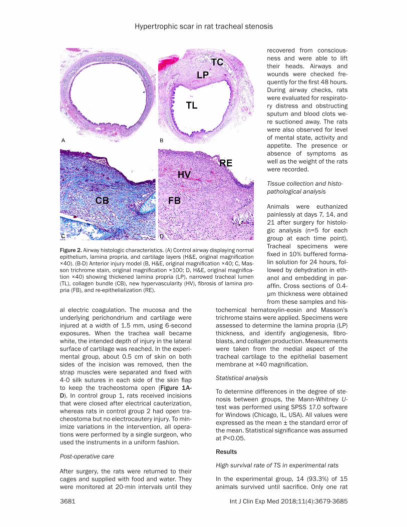
Figure 3. Effect of electrocauterization on rat trachea. A. H&E staining of rat tracheal speci-mens. Control and experimental tracheal ste-nosis are shown (original magnifications ×10 and ×40). Thickness measurements of the lamina propria (LP, black bar) were taken at the thickest locations in each arc segment. Note the progressive thickening of the LP at days 7, 14, and 21 after injury. B. LP mean thicknesses were calculated for each rat group at each time point after injury (days 7, 14, and 21), and the mean thickness of model rats were compared with the controls at each time point. Data demonstrate signifi-cantly increased tracheal thickness of LP in cauterized animals compared with controls (***P=.0001).

Hypertrophic scar in rat tracheal stenosis
3683 Int J Clin Exp Med 2018;11(4):3679-3685
contents of the LP layers. Figure 2A-D shows representative images from a control rat (Fig- ure 2A) and a rat with trachea injured by elec-trocauterization (Figure 2B-D). Lower magnifi-cation images demonstrated (1) anterior nar-rowing of the tracheal lumen, and (2) an incre- ased thickness in the LP (Figure 2B). Higher magnification images demonstrated (1) the destruction of normal epithelium and re-epi-thelialization at the anterior trachea, (2) fibro-sis, infiltration of inflammatory cells, and prolif-eration of fibroblasts in the LP driven by an increase in collagen contents as indicated by trichrome staining, and (3) an increase in blood vessel size and number (Figure 2C and 2D).
Marked increase in LP thickness following electrical cauterization
Histologic examination showed submucosal hypertrophy at the cauterized site resulting in a luminal narrowing of the trachea. Significant differences in thickness of the LP and the cross-sectioned luminal area of the trachea
tube method [10] uses a 10-mm-long stent ma- de from an 8 F feeding tube inserted into the trachea after an incision, and then the wounds are sutured. After 10 days, the stent is removed under general anesthesia. In that study, the thickness of the LP was increased 3-fold and the mortality rate was 15% [10]. The tracheos-tomy cannula method [11] uses a 6 F feeding tube as a tracheostomy cannula that is fixed to peripheral tissues and skin. After 10 days, the tracheal cannula is removed. A previous study showed that the LP thickness increased 4-fold by fibrosis and the mortality rate was 20.83% [11]. Electrocauterization [7] is performed on the anterior 180° of the tracheal mucosa and then incisions are closed. It was reported that after 4 weeks, the LP thickness was increased by 100 µm with a mortality rate of 13.33%. In the scratching method [12], the mucosa of the posterior half of the trachea is damaged until the perichondrium is reached by biomicroscop-ic visualization. Using this method, it was reported that the LP thickness increased 3-fold, equivalent to around 60 µm, without related
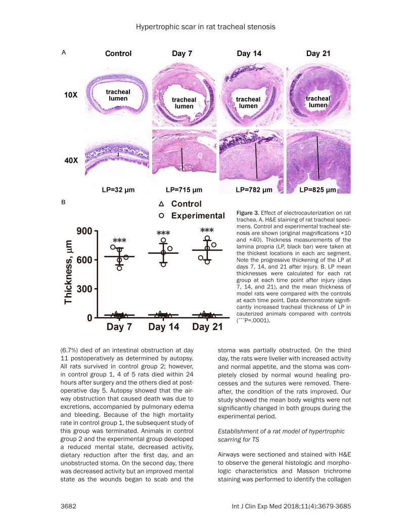
Figure 4. Quantitative analysis of TS. A, B. The degree of stenosis was calcu-lated by the following formula: (1 - the area of the narrow tracheal lumen [B]/the area of the normal tracheal lumen [A]) ×100. C. The results are expressed as the mean ± SD of five cases in the experimental group at each time point. Significant differences (P<0.05; Δ, Day 7 vs. Day 21; #, Day 14 vs. Day 21) are shown.
were observed between the injury group and the control group. Compared with control group 2, the experimental group showed and maintained a signifi-cant increase in LP thick-ness at days 7, 14, and 21 (635.2 ± 87.4 µm vs. 28.0 ± 11.2 µm, 670.8 ± 98.2 µm vs. 30.2 ± 10.5 µm, and 700.6 ± 100.4 µm vs. 30.4 ± 11.5 µm, respectively; P< .0001). Model specimens at days 14 and 21 demon-strated a thickened LP compared with day 7, but without significant differ-ence (Figure 3A and 3B). The cross-sectional area of the model group was nar-rower than that of control group 2 (Figure 4A and 4B), with the greatest differ-ence at day 21 (Figure 4C).
Discussion
Currently, several methods are used to develop TS rat models. The endotracheal

Hypertrophic scar in rat tracheal stenosis
3684 Int J Clin Exp Med 2018;11(4):3679-3685
deaths [12]. Although these models success-fully modeled TS, they had low levels of tissue injury, low levels of scar tissue, and no signifi-cant effect on tracheal ventilation functions, all of which are different from that observed in clinical practice. Recently, the nylon brush me- thod [13] was described, where the mucosa is scraped with a nylon brush through the trache-ostoma. The degree of stenosis was 26.8% at 9 days after scraping, with a low mortality rate of 5.25%. However, it was hard to control the definite extent and depth of the injury using this method, and it produced limited scar tissues.
Compared with the above methods, our meth-od has the following advantages: 1) low mortal-ity rate. There were no postoperative deaths in 15 rats of the experimental group, although one died of an intestinal obstruction as deter-mined by autopsy; thus, its death was related to intraperitoneal anesthesia; 2) marked scar formation. The LP was thickened by the hyper-plasia of fibroblasts, collagen, and granulation tissue. The LP thickness of 608.14 ± 269.24 µm at 3 weeks after surgery, which reached a maximum of 991.02 µm, was much thicker than that reported for other methods; and 3) the operation is simple, and postoperative spe-cial care is not required.
Two key points contributed to the success of our model. First, tracheostomy and the stoma closed on its own 2-3 days after surgery. Swo- llen airways caused by injury or exudates can result in death by obstructing the trachea, which can be prevented by tracheostomy. The most critical period was at 24 hours after sur-gery. If rats survive to this time point, they gen-erally do not suffocate at later stages; however, persistence of the stoma might cause infection of the lower respiratory tract and even death. Therefore, opening the stoma at a certain time-point and the ability to self-heal and close after the critical stages are important factors for the success of our model. Second, the controllabil-ity of the depth and extent of injury. The use of biomicroscopy and transparency of the rat tra-chea allowed the depth of the injury to be evalu-ated in real time to avoid over or insufficient damage. The extent of injury was managed by reshaping electrodes. In addition, it is impor-tant to avoid repeated suction in the trachea by intraoperative control of hemorrhages and to keep the stoma open on postoperative day 1 by
suturing the strap muscles to the skin flap to an appropriate degree of tightness.
Conclusion
This study reports an improved rat model for the study of TS. Ease in animal handling, low material costs, flexibility in experimental desi- gn, and availability of molecular reagents are the benefits of this model. Its limitation was the short follow-up period. Therefore, further research with a longer follow-up period will be necessary to document the changes in tissue hyperplasia over time.
Acknowledgements
This work was supported by grants from the Natural Science Foundation of Guangdong Pro- vince (2017A030313700). We thank Dr. Wen-Bin Lei for helpful discussions and for critically reviewing this manuscript. We thank Edanz Gro- up China (www.liwenbianji.cn/ac), for editing the English text of a draft of this manuscript.
Disclosure of conflict of interest
None.
Address correspondence to: Dr. Wei-Ping Wen, De- partment of Otorhinolaryngology Head and Neck Surgery, The First Affiliated Hospital of Sun Yat-sen University, 2nd Zhongshan Road 58#, Guangzhou 510080, Guangdong, P.R. China. Tel: +86 1380296- 6937; Fax: +86 020 38254221; E-mail: [email protected]
References
[1] D’Andrilli A, Maurizi G, Andreetti C, Ciccone AM, Ibrahim M, Poggi C, Venuta F, Rendina EA. Long-term results of laryngotracheal resection for benign stenosis from a series of 109 con-secutive patients. Eur J Cardiothorac Surg 2016; 50: 105-109.
[2] Tayfun MA, Eren E, Basoglu MS, Aslan H, Oz-turkcan S, Katilmis H. Postintubation laryngo-tracheal stenosis: assessing the success of surgery. J Craniofac Surg 2013; 24: 1716-1719.
[3] Hseu AF, Benninger MS, Haffey TM, Lorenz R. Subglottic stenosis: a ten-year review of treat-ment outcomes. Laryngoscope 2014; 124: 736-741.
[4] Gordin A, Chadha NK, Campisi P, Luginbuehl I, Taylor G, Forte V. An animal model for endotra-cheal tube-related laryngeal injury using hy-

Hypertrophic scar in rat tracheal stenosis
3685 Int J Clin Exp Med 2018;11(4):3679-3685
poxic ventilation. Otolaryngol Head Neck Surg 2011; 144: 247-251.
[5] Eliashar R, Eliachar I, Esclamado R, Gramlich T, Strome M. Can topical mitomycin prevent la-ryngotracheal stenosis? Laryngoscope 1999; 109: 1594-1600.
[6] Kelly NA, Murphy M, Giles S, Russell JD. Sub-glottic injury: a clinically relevant animal mod-el. Laryngoscope 2012; 122: 2574-2581.
[7] Woo JH, Han GC, Kang IG, Kim ST, Cha HE, Kim DY. A new rat model for investigation of sub-glottic stenosis. Acta Otolaryngol 2013; 133: 276-280.
[8] Richter GT, Mehta D, Albert D, Elluru RG. A novel murine model for the examination of ex-perimental subglottic stenosis. Arch Otolaryn-gol Head Neck Surg 2009; 135: 45-52.
[9] Hillel AT, Namba D, Ding D, Pandian V, Elisseeff JH, Horton MR. An in situ, in vivo murine model for the study of laryngotracheal stenosis. JAMA Otolaryngol Head Neck Surg 2014; 140: 961-966.
[10] Bicer YO, Koybasi S, Suslu AE, Kukner A, Tez-can E, Ulas N. Effect of heparin on inflamma-tion: an animal model of tracheal stents. La-ryngoscope 2014; 124: E368-E372.
[11] Koc S, Kiyici H, Sogut E, Eyibilen A, Ekici A, Salman N. Effect of pentoxifylline and 5-fluoro-uracil/triamcinolone on laryngotracheal steno-sis developing as a complication of tracheos-tomy: study in rats. Eur Arch Otorhinolaryngol 2012; 269: 1813-1820.
[12] Ertugrul EE, Cekin IE, Cincik H, Dogru S, Gun-gor A. Effectiveness of topically applied halofuginone in management of subglottic ste-nosis in rats. Otolaryngol Head Neck Surg 2009; 140: 720-723.
[13] Mizokami D, Araki K, Tanaka N, Suzuki H, Tomi-fuji M, Yamashita T, Matsushita K, Shimada H, Shiotani A. Tacrolimus prevents laryngotrache-al stenosis in an acute-injury rat model. Laryn-goscope 2015; 125: E210-E215.