original research article study of dimensions of ... · may predispose to vertebrobasilar...
TRANSCRIPT

Int J Anat Res 2016, 4(3):2526-30. ISSN 2321-4287 2526
Original Research Article
STUDY OF DIMENSIONS OF RETROARTICULAR BONY RINGS INHUMAN ATLAS VERTEBRAEMonika Lalit *1, Sanjay Piplani 2, Poonam Verma 3.
ABSTRACT
Address for Correspondence: Dr Monika Lalit, Assistant Professor, Department of Anatomy,SGRDIMSAR, Vallah, Amritsar 143001, Punjab, India. E-Mail: [email protected]
Background: The third part of vertebral artery on its exit from the FT of atlas is vulnerable for compression byExternal factors like bony outgrowths which may be complete to form the retroarticular bony rings. Such ringsmay predispose to vertebrobasilar insufficiency, Barre- lieou and cervicogenic syndrome especially in neckmovements. The aim of this study is to know the dimensions of Retroarticular Bony Ring as the knowledge aboutsuch dimensions can improve the success rate of surgeries.Materials and Methods: 120 sides on sixty dry adult human atlas vertebrae were obtained in the Department ofAnatomy, Government Medical College, Amritsar, Punjab, India. The Retroarticular Bony Rings (RBR) were identifiedfollowing the criteria used by Mitchell J and Hasan et al. Linear dimensions of Retroarticular Bony Ring (RBRings) like Antero-posterior (Length) Supero-inferior (Height) and Medio-lateral (Width) were measured with thehelp of digital vernier caliper.Results: The complete ponticuli were seen in 10 vertebrae (16.66%). Bilateral retroarticular bony rings have thelowest incidence of 3.33%, 5% on right side and 8.33% on left side. The mean anteroposterior diameter of theright retroarticular canal was 8.79mm and left side was 8.47mm, superoinferior diameter was 5.98mm on rightside and 5.47mm on left side and mediolateral diameter was 4.76mm on right side and 4.42mm on left side.Conclusions: The Knowledge about the dimensions of Bony ring of atlas is important for radiologists,otolaryngologists, neurologists and orthopaedicians as this information may be helpful in avoiding and reducingcomplications such as vertebral artery injury and spinal cord injury during spine surgeries.KEY WORDS: Retroarticular Bony ring, Vertebral artery, Ponticuli, Bony Canal, Atlas vertebrae
INTRODUCTION
International Journal of Anatomy and Research,Int J Anat Res 2016, Vol 4(3):2526-30. ISSN 2321-4287
DOI: http://dx.doi.org/10.16965/ijar.2016.258
Access this Article online
Quick Response code Web site:
Received: 10 Jun 2016 Accepted: 15 Jul 2016Peer Review: 29 Jun 2016 Published (O): 31 Jul 2016Revised: None Published (P): 31 Jul 2016
International Journal of Anatomy and ResearchISSN 2321-4287
www.ijmhr.org/ijar.htm
DOI: 10.16965/ijar.2016.258
*1 Assistant Professor, Department of Anatomy, SGRDIMSAR, Amritsar, Punjab, India.2 Professor, Department of Pathology, SGRDIMSAR, Amritsar, Punjab, India.3 Professor, Department of Anatomy, SGRDIMSAR, Amritsar, Punjab, India.
Superior aspect of posterior arch of atlas ischaracterized by a groove known as sulcusarteriae vertebralis for the passage of third partof vertebral artery. Bony outgrowths may occurand convert this groove into a complete orincomplete ring forming retroarticular bony rings[1]. The bony rings may indicate anomalousossification of the groove by oblique ligament
of atlas; a fibrous tissue present at the lowerborder of posterior atlanto-occipital membrane[1,2], regression [3] or remnants of theproatlas [4]. The Ponticulus Posticus is theposterior osseous bridge that is formed betweenthe posterior margin of superior articularfacets and the posterior arch of the atlas andwhen complete forms the retroarticular bony ring[5-7] and has been variously described by many

Int J Anat Res 2016, 4(3):2526-30. ISSN 2321-4287 2527
Monika Lalit, Sanjay Piplani, Poonam Verma. STUDY OF DIMENSIONS OF RETROARTICULAR BONY RINGS IN HUMAN ATLAS VERTEBRAE.
authors in literature as Kimmerle variant,Canalis arteriae vertebralis, Arcuate foramen,Retrocondylar vertebral artery ring, Retroarti-cular canal, Atlas bridges and Ponticuli [8-10].Whereas the Ponticulus lateralis are the lateralbridges formed as a lateral outgrowth fromlateral margin of the superior articular facets toposterior root of transverse process of the atlas[11,7]. Lateral bridges are reported to be lesscommon than the posterior bridge and some-times may exist as a complete foramen calledas supratransverse foramen [12,7]. Thesebridges predispose to vertebrobasilarinsufficiency, Barre-Lieou and cervicogenicsyndromes especially in severe neck movements[3]. The left VA on the left side has a largerdiameter than the right [13] while the left bonycanal or posterior bridge being smaller than theright [14] and thus makes the left bony ring morevulnerable than the right one. Reduction in theforamen size from the transverse to the arcuateforamen may cause further compression of theVA [15]. The aim of this study was to know thedimensions of Retroarticular Bony Ring as theknowledge about such dimensions can improvethe success rate of surgeries hence preventingdamage to the adjoining vital structures likespinal cord, nerve roots, cranial nerves and thevertebral arteries.
retroarticular Bony Rings Antero-posterior(Length) Supero-inferior (Height) and Medio-lat-eral (Width) diameters were measuredRetroarticular Bony Ring Length: It is the maxi-mum antero-posterior Diameter of the BonyCanal, taken from the posterior part of thegroove on the posterior arch to the root of thesuperior articulating surface and marked as AB.Figure 1.Retroarticular Bony Ring Height: It is the maxi-mum supero-inferior diameter of Bony canal,taken from the floor of the groove to the innerpart of the overlying bony strut and marked asEF. Figure 1.Retroarticular Bony Ring Width: It is the maxi-mum Medio-lateral or transverse diameter of theBony Canal marked as CD. Figure 1.
MATERIALS AND METHODS
120 sides on sixty dry adult human atlasvertebrae belonging to North Indian individualswere obtained by maceration from thecadavers made available for the purpose ofdissection, in the Department of Anatomy,Government Medical College, Amritsar, Punjab,India. All the atlas vertebrae were thoroughlycleaned and numbered from 1-60. Damaged andpathological Atlas vertebrae were excluded fromstudy.The Retroarticular Bony Rings (RBR) wereidentified following the criteria used by Chevrelet al and Mitchell (1998a, 1998b).Linear dimensions of Retroarticular Bony Ring(RB Rings) (Figure 1,2) were measured with thehelp of digital vernier caliper with a least countof 0.02 mm. All the measurements were takendirectly from the bones and then the data wasstored on the computer sheet. For the
Fig. 1: Showing Measuring Of A Complete RetroarticularRing- Antero-Posterior Diameter - AB, Vertcical Height-EF, Transeverse Width- CD Also Showing OsteophyteEncroachments.
Fig. 2: Showing bilateral complete retroarticular bonyring V.A- vertebral artery, SAF- Superior Articular Facet,P- posterior arch, AA- Anterior Arch and a probe passingthrough ft and then bony retroarticular bony ring.
Int J Anat Res 2016, 4(3):2526-30. ISSN 2321-4287 2528
Monika Lalit, Sanjay Piplani, Poonam Verma. STUDY OF DIMENSIONS OF RETROARTICULAR BONY RINGS IN HUMAN ATLAS VERTEBRAE.
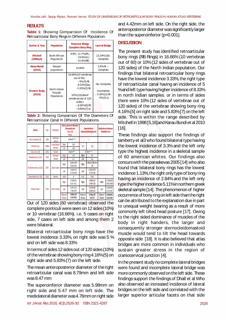
Out of 120 sides (60 vertebrae) observed thecomplete ponticuli were seen on 12 sides (10%)or 10 vertebrae (16.66%). i.e. 5 cases on rightside, 7 cases on left side and among them 2were bilateral.Bilateral retroarticular bony rings have thelowest incidence 3.33%, on right side was 5 %and on left side was 8.33%In terms of sides 12 sides out of 120 sides (10%)of the vertebrae showing bony ring 4.16% (5) onright side and 5.83% (7) on the left side.The mean anteroposterior diameter of the rightretroarticular canal was 8.79mm and left sidewas 8.47 mmThe superoinferior diameter was 5.98mm onright side and 5.47 mm on left side. Themediolateral diameter was 4.76mm on right side
RESULTSTable 1: Showing Comparision Of Incidence OfRetroarticular Bony Rings In Different Population.
Table 2: Showing Comparison Of The Diameters OfRetroarticular Canal In Different Populations.
Pyo & Lowman [8] 1959 American whites
Right 6.4
Left 6.6Unur et al. [9] 2004 Turkish
Right 6.4 Right 5.4Left 6.7 Left 5.4
Karau Bundi et al [30] 2010 Kenyan 6.29 6 5.11 5.16
Author Year Population
8.5mm**** --- -
Mitchell [11] 1998a mixed South Africans
5.3 5.1 -
5.7mm**** -
Paraskevas et al. [14] 2005Northern
Greeks -
Krishnamurthy et al [21] 2007 South Indian Right Right
Present study 2016 North Indian
Right Right
Left Left Left
Bilateral (Mean)
Bilateral (Mean)
Bilateral (Mean)
5.98±0.78 5.56–6.25
4.76±0.56 4.02–5.98
Mediolateral Diameter (PoWidht) mm
Superoinferior Diameter(PoHeight) mm
Antero posterior Diameter (PoLength) mm
Mean±SD Range
9.26±0.464 8.63–9.92
8.14±1.01 6.73–9.43
Right 8.79±0.31 8.21–9.11
- Left Left
4.91±0.670 3.75–5.93
-
5.38±0.2285.04–5.68
8.1mm***
8.47±1.21 7.65–9.87 8.63±1.19 5.21-9.32
5.72±o.78 4.0-7.0
4.44±0.61 3.21-5.50
4.42±0.65 4.21–6.67
5.47±0.98 4.86–6.23
and 4.42mm on left side. On the right side, theanteroposterior diameter was significantly largerthan the superoinferior (p=0.001).
DISCUSSION
The present study has identified retroarticularbony rings (RB Rings) in 16.66% (10 vertebraeout of 60) or 10% (12 sides of vertebrae out of120 sides) of the North Indian population. Ourfindings that bilateral retroarticular bony ringshave the lowest incidence 3.33%, the right typeof retroarticular canal having an incidence of 5% and left type having higher incidence of 8.33%in north Indian samples. or in terms of sidesthere were 10% (12 sides of vertebrae out of120 sides) of the vertebrae showing bony ring4.16% [5] on right side and 5.83% [7] on the leftside. This is within the range described byMitchell in 1998 [5,16]and Karau Bundi et al 2010[16].These findings also support the findings oflamberty et al3 who found bilateral type havingthe lowest incidence of 3.3% and the left onlytype the highest incidence in a skeletal sampleof 60 american whites. Our findings alsoconcurs with the paraskevas 2005 [14] who alsofound that bilateral bony rings has the lowestincidence 1.13%, the right only type of bony ringhaving an incidence of 2.84% and the left onlytype the higher incidence 5.11% in northern greekskeletal sample [14]. The phenomenon of higheroccurrence of bony ring on left side than the rightcan be attributed to the explanation due in partto unequal weight bearing as a result of morecommonly left tilted head posture [17]. Owingto the right sided dominance of muscles of thebody in right handers, the larger andconsequently stronger sternocleidomastoidmuscle would tend to tilt the head towardsopposite side [18]. It is also believed that atlasbridges are more common in individuals whosustain greater stress in the region ofcraniocervical junction [4].In the present study no complete lateral bridgeswere found and incomplete lateral bridge wasmore commonly observed on the left side. Thesefindings support the findings of Dhall et al Whoalso observed an increased incidence of lateralbridges on the left side and correlated with thelarger superior articular facets on that side
Author & Year Population Lateral Bridge
Mitchell (1998a,b)
South African Population
9.8% - 11.7% (Rt.) - 24.6% (Lt.) - 31.8% (Bl)
12.24% (18)- Complete
Posterior Bridge Complete Bony Ring
Karau Bundi (2010)
Kenyan population
14.90% 3.9% Rt. – Complete
Present Study (2016)
North Indian Punjabi
Population
16.66% (10 vertebrae out of 60) -
- 5% (3) Rt. - 8.33% (5)Lt. - 3.33% (2) Bl.
10% (12sidesof vertebrae out of 120
sides ) – 4.16% (5) Rt. - 5.83% (7) Lt.
No- Complete
Incomplete- 3.33% (2) Rt.
- 5% (3) Lt.

Int J Anat Res 2016, 4(3):2526-30. ISSN 2321-4287 2529
Monika Lalit, Sanjay Piplani, Poonam Verma. STUDY OF DIMENSIONS OF RETROARTICULAR BONY RINGS IN HUMAN ATLAS VERTEBRAE.
[19,17]. Whereas Complete lateral bridges,forming supratransverse foramina were foundon right side in 3.9% of the cases in the workdone by Karaubundi et al in 2010 in Kenyanpopulation [16]. The findings of the presentstudy also support those of previous researchthat lateral bridges are less common comparedto the posterior bridges [4,5,11].For the retroarticular canal, the mean antero-posterior diameter was 8.79mm on the right and8.47mm on the left. The mean superoinferiordiameter was 5.98mm on the right and 5.47mmon the left. The mean mediolateral diameter was4.76mm on the right and 4.42mm on the left.These measurements concur with those of theprevious research as shown in Table II. Our re-sults support those of previous workers [5,11,14]that the anteroposterior diameter is significantlylarger than the superoinferior in theretroarticular canal and it has been seen thatthe left vertebral artery was found to be largerthan the right20. Therefore, possible that thevertebral artery is compressed superoinferiorly[5,11].The presence of more bridges on the left sidewith small diameter as compared to right sideare observed in the present study would meanthat VA is more liable for compression [21]. It ispossible that the third segment of the VA maybe a reserve length to allow for neck rotationwithout injury or compression to the artery. Pres-ence of these ponticles may limit this reservelength, predisposing to entrapment of the ar-tery [16].Furthermore, the ossification of ligamentousstructures in various parts of the body may re-sult in clinical problems such as compression toneighboring structures and complications in re-gional surgery [22]. Maintaining the vertebralartery intact constitutes an important concernas presence of Retroarticular bony rings can leadto compression of the vertebral artery in theabsence of arterial disease or may be an aggra-vating factor in case of disease [5,11,14]. Highincidence of this bony rings is associated withvertebro-basilar insufficiency presenting withdizziness, fainting and transient diplopia. Thereare 8 reported cases where surgical removal ofbony bridges alleviate the symptoms of vertebro-basilar insufficiency [23]. It is also revealed that
the vertebral artery occupies 57% of thevertebral artery groove and when a foramen isformed over here, it produces a relatively tightsituation of vertebral artery leading to giddinesson external neck movements due to vertebra-basilar insufficiency [24]. Association betweenthe arcuate foramen and tethering of vertebralartery in it may lead to its dissection fromrepetitive trauma with movement of neck hasalso been reported [25]. This compressionbecomes evidently symptomatic in extrememanipulations of the neck [26]. Cakmak et al(2005) [27] asserted that cervical spineradiography is a simple and useful technique toknow the presence of arcuate foramen andshould be considered if a patient comes withsymptoms like pain in temporal region, pain inback of eye, vertigo, occipital headache,periodic photophobia, paraesthesia of hands orsensation of pressure on hands.In an individual with a lateral bridge of atlas andan associated retroarticular canal may furtherresults in increased compression of thevertebral artery [28] and compromised bloodflow during extreme rotation of head and neckas occurs in manipulation of cervical spine[29,30].
The prevalence of retroarticular bony rings inthe atlas among North Indians are comparableto that in other populations. Thus Knowledgeabout the dimensions of Bony ring of atlas isimportant for radiologists, otolaryngologists,neurologists and orthopaedicians as thisinformation may be helpful in avoiding andreducing complications such as vertebral arteryinjury and spinal cord injury during spinesurgeries.
CONCLUSION
Conflicts of Interests: None
REFERENCES
[1]. William M, Newell RLM, Collin P. The back: cervicalvertebrae. In: Standring S, Ellis H, Haely JC, JohsonD, Williams A (eds), Gray’s Anatomy. 39th edition.Edinburg, London: Elsevier Churchill Livingstone.2005:742-746.
[2]. Romanus T and Tovi A. A variation of the atlas.Roentgenolgic incidence of a bridge over the grooveon the atlas for the vertebral artery. Acta Radiol.Diagn. 1964;2:289-97.

Int J Anat Res 2016, 4(3):2526-30. ISSN 2321-4287 2530
[17]. Dhall U, Chhabra S, Dhal JC. Bilateral asymmetry inbridges and superior articular facets of atlas verte-bra. J Anat Soc.India. 1993;42:23-27.
[18]. Pande BS, Singh I. One sided dominance in upperlimbs of human foetuses as evidenced by asymme-try in muscle and bone weight. J Anat Soc.India.1971;109:457-459
[19]. Hasan M, Shukla S, Siddiqui MS, Singh D. Postero-lateral tunnels and ponticuli in human atlas verte-brae. J Anat 2001;199(3):339-343.
[20]. Thiel, H. Gross morphology and pathoanatomy ofthe vertebral arteries. J. Manipulative Physiol. Ther.1991;14:133-41.
[21]. Krishnamurthy A et al. Arcuate foramen of atlas:Incidence, Phylogenetic and Clinical significance.Romanian journal of morphology and embryology2007;48(3):263-266.
[22]. Prescher A. The craniocervical junction in man, theosseous variations their significance and differen-tial diagnosis. Annals of anatomy. 1997;179:1-19.
[23]. Ercegovac N, Davidovic R. Foramen arcuale atlantiskao etiolski faktor vertebrobazilare insu®cijencijedekompresija arterije vertebralis.Vojnosanitetski pregled. 1970;10:435-441.
[24]. Cacciola F, Phalke U and Goel A. Vertebral artery inrelationship to C1 – C2 vertebrae: An anatomicalstudy. Neurology India. 2004; 52(2):178-184.
[25]. Dahipale V P, Bahetee B H. The Retroarticular verte-bral artery ring of the atlas and its significance. JAnat Soc. India. 2009; 58:149-151.
[26]. Limousin, C. A. Foramen arcuale and syndrome ofBarre- Lieou. Int. Orthop.1980;4:19-23.
[27]. Cakmak O, Gurdal E, Ekinci G, Yildiz E, Cavdar S.Arcuate foramen and its clinical significance. SaudiMed J. 2005;26(9):1409-1413.
[28]. Manjunath K Y. Posterior bridging of the atlas verte-bra in south Indians. Indian J Med Sci.2001;55(9):488-490.
[29]. Parkin PJ, Wallis WE, Wilson JE. Vertebral areteryocclusion following manipulation of the neck. N ZMed. 1978;88:441-443.
[30]. Krueger BR, Okazaki H. Vertebral basilar distribu-tion infarction following chiropractic cervical ma-nipulation. Mayo Clinic Proceedings. 1980;55:322-332.
[3]. Lamberty BG and Zivanovic S. The retro-articularvertebral artery ring of the atlas and its signifi-cance. Acta Anat. 1973;85:113-22.
[4]. Taitz C, Nathan H. Some observations on the poste-rior and lateral bridge of atlas. ActaAnat.1986;127(3):212-217.
[5]. Mitchell J. The incidence and dimensions of theretroarticular canal of the atlas vertebra. Acta Anat1998 (a);163:113-120.
[6]. Le Minor, J. M. & Trost, O. Bony ponticles of the atlas(C1) over the groove for the vertebral artery in hu-mans and primates: Polymorphism and evolution-ary trends. Am.J. Phys. Anthropol.2004;125:16-29.
[7]. Lalit M, Piplani S, Arora A K, Kullar J S, Sharma T.Incidence of Atlas Bridges and Tunnels- Their Phy-logeny, Ontogeny and Clinical Implications. Rev ArgDe Anat Clin. 2014;6(1):26-34.
[8]. Pyo J & Lowman R M. The ponticulus posticus of thefirst cervical vertebra. Radiology. 1959;72:850-854.
[9]. Unur, E, Erdogan N, Ülger H, Ekinci N & Oztürk O.Radiographic incidence of complete arcuate fora-men in Turkish population. Erciyes Med J2004;26:50-54.
[10]. Lalit M, Singla R K, Kullar J S. Bilateral Arcuate Fora-men in a Human Atlas Vertebra - A Case Report.International Journal of Anatomy, Radiology andSurgery, 2013;2(3):3-6.
[11]. Mitchell, J. The incidence of the lateral bridge of theatlas vertebra. J. Anat. 1998b; 193:283-5.
[12]. Chevrel J P, Pineau H, Delmas A. D’are posterieur deI; atlas. Ses variations. Etude morphologique etstatistique. CR Assoc Anat. 1965;50;280-288.
[13]. Sun, J. Y. Foramen arcuale and vertigo. ZhonghuaWai Ke Za Zhi.1990;28:592-4.
[14]. Paraskevas G, Papaziogas B, Tsonidis C & KapetanosG. Gross morphology of the bridges over the verte-bral artery groove on the atlas. Surg Radiol Anat.2005;27:129-136.
[15]. Tubbs R S, Johnson P C, Shoja M M, Loukas M, OakesW J. Foramen Arcuale: Anatomical study and re-view of literature. J Neurosurg Spine. 2007;6(1):31-34.
[16]. Karau Bundi P, Ogengo JA, Hassanali J, Odula PO.Morphometry and variations of bony poticles ofthe atlas vertebrae in Kenyans. Int J Morphol.2010;28(4):1019-1024
How to cite this article:Monika Lalit, Sanjay Piplani, Poonam Verma. STUDY OFDIMENSIONS OF RETROARTICULAR BONY RINGS IN HUMANATLAS VERTEBRAE. Int J Anat Res 2016;4(3):2526-2530. DOI:10.16965/ijar.2016.258
Monika Lalit, Sanjay Piplani, Poonam Verma. STUDY OF DIMENSIONS OF RETROARTICULAR BONY RINGS IN HUMAN ATLAS VERTEBRAE.