ortho-biologics for osteoarthritis - dr. brian cole€¦ · ortho-biologics for osteoarthritis kyla...
TRANSCRIPT
1
234
567
8910
11121314
151617
181920
212223
24252627
282930
313233
34353637
383940
414243
444546
4748
Ortho-Biologics forOsteoarthrit is
Q2 Q3
Kyla Huebner, MSc, MD, PhDa, Rachel Frank, MDb,Alan Getgood, MPhil, MD, FRCS (Tr&Orth)a,*
Q4
Q7
KEYWORDS
� PRP � Autologous conditioned serum � Stem cell � Bone marrow � Osteoarthritis
KEY POINTS Q6
� This study seeks to shed light on the current literature in the use of key ortho-biologics andtheir potential use in the treatment of osteoarthritis.
INTRODUCTION
Osteoarthritis (OA) is a debilitating disease affecting approximately 27 millionAmericans.1 The most common symptoms of OA are pain and physical limitationsthat have a significant effect on people’s quality of life and their social and economicactivities.2,3 Because of the increasing life expectancy, increasing numbers of elderly,and increasing prevalence of obesity in North America, the prevalence of OA willcontinue to increase. There are currently limited options for treatment and preventionof OA, with joint replacement often the ultimate outcome. The cost of joint replace-ments is around $55,000 per person with complication rates of approximately 1%to 10% and mortality rates of 0.25%.4 In order to reduce costs to the medical systemand the risks and costs to patients, we need a better understanding of thediseasepathophysiology, improved early detection, and strategies for disease preventionand early disease management. Ortho-biologics may be one such option for the treat-ment of OA.Ortho-biologics as defined by the American Academy of Orthopaedic Surgeons
(AAOS) are biological substances found naturally in the body that help injuries healmore quickly.5 These substances includes any biologically derived conductive mate-rial that aids in repair and regeneration of bone, muscle, tendons, ligaments andcartilage. There are many treatments that now fit under this overarching term. These
Disclosure Statement: None.a Division of Orthopaedic Surgery, Western University, Fowler Kennedy Sports Medicine Clinic,3M Centre, 1151 Richmond Street, London, Ontario N6A 3K7, Canada; b University of Colo-rado, Boulder, CO, USA Q5
* Corresponding author.E-mail address: [email protected]
CSM995_proof ■ 27 September 2018 ■ 7:57 am
Clin Sports Med - (2018) -–-https://doi.org/10.1016/j.csm.2018.09.002 sportsmed.theclinics.com0278-5919/18/ª 2018 Elsevier Inc. All rights reserved.
Huebner et al2
49
505152
535455
565758
59606162
636465
666768
697071
72737475
767778
798081
82838485
868788
899091
929394
95969798
99
treatments include platelet-rich plasma (PRP), prolotherapy, ozone therapy, autolo-gous conditioned serum (ACS), bone marrow aspirate concentrates (BMACs),adipocyte-derived stem cells, mesenchymal-derived concentrates, amniotic-derivedcell concentrates, cord blood–derived cell concentrates, interleukin therapies, andalpha-2 macrophages. For the purpose of this review, the authors focus on viscosup-plementation, PRP, ACS, BMACs, and other cell-derived therapies, as these arecurrently in clinical use.
Q8
Q9
Q10
VISCOSUPPLEMENTATION
Viscosupplementation consists of hyaluronic acid (HA) treatments injected into thejoint for pain relief and possible antiinflammatory effect.6 HA is an anionic, nonsulfatedglycosaminoglycan found in connective tissues, epithelium, and neural tissue. It isformed in the plasma membrane and is one of the main components of the extracel-lular matrix, contributing to cell proliferation and migration. HA is found within jointsproviding viscoelastic properties to the synovial fluid. In OA, there is a reduction inHA synthesis with increased HA degradation, in turn, leading to a lower molecularweight in the synovium, synovial fluid, and cartilage.7 HA therapy provides relief viavarious pathways, including suppression of proinflammatory cytokines and chemo-kines through the synthesis of antiinflammatory mediators.8 In a systematic reviewby Altman and colleagues, 48 articles were analyzed to evaluate the antiinflammatoryeffect of HA in OA. They found that proinflammatory cytokines (interleukin 1b [IL-1b]),tumor necrosis factor a (TNFa), and interferon g can regulate HA synthase expression.HA binds to cell surface receptors, such as CD44, toll-like receptor (TLR) 2 and 4, lyilin,and intracellular adhesion molecule-1 (ICAM-1). In binding to CD44, it suppressesproinflammatory cytokines, matrix metalloproteinases (MMPs), proteoglycans, andprostaglandin E2 synthesis via CD44 through the downregulation of nuclear factor(NF)-kB. HA also activates the innate immune response via TLR-2. HA treatmentwas shown to bind to TLR-2 and TLR-4 and decrease TNFa, IL-1b, IL-17, MMP13,and inducible nitric oxide. Lyilin is expressed in human articular chondrocytesand synoviocytes; by binding to lyilin HA suppressed the expression of IL-1b andMMP1 and 13. ICAM-1 activates the NF-kB regulatory system activating proinflamma-tory cytokines; HA binds to ICAM-1 and inhibits its action thereby preventinginflammation.9,10
Early studies of HA treatments in OA had mixed results. In a large meta-analysis of89 trials containing 12,667 participants, 71 studies showed a modest effect indecreasing pain, whereas the remainder showed no effect. Fourteen studies had sig-nificant adverse effects related to HA injections. Rutjes and colleagues11 concludedbased on these early studies that HA therapy had a clinically irrelevant benefit with sig-nificant adverse reactions.Miller and Block12 did 2 meta-analyses evaluating 26 articles with a total of 4866
subjects for the safety and efficacy of HA. They found that there was a large treat-ment effect for up to 26 weeks for pain relief and improved Western Ontario andMcMaster Universities Osteoarthritis Index (WOMAC) scores. There were no signifi-cant adverse effects reported in this series of studies.13 In another meta-analysis ofhigh-quality level 1 randomized controlled trials (RCTs), 12 studies consisting of 1794participants were analyzed. Early on, between 1 and 3 months, corticosteroid injec-tions had improved outcomes in the WOMAC score and lower visual analog scale(VAS) scores. However, at 6 months, the effect of HA was better than corticosteroidsin OA.14 In another study of 13 articles, HA was shown to have greater effects upto 1 year compared with nonsteroidal antiinflammatories and corticosteroids.15
CSM995_proof ■ 27 September 2018 ■ 7:57 am

Q1Ortho-Biologics for Osteoarthritis 3
100
101102103
104105106
107108109
110111112113
114115116
117118119
120121122
123124125126
127128129
130131132
133134135136
137138139
140141142
143144145
146147148149
150
Bhandari and colleagues16 reviewed 8 meta-analyses and found that by 26 weeksthere were significant improvements in pain, functional scores, and stiffness afterHA injections in patients with mild to moderate OA. In addition, they found HA tobe well tolerated and safe. Importantly, they observed that HAs with a molecularweight greater than 6000 kDA or greater had the greatest treatment effect on painat 13 weeks and 3000 kDA or greater has the greatest treatment effect on pain at26 weeks. In addition to one-time injections, patients often require multiple treat-ments. A meta-analysis of 7404 patients showed that repeat HA injections weresafe in patients with OA. In 95% of patients who had an adverse event, it was atthe time of the first treatment; there was no increase in frequency or severity ofadverse events with repeat treatments. The adverse event rate was 0.008 with repeatinjections.17
In light of the mixed results in the literature and the changes in AAOS guidelines, aUS and a European consensus were formed to help guide the use of HA in OA. TheEuropean Viscosupplementation Consensus Group determined that, based on anextensive review of the literature, if HA injections were successful previously, a repeatattempt at treatment should be undertaken. They also recommended the use of HAinjections in young patients at high risk of progression of OA and competitive athletesin a possible attempt to slow the progression of OA.18 A similar US task force of rheu-matologists, orthopedic surgeons, physiatrists, sports medicine physicians, andnurses was formed to study HA injections in OA. They reviewed 100 studies that sug-gested HA was superior to placebo treatments. Based on these studies, they came upwith 8 various clinical scenarios by which to use HA injections (3 appropriate uses and5 unclear uses)19 (Table 1).
Table 1Clinical scenarios Q18for the use of HA listed by Bhadra and colleagues
1. Symptomatic adults with mild or moderate OA of the knee who haveclinically and radiologically confirmed disease who have not receivedother therapies for the knee
Appropriate
2. Symptomatic adults with severe mild or moderate OA of the knee whohave clinically and radiologically confirmed disease and have failed othernonpharmacologic or pharmacologic therapies for the knee
Appropriate
3. Symptomatic adults with mild or moderate OA of the knee who haveclinically and radiologically confirmed disease who have incompleteresponse to other therapies for the knee
Appropriate
4. Symptomatic adults with mild or moderate OA of the knee who areintolerant of, have a high-risk of adverse reaction to, or who arecontraindicated for pharmacologic agents for the knee (oral, topical, orintra-articular)
Unclear
5. Symptomatic adults who have mechanical meniscus pathology withunderlying OA of the knee
Unclear
6. Symptomatic adults with OA of the knee who have had a significantadverse reaction to an intra-articular HA product
Unclear
7. Symptomatic adults with OA of the knee who have active inflammatoryarthritis (rheumatoid arthritis, gout, and so forth)
Unclear
8. Symptomatic adults with OA of the knee who have synovitis of the kneewith significant effusion
Unclear
From Bhadra AK, Altman R, Dasa V, et al. Appropriate use criteria for hyaluronic acid in the treat-ment of knee osteoarthritis in the United States. Cartilage 2017;8(3):234–54; with permission. Q19
CSM995_proof ■ 27 September 2018 ■ 7:57 am

Huebner et al4
151
152153154
155156157
158159160
161162163164
165166167
168169170
171172173
174175176177
178179180
181182183
184185186187
188189190
191192193
194195196
197198199200
201
In practice, HA is widely used as a part of the treatment algorithm for mild to mod-erate OA despite the lack of consensus and the current US and Canadian treatmentguidelines. It likely has some benefit in certain patients and is worth a trial of treatmentin those who are candidates.
Q11
PLATELET-RICH PLASMA
As cartilage is nonvascular, its nourishment is based on diffusion. Therefore, intra-articular injections at high concentrations are often the preferred method to aid incartilage regeneration. PRP, which has a higher concentration of platelets thanwhole blood, has been an interesting option for use in OA. PRP is a natural concen-trate of autologous factors obtained by centrifugation or filtration of the patients’blood. It is obtained at a low cost, simple to obtain, and minimally invasive. PRPis thought to work via biologically active proteins (including platelet-derived growthfactor [PDGF], transforming growth factor [TGF], insulinlike growth factor, fibroblastgrowth factor, and vascular endothelial growth factor [VEGF]20) expressed by plate-lets leading to gene expression by binding to transmembrane receptors in targetcells. PDGF is a chemoattractant and stimulator of cell proliferation. TGF is a poly-peptide that is abundant in platelets and bone and plays an important role in woundhealing; it may negatively influence angiogenesis and promotes matrix productionby fibroblasts and stimulates the production of VEGF. VEGF is a family of proteinsthat act through the kinase family expressed on endothelial cells, which stimulateblood vessel formation and exert a trophic effect on endothelial cells. VEGF isalso proinflammatory and stimulates leukocyte adhesion to endothelial cells. As aresult of these growth hormones, cellular recruitment, migration, growth, andmorphogenesis are triggered and inflammation is decreased.21 Therefore, it hasbeen widely used and studied as a noninvasive treatment of cartilage regenerationin OA.As PRP is an autologous product, there is a lot of variability within individual pa-
tients. Differences in patients’ daily platelet levels, procurement methods, concen-tration mechanisms, and exogenous factors to enhance platelet activation can allcontribute to varied PRP preparations. Platelet concentration varies significantly be-tween procurement method and time of draw.22,23 Platelet concentrates have beenrecorded as between 200 � 103 and 1000 � 103 platelets per microliter, with noconsensus existing as to which concentration has the best outcomes. However,concentrations greater than this have been demonstrated to be biologically unfavor-able.23,24 In addition to the variation in draw times and platelet concentration, therecan be variability in leukocytes within the RPR formulation. It is debatable whetherleukocytes are beneficial or detrimental, as they have the potential to aid in healing;however, they can also be the cause of increased injury and adverse reactions.24
Leukocytes adversely increase local inflammation, beneficially produce VEGF,have antimicrobial effects. and are restorative to tissues.25–27 The addition of leuko-cytes to PRP has also been shown to enhance the concentration of growth factors inPRP.27 There are 2 different types of commercially available system for PRP: oneproducing a leukocyte-rich PRP (LR-PRP) and the other producing a leukocyte-poor PRP (LP-PRP). A buffy coat system, which uses a high centrifugation ratefor a longer time, produces LR-PRP.28 Plasma-based systems produce LP-PRP; ituses slower centrifugation or filtration for a shorter time.28 The literature is still spliton the benefit of LR-PRP versus LP-PRP for a given pathology. Exogenous factorscan also be added to PRP formulations, the most common being thrombin.Thrombin activates platelets and is often used in combination with calcium
CSM995_proof ■ 27 September 2018 ■ 7:57 am
Ortho-Biologics for Osteoarthritis 5
202
203204205
206207208
209210211
212213214215
216217218
219220221
222223224
225226227228
229230231
232233234
235236237238
239240241
242243244
245246247
248249250251
252
chloride.22 Thrombin plus calcium chloride was shown to increase the release ofgrowth factors in PRP, releasing 100% of growth factors by 1 hour.29
Preclinical studies have been supportive of the use of PRP for the regeneration ofjoint tissue in OA. PRP increases chondrocyte proliferation and increases the produc-tion of proteoglycans and type II collagen in vitro.30–33 In animal models PRP leads toimproved cartilage regeneration,34 and enhances meniscal cells35 and synovio-cytes.36 PRP has also been shown to have an antiinflammatory effect.37,38 Basedon these studies of the basic biology involved in PRP, there is evidence to supportthat PRP enhances cartilage repair and slows degradation.The initial investigation into the use of PRP injections to treat OA was published in
2008. It was a retrospective observational study of 60 patients, which showed favor-able outcomes after intra-articular PRP injections.39 It was not until 2012 that the firstRCT was published. To the authors’ knowledge since then, 7 systematic reviews/meta-analyses have been published. This section summarizes the current clinic evi-dence for PRP in OA focusing on meta-analyses. Table 2 shows a summary of thesearticles.Chang and colleagues40 in 2014 performed a systematic review and meta-analysis
analyzing the effectiveness of PRP in treating chondral lesions in the knee. The inves-tigators included 8 single-arm studies, 3 quasi-experimental studies, and 5 RCTs con-sisting of 1543 subjects. PRP showed efficacy for 12 months after injection and itseffectiveness was better and more prolonged than HA injections in patients withmild-moderate OA.40 A level 1 systematic review and meta-analysis performed byLaudy and colleagues41 in 2014 compared PRP with HA and placebo. Six RCTsand 4 non-RCTs were included. They found improved functional outcomes ofWOMAC, the VAS, and Lequesne index after PRP injections compared with HA andplacebo.41
In another meta-analysis of PRP in OA, the use of LR-PRP and LP-PRP was inves-tigated and clinical outcomes (WOMAC and International Knee Documentation Com-mittee [IKDC]) and adverse effects were compared. They included 6 RCTs and 3retrospective studies containing 1055 participants. LP-PRP had better WOMAC andIKDC scores than HA or controls, whereas there was no difference in LR-PRP scores.Both LP-PRP and LR-PRP had higher adverse reactions compared with HA and con-trols, being primarily swelling and pain.42
Meheux and colleagues43 performed a systematic review of level 1 RCTs to deter-mine whether PRP improves patient-reported outcomes at 6 and 12 months and todetermine any differences between PRP or HA or placebo treatment at 6 and12 months. After a quality assessment using the modified Coleman methodologyscore, 6 articles were analyzed. All but one study showed significant differences inclinical outcomes between groups for pain and function. Posttreatment PRP scoreswere significantly better than for HA at 3 and 6 months. In addition, PRP injectionsresulted in significant clinical improvements up to 12 months.43 In another systematicreview by Sadabad and colleagues44 in 2016 evaluating 7 studies consisting of 722participants, they found that PRP led to significantly improved WOMAC scorescompared with HA.In the most recent meta-analysis by Dai and colleagues,45 10 RCTs consisting of
1069 participants were used to compare PRP injections with HA at 6 and 12 months.At 6 months there was no difference in clinical outcomes between HA and PRP treat-ments; however, by 12 months PRP treatment resulted in significantly improvedWOMAC, IKDC, and Lequesne scores.45
Overall the body of literature suggests that PRP is a promising therapy for symptomrelief and improved functional outcomes in patients with OA for at least 12 months.
CSM995_proof ■ 27 September 2018 ■ 7:57 am

TabSu ry of meta-analyses looking at PRP
Stu Studies Included Databases Dates ComparisonSampleSize
AverageFollow- up
OutcomeMeasures Results
Ch et al,40
216 Studies� 8 single arm� 3 quasi-experimental� 5 RCTs
MEDLINE 2010–2013 PRP vs HA 1543 12 mo IKDCKOOSWOMAC
PRP significantlyimproved scoresmore than HA.
PRP was moreeffective in lesssevere OA.
La t al,2 Q20
10 Studies� 6 RCTs� 6 non-RCTs
MEDLINEEmbaseCINHALWeb of ScienceCochrane database
2011–2013 PRP vs HAPRP vs
placebo
1110 6 mo WOMACVASNRW Q21
Lequesne
PRP significantlyimproved scoresthan HA.
PRP significantlyimproved scoresmore than placebo.
Rib t al,2
9 Studies� 6 RCTs� 3 prospective
MEDLINEEmbaseCochrane database
2011–2013 LP PRP vsLR PRP
1055 Not reported IKDCWOMACAdverse reactionsVASLequesneTegnerMarxKOOSSF-36MRI
LP-PRP improvedWOMAC scorescompared withplacebo.
There were similaradverse eventsbetween LP-PRP andLR-PRP.
Huebneretal
CSM995_proof
■27
Septem
ber2018
■7:57
am
6
253
254
255
256
257
258
259
260
261
262
263
264
265
266
267
268
269
270
271
272
273
274
275
276
277
278
279
280
281
282
283
284
285
286
287
288
289
290
291
292
293
294
295
296
297
298
299
300
301
302
303
le 2mma
dy
ang014
udy e014
oh e015
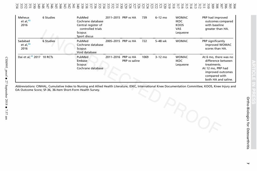
Meheuxet al,43
2016
6 Studies PubMedCochrane databaseCentral register of
controlled trialsScopusSport discus
2011–2015 PRP vs HA 739 6–12 mo WOMACIKDCKOOSVASLequesne
PRP had improvedoutcomes comparedwith baselinegreater than HA.
Sadabadet al,44
2016
6 Studies PubMedCochrane databaseScopusVoid database
2005–2015 PRP vs HA 722 5–48 wk WOMAC PRP significantlyimproved WOMACscores than HA.
Dai et al,45 2017 10 RCTs PubMedEmbaseScopusCochrane database
2011–2016 PRP vs HAPRP vs saline
1069 3–12 mo WOMACIKDCLequesne
At 6 mo, there was nodifference betweentreatments.
At 12 mo, PRP hadimproved outcomescompared withboth HA and saline.
Abbreviations: CINHAL, Cumulative Index to Nursing and Allied Health Literature; IDKC, International Knee Documentation Committee; KOOS, Knee Injury andOA Outcome Score; SF-36, 36-Item Short-Form Health Survey.
Orth
o-Biologics
forOste
oarth
ritis
CSM995_proof
■27
Septem
ber2018
■7:57
am
7
304
305
306
307
308
309
310
311
312
313
314
315
316
317
318
319
320
321
322
323
324
325
326
327
328
329
330
331
332
333
334
335
336
337
338
339
340
341
342
343
344
345
346
347
348
349
350
351
352
353
354
Huebner et al8
355
356357358
359360361
362363364
365366367368
369370371
372373374
375376377
378379380381
382383384
385386387
388389390391
392393394
395396397
398399400
401402403404
405
LP-PRP provided better functional outcomes compared with placebo versus LR-PRP,whereas both have increased adverse events compared with HA or placebo. Furtherwork needs to be done to determine if it has any disease-modifying effects.
AUTOLOGOUS CONDITIONED SERUM
Inflammation has been shown to play a key role in the pathophysiology of OA. Proin-flammatory cytokines and MMPs are upregulated in the synovial fluid and tissue ofpatients with OA,46 including significantly increased levels of IL-1 receptors on chon-drocytes47 and synovial fibroblasts.48 IL-1 receptor antagonist (IL-1Ra) is a competi-tive receptor antagonist and natural inhibitor of IL-1, which blocks IL-1’s signalingactivity.49 It was proposed as a therapeutic agent in the early 1980s.50 Meijer andcolleagues51 created an ortho-biologic based on this known as ACS, marketed asOrthokine. ACS is a process by which venous blood is collected and rapid synthesisof IL-1Ra, IL-4, IL-10, and growth factors are stimulated with glass beads. Orthokinehas been on the market since 1998 and has been used in both animal models and or-thopedic patients. One proposed application is in patients with OA.In a level 1 RCT by Baltzer and colleagues52 in 2008, 376 participants were treated
with ACS, HA, or placebo. Participants were followed for 26 weeks using anintention-to-treat analysis. Outcome measures, VAS, WOMAC, Short-Form 8, andthe global patient assessment, were assessed at baseline, 7, 13, and 26 weeks.The ACS group had improved WOMAC, VAS, and Short-Form 8 scores comparedwith baseline and a larger improvement compared with the HA-treated group. At2 years after treatment, outcomes persisted in the ACS group over the HA and pla-cebo group.Auw Yang and colleagues,53 in a 30-month multicenter RCT, compared ACS with a
saline control in decreasing symptoms of OA. One hundred sixty-seven participantswere treated with either saline or ACS over 3 weeks. Participants completed theVAS, Knee Injury and OA Outcome Score (KOOS), the Knee Society Clinical RatingSystem, and the WOMAC scores at baseline, 3, 6, 9, and 12 months. Adverse eventswere similar between groups. The primary outcome measure of this study was notmet. Both ACS and placebo-treated patients had a significant improvement in all mea-sures. ACS resulted in a significant improvement in the KOOS score compared withplacebo.In observational studies by Baselga Garcia-Escudero and Miguel Hernandez Tril-
los54 and Rutgers and colleagues,55 ACS treatment was compared with placebo in pa-tients with grade I to IV OA. Baselga Garcia-Escudero and Miguel Hernandez Trillos54
found that of 118 patients who had ACS injections, there was a significant improve-ment at 24 months compared with baseline in pain and function scores. Whereas inRutgers and colleagues’55 smaller study of patients who self-selected their treatment,there was no difference between placebo and ACS.In a more recent study looking at 100 patients treated with ACS and followed for a
year, there was an 84% improvement in pain and satisfaction at 6 months and a91% improvement at 12 months after treatment.56 In a level 1 RCT published bySmith57 in 2016, ACS proved to be effective for the treatment of OA in 30 patients.The study was designed as a feasibility study in which patients were randomized toreceive either ACS or placebo. WOMAC scores were the primary outcome, and pa-tients were followed for 1 year. There were no adverse effects from the ACS treat-ments. Furthermore, there was a significant increase in WOMAC scores at 1 yearfrom baseline in the ACS-treated group (78% increase), whereas the placebo grouphad only a 7% increase from baseline. In a subsequent small trial by Zarringam and
CSM995_proof ■ 27 September 2018 ■ 7:57 am
Ortho-Biologics for Osteoarthritis 9
406
407408409
410411412
413414415
416417418419
420421422
423424425
426427428
429430431432
433434435
436437438
439440441442
443444445
446447448
449450451
452453454455
456
colleagues58 examining the role of ACS to prevent surgery in the long-term, therewas no difference in rates of surgery between patients treated with ACS versusthose who were not.There is some preliminary evidence supporting the use of ACS in the treatment of
OA. Unfortunately, studies have yet to reproduce the cytokine changes seen in vitroin human studies59; clinical outcomes are varied across the literature.
BONE MARROW ASPIRATE CONCENTRATE
Cell-based therapies have emerged as a new potential therapeutic approach inmusculoskeletal disease. OA is one of the prominent targets for these therapies. How-ever, most are still in the proof-of-concept phase. BMACs are collected from bonemarrow aspirates and processed immediately for use and have been one of themost popular sources for cell therapy. Bone aspiration is typically performed in apercutaneous fashion and is fast, safe, and associated with low donor site morbidity.Once collected, it is in a single-cell suspension that can be immediately processedand used with minimal manipulation,60,61 therefore, not requiring significant clinical tri-als to gain regulatory approval. These preparations are classified through the US Foodand Drug Administration (FDA) as a 361 product and, hence, are not subject topremarket review and approval, making it easy to access as a treatment. It is mostcommonly collected from the anterior iliac crest, but yields are higher from the poste-rior iliac crest.62 Other areas for harvest include, but are not limited to, the proximaltibia, the proximal humerus, and intercondylar notch. The techniques by which bonemarrow aspirates are collected and processed have a large effect on the number ofnucleated cells. It is key to maintain low aspiration volumes, because bone marrow–derived cells are collected in the first 2 mL of the aspirate and after that are dilutedby the blood volume.63
BMAC is rich in mesenchymal stem cells (MSCs), which play a key role in cartilageregeneration. MSCs have a potential for self-renewal and multipotency toward cells ofthe mesodermal lineage. They have reparative, homing, and trophic propertiescausing them to migrate to areas of damage; once at the site of injury, they releasenumerous factors, including many that help in healing.64 In addition to MSCs,BMAC has recently been shown to have an increased concentration of IL-1Ra protein,which, in combination with the other constituents, may provide antiinflammatory andimmunomodulatory effects.65
In a prospective case series by Wakitani and colleagues,66 24 patients underwent ahigh tibial osteotomy along with BMAC cell transplantation. Their knees were evalu-ated arthroscopically at 42 weeks after treatment, and all regions of cartilage defectswere found to be covered in a white metachromatic tissue. Further histologic andarthroscopic grades showed a significant improvement compared with baseline.However, there were no differences in clinical outcomes. Further studies by Kohand colleagues67 were less successful at demonstrating normal coverage with asecond-look arthroscopy. In a retrospective case series of 37 patients who hadBMAC treatment, patients were found to have higher IKDC and Tegner activity scalescores at 2 years and a 94% satisfaction rate. However, they demonstrated at 2 yearsthat 76% of cartilage defects were still abnormal or severely abnormal. Jo and col-leagues68 in 2014 were able to demonstrate in a small pilot phase I and II study thatBMAC was safe and improved WOMAC scores at 6 months in patients treated withhigh-dose cell numbers (1 � 108). On arthroscopic evaluation there was a hylinelikecap and histologic and arthroscopic scores were higher than pretreatment andcompared with the low-dose cell treatment.
CSM995_proof ■ 27 September 2018 ■ 7:57 am

Q12
Huebner et al10
457
458459460
461462463
464465466
467468469470
471472473
474475476
477478479
480481482483
484485486
487488489
490491492493
494495496
497498499
500501502
503504505506
507
Multiple small studies have demonstrated improved clinical outcomes after BMACtreatment. In a 6-patient series there were no adverse events by 1 year; by 6 monthsparticipants had improved pain and were able to walk further. In addition, T2 relaxationMRIs demonstrated increased cartilage thickness at 6 months compared with pre-treatment MRIs.69 Similarly, Orozco and colleagues70 found increased cartilage onMRI over areas of previous poor cartilage coverage at 1 year (n 5 12). In a furtherstudy, 75 patients also had improved VAS, WOMAC, and Lequesne scores. BMACtherapy improved VAS, IKDC, Short-Form 36, KOOS, and Lysholm in mild to moderate(grade I–III) OA, whereas there was no change in participants with severe grade IVOA.71 BMAC treatment was also found to be safe in a single blinded pilot RCT after6 months of treatment, with VAS scores improved from baseline but no differentcompared with saline controls.72 Sampson and colleagues73 found when BMACwas given in conjunction with PRP in a case series of 125 participants followed for8 weeks that there was an absolute reduction in pain and a 91.7% satisfaction rate.Furthermore, in a comparison of BMAC with placebo to PRP injections, there werelow rates of adverse events and improved LEFS and pain scores compared with base-line and placebo and PRP in 615 patients.74
Lastly, in 2015, Centeno and Bashir75 examined registry data of 373 patients treatedwith a low-cell-count (�4 � 108) or high-cell-count (>4 � 108) BMAC. At 12 months,both low- and high-cell-count treatment groups had better outcomes (IKDC, LEFS,and pain scores) compared with baseline. The higher-cell-count treated group alsohad significantly lower pain scores than the low-cell-count group.75
Despite the high volume of BMAC used clinically, there is a very low level of evi-dence to support its use. Further and more methodologically stringent studies needto be done in order to evaluate the benefit of BMAC for the treatment of OA.
ADIPOSE-DERIVED STROMAL CELL THERAPY
Adipose-derived stromal cell therapy, also known as adipose stromal vascular (ASC)fraction, has gained recent popularity as a treatment that falls under the 361 productas a minimally manipulated product. ASC is collected and isolated in a closeddisposable system. It is most commonly collected from lipo-aspiration of theabdomen but can also be collected from the fat pad in the knee. Once collected,the ASC is processed in cylinders with beads and is filtered and injected into the pa-tients’ joints.76 This process can be done in a single outpatient procedure making itdesirable from a patient perspective. ASC contains a high frequency of adipose-derived stem cells; however, the frequency of stem cells relative to mononuclearcells varies significantly.77
Initial basic science studies have been performed in vitro. For example, in one study,chondrocytes from OA patient donors were cocultured with ASC. Maumus and col-leagues78 found no effect on chondrocyte proliferation but did note a decrease inapoptosis. ASC treatment decreased TGFb secretion by chondrocytes and led tothe induction of human growth factor (HGF), which was reversed with anti-HGF treat-ment. IL-1, TNFa, tissue inhibitor of metalloproteinase 1 and 2, and MMP1 and 9 werenot changed by ASC treatment.78 Further studies compared chondrocytes with syn-oviocytes cocultured with abdominal fat, Hoffa fat pad, or subcutaneous hipfat.67,79–81 There was no difference between the sources of ASC; all decreased levelsof IL-1, TNFa, IL-6, CXCL1, CXCL8, CCL3, and CCL5. This reduction was conditionalon the chondrocytes and synoviocytes producing high levels of inflammatory factors.Furthermore, they demonstrated that these decreases were due to alterations in theprostaglandin E2 and cyclooxygenase 2 pathways.82 Jin and colleagues,79 in 2017,
CSM995_proof ■ 27 September 2018 ■ 7:57 am
Q13
Ortho-Biologics for Osteoarthritis 11
508
509510511
512513514
515516517
518519520521
522523524
525526527
528529530
531532533534
535536537
538539540
541542543544
545546547
548549550
551552553
554555556557
558
harvested chondrocytes from patients with and without OA undergoing abdominalsurgery and treated the chondrocytes with ASC from lipoaspiration. Chondrocytesfrom OA donors had decreased miR-373, which mediated an increase in P2X76,both involved in inflammation. When chondrocytes were stimulated with IL-1b, secre-tion of inflammatory factors increased; this was suppressed by the addition of ASC.Preclinical animal studies have shown some promising results following ASC ther-
apy. New Zealand white rabbits induced with OA were treated with either saline orASC injection collected from the infrapatellar fat pad 12 weeks after induction.83 By20 weeks, radiographic images showed that rabbits had developed OA and thatASC decreased the amount of joint space narrowing, subchondral sclerosis, andosteophytes. The cartilage also showed less signs of degeneration by gross and his-tologic examination after ASC injection.83 When ASC was injected into rabbits with OAand healthy rabbits, there were no adverse effects; both the OA rabbits and healthyrabbits had preserved cartilage on MRI, radiograph, and histopathology.84 Parrilliand colleagues85 compared dosages of ASC (2� 106 vs 6� 106) injected into the rab-bit knee joint with OA. They found increased bone turnover and cartilage repair in bothgroups.Adipose stem cells harvested from rats maintained fibroblast morphology and
differentiated into chondrocytes and stimulated cartilage regeneration when injectedinto the knees of OA rats.86 Mei and colleagues87 demonstrated that ASC therapyversus placebo in a rat model of OA decreased cartilage degeneration seen grosslyand histologically by 8 to 12 weeks after treatment. When xanthan gum was addedto the ASC injection, there were improved results compared with ASC alone as wellas a decrease in IL-1b, TNFa, and MMP3 and 13.88 In culture, chondrocytes exposedto subcutaneous ASC had increased levels of IL-1087 and improved chondrogenesisand immunosuppression.89 ACS was also shown to increase proteoglycan productionin mice.90
In phase I clinical trials of ASC therapy in knee OA, dose-escalation treatments wereall found to be safe, with adverse effects consisting of swelling and pain that werelimited to 24 hours after injection. At the low dose, ASC therapy improved WOMACscores as well.91 Similarly, Russo and colleagues92 found ASC therapy was safe ina trial of 30 participants and had a greater than 10-point improvement in all clinical out-comes (KOOS, IKDC, Lysholm, Tegner, and VAS) by 12 months. In a small study of 6patients, there were no infections after treatment, C-reactive protein remained atbaseline levels, and patients had improved range of motion and timed up-and-go at3 months after treatment, and improvedWOMAC and VAS scores for up to a year aftertreatment.93 Bansal and colleagues81 showed favorable results of ASC treatment inmild grade I to II OA. Ten patients with OA undergoing liposuction were treated withASC and had improvements inWOMAC and 6-minute walk distance up to 2 years aftertreatment. Six patients also had a 0.2-mm increase in cartilage thickness on MRI. In aprospective non-RCT open-label trial, 32 patients with severe grade III to IV OA weretreated with lipoaspirate ASC. VAS, gadolinium MRI, and glycan content wereassessed at baseline and 3, 6, and 12 months. There was a significant improvementin VAS sores at all time points compared with the baseline. MRI studies demonstratedan increase in glycan content.94 In patients with severe OA, stem cells were collectedfrom the Hoffa fat pad and injected into their knees.95 The synovial fluid was thencollected and analyzed with real-time polymerase chain reaction. After exposure toASC, there was an increase in the expression of OPG, PTH1R, and MMP13.95
Koh and colleagues,80 in 2015, published a small case trial of 30 patients who hadASC therapy from lipoaspirate. They followed up on these patients at 2 years assess-ing KOOS, VAS, and Lysholm scores as well as by performing a repeat diagnostic
CSM995_proof ■ 27 September 2018 ■ 7:57 am

Knee OA Ortho-biologic
Algorithm
Systemic disease or Poor healing
Grade III of IV KL or Long-standing
degenerative injury
Consider: Bioidentical hormone
levels and/or microbiome evaluation
Consider: BMC + matrix
Subcortical change or Cell depletion
Follow with LP PRP as dictated by pain and functional outcomes LP PRP + matrix
At 6–12 wk: Evaluate pain and function + movement, stability and
strength
>60% improvement MET
<60% improvement NOT MET
Consider: Retreatment or higher order graft or change
algorithm
Follow every 3–6 mo
No
No
No
Yes
Yes
Yes
Yes
Fig. 1. Proposed algorithm for considering the use of ortho-biologics Q22in OA as per Crane andcolleagues. (Data from Crane DM, Oliver KS, Bayes MC. Orthobiologics and knee osteoar-thritis: a recent literature review, treatment algorithm, and pathophysiology discussion.Phys Med Rehabil Clin N Am 2016;27(4):985–1002).
Huebner et al
CSM995_proof ■ 27 September 2018 ■ 7:57 am
12
559
560561562
563564565
566567568
569570571572
573574575
576577578
579580581
582583584585
586587588
589590591
592593594595
596597598
599600601
602603604
605606607608
609

Ortho-Biologics for Osteoarthritis 13
610
611612613
614615616
617618619
620621622623
624625626
627628629
630631632
633634635636
637638639
640641642
643644645646
647648649
650651652
653654655
656657658659
660
arthroscopic evaluation. Patients had a significant improvement in clinical outcomes.A total of 87.5% of patients had improved or maintained cartilage on arthroscopicevaluation, and most importantly none required a joint replacement over the studyperiod.67
Although promising, these studies have been insufficient to draw conclusions aboutthe efficacy of ASC therapy to adopt it into standard practices. These trials universallylack adequate controls and use a wide variety of approaches, injection regimes, andconcentrations making it challenging to determine what would be the most efficaciousand safest treatment going forward. In order to use evidence-based applications ofASC in OA, these gaps in knowledge must be studied and evaluated further.
DISCUSSION
In this article, the authors summarize what is known about the treatment of OA withregenerative medicine using 5 ortho-biologics: viscosupplementation, PRP, ACS,bone marrow aspirate concentrate and adipose-derived stromal cell therapy. All ofthese treatments have shown some promise in the literature; however, there are stillsubstantial gaps in our knowledge. Guidelines for HA treatments have been lessthan enthusiastic; however, much of the data shows it to be safe and efficacious in pa-tients with OA. Multiple meta-analyses of PRP treatments suggests that PRP is apromising therapy for symptom relief and improved functional outcomes in patientswith OA for at least 12 months after treatment. Results of ACS therapy have beenless conclusive than the use of PRP. Although there is some preliminary promise inthe use of ACS in the treatment of OA, they have yet to reproduce the cytokinechanges seen in vitro in humans. Cell therapies, including BMAC and ASC, are atthe forefront of tissue engineering with lots of potential benefits in OA. These therapiesare stem cell treatments, which are minimally manipulated allowing them to be usedwithout further FDA regulations. With more studies, cell-based therapy may havethe most promise when used appropriately in patients with OA.Rapid advances in tissue engineering will make ortho-biologic therapies, particularly
stem cell therapies, more feasible in changing the landscape of OA treatment. Craneand colleagues96 have suggested that 15 factors will need to be considered going for-ward for both tissue engineering and treatment: tissue, neurohormonal status,vascular supply, growth factors, progenitor cells, matrix, cartilage, synovium, capsule,movement, stability, strength, tissue inflammation, hormones, and microbiome.Based on these criteria, they have proposed an algorithm for considering variousortho-biologic therapies (Fig. 1). Although this is an interesting algorithm, the lack oflevel 1 evidence to support these treatments makes it impossible at this stage touse this algorithm into daily practice.In order to move forward with using these treatments, it is critical that we develop
standardized study regimes that can be compared in large level 1 RCTs, meta-analyses, and systematic reviews.
SUMMARY
There have been large advancements in regenerative medicine in health care since theinitial introduction of bone marrow therapies and PRP in the 1980s.51,96 As regenera-tive medicine progresses, clinicians must make decisions on how best to optimizetheir use and when to use them based on the disease process and patients’ treatmentplan. This review demonstrates that the studies reviewed support that ortho-biologicsare safe and seem to support their use in the treatment of OA for up to 2 years. Thesetreatments are easy to obtain and relatively inexpensive. Ortho-biologics may yield
CSM995_proof ■ 27 September 2018 ■ 7:57 am

Q14
Q15
Q16
Huebner et al14
661
662663664
665666667
668669670
671672673674
675676677
678679680
681682683
684685686687
688689690
691692693
694695696697
698699700
701702703
704705706
707708709710
711
superior results in the treatment of OA relative to more conventional approaches,because of their ability to target repair and regeneration of the underlying cartilagedamage and dampen inflammation leading to this degradation.Future work should be targeting the factors that are most beneficial and effective in
treating OA, determining dosages and timing, in addition to administration methods. Itis of the utmost importance that the medical community comes up with treatment al-gorithms and further trials studying long-term effectiveness.
REFERENCES
1. Foundation TA. 2018. Available at: https://www.arthritis.org/about-arthritis/types/osteoarthritis/what-is-osteoarthritis.php. Accessed February 20, 2018.
2. Woo J, Lau E, Lee P, et al. Impact of osteoarthritis on quality of life in a Hong KongChinese population. J Rheumatol 2004;31(12):2433–8.
3. Michael JW, Schluter-Brust KU, Eysel P. The epidemiology, etiology, diagnosis,and treatment of osteoarthritis of the knee. Dtsch Arztebl Int 2010;107(9):152–62.
4. Sakellariou VI, Poultsides LA, Ma Y, et al. Risk assessment for chronic pain andpatient satisfaction after total knee arthroplasty. Orthopedics 2016;39(1):55–62.
5. AAOS. 2010. Available at: https://orthoinfo.aaos.org/en/treatment/helping-fractures-heal-orthobiologics.
6. Masuko K, Murata M, Yudoh K, et al. Anti-inflammatory effects of hyaluronan inarthritis therapy: not just for viscosity. Int J Gen Med 2009;2:77–81.
7. Moreland LW. Intra-articular hyaluronan (hyaluronic acid) and hylans for thetreatment of osteoarthritis: mechanisms of action. Arthritis Res Ther 2003;5(2):54–67.
8. Stitik TP, Levy JA. Viscosupplementation (biosupplementation) for osteoarthritis.Am J Phys Med Rehabil 2006;85(11 Suppl):S32–50.
9. Altman R, Bedi A, Manjoo A, et al. Anti-inflammatory effects of intra-articular hy-aluronic acid: a systematic review. Cartilage 2018. 1947603517749919. [Epubahead of print].
10. Altman RD, Manjoo A, Fierlinger A, et al. The mechanism of action for hyaluronicacid treatment in the osteoarthritic knee: a systematic review. BMC MusculoskeletDisord 2015;16:321.
11. Rutjes AW, Juni P, da Costa BR, et al. Viscosupplementation for osteoarthritis ofthe knee: a systematic review and meta-analysis. Ann Intern Med 2012;157(3):180–91.
12. Miller LE, Block JE. US-approved intra-articular hyaluronic acid injections aresafe and effective in patients with knee osteoarthritis: systematic review andmeta-analysis of randomized, saline-controlled trials. Clin Med Insights ArthritisMusculoskelet Disord 2013;6:57–63.
13. Strand V, McIntyre LF, Beach WR, et al. Safety and efficacy of US-approved vis-cosupplements for knee osteoarthritis: a systematic review and meta-analysis ofrandomized, saline-controlled trials. J Pain Res 2015;8:217–28.
14. He WW, Kuang MJ, Zhao J, et al. Efficacy and safety of intraarticular hyaluronicacid and corticosteroid for knee osteoarthritis: a meta-analysis. Int J Surg 2017;39:95–103.
15. Euppayo T, Punyapornwithaya V, Chomdej S, et al. Effects of hyaluronic acidcombined with anti-inflammatory drugs compared with hyaluronic acid alone, inclinical trials and experiments in osteoarthritis: a systematic review and meta-analysis. BMC Musculoskelet Disord 2017;18(1):387.
CSM995_proof ■ 27 September 2018 ■ 7:57 am

Ortho-Biologics for Osteoarthritis 15
712
713714715
716717718
719720721
722723724725
726727728
729730731
732733734
735736737738
739740741
742743744
745746747748
749750751
752753754
755756757
758759760761
762
16. Bhandari M, Bannuru RR, Babins EM, et al. Intra-articular hyaluronic acid in thetreatment of knee osteoarthritis: a Canadian evidence-based perspective. TherAdv Musculoskelet Dis 2017;9(9):231–46.
17. Bannuru RR, Brodie CR, Sullivan MC, et al. Safety of repeated injections of so-dium hyaluronate (SUPARTZ) for knee osteoarthritis: a systematic review andmeta-analysis. Cartilage 2016;7(4):322–32.
18. Raman R, Henrotin Y, Chevalier X, et al. Decision algorithms for the retreatmentwith viscosupplementation in patients suffering from knee osteoarthritis: recom-mendations from the EUROpean VIScosupplementation COnsensus Group(EUROVISCO). Cartilage 2018;9(3):263–75.
19. Bhadra AK, Altman R, Dasa V, et al. Appropriate use criteria for hyaluronic acid inthe treatment of knee osteoarthritis in the United States. Cartilage 2017;8(3):234–54.
20. Sundman EA, Cole BJ, Karas V, et al. The anti-inflammatory and matrix restorativemechanisms of platelet-rich plasma in osteoarthritis. Am J Sports Med 2014;42(1):35–41.
21. Anitua E, Sanchez M, Orive G. Potential of endogenous regenerative technologyfor in situ regenerative medicine. Adv Drug Deliv Rev 2010;62(7–8):741–52.
22. Arnoczky SP. Platelet-rich plasma augmentation of rotator cuff repair: letter. Am JSports Med 2011;39(6):NP8–9 [author reply: NP9-11].
23. Mazzocca AD, McCarthy MB, Chowaniec DM, et al. Platelet-rich plasma differsaccording to preparation method and human variability. J Bone Joint Surg Am2012;94(4):308–16.
24. Russell RP, Apostolakos J, Hirose T, et al. Variability of platelet-rich plasma prep-arations. Sports Med Arthrosc Rev 2013;21(4):186–90.
25. Werther K, Christensen IJ, Nielsen HJ. Determination of vascular endothelialgrowth factor (VEGF) in circulating blood: significance of VEGF in various leuco-cytes and platelets. Scand J Clin Lab Invest 2002;62(5):343–50.
26. Moojen DJ, Everts PA, Schure RM, et al. Antimicrobial activity of platelet-leukocyte gel against Staphylococcus aureus. J Orthop Res 2008;26(3):404–10.
27. Castillo TN, Pouliot MA, Kim HJ, et al. Comparison of growth factor and plateletconcentration from commercial platelet-rich plasma separation systems. Am JSports Med 2011;39(2):266–71.
28. DeLong JM, Russell RP, Mazzocca AD. Platelet-rich plasma: the PAW classifica-tion system. Arthroscopy 2012;28(7):998–1009.
29. Marx RE. Platelet-rich plasma (PRP): what is PRP and what is not PRP? ImplantDent 2001;10(4):225–8.
30. Akeda K, An HS, Okuma M, et al. Platelet-rich plasma stimulates porcine articularchondrocyte proliferation and matrix biosynthesis. Osteoarthritis Cartilage 2006;14(12):1272–80.
31. Muraglia A, Ottonello C, Spano R, et al. Biological activity of a standardizedfreeze-dried platelet derivative to be used as cell culture medium supplement.Platelets 2014;25(3):211–20.
32. Wu CC, Chen WH, Zao B, et al. Regenerative potentials of platelet-rich plasmaenhanced by collagen in retrieving pro-inflammatory cytokine-inhibited chondro-genesis. Biomaterials 2011;32(25):5847–54.
33. Kanwat H, Singh DM, Kumar CD, et al. The effect of intra-articular allogenicplatelet rich plasma in Dunkin-Hartley guinea pig model of knee osteoarthritis.Muscles Ligaments Tendons J 2017;7(3):426–34.
CSM995_proof ■ 27 September 2018 ■ 7:57 am
Huebner et al16
763
764765766
767768769
770771772
773774775776
777778779
780781782
783784785
786787788789
790791792
793794795
796797798799
800801802
803804805
806807808
809810811812
813
34. Kwon DR, Park GY, Lee SU. The effects of intra-articular platelet-rich plasma in-jection according to the severity of collagenase-induced knee osteoarthritis in arabbit model. Ann Rehabil Med 2012;36(4):458–65.
35. Ishida K, Kuroda R, Miwa M, et al. The regenerative effects of platelet-rich plasmaon meniscal cells in vitro and its in vivo application with biodegradable gelatin hy-drogel. Tissue Eng 2007;13(5):1103–12.
36. Anitua E, Sanchez M, Nurden AT, et al. Platelet-released growth factors enhancethe secretion of hyaluronic acid and induce hepatocyte growth factor productionby synovial fibroblasts from arthritic patients. Rheumatology (Oxford) 2007;46(12):1769–72.
37. Cole BJ, Karas V, Hussey K, et al. Hyaluronic acid versus platelet-rich plasma: aprospective, double-blind randomized controlled trial comparing clinical out-comes and effects on intra-articular biology for the treatment of knee osteoar-thritis. Am J Sports Med 2017;45(2):339–46.
38. Khatab S, van Buul GM, Kops N, et al. Intra-articular injections of platelet-richplasma releasate reduce pain and synovial inflammation in a mouse model ofosteoarthritis. Am J Sports Med 2018;46(4):977–86.
39. Sanchez M, Anitua E, Azofra J, et al. Intra-articular injection of an autologouspreparation rich in growth factors for the treatment of knee OA: a retrospectivecohort study. Clin Exp Rheumatol 2008;26(5):910–3.
40. Chang KV, Hung CY, Aliwarga F, et al. Comparative effectiveness of platelet-richplasma injections for treating knee joint cartilage degenerative pathology: a sys-tematic review and meta-analysis. Arch Phys Med Rehabil 2014;95(3):562–75.
41. Laudy AB, Bakker EW, Rekers M, et al. Efficacy of platelet-rich plasma injectionsin osteoarthritis of the knee: a systematic review and meta-analysis. Br J SportsMed 2015;49(10):657–72.
42. Riboh JC, Saltzman BM, Yanke AB, et al. Effect of leukocyte concentration on theefficacy of platelet-rich plasma in the treatment of knee osteoarthritis. Am JSports Med 2016;44(3):792–800.
43. Meheux CJ, McCulloch PC, Lintner DM, et al. Efficacy of intra-articular platelet-rich plasma injections in knee osteoarthritis: a systematic review. Arthroscopy2016;32(3):495–505.
44. Sadabad HN, Behzadifar M, Arasteh F, et al. Efficacy of platelet-rich plasmaversus hyaluronic acid for treatment of knee osteoarthritis: a systematic reviewand meta-analysis. Electron Physician 2016;8(3):2115–22.
45. Dai WL, Zhou AG, Zhang H, et al. Efficacy of platelet-rich plasma in the treatmentof knee osteoarthritis: a meta-analysis of randomized controlled trials. Arthros-copy 2017;33(3):659–70.e1.
46. Wassilew GI, Lehnigk U, Duda GN, et al. The expression of proinflammatory cy-tokines and matrix metalloproteinases in the synovial membranes of patients withosteoarthritis compared with traumatic knee disorders. Arthroscopy 2010;26(8):1096–104.
47. Martel-Pelletier J, McCollum R, DiBattista J, et al. The interleukin-1 receptor innormal and osteoarthritic human articular chondrocytes. Identification as thetype I receptor and analysis of binding kinetics and biologic function. ArthritisRheum 1992;35(5):530–40.
48. Sadouk MB, Pelletier JP, Tardif G, et al. Human synovial fibroblasts coexpressIL-1 receptor type I and type II mRNA. The increased level of the IL-1 receptorin osteoarthritic cells is related to an increased level of the type I receptor. LabInvest 1995;73(3):347–55.
CSM995_proof ■ 27 September 2018 ■ 7:57 am
Ortho-Biologics for Osteoarthritis 17
814
815816817
818819820
821822823
824825826827
828829830
831832833
834835836
837838839840
841842843
844845846
847848849850
851852853
854855856
857858859
860861862863
864
49. Dinarello CA, Thompson RC. Blocking IL-1: interleukin 1 receptor antagonistin vivo and in vitro. Immunol Today 1991;12(11):404–10.
50. Dinarello CA. Interleukin-1. Rev Infect Dis 1984;6(1):51–95.
51. Meijer H, Reinecke J, Becker C, et al. The production of anti-inflammatory cyto-kines in whole blood by physico-chemical induction. Inflamm Res 2003;52(10):404–7.
52. Baltzer AW, Moser C, Jansen SA, et al. Autologous conditioned serum (Ortho-kine) is an effective treatment for knee osteoarthritis. Osteoarthritis Cartilage2009;17(2):152–60.
53. Auw Yang KG, Raijmakers NJ, van Arkel ER, et al. Autologous interleukin-1 recep-tor antagonist improves function and symptoms in osteoarthritis when comparedto placebo in a prospective randomized controlled trial. Osteoarthritis Cartilage2008;16(4):498–505.
54. Baselga Garcia-Escudero J, Miguel Hernandez Trillos P. Treatment of osteoar-thritis of the knee with a combination of autologous conditioned serum and phys-iotherapy: a two-year observational study. PLoS One 2015;10(12):e0145551.
55. Rutgers M, Creemers LB, Auw Yang KG, et al. Osteoarthritis treatment usingautologous conditioned serum after placebo. Acta Orthop 2015;86(1):114–8.
56. Barreto A, Braun TR. A new treatment for knee osteoarthritis: Clinical evidence forthe efficacy of Arthrokinex autologous conditioned serum. J Orthop 2017;14(1):4–9.
57. Smith PA. Intra-articular autologous conditioned plasma injections provide safeand efficacious treatment for knee osteoarthritis: an FDA-sanctioned, random-ized, double-blind, placebo-controlled clinical trial. Am J Sports Med 2016;44(4):884–91.
58. Zarringam D, Bekkers JEJ, Saris DBF. Long-term effect of injection treatment forosteoarthritis in the knee by orthokin autologous conditioned serum. Cartilage2018;9(2):140–5.
59. Rutgers M, Saris DB, Dhert WJ, et al. Cytokine profile of autologous conditionedserum for treatment of osteoarthritis, in vitro effects on cartilage metabolism andintra-articular levels after injection. Arthritis Res Ther 2010;12(3):R114.
60. Jager M, Hernigou P, Zilkens C, et al. Cell therapy in bone healing disorders. Or-thop Rev (Pavia) 2010;2(2):e20.
61. Muschler GF, Boehm C, Easley K. Aspiration to obtain osteoblast progenitor cellsfrom human bone marrow: the influence of aspiration volume. J Bone Joint SurgAm 1997;79(11):1699–709.
62. Pierini M, Di Bella C, Dozza B, et al. The posterior iliac crest outperforms the ante-rior iliac crest when obtaining mesenchymal stem cells from bone marrow. J BoneJoint Surg Am 2013;95(12):1101–7.
63. Hernigou P, Homma Y, Flouzat Lachaniette CH, et al. Benefits of small volume andsmall syringe for bone marrow aspirations of mesenchymal stem cells. Int Orthop2013;37(11):2279–87.
64. Caplan AI, Dennis JE. Mesenchymal stem cells as trophic mediators. J Cell Bio-chem 2006;98(5):1076–84.
65. Fortier LA, Potter HG, Rickey EJ, et al. Concentrated bone marrow aspirate im-proves full-thickness cartilage repair compared with microfracture in the equinemodel. J Bone Joint Surg Am 2010;92(10):1927–37.
66. Wakitani S, Imoto K, Yamamoto T, et al. Human autologous culture expandedbone marrow mesenchymal cell transplantation for repair of cartilage defects inosteoarthritic knees. Osteoarthritis Cartilage 2002;10(3):199–206.
CSM995_proof ■ 27 September 2018 ■ 7:57 am

Q17
Huebner et al18
865
866867868
869870871
872873874
875876877878
879880881
882883884
885886887
888889890891
892893894
895896897
898899900901
902903904
905906907
908909910
911912913914
915
67. Koh YG, Choi YJ, Kwon OR, et al. Second-look arthroscopic evaluation of carti-lage lesions after mesenchymal stem cell implantation in osteoarthritic knees.Am J Sports Med 2014;42(7):1628–37.
68. Jo CH, Lee YG, Shin WH, et al. Intra-articular injection of mesenchymal stem cellsfor the treatment of osteoarthritis of the knee: a proof-of-concept clinical trial.Stem Cells 2014;32(5):1254–66.
69. Emadedin M, Aghdami N, Taghiyar L, et al. Intra-articular injection of autologousmesenchymal stem cells in six patients with knee osteoarthritis. Arch Iran Med2012;15(7):422–8.
70. Orozco L, Munar A, Soler R, et al. Treatment of knee osteoarthritis with autologousmesenchymal stem cells: a pilot study. Transplantation 2013;95(12):1535–41.
71. Kim JD, Lee GW, Jung GH, et al. Clinical outcome of autologous bone marrowaspirates concentrate (BMAC) injection in degenerative arthritis of the knee.Eur J Orthop Surg Traumatol 2014;24(8):1505–11.
72. Shapiro SA, Kazmerchak SE, Heckman MG, et al. A prospective, single-blind,placebo-controlled trial of bone marrow aspirate concentrate for knee osteoar-thritis. Am J Sports Med 2017;45(1):82–90.
73. Sampson S, Smith J, Vincent H, et al. Intra-articular bone marrow concentrate in-jection protocol: short-term efficacy in osteoarthritis. Regen Med 2016;11(6):511–20.
74. Centeno C, Pitts J, Al-Sayegh H, et al. Efficacy of autologous bone marrowconcentrate for knee osteoarthritis with and without adipose graft. Biomed ResInt 2014;2014:370621.
75. Centeno CJ, Bashir J. Safety and regulatory issues regarding stem cell therapies:one clinic’s perspective. PM R 2015;7(4 Suppl):S4–7.
76. Coughlin RP, Oldweiler A, Mickelson DT, et al. Adipose-derived stem cell trans-plant technique for degenerative joint disease. Arthrosc Tech 2017;6(5):e1761–6.
77. Garza JR, Santa Maria D, Palomera T, et al. Use of autologous adipose-derivedstromal vascular fraction to treat osteoarthritis of the knee: a feasibility and safetystudy. J Regen Med 2015;4(1).
78. Maumus M, Manferdini C, Toupet K, et al. Adipose mesenchymal stem cells pro-tect chondrocytes from degeneration associated with osteoarthritis. Stem CellRes 2013;11(2):834–44.
79. Jin R, Shen M, Yu L, et al. Adipose-derived stem cells suppress inflammationinduced by IL-1beta through down-regulation of P2X7R mediated by miR-373in chondrocytes of osteoarthritis. Mol Cells 2017;40(3):222–9.
80. Koh YG, Choi YJ, Kwon SK, et al. Clinical results and second-look arthroscopicfindings after treatment with adipose-derived stem cells for knee osteoarthritis.Knee Surg Sports Traumatol Arthrosc 2015;23(5):1308–16.
81. Bansal H, Comella K, Leon J, et al. Intra-articular injection in the knee of adiposederived stromal cells (stromal vascular fraction) and platelet rich plasma for oste-oarthritis. J Transl Med 2017;15(1):141.
82. Manferdini C, Maumus M, Gabusi E, et al. Adipose-derived mesenchymal stemcells exert antiinflammatory effects on chondrocytes and synoviocytes from oste-oarthritis patients through prostaglandin E2. Arthritis Rheum 2013;65(5):1271–81.
83. Toghraie FS, Chenari N, Gholipour MA, et al. Treatment of osteoarthritis with infra-patellar fat pad derived mesenchymal stem cells in Rabbit. Knee 2011;18(2):71–5.
84. Riester SM, Denbeigh JM, Lin Y, et al. Safety studies for use of adipose tissue-derived mesenchymal stromal/stem cells in a rabbit model for osteoarthritis tosupport a Phase I clinical trial. Stem Cells Transl Med 2017;6(3):910–22.
CSM995_proof ■ 27 September 2018 ■ 7:57 am
Ortho-Biologics for Osteoarthritis 19
916917918919920921922923924925926927928929930931932933934935936937938939940941942943944945946947948949950951952953954
85. Parrilli A, Giavaresi G, Ferrari A, et al. Subchondral bone response to injectedadipose-derived stromal cells for treating osteoarthritis using an experimentalrabbit model. Biotech Histochem 2017;92(3):201–11.
86. Latief N, Raza FA, Bhatti FU, et al. Adipose stem cells differentiated chondrocytesregenerate damaged cartilage in rat model of osteoarthritis. Cell Biol Int 2016;40(5):579–88.
87. Mei L, Shen B, Ling P, et al. Culture-expanded allogenic adipose tissue-derivedstem cells attenuate cartilage degeneration in an experimental rat osteoarthritismodel. PLoS One 2017;12(4):e0176107.
88. Mei L, Shen B, Xue J, et al. Adipose tissue-derived stem cells in combination withxanthan gum attenuate osteoarthritis progression in an experimental rat model.Biochem Biophys Res Commun 2017;494(1–2):285–91.
89. Tang Y, Pan ZY, Zou Y, et al. A comparative assessment of adipose-derived stemcells from subcutaneous and visceral fat as a potential cell source for knee oste-oarthritis treatment. J Cell Mol Med 2017;21(9):2153–62.
90. Munoz-Criado I, Meseguer-Ripolles J, Mellado-Lopez M, et al. Human Suprapa-tellar fat pad-derived mesenchymal stem cells induce chondrogenesis and carti-lage repair in a model of severe osteoarthritis. Stem Cells Int 2017;2017:4758930.
91. Pers YM, Rackwitz L, Ferreira R, et al. Adipose mesenchymal stromal cell-basedtherapy for severe osteoarthritis of the knee: a phase I dose-escalation trial. StemCells Transl Med 2016;5(7):847–56.
92. Russo A, Condello V, Madonna V, et al. Autologous and micro-fragmented adi-pose tissue for the treatment of diffuse degenerative knee osteoarthritis. J Exp Or-thop 2017;4(1):33.
93. Fodor PB, Paulseth SG. Adipose derived stromal cell (ADSC) injections for painmanagement of osteoarthritis in the human knee joint. Aesthet Surg J 2016;36(2):229–36.
94. Hudetz D, Boric I, Rod E, et al. The effect of intra-articular injection of autologousmicrofragmented fat tissue on proteoglycan synthesis in patients with knee oste-oarthritis. Genes (Basel) 2017;8(10).
95. Bravo B, Arguello JM, Gortazar AR, et al. Modulation of gene expression in infra-patellar fat pad-derived mesenchymal stem cells in osteoarthritis. Cartilage 2018;9(1):55–62.
96. Crane DM, Oliver KS, Bayes MC. Orthobiologics and knee osteoarthritis: a recentliterature review, treatment algorithm, and pathophysiology discussion. Phys MedRehabil Clin N Am 2016;27(4):985–1002.
CSM995_proof ■ 27 September 2018 ■ 7:57 am

Our reference: CSM 995 P-authorquery-v9
AUTHOR QUERY FORM
Journal: CSM
Article Number: 995
Dear Author,
Please check your proof carefully and mark all corrections at the appropriate place in the proof (e.g., by using on-screen
annotation in the PDF file) or compile them in a separate list. It is crucial that you NOTmake direct edits to the PDF using
the editing tools as doing so could lead us to overlook your desired changes. Note: if you opt to annotate the file with
software other than Adobe Reader then please also highlight the appropriate place in the PDF file. To ensure fast publication of
your paper please return your corrections within 48 hours.
For correction or revision of any artwork, please consult http://www.elsevier.com/artworkinstructions.
Any queries or remarks that have arisen during the processing of your manuscript are listed below and highlighted by flags in
the proof.
Location
in article
Query / Remark: Click on the Q link to find the query’s location in textPlease insert your reply or correction at the corresponding line in the proof
Q1 Please approve the short title to be used in the running head at the top of each right-hand page.
Q2 Are author names and order of authors OK as set?
Q3 This is how your name will appear on the contributor's list. Please add your academic title and any other
necessary titles and professional affiliations, verify the information, and OK
KYLA HUEBNER, MSc, MD, PhD, Division of Orthopaedic Surgery, Western University, Fowler
Kennedy Sports Medicine Clinic, 3M Centre, London, Ontario, Canada
RACHEL FRANK, MD, University of Colorado, Boulder, Colorado
ALAN GETGOOD, MPhil, MD, FRCS (Tr&Orth), Division of Orthopaedic Surgery, Western
University, Fowler Kennedy Sports Medicine Clinic, 3M Centre, London, Ontario, Canada
Q4 The following synopsis is the one that you supplied but lightly copyedited. Please confirm OK. Please note
that the synopsis will appear in PubMed: Osteoarthritis (OA) is a debilitation condition that affects millions
of North Americans. Aside from weight loss, activity modification, and joint replacement, little else has
been effective in delaying the progression of OA or treating the symptoms of OA. Ortho-biologics have
become a popular treatment option in a variety of musculoskeletal conditions, including OA. In this article,
the authors explore the use of 4 key ortho-biologics in the treatment of OA, all of which have shown
promising results in the literature, despite the lack of large randomized controlled trials.
Q5 Please verify the affiliation addresses and provide the missing information (department name, street name,
zip code for affiliation "b").
Q6 Please supply 3 to 5 key points of approximately 25 words each.
Q7 If there are any drug dosages in your article, please verify them and indicate that you have done so by
initialing this query.
(continued on next page)

Q8 Please provide the reference citation that corresponds with “Altman and colleagues” in the sentence
beginning “In a systematic…”
Q9 Please verify the term “lyilin.”
Q10 Originally, the author “Miller and Block” was matched with Ref. 13, so Ref. 13 has been moved
accordingly and Refs. 12, 13 renumbered sequentially. Please verify.
Q11 Please clarify what is meant by the term pathology. Please note that, per Clinics style, the term pathology
refers to the study and description of disease processes, abnormalities, and lesions and is not synonymous
with pathologic conditions.
Q12 Please spell out LEFS.
Q13 If applicable, please spell out PTH1R and OPG.
Q14 Correctly acknowledging the primary funders and grant IDs of your research is important to ensure
compliance with funder policies. We could not find any acknowledgement of funding sources in your text.
Is this correct?
Q15 Please provide the complete accessed date for Ref. 5.
Q16 Please provide volume number or issue number for Ref. 9.
Q17 Please provide page range for Refs. 77, 94.
Q18 Please note that, per Clinics style, numbered lists are only used to show order of importance or
chronological order. Please verify that the numbered list in Table 1 reflects Clinics style.
Q19 Please provide the exact page number in the original publication on which Table 1 appeared.
Q20 The reference details for the author name and year (Laudy et al, 2014; Riboh et al, 2015) in Table 1 is not
provided in the reference list. Please provide the reference details if necessary.
Q21 Please spell out NRW.
Q22 Please spell out “KL” on Fig. 1.
Please check this box or indicate
your approval if you have no
corrections to make to the PDF file ,
Thank you for your assistance.