oscillatory p henomena by department of biochemistry620663/fulltext02.pdf · department of...
TRANSCRIPT

OSCILLATORY P HENOMENA IN C HLOROPLASTS F ROM SPINACH C OTYLEDONS
by
Gertrud Puu Department of Biochemistry
University of Umeå S-901 87 Umeå, Sweden
AKADEMISK A VHANDLING
som m ed t i l ls tånd av rektorsämbetet vid Umeå Universitet för erhållande av f ilosofie doktorsexamen f ramlägges t i l l offentl ig granskning vid Kemiska Inst i tut ionen, sal C, LU 0, onsdagen den 12 januari 1977, kl . 10.00
Umeå 1976


PREFACE
When I some years ago came t o the Department of Biochemistry at the
University of Umeå, I was introduced to a fascinating field of
research, of which I knew nothin g - the biochemistry of chloroplasts .
I am deep ly indebted to Professor Per-Åke Albertsson, at that t ime
head of the department, for introducing me in this f ield. When I ,
more or less by c hance, entered a s t i l l more fascinating field --
that of oscil latory phenomena Professor Albertsson continued to
encourage me and to show i nterest in my work, even after his transfer to Lund. I am ver y grateful that the distance has not restrained him in this.
I am al so very much obl iged to Professor K.G. Paul, head of the
Department o f Medical Chemistry at Umeå, for st imulating and
encouraging comments on my work.
I wish to acknowledge my d ebt to f i l . kand. Hjalmar Westrin for
reading the manuscript with an extraordinary and exemplary c are
and for making many valuable comments.
I also owe Mrs Ingali l l Carlstedt sincere thanks for typing the
manuscript and helping me w ith other practical work and Mrs Gunilla
Nordbrandt for drawing the figures.
I also want to thank al l others, inside and outside the department,
for st imulating discussions and useful advices.
Above a l l , I am most o bliged for the steady encouragement, sometimes mixed with some words of general cri t icism, from m y husband.
During m y ye ars at this department, I have had Ph.D. fel lowships,
part ly from Sven and Lilly Lawski 's Foundation for Research in the
Natural Sciences, partly from the Faculty of Science, University of Umeå. These fel lowships are greatly acknowledged for.


-2-
CONTENTS
Summary 3
I General Introduction 4
II Materials 9
III Methods 11
IV Catalase 14
V On the mechanism o f the oscil lat ions in 24
catalase activity
VI Time s eries of chloroplastic glutathione 50
(reduced)
VII Time s eries of the activit ies of glucose-6- 56
-phosphate dehydrogenase and NADP-1inked
malate dehydrogenase in intact chloroplasts
VIII Presence of glutathione reductase in chloroplasts 59
IX Time s eries of oxidation-reduction potential 61
X General discussion 68
References 76
Appendix I Effects of dithiothreitol on the activities 80 of some enzymes in extracts from d ifferent classes of chloroplasts
Appendix II Statistical t ime series analysis 84

-3-
SUMMARY
!
A suspension of chloroplasts, isolated from spinach cotyledons,
was found to be an oscil latory system, i .e. a system in which one
parameter (or more) varies over t ime in a fairly regular way.
The oscillat ions, with a period of f if teen to twenty minutes,
occured spontaneously in the l ight as well as in the dark and were
self-sustained for several hours.
Catalase activity was the parameter on which the oscilla t ions in
the chi oroplastic system were f irst observed and was also the parameter most intensely studied. Some o ther parameters invest igated
were the amount of glutathione, oxidation-reduction potential and
activit ies of glucose-6-phosphate dehydrogenase and NADP-1inked
malate dehydrogenase.
Oscil lat ions could be induced in previously non-oscil lat ing systems
(e.g. in dialysed chloroplast extracts) by a ddit ion of ascorbate
and glutathione. A mechanism is suggested, buil t upon a respiratory
chain in which the ascorbate and glutathione redox couples are
involved and which is known to occur in young plants . The me chanism
involves cyclic changes in the redox s tate of sulphur groups
(-SH^^-SS- transit ions) in some enzymes, causing changes in the
activities of the enzyme. The proposed feed-back mechanism thus
implies covalent modification of enzymes. The enz ymes involved were found to be localized mainly in the stroma space of the chloroplast .

-4-
I GENERAL INTRODUCTION
'Biological rhythms' and 'biological clocks' are general terms for
phenomena that occur periodically in l iving creatures; i .e. phenomena
that are repeated over and over again with fairly regular intervals. Perhaps the most obvious rhythms are the diurnal (circadian) rhythms,
with a period of about 24 hours (Vor review see l j , but lunar (about 28 days) and annual (about one year) rhythms are also easily
noticed.
Such concepts as biological rhythm and periodicity can be interpre
ted in two ways.
First , we can mean 'a l l or nothing' phenomena. With this we me an
phenomena which a re apprehended or measured to be in one of two
possible states. Sleép and pulse are two examples of such 'al l
or nothing' phenomena.
Secondly, we can have in mind measurable variables that are gradually
changing in a continuous movement between maxima and minima. If the
change is very regular, in the sense that there is a definite t ime profile for the movement over a given period, in the course of which
the variable always ends up at i ts or iginal value, and exactly the
same mov ement is repeated over and over again, we say that we d eal
with an oscil latory phenomenon.
If the changes, on the other hand, are irregular, we s hould prefer
the word ' f luctuations' . However, when d escribing biological rhythms,
the boundary-lines between f luctuations and oscillat ions are indist inct because oscillat ions according to the very s tr ict definition above
are very unlikely to occur with the complex fe ed-back mechanisms ofbiological systems. For this reason, I will use the term oscil la
t ion in a somewhat more l iberal sense, i .e. whenever there is enough
of a systematic periodicity to make i t unlikely that the variat ions

-5-
could be buil t up by a sequence of random numbers.
Rhythmic phenomena in plants were discovered surprisingly early.
PI ini us describes ' the countrymans clock1 [2] . Lupin is this
reliable clock, raising and lowering i ts leaves with a diurnal
rhythm. Carl Linné called this phenomenon ' the sleep of plants ' , and during the eighteenth and nineteenth centuries, this property
of plants was investigated by ma ny prominent scientists, among
others Charles Darwin [Vor review see 3] .
The s leep of plants might be considered an example of an exogenous periodicity, that is a rhythm arising from another periodicity in
the environment - in the described case l ight and d arkness. However, the sleep of plants is an endogenous rhythm. The movements of the
leaves are not dependent on the variations in l ight intensity but
occur in the case of constant l ight as well as in the case of
constant darkness. Such oscillations are also called self-sustained.
Since these early invest igat ions, an increasing amount of biological rhythms have been observed, in man, in animals and in plants, [ l]
I t is often diff icult to judge whether a rhythm is endogenous or
exogenous. There have been speculations on changes in the environment c ausing the biological rhythms: e.g. ebb and f low,
l ight and darkness, changes in cosmic radiat ion or in temperature. However, i t is evident that chemical changes within the system i t self must consti tute the primary mechanism f or biological rhythms,
al though one must not exclude the possibili ty that some known or
unknown external factor interacts with the chemical system. Research has consequently been co ncentrated on chemical changes in organisms
showing a biological rhythm, e .g. in the concentration of a certain
metaboli te or in the activity of enzymes. The important question is ,
how th ese chemical changes are regulated and how th ey influence the organism.
What is , at the chemical level, the ' t rue ' biological clock? So f ar, nobody has succeeded in answering this question. Nor does anybody know whether different organisms, different types of cells or
-6-
different cell organelles possess the same clock, that is display
the same sequence of chemical reactions regulating biological
rhythms. One requirement for the sequences to be identical in al l
types of biological material is that they must be independent of temperature.
The theory of chemical oscil lat ions was founded by Lotka in the
beginning of this century and has since then been further developed
[4,5] . There have also been constructed simple chemical systems,
containing a few o rganic or inorganic compounds, showing oscil lat ions.
Belousov's oscil lat ing system, consist ing of bromate, malonic acid,
sulphuric acid and a catalyst (cerium or manganese ions) is one
example. This system has been subject of intensive studies, experi
mentally as well as theoretically [4,5 J .
The pure chemical oscil lat ing systems are very simple in the sense
of a small number of reacting substances - although i t can be
difficult to find the exact reaction mechanisms.The biological oscil lat ing systems in contrast are very complex in al l respects.
At a level somewhere inbetween, the biochemical oscil lat ions are found. These could be defined as rhythms in a suspension of homo
geneous cells or cell organelles, or in a solution with components of biological origin, where the oscil lat ions can be expressed in
biochemical terms and the system explained at a molecular level
[6 J . A ch aracterist ic feature of biochemical oscillat ions, so far investigated, is a rather short period, in the range from some seconds to about one hour.
The system which is best understood is glycolysis in yeast [4,5,6,7^.
Oscil lat ions in the concentrations of NADH, ATP and al l glycolytic
intermediates can be observed, in cell suspensions as well as in
yeast extracts. The mec hanism f or these oscil lat ions has been
elucidated and the adenine nucleotide system has been identified as a control factor. The most important point of control is the
al losteric enzyme phosphofructokinase. Glycolytic oscil lat ions have also been di scovered in other types of cel ls.
-7-
Oscillat ion phenomena have a lso been discovered in a cell organelle ,
the mitochondrion [4,5,6] . Several oscil lat ing parameters have been investigated, e.g. cation exchange, swelling and redoxstate of compo
nents in the respiratory chain, and al l have turned out to have the
same period, about one m inute. Also in this system the adenine
nucleotide system is one control factor, but the system is far more
complicated than the glycolytic system [4,5,6] . Mitochondrial respirat ion also shows a longer period of oscil lat ion, about 30 minutes [ 8] .
Chloroplasts are plant cell organelles, structurally and functionally
to some extent resembling mitochondria. There are, as far as I know,
no works published on self-sustained biochemical oscil lat ions in
isolated chloroplasts . There are some works published on algae.
Wilson and Calvin, working with Seenedesmus, noticed that the con
centrations of r ibulosediphosphate and phosphoglyceric acid oscil la
ted during some min utes, when the photosynthesizing algae suddenly were exposed to a lowered carbon dioxide pressure (from 1 % to
0.003%) [9] . This is , however, no sustained oscil lat ion.
Bannister induced oscil lat ions in the rate of net oxygen e volution
in Chlorella pyrenoidosa by changing white l ight i l lumination HOJ.
A requi rement for these oscil lat ions to take place is that the dark reaction is kept at a low l evel, ei ther by a low carbon dioxide
pressure or the presence of agents, inhibit ing the carbon cycle.
In the various biochemical oscil lat ing systems described, addit ion
of a substrate or a change in the milieu were often used to put the individual cells or organelles in the same phase of a rhythm; that
is to set the 'biochemical clock ' .
However,some of the systems can display oscil lat ions without being treated in this way. Glycolytic oscil lat ions in yeast cells and the
30 minute period oscil la t ions in the respiratory rate of mitochondria
are two examples of such spontaneously occuring oscil lat ions. Some
preparations of mitochondria did not display cycl ical changes spontaneously, but the oscil lat ions could be induced by a ddit ion of substrate
[ ? ] •

-8-
With this work another cell organelle, the chloroplast , is added
to the l ist of biological systems displaying sustained oscil lat ions.
The oscil lat ions occur spontaneously and are self-sustained for
several hours. The o scil latory behaviour was discovered when measuring
the activity of catalase, and as I for a long t ime thought that the
oscil lat ions were due to some feed-back mechanism a ffecting catalase
solely, the work was concentrated on this enzyme. However, in the
search for a feasible mechanism underlying the oscil lat ions in
catalse activity, I found in the chloroplast system other parameters
displaying oscil la t ions, al l with a period of 10 to 20 min.

-9-
II MATERIALS
Plant material :
Spinach (Viking, Weibulls, Landskrona, Sweden) was grown in a
green-house. The n atural l ight was supplemented with white
ar t ificial l ight 12 hours each day.
A fe w of the experiments were performed in summer-time when no art i
ficial l ight was used and the period of natural i l lumination
exceeded 12 hours per day. Date of harvesting will be given in
these experiments.
Most experiments were done w ith cotyledons, 14-17 days old. When full-grown leaves were used, the age of the leaves was 2-3 months.
With 1ight-treated leaves is meant that the leaves had the normal
12-12 hours l ight-dark periods and were harvested 2-3 hours after
the beginning of an i l lumination period.
With dark-treated leaves is meant that the leaves were protected against l ight for 24 hours before harvesting.
For i l lumination of chloroplast suspensions, a 300 W lamp (Phil ips
Compatalux R 40) was used. The l ight was passed through a heat f i l ter. Intensity of i l lumination was 50 000 lux. A fan was used
to keep t he temperature of the suspensions constant .
Preparative solution:
0.05 M potassium phosphate buffer, pH 7. 8/ 0.4 M sucrose / 1 mM
MgCl2 / 1 mM MnCl^ / 2 mM EDTA (eth ylene-diamine tetraacetic acid).
Assay s olution:
0.05 M HEPES ( N-2-hydroxyethylpiperazine-N-2-ethanesulfonic acid)
buffer , pH 7. 6 / 0.33 M sorbitol / 1 mM MgC^ / 1 mM MnC^ / 2 mM
EDTA

-10-
Chemicals and abbreviations
The following chemicals were purchased from Sign ra:
Brij 58 Polyoxyethylene 20 cetyl ether
GSH Glutathione, reduced
GSSG Glutathione, oxidized (grade III)
DTT Dithiothreitol (Cleland's Reagent)
DTNB 5,5 '- Dithiobis-(2-nitrobenzoic acid) (Ellman's Reagent)
OPT o-Phtaldialdehyde
NEM N-ethylmaleimide
AA L-Ascorbic acid (sodium s alt)
NADP Nicotineamide Adenine Dinucleotide Phosphate
(sodium s al t)
NADPH Reduced NADP (Tetrasodium salt , Type I)
G-6-P D-Glucose-6-phosphate (monosodium salt)
OAA cis-Oxaloacetic acid
Catalase (from bovine l iver . Thymol-free)
From M ERCK:
H2O2 Hydrogen peroxide (Perhydrol 30% P- a-)
Other abbreviation used:
DHA Dehydroascorbate
All other chemicals, not specified in the l ist , were of analytical
grade. The water used was desti l led twice in a quartz apparatus. .

- I I -
III METHODS
Chi oroplast preparation:
30 g of leaves were harvested. After chil l ing the leaves,they were
homogenised with 100 ml preparative solution in a knife blendor
for 5 sec. The homogenate was f i l tered through four layers of perIon
net and the f i l trate was centrifuged for 1 min at 2000 x g. The
remaining plant material on the perlon net was homogenised once
more, f i l tered and centrifuged in the same way. The green pellets
were washed once by resuspension in 20 ml preparative solution,
followed by c entr ifugation. All s teps were performed in a cold-room, 2-4°C.
The preparation procedure was essentially according to Larsson et
al [n,12].
Preparation of intact chloroplasts(class I) :
A modification of a polymer two phase technique, described by
Larsson and Albertsson [ l?] , was used. The c hloroplast pellet was suspended in 5 ml top-phase and 3 ml
bottom-phase of the phase-system with the composit ion described
by Larsson [12]. 50 yl IM KCl was added to the system, which was then mixed. After phase separation, the top phase was removed and replaced by an equal volume of fresh top-phase. No mor e KCl
was added. Extraction with fresh top-phase was done three to five
t imes. The c entrifugation step, used by Larsson to shorten separa
t ion t ime, was omitted in two of the extractions. The bottom-phase was diluted with 15 ml preparative solution and centrifuged for 5 min at 500 x g. The c hloroplast pellet was suspended in assay solution and checked
for intactness by measuring the ratio of absorbance a t 550 and 680 nm
[13] and by phase contrast microscopy.
-12-
Chloroplast extract:
Intact chloroplasts were homogenised with 3 ml 0,2 M potassium-
phosphate-buffer, pH 7.0,containing 0.05% Brij 58, in a Potter-
Elvehjem grinder. The homogenate was centrifuged at 20 000 x g for 20 min. The pellet was treated in the same way tw ice more,
whereupon the l ight green supernatants were pooled. (Detergent was
omitted in some experiments).
Enzyme assays:
All enzyme a ctivit ies were measured photometrically at 22°C with
a Beckman ACTA C I 11 spectrophotometer with a scattered transmission accessory. With this apparatus i t is possible to detect small
changes in absorbance even in turbid solutions. In al l enzyme a ctivity determinations, 1 ml quarts cuvettes with
1 cm l ight path were used. When measuring enzyme activities in chloroplast suspension, a small al iquot (100-250 y l) of the suspen
sion was added to a buffer containing 0.1% Brij 58 in order to break the chloroplast membranes. Half a minute later the substrate
solution was added. In experiments with extracts, no treatment with detergent and hypo
tonic medium was needed.
Specific activity was calculated as activity relative to the amount
of chlorophyll . In experiments with extracts , which contained no or very small amounts of chlorophyll , the calculat ions were mad e relative to the amount of chlorophyll in the intact chloroplasts ,
which were used for extraction.
Katalase (E.C. 1.11.1.6). An UV-assay was used [14] . Decrease in
absorbance at 240 nm w as recorded during 30-60 seconds. Concentration
of hydrogen peroxide in the assay mixture was 12.5 mM.

-13-
Gl^cose-6-£hos£h^te-d[ehyd^rogenase (E.C. 1.1.1.49) was measured
according to Mukerji and Ting [ l5] .
Ma 1^ te_dehydrogenase^ NADP^link ed (E.C. 1.1.1.37) . The assay
described for the NAD-linked enzyme by Mukerji and Ting Jj5^ was used.
Gl^thathione^reducta^e (E.C. 1.6.4.2) Assay procedure described
by Co lman [ l6] was used. However, serum albumin was omitted.
Gluthathione
was determined photometrical ly with DTNB [ l7,18] or f luorimetrical-
ly with OPT [ l 9,20,2l] . Detai ls will be given later.
Chlorophyl 1 was determined according to Arnon[22^ .
Oxidation-reduction-potential
Changes in potential over t ime in a chloroplast suspension or in other systems were measured with a pH meter 28 (Radiometer, Copen
hagen) connected to a recorder (Servogor S. Goerz Electro, Vienna).
A platinum electrode (P 101, Radiometer, Copenhagen) was used as
indicating electrode, and a calomel electrode (K 401, Radiometer,
Copenhagen) was used as reference electrode. All measurements were performed at room t emperature.

-14-
IV CATALASE
Introduction
I t was long taken for granted that the main location for catalase
in green plant cells was in the chloroplast This assumption was
made partly because catalase was assumed to play some r ole in photo
synthesis, part ly because catalase, which is an enzyme c haracterist ic for aerobic cells , should be found in an organelle with such a high
oxygen pressure, as the photosynthesizingchloroplasts have. In spite
of these speculations on the importance of the enzyme, very few investigations were made, as compared to the immense l i terature on
catalases from an imals and micro-organisms.
Gregory [23] purified and characterized the enzyme from spinach. He also investigated the intracellular location and found more than
90 % of the enzyme a ctivity in the soluble fraction and about 5 %
in the chloroplasts . Most of the chloroplastic enzyme was found in
the stroma material . The high activity in the soluble fraction did
not, as Gregory s upposed, originate from cytoplasma but from broken
peroxisomes. Toi bert et al isolated these fragile organelles from
spinach and found about 50 % of the catalase activity in these
particles , about 5 % in the chloroplasts [24] . This observation
was confirmed by Frederick and Newcomb [25] , using a cytochemical
method on tobacco leaves; the peroxidatic oxidation of di amino-
benzidine was used. With this method, no c atalase activity was
detectable in the cytoplasma, in the chloroplastsor in any sub
cellular structure but peroxisomes. Thus i t has been suggested
[_ 25,26 J that catalatic activity, as measured biochemically, in other organelles is due to catalase from broken peroxisomes, as the enzyme b inds readily to other proteins.
However, other authors are of a different opinion, and in this
work further evidence will be given of the presence of catalase in chloroplasts from spinach.
Catalasesof any origin are known to be inactivated by l ight , and this
also holds for the chi oroplast ic enzyme [27,28] . The inactivation
of catalase in a chloroplast suspension in course of t ime }as well as
the reverse reaction in darkness, was investigated by me, during
which I discovered the oscil latory catalase activity reported here.
In the efforts to find a possible mechanism f or this behaviour, further information of the chloroplastic form o f catalase was gained.
-16-
Presence of catalase in chloroplasts
2s_th^e_ c£ta_la^tj_ c_a£ti_vJ_ty due to_a_c M oropj_a s^t j_c_pero^xj_da^se ?
I tested peroxidase activity, in chloroplast suspensions as well
as in extracts, with guaiacol as acceptor and hydrogen peroxide
or tert .-butyl-peroxide as donor [29] .
No a ctivity was detectable. Furthermore, photoinactivation indicates the presence of catalase
rather than of a peroxidase.
J[s_t he_cat a 1 at2c_ a c t^vj_ty due to_a_n o n^en zyma tJ_c__r e a^t^ojn with hydrogen_peroxj_de _?
This possibili ty is excluded. A boi led extract showed no c atalatic
activity. A dialy sed extract showed the same activity as an un-
dialysed.
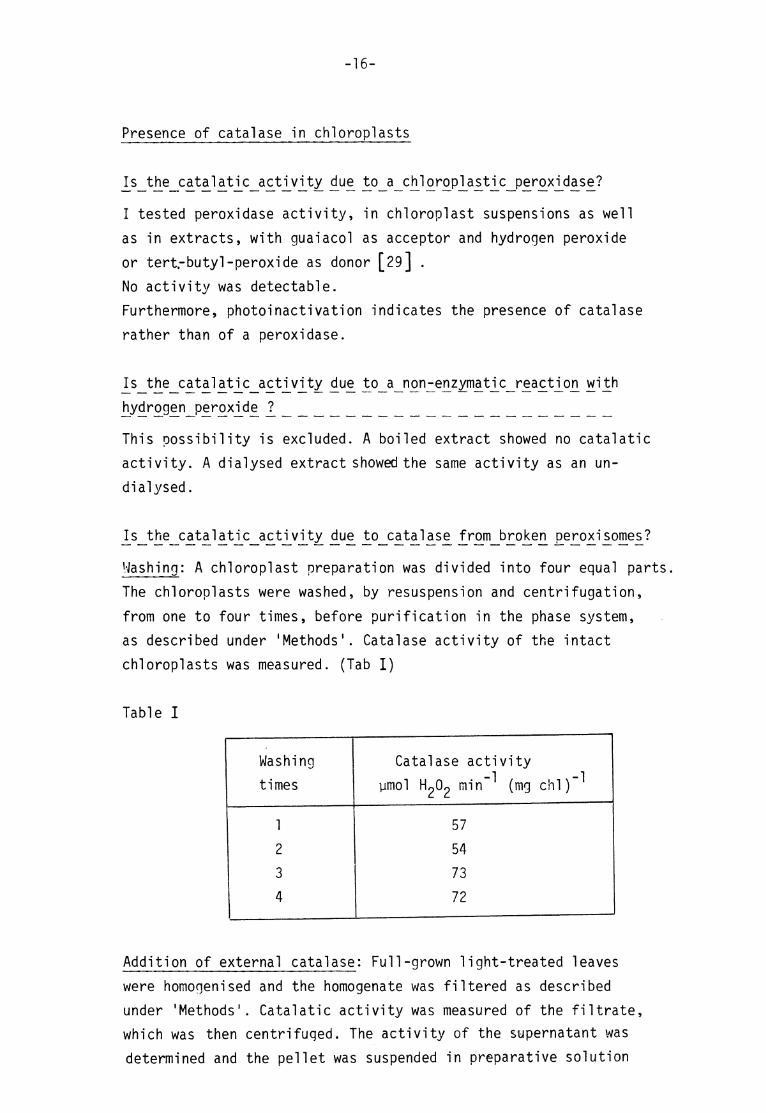
ls_t he_c a_ta 1atjc_a^tj_vity due^ to_c atcnase from_brokeji £eroxi^omes?
Washing: A chlo roplast preparation was divided into four equal parts.
The chloroplasts were washed, by resuspension and centrifugation,
from one to four t imes, before purification in the phase system,
as described under 'Methods' . Catalase activity of the intact chloroplasts was measured. (Tab I )
Table I
Washing Catalase activity t imes ymol min -^ (mg chl)"^
1 57
2 54 3 73 4 72
Addition of external catalase: Full-grown l ight-treated leaves
were homogenised and the homogenate was f il tered as described
under 'Methods' . Catalat ic activity was measured of the f i l trate, which was then centrifuqed. The activity of the supernatant was
determined and the pellet was suspended in preparative solution

-17-
and divided into two equal parts . To one of these parts, bovine l iver
catalase was added. The a ctivity of the chloroplast suspensions was
measured, and the suspensions were then centrifuged. The activity of
the supernatants was determined. The chloroplasts were transferred
to phase systems and extractions with fresh top phase were done three
t imes. Top-phases from the extractions and the supernatants from centri
fugati on o f the diluted bottom-phases were pooled and assayed for
catalase activity.
The pellets consist ing of class I chloroplasts were suspended in
preparative solution and assayed for catalase activity.
Table II
Purification step No a ddit ion
Addition of bovine l iver catalase
total act .x XX spec.act . total act .x spec.act .x x
Filtrate 25000 8250
Supernatant 25000 25400
Chloroplast suspension 247 24 887 85
Supernatant 223 105 926 312
Pooled top-phases and supernatant
24 14 33 24
Intact chloroplasts 4,6 10 4,2 7
x) expressed as ymol min
xx) expressed as ymol min (mg chi) ^
Discussion: As washing d id not decrease the specif ic activity of catalase in intact chloroplasts (Tab I) , the enzyme must e i ther
be found inside the chloroplast or be f irmly bound to i ts envelope (outer membrane).

-18-
The second possibili ty was rejected because of the experiment with
extra catalase added (Tab II) . Most of the activity was removed
from the chloroplasts by c entrifugation. A very small amount was
left in the chloroplast pellet but was removed completely in the
purification of class I chloroplasts with polymer two-phase techni
que. Thus total activity and specific activity were the same,
whether or not the chloroplasts were treated with extra catalase.
The slow 1ight-inactivation of chloroplastic catalase, which will
be shown later, also indicates that the enzyme is inside the
chloroplast , thus being protected against incident l ight.
With the metods used here, much less catalase activity of a spinach
homogenate was found in the intact chloroplasts than in the
chloroplasts prepared with density gradient centrifugation [23,24].
The presence of contaminating peroxisomal components [26] in chloroplast fractions, obtained by d ensity gradient centrifu
gation, could explain the difference.
Time series of chloroplastic catalase activity
_I_n ta ct_c M or o pjasts^ from_d arJ<-treated^ cotyled ons
Preparation of chloroplasts were performed in dim gree n l ight
and as described under 'Methods' . After tempering the chloroplast suspension to 22°C, catalase activity was measured every third
minute. Different chloroplast preparat ions were used for the different t ime series described below.
1. Chloroplast suspension in darkness
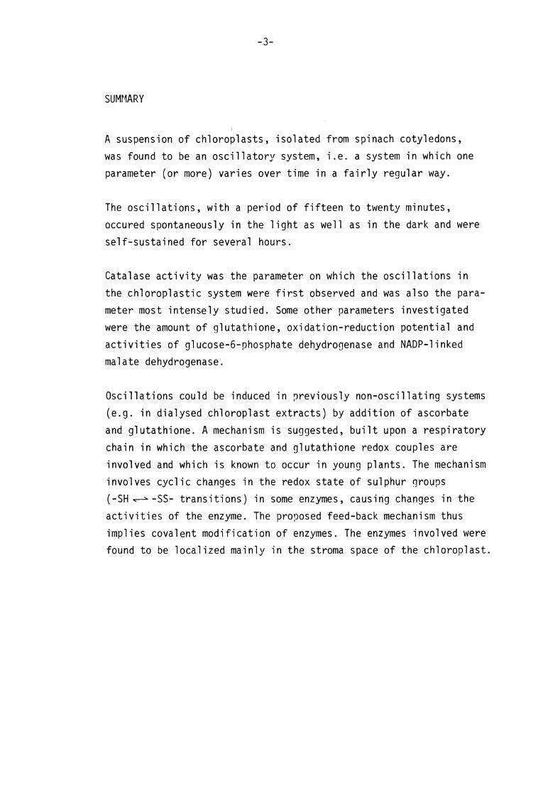
In the experiment shown, (f ig 1) the activity varied between 18
and 25 ymo'l min ^ (mg chl)~jwith a greatest range of variat ion
over a cycle of 6 activity units . There was no trend of increasing
or decreasing activity, nor was there any tendency towards damping
of the oscillat ions.
A visu al est imate of the length o.f Der i od was some 15 to 20 minutes. There were minor activity peaks between the major ones. An inter
pretat ion of this complex pattern is that the oscil lat ion was com
posed by two different simple oscillat ions, one harmonic with a

-19-
period of 7-10 min. , superimposed upon another one with a period of
15-20 min.
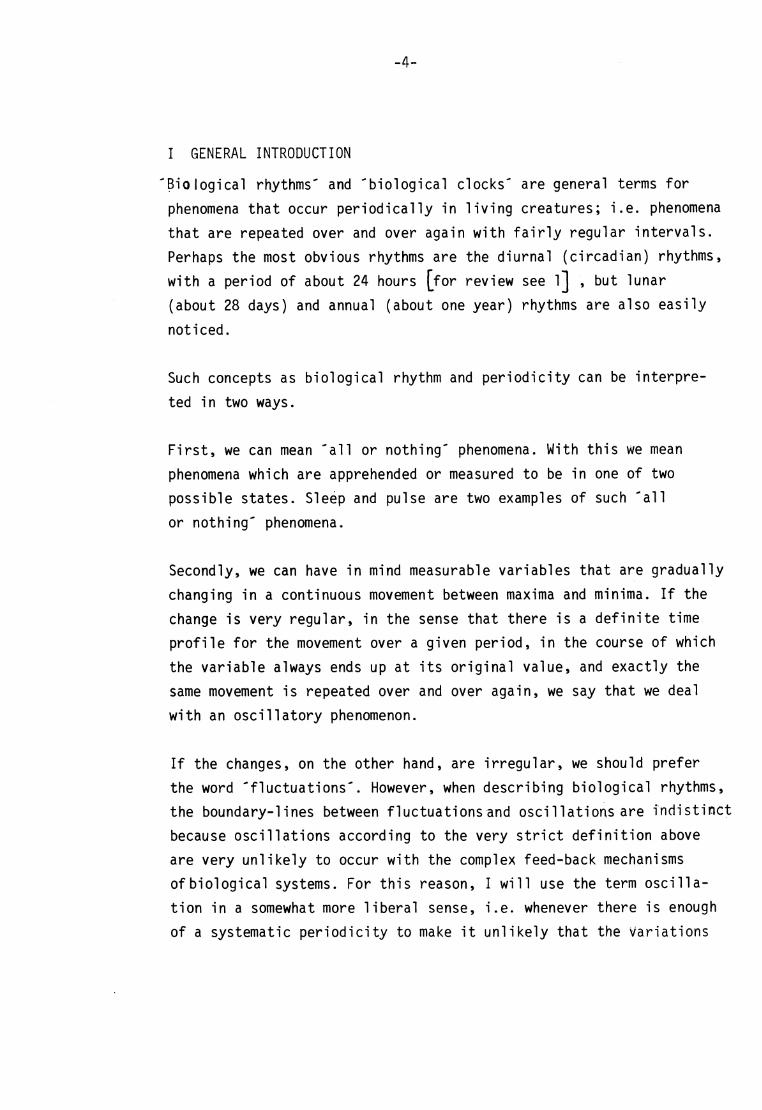
2. Chi oroplast suspension i l luminated.
An example of this is shown in f ig 2. The activity varied between 20
and 40 ymol m i n~^ (m9 chl)"^ • The wider range, as compared
to the experiment shown in fig 1, was due to photoinactivation of the
enzyme. The am plitude of the oscil lat ions, (remaining af ter elimination of the trend present in this case),was about the same as in
the previous experiment (1) and the oscil lat ions had approximately
the same shape and periodicity.
Photoinactivation was usually observed after 30-60 min. of i l lumina
t ion. After this period, a very typical , steep decline in activity
fol lowed. The photoinactivation process proceeded step-wise in an
oscil latory manner. In each 15-20 min. cycle, the maximal activity
value was followed by a great decrease in activity.
In the experiment shown in f ig 2, there were only small oscil lat ions in the activity during the f irst 30 min. of i l lumination. This behaviour is atypical. In other experiments (not shown), oscil lat ions
occured in the entire t ime sequence tested.
Estimation of experimental error_i^ the^ measurement^ of_c atajat2C_
aclnWi ty
1. Two samples were withdrawn from the chloroplast suspension simultaneously and treated as for catalase assay. The cuvettes were
placed in the spectrophotometer in reference and sample posit ions,
respectively. Absorbance was recorded for several minutes and the
greatest inclination of the record was noted. The g reatest inclination obtained in a series of such experiments is called
'maximal experimental error ' . The vertical double arrow in f ig 1
represents the obtained error. The le ngth of the arrow corresponds
to the maximal error for the entire t ime series and equals the width,
20.4 - 21.9 activity units , of the hatched area in the f igure.
Thus the activity of catalase would be expected to vary within this
area, if the variations in activity were due to experimental error
only.

-20-
pmol
J max exp. error
time(min) 90 60 30 10
Fig 1. Time series of catalase activity of chloroplasts, prepared from dark-treated leaves and kept in darkness during the experiment. Vertical arrow indicates maximal experimental error (also shown as hatched area)
1
120 time (min)
Fig 2. Time series of catalase activity of chloroplasts, prepared from dark-treated leaves and illuminated during the experiment. Dotted line indicates that the interval between two points of measurement exceeds three minutes
-21-
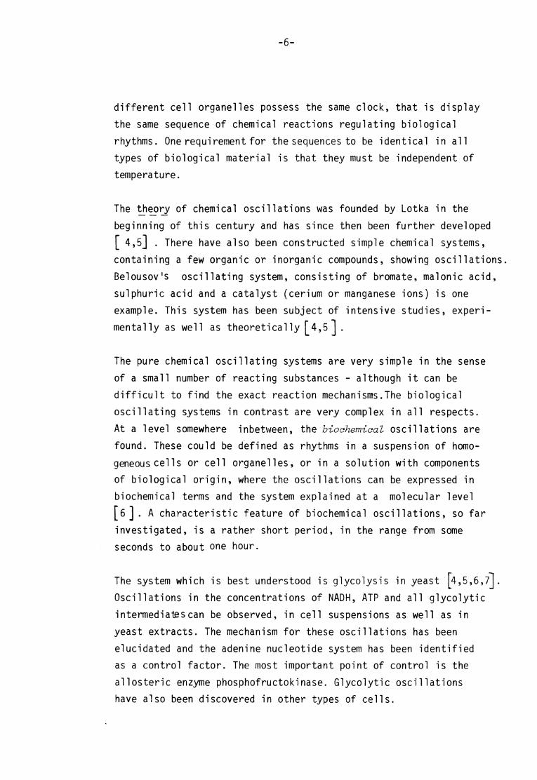
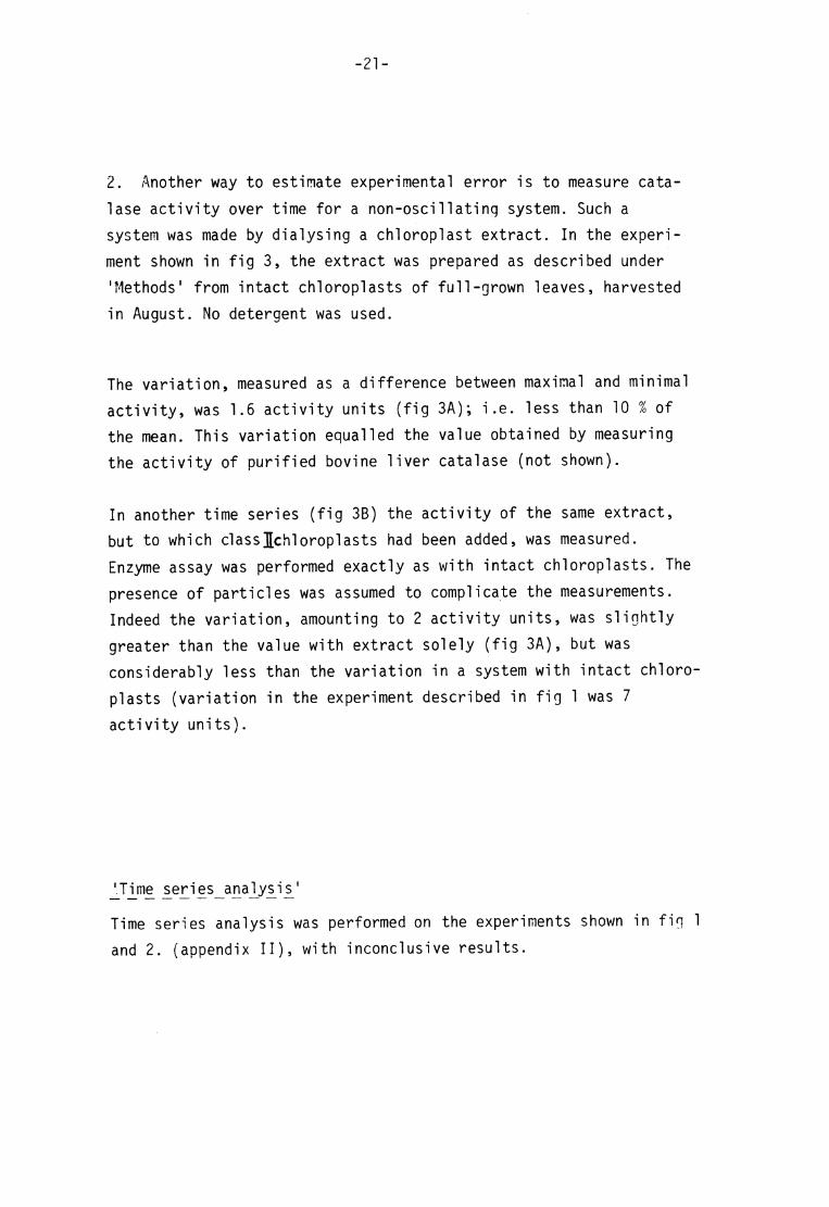
2. Another way t o estimate experimental error is to measure c ata-
lase activity over t ime for a non-oscil lat ing system. Such a system was made by d ialysing a chloroplast extract . In the experi
ment shown in f ig 3, the extract was prepared as described under
'Methods' from intact chloroplasts of full-grown leaves, harvested
in August. No d etergent was used.
The v ariation, measured as a difference between maximal and minimal
activity, was 1.6 activity units (fig 3A); i .e. less than 10 % of the mean. This variation equalled the value obtained by measuring
the activity of purified bovine l iver catalase (not shown).
In another t ime series (f ig 3B) the activity of the same extract ,
but to which class][chloroplasts had been added, was measured.
Enzyme assay was performed exactly as with intact chloroplasts . The
presence of particles was assumed to complicate the measurements.
Indeed the variation, amounting to 2 activity units , was s l ightly
greater than the value with extract solely (fig 3A), but was
considerably less than the variation in a system with intact chloro
plasts (variation in the experiment described in f ig 1 was 7
activity units) .
JJime seri es__ana^ysi s^ '
Time s eries analysis was performed on the experiments shown in f ig 1
and 2. (appendix II) , with inconclusive results .

-22-
prnol pmol
min* H2O2 mirri Ï? rvw
(mgcM^)
20 20
10 10
0 30 time(min) 0 —i » 30 time (min)
Fig 3. Time series of catalase activity of a diatysed chtoroptast extract (A) and of a diatysed chtoroptast extract to which class II chtoroptasts were added (B).
Discussion:
The oscil lat ions in chi oroplastic catalase activity can not be due
to experimental errors solely. The oscil latory pattern is , however,
not perfectly repeated from c ycle to cycle within a t ime series.A
feature which is st i l l more pronounced is that different t ime series experiments have d issimilar oscil latory patterns, although the
period always is the same.
One of the causes of the irregularit ies is that the t ime series of enzyme a ctivit ies are discrete. This is an obvious disadvantage,
escpecially, as in this case, the oeriod of a complete oscil lat ion is short and, due to experimental constraints, there are only a
few - five to seven - measurement points obtainable within each cycle. This means that the exact curve for an individual t ime
series experiment is not completely known.
-23
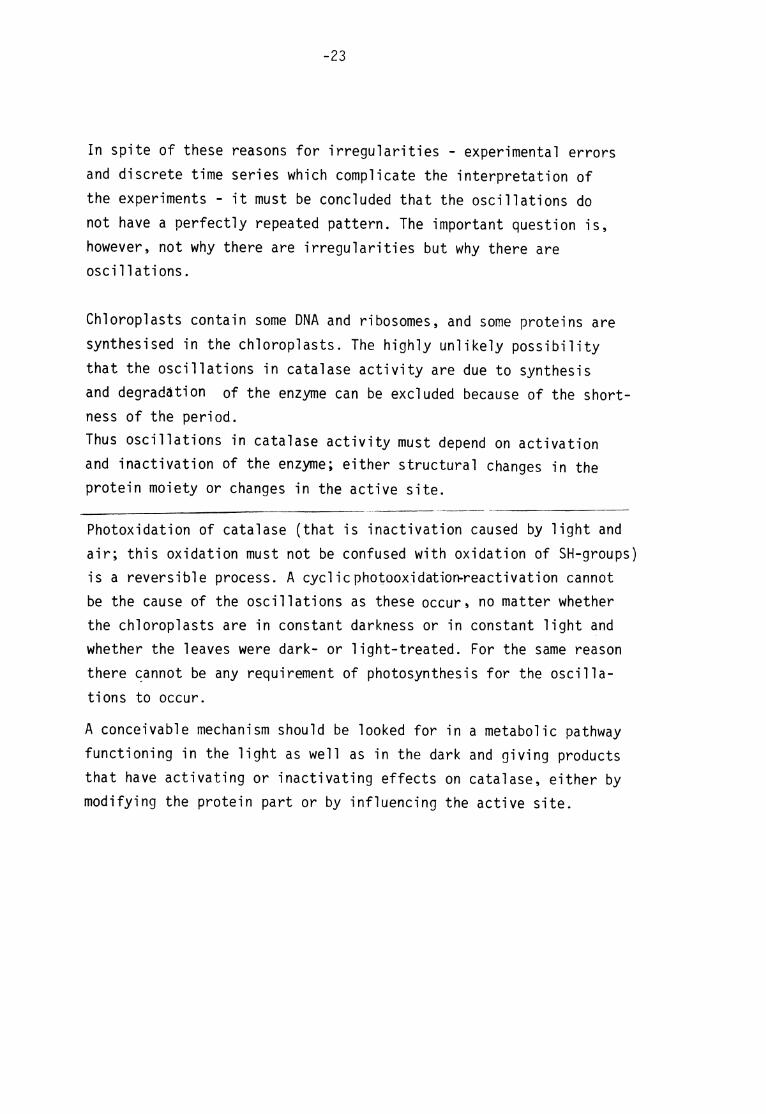
In spite of these reasons for irregularit ies - experimental errors
and discrete t ime series which complicate the interpretation of
the experiments - i t must be concluded that the oscil lat ions do
not have a perfectly repeated pattern. The important question is ,
however, not why th ere are irregulari t ies but why there are oscil lat ions.
Chi oroplasts contain some DNA and ribosomes, and some proteins are
synthesised in the chloroplasts. The highly unlikely possibil i ty
that the oscil lat ions in catalase activity are due to synthesis
and degradation of the enzyme can be excluded because of the shortness of the period.
Thus oscil lat ions in catalase activity must depend on a ct ivation and inactivation of the enzyme; e i ther structural changes in the protein moiety or changes in the active si te.
Photoxidation of catalase ( that is inactivation caused by l ight and
air ; this oxidation must not be c onfused with oxidation of SH-groups) is a reversible process. A cycli cphotooxjdat ion-reactivation cannot
be the cause of the oscil lat ions as these occur» no m atter whether the chloroplasts are in constant darkness or in constant l ight and whether the leaves were dark- or l ight-treated. For the same reason
there cannot be any requirement of photosynthesis for the oscil la
t ions to occur.
A conce ivable mechanism should be looked for in a metabolic pathway functioning in the l ight as well as in the dark and giving products
that have activating or inactivating effects on catalase, ei ther by modifying the protein part or by influencing the active site.

V ON T HE M ECHANISM OF T HE O SCILLATIONS IN C ATALATIC A CTIVITY
In the previous chapter i t was shown that the catalase activity in
intact chi oroplasts from s pinach cotyledons varies over t ime. Some
of the features which should characterize the underlying mechanism
were mentioned. The o bject of the work reported in the present
chapter was to find a possible mechanism of the oscil lat ions.
A cy cl ic oxidation and reduction of -SHgroups of the enzyme was
chosen as a rough working hypothesis. Hermel and Havemann [3o]
have shown that the activity of bovine l iver catalase depends, among other things, on the -SH/SS r atio of the enzyme. They found that , as a rule, the activity increases with decreasing -SH/SS
ratio. A sugg estion of the authors [3l] that the sulphur groups
are involved in the active si te , taking part in the catalatic de
composit ion of hydrogen peroxide, has been cri t icized [32] .
Instead, disulphide groups might be of structural importance [32] .
However, the l i terature dealing with the influence of the -SH/SS
ratio on catalase activity is contradictory. In a study of hetero
geneities in three commercial preparat ions of bovine l iver catalase,
Heidrich found three forms of catalase on ion exchange chromato
graphy, f ive on gel electrophoresis [33] . One form, called fraction
I , corresponded to a -SH form and another (III) to a disulphide
form. Fraction I could be converted to III by o xidation with oxygen,
while the reverse, the reduction of fraction III to I could be done with dithiothreithol but not with mercaptoethanol . The -SH form had
a sl ightly higher specific activity than the -SS-form.
Also catalase from human er ythrocytes separated into three forms on
ion exchange c hromatography, of which one ( the supposed native form)
corresponded to the -SH form and the two others were more o r less
oxidizied forms [34,35] . The oxidized form c ould be reduced with
mercaptoethanol and there was Jlo_dj/ ferenc<5 in activity between the three forms.
-25-
But whatever the truth for catalases of animal origin, my exp eriments
with dithiothreitol treatment of chloroplastic catalase (appendix I)
suggested that the reduced form is le£S_acti_w5 than the oxidized form. Also Schiefer and Kindl found, when studying peroxisomal catalase
from Lens culinari s that treatment with dithiothreitol gives a consider
ably less active enzyme. A s t i l l more pronounced effect was obtained
by mercaptoethanol treatment [36] .
Consequently, my hypothesis is that in the intact chloroplast , catalase alternates between two d ifferent forms, of which one is a more active
di sul phi de form and the other is a less active sulphydryl form.
As catalase is composed by s ubunits, the di sulphide bond c ould be
formed ei ther within each subunit or between different subunits. A
third possibil i ty is that a mixed di sul phi de is formed. Thus
Catalase-S-S-Catalase -» Catalase-SH —* Catalase-S-S-Catalase —*
o r Catalase-S-S-X —* Catalase-SH —> Catalase-S-S-X —>
more a ctive less active more a ctive
To t est this hypothesis , the effects on catalase activity of a naturally
occuring thiol , glutathione (which is the most abundant non-protein
thiol in the cell) and a naturally occuring disulphide, oxidizied
glutathione, were investigated. I also made e xperiments with other
substances which could be involved in or interfere with the oscil la
t ion mechanism. Attempts were also made to reconstitute the oscil la
t ions.
Effects_of reduced glutathione^ ox i_d iz ed _g ]u ta th i_o ne as c orb at e and
^-^thylm£le^imide_oni cataJase_ac_tWrty in_ a_dial_yse<j £hl_oro£last^ extract^
A chl oroplast extract was dialysed in a cold-room over night against
0.1 M potassium phosphate buffer , pH 7. 0. The extract was protected
against l ight. During the experiments, al l enzyme s olutions were kept
in darkness at room tem perature. Additions to the extracts were made
according to table III and catalase activity was assayed. (For experi
ment shown in tab III , leaves harvested in July were used)

-26-
Table III
Activity expressed as ymol H2O2 min 1 (mg chi) 1
Time a f ter
addit ion
(min)
No
addit ion
(control)
GSH
50 yg ml 1
(0.17 mM)
GSSG
100 yg ml 1
(0.17 mM)
Ascorbatex)
100 yg ml 1
(0.5 mM)
NEM X X )
125 yg ml"1
(1.0 mM)
10 36 27 36 23 35
30 37 20 35 28 31
60 34 18 35 29 30
120 35 17 33 28 26
x) Values corrected for hydrogen peroxide decomposit ion by as corbate xx) N-ethylmaleimide
Redu£ed^ g liJta^tMone^ in the concentration used had a pronounced in
hibitory effect on catalase in the dialysed chloroplast extract .
This was expected as i t is well known that thiols inhibit catalases
[37,38] . An a ccepted explanation for this inhibit ion is that thiols by slow autoxidation generate hydrogen peroxide which, instead of
being decomposed by catalase, forms an inactive complex wit h the
enzyme, called compound II.
To a scertain whether the inhibitory effect shown in table III was
due to reduction of disulphide groups of the enzyme o r due to
formation of compound II , or both, the same experim ent was repeated
on extracts containing, together with GSH, 1 mM ethanol or 10 mM EDTA. EDTA pre vents autoxidation of thiols, while ethanol counter
acts formation of complex II [38] . Neither EDTA nor ethanol prevent
ed or even reduced the inactivating effect of GSH on chi oroplast ic catalase.
For a subsequent discussion of other metabolites involved in the regulation of catalase activity, i t can be of interest to note that
catalase in a dialysed extract seemed to be more sensit ive to GSH
treatment than catalase in an undialysed extract . (Not shown in table III)

-27-
0xj[djzed_ £l£tatjpone^ on the other hand, had in this experiment no effect on catalase activity. If the enzyme was in the di sulphide
form when GSSG w as added to the extract , no e ffect was expected. How
ever, when catalase was f irst transferred to sulphydryl form, by
treatment with 0.3 mM GSH in presence of 1 mM ethanol, and thus was
inactivated to about 50 %, i t was possible to reactivate the enzyme to 75 % of the original activity by a ddit ion of GSSG (e quimolar
amount relative to GSH). This effect of GSSG on chloroplastic cata
lase should be compared to results from ex periments on bovine l iver catalase. Seabra and Deutsch[39] found no e ffect of cystine, while
Pi hi et al [38] found, during the f irst twenty minutes af ter the addit ion of cyst ine, an inhibi tory effect . I t was concluded that
this inhibit ion was due to autoxidation of cysteine, delivered
during oxidation of sulphydryl groups of catalase. Catalase formed
a mixed di sul phi de with cystine.
SH SH HS
^ Catalase HS / I I
S —S SH
+ XSSX
SH SH SH
XSS
XSS
ssx Catalase" + XSH I I x SSX S — S
[ss]
Cysteine generated H2O2, which thus gave r ise to Complex II . No
inhibition, and no a ctivation, was noted in the presence of ethanol
or EDTA. I tested the effect of GSSG, in the presence of 1 mM MEM, which is known to block -SH gro ups,on bovine l iver catalase and
found an activating effect (120 % of the control value).
From th ese experiments i t is concluded that the-SH/SS ratio is essential for the regulation of chloroplastic catalase activity.
In tab III, also results from e xperiments with ascorbate and N-
ethylmaleimide are included. (Effects of these compounds will be
discussed more closely in connection with experiments on intact
chloroplast) . In the experiments described here ^ .^o^ate.
immediately decreased catalase activity with about 40 % . However, the inhibitory effect was spontaneously part ly abolished and within
30 min. , the activity raised to 80 % of the control value.

-28-
A compari son with experiments on catalase of animal origin [40J
shows that the course of inactivation is quite different in this
case with chloroplast extract .
NEM which blocks -SH g roups, had a slowly inactivating effect . In
experiments on bovine l iver catalase, I found no inactivation. But
as the chloroplast catalase experiment was made w ith an extract ,
i t is impossible to know whether the inactivation is due to blocking
of essential -SH grou ps of the enzyme o r due to some secondary effect .

-29-
Effects of ascorbate on catalase activity in intact chi oroplasts
Ascorbate is a known inhibitor of catalase. Chance [4lJ showed that ,
as ascorbate slowly generates hydrogen peroxide during autoxidation,
the inhibit ion is due to formation of the inactive catalase
the inhibit ion, under specific conditions, is due to free radical
attack on the protein, which is degraded to smaller peptide fragments
I noted that the inhibit ion of catalase in a chloroplast extract
with ascorbate was highly reversible (Table III) . Is this reversible
inactivation also found for catalase in intact chloroplasts?
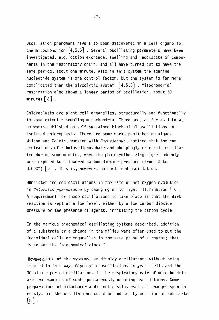
1. Dark-treated leaves. Chloroplasts in the dark.
Catalase activity of a chloroplast suspension, to which no ascorbate
had been a dded, was measured in the usual way dur ing one hour, in
order to make s ure that the system displayed oscillatory catalase
activity. The later part of this t ime series, with an average cata
lase activity of 26.4 ymol min"^ (mg chl)~\ is shown in f ig 4.
At t=70 min, ascorbate was added to the suspension, giving 100 ug
ascorbate ml"^ . A 40 % inhibit ion, which agreed well with the in
hibitory effect found in extracts , treated in the same way ( e.g.
the experiment shown in table III) , could be noted at the f irst
point of measurement. In this case with intact chloroplasts , however,
there was no tendency towards spontaneous reactivation. The activity
continued to oscillate around the lower activity; in average 17.9
activity units .
other hand, demonstrated that
-30-
120 ISO tim* (min)
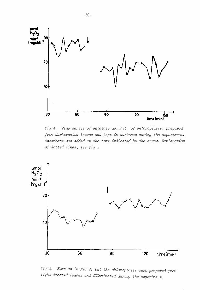
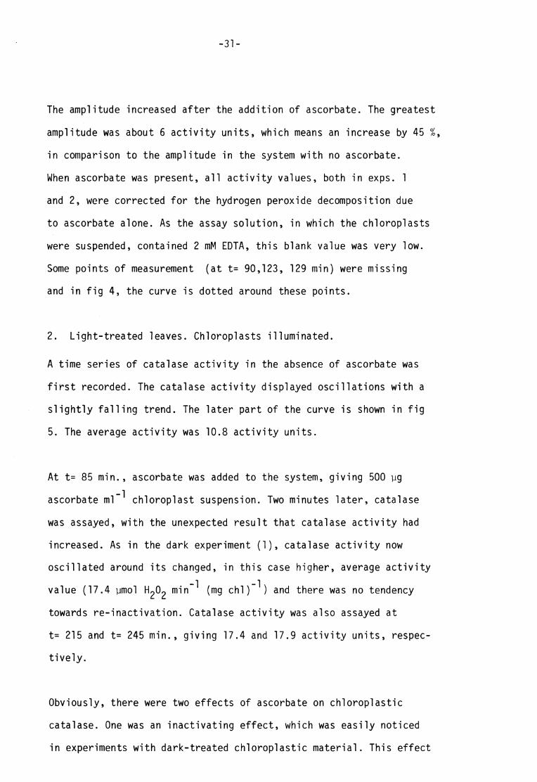
Fig 4. Time series of catalase activity of chloroplasts3 prepared from darktreated leaves and kept in darkness during the experiment. Asaorbate was added at the time indicated by the arrow. Explanation of dotted lines, see fig 2
jjmol
120 time(min)
Fig 5. Same as in fig 4, but the chloroplasts were prepared from light-treated leaves and illuminated during the experiment.
-31-
The am plitude increased after the addit ion of ascorbate. The greatest
amplitude was about 6 activity units, which means an increase by 45 %,
in comparison to the amplitude in the system with no a scorbate.
When a scorbate was present, al l activity values, both in exps. 1
and 2, were corrected for the hydrogen peroxide decomposit ion due
to ascorbate alone. As the assay solution, in which the chloroplasts
were suspended, contained 2 mM EDTA, this blank value was very low.
Some points of measurement (at t= 90,123, 129 min) were m issing
and in fig 4, the curve is dotted around these points .
2. Light-treated leaves. Chloroplasts i l luminated.
A t ime series of catalase activity in the absence of ascorbate was
f i rs t recorded. The c atalase activity displayed oscil lat ions with a
sl ightly fall ing trend. The later part of the curve is shown in f ig
5. The average activity was 10.8 activity units .
At t= 85 min. , ascorbate was added to the system, giving 500 yg
ascorbate ml~^ chloroplast suspension. Two minutes later, catalase
was assayed, with the unexpected result that catalase activity had
increased. As in the dark experiment (1), catalase activity now
oscil lated around i ts changed, in this case higher, average activity
value (17.4 ymol ^02 min~^ (mg chl)"^) and there was no tendency
towards re-inactivat ion. Catalase activity was also assayed at
t= 215 and t= 245 min. , giving 17.4 and 17.9 activity units, respec
t ively.
Obviously, there were two e ffects of ascorbate on chloroplastic
catalase. One wa s an inactivating effect , which was easily noticed
in experiments with dark-treated chloroplastic material . This effect

-32-
should also be taken into account for l ight-treated chloroplastic
material . The other was an activating effect , which could be
noticed only for l ight-treated chloroplasts.(Does ascorbate reverse
the photoxidation of catalase^ In the presence of ascorbate, cata-
lase activity was found at the same level, about 17-18 activity
units, both in experiment 1 and 2. In experiment 1, however, the
amplitudes of the oscillat ions were mu ch g reater than in experiment
2 .
In at tempts to reactivate photoxidizied catalase in dialysed and
undialysed extracts, which were treated with different amounts o f
ascorbate in the presence or the absence of methanol or EDTA, the
results obtained were ambiguous and diff icult to interpret . The
inconclusive results could be part ly explained by the competit ion
between an activating and an inactivating effect of ascorbate, but
the problem d eserves a deeper study, which is beyond the scope of
the present one.
In this context, the main purpose of studying the effect of ascorbate
on catalase in chloroplasts was to find out whether ascorbate might
be involved in the mechanism underlying the oscil lat ions in catalase
activity. The r esults indicate that ascorbate is one possible re
gulator.
Effect _°f N - e t hy Imefl e i mi de_on catalase_activit^ in_intact chloro^.
pla_sts _
Previously, I have suggested that a cyclic oxidation and reduction
of -SH groups in catalase can be a cause of the oscilla t ions in
enzyme a ctivity. The -SH/SS r atio of the enzyme is assumed to be
determined by the -SH/SS r atio in the environment, and this ratio
-33-
in i ts turn is assumed to be ma inly determined by low molecu lar weight
thiols and disulphides, e.g. reduced and oxidized glutathione. Thus,
the oscil lat ions should cease, i f a -SH blocking agent was added to
the system.
Desired properties of such an agent are: a) i t should be able to
penetrate the chloroplastic outer membrane ( the envelope), without
damaging the membranes; b) i t must be specific for -SH g roups; c)
the adduct formed with thiols must be s table; d) the agent must be
stable in the system tested.
Of known -SH a gents, one of the most specific ones, namely N-ethyl-
maleimide (NEM), seemed to be the most promising one f or my ex periments,
as the agent is known to penetrate the erythrocyte membrane, without
affect ing the survival of the cells ^43,44^ .
As thiols, e.g. GSH, are strongly nucleophil ic agents, they can add
to the C=C bond of NEM in the following manner:
S 0 N II C C
HC ^ N - CH2-CH3 + GSH > 1 N-CH2-CH3
^ / H " /
S /S " NEM 0 G ' 0
giving a product of glutathione in sulphide form, and N-ethylsuccin-
imide.
There are disadvantages with this -SH a gent. I t is not very stable
in water solutions ^45^ . The addition of GSH to NEM has in experi
ments with rabbit red cells and hemolysates turned out to a reversib
le reaction j 441 }
-34-
suggesting that the desired property c) is not always fulfi l led.
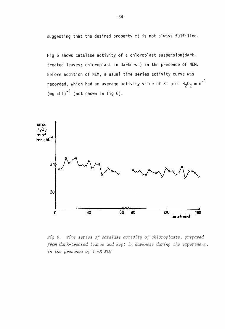
Fig 6 shows catalase activity of a chloroplast suspension(dark-
treated leaves; chloroplast in darkness) in the presence of NEM.
Before addit ion of NEM, a usual t ime series activity curve was
recorded, which had an average activity value of 31 ymol min ^
(mg chl)"^ (not shown in f ig 6).
pmol H202 mir»*1
(mg chi)
120 ISO
Fig 6. Time series of catalase activity of chloroplasts, prepared from dark-treated leaves and kept in darkness during the experiment, in the presence of 1 mM N EM
-35-
At t= O N EM was added to the suspension to give a concentration
of 1 mM. After about 30 min, a small decrease in activity was noted,
which was in agreement with the effect of NEM on catalase of a
dialysed chloroplast extract (Table III) . However, in this case,
the enzyme was not further inactivated. The amp litude was some
what decreased after a couple of hours, but i t is quite obvious
that the oscil lat ions did not cease in the presence of NEM. Micro
scopic observations on the suspension after the experiment showed
that the previously intact chi oroplasts were destroyed by the NEM-
treatment. However, intactness is not a requirement ' f or oscilla t ions
to occur*, rather i t should be an advantage for this experiment, i f
al l thiols and -SH enzym es were in solution, easily avai lable for
the -SH a gent.
Thus,either the hypothesis that thiol groups are involved in the
mechanism of the oscilla t ions is wrong, or NEM and/or the adducts
of NEM and -SH com pounds a re highly unstable under these conditions.

-36-
Reconsti tution of oscillat ions in chi oroplast ic catalase activity
As the attempt to cease the oscil lat ions in catalase activity of
chloroplasts fai led, a reverse experiment i .e. to induce oscil lat ions
in a previously non-oscil lat ing chloroplastic system, was performed
to test the hypothesis that -SH*-*SS t ransit ions are involved in
the mechanism, underlying the oscil lat ions.
The exp eriments on catalase activity in a chloroplast extract , to
which g lutathione or ascorbate had been added, as well as the
experiments on catalase activity of intact chloroplasts in the
presence of ascorbate, strongly suggest that glutathione and ascor
bate are possible regulators of catalase activity. The reduced forms
of the two s ubstances have inactivating effects, but the course of
inactivation differ greatly. Furthermore, i t is known that reduced
glutathione reduces oxidized ascorbate (dehydroascorbate) [46,47J .
2 GSH + DHA —> GSSG + AA
This reaction is catalysed by an enzyme, glutathione dehydrogenase,
(glutathione: dehydroascorbate oxidoreductase, E.C. 1.8.5.1.), which
is found in plant t issues ^46,47^ .
Is i t possible to s tart the oscil lat ions, simply by adding these
metaboli tes to a non-oscil lat ing chloroplastic system?
A dialy sed chloroplast extract , with a catalase activity of 25 ymol
H2O2 min (mg chi) , was used for the experiment. At t= 0 min. ,
ascorbate, GSH an d GSSG wer e added to the extract , to give 100, 50
and 100 yg ml"" ' , respectively.

-37-
The f irst sample was taken half a minute later and assayed for
catalase activity, and a 40 % decrease in activity was noted
(f ig 7). Minimal activity, 11.9 activity units, was reached at
t= 6 min. , after which the activity increased. Maximal activity
was reached at t= 93 min. (27.3 activity units) , whereupon a
decreasing trend was noticed again. The f i rst high frequency
oscil lat ion was observed af ter about 30 min. , and the amplitude
reached i ts maximal value after about one hour.
t max exp. error
90 time (min) 60 30 0
Fig 7. Time series of catalase activity of a dialysed chloroplast extract, to which GSSG3 GSH and ascovhate were added at t—0 min

-38-
The experiment showed that i t was possible to start the oscil lat ions
in catalase activity by a ddit ion of the glutathione redox couple and
ascorbate. However, the oscil latory curve obtained was dist inguished
from cu rves obtained in experiments with intact chloroplasts. Further
more, in reconsti tution experiments on other extracts, differing from
the extract in the described experiment as regards pretreatment of the
leaves and protein concentration in the extract , the oscillatory
curves obtained were divergent from the pattern of the curve shown in
f ig 7. The amplitude as well as the t ime needed for reactivation were
dissimilar for the different experiments.
There were a lso features in common for the different experiments.
The rapid decrease in catalase activity, reminding of the effect
of ascorbate solely, was found in al l experiments. Also the f irst
marked oscillat ion in activity was noted about half an hour after
the addit ions were made.
The glutathione and ascorbate redox couples thus seem to be impor
tant metabolites for the mechanism underlying the oscil latory
catalatic activity in the chloroplast extract .
DISCUSSION
On_regij lat20ii i_n_general_of <:ata]_ase_iin chjoroplasty
The enzyme was inactivated in the presence of reduced glutathione.
This inactivation was not due to complex II formation. As the
inactivated enzyme wa s part ly reactivated when oxidized gluta
thione was added, the -SH/SS ratio of the enzyme seems to be of
importance for determining the activity of the enzyme. Thus, i f
this interpretation is correct , the chloroplastic catalase should
be an interconvertible enzyme, i .e. an enzyme of which the
activity is regulated by a covalent modification of the protein

-39-
(for reviews on covaient modification, see ref 48 and 49).
Regulation by c ovalent modification of sulphur groups is known f or
is activated by r eduction of a disulphide group to sulphydryl
groups. For xanthine oxidase (from rat l iver) , a change in -SH/SS
ratio of the enzyme me ans a change in substrate specifici ty. The
enzyme in sulphydryl form is a dehydrogenase, in disulphide form
an oxidase.
I t has also been proposed [48,5o] that covalent modification of
sulphur groups is a way of regulating the activities of several
chloroplastic enzymes. Many of the chloroplastic enzymes, e.g.
some enzy mes in the Calvin cycle, are in vivo stimulated by l ight
and i n vitro activated by d ithiothreitol [50] . These enzymes are
thus more active in the -SH form than in the -SS-form. On the other
hand, glucose-6-phosphate dehydrogenase, which i n vivo is inacti
vated by l ight, is in vitro inactivated by d ithiothreitol |^5o] .
This enzyme is the key enzy me f or the oxidative pentose phosphate
cycle, which is a metabolic pathway supposed to dominate in non-
photosynthesizing chioroplasts .
I t is not known how t he modif ication of sulphur groups in the
chloroplastic enzymes occur. Hatch and Turner found in pea seeds
an enzyme, a protein disulphide reductase, which uti l izied NADPH
•sfc) Of course there are several enzymes which are dependent on -SH groups for activity, but this does not mean that
al l such enzymes in vivo are regulated by co valent modi
f ication.
two o ther enzymes Pyruvate formate lyase from E. coli

-40-
to reduce protein -SS-groups [5lJ . Another, perhaps more possible
mechanism, as i t would explain both the reduction step and the
oxidation step, is that the transit ion occur via thiol-disulphide
exchange, in which low molecu lar weight sulphur compounds are
involved. At physiological pH, thiol-disulphide exchange o ccurs
spontaneously, but the participation of an enzyme, a thiol trans
ferase [52] , seems more probable.In analogy with other interconver
t ible enzyme s ystems, the thiol transferase should be called the
'converter enzyme ' . Converter enzymes are usually in their turn
regulated by more o r less complicated mechanisms [48,49] .
Thus, the low a ctivity of catalase found in chloroplasts isolated
from l ight-treated leaves is due part ly to the photoxidation of
the enzyme and partly to a high -SH/SS r atio of the enzyme.
Xanthine oxidase changes substrate specifici ty when the -SH groups
of the enzymeare conve rted to disulphide form. Does also catalase
change i ts specifici ty, from catalatic activity in the disulphide
form to peroxidatic activity in the sulphydryl form? [53] . As shown
in chapter IV no peroxidatic activity was detectable in the chloro-
plast ic systems. This result does not, however, exclude the possi
bil i ty of a changed substrate specifici ty. In al l experiments, de
scribed in chapter IV, guaiacol was used as acceptor. Other sub
strates should be tested.
#) Professor Anatole Purmal suggested me this possibil i ty

-41-
The results from the experiments, in which ascorbate was added to
chloroplast extracts or chloroplast suspensions, indicate that also
ascorbate could be a regulator of catalase. As regards the inacti
vating effect , the course of the inactivat ion - a very rapid
decrease in activity immediately after the addit ion of ascorbate
followed, in the experiments with extracts , by a spontaneous reacti
vation - suggests that the effect is neither due to complex II
formation, nor to free radical attack. The mec hanism is obscure.
If ascorbate could reduce -SS- groups of the enzyme d irectly, the
inactivating effect could be explained by an increased -SH/SS r atio
in the enzyme; the inactivating effect of ascorbate would thus be
the same a s the inactivating effect of GSH. However, al though the
oxidation of GSH or protein -SH groups by dehy droascorbate occurs
readily ^47^ , the reverse reaction is not known to take place.
The regulatory properties of ascorbate on catalase are st i l l more
confusing when also the effects of ascorbate on catalase in chloro-
plasts Ttght-treated leaves are taken into account. One spe cu
lative hypothesis already mentioned is that ascorbate reverses the
photoxidation effect in some way. Another, not less speculative,
hypothesis is that catalase in chi oroplasts from l ight-treated
leaves, predominantly in -SH fo rm, does have a changed substrate
specifici ty; i .e. a peroxidatic activity, using ascorbate as sub
strate. Thus, in the experiment shown in f ig 5, decomposit ion of
hydrogen peroxide due to remaining catalatic activity was measured
before the addition of ascorbate. After the addit ion, decomposit ion
of hydrogen peroxide due to an even more decreased catalatic activity
plus decomposit ion of hydrogen peroxide due to a much higher peroxi-

-42-
datic activity (HgC^ + AA-t H^O + DHA) were measured. The concentra
t ion of ascorbate in the chloroplast suspension was high enough to
give a reasonable substrate concentration in the assay solution.
Of course, the same result would be obtained if the peroxidatic acti
vity was due to a ' true ' peroxidase, that is not the modified cata-
lase, but a l ight-st imulated peroxidase.
From a teleologica! point of view, the hypothesis of a l ight-st imula
ted peroxidatic activity is very at t ractive. If catalase, with cata-
lat ic activity solely, was the only hydrogen peroxide decomposit ing
enzyme in the chloroplast , the capacity of protection against hydrogen
peroxide would be much less in the l ight than in the dark. The
photosynthesizing chloroplast , which has a high oxygen pressure and
in which hydrogen peroxide can be produced from e.g. photosystem
I in the absence of NADP, certainly 'needs ' this protection more
than the chloroplast in the dark. A l ig ht-st imulated peroxidase
could thus be the desired protective agent.
Or^th^oscil 2ator7_mecha£i sm
The r esults from the experiments with reconsti tution of oscil lat ions
in catalase activity strongly indicate, that the glutathione and
ascorbate redox couples are of importance for the oscil lat ions in
activity.
To si mplify the discussion I will f i rst assume that the catalase
activity is regulated only by the -SH/SS ratio of the enzyme and
the ascorbate/dehydroascorbate rat io in the environment. Each o f
these ratios is assumed to display a simple, sinusoidal oscil lat ion.
-43-
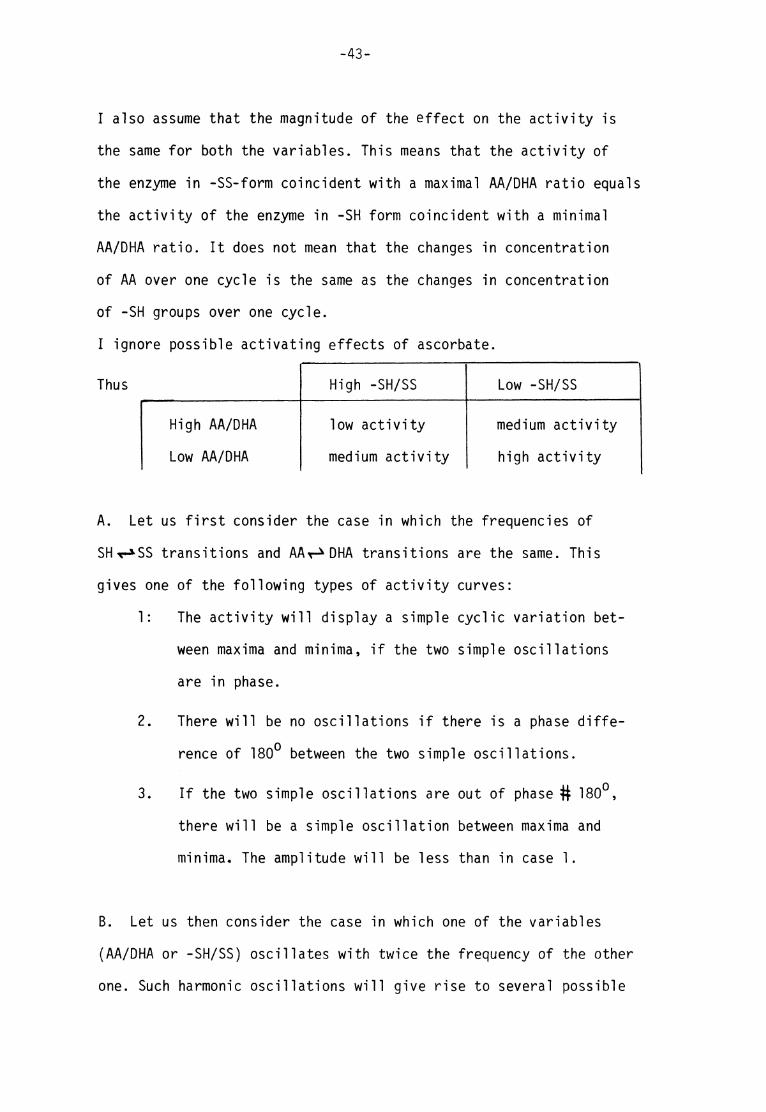
I also assume that the magnitude of the effect on the activity is
the same f or both the variables. This means that the activity of
the enzyme in -SS-form coincident with a maximal AA/DHA ra t io equals
the activity of the enzyme i n -SH form coincident with a minimal
AA/DHA r at io. I t does not mean that the changes in concentration
of AA over one cycle is the same as the changes in concentration
of -SH gr oups over one cycle.
I ignore possible activating effects of ascorbate.
Thus High -SH/SS Low -SH /SS
High AA/DHA low a ctivity medium a ctivity
Low A A/DHA medium a ctivity high activity
A . Let us f i rst consider the case in which the frequencies of
SHv-*SS transit ions and AAr-^DHA transit ions are the same. This
gives one of the following types of activity curves:
1: The activity will display a simple cyclic variation bet
ween ma xima and minima, i f the two si mple oscillat ions
are in phase.
2. There will be no o scil lat ions if there is a phase diffe
rence of 180° between the two sim ple oscil lat ions.
3. If the two sim ple oscil lat ions are out of phase # 180°,
there will be a simple oscillat ion between maxima and
minima. The amp litude will be less than in case 1.
B. Let us then consider the case in which one of the variables
(AA/DHA or -SH/SS) oscillates with twice the frequency of the other
one. Such harmonic oscil la t ions will give r ise to several possible

-44-
shapes of the activity curve, but never a non-oscil lat ing curve.
1. One extreme case is when the minimal value of the
oscillat ion with lower frequency coincides with one
of the minima of the higher frequency oscil lat ion.
As a consequence, the maximal value of the lower
frequency oscil lat ion coincides with the second
minimum of the higher-frequency oscil lat ion.
The sha pe of one cycle will be:
Thus within one cycle there are
two ma ximal values at the same
level and two minima l values at
very different levels. The curve
is symmetrical .
2. The other extreme case is when the maximal value
of the lower frequency oscil lat ion coincides with
one of the maximal values of the other simple
oscillat ion. Thus the minimal value of the lower
frequency oscil lat ion will coincide with the second
maximal value of the higher frequency oscil lat ion.
The shape of one cycle will be:
Thus within one cycle there are
two mini mal values at the same
level and two max imal values at
very different levels. The curve
is symmetrical .
-45-
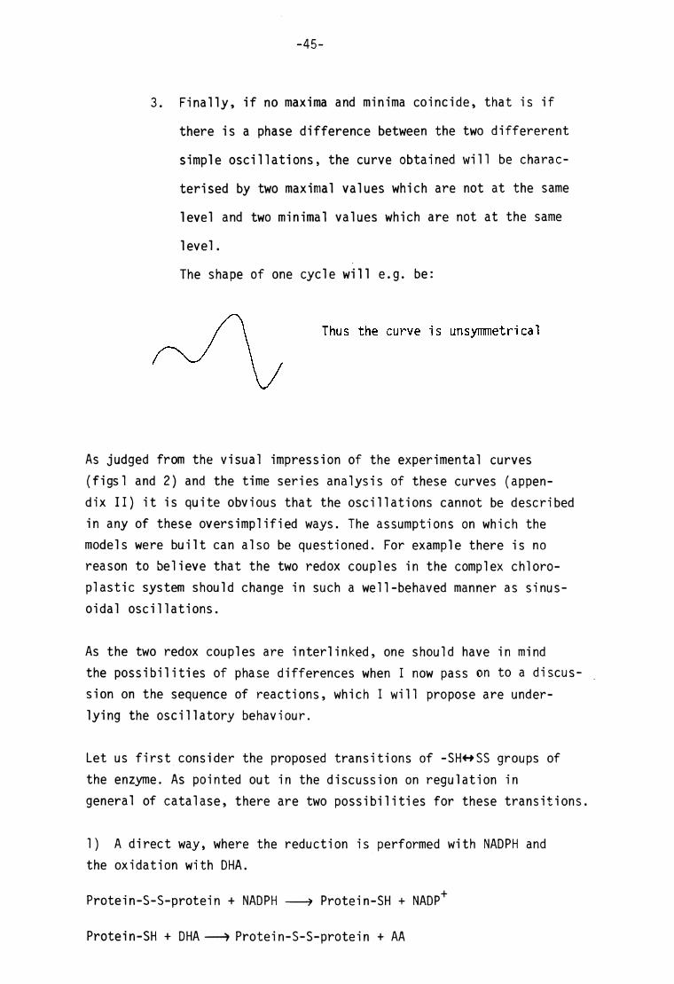
3. Finally, i f no ma xima and minima coincide, that is if
there is a phase difference between the two d iffererent
simple oscil lat ions, the curve obtained will be charac
terised by two max imal values which a re not at the same
level and two minima l values which are not at the same
level.
The shape of one cycle will e.g. be:
Thus the curve is unsymmetrical
As judged from the visual impression of the experimental curves
(f igsl and 2) and the t ime series analysis of these curves (appen
dix II) i t is quite obvious that the oscil la t ions cannot be d escribed in any of these oversimplified ways. The assum ptions on which the
models were buil t can also be questioned. For example there is no reason to believe that the two redox couples in the complex chloro-
plast ic system should change in such a well-behaved manner as sinusoidal oscil lat ions.
As the two redox couples are interl inked, one should have in mind the possibili t ies of phase differences when I now pa ss on to a discus
sion on the sequence of reactions, which I will propose are underlying the oscillatory behaviour.
Let us f i rst consider the proposed transi t ions of -SH<*SS groups of
the enzyme. As pointed out in the discussion on regulation in
general of catalase, there are two possibil i t ies for these transit ions.
1) A di rect way, where the reduction is performed with NADPH an d the oxidation with DHA.
Protein-S-S-protein + NADPH > Protein-SH + NADP+
Protein-SH + DHA—» Protein-S-S-protein + AA
-46-
The reductase ( the protein disulphide reductase reported by Hatch
and Turner or glutathione reductase - the two enzymes m ight actually
be the same [54] ) and the glutathione dehydrogenase must both be
able to use catalase as substrate. As writ ten here, the -SS-form of the
enzyme shou ld coincide with a high AA/DHA r at io.
2) An indirect way, with the part icipation of glutathione or other
low molecu lar weight sulphur compounds, i . e. a thiol-disulphide
exchange probably catalysed by a thiol transferase. Thus the GSH/
GSSG r at io must vary over t ime and the -SH/SS r atio of the enzyme
reflects the GSH/GSSG r at io. With this mechanism, the enzyme in disulphide form might be a mixed disulphide and this form might not be coinci
dent with a high AA/DHA r at io. The two mechan isms do not differ in
principle and I will not exclude the possibili ty that both are involved in the -SH*—»SS t ransit ions. However, I favour the second
one and the following discussion concerns that mechanism.
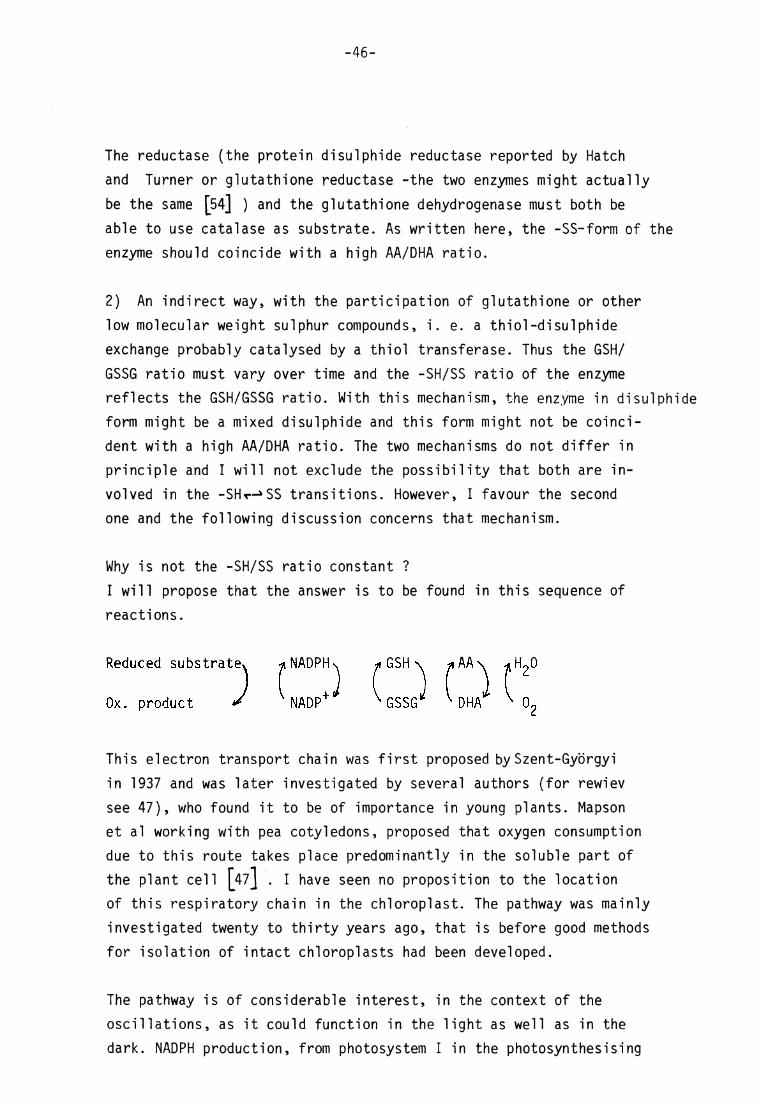
Why is not the -SH/SS r atio constant ?
I will propose that the answer is to be found in this sequence of
reactions.
Reduced substrat" m«™,. ^
This electron transport chain was f irst proposed by Szent-Györgyi
in 1937 and was later investigated by s everal authors (for rewiev
see 47), who found i t to be of importance in young plants. Mapson et al working with pea cotyledons, proposed that oxygen consumption
due to this route takes place predominantly in the soluble part of
the plant cell [47] . I have seen no proposit ion to the location of this respiratory chain in the chloroplast . The pathway was mainly
investigated twenty to thir ty years ago, that is before good methods
for isolat ion of intact chloroplasts had been d eveloped.
The pathway is of considerable interest , in the context of the
oscil lat ions, as i t could function in the l ight as well as in the
dark. NADPH pro duction, from photosystem I in the photosynthesising
Ox. product

-47-
chloroplasts , from substrates emanating from starch in the dark,
together with oxidation of ascorbate are the driving forces.
I t should be borne in mind that in the chloroplast there are several
pathways by which NADPH, GSH an d AA are oxidized. This means that
the GSH/GSSG and AA/DHA r atios could change (decrease) in a rather
unpredictable way. However, there is some point of control which
makes the period of the oscil lat ions fairly constant.
An idealised model for oscil lat ions due to changes in the ratios of
the two redox couples, bui I t upon the assumptions that some of the
enzymes involved in the electron transport chain are sensi t ive to
-SH/SS r at io, is the following:
HS-Enzymes- | 234
-S-S-Enzymes-j 234
NADPH
H2O
Ox. product
E-,(SH?)
i \¥S H ?)
DHA 02 ,H202
Red. substrate
HS-enzymes, 0 ~ , and -S-S-enzymes _ s tand for i jL ) J )4 ) « « . n
al l the chloroplastic enzymes which have sulphur groups that are

-48-
reducable and oxidizable via thiol-disulphide exchange. Some of the
enzymes might be more a ct ive in -SH-form while others might be more
active in -SS-form.
E-j and E2 stand for the ascorbate oxidizing enzymes^ primarily ascor-
bate oxidase or ascorbate peroxidase ( the catalase in sulphydryl
form ?).The enzymes a re assumed to be more active in -SH-form.
E^ stands for glutathione dehydrogenase (more a ctive in -SH-form ?),
E4 f or glutathione reductase (more active in -SS-form ?) and Eg.. .n
for NADPH producing enzymes of which some might be more a ctive in -SS-form, others in - SH-form. Finally, E^ stands for thiol trans
ferase, for which I will not propose any regulatory mechanism.
Thus when the enzymes a re in-SH-forms, the ascorbate is oxidized
(E-j, E^). The oxidized product DHA i s re-reduced by G SH (E g). As the oxidized glutathione via thiol disulphide exchange transfer
the enzymes to disulphide form (E t t) , the oxidation of AA (and GSH
formed from the exchange ?) ceases and the most important reaction
is the reduction of GSSG by NADPH (E ^), regenerating GSH an d conse
quently a reduction of the enzyme -SS-g roups (E t t)-
I t must be s tressed that this is just a suggestion of how t he oscil
lat ions in catalase activity could arise. The model will be further
discussed in the last chapter, after describing experiments on other
parameters in the chi oroplast ic system.
I will make some comments on the experiments performed on c atalase activity, described in this chapter. I found that the inactivating effect of GSH on c atalase was less pronounced in an undialysed extract
then in a dialysed.This might be due to the presence of AA an d DHA in the undialysed extract , thus permitt ing the oxidation of GSH to
GSSG. The r eduction of the formed GSSG occur s only as long as there
are NADPH producing substrates left in the extract .
The rapid inactivation of catalase by a scorbate followed by r eacti
vation in the dialysed extract ( table III) could be explained by a
steady oxidation of AA. The t ime course of inactivation when ascorbate

-49-
was added to a suspension of chloroplasts (f ig 4) was different.
As GSH an d NADPH producing substrates were present in this system,
ascorbate was oxidized and reduced in a cycl ic way.
In the reconsti tuted oscilla t ing system (fig 7) one essential part
- the GSSG red ucing step - of the oscil latory system is missing.
This means that the oscil lat ions in catalase activity in this case
reflect variat ions in the AA/DHA r at io and a decreasing GSH/GSSG
rat io. When there is no G SH l ef t , the oscil lat ions should stop.
The mo del is an attempt to describe a simple sequence of events that
could influence the activity of catalase. I t can by no mea ns be excluded that other components in the chloroplastic system are in
volved. One such component is hydrogen peroxide. However, I tested
catalase activity of a chloroplast suspension to which methanol
had been added and the oscil lat ions did not cease, indicating that complex II formation cannot be the only, but perhaps one, explana
t ion to the oscil lat ions in activity.
A cy clic oxidation-reduction of SH-groups has also been proposed
to be the cause of high frequency oscil lat ions in activity of creatine kinase, in muscle extract as well as in a solution of purified enzyme, reported by Ch etverikova [55] .

-50-
VI TIME SE RIES O F C HLOROPLASTIC G LUTATHIONE (REDUCED)
In the previous discussions I have mentioned several t imes that
a cycl ic variat ion of catalase activity could be due to a cyclic
variation in the -SH/SS r atio of the enzyme. One p ossible way t o
regulate this -SH/SS r atio is via thi ol-di sul phi de exchange with
low mole cular weight sulphur compounds in the chloroplasts, in the following called glutathione. The -SH/SS r atio of the enzyme
at one moment should reflect the GSH/GSSG r at io at the preceding
moment. Thus, does the GSH/GSSG r at io change over t ime in about
the same manner a s catalase activity? To test this , t ime series
experiments of GSH were performed. I assume that a decrease in the concentration of GSH i s corresponded by an increase in the
concentration of di sulphide, ei ther as GSSG or as mixed di sul phi des.
Determination with DTNB [ l7,18]
DTNB, which is a disulphide and thus reacts with thiols via thi ol-
di sulphide exchange, is not specific for GSH.
The ar omatic thiol formed is coloured.
In the experiment shown in fig 8, the thiol content of an i l lumi
nated chloroplast suspension was measured with irregular intervals.
The c hloroplasts were prepared from d ark-treated leaves, and the
i l lumination of the chloroplast suspension started at t=0 min.
At each point of measurement, two 100 yl samples were taken from the suspension. The samples were added to two cu vettes , containing 0.9 ml 0.1 M potassium phosphate buffer pH 7 .0, 0.05 % Brij 58.
One of the cuvettes was placed in reference posit ion in the spectrophotometer. To t he other, 25 yl 8 mM DTNB ( i n 0.5 M potassium phosphate buffer, pH 7. 0) was added. The cuvette was placed in sample posit ion immediately af ter mixing and absorbance at 412 nm
was noted (AQ) .
S S S-S-R S

-51-
jjg GSH (mg chi)
90 time (min) 60 30 10
Fig 8. Time series of reduced glutathioneGSH^ determined with DTNB> i n illuminated chloroplastsy prepared from dark-treated leaves. With this method> the concentration of non-protein thiols is obtained. The obtained values were converted to amount of GSH3
in order to permit comparison to the experiments shown in figs 9 and 10.
90 time (min) 30 60 10
Fig 9. Time series of the amount of reduced glutathione, GSH, determined with OPT, in a ohlorovlast suspension, kept in darkness during the experiment (Dark-treated leaves)

-52-
After 3 min, absorbance was read off again (Ag). The difference in
absorbance (Ag - AQ) is assumed to be due to the aromatic thiol
formed via thiol disulphide exchange. The c oncentration of thiols
in the chloroplast suspension was calculated and in f ig 8, these
concentrations were converted to the amount of GSH per mg chlorophyll (al though the method is not GSH sp ecific) .
To check the reliabi l ity of the method,measurements of two st andard
series with known concentrations of GSH were performed. In one of the series, GSH w as measured in the presence of chi oroplasts. The
measurements were performed e xactly as described above. The absor
bance d ifferences (Ag - AQ) obtained were accordant to the theoretical values. The disadvantage with this method is that because of experimental constraints there was a large interval
between the points of measurement. Thus the shape of the t ime series
curve is unknown. Furthermore the absorbance differences obtained
were small .
petermjnati_o£ wit_h_0PT [ 19,20,21 ]
This method is a f luorimetrie one. I t is specific for GSH. Other non
protein thiols give very low fl uorescence intensities with OPT. To
el iminate the r isk of contributory fluorescence from proteins, which
also give fluorescent product with OPT [56] , the chloroplast suspen
sion was treated by perchloric acid before the addit ion of OPT w as
made.
In the experiment shown in f ig 9 chi oroplasts from d ark-treated
leaves were used and the chloroplast suspension was in darkness
during the t ime series experiment. All s teps were performed at room temperature. At each point of measurement, 0.2 ml of the chloro
plast suspension was carefully mixed w ith 1.8 ml 4% perchloric acid,
also containing 0.4 mM EDTA. After centrifugation for 10 min at 1000 X g , 1.25 ml of the supernatant was added to 0.75 ml 0.5 M Tris-HCl buffer» pH 8 .3. This solution contained NaOH to
neutralize the perchloric acid. Final pH was 8.5. 0.1 ml 1% OPT ( in methanol) was added and after 1 min, the fluorescence was
read off , using Zeiss spectrophotometer PMQ II with a f luorescence attachment ZFM 4. A H g lamp with a monochromatic f i l ter M 365 were
used for excitation. Emission wave-length was 425 nm.

-53-
In the experiment shown in f ig 10, chloroplasts from 1 ight-treated
leaves were used and the chloroplast suspension was also i l luminated
during the t ime series experiment.
0.5 ml sample was with-drawn from the suspension every third minute,
mixed with 2 ml ice-cold 6 % perchloric acid, containing 1 mM EDTA,
and the mixture was centrifuged in a cold-room at 1000 x g for 10 min.
2.0 ml of the supernatant was added to 1.1 ml of the solution described
in the previous experiment, giving a f inal pH of 8.0. The f luorimetrie
assay was performed as described above.
Calibration curves were used to determine the amounts of GSH. The
standard solutions, with known amounts of GSH, were treated in the
same way a s the chloroplast samples, i .e . treated with perchloric
acid. As in the case with DTNB, also standard series of GSH in the
presence of chloroplasts were assayed. The f luorescences of such
series were mu ch less than expected. The presence of chi oroplastic
material gave r ise to quenching [ 20J , and the quenching increased
with decreasing GSH/chlorophyll rat io. Thus the fluorescence values
obtained in the measurements of GSH in chloroplasts were c orrected
for the quenching effect . As a consequence, the absolute values of
GSH in f igs 9 and 10 are uncertain. However, the correct ion factor
does not affect the shape of the t ime series curves.

-54-
pgGSH (mg chi]
101
50
10 30 50 time (min)
Fig 10. Scone as in fig 9, but the ahloroplasts were prepared from light-treated, leaves and i lluminated during the experiment.
Discussion
The results indicate that there are drast ic changes in the GSH
content in intact chi oroplasts, regardless of pretreatment of the
leaves and l ight- or dark-treatment of the chloroplast suspensions.
Provided that -the measuremen ts were rel iable, one can conclude from
the experiments that the changes occur with about the same frequency
as the changes in catalase activity. The results should however be
interpreted with great cautiousness because of the small absorbance
and f luorescence values obtained.
The ab solute values of non-protein SH, determined with DTNB ( f ig 8;
the absolute values obtained with OPT ar e uncertain due to the
correction factor) are in the range 5-50 nmol SH pe r mg chlorophyll .
Calculated on protein basis (protein/chlorophyll rat io of 10) the
range corresponds to 0.5 - 5 nmol (mg protein)"1 .
-55-
Ir i mitochondria, the amount is 5 nmol(mg protein)"^ [57] . The
obtained values for the chloroplasts,corresponding to an intrachloro-
plastic concentration of 2.5 - 25 yM, are in comparison to other
cell organelles not surprisingly small[57] .
A rough estimation of protein sulphur groups, assuming an average
molecular weight of 100 000 and each 100 000 unit having two
sulDhur groups, is 200 nmol (mg c hi) \
The co nclusion from these estimations is that there is no e xcess
of glutathione as compared to protein sulphur groups in the chloro-
plast .
-56-
VII TIME SE RIES O F T HE A CTIVITIES O F G LUCOSE-6-PHOSPHATE D EHYDRO
GENASE A ND N ADP-LINKED MALATE DEHYDROGENASE IN INTACT C HLORO-PLASTS
An implication of the experiments and discussions presented so far
is that catalase might not be the only enzyme in the chloroplast
system with an oscil latory activity. Other -SH o r -SS- dependent
enzymes, provided their sulphur groups are fairly exposed to the
environment, could perhaps display oscil lat ions in activity as well .
To t est this, the activit ies of two enzymes, of which one is inactivated and the other one is activated by D TT (appe ndix I) were m easured in t ime series experiments.
Glucos^-ö^hos^ha^te dehydrogenase^
Dark-treated cotyledons were used for preparation of intact chloro-
plasts. The c hloroplast suspension was kept in darkness during the
experiment. 0.15 ml of the suspension was withdrawn every fourth
minute and assayed for activity as described under 'Methods . As
shown in f ig 11 the activity displayed an oscil latory behaviour.
The oscil lat ions were rather small - the range of variation within
each cycle was about 15 to 20 activity units - and irregular. Maxi
mal experimental error, determined in the same way as described for catalase (chapter!/ method 1) , was very small .
Apart from the f irst two p oints of measurement there was a sl ightly
decreasing trend in activity. This trend was noticed in other experiments as well . Maximal activity, 127 activity units , was obtained at t= 6 min. , minimal activity, 87 activity units , at t= 103 min.
NADP-1 jnke<d maj_ate _dehydrogenase
Light-treated cotyledons were used to prepare intact chloroplasts.
The c hloroplast suspension was in this experiment not i l luminated as
described under 'Methods' but only kept in the normal l ighting of the room. The intensity of i l lumination, 150 lux, was much less than in
the experiments on catalase activity of i l luminated chloroplasts .

-57-
nmol . NADPH min-1 (mg chi)"
100-, max exp error
90 time (min)
Fig 11. Time series of the activity of glucose-6-phosphate dehydrogenase in a suspension of ohloroplasts, prepared from d ark-treated leaves. The ohloroplasts were kept in darkness during the experiment.
* max exp error
200
100
time (min)
Fig 12. Time series of the activity of NADP-linked malate dehydrogenase in an illuminated suspension of ohloroplasts} prepared from light-treated leaves.

At the t imes indicated in f ig 12, 0.15 ml was withdrawn from the
suspension and assayed for activity as described under 'Methods' . The interval between the measurements was 3.5 min. As shown in f ig
12 the activity in the beginning of the experiment was rather low,
about 50 activity units , but increased firmly. As most the activity
was 390 units .
The enzy me di splayed a pronounced oscil latory activity. During the
later part of the t ime series the range of variat ion within each
cycle was 100 to 180 activity units. The oscil lat ions were simple and the length of period was 10 to 15 min.
Maximal experimental error was determined in the same way as describ
ed for catalase (chapter IV, method 1).
Discussion
The obser ved oscil lat ions in activity for the two enzy mes investi
gated do not necessari ly mean that the underlying mechanism is the
same a s for catalase. Glucose-6-phosphate dehydrogenase is regula
ted mainly by the NADPH/NADP+ ratio [58, 59] . If this ratio changes
over t ime i t could be at least a contributory cause of the oscil la
t ions. Lendzian and Bassham have however shown, for chloroplasts
from full-grown leaves, that the ratio is constant under constant
l ight conditions J58, 59] .
It is more d ifficult to find any other possible explanation but
-SHr->SS transit ions to the pronounced oscillatory behaviour of
NADP-1inked malate dehydrogenase.
Apart from the 10-15 min. period oscil lat ions in activity of the two
enzymes i t is also of interest to note that there are trends, in
opposite directions, in the two t ime series experiments. Provided
the -SH/SS ratios are the most important factors for the activit ies,
this finding could be interpreted as a tendency towards a more
reduced chloroplastic system , as concerns the state of the sulphur
groups in the enzymes, in the dark as well as in the l ight. No such tendency washowever found in the t ime series experiments on gluta
thione (chapter VI) or catalase (chapterIV). The discrepance will
be f urther discussed in chapter X (gen eral discussion).
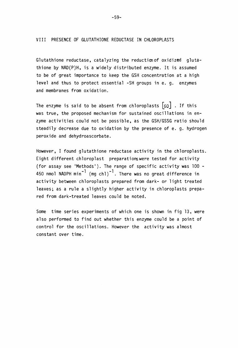
-59-
VIII PRESENCE O F G LUTATHIONE R EDUCTASE IN C HLOROPLASTS
Glutathione reductase, catalyzing the reduction of oxidized gluta
thione by NAD(P)H, is a widely distr ibuted enzyme. I t is assumed
to be of great importance to keep the GSH con centration at a high
level and thus to protect essential -SH groups in e. g. enzymes
and membranes from oxidation.
The enz yme is said to be absent from c hloroplasts M . If this was t rue, the proposed mechanism f or sustained oscil lat ions in en
zyme activit ies could not be possible, as the GSH/GSSG r at io should
steadily decrease due to oxidation by the presence of e . g. hydrogen
peroxide and dehydroascorbate.
However, I found glutathione reductase activity in the chloroplasts.
Eight different chloroplast preparationswere tested for activity
(for assay see 'Methods') . The r ange of specific activity was 100 -
450 nmol NADPH min"^ (mg chl)"^. There was no g reat difference in activity between chloroplasts prepared from d ark- or l ight treated
leaves; as a rule a slightly higher activity in chloroplasts prepared from d ark-treated leaves could be noted.
Some t ime series experiments of which one is shown in f ig 13, were
also performed to find out whether this enzyme cou ld be a point of
control for the oscil lat ions. However the activity was almost
constant over t ime.
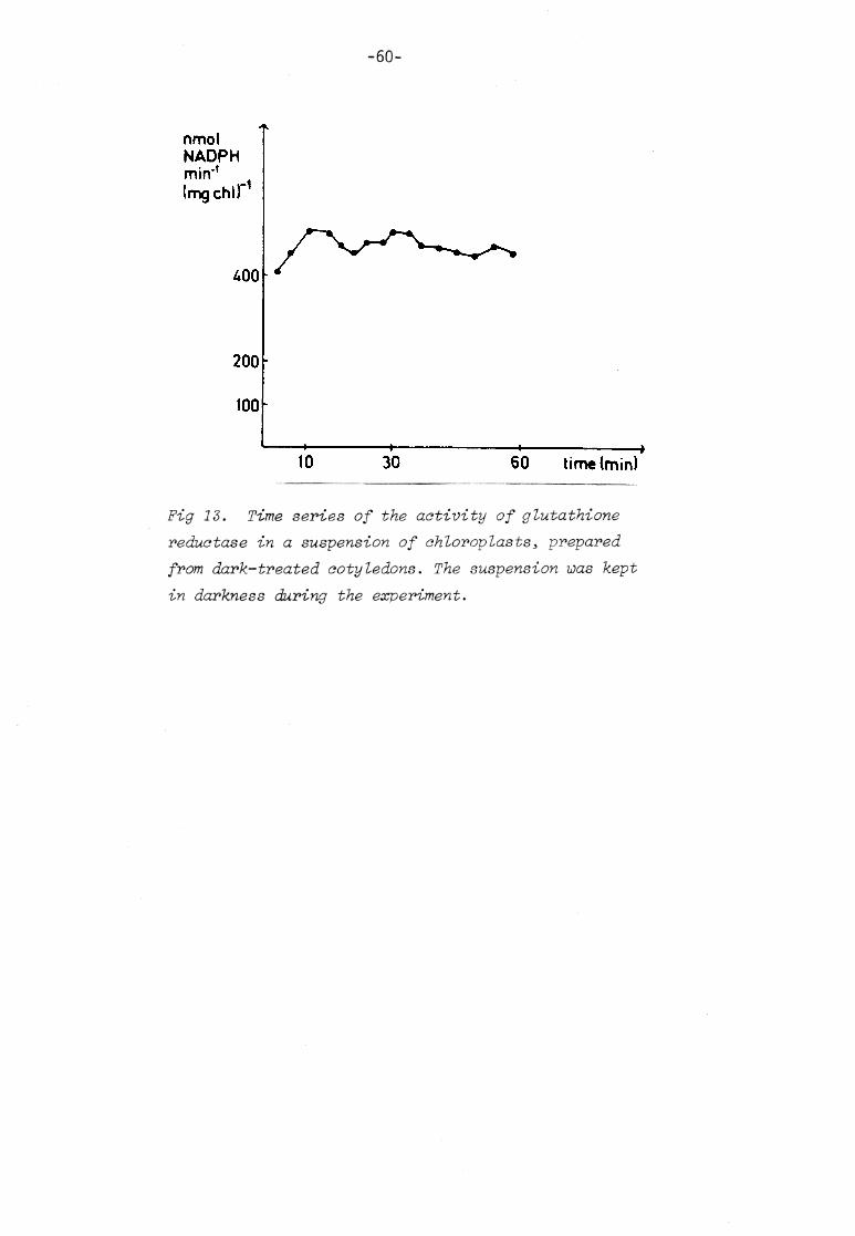
-60-
nmol NADPH min'1
(mg chi)'
200
100
10 30 60 time (min)
Fig IS. Time series of the activity of glutathione reductase in a suspension of chloroplasts, prepared from dark-treated cotyledons. The suspension was kept in darkness during the experiment.
IX TIME SE RIES O F O XIDATION - REDUCTION P OTENTIAL
The t ime series experiments described so far have a l l been a ff l icted
with the disadvantage of being discrete. One me thod to obtain a
continuous curve is to measure the oxidation-reduction potential .
Originally, I hoped to be able to detect changes in the potential
in the extra-chloroplastic solution, as I thought that e.g. ascorbate
and oxidized glutathione could penetrate the membranes and function
as communicators between the chloroplasts. I t soon turned out,
however, that i t was impossible to dist inguish between such sub
stances - if there are any - and material arising from decomp osing
chloroplasts . Thus the experiments to be shown were a l l performed
on chloroplast homogenates or on stroma material .
I will take no notice of the absolute values of the redox-potential
as i t is without meaning to talk about the redox state of such a
complex system as the chloroplastic one. The object of the investi
gations was only to find out whether there were any cyclic changes
in the potential , and if there were, whether the period was accordant to the periods of other parameters invest igated.
Thiol- containing solutions can cause troubles in potential measure
ments, as the thiols form a stable complex w ith the metal of the
electrode [61 J . I noticed no such disturbances in the experiments
to be described.
Oxidation-reduction potential of chloroplast homogenates
Spontaneous^ ^S£i l_latjons_
In f ig 14, a continuous t ime series curve of the oxidation reduction potential of a chloroplast homogenate is shown. The chloroplasts were
prepared from 1ight-treated cotyledons and suspended in 15 ml 0.33 M
sorbitol - 0.05 M HEPES b uffer, pH 7. 6. The suspension was kept in darkness during the experiment. At the beginning of the experiment,
the chloroplasts were mainly intact , but in the sections shown (fig 14 A rep resenting t= 120 - 175 min, 14 B representing t= 300 - 350 min)

-62-
•
lOmV
10min
•
lOmV
ir— > 10min
Fig 14. Continuous time series of oxidation-reduction potential of a chloroplast homogenate5 kept in darkness during the experiment. The chloroplasts were prepared from light-treated cotyledons. Upper curve (fig 14 A) represents t=120-175 min> the lower curve (fig 14 B) t=300-350 min. Recording of redox-potential started at t-0 min.

-63-
the suspension was, due to spontaneous dis integration, a chloroplast
homogenate c onsist ing of stroma material and class II (broken) chloro-
plasts , as judged from phase contrast microscopy. As shown in the figu
the redox potential changed over t ime in a cyclic manner. The ampli
tude and shape of the curve changed but the period was the same,
15 - 16 min, in the entire t ime series investigated.
Chi oroplasts from l ight-treated cotyledons were a lso used for the
experiment shown in f ig 15. After measuring the potential for one
hour, the chi oroplasts were disintegrated on purpose by f reezing and
thawing twice. After that treatment, the potent ial was measured again
and the homogenate was i l luminated (50 000 lux). The section shown in f ig 15 represented t= 0 - 110 min, where t= 0 was the start of
i l lumination. This system also displayed pronounced oscil lat ions in
redox potential . The shape of the curve was about the same during
the two h ours of recording. The period however was much shorter at the end of the experiment and this was probably due to a marked
increase in temperature caused by the lamp. Because of this r ise in
temperature, the homogenate was put into darkness and the redox potential recording continued for several hours. During the f irst
six hours in darkness there were o nly minor oscil lat ions. After that
period, the oscilla t ions increased in amplitude. The shape of the oscil lat ions were irrregular compared to the curves shown in f igs 14 and 15, but the period was in the same r ange.
10min
ft
Fig 15. Same as in fig 14but the homogenate was illuminated,. Section shown represents t-0-110 min.
-64-
Jnduced oscil lat ions
Some chloroplast preparations displayed no o r very small oscil lat ions
in redox potential . Such preparations could be brought to an oscilla
tory behaviour by adding ascorbate or ascorbate and glutathione. An
example of such an induced system is shown in f ig 16. The chloro
plast homogenate (chloroplasts in darkness; l ight-treated leaves were used) had for four hours displayed no oscil lat ions in potential .
Ascorbate was then added to the system (at t= 0 min), giving 25 yg
ascorbate ml~\ and the f irst oscil lat ion could be noted af ter about two hours. The section shown in f ig 16 represented t= 240 - 380 min.
Induced oscil lat ions were always irregular.
Induced oscil lat ions in stroma material . Time s eries of oxidation-reduction potential and GSH.
The expe riments on redox potential so far described were a l l per
formed with chloroplast homogenates. I t turned out that oscil lat ions
were inducable in solutions containing stroma material solely, indicating that enzymes involved in the mechanism f or the oscil lat ions
are localized primarily in the soluble part of the chloroplast . Stroma material systems have a great advantage in comparison to homo
gena tes, as GSH in such systems can be determined photometrically
without disturbances from c hlorophyll and other pigments. Such
systems were used to find out whether there was a connection between changes in redox potential and the redox s tate of glutathione.
Measurement of_GSH: Glutathione was determined photometrically with DTNB (s ee chapter VI) by adding 0.5 ml of the stroma- containing
solution to 2.0 ml 0.5 M Tris-HCl buffer , pH 8. 4. After measuring
the absorbance at 412 nm (A Q) , 1.0 ml 0 .5 mM DTNB w as added and the
absorbance was read off af ter 2 min (A^). - AQ) , corrected for dilution, was used to calculate the amount of GSH in the system.
An e xperiment is shown in f ig 17. A chl oroplast homogenate, prepared from l ight-treated leaves, was used as start ing material . Broken
chloroplasts were removed. 15 mg ascorbate and 4 mg GSH were added t o the 15 ml system at t= 0 min. Oxidation-reduction potential was
recorded and the section shown in f ig 17 A rep resents t= 40 - 120 min. The potential decreased in an oscil latory manner during this
period (note that in f ig 17 A the E' scale is reversed as compared

-65-
50mV 10min
Fn^n-t' • T%1e- terleS °f ,the oxi^ion-reduction potential of a homo-I e %nwirioh Me osczllations were induced by addition of ascorbate.
time (min)
mg GSH
4,0
30
2j0,
120 KO 160 180 time(min)
Fig 17. Oxidation-reduction potential (left curve,A) of a stroma-containing solution after addition of ascorbate and GSH, followed by determination of GSH ( right curve,B).
to f igs 14 - 16). The measurement was stopped at t= 120 min and GSH w as
determined during the following 55 min. As shown in f ig 17 B the amount
of GSH in the system increased markedly in an oscil latory manner d uring this period.
The exper iment clearly indicated that great changes in the redox s tate
of glutathione occured when ascorbate and GSH were added to the system.
The amou nt of GSH, two hours after the addit ion, was about half the

-66-
amount add ed. GSH had obviously been oxidized. , probably by
dehydro-ascorbate formed from oxidation of ascorbate. The re-
reduction of GSSG ( f ig 17 B) should be c aused by NADPH. I t is an
enigma which substrate could be used for the NADPH pro duction. Is
the content of starch and other oxidizable substrates really high
enough in this system? I cannot exclude the possibli ty that sorbi
tol , which is a hexitol and present in the assay solution, was
oxidized thus giving glucose (or fructose) and NAD(P)H, provided
sorbitol oxidizing enzymes were present. Furthermore one should
have in mind that dehydroascorbate might be hydrolysed and further
metaboli sed.
Tests on enzyme dependence
To m ake sure that induced oscil lat ions are not due to some artefact
emanating e.g. from d isturbances on the electrode surface, prolonged
t ime series on oxidation-reduction potential of solutions containing
ascorbate and the glutathione redox couple but no c hloroplast ic material , were performed. No o scil lat ions were noticed in any of these experiments. Analysis of GSH an d ascorbate showed that the amounts of the two su bstances decreased in a monotonie way, probably
due to oxidation and perhaps also hydrolysis.
Enzyme dependence was also investigated in experiments on induced
oscil lat ions in stroma-containing solutions. Such a solution was
divided into two equal parts . One tu be was put in boil ing water for
30 min and then rapidly chil led. Ascorbate and GSH wer e added to the
two s olutions. The heat-treated solution displayed no o scil lat ions
in redox potential while the untreated solution did.
Discussion
The sp ontaneously occuring oscil lat ions in redox potential in chloro-
plast homogenates are of course of greatest interest in connection to the spontaneously occuring oscil lat ions in enzyme a ctivit ies
described previously. The t ime series of redox potential tel l us
nothing about what is actual ly happening in the system but they do
tell us that there are events repeating periodically and that the period is the same as estimated for the discrete t ime series of enzyme a ctivit ies.

-67-
A remarkable feature of the spontaneously occuring redox potential
oscil lat ions is the very rapid decrease in E' in each cycle. This
behaviour was most pronounced d uring the f irst hours of recording
(f igs 14, 15). There may be somelabile (membrane-bound?) component
which is destroyed a fter a couple of hours. The behaviour could never
be repeated in experiments on induced oscil lat ions.
As mentioned before, not al l chloroplast preparations displayed
oscilla t ions spontaneously. This f inding is in contrast to experiments
on enzyme activities. The ca use of this lack of oscil lat ions is
obscure. One re ason could be that the concentration of chloroplasts
in the suspension usually was lower in the redox potential measurements as compared to enzyme a ctivity experiments, as a larger total volume was necessary. Furthermore i t seemed that oxygen pressure
was cri t ical to obtain any oscillat ions, thus making the measurements
somewhat t r icky.
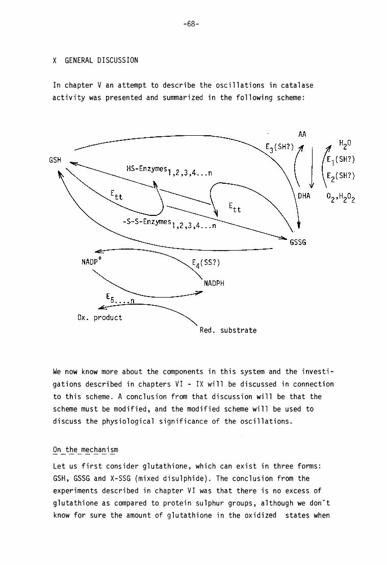
-68-
X GENERAL DISCUSSION
In chapter V an attempt to describe the oscil lat ions in catalase
activity was presented and summarized in the following scheme:
E3(SH?)
HS-Enzymes-, 2 3 4_n
-S-S-Enzymes 2,3,4. . .n
E4(SS?)
NADPH
Ox. product
H2O
E-| ( SH? )
E2(SH?)
O2» ̂ 2^2
Red. substrate
We no w kn ow mo re a bout the components in this system and the investi
gations described in chapters VI - IX will be discussed in connection
to this scheme. A conclusi on from that discussion will be that the
scheme must be m odified, and the modified scheme will be used to discuss the physiological significance of the oscil lat ions.
On_the_mechanism
Let us f i rs t consider glutathione, which can exist in three forms:
GSH, GSSG and X-SSG (mixed disulphide). The conclusion from the
experiments described in chapter VI was that there is no excess of
glutathione as compared to protein sulphur groups, al though we d on' t know fo r sure the amount of glutathione in the oxidized states when

-69-
the maximal values of GSH were observed.
Furthermore we d on' t know fo r sure the number of proteins having
sulphur groups exposable for thiol-disulphide exchange. Some experi
ments (chapter VII) indicate that the property might not be unusual.
The co nclusion that there is no excess of glutathione as compared to enzyme (protein) sulphur groups has several implications. First , one
cannot expect that the variations in enzyme a ctivit ies due to
-SH<—'SS transit ions are very great within one cycle. Nor i s i t
surprising that the oscil lat ions in enzyme a ctivit ies are rather
irregular. The total number of enzyme (E, 9 7 ) sulphur groups I 5 C . ) O • • • I I affected should be c onstant for different cycles, but there should be an element of randomness as to the number of molecules of a certain
enzyme s pecies (E-j, E2 o r Eg e tc) affected.
A th ird important implication is that the amounts of GSH an d GSSG,
formed from thiol-disulphide exchange w ith protein -SH- and -SS-groups, respectively, are not ignorable as compared to the amounts
formed from reduction by NADPH a nd oxidation by D HA, respectively. This puts us into a dilemma, a s we know from the experiments on glutathione reductase (chapter VIII) that the activity of this enzyme
is at an almost constant level. Why th en should GSSG, despite the high
constant activity of glutathione reductase, sometimes be used mainly for thiol-disulphide exchange, sometimes be reduced by NADPH?
First , as at least some NAD P-linked enzymes d isplay oscillatory
activit ies, the NADPH/NADP+ ratio may no t be constant . During one
part of the pathway de picted in the scheme, the NADPH conc entrat ion
is too low t o permit reduction of GSSG. If this was t rue, the NADPH/
NADP+ ratio in chloroplasts from cotyledons should differ markedly
from c hloroplasts from fu ll-grown spinach leaves [58,59] .
Secondly, the thiol- transferase may have some reg ulatory property
which mak es the enzyme m uch more a ctive during that part of the
pathway corr esponding to GSH oxid ation by D HA in comparison to the
"opposité1 part of the pathway. This suggestion is self-contradictory

-70-
as the thiol-transferase, according to the scheme, must be a ctive
in both parts of the pathway.
A th ird possibil i ty is that the concentration of GSSG i s , due to
glutathione reductase, always at a low l evel . The oxidation of
protein-SH groups is then not performed by thiol-disulphide exchange as described in the scheme but by d irect oxidation with DHA.
^3 DHA + XSH + YSH AA + XSSY
Thus whereas, in the previous scheme, XSH an d YSH both symbolized
GSH, in the revised scheme, one of the components is protein-SH (although one cannot exclude that GSSG an d protein-S-S-protein are formed as well) .
AA
HS-Enzymes
GSS-Enzymes
DHA 02 ,H202
E-| (SH?)
\ E 2 < S H ? )
NADP+ NADPH
Ox. product Red. substrate

Apart from solving the GSSG proble m with this revision, the scheme in
i ts modified form has other advantages. As the thiol-transferase works
at just one of the protein SHr—»SS transit ions, the converter enzyme
i tself might be regulated by such transit ions, result ing in amplifi
cation.
We no w look at the sequence of reactions in this modified scheme and
start at a state where t he enzymes (E, ? - ) mainly exist in I ) L ^ J • • • • i I GSSE-form. As mu ch of the glutathione is protein-bound, there is a
rather low co ncentration of GSH. E-j and E2 a re in the assumed less active -S-S-form, result ing in a slow production of DHA.
Due t o the lack of DHA, the main route for GSH i s a thiol-disulphide
exchange w ith -SS-enzymes, 0 . GSSG del ivered from the exchange I ) k • • • i l is momentari ly reduced by NADPH, thus regenerating GSH. If the thiol-
transferase (E^) is actually activated by t he transit ion from -SS-to -SH for m, the rate of thiol-disulphide exchange accelerates.
The enzymes (E, 9 7 ) are now ge tt ing more reduced as regards the i $ L ) j • • • n sulphur groups and the production of DHA in creases. We a rr ive at a
point where the concentrations of DHA, GSH an d protein-SH are high
and where the glutathione dehydrogenase s tarts to work. The main products are ascorbate and GSS-proteins.
I will stress again that this scheme is a suggestion of how t he oscil lat ions in the chloroplastic system can occur. Furthermore, i t
is a minimal model. Other components may co ntribute, making the
mechanism mor e complex. One such component is l ipoic acid, a known
chloroplastic component. This sulphur compound in i ts reduced state
is a dithiol . I t is usually protein-bound, and if also membrane-bound, the absence of thylakoid membranes in the experiments on
chloroplast extracts (chapter V an d IX) might explain the dissimila
ri t ies between spontaneous and induced oscil lat ions.
Another explanation to these dissimilarit ies is that glutathione and
ascorbate were in excess in the experiments with induced oscil lat ions.
Thus the oscil latory mechanism in these cases could be d ifferent from
the one depicted in the revised scheme and more resemble the mechanism

-72-
in the original scheme. The SHt—»SS transit ions might then have
been mainly due to thiol di sulphide exchanges, not necessari ly
enzyme-catalysed.
A typ ical feature for oscilla t ing systems is feed-back mechanisms.
Both schemes depicted do contain such a mechanism. In contrast to e.g. oscil lat ions in glycolysis in yeast , where al loster ic enzymes
are involved in the feed-back mechanism, the chi oroplast ic system
is suggested to have a feed-back mechanism result ing in covalent
modification of enzymes. However, the regulation of the three enzymes
E-j , a nd E3 which are the most important ones for the oscil la t ions
according to the schemes, must be investigated.
On t he physiological significance
The metabolic pathways of carbohydrates in the chi oroplasts in the
l ight are different from the metabolic pathways of carbohydrates in
the dark; mainly the reductive and oxidative pentose phosphate cycle,
respectively. There are many proposals to the regulation of the
chi oroplast ic enzymes involved in the different pathways [for a recent review see 62J . One is that , as protons migrate into the
thylakoid membranes upon i l lumination, pH and concentration of mag
nesium ions increase in the stroma, thus activating the key enzy me
of the Calvin cycle, r ibulose diphosphate carboxylase. Another pro
posal is that the change ( increase) in the NADPH/NADP rat io upon
i l lumination affects key enzy mes [58,59] . Sti l l another, mentioned
in the discussion of chapter V, is that SHr-^SS t ransit ions in essential enxymes are important ways to switch the metabolism from
one pathway to another jjSOj .
In the t ime series experiments on glucose-6-phosphate dehydrogenase and NADP-1inked malate dehydrogenase we noticed trends in opposite
directions. The former enzyme, assumed to be more a ctive in -SS-form, had a decreasing activity, and the lat ter enzyme, assumed to be more
active in -SH-form, had an increasing activity, irrespective of
constant dark and l ight conditions, respectively. These f indings can
perhaps be connected with the fact that leaves used for chloroplast preparation were always harvested in the morning. Is there an

-73-
endogenous rhythm result ing in reduction of enzyme s ulphur groups
during the day, and oxidation of the same groups during the night, regardless of l ight conditions, thus fulfi l l ing a request that the enzymes involved in carbon dioxide f ixation are more active in daytime and the starch degrading enzymes more active in the night?
If there is such an endogenous rhythm i t means that there is one
way of regulation independent of external factors, working together
with other ways of regulation (ion transport , NADPH/NADP+) depending on external factors ( l ight, darkness).
The function of the high frequency oscil lat ions reported and discus
sed in this work would be to build up a diurnal rhythm.
Do t he results and the proposed mechanism permit this very tempting
interpretation? I t implicates that within each cycle in the begin
ning of the day, reduction of enzyme -S S-groups is more e ffective
than oxidation of -SH-groups. As the enzymes are getting more reduced,
the production of DHA should be enhanced for each cycle, due to
activation of E-j and E^. Is DHA re-red uced by any other -SH com pound but enzyme -SH-groups, e.g. GSH so lely, or is DHA not fully re-
reduced, result ing in a decreased AA/DHA r atio? The lack of trends in the t ime series experiments on GSH (c hapter VI) could implicate
that , as less glutathione is enzyme-bound for each cycle, more and
more of the substance is found as GSSG. The formation of the di-
sulphide could be coupled to reduction of DHA. On the other hand,
the lack of trends in the experiments on catalase activity (fig 1) could implicate that the AA/DHA r atio is decreasing for each cycle.
The inactivating effect on catalase due to -SS—» SH tr ansi t ion is counterbalanced by a decreasing concentration of ascorbate.
Thus the scheme in i ts present form is too simple to explain a
diurnal rhythm. Some a ddit ions must be made, e .g. some regulatory
properties of glutathione dehydrogenase. Furthermore, when the period
is as long as about 24 hours, synthesis and degradat ion could influen
ce the total amounts o f the involved components.

-74-
If the physiological function of the oscil lat ions is to build up
a diurnal rhythm with implications on the regulation of essential
enzymes, the oscil lat ions should be temperature independent. The
t ime series experiment on oxidation-reduction potential shown in f ig 15 (p 63) indicates that the period is shortened, probably
by an increase in temperature. On the other hand, some t ime series
experiments on enzyme (catalase) activity of chloroplast suspensions kept at 0°C (not shown) resulted in oscil latory curves with a period
fairly in accordance to experiments on suspensions kept at room
temperature.
Furthermore the oscil lat ions should occur in chi oroplasts from f ul l-grown leaves too, if the physiological function is to build up a diurnal rhythm. However the electron transport chain (p 46) upon
which the proposed mechanism is buil t , is most important in young leaves [47] . The physiological role of the oscil lat ions could thus
be one of development.
I t should be borne in mind that the oscil latory phenomena reported
in this work concern suspensions of an isolated cell organelle. In
the intact plant cell , where the chloroplasts are in contact with
other cell organelles and cytoplasm the behaviour might be different.
This should be investigated, e.g. by s tudying the so called class
III chloroplasts (chloroplasts surrounded by a cytoplasmic layer [ l l] .)
If the oscil lat ions occur in the intact leaves, are the chloroplasts
synchronized in any way or does each chloroplast have a rhythm of i ts
own? During the isolation procedure the chloroplasts may be forced
into synchronization, as there are inevitable temperature transit ions, and for l ight-treated material also l ight-dark transit ions, during
the purif ication.
The c hloroplasts might also loose some essential component during
the isolat ion procedure, result ing in art if icial oscil lat ions. This
is a general problem when working with isolated cell structures.
However the purification method used is rapid, and oscil lat ions can
be induced by adding substances rather than by e l iminating substances.

-75-
If the oscillat ions do o ccur in vivo, i t is of course not sure that
the oscillat ions are important for the metabolism in the chloroplasts . Instead, they may a rise just as a consequence of some o ther, more
important metabolic feature. Even i f the oscil lat ions are of no
physiological significance for the metabolism of the organelle, they
s t i l l are of great significance for the biochemist , as a tool for studying regulatory processes. The works on oscil lat ions in glycoly
sis in yeast do show t his importance [4,5,6,7].
With this work, the presence and some properties of the oscil lat ions
in the chloroplasts were shown. As the preceding discussions have
indicated, the system must be further studied.

1
2
3
4
5
6
7
8
9
10
11
12
13
14
15
-76-
REFERENCES
Bünning,E.(1973) The Physiological Clock, Springer, New Yo rk
Plinius. Natural History book XVIII (for example Mc Graw
Hill , 1964, pi68)
Sweeney,B.M.(1969)Rhythmic Phenomena in Plants, Academic
Press, London
'Biological and Biochemical Oscil lators ' (1973) A Co lloquium
of the Johnson Research Foundation (Chance,B., Pye,E.K.,
Ghosh,A.K. and Hess,B.,eds) Academic Press, New York, London
Faraday Symposia of The Chemical Society (1974) No. 9, 'Physical Chemistry of Oscil latory Phenomena'
Hess,B. and Boiteux,A. (1971) Ann. Rev. Biochem. 40, 237-
258
Hess,B. (1973) jjn Rate Control of Biological Processes (ed.
D.D.Davies) Symposia of the Society for Experimental Biolo
gy nr XXVII, pp 105-131
Cristea,E. (1966) Arch. Biochem. Biophys. 113, 273-280
Wilson,A.T. and Calvin,M. (1955) J . Am. Chem. Soc. 20,
5948-5957
Bannister,T.T. (1965) Biochtm. Biophys.Acta 109, 97-107
Larsson,C., Collin,C. and Albertsson,P.A. (1971) Ibid 245,
425-438
Larsson,C. and Albertsson,P.Å. (1974) Ibid 357,412-419
Karlstam,B. and Albertsson,P.Å. (1970) Ibid 216, 220-222
Aebi,H. (1974) ijn Meth ods of Enzymatic Analysis , (2nd edi-tion)(Bergmeyer,H.U.,ed) pp 673-684. Verlag Chemie, Wein
heim; Academic Press, New Yo rk and London
Mukerji jS.K. and Ting,J .P. (1968) Phytochem. 1_, 903-911
Colman,R.F. (1971) i j i Methods in Enzymology (Colowick,S.P. and Kaplan,N.O., editors-in-chief) Vol XVIIB (Tabor,H. and
Tabor,C.W.,eds) pp 500-503, Academic Press, New Yo rk and London

17
18
19
20
21
22
23
24
25
26
27
28
29
30
31
32
33
34
35
36
-77-
Ellman,G.L. (1959) Arch. Biocbem. Biophys. 8£, 70-77
Jocelyn,P.C. (1962) Biochem. J . 85, 480-485
Cohn,V.H. and Lyle,0. (1966) Anal. Biochem. 434-440
McNeil ,T.L. and Beck,L.V. (1968) Ibid 22, 431-441
Jocelyn,P.C. and Kamminga,A. (1970) Ibid J7, 417-421
Arnon,D.I. (1949) Plant Physiol . 24, 1-15
Gregory,R.P.F. (1968) Biochim. Biophys. Acta 159, 429-439
TolbertjN.E., Oeser,A., Kisaki,T. , Hageman,R.H. and Yama-
zaki,R.K. (1968) J . B i o1. Chem. 243, 5179-5184
Frederick,S.E. and Newcomb,E.H. (1969) 0. Cell Biol. 43, 343-353
Tolbert ,N.E. (1971) Ann. Rev. Plant Physiol . 22^, 45-74
Lavorel ,J. (1956) Biochim. Biophys. Acta 22^, 226-237
Björn,L.O. (1969) Photochem. Photobiol. ^0, 125-129
Pli t ter,J . (1974) ji ji Methods of Enzymatic A nalysis (2nd
edit ion)(Bergmeyer,H.U.,ed) 685-690, Verlag Chemie, Wein
heim, Academic Press, New Yo rk and London
Hermel,H. and Havemann,R. (1966) Biochim. Biophys.Acta 128,
272-282
Hermel,H. and Havemann,R. (1966) Ibid J28, 283-295
Deisseroth,A. and Dounce,A.L. (1970) Physiol . Rev. 50,
319-375
Heidrich,H.G. (1968) Z. Physiol . Chem. 349, 873-880
Cantz,M., Mörikofer-Zwez,S. ,Bossi ,E., Kaufmann,H., von
Wartburg,J.P. and Aebi,H. (1968) Experi enti a 24, 119-121
Mörikofer-Zwez,S. , Cantz,M., Kaufmann,H., von Wartburg,J.P.
and Aebi,H. (1969) Europ. J . Biochem. Vl_, 49-57
Schiefer,S. and Kindl,H. (1976) Abstracts , Tenth Inter
nat ional Congress of Biochemistry, Hamburg

-78-
37 Keilin,D. and Nicholls,P. (1958) Biochim. Biophys. Acta, 28, 225
38 Pihl ,A., Lange,R. and Evang,A. (1961) Acta Chem. Scand.
J_5, 1271-1279
39 Seabra,A. and Deutsch,H.F. (1955) J . Biol. Chem. 214,
447-454
40 0rr ,C.W.M. (1967) Biochem. 6, 2995-2999
41 Chance,B. (1950) Biochem. J . 46, 387-402
42 Orr,C.W.M. (1967) Biochem. 6, 3000-3006
43 Jocelyn,P.C. (1972) Biochemistry of the SH Gro up, p 250,
Academic Press, London
44 Beutler,E. , Srivastava,S.K. and West,C. (1970) Biochem.
Biophys. Res. Comm. 38, 341-347
45 See ref 43, p 143
46 Ibid pp 102-103, 199, 279
47 Mapson,L.W. (1959) in Glutathione (Crook,E.M.,ed)
Biochemical Society Symposia nr 17, pp 28-42
48 Holzer,H.and Duntze,W. (1971) Ann. Rev. Biochem. 40, 345-
374
49 Holzer,H. (1973) in Rate Control of Biological Processes
(ed D.D. Davies) Symposia of the Society for Experimental
Biology nr XXVII, pp 319-331
50 Anderson,L.E. (1974) Proc. 3rd Int . Congr. Photosynthesis
(Avron,M.,ed), 1393-1405
51 Hatch,M.D. and Turner,J .F. (1959) Biochem. J . 76, 556-562
52 AskelöfjP. , Axelsson,K., Eriksson,S. and Mannervik,B. (1974) FEBS Lett 38^, 263-267
53 Azizov,Yu.M., Likhtenshtein,G.J. and Purmal,A.P. (1972)
Biokhimiya 37_, 620-628 (not seen)
54 Eriksson,S. (1974) Ph.D. Dissertat ion, University of
Stockholm, Sweden, p 17
-79-
55 Chetverikova,E.P. (1973) See ref 4, pp 347-362
56 Weidekamm,E., Wallach,D.F.H. and Flückiger,R. (1973)
Anal. Biochem. 54, 102-114
57 See ref 43, p 222
58 Lendzian,K.J. and Bassham,J.A. (1975) Biochim. Biophys.
Acta 396, 260-275
59 Lendzian,K.J. and Bassham,J.A. (1976) Ibid 430, 478-489
60 See ref 43, p 282
61 See ref 43, p 55
62 Kelly,G.J. , Latzko,E. and Gibbs,M. (1976) Ann. Rev.
Plant Physiol . 27, 181-205
-80-
APPENDIX I
EFFECTS O F D ITHIOTHREITOL O N T HE A CTIVITIES O F S OME E NZYMES
IN E XTRACTS F ROM D IFFERENT C LASSES O F C HLOROPLASTS
Anderson et al . [ i] have shown that several chloroplastic enzymes
which in vivo are st imulated by l ight , in vitro are st imulated by
dithiothreitol (DTT), while enzymes, inactivated by light in vivo, are inactivated by DTT i n vitro.
The aim of this work was to investigate such effects on two morp hologically different types of chloroplasts, class I chloroplasts
( intact) and class III chloroplasts (chloroplasts surrounded by a
cytoplasmic layer [22). Some of the enzymes studied, e.g. NADP-
1 inked malate dehydrogenase and glucose-6-phosphate dehydrogenase,
have been studied before j j ,3,4] , while catalase has not. In this appendix, only results concerning enzymes mentioned in the thesis
are given.
Materials and methods
The materials and methods used in this study were essential ly the
same as those described in the thesis.
Class III chloroplasts were prepared according to Larsson and Alberts-
son [5] . Class I and class III chloroplasts were, for each experiment,
prepared simultaneously and from the same plant material .
Some e xperiments were performed with chloroplasts, purified by the rapid batch technique using polymer two phase systems [5] . In other
experiments, the separation of chloroplasts into different classes
was performed with counter current distr ibution (CCD) technique with
polymer two phase systems [2] , a process which is much more t ime-
consuming.

-81-
When using the CCD techn ique l ight- or dark-treatment means not
only that l ight- or dark-treated cotyledons were used as plant
material , but also that the chloroplasts were i l luminated or kept
in the dark during the CCD-'runs ' .
Chloroplast extracts were mad e from the purified class I and class
III chloroplasts. Protein concentration was determined, using a
f luorimetrie method [6] . Specific activit ies are expressed as en
zyme activit ies relative to the amount of protein. To con vert the
specific activities on protein basis in the extracts to the amount
of chlorophyll , the activity values of class I chloroplasts should be multiplied by a factor of 3 , of class III chloroplasts by a factor
of 10.
After assaying the enzymes (before DTT) the extracts were treated with 1 mM DTT a t 4°C over night and in darkness and a ssayed again
(after DTT).
Results and discussion
The activit ies, before and after DTT tr eatment, of three enzymes are
l isted in the following tables. The results were obtained from f our
experiments, performed on materials pretreated in different ways
( l ight/darkness, CCD/batch).
CATALASE
Class I Class III
activity before DTT
activity af ter DTT
activity before DTT
activity after DTT
Light CCD 7
batch 16
9
69
6
9
Darkness
CCD
batch
22
15
94
72
27
7
Activity expressed as ymol min ^ (mg protein)"^
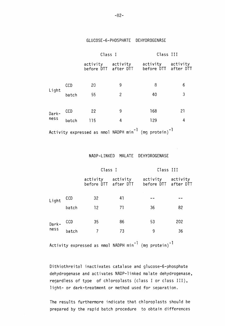
-82-
GLUCOSE-6-PHOSPHATE DEHYDROGENASE
Class I
activity activity before DTT after DTT
Class III
activity activity before DTT after DTT
Light CCD
batch
20
55
8
40
6
3
Darkness
CCD
batch
22
115
9
4
168
129
Activity expressed as nmol NADPH min ^ (mg protein) ^
NADP-LINKED MALATE DEHYDROGENASE
21
4
Class I
activity activity before DTT af ter DTT
Class III
activity activity before DTT af ter DTT
Light CCD
batch
32
12
41
71 36 82
Darkness
CCD 35
batch 7
86
73
53
9
202
36
Activity expressed as nmol NADPH min ^ (mg protein) ^
Dithiothreitol inactivates catalase and glucose-6-phosphate
dehydrogenase and activates NADP-1inked malate dehydrogenase,
r e g a r d l e s s o f t y p e o f c h l o r o p l a s t s ( c l a s s I o r c l a s s I I I ) ,
l ight- or dark-treatment or method used for separation.
The results furthermore indicate that chloroplasts should be
prepared by the rapid batch procedure to obtain differences

-83-
due to l ight- and dark-treatment in the activities of the two dehydrogenases in class I chloroplasts.
References APPENDIX I :
1. Anderson,L.E. (1974) Proc. 3rd Int . Congr. Photosynthesis
(Avron,M. ed.) 1393-1405
2. Larsson,C., Collin,C. and Albertsson,P.Å. (1971) Biochim.
Biophys. Acta 245, 425-438
3. Ting,J.P. and Rocha,V. (1971) Arch. Biochem. Biophys. 147, 156-164
4. Johnson,H.S. (1971) Biochem. Biophys. Res. Comm. 43^, 703-709
5. Larsson,C. and Albertsson,P.Å. (1974) Biochim. Biophys. Acta
357, 412-419
6. Weidekamm,E., Wallach,D.F.H. and Flückiger,R. (1973) Anal.
Biochem. 54, 102-114
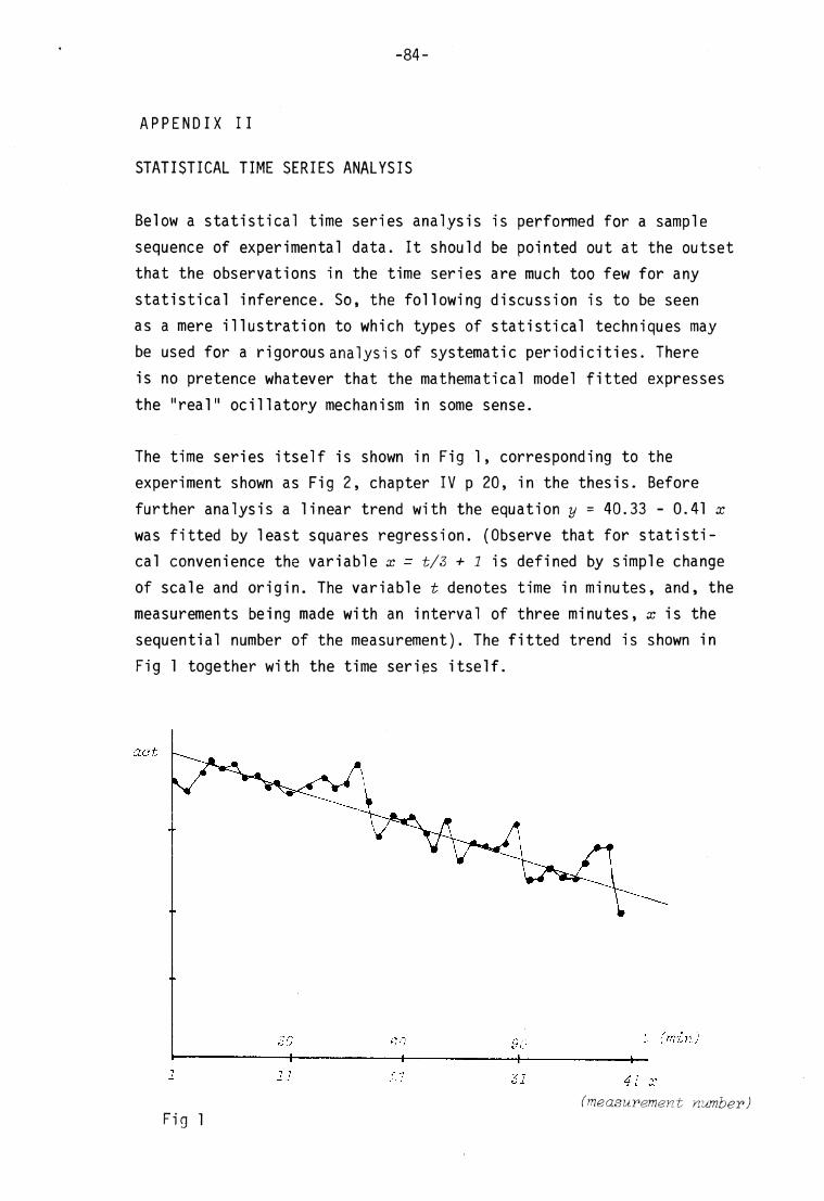
-84-
A P P E N D I X I I
STATISTICAL TIME SE RIES A NALYSIS
Below a s tat ist ical t ime series analysis is performed for a sample
sequence of experimental data. I t should be pointed out at the outset
that the observations in the t ime series are much too few for any
stat ist ical inference. So, the following discussion is to be seen
as a mere i l lustration to which types of s tat is t ical techniques may
be used for a r igorous analysis of systematic periodicit ies. There
is no pretence whatever that the mathematical model f i t ted expresses
the "real" ocil latory mechanism in some sense.
The t ime series i tself is shown in Fig 1, corresponding to the experiment shown as Fig 2, chapter IV p 20, in the thesis. Before
further analysis a linear trend with the equation y = 40.33 - 0.41 x was f i t ted by least squares regression. (Observe that for s tat is t ical convenience the variable x = t/3 + 1 is defined by si mple change
of scale and origin. The variable t denotes t ime in minutes, and, the
measurements being made with an interval of three minutes, x is the
sequential number of the measurement). The f i t ted trend is shown in
Fig 1 together with the t ime series i tself .
act
t (min) 6 0
1 11 21 31 41 x (measurement number)
Fig 1

-85-
After el imination of the trend, different techniques were tr ied to analyse regular periodicit ies. In Fig 2 is shown the so called
covariance function for the resulting residual t ime series when the
trend is eliminated. I t shows the covariance between activity at any
measurement point and activity with different lags in terms of measure
ment numbers. For zero lag the highest covariance is obtained. This
is natural as there is no more perfect correlat ion obtainable than
that when a variable is correlated with i tself. The "covariance" then
is simply a variance.
More interesting is that the covariance function has a peak (with
posit ive covariance) at a lag of about 8 observation points. With this lag peaks seem to be correlated with peaks and throughs with
throughs. This would indicate a complete cycle of about 8 measurement
points, corresponding in terms of t ime to 24 minutes. The negative
covariance with intermediate lags indicates that peaks are then correlated with throughs and vice versa.
From th e covariance function the spectral density for different frequencies was calculated. The result is shown in Fig 3. As is seen
a frequency of about 2TT/7 is dominant. The length of a complete
cycle is , of course, obtained as 2 TT divide d by t he frequency. Hence,
the predominant period in the t ime series would extend over 7 measure
ment points . This is in not too bad an accordance with what was seen
from the covariance function - - and with what c ould be seen by mere
inspection of the t ime series i tself .
The t ime series remaining after elimination of the trend was also
analyzed in a different way. A reg ression hyperplane was f i t ted by
least squares for activity (measured as deviation from t he trend) at
a certain measurement point as dependent variable, and activit ies at the previous measurement point , the two preceding measurement points, the three preceding ones etc, as independent variables. I t was seen
that the sum of squared residuals was signif icantly decreased with
the inclusion of addit ional independent variables up to the number of three. After that only small improvements of f i t were obtained from increasing complexity.
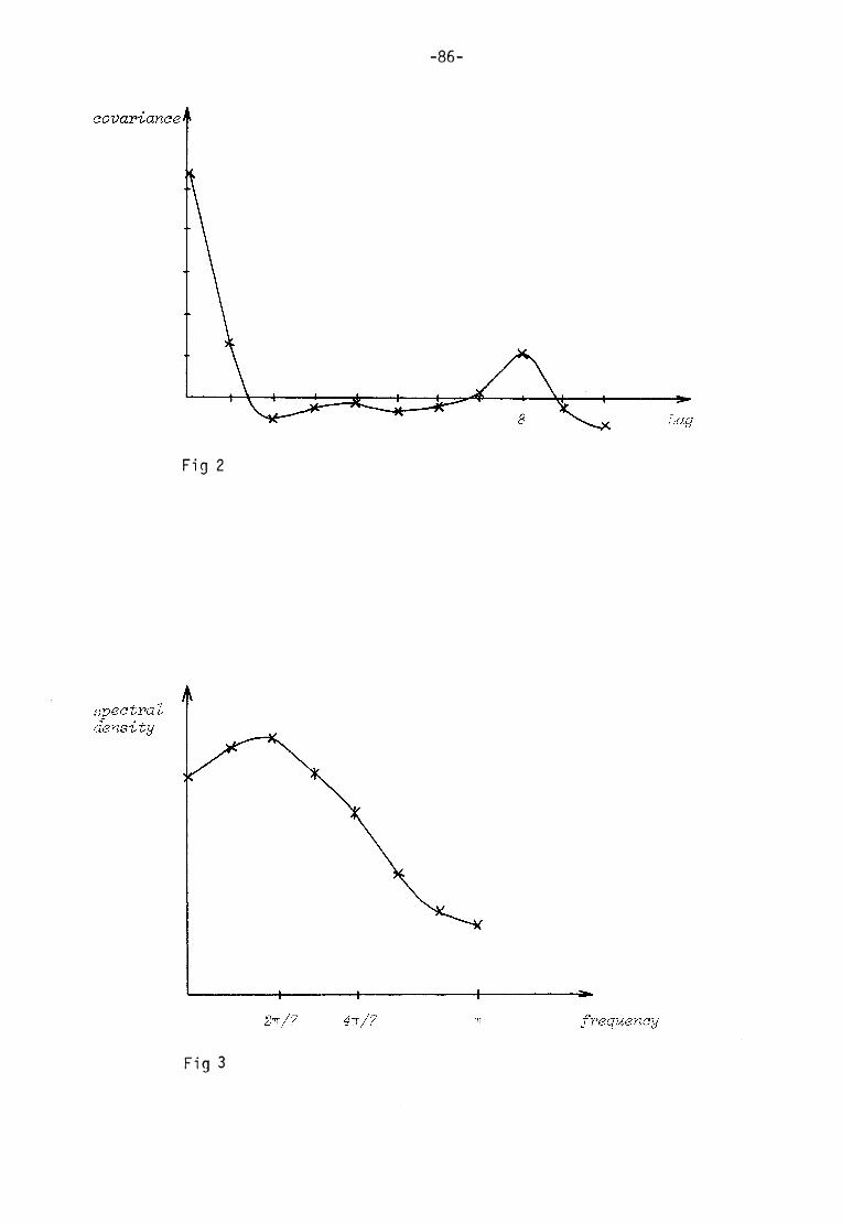
-86-
Lag
F i g 2
spectral density
frequency TT

-87-
The regression hyperplane with three independent variables is
y ( x ) = 0 . 2 2 y ( x - l ) - 0 . 1 8 y ( x - 2 ) + 2 . 2 1 y ( x - Z )
This third order difference equation (which could simply be trans
formed to a differential equation of the third order having the
same s olut ion) together with the trend actually "explains" more
than 90 percent of the variation in activity. But, i t should be
stressed once more, the l imited number o f observations in the t ime
series, does not admit any stat ist ical inference. — A high coeffic i e n t o f d e t e r m i n a t i o n i s a l w a y s p o s s i b l e t o o b t a i n b y f i t t i n g a suff iciently complex model.
Another comment should be added. What has been checked is whether
a l inear difference, or differential , equation with constant coeff icients could account for the oscil lat ions observed. If the mechanism
is non-linear -- however sim ple - - the simple methods of analysis
available can not be used to identify i t . No mor e is i t possible to guess i ts character by inspection when there is a discrete
t ime series with measurement intervals as large as in the present
case.
There are other tests for detect ion of whether a seeming o scil lat ion could be built up by a random process, such as the test of runs of
signs, but there is no point in going further at present for reasons
already given.
Time s eries analysis were also performed on the t ime series shown
in Fig 1 , p 20 (chapter IV) in the thesis. The resulting covariance
function and spectral density are shown in Figs 4 and 5.
Aoknoijle^ements. I am most grateful to f i l kand Lennart Nilsson and f i l kand Anders Nylén at the Department of Mathematical Statist ics
for performing the analysis. I am al so deeply indebted to my husband
for taking part in that work and for helping me in understanding
the obtained results and putt ing them into words.
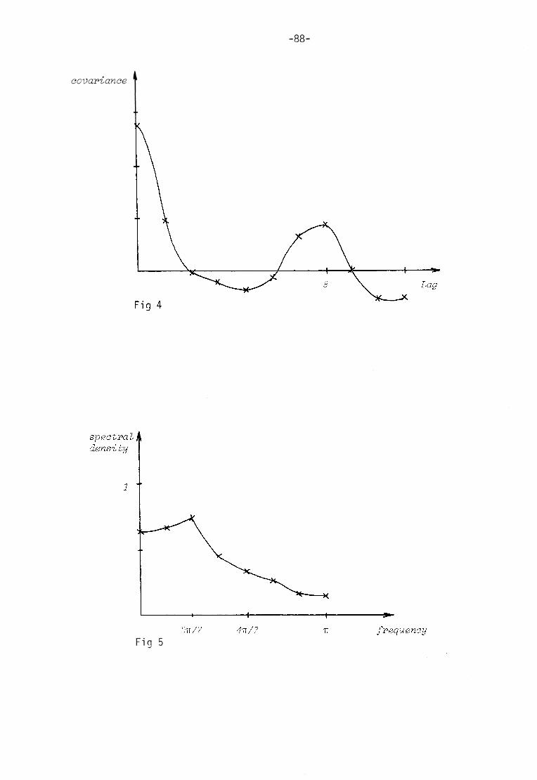
covariance
-88-
Lag
spectral density
frequency TT
F i g 5
