osmoregulated periplasmic glucans are needed for competitive growth and biofilm formation by...
TRANSCRIPT
R E S E A R C H L E T T E R
Osmoregulated periplasmicglucans areneeded for competitivegrowthand bio¢lmformation bySalmonella enterica serovarTyphimurium in leafy-greenvegetablewashwaters andcolonization inmiceLiu Liu1,2,5, Shawn Tan1, Won Jun1,3, Allen Smith4, Jianghong Meng2 & Arvind A. Bhagwat1
1Produce Quality and Safety Laboratory, Henry A. Wallace Beltsville Agricultural Research Center, Agricultural Research Service, USDA, Beltsville, MD,
USA; 2Department of Food Science and Nutrition, University of Maryland, College Park, MD, USA; 3Department of Plant Science and Landscape
Architecture, University of Maryland, College Park, MD, USA; 4Diet Genomics and Immunology Laboratory, Henry A. Wallace Beltsville Agricultural
Research Center, Agricultural Research Service, USDA, Beltsville, MD, USA; and 5College of Food Science and Technology, Northwest A&F University,
Yangling, China
Correspondence: Arvind A. Bhagwat,
Produce Quality and Safety Laboratory, Henry
A. Wallace Beltsville Agricultural Research
Center, Agricultural Research Service, USDA,
10300 Baltimore Avenue, Building 002,
BARC-W, Beltsville, MD 20705-2350, USA.
Tel.: 11 301 504 5106; fax: 11 301 504
5107; e-mail: [email protected]
Present addresses: Shawn Tan, Ngee Ann
Polytechnic, Singapore 599489, Singapore.
Won Jun, Food Safety Laboratory, USDA-ARS,
Beltsville, MD 20705, USA.
Received 30 October 2008; accepted 13
November 2008.
First published online 24 December 2008.
DOI:10.1111/j.1574-6968.2008.01462.x
Editor: Stephen Smith
Keywords
food safety; biofilms; glucans; vegetable wash
waters; food microbiology; low osmotic
environments.
Abstract
Osmoregulated periplasmic glucans (OPGs) are major periplasmic constituents of
Gram-negative bacteria. The role of OPGs has been postulated in symbiotic as well
as pathogenic host–microorganism interactions. Here, we report the role of OPGs
from Salmonella enterica serovar Typhimurium during growth and biofilm
formation in leafy-green vegetable wash water. The opgGH mutant strain, which
was defective in OPG biosynthesis, initiated the growth at a slower rate in wash
waters obtained from spinach, lettuce and green collard and severely impaired
biofilm formation. The lack of OPG synthesis did not influence biofilm formation
by the opgGH mutant in low-nutrient low-osmolarity laboratory media. In
coculture experiments initiated with equal proportions of cells, the opgGH mutant
was outnumbered by the wild-type strain under the planktonic as well as the
biofilm growth conditions. The opgGH mutant strain poorly colonized mouse
organs when introduced orally along with the wild-type strain. This is the first
report demonstrating the role of OPGs of Salmonella in competitive colonization
of biofilms, planktonic cultures and mouse organs.
Introduction
Salmonella are common agents of gastrointestinal-based
diseases in humans and have been recognized as a major
foodborne hazard. In humans, the organism is most com-
monly acquired following ingestion of contaminated food
or water (D’Aoust et al., 2001). Traditionally, poultry
products have been documented as a major source of
contamination in many developed countries (Hald et al.,
2004). However, in recent years, Salmonella infections
associated with raw vegetables have occurred with increased
frequency, particularly involving fresh produce (Horby
et al., 2003; CDC, 2005; Singh et al., 2007). While the
specific sources of contamination have not been identified,
fresh produce is grown in natural habitats for Salmonella
reservoirs (such as birds, amphibians, reptiles and poultry).
One possible source for foodborne infections is the quality
of water, either in the liquid phase used to wash the produce
or in the form of ice used in shipping or storage (Bhagwat,
2006). Irrigation water quality is also of significant
FEMS Microbiol Lett 292 (2009) 13–20 Journal compilation c� 2008 Federation of European Microbiological SocietiesPublished by Blackwell Publishing Ltd. No claim to original US government works
importance as it may be responsible for for carrying micro-
organisms from the field to the fork (Wachtel et al., 2002;
Steele & Odumeru, 2004; Duffy et al., 2005). In several
instances, foodborne illnesses have been traced to poor or
unsanitary postharvest practices, specifically to nonpotable
cooling water and ice (Harris et al., 2003; Steele & Odumeru,
2004). Salmonella, as a consequence of its lifestyle, endures
extended periods of nutrient deprivation in natural aquatic
and terrestrial environments while retaining its pathogenic
potential (Foster & Spector, 1995; D’Aoust et al., 2001). A
number of environmental factors, including nutrient depri-
vation, osmolarity and availability of oxygen, have been
implicated in modulating the virulence of Salmonella,
implying an empirical relationship between survival in
nature and survival in the host organism (Arricau et al.,
1998; Barak et al., 2005).
Relatively little is known about how Salmonella survive in
nutrient-deprived, low-osmolarity environments such as
irrigation and vegetable wash waters (Barrow et al., 1996;
Casani et al., 2005). Growth of cells requires that the
cytoplasm contains essential constituents with a total osmo-
larity of about 300 mosmol L�1 (Kennedy, 1996). When cells
are grown in a low-osmolarity medium, swelling and
rupturing of the cytoplasmic membrane is prevented by the
osmolarity of the periplasmic space, which is mainly due to
anionic short glucose chains, referred to in the literature
as membrane-derived oligosaccharides (MDOs) or osmo-
regulated periplasmic glucans (OPGs) as per the new
nomenclature (Miller et al., 1986; Kennedy, 1996; Bohin,
2000; Bohin & Lacroix, 2007). When cells of Escherichia coli
are grown in a low-osmolarity medium (c. 30 mosmol L�1),
OPGs may represent as much as 5–7% (dry weight) of the
cells, and may constitute a considerable fraction of the fixed
anions in the periplasm (Miller et al., 1986). OPGs are
thought to play an important, but poorly understood, role
in host–microorganism interactions involving specific plant
and animal hosts (Bhagwat et al., 1996; Page et al., 2001;
Arellano-Reynoso et al., 2005). In E. coli, the roles of OPGs
in cell-to-cell signaling, chemotaxis, regulation of polysac-
charide synthesis and resistance to sodium dodecyl sulfate
(SDS) have been postulated (Ebel et al., 1997; Bohin, 2000;
Rajagopal et al., 2003).
We recently observed that the opgGH (previously referred
to as mdoGH) mutant of Salmonella enterica serovar Typhi-
murium was compromised in mouse virulence and required
a 2-log higher oral dose to achieve a lethal dose 50% (LD50)
comparable to that of the wild-type strain (Bhagwat et al.,
2009). We wanted to investigate the role of OPGs in the
growth and competence of Salmonella in environments
critical for foodborne outbreaks such as wash waters
obtained from leafy-green vegetables. Because bacteria often
adhere to surfaces and form biofilm communities in their
natural settings (Donlan & Costerton, 2002; Burmolle et al.,
2006), we investigated the biofilm-forming ability of the
opgGH mutant strain under low-nutrient low-osmolarity
conditions. Lastly, we checked whether the inability to
synthesize OPGs would compromise the strain’s competitive
colonization potential in mouse tissue as well as under
biofilm and planktonic conditions.
Materials and methods
Bacterial strains and culture conditions
Salmonella enterica serovar Typhimurium wild-type strain
SL1344 (Nal-r) and the opgGH null mutant SG111 (Km-r)
(Bhagwat et al., 2009) were grown in Luria–Bertani (LB)
medium at 37 1C in a shaking incubator at 220 r.p.m. When
required, the medium was supplemented with kanamycin
(50 mg mL�1), nalidixic acid (10mg mL�1) or streptomycin
(50 mg mL�1). Salmonella–Shigella agar and brilliant green
agar (Difco, Franklin Lakes, NJ), Salmonella semi-selective
indicator media with appropriate antibiotics, were used to
isolate Salmonella from mouse tissue. The osmolarity of
growth media (mosmol L�1) was measured using a Wescor
vapor pressure osmometer (model 5500, Wescor Inc.,
Logan, UT).
The growth rates of the wild type and opgGH mutant were
determined in different growth media such as LB broth and
low-nutrient no-salt (LNNS) media (which are 1 : 20 diluted
LB broth without NaCl) having osmolarity values of 407� 4
and 31� 3 mosmol L�1, respectively (1/� denote SD of
mean). Growth was measured using a Bioscreen C auto-
matic turbidometric analyzer (GrowthCurves USA, NJ).
Starter cultures were prepared by inoculating a single colony
of the appropriate strain into LB broth, followed by over-
night incubation at 37 1C. This culture was diluted 1 : 10 000
into fresh media of varying osmolarities, and 250mL per well
was transferred into a 100-well honeycomb Bioscreen plate.
Growth was analyzed at 37 1C with continuous shaking, and
for each sample, data were collected from five replicate wells.
In the initial experiments, growth was also measured by
performing viable cell counts on LB agar media to ensure
that the OD reflects viable cell numbers appropriately
(Mellefont et al., 2005). To assess the effect of osmotic
stress on growth, media were supplemented with varying
amounts of salt (NaCl or KCl) or buffered with HEPES
(50 mM, pH 7.1).
Biofilm formation
The overnight-grown cultures were diluted 1 : 10 000 in
fresh media or vegetable wash waters, placed in sterile
polystyrene microplates at 100mL per well and incubated
for 24 h static at 30 1C for use in biofilm studies (O’Toole &
Kolter, 1998). Eight wells were inoculated for each sample.
After a 24-h incubation, microplate cultures were aspirated
FEMS Microbiol Lett 292 (2009) 13–20Journal compilation c� 2008 Federation of European Microbiological SocietiesPublished by Blackwell Publishing Ltd. No claim to original US government works
14 L. Liu et al.
and washed five times with 450 mL sterile distilled water with
an Elx50 plate washer (BioTek Instruments Inc., Winooski,
VT). The plates were air dried, and 150 mL of Protocol
crystal violet solution (0.41% w/v dye, 12.0% ethanol and
0.1% phenol in water; Fisher Scientific Company, LLC,
Kalamazoo, MI) was added per well and incubated at room
temperature for 45 min. The wells were then aspirated and
washed five times with 450 mL sterile distilled water. After
allowing the plates to air dry, 200mL of 95% ethanol was
added to each well. A multichannel pipettor was used to mix
the contents of the wells and to dissolve the crystal violet dye.
A600 nm was then recorded for each well using a microquant
microplate spectrophotometer (BioTek Instruments Inc.).
The average absorbance of eight control wells (that had
contained culture medium only) was subtracted from each
sample well to determine the amount of biofilm present.
In order to determine viable counts, biofilms from
individual wells (before crystal violet staining) were sus-
pended in 100 mL of saline and 100 mg glass powder (Sigma
Chemical Co., St. Louis, MO). The suspended biofilms were
recovered in three washes with 100mL saline. The suspen-
sion was vortexed vigorously for 1 min, and 10-fold serial
dilutions were plated on selective media to determine viable
cell counts. The detection limit was 103 cells per well.
Preparation of vegetable wash water
The vegetable rinse water was prepared as described pre-
viously (Bhagwat, 2004). Briefly, fresh spinach (Spinacia
oleracea), lettuce (Lactuca sativa) and collard green (Brassica
oleracea) vegetables were obtained from local grocery stores.
The produce (250 g) was sliced to c. 5 cm� 5 cm and washed
for 30 min by gentle shaking in a plastic container
(33 cm� 22 cm� 8 cm) containing 500 mL of deionized
distilled water. The vegetable rinse solution was decanted
from the tray and was filtered through a glass wool column
placed in a 50-mL syringe. The filtered vegetable rinse-water
was centrifuged for 10 min at 4000 g at room temperature.
The supernatant was made bacteria-free by filtering through
a 0.22-mm nylon filter and was used in biofilm experiments.
The pH and osmolarity values were measured as described.
Mouse virulence studies
Five-week-old male BALB/c mice were purchased from the
Small Animals Division of the National Cancer Institute
(Frederick, MD). Mice were housed in an AllenTown Caging
Biocontainment-isolator rack, four to five per cage, and
provided with Harland-Teklad rodent chow and deionized
water ad libitum. Mice were acclimated 1 week before use,
and all animal protocols were approved by the Institutional
Animal Care and Use Committee. Animals were fasted for
c. 12 h before being inoculated with 0.2 mL of an S. enterica
serovar Typhimurium suspension (in 0.9% NaCl) by an oral
gavage. Bacterial strains were grown in LB medium at 37 1C
without shaking for 16–18 h, suspended in saline and
adjusted to the appropriate cell density before oral infection.
Viable cell counts were confirmed by retrospective spread
plating onto LB agar plates and incubating the plates over-
night at 37 1C.
To analyze colonization of individual organs by each
bacterial strain, mice were sacrificed 6 days postinfection.
Individual organs (liver, spleen and intestine) were dis-
sected, weighed and homogenized in LB medium. Cell
counts were determined by spread plating appropriate
dilutions onto Brilliant green agar plates (Difco) containing
streptomycin (50mg mL�1) or kanamycin (25mg mL�1).
Individual colonies were counted after an overnight incuba-
tion at 37 1C, and statistical analysis was performed using
ANOVA with post hoc analysis for multiple comparisons or the
Mann–Whitney nonparametric test. A value of Po 0.05 was
considered significant.
Statistical analysis
For statistical analyses, SIGMASTAT 3.0 software (Ashburn, VA)
was used. Data were analyzed by the one-way ANOVA test
to determine statistical differences between the means of
treatments.
Results
OPGs are needed to achieve optimal growthrates in low-nutrient and low-osmolarity media
We examined the contributions of OPGs in growth and
biofilm formation by S. enterica serovar Typhimurium
strains in LNNS media (Fig. 1a). Upon 1 : 10 000-fold
dilution of stationary-phase culture to a fresh LNNS med-
ium, wild-type cells had a ‘relative OD lag time’ of
400� 22 min as compared with 585� 31 min required by
the opgGH mutant (Fig. 1a, filled symbols). The delay in
initiating growth was a phenomenon specific to low osmo-
larity of the medium, as increasing the osmolarity from 31
to 240 mosmol L�1 by addition of NaCl (0.155 M final
concentration) restored normal growth in the opgGH
mutant strain (Fig. 1a, open symbols).
We then compared the growth potential of wild-type and
opgGH mutant strains in vegetable wash waters obtained
from leafy-green vegetables such as spinach, collard green
and lettuce (Fig. 1b–d). Similar to what was observed with
LNNS growth medium, lack of OPG synthesis in the opgGH
mutant severely affected its ability to initiate growth in
vegetable wash waters that had osmolarity values of
25–29 mosmol L�1 (Table 1). The observed delay to initiate
growth by the opgGH mutant does not appear to be related
to its ability to utilize certain nutrients in vegetable wash
waters. By the time strains reached the stationary growth
FEMS Microbiol Lett 292 (2009) 13–20 Journal compilation c� 2008 Federation of European Microbiological SocietiesPublished by Blackwell Publishing Ltd. No claim to original US government works
15OPGs are needed for biofilm formation by S. Typhimurium
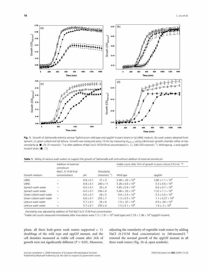
phase, all three leafy-green wash waters supported c. 11
doublings of the wild type and opgGH mutant, and the
cell densities measured as viable cell counts after 24 h of
growth were not significantly different (P4 0.05). Moreover,
adjusting the osmolarity of vegetable wash waters by adding
NaCl (0.155 M final concentration) to 240 mosmol L�1
restored the normal growth of the opgGH mutant in all
three wash waters (Fig. 1b–d, open symbols).
Fig. 1. Growth of Salmonella enterica serovar Typhimurium wild-type and opgGH mutant strains in (a) LNNS medium, (b) wash waters obtained from
spinach, (c) green collard and (d) lettuce. Growth was measured every 15 min by measuring A600 nm using a BioScreen growth chamber either at low
osmolarity (�, ’; 25–31 mosmol L�1) or after addition of NaCl to 0.155 M (final concentration) (�, &; 240–255 mosmol L�1). Wild type (�, �) and opgGH
mutant strain (’, &).
Table 1. Ability of various wash waters to support the growth of Salmonella with and without addition of external osmoticum
Growth medium
Addition of external
osmoticum
(NaCl, 0.15 M final
concentration)� pH
Osmolarity
(mosmol L�1)
Viable count after 24 h of growth in pure culture (CFU mL�1)w
Wild type opgGH
LNNS � 6.8� 0.1 31� 3 5.48� .03� 108 5.85�1.1� 108
LNNS 1 6.8� 0.1 240� 11 5.28� 0.6� 108 5.3�0.6� 108
Spinach wash water � 6.0� 0.1 29� 4 5.85� 0.6� 108 6.6�0.7� 108
Spinach wash water 1 6.0� 0.1 244� 6 5.48� .03� 108 7.22�1.1� 108
Green collard wash water � 6.6� 0.1 28� 5 9.9� 2.0� 108 5.3�3.0� 108
Green collard wash water 1 6.6� 0.1 255� 7 1.3� 0.5� 108 1.1�0.27� 108
Lettuce wash water � 5.7� 0.1 26� 6 1.9� .07� 108 0.9� .04� 108
Lettuce wash water 1 5.7� 0.1 250� 4 1.5� 0.1� 108 1.4� .2� 108
�Osmolarity was adjusted by addition of 5 M NaCl to 0.15 M final concentration.wViable cell counts measured immediately after inoculation were 7.3� 1.91�104 (wild type) and 7.29� 1.98� 104 (opgGH mutant).
FEMS Microbiol Lett 292 (2009) 13–20Journal compilation c� 2008 Federation of European Microbiological SocietiesPublished by Blackwell Publishing Ltd. No claim to original US government works
16 L. Liu et al.
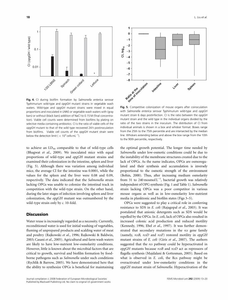
OPGs and biofilm formation
Attachment of Salmonella to food-processing surfaces and
subsequent development of biofilms may have significant
economic and public health consequences (Donlan &
Costerton, 2002; Burmolle et al., 2006; Agle, 2007). It is
suggested that Salmonella biofims adapt structurally to
changes in the medium osmolarity and nutrients (Lapidot
et al., 2006; Mangalappalli-Illathu et al., 2008). We examined
biofilm formation by wild-type and opgGH mutant strains
in LNNS medium (Fig. 2). In general, the opgGH mutant
strain appeared to form less biofilm than the wild type in
LNNS medium, but the differences were not statistically
significant (P4 0.05). However, the opgGH mutant formed
significantly reduced or no biofilms in leafy-green wash
waters (Fig. 2). Biofilm formation by the opgGH strain in
wash waters obtained from green collard and lettuce was
below the detection limit (A600 nm after crystal violet stain-
ing o 0.05). In spite of the fact that the opgGH mutant was
able to support a number of cell divisions and was able to
grow in vegetable wash waters, it formed significantly lower
quantities of biofilm in spinach wash waters. In general,
wild-type Salmonella cells formed reduced quantities of
biofilm in vegetable wash waters in comparison with LNNS
medium.
OPGs and competitive growth in free-living andbiofilm states
We examined how the initial delay in resuming growth of
the mdoGH mutant might affect the strains’ ability to
compete in biofilm and free-living settings when coinocu-
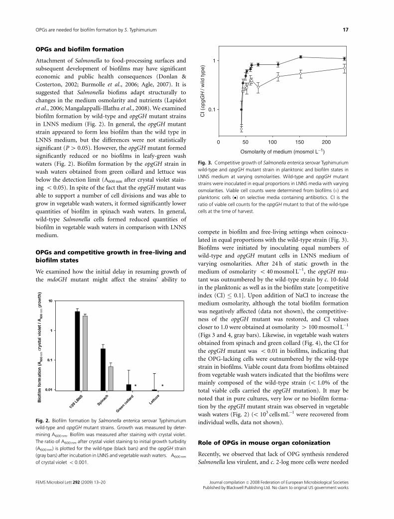
lated in equal proportions with the wild-type strain (Fig. 3).
Biofilms were initiated by inoculating equal numbers of
wild-type and opgGH mutant cells in LNNS medium of
varying osmolarities. After 24 h of static growth in the
medium of osmolarity o 40 mosmol L�1, the opgGH mu-
tant was outnumbered by the wild-type strain by c. 10-fold
in the planktonic as well as in the biofilm state [competitive
index (CI) � 0.1]. Upon addition of NaCl to increase the
medium osmolarity, although the total biofilm formation
was negatively affected (data not shown), the competitive-
ness of the opgGH mutant was restored, and CI values
closer to 1.0 were obtained at osmolarity 4 100 mosmol L�1
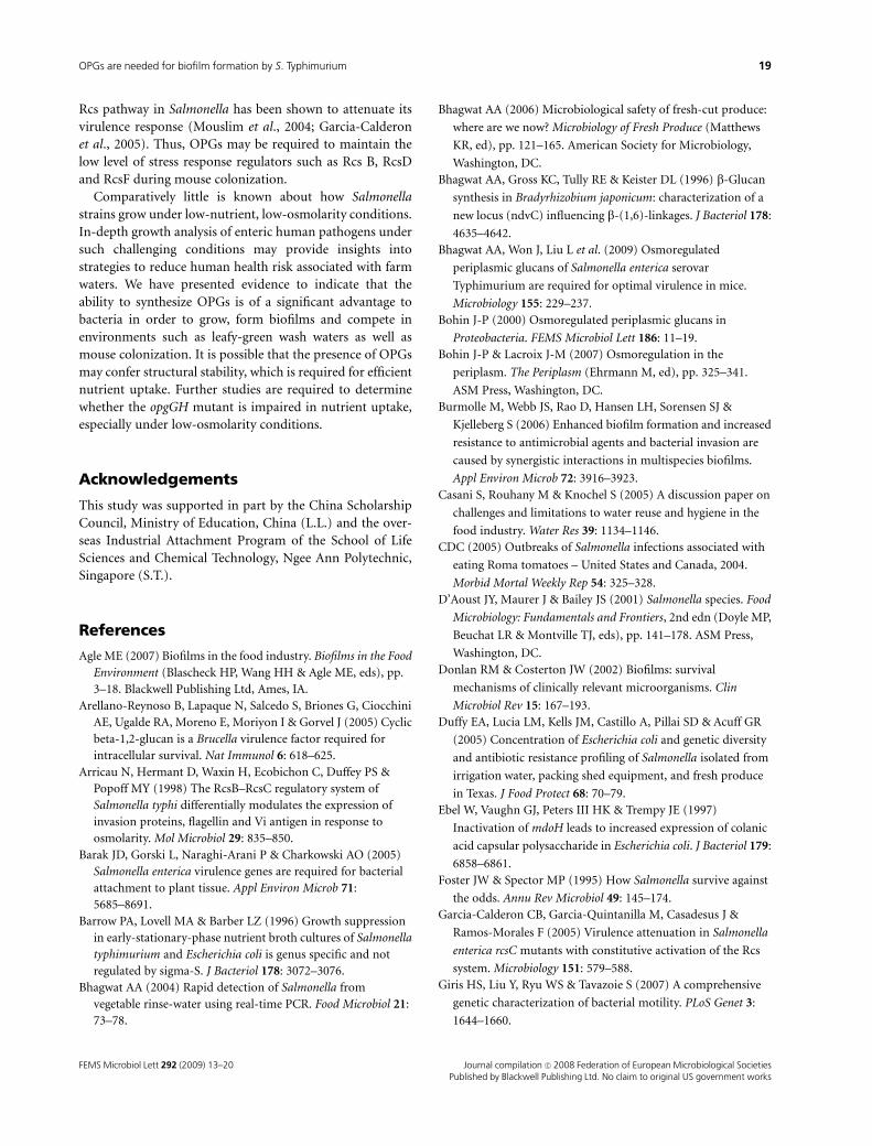
(Figs 3 and 4, gray bars). Likewise, in vegetable wash waters
obtained from spinach and green collard (Fig. 4), the CI for
the opgGH mutant was o 0.01 in biofilms, indicating that
the OPG-lacking cells were outnumbered by the wild-type
strain in biofilms. Viable count data from biofilms obtained
from vegetable wash waters indicated that the biofilms were
mainly composed of the wild-type strain (o 1.0% of the
total viable cells carried the opgGH mutation). It may be
noted that in pure cultures, very low or no biofilm forma-
tion by the opgGH mutant strain was observed in vegetable
wash waters (Fig. 2) (o 103 cells mL�1 were recovered from
individual wells, data not shown).
Role of OPGs in mouse organ colonization
Recently, we observed that lack of OPG synthesis rendered
Salmonella less virulent, and c. 2-log more cells were needed
Fig. 2. Biofilm formation by Salmonella enterica serovar Typhimurium
wild-type and opgGH mutant strains. Growth was measured by deter-
mining A600 nm. Biofilm was measured after staining with crystal violet.
The ratio of A600 nm after crystal violet staining to initial growth turbidity
(A600 nm) is plotted for the wild-type (black bars) and the opgGH strain
(gray bars) after incubation in LNNS and vegetable wash waters. �A600 nm
of crystal violet o 0.001.
Osmolarity of medium (mosmol L−1)
0 50 100 150 200
CI (
opgG
H /
wild
type
)
0.1
1
Fig. 3. Competitive growth of Salmonella enterica serovar Typhimurium
wild-type and opgGH mutant strain in planktonic and biofilm states in
LNNS medium at varying osmolarities. Wild-type and opgGH mutant
strains were inoculated in equal proportions in LNNS media with varying
osmolarities. Viable cell counts were determined from biofilms (�) and
planktonic cells (�) on selective media containing antibiotics. CI is the
ratio of viable cell counts for the opgGH mutant to that of the wild-type
cells at the time of harvest.
FEMS Microbiol Lett 292 (2009) 13–20 Journal compilation c� 2008 Federation of European Microbiological SocietiesPublished by Blackwell Publishing Ltd. No claim to original US government works
17OPGs are needed for biofilm formation by S. Typhimurium
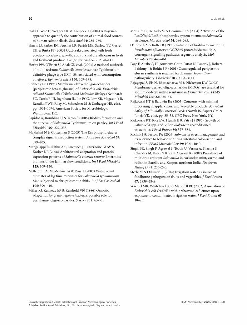
to achieve an LD50 comparable to that of wild-type cells
(Bhagwat et al., 2009). We inoculated mice with equal
proportions of wild-type and opgGH mutant strains and
examined their colonization in the intestine, spleen and liver
(Fig. 5). Although there was variation among individual
mice, the average CI for the intestine was 0.0001, while the
values for the spleen and the liver were 0.08 and 0.09,
respectively. The data indicated that the Salmonella strain
lacking OPGs was unable to colonize the intestinal track in
competition with the wild-type strain. On the other hand,
during the later stages of infection involving spleen and liver
colonization, the opgGH mutant was outnumbered by the
wild-type strain only by c. 10-fold.
Discussion
Water reuse is increasingly regarded as a necessity. Currently,
reconditioned water is used for initial washing of vegetables,
fluming of unprepared products and scalding water of meat
and poultry (Rajkowski et al., 1996; Rajkowski & Baldwin,
2003; Casani et al., 2005). Agricultural and farm wash waters
are likely to have low-nutrient low-osmolarity conditions.
However, little is known about the microbial factors that are
critical to growth, survival and biofilm formation by food-
borne pathogens such as Salmonella under such conditions
(Rychlik & Barrow, 2005). We have demonstrated here that
the ability to synthesize OPGs is beneficial for maintaining
the optimal growth potential. The longer time needed by
Salmonella under low-osmotic conditions could be due to
the instability of the membrane structures created due to the
lack of OPGs. As the name indicates, OPGs are osmoregu-
lated and their synthesis and accumulation is inversely
proportional to the osmotic strength of the environment
(Bohin, 2000). Thus, after increasing medium osmolarity
from 31 to 240 mosmol L�1, bacterial growth was relatively
independent of OPG synthesis (Fig. 1 and Table 1). Salmonella
strain lacking OPGs was a poor competitor in various
mouse organs as well as in low-osmolarity low-nutrient
media in planktonic and biofilm states (Figs 3–5).
OPGs were suggested to play a critical role in conferring
resistance to SDS in E. coli (Rajagopal et al., 2003). It was
postulated that anionic detergents such as SDS would be
repelled by the OPGs. In E. coli, lack of OPGs also resulted in
increased colonic acid production and reduced motility
(Kennedy, 1996; Ebel et al., 1997). It was further demon-
strated that secondary mutations in the rcs gene family
(namely, rcsB, rcsD and rcsF) restored motility in opgGH
mutant strains of E. coli (Giris et al., 2007). The authors
suggested that the rcs pathway could be hyperactivated in
opgGH mutants because rcsB and rcsD act as repressors of
flagella synthesis (Majdalani & Gottesman, 2005). Based on
what is observed in E. coli, the Rcs pathway might be
overactivated under low-osmolarity conditions in the
opgGH mutant strain of Salmonella. Hyperactivation of the
CI (
opgG
H /
wild
type
)
0.01
0.1
1
1/20
LBNS
Spinac
h
Green
colla
rd
Lettu
ce
*
Fig. 4. CI during biofilm formation by Salmonella enterica serovar
Typhimurium wild-type and opgGH mutant strains in vegetable wash
waters. Wild-type and opgGH mutant strains were mixed in equal
proportions and inoculated in LNNS or vegetable wash waters with (gray
bars) or without (black bars) addition of NaCl to 0.15 M (final concentra-
tion). Viable cell counts were determined from biofilms by plating on
selective media containing antibiotics. CI is the ratio of viable cells of the
opgGH mutant to that of the wild type recovered 24 h postinoculation
from biofilms. �Viable cell counts of the opgGH mutant strain were
below the detection limit (o 102 cells mL�1).
Fig. 5. Competitive colonization of mouse organs after coinoculation
with Salmonella enterica serovar Typhimurium wild-type and opgGH
mutant strain 6 days postinfection. CI is the ratio between the opgGH
mutant strain and the wild type in the individual organs divided by the
ratio of the two strains in the inoculum. The distribution of CI from
individual animals is shown in a box and whisker format. Boxes range
from the 25th to the 75th percentile and are intersected by the median
line. Whiskers extending below and above the box range from the 10th
to the 90th percentile, respectively.
FEMS Microbiol Lett 292 (2009) 13–20Journal compilation c� 2008 Federation of European Microbiological SocietiesPublished by Blackwell Publishing Ltd. No claim to original US government works
18 L. Liu et al.
Rcs pathway in Salmonella has been shown to attenuate its
virulence response (Mouslim et al., 2004; Garcia-Calderon
et al., 2005). Thus, OPGs may be required to maintain the
low level of stress response regulators such as Rcs B, RcsD
and RcsF during mouse colonization.
Comparatively little is known about how Salmonella
strains grow under low-nutrient, low-osmolarity conditions.
In-depth growth analysis of enteric human pathogens under
such challenging conditions may provide insights into
strategies to reduce human health risk associated with farm
waters. We have presented evidence to indicate that the
ability to synthesize OPGs is of a significant advantage to
bacteria in order to grow, form biofilms and compete in
environments such as leafy-green wash waters as well as
mouse colonization. It is possible that the presence of OPGs
may confer structural stability, which is required for efficient
nutrient uptake. Further studies are required to determine
whether the opgGH mutant is impaired in nutrient uptake,
especially under low-osmolarity conditions.
Acknowledgements
This study was supported in part by the China Scholarship
Council, Ministry of Education, China (L.L.) and the over-
seas Industrial Attachment Program of the School of Life
Sciences and Chemical Technology, Ngee Ann Polytechnic,
Singapore (S.T.).
References
Agle ME (2007) Biofilms in the food industry. Biofilms in the Food
Environment (Blascheck HP, Wang HH & Agle ME, eds), pp.
3–18. Blackwell Publishing Ltd, Ames, IA.
Arellano-Reynoso B, Lapaque N, Salcedo S, Briones G, Ciocchini
AE, Ugalde RA, Moreno E, Moriyon I & Gorvel J (2005) Cyclic
beta-1,2-glucan is a Brucella virulence factor required for
intracellular survival. Nat Immunol 6: 618–625.
Arricau N, Hermant D, Waxin H, Ecobichon C, Duffey PS &
Popoff MY (1998) The RcsB–RcsC regulatory system of
Salmonella typhi differentially modulates the expression of
invasion proteins, flagellin and Vi antigen in response to
osmolarity. Mol Microbiol 29: 835–850.
Barak JD, Gorski L, Naraghi-Arani P & Charkowski AO (2005)
Salmonella enterica virulence genes are required for bacterial
attachment to plant tissue. Appl Environ Microb 71:
5685–8691.
Barrow PA, Lovell MA & Barber LZ (1996) Growth suppression
in early-stationary-phase nutrient broth cultures of Salmonella
typhimurium and Escherichia coli is genus specific and not
regulated by sigma-S. J Bacteriol 178: 3072–3076.
Bhagwat AA (2004) Rapid detection of Salmonella from
vegetable rinse-water using real-time PCR. Food Microbiol 21:
73–78.
Bhagwat AA (2006) Microbiological safety of fresh-cut produce:
where are we now? Microbiology of Fresh Produce (Matthews
KR, ed), pp. 121–165. American Society for Microbiology,
Washington, DC.
Bhagwat AA, Gross KC, Tully RE & Keister DL (1996) b-Glucan
synthesis in Bradyrhizobium japonicum: characterization of a
new locus (ndvC) influencing b-(1,6)-linkages. J Bacteriol 178:
4635–4642.
Bhagwat AA, Won J, Liu L et al. (2009) Osmoregulated
periplasmic glucans of Salmonella enterica serovar
Typhimurium are required for optimal virulence in mice.
Microbiology 155: 229–237.
Bohin J-P (2000) Osmoregulated periplasmic glucans in
Proteobacteria. FEMS Microbiol Lett 186: 11–19.
Bohin J-P & Lacroix J-M (2007) Osmoregulation in the
periplasm. The Periplasm (Ehrmann M, ed), pp. 325–341.
ASM Press, Washington, DC.
Burmolle M, Webb JS, Rao D, Hansen LH, Sorensen SJ &
Kjelleberg S (2006) Enhanced biofilm formation and increased
resistance to antimicrobial agents and bacterial invasion are
caused by synergistic interactions in multispecies biofilms.
Appl Environ Microb 72: 3916–3923.
Casani S, Rouhany M & Knochel S (2005) A discussion paper on
challenges and limitations to water reuse and hygiene in the
food industry. Water Res 39: 1134–1146.
CDC (2005) Outbreaks of Salmonella infections associated with
eating Roma tomatoes – United States and Canada, 2004.
Morbid Mortal Weekly Rep 54: 325–328.
D’Aoust JY, Maurer J & Bailey JS (2001) Salmonella species. Food
Microbiology: Fundamentals and Frontiers, 2nd edn (Doyle MP,
Beuchat LR & Montville TJ, eds), pp. 141–178. ASM Press,
Washington, DC.
Donlan RM & Costerton JW (2002) Biofilms: survival
mechanisms of clinically relevant microorganisms. Clin
Microbiol Rev 15: 167–193.
Duffy EA, Lucia LM, Kells JM, Castillo A, Pillai SD & Acuff GR
(2005) Concentration of Escherichia coli and genetic diversity
and antibiotic resistance profiling of Salmonella isolated from
irrigation water, packing shed equipment, and fresh produce
in Texas. J Food Protect 68: 70–79.
Ebel W, Vaughn GJ, Peters III HK & Trempy JE (1997)
Inactivation of mdoH leads to increased expression of colanic
acid capsular polysaccharide in Escherichia coli. J Bacteriol 179:
6858–6861.
Foster JW & Spector MP (1995) How Salmonella survive against
the odds. Annu Rev Microbiol 49: 145–174.
Garcia-Calderon CB, Garcia-Quintanilla M, Casadesus J &
Ramos-Morales F (2005) Virulence attenuation in Salmonella
enterica rcsC mutants with constitutive activation of the Rcs
system. Microbiology 151: 579–588.
Giris HS, Liu Y, Ryu WS & Tavazoie S (2007) A comprehensive
genetic characterization of bacterial motility. PLoS Genet 3:
1644–1660.
FEMS Microbiol Lett 292 (2009) 13–20 Journal compilation c� 2008 Federation of European Microbiological SocietiesPublished by Blackwell Publishing Ltd. No claim to original US government works
19OPGs are needed for biofilm formation by S. Typhimurium
Hald T, Vose D, Wegner HC & Koupeev T (2004) A Bayesian
approach to quantify the contribution of animal-food sources
to human salmonellosis. Risk Anal 24: 255–269.
Harris LJ, Farber JN, Beuchat LR, Parish ME, Suslow TV, Garret
EH & Busta FF (2003) Outbreaks associated with fresh
produce: incidence, growth, and survival of pathogens in fresh
and fresh-cut produce. Compr Rev Food Sci F 2: 78–141.
Horby PW, O’Brien SJ, Adak GK et al. (2003) A national outbreak
of multi-resistant Salmonella enterica serovar Typhimurium
definitive phage type (DT) 104 associated with consumption
of lettuce. Epidemiol Infect 130: 169–178.
Kennedy EP (1996) Membrane-derived oligosaccharides
(periplasmic beta-D-glucans) of Escherichia coli. Escherichia
coli and Salmonella Cellular and Molecular Biology (Neidhardt
FC, Curtis R III, Ingraham JL, Lin ECC, Low KB, Magasanik B,
Reznikoff WS, Riley M, Schaechter M & Umbarger HE, eds),
pp. 1064–1074. American Society for Microbiology,
Washington, DC.
Lapidot A, Rombling U & Yaron S (2006) Biofilm formation and
the survival of Salmonella Typhimurium on parsley. Int J Food
Microbiol 109: 229–233.
Majdalani N & Gottesman S (2005) The Rcs phosphorelay: a
complex signal transduction system. Annu Rev Microbiol 59:
379–405.
Mangalappalli-Illathu AK, Lawrence JR, Swerhone GDW &
Korber DR (2008) Architectural adaptation and protein
expression patterns of Salmonella enterica serovar Enteritidis
biofilms under laminar flow conditions. Int J Food Microbiol
123: 109–120.
Mellefont LA, McMeekin TA & Ross T (2005) Viable count
estimates of lag time responses for Salmonella typhimurium
M48 subjected to abrupt osmotic shifts. Int J Food Microbiol
105: 399–410.
Miller KJ, Kennedy EP & Reinhold VN (1986) Osmotic
adaptation by gram-negative bacteria: possible role for
periplasmic oligosaccharides. Science 231: 48–51.
Mouslim C, Delgado M & Groisman EA (2004) Activation of the
RcsC/YojN/RcsB phosphorelay system attenuates Salmonella
virulence. Mol Microbiol 54: 386–395.
O’Toole GA & Kolter R (1998) Initiation of biofilm formation in
Pseudomonas fluorescens WCS365 proceeds via multiple,
convergent signalling pathways: a genetic analysis. Mol
Microbiol 28: 449–461.
Page F, Altabe S, Hugouvieux-Cotte-Pattat N, Lacorix J, Robert-
Baidouy J & Bohin J-P (2001) Osmoregulated periplasmic
glucan synthesis is required for Erwinia chrysanthemi
pathogenicity. J Bacteriol 183: 3134–3141.
Rajagopal S, Eis N, Bhattacharya M & Nickerson KW (2003)
Membrane-derived oligosaccharides (MDOs) are essential for
sodium dodecyl sulfate resistance in Escherichia coli. FEMS
Microbiol Lett 223: 25–31.
Rajkowski KT & Baldwin EA (2003) Concerns with minimal
processing in apple, citrus, and vegetable products. Microbial
Safety of Minimally Processed Foods (Novak JS, Sapers GM &
Juneja VK, eds), pp. 35–52. CRC Press, New York, NY.
Rajkowski KT, Rice EW, Huynh B & Patsy J (1996) Growth of
Salmonella spp. and Vibrio cholerae in reconditioned
wastewater. J Food Protect 59: 577–581.
Rychlik I & Barrow PA (2005) Salmonella stress management and
its relevance to behaviour during intestinal colonisation and
infection. FEMS Microbiol Rev 29: 1021–1040.
Singh BR, Singh P, Agrawal S, Teotia U, Verma A, Sharma S,
Chandra M, Babu N & Kant Agarwal R (2007) Prevalence of
multidrug resistant Salmonella in coriander, mint, carrot, and
radish in Bareilly and Kanpur, northern India. Foodborne
Pathog Dis 4: 233–240.
Steele M & Odumeru J (2004) Irrigation water as source of
foodborne pathogens on fruits and vegetables. J Food Protect
67: 2839–2849.
Wachtel MR, Whitehead LC & Mandrell RE (2002) Association of
Escherichia coli O157:H7 with preharvest leaf lettuce upon
exposure to contaminated irrigation water. J Food Protect 65:
18–25.
FEMS Microbiol Lett 292 (2009) 13–20Journal compilation c� 2008 Federation of European Microbiological SocietiesPublished by Blackwell Publishing Ltd. No claim to original US government works
20 L. Liu et al.