osteochondral#fracture#in#40m50%# - bonefixbonefix.co.nz/portals/160/images/1 patellar...
TRANSCRIPT


• Is a malady commonly seen in the orthopaedic office. • MPFL to be the major medial so: ;ssue stabilizer, providing 53% of the
total restraining force.
• Symptoms are occasionally preceded by a trauma;c event but more commonly are insidious in onset.
• Lateral transla;on of the patella is the most common direc;on of patellar subluxa;on and is usually associated with malalignment of the lower extremity. In pa;ents with lateral subluxa;on, the trochlea has a structural role in centralizing the patella during knee flexion.
• Osteochondral fracture in 40-‐50%
• Mean Q-‐angle values approach 10 degrees in men and 15 degrees in women.

• Tenderness over the medial epicondyle (BasseS’s sign) may represent an injury to the MPFL in pa;ents with acute or recurrent patellar disloca;ons
• J sign: In pa;ents with patellar subluxa;on, however, the patella travels from a central posi;on within the femoral trochlea at 30 degrees of flexion to a laterally subluxated posi;on in full xtension.
• The lateral excursion during terminal knee extension, referred to as the J sign, is pathognomonic of lateral patellar subluxa;on.
• The ini;al radiographic evalua;on of the patellofemoral joint should include standard anteroposterior and lateral weight-‐bearing views as well as an axial radiograph.

• CT allows axial cuts of the patellofemoral ar;cula;on at angles less than 20 degrees of knee flexion.
• This enhances the detec;on of subluxa;on as the patella loses the stabilizing func;on of the lateral femoral condyle.
• An axial CT image demonstra;ng the femoral trochlear groove is superimposed on an axial image of the ;bial tubercle. tubercle. Values greater than 9 mm is significant
• Redisloca;on a:er non op: 15-‐44%

JBJS 2011;93-‐B:1341–7. • Patella subluxa+on assessed on dynamic MRI has previously been shown to be associated with
anterior knee pain. • Patella engagement (% of patella car+lage overlapping with trochlea car+lage) had the • strongest rela+onship with subluxa+on. Patellae with > 30% engagement tended not to • sublux; those with < 30% tended to sublux.
• Other factors that were associated with subluxa+on included the +bial tubercle-‐trochlea notch distance, vastus medialis obliquus distance from patella, patella alta, and the bony and car+laginous sulcus angles in the superior part of the trochlea.
• No rela+onship was found between subluxa+on and sulcus angles for car+lage and bone in the middle and lower part of the trochlea, car+lage thicknesses and Wiberg classifica+on of the patella.
• This study indicates that patella engagement is a key factor associated with patellar • Subluxa+on.

Acute Trauma+c Disloca+on Am J Sports Med July 2000 vol. 28 no. 4 472-‐479
History taking is important: a. Trivial or significant injury b. Requires Hospital or self reduc;on • c. Bilateral, Other joints d. Family e. Ligament laxity syndrome • Ini;al disloca;on and treatment
• Impairments: Walk, stairs, etc, Locking, giving,
• Pain or instability is the problem:
Clinical Gait : What suggest internal torsion: Gait and foot progression angle Patellar squint Excessive internal rota;on Small Patella J Sign Size Lateral transla;on Apprehension test: knee in 30* on examiners; explain the test; note the pa;ent; gentle lateral push

How to look for crepitus? Feel with ac;ve movements
Is anterior or retropatellar pain is important? Yes. When present careful above medial and distal transposi;on. [anteriorisa;on is indicated]
ClaSerworthy: Arthroscope and then decide on the type of anteriorisa;on depending on car;lage loss in the patella.

X ray • AP
• Lateral in 30* flexion
• Axial or skyline view of PFJ
• Merchant view: 45* flexion of the X ray tube at 30* to the horizontal: Sulcus angle and congruent angle
• Laurin view: Knee flexed 20*and cassette held by the patient
Merchant
Laurin

Assessment of patellar position
Blumensaat’s line • Lateral X ray in 30* flexion • Line projected anteriorly from the intercondylar notch • Lower pole of the patella at this line
Insall Salvati Method [Lat X ray 30*] • T[Tendon]/P[patella] = 1.02 +/-0.13. Should not be more than 20%
• 1.2 = Patella alta • 0.8 = Patella Baja
• Not accurate: difficult to define the tibial tuberosity. Non-articular patella may be beaked.

½ = 0.95

• A true lateral with the posterior borders of the femoral condyle overlapping is needed to assess the trochlear groove depth; normally 7–8mm measured lcm from its upper limit.
• < 5mm is considered dysplastic.
• Dejours’s trochlear morphology: • (A) normal knee, the sulcus line is the
trochlear floor
• (B) Type I dysplasia. The medial • femoral condyle is deficient. The
sulcus line joins the the medial condyle
• (C) Type II dysplasia. The crossing of the two condylar outlines of the trochlear floor is symmetrical but
• situated distally.
• (D) Type III dysplasia: the crossing of the two condylar outlines with asymmetry of the outline of the trochlear
Sky line view
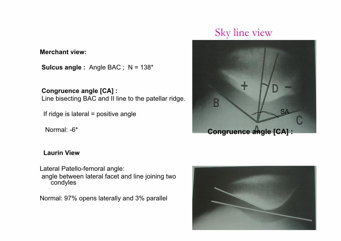
Merchant view: Sulcus angle : Angle BAC ; N = 138* Congruence angle [CA] : Line bisecting BAC and II line to the patellar ridge. If ridge is lateral = positive angle Normal: -6* Laurin View Lateral Patello-femoral angle: angle between lateral facet and line joining two
condyles Normal: 97% opens laterally and 3% parallel
Congruence angle [CA] :
SA

A. Insall ra;o 0.8 -‐1.2 [Lig/patella] with knee in 30* of flexion B. Blackburne-‐Peel :0.8-‐1.1

Normal values
Normal Patella Pain Patella instability Insall T/P 1.06 1.09 1.30
Sulcus angle 138* 138* 153*
Congruence angle -6.7 -9.2 +16.6
Lateral P-F angle 20* 39* 3*
P-F index 1.4 1.4 3.2
CT
Congruence angle 13* -5.7* +4.2
Patella tilt angle 15* 14* 4.85
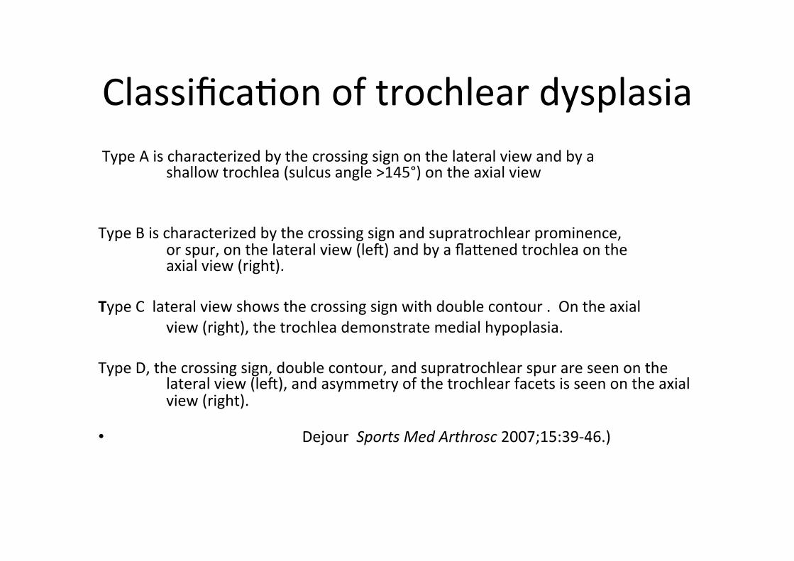
Classifica;on of trochlear dysplasia Type A is characterized by the crossing sign on the lateral view and by a shallow trochlea (sulcus angle >145°) on the axial view Type B is characterized by the crossing sign and supratrochlear prominence, or spur, on the lateral view (le:) and by a flaSened trochlea on the axial view (right). Type C lateral view shows the crossing sign with double contour . On the axial view (right), the trochlea demonstrate medial hypoplasia. Type D, the crossing sign, double contour, and supratrochlear spur are seen on the lateral view (le:), and asymmetry of the trochlear facets is seen on the axial view (right). • Dejour Sports Med Arthrosc 2007;15:39-‐46.)

TT-‐TG distance TT-SF: Tibial tuberosity – Trochlear groove distance
This distance is more accurate measurement than Q angle
> 20 mm is pathological

What are the changes on MRI. These changes required for ACC
• Why medial: Reloca;on injury
• Axial fat suppressed T2-‐weighted MR image
• Shows offset bone bruises in the medial aspect of the patella and in the lateral aspect of the lateral femoral condyle , a pathognomonic sign of patellar disloca+on.
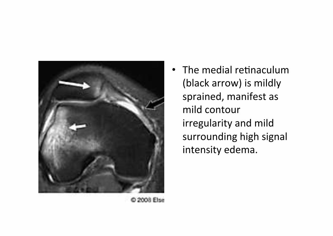
• The medial re;naculum (black arrow) is mildly sprained, manifest as mild contour irregularity and mild surrounding high signal intensity edema.

MPFL in Patellar disloca;on Am J Sports Med August 2009 vol. 37 no. 8 1513-‐1521
• 42 cases, 7 year follow up
• MPFL rupture: Femoral in 35 pa;ents, midsubstance in 11, and patellar in 7.
• At follow-‐up, 15 pa;ents [40%] • [13 femoral, 1 patellar, 1 midsubstance]
• Control MRI showed full-‐thickness patellofemoral car;lage lesions in 50% of the pa;ents, unrelated to MPFL injury loca;on
• An MPFL avulsion at the femoral aSachment in primary trauma;c patellar disloca;ons predicts subsequent patellar instability. The authors suggest that MPFL injury loca;on be taken into account when planning treatment of primary trauma;c patellar disloca;on


• MPFL is in between ME and adductor tubercle.
• Gracillis tendon and select isometric point
• Brace for 6 weeks
• No patella alta or PTT distance less than 20

Surgeries
• Patella alta
• Or
• PTT >20
• Distal surgeries: Goldthwaite before the growth
• Transfer ;bial tubercle a:er the growth
Fulkerson: JOASS January 2011, Vol 19, No 1
• Medial patellofemoral ligament reconstruc;on is recommended for patellofemoral instability in the presence of trochlear dysplasia in pa;ents without patella alta or increased ;bial tubercle–trochlear groove distance.
• Trochleoplasty should be reserved for severe dysplasia in which patellofemoral stability cannot otherwise be obtained.

• The gracilis or semitendinosus tendon is looped through the longitudinal patellar tunnel, passed under the fascia and fixed in a drill hole in the medial femoral condyle with an interference screw
Natural course
• Fithian et al2 reported recurrence in 49% of pa;ents with at least two prior instability events.
• Trochlear dysplasia is es;mated to occur in <2% of the popula;on;however, it is present in up to 85% of pa;ents with recurrent patellar instability.

• Predisposing factors (eg, hyperlaxity, rota;onal malalignment, lateral extensor mechanism vector) are known to cause recurrent patellar subluxa;on or disloca;on, a lateralized patellar res;ng posi;on, and distor;on of trochlear morphology.
• Lateral patellar posi;oning may limit the development of normal trochlear depth and morphology.
• Persistent lateral patellar tracking can result in a flaSened lateral trochlea and can indirectly create a shallow groove. This scenario is sugges;ve of a developmental process.
• Patellar disloca;on typically occurs in persons with several anatomic risk factors. Patellar disloca;on likely is caused by a combina;on of congenital and developmental factors.

Fulkerson anterior tibial osteotomy 88A Aug
• The obliquity of the osteotomy gives more anteriorisation or more medial transfer of the tubercle.
• Less oblique osteotomy giving more medialisation and less anteriorisation