osteogenically-induced exosomes stimulate osteogenesis of
TRANSCRIPT

Osteogenically-induced exosomes stimulate osteogenesisof human adipose-derived stem cells
Mengru Zhu . Yang Liu . Hongzhi Qin . Shuang Tong . Qiang Sun .
Ting Wang . Hua Zhang . Mengying Cui . Shu Guo
Received: 5 April 2020 / Accepted: 21 September 2020 / Published online: 20 November 2020
� The Author(s) 2020
Abstract Exosomes exhibit great therapeutic poten-
tial in bone tissue engineering. The study aimed to
investigate whether the exosomes derived from human
adipose-derived stem cells (hADSCs-Exos) during
different time-span of osteogenic differentiation could
promote osteogenesis. The appropriate concentrations
of hADSCs-Exos to enhance the proliferation, migra-
tion and osteogenesis of hADSCs-Exos were also
examined. PKH67 labelled hADSCs-Exos was used to
detect the internalization ability of hADSCs. The
osteogenic differentiation abilities of hADSCs after
treatment with hADSCs-Exos was evaluated by
Alizarin red staining (ARS). The proliferation and
migration of hADSCs was examined by cell counting
kit-8 and wound healing assay, respectively. The
expression of exosomal surface markers and osteo-
blast-related protein of hADSCs was assessed by
Western blot. PKH67-labelled exosomes were inter-
nalized by hADSCs after 4 h incubation. ARS showed
that the amount of mineralized nodules in Exo1-14d
group was significantly higher than that in Exo15-28d
group. hADSCs-Exos could promote the proliferation
and migration capacity of hADSCs. Western blot
analysis showed that after hADSCs-Exos treatment,
ALP and RUNX2 were significantly enhanced. Spe-
cially, the Exo1-14d group of 15 lg/mL significantly
upregulated the expression of RUNX2 than the other
exosomes treated groups. Our findings suggest that
exosomes secreted by hADSCs during osteogenic
induction for 1–14 days could be efficiently internal-
ized by hADSCs and could induce osteogenic differ-
entiation of hADSCs. Moreover, administration of
Exo1-14d at 15 lg/mL promoted the proliferation and
migration of hADSCs. In conclusion, our research
confirmed that comprised of hADSCs-Exos and
hADSCs may provide a new therapeutic paradigm
for bone tissue engineering.
Keywords Human adipose-derived stem cells �Exosomes � Osteogenic differentiation � Bone tissueengineering
Abbreviations
hADSCs Human adipose-derived stem cells
Mengru Zhu and Yang Liu have contributed equally to this
work.
M. Zhu � H. QinDepartment of plastic surgery, The First Affiliated
Hospital of Dalian Medical University, 222 Zhongshan
Road, Dalian 116011, China
Y. Liu
School of Chemical Engineering, Dalian University of
Technology, No. 2 Linggong Road, Dalian 116024, China
S. Tong � Q. Sun � T. Wang � H. Zhang �M. Cui � S. Guo (&)
Department of Plastic surgery, The First affiliated
Hospital of China Medical University, No 155 Nanjing
North Street, Shenyang 110002, China
e-mail: [email protected]
123
Cell Tissue Bank (2021) 22:77–91
https://doi.org/10.1007/s10561-020-09867-8(0123456789().,-volV)( 0123456789().,-volV)
hADSCs-
Exos
Human adipose-derived stem cells-
derived exosomes
BMSCs Bone marrow-derived mesenchymal
stem cells
MSCs Mesenchymal stem cells
TEM Transmission electron microscopy
PM Proliferation medium
OM Osteogenic medium
ARS Alizarin red staining
DLS Dynamic light scattering
ALP Alkaline phosphatase
ECM Extracellular matrix
EVs Extracellular vesicles
CCK-8 Cell Counting Kit-8
FBS Fetal bovine serum
PBS Phosphate-buffered saline
DAPI 6-Diamidino-2-phenylindole
RUNX2 Runt-related transcription factor 2
Introduction
Globally, a large number of people have suffered from
bone defects owing to trauma, infection osteonecrosis,
congenital deformities, resection of tumors and other
bone diseases (Chen et al. 2010; Seong et al. 2010). To
date, bone grafting is considered as the ‘‘gold
standard’’ for treatment of bone defects, including
autologous bone grafts, allogeneic bone grafts, and
bone-graft substitutes (Dimitriou et al. 2011). How-
ever, all these techniques have certain limitations,
such as donor-site defect, poor bone quality and
limited availability of grafting material (Du et al.
2019; Lord et al. 1988).
Recently, the remarkable development of bone
tissue engineering has brought the dawn to the therapy
of bone defects (El-Rashidy et al. 2017). Mesenchy-
mal stem cells (MSCs) are a population of self-
renewing multipotent cells that have been proposed as
promising candidates for regenerative medicine (An
et al. 2019; Xie et al. 2016; Zuk et al. 2002), especially,
bone marrow-derived mesenchymal stem cells
(BMSCs), are regarded as one of the most widely
applied stem cell with great clinical potential for
regenerative medicine and cell-based therapies.
Osteogenic differentiation of BMSCs facilitate the
recovery of osteoporosis has been widely reported
(Huo et al. 2018; Oryan et al. 2017). Nevertheless,
BMSCs exert drawbacks that may hinder their clinical
applications with regards to insufficient cell number
and complex harvesting procedures. Alternatively,
human adipose-derived stem cells (hADSCs) are
considered an ideal cell source (Kim et al. 2007;
Schaffler and Buchler 2007), which can be extensively
extracted from discarded adipose tissue. Compared
with BMSCs, ADSCs have the following advantages,
including abundant sources, easy access, low immuno-
genicity, rapid proliferation with multilineage poten-
tial and few ethical concerns (An et al. 2019). More
importantly, the osteogenic capacity of hADSCs
appears to be minimally affected by aging, which
enhancing the clinical applicability for the gerontal
patient (Liao and Chen 2014; Shi et al. 2005; Wu et al.
2013).
Emerging evidences have shown that hADSCs can
differentiate into osteoblasts participating the bone
regeneration in vivo and in vitro (Chen et al. 2010; Xu
et al. 2017). They promote osteogenesis mainly by two
approaches. Firstly, differentiated hADSCs to target
cells that participate in osteogenesis; secondly, differ-
entiated hADSCs secrete a numerous of cytokines and
extracellular vesicles (EVs) taking part in cell-to-cell
communication via paracrine manner to promote cell
proliferation, migration of various cell lines that are
mandatory to promote osteogenesis. The latter has
been confirmed to contribute more significantly than
the capacity of directly differentiating in tissue
regeneration (Ma et al. 2019; Phinney and Prockop
2007).
EVs released from hADSCs are involved in tissue
regeneration and contribute to the paracrine effect of
hADSCs (Cooper et al. 2018; Liu et al. 2020; Wong
et al. 2019). Exosomes are a type of EVs with a
diameters range between 30 and 150 nm (Mathivanan
et al. 2010; Tkach and Thery 2016), containing pivotal
functional biomolecules (DNA, proteins, mRNA,
microRNA and lipids) which modulate biological
behavior by horizontally transferring to recipient cells
(Hoshino et al. 2015). Current research about exosome
exhibit a broad range of therapeutic potentials,
including cancer, regenerative medicine and immune
disorders (Shimasaki et al. 2018; Urbanelli et al.
2015), and it is clear that exosomes are an important
avenue in the field of bone regeneration (Fang et al.
2019; Li et al. 2018b; Qin et al. 2016). The application
of hADSCs-derived exosomes (hADSCs-Exos) may
provide a novel strategy for bone tissue engineering
123
78 Cell Tissue Bank (2021) 22:77–91
and regeneration, since it has been found that
hADSCs-Exos could promote BMSCs osteoinductive
capacity under the condition of supplementing osteo-
genic medium in vivo and in vitro (Li et al. 2018a).
Nevertheless, considering prominent potential of
hADSCs in osteogenesis, the further research of
effects on combination of hADSCs-Exos with
hADSCs for bone regeneration is required. Therefore,
it is of great significance and value to evaluate the
appropriate stage and concentration of exosomes
during the osteoinductive process.
In the present study, we aimed to determine
whether exosomes from hADSCs during osteogenic
induction become internalized by target hADSCs and
influence hADSCs proliferation, migration and osteo-
genic differentiation in a stage-and concentration-
dependent manner. The combination application of
hADSCs and hADSCs-Exo is expected to provide new
ideas for stem cell therapy to stimulate osteogenesis.
Materials and methods
Isolation and culture of hADSCs
This study was approved by the Research Ethical
Committee of the First Hospital of China Medical
University. Human abdominal subcutaneous adipose
tissue acquired with informed consent were digested
by 0.1% type I collagenase (Worsington, USA) in a
37 �C water bath for 45 min. The digestion was
terminated using hADSCs culture medium (Cyagen
Biosciences, USA) containing 10% fetal bovine serum
(FBS, Gibco, USA) and was centrifuged at 1200 rpm
for 5 min. The cell pellet was then suspended
in hADSCs proliferation medium (PM, hADSCs cul-
ture medium containing 10% FBS, 1% penicillin-
treptomycin, and 1% glutamine [Cyagen Biosciences,
USA]) and cultured into a 75-cm2 cell culture flask
(Corning, USA). The cells were incubated at 37 �C in
5% CO2 incubator (Thermo Forma, USA), and were
passaged until passage 4 for subsequent experiments.
Characterization of hADSCs
The multi-differentiation of hADSCs was induced by
adipogenic, osteogenic and chondrogenic induction
medium (all from Cyagen Biosciences, USA), respec-
tively. The adipogenic induction was confirmed by Oil
red O staining at day 14, the calcium mineral deposits
were measured by Alizarin red staining (ARS) at day
21, and the chondrogenic induction was confirmed by
Alcian blue staining at day 28 (all from Cyagen
Biosciences, USA). An optical microscope (Olympus,
Tokyo, Japan) was used for observation and image
capture.
The expression of cell surface antigens was
analyzed by flow cytometry. Cell suspension was
collected and centrifuged at 1000 rpm for 5 min,
washed with phosphate-buffered saline (PBS, Gibco,
USA). The CD34, CD90 and CD105 antibodies (BD
Bioscienses, USA) were added to the cells and
incubated at room temperature for 30 min in the dark.
The cell suspension was then washed with PBS and
detected by flow cytometer (BD, USA, Aria II). The
cells without labelling were regarded as control group.
hADSCs-Exos extraction and identification
Exosomes were isolated from conditioned medium of
hADSCs in vitro. The exosomes-depleted FBS,
obtained by ultracentrifugation at 100,000 g for 3 h
as previously described (Nabet et al. 2017), was used
in all conditions in this study. To induce osteogenic
differentiation, hADSCs were seeded in osteogenic
induction media (OM) for 28 days. Exosomes from
different time-span were isolated from supernatants of
undifferentiated hADSCs in PM (Exo0d), and the
differentiated hADSCs during day 1 to 14 (Exo1-14d)
and day 15 to 28 (Exo15-28d), and purified by
sequential centrifugation. The conditioned medium
was collected every 3 days and centrifuged at 2000 g
for 30 min, and then at 20,000 g in a sterile Ultra-
ClearTM tube (Beckman Coulter, USA) for 1 h before
being passed through a 0.22 lm filter (Millipore,
USA) to eliminate some large particles. Afterward, the
filtered solution was then ultracentrifuged at
100,000 g for 1 h. Then, the exosomes were washed
with PBS and again pelleted at 100,000 g for 70 min
to remove protein contamination. Finally, the pelleted
exosomes were resuspended in PBS. All procedures
were performed at 4 �C.For exosomes identification, transmission electron
microscopy (TEM) was performed to monitor the
morphology of exosomes. In brief, 10 lL of Exo-
somes (Exo0d, Exo1-14d and Exo15-28d), were fixed
with 4% neutral paraformaldehyde (Sigma, USA) at
room temperature for 20 min by an Ultrasonic
123
Cell Tissue Bank (2021) 22:77–91 79
cleaning apparatus (KQ-250, KunShan, China).
Thereafter, the fixed exosomes were dropped onto
carbon-coated electron microscope copper mesh, and
air-dried for 15 min. Micrographs were obtained using
a JEM-2000EX TEM (JEOF, Japan). Dynamic light
scattering (DLS) was used to evaluate the size of
hADSCs-Exos. Briefly, 150 lL of exosomes suspen-
sion was diluted in PBS and dispersed at 70 kw for
30 min at room temperature. The hydrated particle
size was determined by laser doppler micro-elec-
trophoresis (Malvern Nano ZS, UK). Finally, the
markers (CD9 and CD63) expression was detected by
Western blot (all from abcam, UK). Exosome protein
was quantified using the bicinchoninic acid assay kit
(Solarbiol, China). Total protein was examined to
confirm the purity of the exosomes.
Exosomes labelling and confocal microscopy
For exosomes internalization assay, Exo0d, Exo1-14d
and Exo15-28d were labelled with PKH67 Green
Fluorescent Cell Linker Kit (Sigma, USA). Firstly,
15 lg exosomes suspended in 500 lL Dilut C, mixed
with equal volume of PKH67 dye were incubated at
37 �C for 8 min. The labelling reaction was termi-
nated by ultracentrifugation at 100,000 g for 1 h and
resuspended in PBS. Thereafter, the labelled exo-
somes were incubated with serum-free hADSCs with
seeding intensity of 2 9 104 cells/cm2 in 24-well plate
in dark for 4 h. The preparations were then fixed with
4% paraformaldehyde for 30 min, after that, the cell
nucleus were stained with 6-Diamidino-2-phenylin-
dole (DAPI, Sigma, USA) for 20 min. Exosomes
internalized by hADSCs was visualized with confocal
microscope (Leica SP8, Germany).
Exosomes treatment of hADSCs and ARS
To determine the optimal time-span of extracted
hADSC-Exos during hADSCs osteogenic differentia-
tion, the hADSCs of passage 4 were seeded into the
24-well plates, and was cultured in PM. After getting
70–80% confluence, the culture medium was replaced
by PM containing exosomes (Exo0d, Exo1-14d and
Exo15-28d) from different time-span of hADSCs
osteogenesis induction. hADSCs cultured in exo-
somes-free PM and OM were set as negative and
positive controls, respectively.
The mineralized deposits were evaluated by ARS
after incubation for 21 days. After fixation by 4%
paraformaldehyde for 30 min, the cells were washed
three times with PBS before staining with Alizarin red
S for 5 min, and then the samples were washed again
with PBS. For ARS quantitation the mineralized
deposits were dissolved in 10% cetylpyridinium
chloride (Sigma, USA) in dark for 30 min at room
temperature. The solution was transferred to a 96-well
plate with 100 lL/well. The optical density (OD)
values was measured at 562 nm using a microplate
reader (BioTek EL808, USA).
Cell proliferation assay
On the basis of the ARS results, Exo1-14d was selected
as the optimal time-span for hADSCs-Exos isolation
during hADSCs osteogenic induction in the following
experiments. Cell Counting Kit-8 (CCK-8) assay
(Dojindo, Kyushu Island, Japan) was carried out to
the detect the optimal concentration of hADSC-Exos
on the proliferation of hADSCs. In brief, hADSCs
were seeded into 96-well plate (5 9 103 cells/well) in
PM without serum. Subsequently, when the cells
grown into 60% confluence, the cells were exposed to
PM containing different concentrations of Exo1-14d
(10, 15 and 20 lg/mL) at 100 lL/well for 24, 48 and
72 h. Next, CCK-8 solution (10 lL/well) was added toeach well and incubated in dark at 37 �C for 3 h. The
absorbance at 450 nm was measured by spectropho-
tometric methods. Each experiment was repeated for
at least 3 times.
Cell migration assay
Cell migration was investigated with wound healing
assay. Firstly, hADSCs were seeded into 24-well
plates (2 9 104 cells/cm2) and incubated with PM.
After the cells fully attached, the confluent monolayer
was scratched by a sterile pipette tip. Each well was
washed with PBS to remove the cellular debris, and
then supplemented with 1 mL of non-serum condi-
tioned medium containing 10, 15 and 20 lg/mL of
Exo1-14d. The cells were photographed at 0, 12, and
24 h using microscope (Leica DMI4000B, Germany).
The migration area ratio was measured by Image J
software (National Institutes of Health, USA). The
migration area ratio = (A0 - An)/A0, where A0 rep-
resents the initial wound area (t = 0 h), and An
123
80 Cell Tissue Bank (2021) 22:77–91

represents the residual area of the wound at the assess
point (t = n h).
Alkaline phosphatase (ALP) staining and activity
analysis
After hADSCs were incubated with 10, 15 and 20 lg/mL of Exo1-14d for 7 and 14 days, the ALP staining
was performed using a BCIP/NBT ALP staining Kit
(Beyotime Technology, China) according to the
manufacturer’s instructions. The cells were incubated
at room temperature for 2 h in dark, and washed twice
with distilled water. The samples were observed by
microscope (Leica DMI4000B, Germany). Mean-
while, ALP activity was evaluated using an ALP
Assay Kit (Beyotime Technology, China). In brief, the
cells were lysed by 1% Triton X-100 (Sigma, USA),
and then centrifuged at 1200 rpm for 10 min. The
ALP activity was measured by detecting the OD at
405 nm.
Western blot analysis
Western blot was conducted to measure the exosomal
and osteoblast-related proteins. hADSCs were seeded
with PM in 6-well plate (2 9 104 cells/cm2). Until
reaching 80% confluence, the cells were incubated
with Exo1-14d at concentrations of 10, 15 and 20 lg/mL for 14 days. The cells or exosomes were lysed
with RIPA lysis buffer along with the Protease
inhibitor (Sigma-Aldrich, USA). Protein extracts were
directly heated at 100 �C for 7 min in 9 5 loading
buffer (Life Technology, USA) and separated on 10%
sodium dodecyl sulfate–polyacrylamide gel elec-
trophoresis (SDS-PAGE) gels (Solarbiol, China) then
transferred to the polyvinylidene difluoride (PVDF)
membrane (Solarbiol, China). The membranes were
blocked with 5% non-fat milk, and incubated at 4 �Covernight with primary antibodies CD9 and CD63
(1:500, Abcam, UK), anti-RUNX 2, and anti-ALP
(1:200, Santa Cruz), respectively. Then the mem-
branes were incubated with goat anti-mouse secondary
antibodies (1:1000, Abcam, UK). The results were
visualized using enhanced chemiluminescence
reagent (Beyotime Biotechnology, China) and imaged
by Image Analysis System (Tanon, Japan).
Statistics analysis
All data are expressed as mean ± SD, and performed
in triplicate. Differences between groups were ana-
lyzed by one-way analysis of variance (ANOVA). To
compare the samples pair-to-pair, the Bonferroni Test
or LSD Test was used when the variance was
homogeneous otherwise the Dunnett’s T3 Test was
used in this study. SPSS 20.0 was used for all
statistical analysis and differences were considered
to be statistically significant as a result of p\ 0.05.
Results
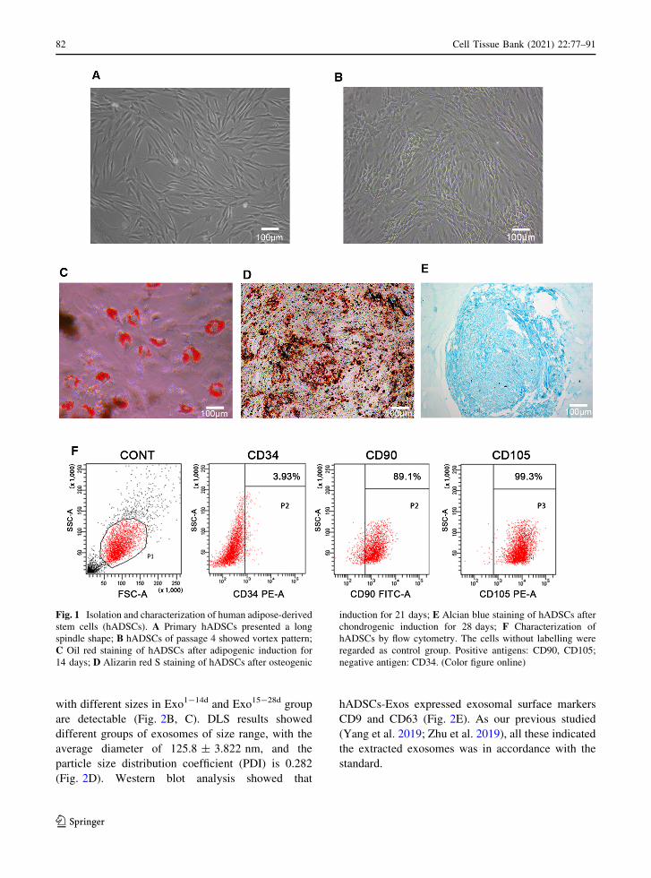
Characterization of hADSCs
Morphology of hADSCs was observed by inverted
microscope. Primary hADSCs substantially adhered to
the dish for 48 h, and then displayed typical fibrob-
lastic-shaped or polygon in morphology (Fig. 1A). It
can be seen that with the increase of the cell density the
hADSCs exhibited a vortex distribution at passage 4
(Fig. 1B).
To evaluate multilineage differentiation of
hADSCs, the cells of passage 4 were incubated in
adipogenic, osteogenic and chondrogenic induction
medium, respectively. The oil red staining results after
incubation in adipogenic medium for 14 days showed
that there were aggregated lipid droplets (Fig. 1C).
After incubation for 21 days, ARS results indicated
that there were a large number of calcified nodules
(Fig. 1D). Alcian blue staining results showed a blue
internal acid mucopolysaccharide in the cartilage
tissue after incubation in chondrogenic induction
medium for 28 days (Fig. 1E).
Flow cytometry analysis of hADSCs of passage 4
confirmed that cells were positive for CD 90 (89.1%),
CD105 (99.3%), and were negative for CD34 (3.93%)
(Fig. 1F).
Identification of hADSCs-Exos
TEM showed that exosomes extracted from condi-
tioned medium of hADSCs proliferation and different
time-span (Exo0d, Exo1-14d and Exo15-28d) of osteo-
genic induction possessed typical cup-shaped mor-
phology. The morphology of exosomes in Exo0d group
(Fig. 2A) was uniform and round. While exosomes
123
Cell Tissue Bank (2021) 22:77–91 81
with different sizes in Exo1-14d and Exo15-28d group
are detectable (Fig. 2B, C). DLS results showed
different groups of exosomes of size range, with the
average diameter of 125.8 ± 3.822 nm, and the
particle size distribution coefficient (PDI) is 0.282
(Fig. 2D). Western blot analysis showed that
hADSCs-Exos expressed exosomal surface markers
CD9 and CD63 (Fig. 2E). As our previous studied
(Yang et al. 2019; Zhu et al. 2019), all these indicated
the extracted exosomes was in accordance with the
standard.
Fig. 1 Isolation and characterization of human adipose-derived
stem cells (hADSCs). A Primary hADSCs presented a long
spindle shape; B hADSCs of passage 4 showed vortex pattern;
C Oil red staining of hADSCs after adipogenic induction for
14 days; D Alizarin red S staining of hADSCs after osteogenic
induction for 21 days; E Alcian blue staining of hADSCs after
chondrogenic induction for 28 days; F Characterization of
hADSCs by flow cytometry. The cells without labelling were
regarded as control group. Positive antigens: CD90, CD105;
negative antigen: CD34. (Color figure online)
123
82 Cell Tissue Bank (2021) 22:77–91

Internalization of exosomes in hADSCs
To investigate whether hADSCs-Exos could enter into
the hADSCs, the PKH67-labeled hADSCs-Exos
(Exo0d, Exo1-14d and Exo15-28d) were co-cultured
with native hADSCs for 4 h, and evaluated by
confocal microscopy analysis. The images demon-
strated that the exosomes (green) were internalized by
hADSCs and distributed in the cytoplasm. Moreover,
it is revealed that hADSCs-Exos tended to accumulate
in the perinuclear region (Fig. 3).
Optimal time-span of hADSCs-Exos for hADSCs
osteogenic differentiation
To study the osteoinductive activity of the exosomes
isolated from hADSCs at different time-span of
osteogenic induction, ARS staining was used as an
indicator. The ARS (Fig. 4A) and quantitative anal-
ysis of calcium nodule (Fig. 4B) showed that after co-
culturing for 21 days, only very few mineralized
nodules were formed in the negative group (PM) and
Exo0d group (APM = 0.091 ± 0.012, AExo0d =
0.172 ± 0.064), while the higher magnification
panels of calcified nodules (AExo1-14d =
1.851 ± 0.064) were observed in Exo1-14d group
(Fig. 4A). Moreover, only a small amount of dispersed
mineralized nodules (AExo15-28d = 1.290 ± 0.003)
were observed in Exo15-28d group. The calcium
nodule content in the OM group (AOM =
2.544 ± 0.091) was the highest among all the groups.
There were significantly more calcium nodules in the
Exo1-14d group than that in PM and Exo15-28d
(p\ 0.001). The above results indicated that
Exo1-14d could promote osteogenic differentiation.
Consequently, time-span of day 1–14 during osteo-
genic induction (Exo1-14d) was used in the following
studies.
Fig. 2 Extraction and identification of exosomes derived from
human adipose-derived stem cells (hADSCs-Exos) during
different time-spans of osteogenesis induction. The representa-
tive images of hADSCs-Exos during time-span of osteogenesis
induction at Day 0 (Exo0d, A), Day 1 to 14 (Exo1-14d, B), andDay 15 to 28 (Exo15-28d, C) were observed by transmission
electron microscopy (TEM). Black arrows indicated the
exosomes. Scale bar = 200 nm. D The particle size distribution
of hADSCs-Exos was measured by dynamic light scattering
(DLS) analysis, and the mean size of hADSCs-Exos was
125.8 ± 3.822 nm. E Western blot analysis showed that the
specific surface markers of exosomes (CD9 and CD63) were
detected
123
Cell Tissue Bank (2021) 22:77–91 83

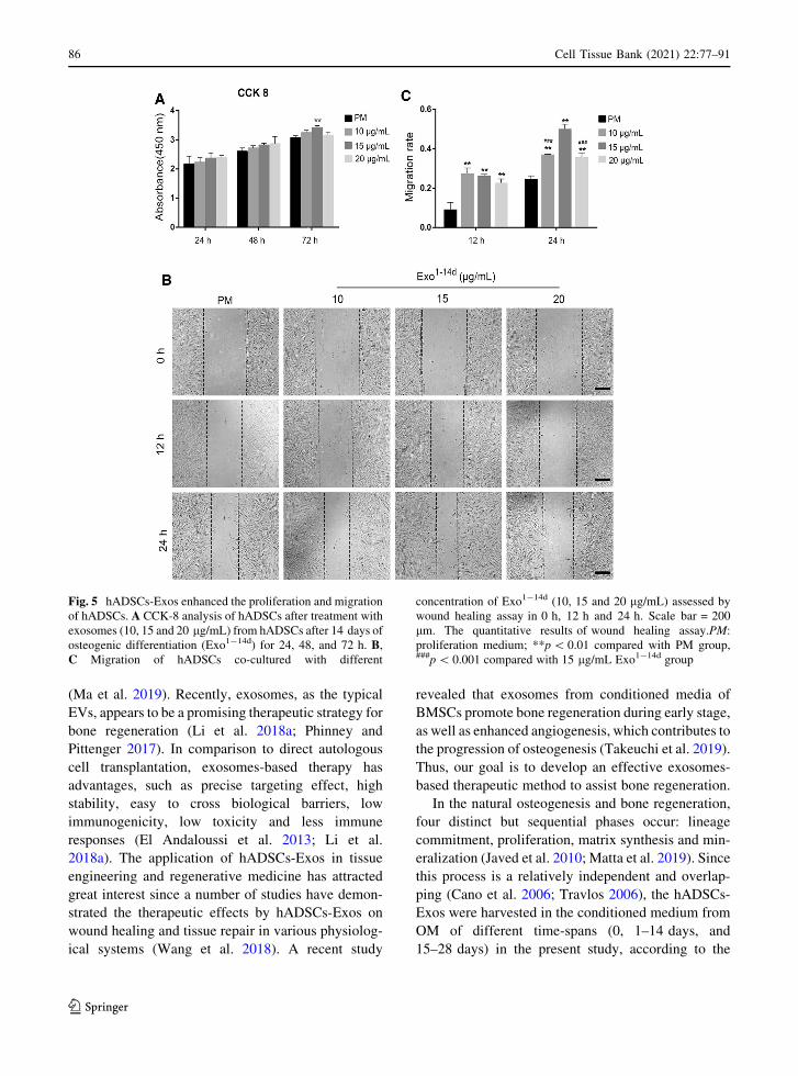
Effects of hADSCs-Exos on hADSCs
proliferation, migration and osteogenic
differentiation
To explore the effects of hADSCs-Exos on hADSCs
proliferation, cells were treated with Exo1-14d at
concentrations of 10, 15 and 20 lg/mL for 24, 48 and
72 h using CCK-8 method. As shown in Fig. 5A, cell
viability was enhanced in a concentration-and time-
dependent trend after treatment for 24 and 48 h.
However, there were no significant differences among
all groups. After treatment with hADSCs-Exos for
72 h, only cells treated with Exo1-14d at a concentra-
tion of 15 lg/mL showed significantly greater prolif-
eration than that without hADSCs-Exos treatment
(p\ 0.01).
To examine whether hADSCs-Exos exhibited bio-
logical effects relevant to migration of recipient
hADSCs, wound healing assay was performed. The
results showed that hADSCs-Exos significantly pro-
moted the migration of hADSCs (Fig. 5B, C,
p\ 0.01). However, the cell migration ability showed
a decreasing trend with the increase of exosome
concentration for 12 h, without significant differences
(p[ 0.05). Furthermore, after treatment with 15 lg/mL Exo1-14d of for 24 h, cells exhibited greater
migration ability than treated with 10 and 20 lg/mL
(p\ 0.001).
After 7 and 14 days of treatment with 10, 15 and
20 lg/mL of Exo1-14d, the ALP activity was deter-
mined as an indicator of osteogenesis (Fig. 6A–C). All
experimental groups of the treated hADSCs revealed
an elevation in staining and activity of ALP in
comparison to that of PM without hADSCs-Exos.
However, only treatments with 15 and 20 lg/mL
Exo1-14d (p\ 0.001) induced a significant increase in
Fig. 3 Internalization of exosomes in hADSCs. The PKH67-
labelled exosomes (Exo0d, Exo1-14d, and Exo15-28d, green)
from hADSCs were detected in the cytoplasm of hADSCs after
incubation for 4 h by confocal microscopy, respectively. The
nucleus of hADSCs were stained with DAPI (blue). Scale
bar = 50 lm. (Color figure online)
123
84 Cell Tissue Bank (2021) 22:77–91

ALP activity than that in the control group in PM at
day 7. Moreover, all treatments with hADSCs-Exos
had markedly greater ALP activity levels compared
with PM at 14 days treatment (p\ 0.001). The ALP
activities in cells induced with the standard osteogenic
medium (OM) was the highest among all groups.
Consistently, the expression of osteoblast-related
proteins (ALP and RUNX2) in hADSCs was also
significantly upregulated when treated with exosomes
on day 14 in comparison to that of PM (Fig. 6D–F)
(p\ 0.05). No significant differences could be detected
between the exosomes-treated group and the control
group in OM in terms of ALP protein expression level.
The expression of RUNX2 after treated with 15 lg/mL
Exo1-14d was significantly higher than that by 20 lg/mL Exo1-14d (p\ 0.01), and even no differences were
found in comparison with the OM group.
Discussion
MSCs have been widely studies as a promising
candidate for regenerative medicine owing to their
self-renewal capability and multilineage differentia-
tion potential of osteogenesis, chondrogenesis, adipo-
genesis and neurogenesis (Huo et al. 2018; Majidinia
et al. 2018). To date, hADSCs have become an
attractive source in bone tissue engineering due to
their stronger osteogenic differentiation ability com-
pared with MSCs derived from the other sources, as
well as easy acquisition and abundance in number
(Damien and Parsons 1991; Kim et al. 2019;
Rodriguez et al. 2005). In our study, hADSCs were
extracted from adipose tissue obtained from healthy
young female with abdominal liposuction. The pri-
mary hADSCs were isolated and characterized by
surface antigen and multi-differentiation potentials.
Growing evidence showed that paracrine effect has
been considered as the predominant mechanism for
the role of MSCs in tissue repair (Li et al. 2018b).
hADSCs, as an principal member ofMSCs, participate
in osteogenesis by this paracrine manner as well.
Recent studies have revealed that during bone regen-
eration, hADSCs not only participate in osteogenesis
via directly differentiation, but also by releasing a
numerous of cytokines and EVs affecting this process
Fig. 4 Alizarin red S staining (ARS) assays of hADSCs after
osteoinduction to determine the optimal time-span for exo-
somes. A: ARS staining of hADSCs incubated with PM, OM
and different time-span of osteogenic induction exosomes
(Exo0d, Exo1-14d and Exo15-28d) on day 21. Scale bar = 200
lm.A’ The higher magnification panels of black box in Fig. 4A.
The black arrow indicated the calcified nodules.BQuantification
of ARS of hADSCs treated with or without exosomes. PM:
proliferation medium; OM: osteogenic medium. ***p\ 0.001
compared with PM group, DDDp\ 0.001 compared with OM
group, ###p\ 0.001 represents significant differences between
compared groups. (Color figure online)
123
Cell Tissue Bank (2021) 22:77–91 85
(Ma et al. 2019). Recently, exosomes, as the typical
EVs, appears to be a promising therapeutic strategy for
bone regeneration (Li et al. 2018a; Phinney and
Pittenger 2017). In comparison to direct autologous
cell transplantation, exosomes-based therapy has
advantages, such as precise targeting effect, high
stability, easy to cross biological barriers, low
immunogenicity, low toxicity and less immune
responses (El Andaloussi et al. 2013; Li et al.
2018a). The application of hADSCs-Exos in tissue
engineering and regenerative medicine has attracted
great interest since a number of studies have demon-
strated the therapeutic effects by hADSCs-Exos on
wound healing and tissue repair in various physiolog-
ical systems (Wang et al. 2018). A recent study
revealed that exosomes from conditioned media of
BMSCs promote bone regeneration during early stage,
as well as enhanced angiogenesis, which contributes to
the progression of osteogenesis (Takeuchi et al. 2019).
Thus, our goal is to develop an effective exosomes-
based therapeutic method to assist bone regeneration.
In the natural osteogenesis and bone regeneration,
four distinct but sequential phases occur: lineage
commitment, proliferation, matrix synthesis and min-
eralization (Javed et al. 2010; Matta et al. 2019). Since
this process is a relatively independent and overlap-
ping (Cano et al. 2006; Travlos 2006), the hADSCs-
Exos were harvested in the conditioned medium from
OM of different time-spans (0, 1–14 days, and
15–28 days) in the present study, according to the
Fig. 5 hADSCs-Exos enhanced the proliferation and migration
of hADSCs. A CCK-8 analysis of hADSCs after treatment with
exosomes (10, 15 and 20 lg/mL) from hADSCs after 14 days of
osteogenic differentiation (Exo1-14d) for 24, 48, and 72 h. B,C Migration of hADSCs co-cultured with different
concentration of Exo1-14d (10, 15 and 20 lg/mL) assessed by
wound healing assay in 0 h, 12 h and 24 h. Scale bar = 200
lm. The quantitative results of wound healing assay.PM:
proliferation medium; **p\ 0.01 compared with PM group,###p\ 0.001 compared with 15 lg/mL Exo1-14d group
123
86 Cell Tissue Bank (2021) 22:77–91

diverse phases of osteogenesis. The morphology and
size distribution of hADSCs-Exos was investigated by
TEM and DLS analysis. The exosomes extracted from
three stages were not significantly different in size.
The extracted hADSCs-Exos all exhibited typical
morphology of exosomes and the mean diameter of
hADSCs-Exos is 125.8 ± 3.822 nm. Our results
revealed the diversity of hADSCs-Exos in morphol-
ogy, which may relate to their multiple functions in
different periods, consistent with previous report
(Zabeo et al. 2017). Furthermore, hADSCs-Exos
Fig. 6 hADSCs-Exos promoted osteogenic differentiation of
hADSCs in vitro. A Alkaline phosphatase (ALP) staining of
hADSCs after incubation with exosomes (10, 15 and 20 lg/mL)
from hADSCs after 14 days of osteogenic (Exo1-14d) on day 7
and day 14. Scale bar = 200 lm; B, C ALP activity normalized
against the total protein level on day 7 and 14, respectively;
D Western blot showed the expression of osteogenic-related
proteins ALP and RUNX2 on day 14;E, FRelative protein level
of ALP and RUNX2 on day 14, respectively. PM: proliferation
medium; OM: osteogenic medium; *p\ 0.05, **p\ 0.01, and
***p\ 0.001 compared with PM group, respectively.Dp\ 0.05 and DDp\ 0.01 compared with OM group, respec-
tively; ##p\ 0.01 represents significant differences between
compared groups
123
Cell Tissue Bank (2021) 22:77–91 87

positively expressed exosome-specific protein mark-
ers CD9 and CD63 by western blot analysis.
Exosomes can be taken up by recipient cells in the
local microenvironment following release. Moreover,
hADSCs-Exos implement intercellular communica-
tion through target cell internalization (Liu et al. 2019;
Tkach and Thery 2016). Fluorescence microscopy
analysis showed that the hADSCs-Exo of Exo0d,
Exo1-14d and Exo15-28d labelled with PKH-67 were
taken up by the native hADSCs and mainly distributed
in the perinuclear region. Interestingly, we observed
that hADSCs-Exos labeled in green were definitely
internalized by the hADSCs within 4 h, and this
process was high-efficient than that of BMSCs, which
taking almost 48 h (Li et al. 2018a). It seems that
hADSCs-Exos were more prone to be internalized by
hADSCs as their ‘‘parent’’ cells, but not cells from
other sources. We speculate that it may be related to
the homology of the recipient cells. Numerous direct
evidence exists to suggest that the binding of exo-
somes to recipient cells involves a variety of endocytic
pathways, including phagocytosis, macropinocytosis
and plasma or endosomal membrane fusion (Mulcahy
et al. 2014). The decisive factor for this process may
depending on the type of recipient cell and exosomes
constituents (Naslund et al. 2014). In another report,
they found that only a subpopulation of MSCs
internalized exosomes labelled with PKH67, and
explained that it may be due to the heterogeneity of
MSCs in terms of some certain surface receptors, as
well as the different phase of the cell cycle (Wang
et al. 2018). Despite all this, the underlying mecha-
nisms of hADSCs-Exo internalized by hADSCs
should be further illuminated.
The ability to mineralize is the main functional
characteristic of osteoblasts in vitro. hADSCs can be
induced to differentiate into osteoblasts that are able to
mineralize their extracellular matrix (ECM) and
express proteins associated with bone phenotypes
(Halvorsen et al. 2001). The osteogenic differentiation
capacities of the exosome-treated hADSCs (Exo0d,
Exo1-14d and Exo15-28d) as detected by ARS, the
results indicated that hADSCs-Exo0d without osteo-
genic-induction could not effectively induce hADSCs
into osteoblasts in vitro. However, both hADSCs-
Exo1-14d and Exo15-28d group exhibited remarkable
improvement in mineralization of hADSCs. Espe-
cially values in the group Exo1-14d were significantly
increased in comparison with those in the Exo0d, and
Exo15-28d group.
During the early stage of osteogenic differentiation,
mixed cells at diverse differentiative stages secrete a
great quantity of exosomes involving the information
exchange of hADSCs, thus establishing a positive-
feedback loop during osteogenesis (Cui et al. 2016;
Yeo et al. 2013). Actually, in the late stage of
osteogenesis, as a result of cellular senescence, contact
inhibition and cell differentiation, the capacity of
hADSCs secreting exosomes attenuated, meanwhile
the function of hADSCs-Exos may also changed
(Gudbergsson et al. 2016; Hayes et al. 2005; Li et al.
2018a; Steinman et al. 2003). Some other issues, such
as the extraction amount and labor-saving, also need to
be taken into consideration for exosomes clinical
application in the future. In the present study, the
conditioned medium was collected every 3 days,
which greatly increased the extraction amount in a
limited time. Besides, the time interval of collecting
exosomes is 1 to 14 days, which has competitive
advantages (e.g. time saving, cost-effective etc.) over
prolonged periods. To maximize the efficiency of
hADSCs-Exos in osteogenesis, the Exo1-14d was
applied to the follow-up research.
Since the proliferative and migratory capacity of
transplanted hADSCs are one of the most important
processes to promote tissue regeneration (Monaco
et al. 2011), we investigated the effects of exosomes
derived from osteogenically induced hADSCs on their
‘‘parent’’ cells. After treated with exosomes, the
proliferation and cell migration of hADSCs were
significantly promoted compared with the negative
control group (PM without exosomes). Only Exo1-14d
(15 lg/mL) dramatically improved the proliferation of
hADSCs at 72 h. Moreover, the migration of hADSCs
was enhanced in group Exo1-14d (10, 15 and 20 lg/mL) compared with group PM. In addition, with the
increasing concentration of hADSCs-Exos, the cell
migratory capacity gradually increased. Moreover, the
group Exo1-14d (15 lg/mL) showed the peak of
capacity compared with other experimental groups,
when the concentration was over 15 lg/mL, the cell
migration decreased. We speculate that this may be
related to the dysregulation of functional protein
activity.
After treated with hADSCs-Exos for 7 and 14 days,
ALP staining and activity of hADSCs were signif-
icantly enhanced. RUNX2 is a key transcription factor
123
88 Cell Tissue Bank (2021) 22:77–91

for osteogenesis which acting as early indicators of
osteogenesis (Chen et al. 2015; Komori 2003; Komori
et al. 1997). Increased expression of RUNX2 as well
as ALP activity and mineralization is an essential
requisite for bone regeneration(Qi et al. 2016). In our
study, RUNX2 and ALP protein expression levels
were also analyzed by Western blot. And the results
showed the 15 lg/mL hADSCs-Exos after 14 days
osteogenically induction significantly promoted the
osteogenesis of hADSCs by enhancing ALP activity,
extracellular mineralization nodules, and expression
of the osteoblastogenesis-related proteins, such as
ALP and RUNX2.
The hADSCs-Exos might regulate the osteogenesis
of hADSCs by transmitting a variety of signal
molecules including mRNAs, miRNAs, non-coding
RNAs and proteins to surrounding cells. Takeuchi
et al. found that MSC-Exos contain miRNAs that may
enhance VEGF secretion from the recipient cells,
which contributes to bone regeneration (Takeuchi
et al. 2019). Chen et al. demonstrated that exosomes
derived from miR-375-overexpressing hADSCs pro-
moted bone regeneration (Chen et al. 2019). However,
we did not identify the active component(s) affect
osteogenesis in this study, and thus, the regulated
mechanism require further investigation.
Considering all of the above experimental results,
osteogenically-induced hADSCs-Exos promote
hADSCs proliferation, migration and osteogenic dif-
ferentiation in vitro. hADSCs-Exos at concentration of
15 lg/mL at 1–14 days during osteogenic induction
might be optimal. To our best knowledge, this is the
first report to apply different stages and concentration
of osteogenically-induced hADSCs-Exos to induce
the hADSCs osteogenesis in vitro. In conclusion, we
suggested that hADSCs-Exo may be an ideal substi-
tute for improving the deficiency of traditional
hADSCs therapy in the bone repair. Strategies to load
new bone tissue engineering scaffolds with exosomes
can be applied for future clinical transformation
therapy.
There still existed some limitations about our study.
First, the evaluation of bone formation could be
performed in vivo. Second, the mechanisms of
hADSCs-Exos on promoting osteogenesis and inte-
gration of remains further studied.
Acknowledgments This work was supported by grants from
the funds for the National Natural Science Foundation of China
(No. 51872332; No. 31200740), the National Natural Science
Foundation of Liaoning province (No. 20170541040).
Funding No competing financial interests exist.
Compliance with ethical standards
Ethical approval Human abdominal subcutaneous adipose
tissue acquired with informed consent was approved by the
Research Ethical Committee of the First Hospital of China. This
article does not contain any studies with animals performed by
any of the authors.
Open Access This article is licensed under a Creative Com-
mons Attribution 4.0 International License, which permits use,
sharing, adaptation, distribution and reproduction in any med-
ium or format, as long as you give appropriate credit to the
original author(s) and the source, provide a link to the Creative
Commons licence, and indicate if changes were made. The
images or other third party material in this article are included in
the article’s Creative Commons licence, unless indicated
otherwise in a credit line to the material. If material is not
included in the article’s Creative Commons licence and your
intended use is not permitted by statutory regulation or exceeds
the permitted use, you will need to obtain permission directly
from the copyright holder. To view a copy of this licence, visit
http://creativecommons.org/licenses/by/4.0/.
References
An Y, Zhao J, Nie F, Wu Y, Xia Y, Li D (2019) Parathyroid
hormone (PTH) promotes ADSC osteogenesis by regulat-
ing SIK2 and Wnt4. Biochem Biophys Res Commun
516:551–557. https://doi.org/10.1016/j.bbrc.2019.06.084
Cano J, Campo J, Moreno LA, Bascones A (2006) Osteogenic
alveolar distraction: a review of the literature. Oral Surg
Oral Med Oral Pathol Oral Radiol Endod 101:11–28.
https://doi.org/10.1016/j.tripleo.2005.04.015
Chen Q et al (2010) Adipose-derived stem cells modified
genetically in vivo promote reconstruction of bone defects.
Cytotherapy 12:831–840. https://doi.org/10.3109/
14653249.2010.495980
Chen S et al (2019) Exosomes derived from miR-375-overex-
pressing human adipose mesenchymal stem cells promote
bone regeneration. Cell Prolif 52:e12669. https://doi.org/
10.1111/cpr.12669
Chen YJ, Zhang X, Wu ZS, et al (2015) Autocrine human
growth hormone stimulates the tumor initiating capacity
and metastasis of estrogen receptor-negative mammary
carcinoma cells. Cancer Lett 365:182–189. https://doi.org/
10.1016/j.canlet.2015.05.031
Cooper DR et al (2018) human adipose-derived stem cell con-
ditioned media and exosomes containing MALAT1 pro-
mote human dermal fibroblast migration and ischemic
wound healing. Adv Wound Care 7:299–308. https://doi.
org/10.1089/wound.2017.0775
Cui Y, Luan J, Li H, Zhou X, Han J (2016) Exosomes derived
from mineralizing osteoblasts promote ST2 cell osteogenic
123
Cell Tissue Bank (2021) 22:77–91 89
differentiation by alteration of microRNA expression.
FEBS Lett 590:185–192. https://doi.org/10.1002/1873-
3468.12024
Damien CJ, Parsons JR (1991) Bone graft and bone graft sub-
stitutes: a review of current technology and applications
Journal of applied biomaterials : an official journal of the
Society for. Biomaterials 2:187–208. https://doi.org/10.
1002/jab.770020307
Dimitriou R, Jones E, McGonagle D, Giannoudis PV (2011)
Bone regeneration: current concepts and future directions.
BMC Med 9:66. https://doi.org/10.1186/1741-7015-9-66
Du W, Su L, Zhang N, Wang H (2019) Exosomes derived from
preadipocytes improve osteogenic differentiation, poten-
tially via reduced miR223 expression. Mol Med Reports
19:951–958. https://doi.org/10.3892/mmr.2018.9760
El-Rashidy AA, Roether JA, Harhaus L, Kneser U, Boccaccini
AR (2017) Regenerating bone with bioactive glass scaf-
folds: A review of in vivo studies in bone defect models.
Acta Biomater 62:1–28. https://doi.org/10.1016/j.actbio.
2017.08.030
El Andaloussi S, Lakhal S, Mager I, WoodMJ (2013) Exosomes
for targeted siRNA delivery across biological barriers. Adv
Drug Deliv Rev 65:391–397. https://doi.org/10.1016/j.
addr.2012.08.008
Fang S, Li Y, Chen P (2019) Osteogenic effect of bone marrow
mesenchymal stem cell-derived exosomes on steroid-in-
duced osteonecrosis of the femoral head. Drug Des
Develop Ther 13:45–55. https://doi.org/10.2147/DDDT.
S178698
Gudbergsson JM, Johnsen KB, Skov MN, Duroux M (2016)
Systematic review of factors influencing extracellular
vesicle yield from cell cultures. Cytotechnology
68:579–592. https://doi.org/10.1007/s10616-015-9913-6
Halvorsen YD et al (2001) Extracellular matrix mineralization
and osteoblast gene expression by human adipose tissue-
derived stromal cells. Tissue Eng 7:729–741. https://doi.
org/10.1089/107632701753337681
Hayes O, Ramos B, Rodriguez LL, Aguilar A, Badia T, Castro
FO (2005) Cell confluency is as efficient as serum starva-
tion for inducing arrest in the G0/G1 phase of the cell cycle
in granulosa and fibroblast cells of cattle. Anim Reprod Sci
87:181–192. https://doi.org/10.1016/j.anireprosci.2004.11.
011
Hoshino A et al (2015) Tumour exosome integrins determine
organotropic metastasis. Nature 527:329–335. https://doi.
org/10.1038/nature15756
Huo S et al (2018) Insight into the Role of Long Non-coding
RNAs During Osteogenesis in Mesenchymal Stem Cells.
Curr Stem Cell Res Ther 13:52–59. https://doi.org/10.
2174/1574888X12666171115124112
Javed A, Chen H, Ghori FY (2010) Genetic and transcriptional
control of bone formation. OralMaxillofac Surg Clin North
America 22:283–293. https://doi.org/10.1016/j.coms.
2010.05.001
Kim I, Lee SS, Kim SHL, Bae S, Lee H, Hwang NS (2019)
Osteogenic effects of VEGF-overexpressed human adi-
pose-derived stem cells with whitlockite reinforced cryo-
gel for bone regeneration. Macromol Biosci 19:e1800460.
https://doi.org/10.1002/mabi.201800460
Kim WS, Park BS, Sung JH, Yang JM, Park SB, Kwak SJ, Park
JS (2007) Wound healing effect of adipose-derived stem
cells: a critical role of secretory factors on human dermal
fibroblasts. J Dermatol Sci 48:15–24. https://doi.org/10.
1016/j.jdermsci.2007.05.018
Komori T (2003) Requisite roles of Runx2 and Cbfb in skeletal
development. J Bone min metab 21:193–197. https://doi.
org/10.1007/s00774-002-0408-0
Komori T et al (1997) Targeted disruption of Cbfa1 results in a
complete lack of bone formation owing to maturational
arrest of osteoblasts. Cell 89:755–764. https://doi.org/10.
1016/s0092-8674(00)80258-5
Li W et al (2018a) Tissue-engineered bone immobilized with
human adipose stem cells-derived exosomes promotes
bone regeneration. ACS Appl Mat Interfaces
10:5240–5254. https://doi.org/10.1021/acsami.7b17620
Li Y et al (2018b) Mesenchymal stem cells-derived exosomes: a
possible therapeutic strategy for osteoporosis. Curr Stem
cell Res Ther 13:362–368. https://doi.org/10.2174/
1574888X13666180403163456
Liao HT, Chen CT (2014) Osteogenic potential: comparison
between bone marrow and adipose-derived mesenchymal
stem cells world. Journal Stem Cells 6:288–295. https://
doi.org/10.4252/wjsc.v6.i3.288
Liu CY et al (2020) Effect of exosomes from adipose-derived
stem cells on the apoptosis of Schwann cells in peripheral
nerve injury. CNS Neurosci Ther 26:189–196. https://doi.
org/10.1111/cns.13187
Liu Z, Xu Y, Wan Y, Gao J, Chu Y, Li J (2019) Exosomes from
adipose-derived mesenchymal stem cells prevent car-
diomyocyte apoptosis induced by oxidative stress. Cell
death discov 5:79. https://doi.org/10.1038/s41420-019-
0159-5
Lord CF, Gebhardt MC, Tomford WW, Mankin HJ (1988)
Infection in bone allografts Incidence, nature, and treat-
ment. J Bone Jt Surg American 70:369–376
Ma T, Fu B, Yang X, Xiao Y, Pan M (2019) Adipose mes-
enchymal stem cell-derived exosomes promote cell pro-
liferation, migration, and inhibit cell apoptosis via
Wnt/beta-catenin signaling in cutaneous wound healing.
J Cell Biochem 120:10847–10854. https://doi.org/10.1002/
jcb.28376
Majidinia M, Sadeghpour A, Yousefi B (2018) The roles of
signaling pathways in bone repair and regeneration. J Cell
Physiol 233:2937–2948. https://doi.org/10.1002/jcp.26042
Mathivanan S, Ji H, Simpson RJ (2010) Exosomes: extracellular
organelles important in intercellular communication.
J Proteom 73:1907–1920. https://doi.org/10.1016/j.jprot.
2010.06.006
Matta C et al (2019) Osteogenic differentiation of human bone
marrow-derived mesenchymal stem cells is enhanced by an
aragonite scaffold. Differentiation 107:24–34. https://doi.
org/10.1016/j.diff.2019.05.002
Monaco E, Bionaz M, Hollister SJ, Wheeler MB (2011)
Strategies for regeneration of the bone using porcine adult
adipose-derived mesenchymal stem cells. Theriogenology
75:1381–1399. https://doi.org/10.1016/j.theriogenology.
2010.11.020
Mulcahy LA, Pink RC, Carter DR (2014) Routes and mecha-
nisms of extracellular vesicle uptake. J Extracell Vesicles.
3:24641. https://doi.org/10.3402/jev.v3.24641
Nabet BY et al (2017) Exosome RNA unshielding couples
stromal activation to pattern recognition receptor signaling
123
90 Cell Tissue Bank (2021) 22:77–91

in cancer. Cell 170:352–366. https://doi.org/10.1016/j.cell.
2017.06.031
Naslund TI, Paquin-Proulx D, Paredes PT, Vallhov H, Sandberg
JK, Gabrielsson S (2014) Exosomes from breast milk
inhibit HIV-1 infection of dendritic cells and subsequent
viral transfer to CD4? T cells. AIDS 28:171–180. https://
doi.org/10.1097/QAD.0000000000000159
Oryan A, Kamali A, Moshiri A, Baghaban Eslaminejad M
(2017) Role of Mesenchymal Stem Cells in Bone Regen-
erative Medicine: What Is the Evidence? Cells Tissues Org
204:59–83. https://doi.org/10.1159/000469704
Phinney DG, Pittenger MF (2017) Concise Review: MSC-
Derived Exosomes for Cell-Free. Ther Stem Cells
35:851–858. https://doi.org/10.1002/stem.2575
Phinney DG, Prockop DJ (2007) Concise review: mesenchymal
stem/multipotent stromal cells: the state of transdifferen-
tiation and modes of tissue repair–current views. Stem
Cells 25:2896–2902. https://doi.org/10.1634/stemcells.
2007-0637
Qi X et al (2016) Exosomes secreted by human-induced
pluripotent stem cell-derived mesenchymal stem cells
repair critical-sized bone defects through enhanced
angiogenesis and osteogenesis in osteoporotic rats. Int J
biol Sci 12:836–849. https://doi.org/10.7150/ijbs.14809
Qin Y, Sun R, Wu C, Wang L, Zhang C (2016) Exosome: a
novel approach to stimulate bone regeneration through
regulation of osteogenesis and angiogenesis. Int J Mol Sci
17(5):712. https://doi.org/10.3390/ijms17050712
Rodriguez AM, Elabd C, Amri EZ, Ailhaud G, Dani C (2005)
The human adipose tissue is a source of multipotent stem
cells. Biochimie 87:125–128. https://doi.org/10.1016/j.
biochi.2004.11.007
Schaffler A, Buchler C (2007) Concise review: adipose tissue-
derived stromal cells-basic and clinical implications for
novel cell-based therapies. Stem Cells 25:818–827. https://
doi.org/10.1634/stemcells.2006-0589
Seong JM, Kim BC, Park JH, Kwon IK, Mantalaris A, Hwang
YS (2010) Stem cells in bone tissue engineering. Biomed
Mat 5:062001. https://doi.org/10.1088/1748-6041/5/6/
062001
Shi YY, Nacamuli RP, Salim A, Longaker MT (2005) The
osteogenic potential of adipose-derived mesenchymal cells
is maintained with aging. Plast Reconstr Surg 116:
1686–1696. https://doi.org/10.1097/01.prs.0000185606.
03222.a9
Shimasaki T, Yamamoto S, Arisawa T (2018) Exosome research
and co-culture Study. Biol pharm bulletin 41:1311–1321.
https://doi.org/10.1248/bpb.b18-00223
Steinman RA, Wentzel A, Lu Y, Stehle C, Grandis JR (2003)
Activation of Stat3 by cell confluence reveals negative
regulation of Stat3 by cdk2. Oncogene 22:3608–3615.
https://doi.org/10.1038/sj.onc.1206523
Takeuchi R, Katagiri W, Endo S, Kobayashi T (2019) Exosomes
from conditioned media of bone marrow-derived mes-
enchymal stem cells promote bone regeneration by
enhancing angiogenesis. PLoS ONE 14:e0225472. https://
doi.org/10.1371/journal.pone.0225472
Tkach M, Thery C (2016) Communication by extracellular
vesicles: where we are and where we need to go. Cell
164:1226–1232. https://doi.org/10.1016/j.cell.2016.01.043
Travlos GS (2006) Normal structure, function, and histology of
the bone marrow. Toxicol Pathol 34:548–565. https://doi.
org/10.1080/01926230600939856
Urbanelli L, Buratta S, Sagini K, Ferrara G, LanniM, Emiliani C
(2015) Exosome-based strategies for diagnosis and ther-
apy. Recent Pat CNS drug discov 10:10–27. https://doi.org/
10.2174/1574889810666150702124059
Wang X, Omar O, Vazirisani F, Thomsen P, Ekstrom K (2018)
Mesenchymal stem cell-derived exosomes have altered
microRNA profiles and induce osteogenic differentiation
depending on the stage of differentiation. PLoS ONE
13:e0193059. https://doi.org/10.1371/journal.pone.0193
059
Wong DE, Banyard DA, Santos PJF, Sayadi LR, Evans GRD,
Widgerow AD (2019) Adipose-derived stem cell extra-
cellular vesicles: a systematic review journal of plastic,
reconstructive and aesthetic surgery. JPRAS 72:
1207–1218. https://doi.org/10.1016/j.bjps.2019.03.008
WuW, Niklason L, Steinbacher DM (2013) The effect of age on
human adipose-derived stem cells. Plast Reconstr Surg
131:27–37. https://doi.org/10.1097/PRS.0b013e31827
29cfc
Xie Q et al (2016) The role of miR-135-modified adipose-
derived mesenchymal stem cells in bone regeneration.
Biomaterials 75:279–294. https://doi.org/10.1016/j.
biomaterials.2015.10.042
Xu WL et al (2017) In Situ Release of VEGF enhances osteo-
genesis in 3D porous scaffolds engineered with osterix-
modified adipose-derived stem cells. Tissue Eng Part A
23:445–457. https://doi.org/10.1089/ten.TEA.2016.0315
Yang S, Guo S, Tong S, Sun X (2019) promoting osteogenic
differentiation of human adipose-derived stem cells by
altering the expression of exosomal miRNA. StemCells Int
2019:1351860. https://doi.org/10.1155/2019/1351860
Yeo RW, Lai RC, Zhang B, Tan SS, Yin Y, Teh BJ, Lim SK
(2013) Mesenchymal stem cell: an efficient mass producer
of exosomes for drug delivery. Adv Drug Deliv Rev
65:336–341. https://doi.org/10.1016/j.addr.2012.07.001
Zabeo D, Cvjetkovic A, Lasser C, Schorb M, Lotvall J, Hoog JL
(2017) Exosomes purified from a single cell type have
diverse morphology. J Extracell Vesicles 6:1329476.
https://doi.org/10.1080/20013078.2017.1329476
Zhu M, Guo S, Liu Y, Tong S, Zhang H, Cui M, Y., Chen Y,
(2019) In vitro study of different stages of ADSC-Exos in
the promotion of osteogenic differentiation Chin. J Aesth
Plast Surg 30:101–105. https://doi.org/10.3969/j.issn.
1673-7040.2019.05.000
Zuk PA et al (2002) Human adipose tissue is a source of mul-
tipotent stem cells. Mol Biol Cell 13:4279–4295. https://
doi.org/10.1091/mbc.e02-02-0105
Publisher’s Note Springer Nature remains neutral with
regard to jurisdictional claims in published maps and
institutional affiliations.
123
Cell Tissue Bank (2021) 22:77–91 91