osteomyelitis: a review of diagnosis based on crp...
TRANSCRIPT

OSTEOMYELITIS:
A REVIEW OF DIAGNOSIS BASED ON CRP
Author of the thesis:
Griselda March Sorribes
Director: C. Padrós
Bachelor’s degree in Podiatry
June 2015, Barcelona
G. March
2
INDEX
INDEX OF FIGURES ......................................................................................... 3
ABBREVIATIONS INDEX................................................................................... 4
ABSTRACT ........................................................................................................ 5
1. INTRODUCTION ......................................................................................... 7
2. DIABETIC FOOT SYNDROME .................................................................... 9
2.1 DIABETIC FOOT INFECTION ............................................................ 10
2.2 DIAGNOSIS OF BONE INFECTION ................................................... 11
3. ACUTE REACTIVE PROTEIN ................................................................... 13
3.1 C REACTIVE PROTEIN (CRP) ........................................................... 16
3.2 ERITRHOCYTE SEDIMENTATION RATE (ESR) ............................... 18
3.3 ALBUMIN ............................................................................................ 19
3.4 LEUKOCYTOSIS ................................................................................ 20
4. OBJECTIVES ............................................................................................ 22
4.1 GENERAL OBJECTIVE ...................................................................... 22
4.2 SPECIFIC OBJECTIVES .................................................................... 22
5. MATERIAL AND METHODS ..................................................................... 23
5.1 ACTING PROCESS ............................................................................ 24
5.1.1 Variables to consider .................................................................... 25
5.1.2 Statistical analysis ........................................................................ 25
6. RESULTS .................................................................................................. 26
6.1 ANALYTICAL RESULTS ..................................................................... 27
6.2 SURGICAL PROCEDURES RESULTS .............................................. 33
6.3 GENERAL RESULTS .......................................................................... 33
7. DISCUSSION ............................................................................................ 36
8. CONCLUSIONS ........................................................................................ 38
9. REFERENCES .......................................................................................... 39
10. AKWNOWLEDGEMENTS ...................................................................... 43
G. March
3
INDEX OF FIGURES
Fig. 2.1 Algorithms of food ulceration in diabetic patients. ............................... 10
Fig. 3.2 Increase of acute phase reactants during an inflammation process. .. 13
Fig. 3.3 Alterations in some acute phase reactants following tissue injury ...... .15
Fig. 6.4 Results of blood test of all diabetic foot patients diagnosed with OM...26
Fig. 6.1.1.5 CRP results of the studied patients. .............................................. 28
Fig. 6.1.2.6 Albumin results of the studied patients……………………………... 29
Fig. 6.1.3.7 Alkaline phosphatase results of the studied patients. .................... 30
Fig. 6.1.4.8 Leukocytes results of the studied patients. ................................... 31
Fig. 6.1.4.9 Neutrophils results of the studied patients. ................................... 31
Fig. 6.1.4.10 Erythrocytes results of the studied patients. ................................ 32
Fig. 6.1.4.11 Hemoglobin results of the studied patients. ................................ 33
Fig. 6.3.12 Age and sex group of the studied patients. .................................... 34
Fig. 6.3.13 Associated comorbidities of the studied patients. .......................... 34
Fig. 6.3.14 CRP levels before and after antibiotic teraphy ............................... 35
G. March
4
ABBREVIATIONS INDEX
OM: Osteomyelitis
CRP: C Reactive Protein
ESR: Erythrocyte Sedimentation Rate
CBC: Complete Blood Count
DM: Diabetes Mellitus
MRI: Magnetic Resonance Imaging
PET: Positron Emission Proton

G. March
5
ABSTRACT
In diabetic patients with foot infection, osteomyelitis shows up in 20% of cases
and highly increase the probability to require a lower-extremity amputation.
Unfortunately, there are no agreed guidelines for its diagnosis and early diagnosis
is necessary to ensure appropriate treatment. Nevertheless, it may take several
weeks for bone infection to produce defects on x-ray and bone biopsy may
occasionally be misleading. MRI is the gold standard diagnostic technique for
osteomyelitis (OM) but because of its low availability other noninvasive and
reliable diagnostic markers are studied; within these, C reactive protein (CRP) is
the principal; the levels of this protein rise highly in response of an infection. In
this thesis, an initial comprehensive literature search is carried out on topics
related to acute phase reactants, diabetic foot infection and diagnosis of OM.
Medical records of 30 patients with bone infection who attended Medical
Emergency from Diabetic Foot Unit during a period of 1,5 years are searched and
studied. The aim of this study is to compare statistically serum levels of CRP to
see if it is an OM indicator; moreover, to know if there is another factor, besides
this mentioned, which is also altered. Finally, the validity of CRP is shown to have
diagnostic value front of an OM.
Keywords: diabetic foot, osteomyelitis, C reactive protein, amputation, acute
phase reactants
G. March
6
RESUM
L’osteomielitis està present en un 20% dels casos d’infecció del peu en persones
diabètiques i augmenta la probabilitat de requerir una amputació de les
extremitats inferiors. Desafortunadament, no hi ha pautes acordades pel seu
diagnòstic. Un diagnòstic exacte és necessari per garantir un tractament
adeqüat. No obstant, la infecció òssia pot tardar diverses setmanes en produir
defectes en una radiografia simple i la biopsia òssia pot ser enganyosa. La RM
és l’estàndard d’or per a la osteomielitis (OM) però, a causa de la seva baixa
disponibilitat, altres marcardors diagnòstics no invasius i confiables han sigut
estudiats. Dins d’aquests, la proteïna C reactiva (PCR) és la principal; els nivells
d’aquesta s’eleven altament en presència d’infecció. En aquest treball, es du a
terme inicialment una recerca bibliogràfica completa sobre temes relacionats
amb reactants de fase aguda, infecció del peu diabètic i diagnòstic de la OM. Es
busquen les històries clíniques de 30 pacients de la Unitat de Peu Diabètic amb
infecció òssia que van assistir a Urgències en un període comprès entre el
setembre de 2013 i l’abril de 2015. Seguidament, es du a terme un estudi dels
anàlisis sanguinis dels pacients. L’objectiu d’aquest estudi és comparar els
nivells sèrics de PCR per veure si és un indicador de l’OM; tanmateix, saber si hi
ha un altre factor, apart de l’esmentat anteriorment, que també s’alteri. A
continuació, les dades són analitzades estadísticament. Finalment, la validesa
de la PCR es demostra que té un valor diagnòstic davant una OM.
Paraules clau: peu diabètic, osteomielitis, proteïna C reactiva, amputació,
reactants de fase aguda

G. March
7
1. INTRODUCTION
Diabetic foot ulcers cause significant morbidity and are responsible of a large
number of hospitalizations. It has been reported that approximately 20% of
diabetic patients develop foot ulcers at some point in their lives. However, other
studies indicate that between 50% and 95% of cases of amputations of lower
extremities due to non-traumatic issues correspond to diabetic patients1. Around
40% of diabetic patients who have been amputated require subsequent
amputation in the next five years from the initial event, reporting morbidity of 50%
in the first three years2.
Many studies have shown that diabetic patients have immune and tissue repair
disorders, such as leukocyte and platelet dysfunction, basal membrane thinning
and atherosclerosis of small vessels, abnormalities in the function of neutrophils
and fibroblasts, peripheral neuropathy and tissue hypoxia which interact with
each other in the genesis of the diabetic foot. Ischemia contributes a 30-40% in
the formation of ulcers3.
In most cases, the main determinant in the decision to carry out an amputation in
diabetic foot is the presence of osteomyelitis. This, which involves bone infection
both cortical and bone marrow, is a serious and common problem in diabetic
patients because it is precisely a consequence of diabetes complications,
especially neuropathy and, to a lesser degree, the vasculopathy, the
immunological and healing defects.
However, the diagnosis of osteomyelitis in this group of patients still remains a
challenge. The classic signs and symptoms of infection may be absent or masked
by coexisting vascular disease or neuropathy. In patients with suspected
osteomyelitis, the radiography is the initial study. However, radiographic changes
may take two weeks to appear. The gold standard for diagnosis of osteomyelitis
is bone biopsy. Yet, this invasive procedure is not always practical in patients with
severe peripheral vascular disease and diabetes, that’s the reason why
noninvasive and reliable markers are looked for4.
Magnetic resonance imaging (MRI) has been recognized for early diagnosis with
a sensitivity between 77-100% and a specificity of 79 to 100% according to
different series of studies3-5. The disadvantages of this study are: its high cost,

G. March
8
low availability and the decreasing of specificity in cases of previous surgery,
neuropathic osteoarthropathy (Charcot) and other inflammatory diseases such as
rheumatoid arthritis.
During the acute phase of inflammation around 50 glycoproteins can be identified
as reactants. For many years, the erythrocyte sedimentation rate (ESR) has been
used as an acute phase reactant and marker of inflammation. ESR is an indirect
way of measuring concentrations of plasma proteins of acute inflammation
(though many of the physicochemical factors that affect ESR are unknown).
In osteomyelitis, acute or chronic, ESR is usually high and decreases front of a
favorable response to treatment, thus has been considered as a useful marker
for the diagnosis and monitoring of diabetic foot.
Moreover, C-reactive protein (CRP) is another marker of acute inflammation. An
increase in serum concentrations of this peptide has been observed during
inflammation and tissue necrosis. Generally, the CRP values reflect the severity
of inflammation or tissue injury; rises few hours after the acute process had begun
and is normalized within few days after treatment6.
The aim of this research is to compare serum levels of CRP in patients with
diabetic foot and, at the same time, see how others parameters of blood analysis
are modified in the presence of osteomyelitis.
G. March
9
2. DIABETIC FOOT SYNDROME
The diabetic foot syndrome is considered by the World Health Organization
(WHO) as: the presence of ulceration, infection and/or gangrene of the foot
associated with diabetic neuropathy and various degrees of peripheral vascular
disease, result from the complex interaction of various factors induced by a
maintained hyperglycemia.
The risk of ulceration and amputation in diabetic patients is much higher
compared to non-diabetic patients: the risk of a diabetic patient of developing
diabetic foot ulcers is greater than 25% and it is estimated that every 30 seconds
an amputation of the lower limbs is performed somewhere in the world as a result
of diabetes7.
The development of diabetic foot includes a multifactorial etiology; neuropathic,
vascular and infectious (immunopathy), which because of an external or internal
trauma, develop a foot injury. The main cause of ulcers is diabetic polyneuropathy
due to the risk posed by the loss of sensitivity to the same trauma. Moreover,
there are other etiological factors that increase the risk of foot ulcers, such as
structural deformities, limited joint mobility and peripheral vascular disease8.

G. March
10
Fig. 2.1 Algorithms of food ulceration in diabetic patients.
2.1 DIABETIC FOOT INFECTION
The presence of infection is an aggravating factor of these injuries but usually not
the cause of the injury, except for trauma injuries9. Not all the diabetic foot ulcers
are infected, but when the infection appears both the member and, sometimes,
the patient's life are endangered.
It is considered that the chronic wound is infected when observed local ischemia,
abnormal color, smell foul, friable granulation tissue and/or presence of an
intense pain not justifiable10.
Diabetes Mellitus
Peripheral Neuropathy
Less perception of pain,
temperature...
Small loss of muscle mass
Deformities
Increase of the plantar pressure
Autonomic Neuropathy
Less sweat
Dry Skin
Formation of corns
Risk foot
ULCER
Alteration of blood flow
Distension foot veins
Peripheral arterial disease
Trauma

G. March
11
It is accepted as clinical criteria the purulent infection or, at least, two signs or
symptoms of inflammation (heat, redness, tumor pain and induration). Also, the
presence of friable tissue, the cavitation under the surface of the wound and the
foul odor suggest the presence of infection. General symptoms of infection are
usually absent, but if they appear, they are suggesting the presence of a serious
infection11.
Diabetic foot infections affect bone and soft tissue causing necrotizing infections
and osteomyelitis. Osteomyelitis is the septic complication most common in the
diabetic foot syndrome. It is estimated that between 50% and 60% of infections
in diabetic foot ulcers occur with bone infection, and of these, 10% to 30% require
amputation12.
The triggering cause of amputation in diabetic foot is infection more than
ischemia, causing 90% of them, especially for a delayed diagnosis and treatment.
2.2 DIAGNOSIS OF BONE INFECTION
The main complication of acute episode is, undoubtedly, infection and, in many
cases, the septic episodes worsen by a delayed diagnosis. One of the main
challenges offered by the treatment of osteomyelitis in the diabetic foot is the
difficulty of its diagnosis, especially when it comes to chronic OM.
When bone infection is associated with a septic process of soft tissue that
manifests itself with a picture of acute oozing, swelling, erythema and smelly, the
diagnosis of infection is easier, although the treatment is more complex and the
prognosis uncertain12. However, when the bone infection occurs in patients with
longstanding neuropathic ulcers that do not present any symptoms, diagnosis is
more complex and the therapeutic action is delayed.
Early diagnosis of osteomyelitis still remains a problem and a difficult challenge.
There is so much controversy currently in determining tests for the diagnosis of
diabetic foot bone infection. All authors agree that the histopathological study is
the "gold standard", but is not generally used because obtaining bone tissue
through a surgical debridement is aggressive and, therefore, involves risks for the
patient13.

G. March
12
Imaging studies may be useful to define more clearly the deep purulent
collections in soft tissue and are usually required to detect pathological signs of
the bone. However, conventional radiology has a very low specificity and
sensitivity and does not provides data for the early detection of bone infection in
the first two weeks14.
Meanwhile, most authors claim that are more accurate the studies with
radioisotopes, PET (Positron Emission Proton) or MRI (Magnetic Resonance
Imaging) but they are very expensive, take too long to make an early diagnosis
and are not usually available in all clinics15-17.
The diagnosis of OM should be fast because an infection in a diabetic patient can
progress in hours and if not diagnosed in time, can reach to minor or severe
amputations18. That is why the diagnosis should be primarily clinical, depending
on the presence of local and systemic signs and symptoms of patients with
diabetic ulcers. However, most of these patients have neuropathy and do not
have any sensitivity to pain.
This is why new methods which are non-invasive and technically easy have been
proposed: the study of acute phase reactants, which rise in presence of infection
and inflammation. They are considered, therefore, useful markers for the
diagnosis and monitoring of OM.
As laboratory tests to establish a diagnosis of infection we can use the leukocyte
count, erythrocyte sedimentation rate and C-reactive protein (the last one is
considered more sensitive than ESR for the diagnosis of OM19).

G. March
13
3. ACUTE REACTIVE PROTEIN
Acute phase reactants are proteins markers which elevate during an acute or
chronic infectious process, enabling their quantification and detection by blood
tests. So, the infection acts as a powerful stimulus in the increase of these
reactants; bacterial infections present stronger stimuli than parasitic or viral
infections. The fact that some acute phase reactants increase in a higher
proportion in some infections is helpful for us to know the prognosis and also to
evaluate the effectiveness or not of the treatment used, even if it does not give
information on the possible etiology.
This acute phase reaction is characterized by a decrease or increase in the
synthesis of transport proteins produced by the liver. Are called acute phase
negative proteins (albumin and prealbumin) and positive proteins (CRP,
complement C3 and fibrinogen) respectively. This phenomenon is induced by
cytokines or interleukins generated where the damaged tissue is, mainly by
macrophages and monocytes.
Fig. 3.2 Increase of acute phase reactants during an inflammation process.20
The functions of acute phase proteins include: opsonize, fix minerals, inhibit
protease, increase blood clotting, remove foreign material and
immunomodulation.

G. March
14
Even though CRP is still not an antibody, works as if it was for the abitily to join
to bacteria through phosphorylcholine, a common constituent of microbial
membranes. In addition to the binding capacity and opsonitzation of bacteria,
CRP can activate the complement cascade through the alternate route.
Besides, ESR is not an acute phase protein but it has been determined that the
increase of fibrinogen is frequently associated with ESR increases and has also
been demonstrated the presence of an inhibitory effect from serum albumin21.
Some clinical episodes are clearly bacterial and do not require special efforts for
its correct diagnosis but, sometimes, the clinic is masked by the associated
diseases that present patients with diabetic foot and we have to rely on laboratory
tests to evaluate erythrocyte sedimentation rate (ESR), C-reactive protein (CRP),
white blood cells and differential count and the microbiological cultures. However,
none of nonspecific laboratory tests (ESR, CRP and white blood cells) may affirm
or discard safely the diagnosis of an infectious disease such as osteomyelitis22.
The acute phase reactants are a heterogeneous group of proteins that are
synthesized in the liver and their amount increase rapidly in the presence of
inflammation and tissue necrosis and in response to cytokine stimulation. They
include coagulation proteins such as fibrinogen and prothrombin; transport
proteins such as haptoglobin, transferrin and ceruloplasmin; components of the
complement such as C3 and C4; protease inhibitors and various proteins such
as albumin, fibronectin, C-reactive protein and amyloid A protein.
The most frequently used acute phase reactants in clinical are erythrocyte
sedimentation rate (ESR) and C-reactive protein (CRP). CRP levels reflect
changes in inflammatory activity more quickly than the ESR, thus probably CRP
is a good test for assessing early stages of inflammation. However, ESR only
takes an hour to work and is technically simple whereas the determination of CRP
has greater technical complexity. ESR may be elevated even in the absence of
disease, may increase with age, in presence of anemia and generally in females
is higher than males23.
G. March
15
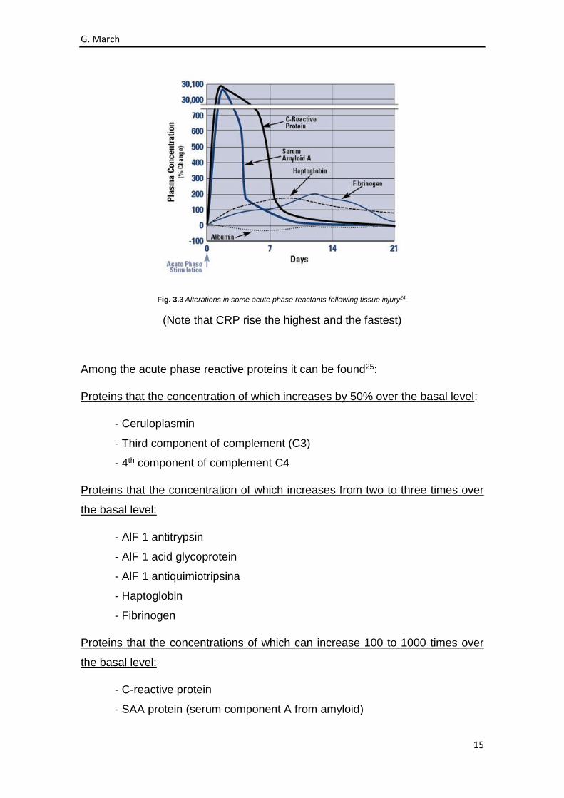
Fig. 3.3 Alterations in some acute phase reactants following tissue injury24.
(Note that CRP rise the highest and the fastest)
Among the acute phase reactive proteins it can be found25:
Proteins that the concentration of which increases by 50% over the basal level:
- Ceruloplasmin
- Third component of complement (C3)
- 4th component of complement C4
Proteins that the concentration of which increases from two to three times over
the basal level:
- AlF 1 antitrypsin
- AlF 1 acid glycoprotein
- AlF 1 antiquimiotripsina
- Haptoglobin
- Fibrinogen
Proteins that the concentrations of which can increase 100 to 1000 times over
the basal level:
- C-reactive protein
- SAA protein (serum component A from amyloid)
G. March
16
3.1 C REACTIVE PROTEIN (CRP)
Among the acute phase reactants, CRP is the most widely used due to the
features that presents by its high sensitivity and its kinetics of rapid evolution.
Along with ESR, are the most used to detect and monitor acute phase responses.
CRP was discovered in 1930 by William Tillet and Thomas Francis in the
Rockefeller Institute and its name is due to the ability to precipitate C
polysaccharide from Streptococcus pneumoniae. It consists of five identical
subunits not glycosylated, covalently associated in a ring configuration with a
cyclic pentameric symmetry26.
It is an acute phase protein which, in the presence of Ca+2 ions, reacts with
polysaccharide C (substance C is increased) of the pneumococcal cell wall
(rough way) and causes precipitation, migrating with beta globulin fraction from
serum in the electrophoresis. It stimulates phagocytosis.
It has also been shown that activates the classical pathway of complement, it is
set to T lymphocytes and inhibits clot retraction and platelet aggregation. It may
constitute a mechanism of protection phylogenetically old and non specific. Acts
as opsonin, interacting with lymphocytes and thrombocytes.
Presents a significant homology with IgG; has a similar peptide sequence
although it is antigenically differents, suggesting a common ancestral origin.
It belongs to the proportion of beta globulins, approximate molecular weight of
120,000 daltons, and its concentration tends to keep correlation with erythrocyte
sedimentation rate, increases in most inflammatory and neoplastic diseases and
it is detected in serum (increases CRP test). It is relatively thermostable, probably
produced in the liver in response to biochemical signals that provide from the
damaged or dead cells and is inactivated adding oxalate or citrate27.
The concentration of this protein is very low in normal conditions, increases
rapidly due to an acute inflammatory reaction, so abnormal concentrations can
be detected within few hours of the start of this reaction. The concentration
achieved by these proteins reflects the intensity of the underlying inflammatory

G. March
17
process. During very intense inflammatory reactions, it can quickly reach a
concentration thousand times higher than the basal and in a quickly way28.
It is also particularly effective in monitoring patient response to antibiotic
treatment. It is more sensitive than ESR to evaluate the response to treatment.
CRP is selectively deposited at the place of inflammation and can be degraded
in fragments by neutrophil proteinase in the same place.
The production of CRP may be affected by the products of fibroblasts, which are
activated in combination with dexamethasone and it is also affected by heavy
metals (cadmium, mercury, lead, copper, zinc, nickel).
CRP, orosomucoide, leukocytes and ESR have a value on early diagnosis.
PMN-elastase, alpha 1 antritripsina, ferroxidasa and haptoglonina and ESR are
markers for a delayed use29.
CRP shows a considerable rise in acute bacterial infections, while it is moderate
or absent in viral infections. This determines its use for the detection of a hidden
infection; to demonstrate the presence of a bacterial infection, especially when
the microbiological diagnosis is difficult or when the obtention of the result is
delayed; and to monitor drug therapy30.
CRP is an important factor within the elements of the acute phase response due
to the quickly increase of its concentration in a variety of inflammatory or tissue
damage stadiums.
It is a marker of inflammation, sensitive but not specific, which clearly increases
after any type of inflammatory stimulus. C-reactive protein levels rise since the 6-
12 hours of the beginning of the inflammatory process and normalize in the
absence of complications in two days. If the infection is not controlled, the levels
remain high. It may be useful, therefore, as a marker of bacterial infection and to
monitore therapeutic response.
In healthy persons, CRP levels in plasma are usually less than 1mg/L, less than
3mg/L in 90% of cases and less than 10mg/L in 96%. A normal CRP reflects the
absence of an inflammatory process, an inflammation of less than 12 hours of
evolution or a few inflammatory processes that do not elevate its values22.
G. March
18
However, CRP has a high diagnostic value because, when compared to other
markers of inflammation, it is elevated in all determinations where, for example,
the ESR is abnormally high. ESR increases when there is an absence of
inflammation, when there is an anemia due to a decrease of erythrocytes, during
pregnancy due to the increase of fibrinogen and in necrosis due to the loss of
albumin and the relative increase of the globulins; while the CRP has not a
variable range between normal and abnormal concentration and is not influenced
by anemia or others alterations in serum proteins31.
At the same time, CRP has shown great utility when monitoring patients in order
to know the effectiveness of treatment once the diagnosis of infection is done.
The serum levels of CRP decrease rapidly when antibiotic treatment is right,
keeping high when the treatment is ineffective32.
3.2 ERITRHOCYTE SEDIMENTATION RATE (ESR)
It is a routine test which measures the speed of red blood cells contained when
a blood sample is precipitated to the bottom of a milimetred pipette. Under normal
conditions, during the first hour, the precipitated volume is maximum 15mm for
men and 20mm for women.
The increase of ESR primarily reflects fibrinogen level and, to a lesser extent, the
level of other acute reactive proteins. The increase of these very asymmetrical
molecule proteins, promotes the breakdown of red blood cells forming piles of
coins, which increase their weight without increasing proportionately its surface
and settle faster.
The elevation of the ESR for an inflammatory process is completely nonspecific
and independent of the etiology of it. ESR is also less sensitive to the speed of
response to a correct treatment33.
The ESR, same way as CRP, is a nonspecific inflammatory marker that may be
useful in the diagnosis of a bacterial infection. A disadvantage compared to CRP
is that its rise in relation to inflammatory episodes is slow, both at the time of the
debut and during the referral process and also when monitoring therapeutic
response. Moreover, their values vary according to age, sex of the patient and

G. March
19
depend on series of reactions related to red blood cells, fibrinogen, lipids and
immunoglobulins.
Pathological values are considered when are upper than 25-30mm during the first
hour34.
The sedimentation rate mainly reflects the modifications of plasma proteins and
those often accompany most acute and chronic infections, tumors and
degenerative diseases.
ESR is a nonspecific response to tissue deterioration and indicates the presence
of disease, although it does not calibrate the severity of this31.
The increase of erythrocyte sedimentation rate is characterized by infections. It
is explained by the increase of fibrinogen and other acute phase reactants, which
are large and asymmetric molecules that cause a sink effect of repulsive forces
between blood cells red.
3.3 ALBUMIN
It is a plasmatic protein and its quantification is clinically useful.
It is the most abundant protein in blood plasma, with a concentration between 35-
52g/L. It covers up to two thirds of total plasma protein content.
Unlike the total protein, albumin reports the value of all globulins, with a
combination of other fractions which can increase individually in severe
conditions35.
Its half-life in plasma is about 20 days. It catabolitzes in various tissues. Albumin
is increased during infections, trauma and surgical operations.
The main functions are maintaining the pressure plasma, using as storage of
amino acids and transporting different ligands. Among the substances which are
transported by albumin there are: hormones (thyroxine, estrogen), fatty acids,
bilirubin, penicillin, cortisol, cumadina, calcium and magnesium.
The main changes of plasma albumin are hyperalbuminemia, hypoalbuminemia
and analbuminemia.

G. March
20
The hiperalbuminemia may be an artifact, for example, consequence of an
excessive venous blood ecstasy during the extraction process, or an excessive
parenteral infusion of albumin or a real effect on dehydration.
The hypoalbuminemia may be physiological or pathological. The physiological
can be because of the posture or pregnancy. With an upright posture, albumin
increases its concentration whereas in pregnancy the concentration is reduced.
When is pathological it may decrease due to a decline in its synthesis, an
increase of catabolism, a reduced absortion of intestinal amino acids, an increase
of distribution volume or an increase of the losses. Several diseases produce a
decrease in albumin synthesis, such as malnutrition and liver processes36.
After a trauma or a sepsis, occurs an increase in the permeability of the blood
that leads to a redistribution of albumin, although in these situations there is also
an increased catabolism.
The increase in the proteic losses can be produced by urinary tract (nephrotic
syndrome, glomerulonephritis, diabetes...) by feces (a protein losing enteropathy)
or by the skin (burns).
The measure of the concentration of albumin is a useful nutritional marker when
monitoring during a long-term (several weeks or months) patients who receive
nutritional support. However, its measure is also useful as a liver function
parameter; (it is usually normal in episodes of acute hepatitis and decreased in
chronic hepatitis)37.
3.4 LEUKOCYTOSIS
Is one of the most characteristic abnormalities of infectious diseases. Under
normal conditions they are found between 4,000-10,000 leukocytes per mm3 of
blood, in presence of infection they can reach up to 20,000 or 30,00028.
The leucocytary formula consists in the determination of the percentage of each
type of leukocytes - neutrophils, basophils, eosinophils, lymphocytes and
monocytes - in relation to the total. This data can be very important in the

G. March
21
diagnosis of many infectious diseases in which the concentration of certain types
of white blood cells increases significantly in relation to others.
In general, leukocytosis is a very variable element and the relation between
hyperleukocytosis and clinical signs of infection is not found in 60% of cases,
giving many false positives38.
Table I
Analitical Data
Normal Values:
-Leukocytes (4-10, 5) 10 (9)/L
-Neutrophils (2-7, 5) V.N. absolut
-Lymphocytes (1, 2-4, 5) V.N. absolut
-ESR (1-30) mm
-CRP (<6mg/dl)
-Albumin (3, 5 – 5,2g/dl)
-Total protein (6-8g/dl)
G. March
22
4. OBJECTIVES
4.1 GENERAL OBJECTIVE
To assess the effectiveness of CRP in the diagnosis of osteomyelitis in diabetic
foot patients.
4.2 SPECIFIC OBJECTIVES
To validate if albumin, leukocytes and ESR are also useful parameters in
the diagnosis of OM in diabetic foot.
To precise which others parameters of the blood test are altered in
presence of OM, besides the acute phase reactants.
To analyze the characteristics more frequently found in OM patients.

G. March
23
5. MATERIAL AND METHODS
Medical records collected in the database of Diabetic Foot Unit (UFPD) from
Hospital de Bellvitge are randomly reviewed. Only patients who were attended at
Emergency Unit diagnosed with osteomyelitis in the period between September
2013 and April 2015 are included in the study.
Records of diabetic patients hospitalized or treated in outpatient UFPD from
internal medicine with diagnosis of <<osteitis>>, <<soft tissue infection>> or
<<lower limbs cellulitis>> are obtained. Diagnosis of osteomyelitis is considered
by the histopathological test.
Those patients with no report of CRP, complete blood count and with no medical
story in the period of analysis are excluded.
Inclusion criteria
- Diabetic patients who were attended at Emergency Unit with foot infection.
- Diabetic patients from UFPD from Hospital de Bellvitge who were attended
at Emergency Unit.
- Patients from UFPD from Hospital de Bellvitge diagnosed with OM, osteitis
or soft tissue infection and admitted to Hospital de Bellvitge.
- With blood test before treatment.
- CRP and complete blood test report.
Exclusion criteria
- Patients who do not have any blood test at the beginning of treatment.
- CRP value was not reported in blood test.
- Blood test after the antibiotic treatment.
G. March
24
5.1 ACTING PROCESS
30 patients from the Diabetic Foot Unit of Hospital de Bellvitge are included in the
study. They presented an episode of osteomyelitis and attended Urgency Unit in
a period between 3th September 2013 and 3th April 2015.
First, the medical records of the selected patients are accessed, looking for the
blood analysis and subsequents clinical notes.
Once achieved all analytical tests from patients, a statistical study of all the
factors that are usually required in a case of OM is done. The aim of this study is
to know if CRP is a valid method to diagnosticate OM and also if there is another
factor, among those mentioned, which is also altered in most patients.
A measurement of the following parameters is done:
1- CRP
2- ALBUMIN
3- LEUKOCYTES
4- COMPLETE BLOOD COUNT (CBC)
The ESR has not been studied because in the analytical profile of the studied
patients is not required.
All 4 variables are measured and observed in each patient because bone
infection is a sufficiently important process for do not eliminate all the
complementary elements that we have available for its detection and control.
Later, a monitoring of the clinical courses of patients is done. The purpose of this
is to obtain more results and relationships with other co-morbidities which are
associated with DM and, therefore, make a broader discussion and obtain more
accurate results.

G. March
25
5.1.1 Variables to consider
General
- Age
- Sex
- Smoking
- Nephropathy
- Retinopathy
- Previous amputations
- Other associated complications
From surgery
- Surgical technique used
Analytical variables
- CRP: values pre and post surgical (comparison)
- Complete blood count pre-surgical
- Protein levels pre-surgical
5.1.2 Statistical analysis
All these data series are collected from each patient in a file. Subsequently, this
file is included in a database which will help us to analyze the obtained data.
A descriptive analysis is performed for the variables of the study. This consists in
obtaining mesures of central tendency and dispersion for quantitative variables,
using for the averages +/- their standard deviation.
All statistical analysis are performed using Microsoft Office Excel 2013.
From here, it is intended to reach the above objectives and to be able to establish
a later discussion about if acute phase reactants are useful or not to detect and
confirm the presence of OM.

G. March
26
6. RESULTS
Once the results are obtained, we observe which of the matching parameters are
altered in between all patients.
Fig. 6.4 Results of blood test of all diabetic foot patients diagnosed with OM.
-1100%
-900%
-700%
-500%
-300%
-100%
100%
300%
500%
700%
900%
1100%
1300%
1500%
1700%
1900%
2100%
2300%
2500%
2700%
Ure
a
Cre
atin
ine
Glo
mer
ula
r Fi
ltra
tio
n R
ate
Sod
ium
Ion
; c.s
ub
st.
Po
tass
ium
Ion
; c.s
ub
st.
Glu
cose
; c.s
ub
st.
Ala
nin
e-am
ino
tran
sfer
ase
; c.c
at.
Alb
um
in; c
.mas
sa (
CR
M 4
70
)
Gam
ma-
glu
tam
yltr
ansf
eras
e; c
.cat
.
Alk
alin
e P
ho
sph
atas
e; c
.cat
.
C R
eact
ive
Pro
tein
; c.m
assa
(C
RM
47
0)
Eryt
hro
cyte
s c.
no
m
Hem
ogl
ob
in; c
.mas
sa
Eryt
hro
cyte
s; f
r.vo
l (h
em
atò
crit
)
Eryt
hro
cyte
s; v
ol e
ntí
lic (
VC
M)
Hem
ogl
ob
in; m
assa
en
tilic
a (H
CM
)
Hem
ogl
ob
in; c
.mas
sa (
CH
CM
)
Eryt
hro
cyte
vo
lum
e; w
idth
re
lati
ve d
istr
ibu
tio
n
Pla
tele
ts c
.no
m
Pla
tele
ts; v
ol e
nti
lic (
VP
M)
Leu
kocy
tes
c.n
om
Ne
utr
op
hils
c.n
om
Lym
ph
ocy
tes
c.n
om
Mo
no
cyte
s c.
no
m
Eosi
no
ph
ils c
.no
m
Bas
op
hils
c.n
om
Tiss
ue
fact
or-
ind
uce
d c
oag
ula
tio
n (
pro
thro
mb
in…
Average ± SD (% from the normal range)
G. March
27
In figure 4, all factors are captured to observe which are altered. To achieve this,
0% represents the minimum normal level and 100% the maximum, this way all
factors are in the same scale and can be compared on the same chart. If the
round red is located outside the minimum or maximum, the average of the factor
in question is altered.
The factors that are altered in the majority of the studied patients during a bone
infection are clearly visible in this figure.
Creatinine, glucose and gamma-glutamyltransferase are above their standard
values. The fact that glucose displays high in diabetic patients is logical. Just as
creatitina also means that these patients often present a nephropathy. The
gamma-glutamyltransferase is used to detect diseases of the liver so, its high
levels are not useful for this study.
At the same time, alkaline phosphatase and CRP are found altered. It will be
discussed later.
From complete blood count, not all factors are included because they are not
requested in analytical profiles of all patients; however, from the requested
parameters of the CBC of all patients, erythrocytes, hemoglobin and neutrophils
are altered. Leukocytes are slightly higher. It will also be discussed with more
detail in the following sections.
Albumin is also slightly altered.
6.1 ANALYTICAL RESULTS
According to the histopathological results, the levels of CRP and the complete
blood count are compared. It is proved that they have an important role in front
of an infectious process. In the same way, albumin and alkaline phosphatase are
also specifically studied because they have shown altered and it could also
influence the diagnosis of OM.

G. March
28
6.1.1. C reactive protein
It is observed that patients with OM have a CRP much higher than the ones with
no marrow affectation.
Fig. 6.1.1.5 CRP results of the studied patients.
In the population of the 30 studied patients, CRP serum levels are in all patients
above the normal range.
The CRP of healthy patients should be between 0 and 5 mg/L whereas the
average of CRP in our patients is 69,81mg/L. This indicates that, in presence of
an infection, the protein is altered and its blood levels soar. The hypotesis of this
thesis is resolved in the above figure.
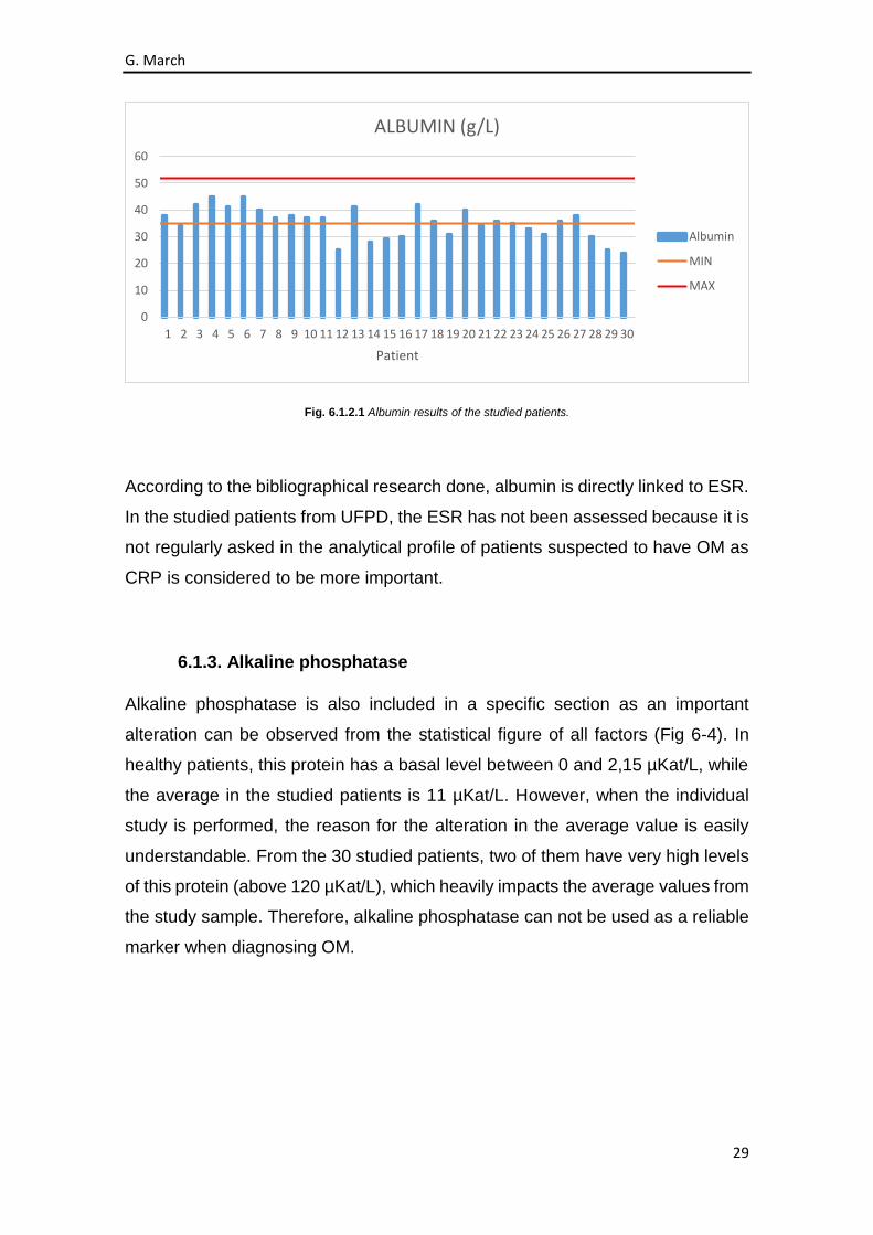
6.1.2. Albumin
The albumin is also studied because it has been altered in the studied patients.
In healthy patients this protein has a basal level between 35 and 53 g/L while in
the studied patients this protein is below levels, with an average of 35 g/L (the
minimum value of the protein).
0
50
100
150
200
250
300
1 2 3 4 5 6 7 8 9 10 11 12 13 14 15 16 17 18 19 20 21 22 23 24 25 26 27 28 29 30
Patient
CRP (mg/L)
CRP
MIN
MAX
G. March
29
Fig. 6.1.2.1 Albumin results of the studied patients.
According to the bibliographical research done, albumin is directly linked to ESR.
In the studied patients from UFPD, the ESR has not been assessed because it is
not regularly asked in the analytical profile of patients suspected to have OM as
CRP is considered to be more important.
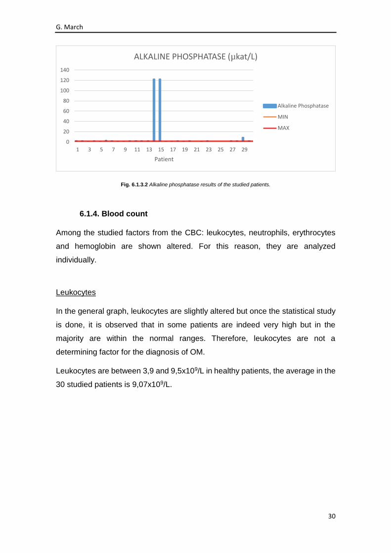
6.1.3. Alkaline phosphatase
Alkaline phosphatase is also included in a specific section as an important
alteration can be observed from the statistical figure of all factors (Fig 6-4). In
healthy patients, this protein has a basal level between 0 and 2,15 µKat/L, while
the average in the studied patients is 11 µKat/L. However, when the individual
study is performed, the reason for the alteration in the average value is easily
understandable. From the 30 studied patients, two of them have very high levels
of this protein (above 120 µKat/L), which heavily impacts the average values from
the study sample. Therefore, alkaline phosphatase can not be used as a reliable
marker when diagnosing OM.
0
10
20
30
40
50
60
1 2 3 4 5 6 7 8 9 10 11 12 13 14 15 16 17 18 19 20 21 22 23 24 25 26 27 28 29 30
Patient
ALBUMIN (g/L)
Albumin
MIN
MAX
G. March
30
Fig. 6.1.3.2 Alkaline phosphatase results of the studied patients.
6.1.4. Blood count
Among the studied factors from the CBC: leukocytes, neutrophils, erythrocytes
and hemoglobin are shown altered. For this reason, they are analyzed
individually.
Leukocytes
In the general graph, leukocytes are slightly altered but once the statistical study
is done, it is observed that in some patients are indeed very high but in the
majority are within the normal ranges. Therefore, leukocytes are not a
determining factor for the diagnosis of OM.
Leukocytes are between 3,9 and 9,5x10⁹/L in healthy patients, the average in the
30 studied patients is 9,07x10⁹/L.
0
20
40
60
80
100
120
140
1 3 5 7 9 11 13 15 17 19 21 23 25 27 29
Patient
ALKALINE PHOSPHATASE (µkat/L)
Alkaline Phosphatase
MIN
MAX

G. March
31
Fig. 6.1.4.3 Leukocytes results of the studied patients.
Neutrophils
Neutrophils, same way as leukocytes, showed a slight alteration in the general
graph because there is a lot of variability between patients. Some patients have
levels above the maximum normal values, while there are patients whose levels
are below the minimum. The majority of the studied patients present neutrophils
within normal ranges, and therefore, it is not a valuable parameter when
diagnosing OM. In healthy patients the levels of neutrophils are between 1,5 and
5,7x10⁹/L while in these patients the average is 6,43x10⁹/L.
Fig. 6.1.4.4 Neutrophils results of the studied patients.
0
5
10
15
20
25
1 2 3 4 5 6 7 8 9 10 11 12 13 14 15 16 17 18 19 20 21 22 23 24 25 26 27 28 29 30
Patient
LEUKOCYTES (x109/L)
Leukocytes
MIN
MAX
0
5
10
15
20
1 2 3 4 5 6 7 8 9 10 11 12 13 14 15 16 17 18 19 20 21 22 23 24 25 26 27 28 29 30
Patient
NEUTROPHILS (x109/L)
Neutrophils
MIN
MAX

G. March
32
Erythrocytes
In the individual graph it is shown how erythrocytes, which are altered in the graph
of all factors, are in almost all patients below the minimum range of normality.
This fact has sufficient validity to indicate that studied patients with osteomyelitis
present very low values of red blood cells. A relationship between bone infection
and low concentration of erythrocytes could be established.
Erythrocytes levels in healthy patients are between 4,3 and 5,6x10¹²/L while in
the studied patients, the average of erythrocytes is 3,74x10¹²/L.
Fig. 6.1.4.5 Erythrocytes results of the studied patients.
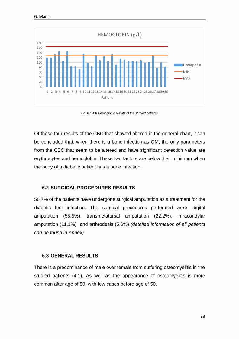
Hemoglobin
Simultaneously, hemoglobin is shown altered in the general graph. In the
individual study, abnormally low levels in almost all patients can be observed. As
well as with erythrocytes, it could be affirmed that there is an existing relationship
between diagnosis of infection and hemoglobin concentration in blood.
The average of hemoglobin in the studied patients is 107g/L while parameters in
healthy patients are between 130 and 165g/L.
0
1
2
3
4
5
6
1 2 3 4 5 6 7 8 9 10 11 12 13 14 15 16 17 18 19 20 21 22 23 24 25 26 27 28 29 30
Patient
ERYTHROCYTES (x1012/L)
Erythrocytes
MIN
MAX
G. March
33
Fig. 6.1.4.6 Hemoglobin results of the studied patients.
Of these four results of the CBC that showed altered in the general chart, it can
be concluded that, when there is a bone infection as OM, the only parameters
from the CBC that seem to be altered and have significant detection value are
erythrocytes and hemoglobin. These two factors are below their minimum when
the body of a diabetic patient has a bone infection.
6.2 SURGICAL PROCEDURES RESULTS
56,7% of the patients have undergone surgical amputation as a treatment for the
diabetic foot infection. The surgical procedures performed were: digital
amputation (55,5%), transmetatarsal amputation (22,2%), infracondylar
amputation (11,1%) and arthrodesis (5,6%) (detailed information of all patients
can be found in Annex).
6.3 GENERAL RESULTS
There is a predominance of male over female from suffering osteomyelitis in the
studied patients (4:1). As well as the appearance of osteomyelitis is more
common after age of 50, with few cases before age of 50.
0
20
40
60
80
100
120
140
160
180
1 2 3 4 5 6 7 8 9 10 11 12 13 14 15 16 17 18 19 20 21 22 23 24 25 26 27 28 29 30
Patient
HEMOGLOBIN (g/L)
Hemoglobin
MIN
MAX

G. March
34
Fig. 6.3.7 Age and sex group of the studied patients.
The main comorbidity reported has been smoking (53,33% of the patients),
followed by nephropathy (33,33% of the studied sample).
Dyslipidemia was presented only in 20% of the patients. Other comorbidities
reported were: previous history of amputation, retinopathy, heart disease and
hypertension. (detailed information of all patients can be found in Annex).
Fig. 6.3.8 Associated comorbidities of the studied patients.
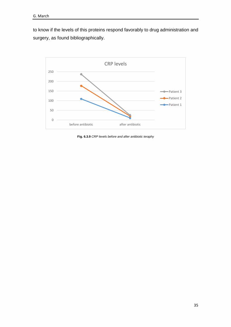
Finally, a follow-up of three of the total studied patients is included. The aim of
this, is to compare the levels of CRP when OM is diagnosed with the levels when
patients are already with pharmacological or surgical treatment. This will help us
0%
10%
20%
30%
40%
50%
60%
70%
0-19 20-39 40-59 60-79 80-99
Age group of the patients Sex of the patients
Male Female
0% 10% 20% 30% 40% 50% 60%
Retinopathy
Heart Disease
Dyslipemia
Previous Amputation
HTA
Nephropathy
Smoking
Comorbidities
G. March
35
to know if the levels of this proteins respond favorably to drug administration and
surgery, as found bibliographically.
Fig. 6.3.9 CRP levels before and after antibiotic teraphy
0
50
100
150
200
250
before antibiotic after antibiotic
CRP levels
Patient 3
Patient 2
Patient 1
G. March
36
7. DISCUSSION
Although the number of studied cases has been reduced to obtain a statistically
significant difference, the tendency to higher levels of CRP in all patients with OM
report compared to those without bone infection has been demonstrated. This
finding is similar to other series already reported, indicating that CRP is an
important factor in the diagnosis of osteomyelitis.
However, further studies with significant sample of patients to affirm with greater
validity the hypothesis should be reported. This thesis is just an initial study where
the trend of CRP in front of an episode of osteomyelitis is observed, but more
detailed investigation on the observed relationships should be carried out.
It is also important to note that CRP values decrease drastically once the infection
is treated, both pharmacologically and surgically, which helps us to monitor the
evolution of OM and the effectiveness of the treatment given.
On the other hand, the undertaken research has not been able to assess the
diagnostic value of ESR because it was not requested in the analytical profiles of
the studied patients, which falls beyond the scope of our work.
Albumin and leukocytes have shown to present a lot of variability between
patients. Therefore, neither of these proteins have demonstrated their
effectiveness when diagnosing osteomyelitis.
From the complete blood count, erythrocytes and hemoglobin are below normal
levels in almost all patients with OM. This could be relevant when proposing
further studies that specifically evaluate these two factors as they seem to have
a close relationship with the appearance of OM.
Patients with diabetic foot have severe cardiovascular risk factors such as
smoking, excess weight and presence of complications associated with diabetes
(retinopathy, nephropathy, hypertension and amputation).
It has been observed that 20% of patients refer to have dyslipidemia or are
treated with a statin. A remarkable feature is the antecedent of previous
amputation in a considerable proportion of the population. The history of foot
ulcers and amputation significantly increases the likelihood of subsequent ulcers.
G. March
37
These whole series of complications, both micro and macrovascular as well as
previous episodes of amputation reflect the serious systemic deteriorations of the
selected population, so there is a worse prognosis on function and morbidity.
About epidemiology, there is higher incidence of suffering osteomielitis after the
age of 50 with higher prevalence in males.
Due to the individual and social impact that diabetic foot causes, it is required to
implement prevention programs and take better care of risk patients in order to
avoid adopting extreme measures in late situations. These educational measures
should be directed to patients, families and doctors (both first contact and
specialists of different areas that are in contact with diabetic patients).
To sum up, Magnetic Resonance Imaging (MRI) is the study that has shown to
have greater utility in the noninvasive development of osteomyelitis in the diabetic
foot. However, its low availability in our environment is a limiting factor and that’s
why other indirect markers have been looked for. C-reactive protein may be a
support diagnosis when osteomyelitis is suspected, but it has to be considered
or interpreted in the clinical context of the patient and in conjunction with
radiological studies.
If acting on the diagnosis of almost all diabetic foot patients who present bone
infection was possible, the decrease in the prevalence of amputations would be
a reality. This would improve the life quality of diabetics.
Currently, it is just an ideal instead of the goal of many Medical teams. To achieve
this, it is necessary to raise awareness on this health problem and, therefore,
keep working to implement widely agreed guidelines for early and accurate
diagnosis.

G. March
38
8. CONCLUSIONS
In the analyzed population there is a delay in the diagnosis of osteomyelitis due
to the difficulties that diabetic foot infection involves.
C-reactive protein has shown to have high diagnostic value for OM.
The ESR has not been evaluated in the studied patients because it is not
requested in the analytical profile of the studied Unit, which implies that it is not
worth considering when diagnosing OM.
Albumin and leukocytes are not value parameters to diagnose OM. From CBC,
hemoglobin and erythrocytes appear to be related to the onset of OM.
OM appears more frequently in male and after the age of 50. Smoking is the main
comorbidity of these patients followed by nephropathy.
G. March
39
9. REFERENCES
1. Got I. “Necessary multidisciplinary management of diabetic foot”. J Mal Vasc
2001, 36, pp. 130-134.
2. Meijer JW, Trip J, Jaegers SM, Links TP, Smith Al, Groothoff Jw et al. “Quality
of life in patients with diabetic foot ulcers”. Diabril Rehabil 2001, 23, pp. 336-340.
3. “Consensus development conference on diabetic foot wound care”. American
Diabetes Association. Adv Wound Care 1999, 12, pp. 353-361.
4. Fejfarova V, Hosova J, Striz I, Kalanin J, Skibova J. “Analysis of the
inflammation reaction and selected indicators of immunity in patients with an
infected diabetic ulcer”. Cas Lek Cesk 2002, 141, pp. 483-486.
5. Morrisson WB, Ledermann HP. “Work-up, of the diabetic foot”. Radiol Clin N
Am 2002, 40, pp. 1171-1192.
6. Lew DP, Waldvogel FA. “Osteomyelitis”. Lancet 2004, 364, pp. 369-379.
7. Bakker K, Van Houtum W. H, Riley P. C. “The International Diabetes
Federation focuses on the diabetic foot”. Curr. Diab. Rep. December, 2005, 5 (6),
pp. 436-440.
8. Schaper N. C, Nabuurs-Franssen M. H. “The diabetic foot: pathogenesis and
clinical evaluation”. Semin. Vasc. Med. May, 2002, 2 (2), pp. 221-228.
9. Senneville, E. “Infection and diabetic foot”. Rev. Med. Interne. September,
2008, 29 (2), pp. 3.243-3.248.
10. Bjarsholt T, Kirketerp-Moller K, Jensen P. O; et al. “Why chronic wounds will
not heal: a novel hypothesis”. Wound Repair Regen. January-February, 2008, 16
(1), pp. 2-10.
11. Schaper, N. C. “Diabetic foot ulcer classification system for research
purposes: a progress report on criteria for including patients in research studies”.
Diabetes Metab. Rev. May-June, 2004, 20 (1), pp. 590-595.
12. Aragón-Sánchez, J. “Treatment of diabetic foot osteomyelitis: a surgical
critique”. Int. J. Law. Extrem. Wounds. March, 2010, 9 (1), pp. 37-59.
G. March
40
13. Berendt A. R, Peters E. J, Bakker K; et al. “Diabetic foot osteomyelitis: a
progress report on diagnosis and a systematic review of treatment”. Diabetes
Metab. Res. Rev. May-June, 2008, 24 (1), pp. 5.145-5.161.
14. Lavery L. A, Armstrong D. G, Peters E. J; et al. “Probe-to-bone test for
diagnosing diabetic foot osteomyelitis: reliable or relie?” Diabetes Care.
February, 2007, 30 (2), pp. 270-274.
15. Schwegler B, Stumpe K. D, Weishaupt D; et al. “Unsuspected osteomyelitis
is frequent in persistent diabetic foot ulcer and better diagnosed by MRI than by
18F-FDG PET or 99mTc-MoAb”. J. Intern. Med. January, 2008, 263 (1), pp. 99-
106.
16. Rozzanigo U, Tagliani A, Vittorini E; et al. “Role of magnetic resonance
imaging in the evaluation of diabetic foot with suspected osteomielitis”. Radiol.
Med. October, 2008, pp. 25.
17. Strobel K, Stumpe K. D. “PET/CT in musculoskeletal infection”. Semin.
Musculoskelet. Radiol. December, 2007, 11 (4), pp. 353-364.
18. Aragón-Sánchez J, Quintana-Marrero Y, Lázaro-Mártinez, J. L; et al.
“Necrotizing soft-tissue infections in the feet of patients with diabetes: outcome
of surgical treatment and factors associated with limb loss and mortality”. Int. J.
Low Extrem Wounds. September, 2009; 8 (3), pp. 141-146.
19. Morales J J, Cabo J, Fernández Sabaté A, Clos R, Villena M, Ariza J. “The
biological test used in acute-phase of inflamation in bone infection”. Eur J Orthop
Surg Traumatol. 1995, 5, pp. 33-36.
20. Joan M. et al. “The role of C-reactive protein in the evaluation and
management of infants with suspected sepsis”. Adv Neonatal Care. 2003, 3 (1).
21. Campos I, Sotelo E, Gutiérrez H. “Comportamiento de los reactantes de fase
aguda en pacientes desnutridos y eutróficos, con y sin infección”. Arch ven puer
pediatr. 2001, 64 (2), pp. 87-94.
22. Ciampolini J, Harding KG. “Pathophysiology of chronic bacterial osteomielitis.
Why do antibiotics fail so often?” Postgrad Med J 2000, 76 (898), pp. 479-483.
23. DuClos T. “Function of C-reactive protein”. Ann Med. 2000, 32, pp. 274-278.
G. March
41
24. Morris, A. M. “Should we be testing procalcitonin in critically ill patients?” Blog,
2009. Available in: http://www.idologist.com/Blog/2009/04/23/should-we-be-
testing-procalcitonin-in-critically-ill-patients/ Accessed February 20, 2015.
25. Pascual Gómez E. “Pruebas de laboratorio en el diagnóstico de
enfermedades reumáticas”. Patología reumática básica. Medicine. 1982, pp. 25-
39.
26. Jenny J-Y, Gaudias J, Bourguignat A, Férard G, Kempf I. “La protéine C-
réactive et la transthyrétine dans le diagnostic précoce de l’infection aprés
fracture ouverte des membres inférieurs (etude préliminaire)”. Rev Chir Orthop.
1999, 85, pp. 321-327.
27. Kushner I. “C reactive protein in rheumatology”. Arthritis Rheum. 1999, 1 (34),
pp. 1065-1068.
28. Berliner S et al. “Agregation of white cells and C-reactive protein: relation
between these two indices in acute phase reaction”. J Clin Path. January 1987,
40 (1), pp. 103-106.
29. Escrivá F, Jiménez A. “Proteínas plasmáticas”. In: Herrera E ed. Biología
molecular y bioquímica fisiológica. Madrid: Mc Graw Hill Interamericana. 1991,
pp. 1285-1295.
30. Guillén et al. “Reactantes de fase aguda y su impacto en el estado
nutricional”. Rev Med Cie. 2008, 14 (1), pp. 12-18.
31. Marín et al. “Comparación de dos métodos automatizados para la
determinación de Proteína C reactiva en pacientes pediátricos”. Rev. Méd. Hosp.
Nac. (Costa Rica) Niños. 2011, 37 (1-2), pp. 29-31.
32. Schwart CH. “Metabolismo hepático”. In: Kelley W, De vita V, DuPont H et al
eds. Medicina Interna. Buenos Aires: Panamericana. 1990, pp. 491-502.
33. Verges G et al. “Enfermedades infecciosas. Pruebas diagnósticas”.
Enciclopedia de medicina y salud. Barcelona: Sigma. 1991, (7), pp. 65-73.
34. Javayolas M, Montreal M. “Tratamiento antibiótico por vía oral de la
osteomielitis bacteriana del adulto”. Med Clín, 1999, pp. 488-489.
G. March
42
35. Voet D, Voet J G. “El sistema endocrino. Comunicaciones bioquímicas:
Hormonas y neurotransmisores”. Bioquímica. Barcelona: Omega. 1992, pp.
1234-1247.
36. Borque L, González J M. “Proteínas del plasma sanguíneo”. Bioquímica
clínica. Buenos Aires: Mc Graw-Hill Interamericana. 1998, pp. 191-204.
37. Gibbs J. et al. “Preoperative serum albumin level as a predictor of operative
mortality and mobidity”. Results from the national V A surgical risk study. 1999,
Arch Surg 134 (1): pp. 36-42.
38. Evello J. “Infección herida quirúrgica, infección estafilocócica”. Enfermedades
infecciosas, patogénesis y diagnóstico. Barcelona: Salvat 1983, pp. 564-572.

G. March
43
10. AKWNOWLEDGEMENTS
I would like to express my deepest gratitude to my supervisor, Carolina Padrós,
whose expertise, understanding and patience added considerably to my graduate
experience.
I’d also like to thank the other members of the Podiatry Department of the
Univeristat of Barcelona for the assistance they provided during all these years
and helping me to develop my background in podiatry.
A special thanks to my parents and sister. They were –and always are -
supporting me and encouraging me with their best wishes. I would never have
been able to finish this project without your guidance, help and support.
Finally, I must acknowledge Gerard, he was always there cheering me up and
stood by me through the good times and bad.

G. March
44
ANNEXES
G. March
45
INDEX
1. FACTORS OF THE BLOOD TEST ............................................................ 46
2. OTHER FACTORS .................................................................................... 55

G. March
46
1. FACTORS OF THE BLOOD TEST
Factor Urea Creatinine Glomerular Filtration Rate
1 6,2 66 90
2 8 60 90
3 3,6 56 90
4 4,9 57 90
5 4,2 59 90
6 4,1 65 90
7 7,9 129 52
8 6,6 90 74
9 11,5 123 74
10 6,4 94 89
11 7,1 86 76
12 6,7 130 38
13 5 75 90
14 7,5 146 47
15 9,3 137 47
16 4,9 106 66
17 3,6 56 90
18 13,1 510 10
19 7,2 138 49
20 18,1 320 16
21 7,5 102
22 19 282 20
23 19,7 121 51
24 6,6 88 80
25 7 99 69
26 7,5 119
27 6 73 90
28 22,1 625 8
29 17 153 39
30 2,9 72 90
G. March
47
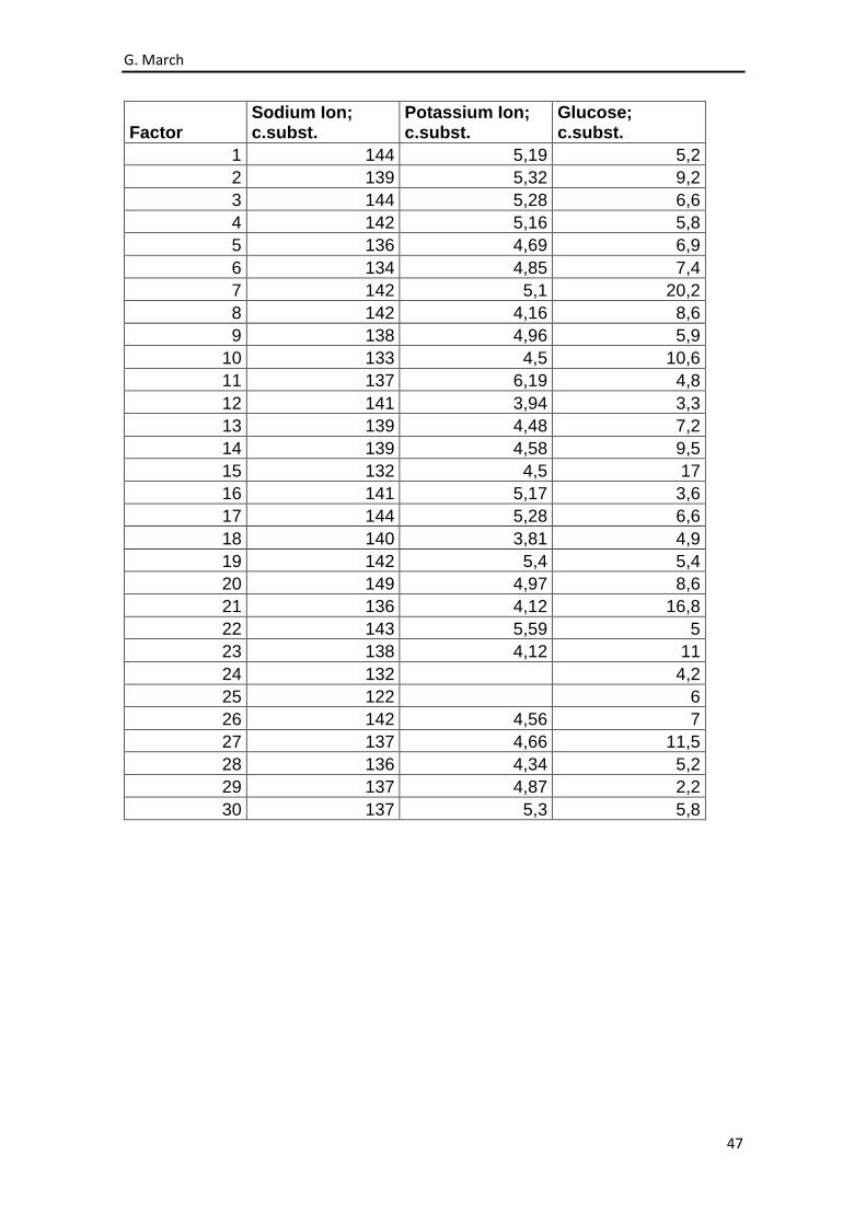
Factor Sodium Ion; c.subst.
Potassium Ion; c.subst.
Glucose; c.subst.
1 144 5,19 5,2
2 139 5,32 9,2
3 144 5,28 6,6
4 142 5,16 5,8
5 136 4,69 6,9
6 134 4,85 7,4
7 142 5,1 20,2
8 142 4,16 8,6
9 138 4,96 5,9
10 133 4,5 10,6
11 137 6,19 4,8
12 141 3,94 3,3
13 139 4,48 7,2
14 139 4,58 9,5
15 132 4,5 17
16 141 5,17 3,6
17 144 5,28 6,6
18 140 3,81 4,9
19 142 5,4 5,4
20 149 4,97 8,6
21 136 4,12 16,8
22 143 5,59 5
23 138 4,12 11
24 132 4,2
25 122 6
26 142 4,56 7
27 137 4,66 11,5
28 136 4,34 5,2
29 137 4,87 2,2
30 137 5,3 5,8

G. March
48
Factor
Alanine-aminotransferase; c.cat.
Albumin; c.massa (CRM 470)
Gamma-glutamyltransferase; c.cat.
1 0,23 38 0,52
2 0,41 34 1,46
3 0,22 42 1,29
4 0,4 45 0,3
5 0,51 41 1,42
6 1,13 45 9,98
7 0,17 40 0,62
8 0,08 37 0,24
9 0,08 38 0,24
10 0,31 37 0,89
11 0,11 37 0,55
12 0,11 25 0,35
13 0,25 41 0,76
14 0,38 28 0,73
15 0,38 29 0,74
16 0,08 30 0,16
17 0,22 42 1,29
18 0,63 36 0,95
19 0,61 31 0,34
20 0,21 40 0,47
21 0,36 34
22 0,23 36 0,74
23 1,82 35 3,59
24 0,44 33
25 1,12 31 1,99
26 0,4 36
27 0,33 38 0,38
28 0,12 30 0,55
29 0,38 25 5,23
30 0,2 24 0,65

G. March
49
Factor
Alkaline Phosphatase; c.cat.
C Reactive Protein; c.massa (CRM 470)
Erythrocytes c.nom
1 1,98 67,2 3,89
2 2,04 17,1 4,72
3 1,13 23,7 4,15
4 1,29 2,2 4,77
5 1,2 60,5 3,79
6 2,82 17,9 4,61
7 1,5 27,5 2,97
8 0,37 54,8 3,59
9 0,37 88,1 3,2
10 1,61 175,8 4,72
11 1,79 25,2 2,98
12 1,32 26,7 2,78
13 1,66 40,1 4,47
14 122 193,4 3,71
15 122 252 4,3
16 0,6 108,2 3,75
17 1,13 23,7 4,15
18 1,94 15,9 3,02
19 1,25 61,7 3,94
20 1,84 29,9 3,85
21 193 3,7
22 0,88 75,2 3,53
23 1,62 67,2 3,53
24 76,5 3,69
25 53,1 3,35
26 69,8 3,5
27 1,31 10,7 4,56
28 1,76 60 2,48
29 8,75 109,4 3,45
30 2,07 67,7 3,07

G. March
50
Factor Hemoglobin; c.massa
Erythrocytes; fr.vol (hematòcrit)
Erythrocytes; vol entílic (VCM)
1 119 38,1 98
2 118 37,8 80
3 132 41,2 99
4 144 44,4 93
5 105 0,323 85
6 144 0,436 95
7 83 0,27 91
8 82 0,257 71
9 70 0,219 68
10 134 0,401 85
11 97 0,291 98
12 82 0,25 90
13 130 0,377 84
14 108 31 84
15 124 35,6 83
16 104 32 85
17 132 41,2 99
18 90 0,282 94
19 114 0,358 91
20 110 0,318 83
21 105 32 87
22 104 0,323 91
23 103 0,312 88
24 107 0,319 87
25 97 0,272 81
26 100 30,8 87
27 129 0,395 87
28 77 0,236 95
29 99 30,6 89
30 81 26 85

G. March
51
Factor
Hemoglobin; massa entilica (HCM)
Hemoglobin; c.massa (CHCM)
Erythrocyte volume; width relative distribution
1 31 312 14,9
2 25 312 15
3 32 320 13,8
4 30 324 12,5
5 28 324 16,9
6 31 329 16,1
7 28 309 18
8 23 318 24,3
9 21 300 21
10 28 334 17,8
11 33 335 17,3
12 30 330 17,8
13 29 344 15,9
14 29 348 12
15 29 348 12,3
16 28 325 13,1
17 32 320 13,8
18 30 318 20
19 29 318 15,8
20 29 345 18,1
21 29 328 13,9
22 29 320 16,9
23 29 330 18,9
24 29 335 16,2
25 29 355 15,4
26 28 325 13,2
27 28 327 16,1
28 31 328 17,1
29 29 324 15,4
30 26 312 14,7

G. March
52
Factor Platelets c.nom Platelets; vol entilic (VPM)
Leukocytes c.nom
1 338 11,4 6,5
2 420 11,4 11,3
3 161 11,7 5,1
4 195 10,2 7,4
5 362 7,7 7,9
6 134 10 7,2
7 168 9,5 6,4
8 378 7,8 6,5
9 621 7,2 7,2
10 348 10,6 15,5
11 357 9,7 8,7
12 96 8,6 2,3
13 332 10,1 9,7
14 334 9,3 8,7
15 449 9,1 19,8
16 304 11,5 9,1
17 161 11,7 5,1
18 165 7,8 5,5
19 301 8,6 8
20 260 8,7 12,4
21 230 10,9 9
22 244 10,5 10,1
23 221 9,6 2,6
24 376 8,4 10,9
25 848 7,9 11,5
26 276 10,9 11,7
27 311 8,6 8,5
28 159 7,9 4,2
29 483 10,5 20,9
30 581 10 12,3

G. March
53
Factor Neutrophils c.nom
Lymphocytes c.nom
Monocytes c.nom
1 4,1 1,7 0,51
2 5,9 4,1 0,69
3 2,8 1,4 0,63
4 3,9 2,5 0,8
5 5,6 1,4 0,54
6 5,3 1,2 0,5
7 5,6 0,6 0,19
8 4,4 1,5 0,47
9 5,2 1,4 0,36
10 11,6 2,1 1,5
11 6,5 1,6 0,25
12 1,4 0,7 0,07
13 6,3 2,4 0,63
14 6,3 1,2 1,02
15 17,4 0,9 1,4
16 6,4 1,6 0,86
17 2,8 1,4 0,63
18 3,6 1,2 0,42
19 4,9 2,1 0,5
20 8,8 2,3 0,91
21 6,4 1,5 0,8
22 7 1,6 0,76
23 2,4 0,1 0,05
24 8,1 1,4 1,18
25 8,3 1,6 1,29
26 8,6 1,9 1,06
27 5,4 2,3 0,61
28 2,6 0,8 0,47
29 16,2 2,6 1,81
30 9,2 2,2 0,83
G. March
54
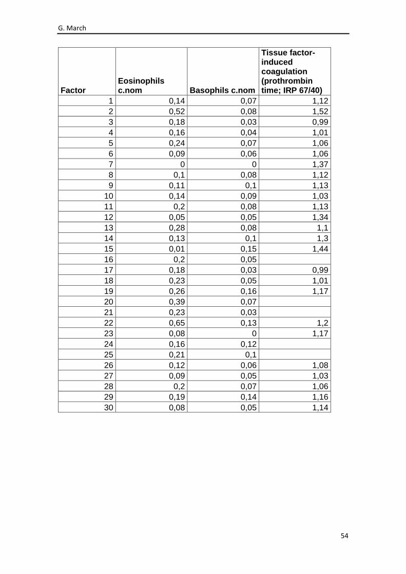
Factor Eosinophils c.nom Basophils c.nom
Tissue factor-induced coagulation (prothrombin time; IRP 67/40)
1 0,14 0,07 1,12
2 0,52 0,08 1,52
3 0,18 0,03 0,99
4 0,16 0,04 1,01
5 0,24 0,07 1,06
6 0,09 0,06 1,06
7 0 0 1,37
8 0,1 0,08 1,12
9 0,11 0,1 1,13
10 0,14 0,09 1,03
11 0,2 0,08 1,13
12 0,05 0,05 1,34
13 0,28 0,08 1,1
14 0,13 0,1 1,3
15 0,01 0,15 1,44
16 0,2 0,05
17 0,18 0,03 0,99
18 0,23 0,05 1,01
19 0,26 0,16 1,17
20 0,39 0,07
21 0,23 0,03
22 0,65 0,13 1,2
23 0,08 0 1,17
24 0,16 0,12
25 0,21 0,1
26 0,12 0,06 1,08
27 0,09 0,05 1,03
28 0,2 0,07 1,06
29 0,19 0,14 1,16
30 0,08 0,05 1,14

G. March
55
2. OTHER FACTORS
Patient Age Sex Comorbidities Smoke Previous Amputation
1 70 M Smoke Yes
2 73 M Smoke Yes
3 65 M Cirrhosis Yes
4 63 M
5 66 M Smoke Yes
6 59 M Cirrhosis
7 64 M HTA Yes
8 59 F Smoke, EPOC Yes Yes
9 32 F Yes
10 38 M No AMC
11 75 F Smoke Yes Yes
12 54 M Smoke Yes Yes
13 60 M Smoke Yes
14 55 M Smoke Yes Yes
15 57 M Smoke Yes Yes
16 58 M Smoke, obesity Yes
17 80 F
18 72 M HTA, DLP
19 52 M Smoke, Alcohol Yes Yes
20 60 F HTA, DLP
21 74 M Smoke, Obesity, HTA, Alcohol
Yes
22 65 M HTA, DLP, DBM
23 60 M HTA, DLP, DM
24 70 M Smoke Yes
25 65 F Smoke Yes
26 67 M
27 80 M
28 58 M Smoke Yes
29 73 M
30 71 M Smoke Yes

G. March
56
Patient Dyslipemia Nephropathy Retinopathy HTA Heart Disease
1
2
3
4
5 Yes Yes
6 Yes
7
8 Yes Yes Yes
9 Yes Yes
10 Yes Yes
11 Yes Yes
12 Yes
13
14 Yes Yes Yes Yes
15 Yes Yes
16 Yes Yes Yes
17 Yes Yes
18 Yes Yes
19 Yes Yes
20 Yes
21 Yes
22 Yes
23 Yes
24 Yes
25
26 Yes
27
28 Yes
29
30
G. March
57
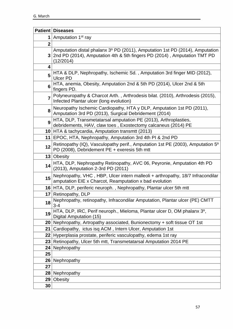
Patient Diseases
1 Amputation 1st ray
2
3 Amputation distal phalanx 3º PD (2011), Amputation 1st PD (2014), Amputation 2nd PD (2014), Amputation 4th & 5th fingers PD (2014) , Amputation TMT PD (12/2014)
4
5 HTA & DLP, Nephropathy, Ischemic Sd. , Amputation 3rd finger MID (2012), Ulcer PD
6 HTA, anemia, Obesity, Amputation 2nd & 5th PD (2014), Ulcer 2nd & 5th fingers PD.
7 Polyneuropathy & Charcot Arth. , Arthrodesis bilat. (2010), Arthrodesis (2015), Infected Plantar ulcer (long evolution)
8 Neuropathy Ischemic Cardiopathy, HTA y DLP, Amputation 1st PD (2011), Amputation 3rd PD (2013), Surgical Debridement (2014)
9 HTA, DLP, Transmetatarsal amputation PE (2013), Arthroplasties, debridements, HAV, claw toes , Exostectomy calcaneus (2014) PE
10 HTA & tachycardia, Amputation transmtt (2013)
11 EPOC, HTA, Nephropathy, Amputation 3rd 4th PI & 2nd PD
12 Retinopathy (IQ), Vasculopathy perif., Amputation 1st PE (2003), Amputation 5º PD (2008), Debridement PE + exeresis 5th mtt
13 Obesity
14 HTA, DLP, Nephropathy Retinopathy, AVC 06, Peyronie, Amputation 4th PD (2013), Amputation 2-3rd PD (2011)
15 Nephropathy, VHC , HBP, Ulcer intern malleoli + arthropathy, 18/7 Infracondilar amputation EIE x Charcot, Reamputation x bad evolution
16 HTA, DLP, periferic neuroph. , Nephropathy, Plantar ulcer 5th mtt
17 Retinopathy, DLP
18 Nephropathy, retinopathy, Infracondilar Amputation, Plantar ulcer (PE) CMTT 3-4
19 HTA, DLP, IRC, Perif neuroph., Mieloma, Plantar ulcer D, OM phalanx 3º, Digital Amputation (15)
20 Nephropathy, Artropathy associated, Bunionectomy + soft tissue OT 1st
21 Cardiopathy, ictus isq ACM , Intern Ulcer, Amputation 1st
22 Hyperplasia prostate, periferic vasculopathy, edema 1st ray
23 Retinopathy, Ulcer 5th mtt, Transmetatarsal Amputation 2014 PE
24 Nephropathy
25
26 Nephropathy
27
28 Nephropathy
29 Obesity
30