osteopontin as a means to cope with environmental insults...
TRANSCRIPT

The Journal of Clinical Investigation | May 2001 | Volume 107 | Number 9 1055
PERSPECTIVE SERIES
E. Helene Sage, Series Editor
Matricellular proteins
Osteopontin (OPN) is a phosphorylated acidic glyco-protein that has been implicated in a number of phys-iological and pathological events, including mainte-nance or reconfiguration of tissue integrity duringinflammatory processes. As such, it is required forstress-induced bone remodeling and certain types ofcell-mediated immunity. It also acts in dystrophic cal-cification, coronary restenosis, and tumor cell metas-tasis. An RGD-containing protein, OPN exists both asan immobilized ECM molecule in mineralized tissuesand as a cytokine in body fluids; it is not a significantpart of typical nonmineralized ECM.
OPN can engage a number of receptors, including theintegrins αv(β1, β3, or β5) and (α4, α5, α8, or α9)β1, and itmay also be a ligand for certain variant forms of CD44,specifically v6 and/or v7, but possibly only in conjunc-tion with a β1 integrin (1). These receptors directly orindirectly activate cellular signaling pathways, allowingOPN to mediate cell-matrix, and possibly cell-cell,interactions. Several studies have demonstratedthat OPN delivers a prosurvival, antiapoptoticsignal to the cell. Here, we argue that OPN influ-ences cellular functions in a unique manner, bymimicking key aspects of an ECM signal outsidethe confines of the ECM. We will explore this ideaby reviewing recent data concerning OPN signal-ing and the consequences of OPN deficiency inseveral settings, notably inflammatory processesinvolving immune cells and bone cells.
OPN-integrin interactions: consequences ofcleavage by thrombinFigure 1 illustrates some of the features of theOPN molecule. The presence of a conservedthrombin cleavage site suggests that certain phys-iological processes employing OPN depend uponits cleavage by thrombin. Some of these adhesiveinteractions involve the RGD sequence, which isfound in various ECM proteins and binds direct-ly to many integrins. Both RGD-dependent and
RGD-independent OPN-receptor interactions are mod-ulated by thrombin cleavage of OPN. For instance,thrombin-cleaved OPN, but not intact OPN, can sup-port RGD-dependent migration of melanoma cells (2).Likewise, K562 erythroleukemia cells bind via activat-ed α5β1 to the RGD sequence in thrombin-cleavedOPN. A non–RGD-dependent interaction with α9β1
offers yet another example: only after cleavage bythrombin can human OPN interact with α9β1 via thesequence SVVYGLR, which is located between the RGDsequence and the thrombin cleavage site (3). This bind-ing motif is also responsible for the RGD-independentbinding of the J6 T-cell line to activated α4β1, but in thelatter case, cleavage by thrombin is not required forbinding of OPN by activated integrin (4). Adhesion ofB lymphocytes via αvβ3 also occurs via a cryptic bind-ing site masked in intact OPN, and TPA-activated B lymphocytes attach more effectively to thrombin-cleaved OPN than to full-length OPN (5). In contrast,
Osteopontin as a means to cope with environmental insults:regulation of inflammation, tissue remodeling, and cell survival
David T. Denhardt,1 Masaki Noda,2 Anthony W. O’Regan,3 Dubravko Pavlin,4
and Jeffrey S. Berman3
1Department of Cell Biology and Neuroscience, Rutgers University, Piscataway, New Jersey, USA2Medical Research Institute, Tokyo Medical and Dental University, Tokyo, Japan3The Pulmonary Center, Boston University School of Medicine, Boston, Massachusetts, USA4Departments of Orthodontics and Cellular and Structural Biology, University of Texas Health Science Center at San Antonio, San Antonio, Texas, USA
Address correspondence to: David T. Denhardt, Nelson Biological Laboratories, 604 Allison Road, Piscataway, New Jersey 08854, USA.Phone: (732) 445-4569; Fax: (732) 445-0104; E-mail: [email protected].
Figure 1Some features of the OPN protein. Indicated sites of O-glycosylation and phos-phorylation are intended to be representative; both vary (phosphorylation inparticular) with the source of the protein. Numbering of the amino acids is basedon the human protein, and the signal sequence is not illustrated.

binding of activated platelets via αvβ3 to the RGDsequence occurs to an equivalent extent with full-length or thrombin-cleaved OPN.
Senger and colleagues (6) demonstrated that interac-tion of thrombin-cleaved OPN with αvβ3 mediatesendothelial cell migration during angiogenesis. Theyshowed that VEGF/vascular permeability factor notonly induces OPN and αvβ3 expression in microvascu-lar endothelial cells but also stimulates cleavage of OPNby thrombin. As a result of thrombin cleavage, thereceptor binding sites found on the NH2-terminal sideof intact OPN are separated from interacting sites onthe COOH-terminal side, thus exposing cryptic bindingsites. These structural changes in the protein would beexpected also to alter the signal transduction pathwaysstimulated in the cell, and indeed, the resulting OPNfragments are strongly chemotactic for the endothelialcells and may help promote new blood vessel formation.Because thrombin cleavage unmasks alternate crypticsites, we suggest that the intact protein is a better mimicof ECM-generated signals than the cleaved protein.
Another role for thrombin-mediated cleavage of OPNmay be seen in the coordination of inflammation withblood coagulation. The coagulation cascade is active atsites of inflammation, where thrombin appears to beactivated. The level of procoagulant activity may influ-ence the severity of inflammation, perhaps mediated byOPN. For example, mouse strains that are deficient inprocoagulant activity exhibit decreased granuloma for-mation during delayed-type hypersensitivity (DTH)
reactions (7). Heparin, which inactivates thrombin, alsoinhibits DTH responses in humans and rats, in part,perhaps, because it blocks the cleavage of OPN.
In addition to proteolytic cleavage, OPN-receptorinteractions may also be determined at the transcrip-tional and posttranslational levels, since three splicevariants have been identified and OPN is subject toboth phosphorylation and glycosylation at multiplesites; some studies suggest that specific forms of OPNmay have distinct functions (for reviews, see refs. 8, 9).
The emerging role of CD44 variants as OPN receptorsCD44, a cell surface glycoprotein that serves as an adhe-sion molecule in cell-substrate or cell-cell interactions,is strongly upregulated in acute and chronic inflamma-tion. Its ligands include OPN and the ECM moleculeshyaluronic acid and chondroitin sulfate, all of whichcan inhibit the cell-cell interactions that lead tomacrophage fusion (10). The widely expressed standardform of this transmembrane protein is CD44s, but anumber of splice variants are known that differ in thecombinations of additional exons represented in theirextracellular region. These CD44 isoforms serve diversefunctions. CD44v6 expression on multiple myelomacells is increased in the bone marrow microenviron-ment, where it aids in the homing and adhesion of thecells (11). The CD44v7 variant isoform appears to medi-ate inflammatory bowel disease (12). In an experimen-tal colitis model, a reduction in the initial inflammato-ry response in CD44v7-null mice correlates withincreased cell apoptosis in the inflamed mucosa, and ithas been suggested that upregulation of CD44v7 inresponse to CD40 ligation protects leukocytes fromactivation-induced cell death. It is particularly intrigu-ing that CD44v6 and CD44v7, which can both act inDTH reactions, appear to be the principal isoforms ableto bind OPN (1). These data suggest that the regionencoded by v6/v7 promotes effector lymphocyte sur-vival, thus prolonging inflammatory processes. We pro-pose therefore that OPN is an activator of theCD44v6/v7 survival signal — a signal that may also bedelivered by the ECM, for instance by hyaluronic acid.
In an effort to identify antiapoptotic genes, Lin andcolleagues found that OPN is induced in hematopoi-etic cells by IL-3 and GM-CSF signaling, both of whichare dependent upon a common β subunit shared by theIL-3 and GM-CSF receptors (13). They then showedthat recombinant OPN can synergize with GM-CSF topromote the survival and growth of IL-3–dependentmouse bone marrow cells and the pro–B cell line Ba/F3.This effect seems to involve paracrine or autocrine sig-naling by OPN through CD44, since it can be blockedusing antibodies to CD44, but not to the αv integrin.The survival pathway activated by OPN in Ba/F3 cellsdoes not involve activation of NF-κB, as has beenreported in endothelial cells (see ref. 8).
1056 The Journal of Clinical Investigation | May 2001 | Volume 107 | Number 9
PERSPECTIVE SERIES
E. Helene Sage, Series Editor
Matricellular proteins
Figure 2Pro- and anti-inflammatory actions of OPN in cell injury and infection.The left side of the diagram summarizes proinflammatory and anti-inflammatory events believed to be regulated by OPN. The right side sum-marizes known or predicted events in the OPN-null mouse. The centerillustrates the wounding of the epithelium or endothelium, which is fol-lowed by macrophage-mediated containment of an infecting agent and,finally, repair of the injury. MMP-2, matrix metalloproteinase 2; M,macrophage. See text for further details.

OPN functions in inflammation and immunityRecent research has defined a role for OPN in regulatinginflammatory cell accumulation and function at sites ofinflammation and repair (reviewed in ref. 7). A variety ofinflammatory mediators and growth factors, includingIL-1, TNF-α, and PDGF, stimulate OPN transcription,often via activation of protein kinase C (reviewed in ref.14). While the exact role of OPN in immune responsesin vivo is unclear, it appears to be critical for macrophagerecruitment and production of certain cytokines duringcell-mediated immunity. Other studies suggest thatOPN exerts anti-inflammatory effects and influences tis-sue repair at sites of inflammatory responses.
OPN is widely expressed by a variety of inflamma-tory cells in culture, including T cells, macrophages,and NK cells (reviewed in refs. 14, 15). It was identi-fied as early T-cell activation gene-1 (Eta-1), whosemRNA transcript is abundant in mouse T cells acti-vated by concanavalin-A. Expression of OPN isenhanced in a variety of inflammatory processes,ranging from infection of macrophages by mycobac-teria to the granulomas of tuberculosis to atheroscle-rosis. In particular, extensive OPN expression is foundin T cells and macrophages in granulomatous dis-eases such as sarcoidosis.
As diagrammed in Figure 2, OPN has both pro- andanti-inflammatory actions. As a proinflammatoryagent, it is chemotactic for, supports adhesion of, andmodulates the function of T cells and mono-cytes/macrophages. OPN induces chemotaxis andhaptotaxis of T cells and macrophages in vitro, func-tioning as a typical chemoattractant (15). OPN inject-ed subcutaneously results in the accumulation ofmacrophages at the site of injection. Interestingly, asimilar cutaneous response to subcutaneous injectionof the formyl-peptide fMLP is associated with signif-icant expression of OPN by recruited macrophagesand can be inhibited by pretreatment with anti-OPNantibodies (cited in ref. 7), suggesting that, in addi-tion to its own direct chemoattractant effects, OPNcan facilitate macrophage migration to otherchemoattractants. The basis of this latter effect isunknown, but, as discussed below, recent studies sug-gest that OPN interacts directly with the intracellularmachinery of cell migration and modulates theexpression of matrix metalloproteinases required formovement through the ECM. These findings revealan essential role for OPN in macrophage motility.
Polarization of Th cells to the Th1 or Th2 pheno-types, a critical aspect of cell-mediated immunity, isinfluenced by production of early cytokines, includingOPN, which interacts with integrins and CD44 toenhance Th1 and inhibit Th2 cytokine expression.Ashkar et al. (16) report that in cultured mouse peri-toneal macrophages, OPN-integrin interactioninduces IL-12, a cytokine that drives Th1 responses. Inthe same cells, interaction of OPN with CD44 prevents
LPS-stimulated production of the Th2 cytokine IL-10(16). The integrin-mediated response, but not theCD44-mediated response, requires that OPN be phos-phorylated. O’Regan et al. (17) show that OPN canenhance T cell–dependent IL-12 production fromhuman PBMCs, in part via its ability to regulate CD3-induced expression of IFN-γ and CD40L by T cells.Coupled with the in vivo data described below, theseresults suggest that OPN acts as a Th1 cytokine and isimportant in early Th1 responses.
OPN exerts an anti-inflammatory effect by inhibitingthe expression of nitric oxide (NO). In vitro, OPN down-regulates inducible NO synthase (iNOS) and reducesNO production by macrophages and kidney tubuleepithelial cells (8, 18). During sepsis, OPN expression isincreased in the vasculature, where it attenuates iNOSactivity and blocks the production of NO metabolites(19). NO stimulates expression of OPN, which, in turn,inhibits iNOS transcription and reduces NO produc-tion, thus establishing an autoregulatory loop (20).
Both in rheumatoid arthritis and, to a lesser extent, inosteoarthritis, OPN expression is elevated in the syn-ovial fluid of the joints (21, 22), where it represses pro-duction of NO and prostaglandin E2. In the inflamedjoint, macrophages are present in abundance, but onlysome of them express OPN. Among the agents thatmight be responsible for increased OPN expression areNO and IL-1. In rheumatoid arthritis, OPN is expressedpredominantly by synovial fibroblasts attached to thecartilage at sites of invasion. Proinflammatory actionsof OPN include its ability to stimulate collagenase 1(matrix metalloproteinase 1) expression and activateinvasive behavior of macrophages and articular chon-drocytes. In addition, however, OPN may act in an anti-inflammatory fashion, by virtue of its ability to inhibitproduction of the proinflammatory mediators NO andprostaglandin E2, and may thus reduce the extent of car-tilage damage and help maintain tissue integrity.
Compared with wild-type mice, OPN-null mice havea defective Th1 response and are more sensitive toinfection by Herpes simplex and Listeria monocytogenes(16). Although DTH, as assessed in the footpad fol-lowing ocular infection with H. simplex, appeared to beimpaired in one set of studies, Bonvini et al. (23) foundno such effect. In the latter study, mice were immu-nized with rabbit IgG to induce anti–globular base-ment membrane nephritis; subsequent challenge to thefootpad resulted in comparable responses from bothwild-type and OPN-null animals. These conflictingresults suggest that some but not all cellular immunereactions are dependent upon OPN.
OPN is expressed in human and murine granulo-matous responses of diverse etiology (15, 16), andwork with OPN-null mice suggests that this proteinis required for functional granuloma formation.Macrophages from these mutant mice fail to formskin granulomas when challenged intradermally
The Journal of Clinical Investigation | May 2001 | Volume 107 | Number 9 1057
PERSPECTIVE SERIES
E. Helene Sage, Series Editor
Matricellular proteins

with polyvinyl pyrrolidone or to accumulate normallevels of macrophages in pulmonary granulomas.Mycobacterium bovis bacille Calmette-Guérin prolif-erates more aggressively in granulomas and inmacrophages from OPN-null mice than in wild-typecontrols, indicating that OPN helps blunt the courseof the infection (24). Similarly, in humans with adefective IFN-γ receptor 1, OPN expression inmycobacterial granulomas is impaired. In thesepatients, as in OPN-null mice, mycobacterial infec-tion takes a much more severe course (25).
OPN in tissue and bone remodelingMice deficient in OPN exhibit aberrant wound heal-ing, characterized by normal wound strength butabnormal macrophage debridement and abnormalmaturation of collagen bundles (26). OPN-null micealso exhibit less macrophage infiltration and collagendeposition in the kidney in a model of interstitial renalfibrosis (reviewed in ref. 8). The progressive hypertro-phy of rat pulmonary arteries in organ culture, result-ing from the induction of tenascin-C by matrix metal-loproteinases and consequent enhanced smoothmuscle cell proliferation, can be reversed by inhibitionof metalloproteinase activity (27). Further, apoptosisof the smooth muscle cells, which results from theinhibition of matrix metalloproteinase activity, is sup-pressed by OPN, suggesting that OPN suppressesfibrosis following inflammation, perhaps because ofits ability to support cell survival. In addition, as inmineralized tissues, the presence of OPN may be a piv-otal regulator of dystrophic calcification, a deleteriousconsequence of inflammatory remodeling.
Injury to the endothelial lining of the vasculaturemay lead to pathological calcification. During inflam-mation resulting from such injury, growth factors andinflammatory mediators released by platelets induceleukocyte invasion and the proliferation of residentcells. Various cytokines, OPN included, are upregulat-ed by smooth muscle cells and macrophages (reviewedin ref. 7). Although tissue integrity is generallyrestored, occasionally the process becomes pathologi-cal, resulting in excessive cell proliferation and thedeposition of an ECM that in time becomes calcified.What, if any, contribution OPN might make to thisprocess remains to be established, but since OPN canfunction as a negative regulator of calcification (9), itis possible that calcification of atherosclerotic lesionsis augmented in its absence.
Although it is not required for normal bone forma-tion and development, the presence of OPN on thebone surface is critical for the remodeling of maturebone. The abundance and distribution of aspartate andphosphorylated serine residues in OPN cause it to bindstrongly to the calcium phosphate crystals in mineral-ized tissues and to inhibit crystal growth (9, 28). Cer-tain functions of OPN require it to be phosphorylated,
a fact of interest because OPN phosphorylation may becontrolled by extracellular phosphatases and kinases.Extracellular phosphate induces OPN expression in theosteoblast-like MC3T3 cells (29). This regulation maybe a control mechanism that ties an increase in OPNexpression to the cessation of osteoblast proliferationand the onset of differentiation, events that coincidewith the induction of alkaline phosphatase. Inductionby phosphate could also account for high levels of OPNexpression in osteoclasts involved in resorbing bonematrix and solubilizing bone mineral.
Using an OPN-null mouse model (8), Noda and col-leagues (30) have shown that ovariectomized mice donot lose bone mineral to nearly the same extent as con-trol animals. Four weeks after ovariectomy, the wild-typemice had lost 58% of their trabecular bone volume in theproximal tibia, while the OPN-null mice had lost only12%. Resorption of ectopic bone is also substantiallyimpaired in the absence of OPN (31). Calvaria bone discsfrom wild-type mice implanted intramuscularly in wild-type mice are resorbed much more rapidly than bonediscs from OPN-null mice implanted in OPN-null mice.Vascularization of the implanted bone discs and thenumber of adherent osteoclasts are also much reducedin the absence of OPN. In a tail-suspension model of dis-use osteoporosis, the OPN-null femur is resistant to theloss of bone mineral, and these mice do not exhibit thereduction in bone formation rate seen in wild-type, tail-suspended mice, suggesting that they are defective notjust in osteoclast function, but also in the coupling ofmechanical stress to osteoblast function (32).
OPN signaling and CD44Osteoclasts share their lineage with macrophages, andboth cell types are specialized for resorption, produc-ing and responding to many of the same factors,including OPN. Osteoclast motility on bone isimpaired in the absence of OPN (33), apparently as aresult of decreased surface expression of CD44 and theabsence of an association between the actin-bindingprotein gelsolin and mDia1, a Rho effector protein andtranscriptional activator that mediates some of theeffects of serum response factor. This association nor-mally occurs during the formation of podosomes andis responsible for the αvβ3-mediated activation by OPNof gelsolin-associated Src, which in turn results inenhanced phosphatidylinositol 3-kinase activity andactin filament formation.
The involvement of CD44 is intriguing in light of theevidence that OPN found in migrating fibroblasts canassociate with CD44 and the ERM (ezrin, radixin,moesin) proteins just inside the plasma membrane,notably at the leading edge in filopodia-like structures(34). This perimembranous distribution is distinctfrom the perinuclear and punctate cytoplasmic stain-ing pattern seen in nonmigrating cells, which is pre-sumed to represent OPN protein in the secretory path-
1058 The Journal of Clinical Investigation | May 2001 | Volume 107 | Number 9
PERSPECTIVE SERIES
E. Helene Sage, Series Editor
Matricellular proteins
way. The ERM proteins mediate interactions betweenthe plasma membrane and cortical actin filaments, reg-ulating formation of surface structures such asmicrovilli, filopodia, and membrane ruffles. Theiractivity is controlled by phosphatidylinositol 4,5-bis-phosphate and by phosphorylation by various tyrosineand serine/threonine kinases. The CD44/OPN/ERMcomplex appears necessary for cell migration, since
fibroblasts from CD44-null or OPN-null mice exhibitimpaired migration and attach less efficiently tohyaluronan-coated beads. Intracellular OPN fails tolocalize to the perimembranous regions in the absenceof CD44, suggesting that CD44 is required for OPN toform the perimembranous complex. The perinuclearand perimembranous localizations appear to be mutu-ally exclusive, but it remains unclear whether the per-imembranous pool of OPN is free in the cytoplasm orcontained within secretory vesicles.
Regulation of OPN expression in boneIn some bone cell types OPN expression is stronglyenhanced by mechanical stimuli (35). In culturedosteoblastic cells, OPN is expressed early during differ-entiation, and expression is maintained at a high levelthroughout the later stages of in vitro mineralization.OPN is also produced by osteoblasts involved in endo-chondral ossification during development. However, invivo osteoinduction in mouse alveolar bone duringtooth movement results in downregulation of OPN inosteoblasts that are stimulated to differentiate and syn-thesize matrix by mechanical loading (36–38). A studyof experimental tooth movement in rats showed a rapidenhancement of OPN expression in osteocytes andosteoblasts on the pressure (compression) side followedby osteoclast recruitment and bone resorption (39).
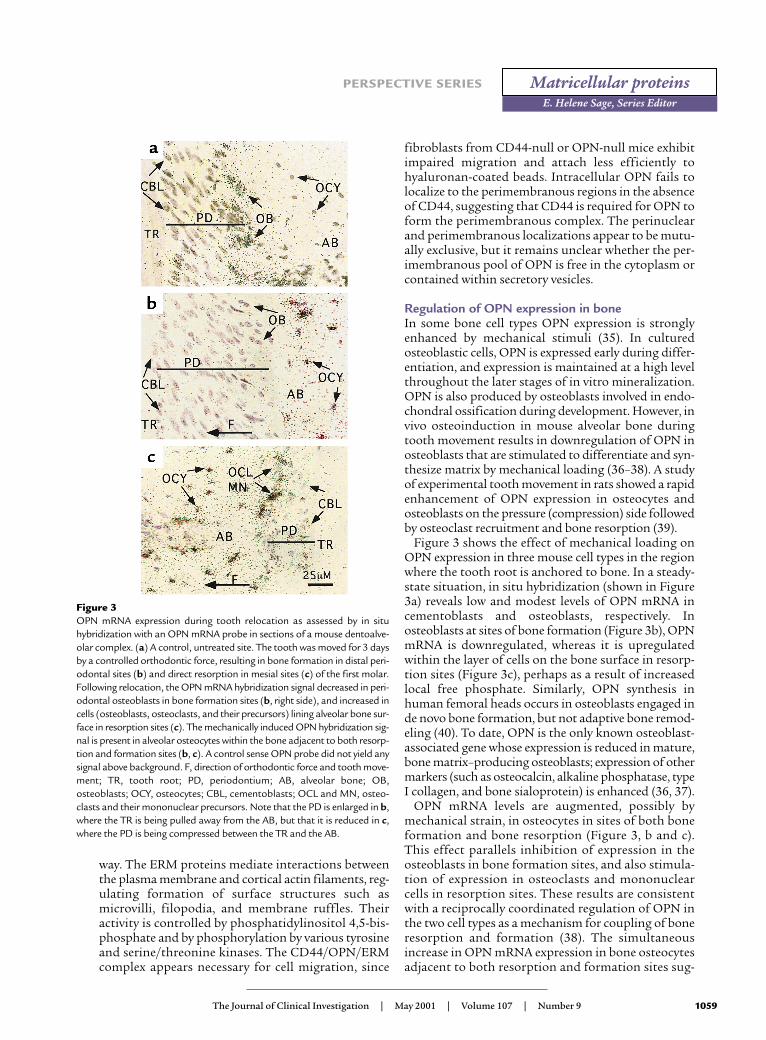
Figure 3 shows the effect of mechanical loading onOPN expression in three mouse cell types in the regionwhere the tooth root is anchored to bone. In a steady-state situation, in situ hybridization (shown in Figure3a) reveals low and modest levels of OPN mRNA incementoblasts and osteoblasts, respectively. Inosteoblasts at sites of bone formation (Figure 3b), OPNmRNA is downregulated, whereas it is upregulatedwithin the layer of cells on the bone surface in resorp-tion sites (Figure 3c), perhaps as a result of increasedlocal free phosphate. Similarly, OPN synthesis inhuman femoral heads occurs in osteoblasts engaged inde novo bone formation, but not adaptive bone remod-eling (40). To date, OPN is the only known osteoblast-associated gene whose expression is reduced in mature,bone matrix–producing osteoblasts; expression of othermarkers (such as osteocalcin, alkaline phosphatase, typeI collagen, and bone sialoprotein) is enhanced (36, 37).
OPN mRNA levels are augmented, possibly bymechanical strain, in osteocytes in sites of both boneformation and bone resorption (Figure 3, b and c).This effect parallels inhibition of expression in theosteoblasts in bone formation sites, and also stimula-tion of expression in osteoclasts and mononuclearcells in resorption sites. These results are consistentwith a reciprocally coordinated regulation of OPN inthe two cell types as a mechanism for coupling of boneresorption and formation (38). The simultaneousincrease in OPN mRNA expression in bone osteocytesadjacent to both resorption and formation sites sug-
The Journal of Clinical Investigation | May 2001 | Volume 107 | Number 9 1059
PERSPECTIVE SERIES
E. Helene Sage, Series Editor
Matricellular proteins
Figure 3OPN mRNA expression during tooth relocation as assessed by in situhybridization with an OPN mRNA probe in sections of a mouse dentoalve-olar complex. (a) A control, untreated site. The tooth was moved for 3 daysby a controlled orthodontic force, resulting in bone formation in distal peri-odontal sites (b) and direct resorption in mesial sites (c) of the first molar.Following relocation, the OPN mRNA hybridization signal decreased in peri-odontal osteoblasts in bone formation sites (b, right side), and increased incells (osteoblasts, osteoclasts, and their precursors) lining alveolar bone sur-face in resorption sites (c). The mechanically induced OPN hybridization sig-nal is present in alveolar osteocytes within the bone adjacent to both resorp-tion and formation sites (b, c). A control sense OPN probe did not yield anysignal above background. F, direction of orthodontic force and tooth move-ment; TR, tooth root; PD, periodontium; AB, alveolar bone; OB,osteoblasts; OCY, osteocytes; CBL, cementoblasts; OCL and MN, osteo-clasts and their mononuclear precursors. Note that the PD is enlarged in b,where the TR is being pulled away from the AB, but that it is reduced in c,where the PD is being compressed between the TR and the AB.

gests that OPN exerts a reciprocal paracrine effect oncells on the bone surface, leading to stimulation andrepression of its synthesis in osteoclasts andosteoblasts, respectively. The dendritic networkbetween osteocytes and bone lining cells may help tointegrate these opposing effects. Additional mecha-nisms, possibly including mineral exposure, may benecessary to determine which phase of the remodelingcycle — resorption or formation — will prevail on a par-ticular area of bone surface exposed to an OPN-medi-ated signal initiated by osteocytes.
SummaryOPN is a multifunctional cytokine and adhesion pro-tein that contains an integrin-binding RGD sequenceand additional sequences that interact with CD44v6/7or other adhesive receptors. Its expression is increasedin response to early proinflammatory cytokines and tomechanical strain in bone. The function of the secret-ed protein may be altered by extracellular enzymes,including thrombin and kinases. The study of OPN-null mice has revealed roles for OPN in a broad rangeof homeostatic (bone remodeling, tissue debridement)and pathologic (cellular immunity, wound healing,cancer metastasis) processes. While these processesseem disparate, they are linked by several commonthemes, including enhanced expression of OPN inresponse to stress or tissue injury, and stimulation ofcell motility and cell survival pathways via interactionsof OPN with adhesive receptors.
OPN is chemotactic for various cell types, notablymonocytes/macrophages, which are attracted to sitesof infection and inflammation. It is essential for cell-mediated immunity and a normal Th1 cytokineresponse during granuloma formation. OPN servesboth to attach bone cells to bone matrix and to gener-ate intracellular signals essential for normal osteoclastmotility on bone; it may mediate osteocyte recognitionof bone strain. OPN activates intracellular signalingpathways and regulates gene expression as a conse-quence of its interactions with its various receptors.The best-characterized is the integrin-stimulated FAK-Src-Rho pathway, which alters gelsolin function andpodosome formation in osteoclasts. Identification anddissection of the signal transduction pathways andtheir targets are complicated by the fact that OPN canengage more than one type of receptor on the cell. Forthis reason, it is important to ascertain which receptorsare in play in any given experimental system.
There is compelling evidence that soluble OPN can ina variety of situations help cells survive an otherwiselethal insult. Remarkably, this survival signaling ismediated by receptors that are generally considered tobe receptors for ECM components. We suggest thatOPN delivers an antiapoptotic “ECM-like” signal viamultiple ligand-receptor interactions to cells, bothadherent and nonadherent.
AcknowledgmentsResearch in the authors’ laboratories has been gen-erously supported by grants from NIH (ES-06897and AR-44434 to D.T. Denhardt, P50-HL56386 andHL-63339 to J.S. Berman, HL-04343 to A.W. O’Re-gan, and DE-11005 to D. Pavlin), and from theJapanese Ministry of Education to M. Noda. Sincereapologies to those whose papers have not been citedbecause of space constraints.
1. Katagiri, Y.U., et al. 1999. CD44 variants but not CD44s cooperate withbeta1-containing integrins to permit cells to bind to osteopontin inde-pendently of arginine-glycine-aspartic acid, thereby stimulating cellmotility and chemotaxis. Cancer Res. 59:219–226.
2. Smith, L.L., and Giachelli, C.M. 1998. Structural requirements for α9β1-mediated adhesion and migration to thrombin-cleaved OPN. Exp. CellRes. 242:351–360.
3. Yokosaki, Y., et al. 1999. The integrin α9β1 binds to a novel recognitionsequence (SVVYGLR) in the thrombin-cleaved amino-terminal fragmentof osteopontin. J. Biol. Chem. 274:36328–36334.
4. Barry, S.T., Ludbrook, S.B., Murrison, E., and Horgan, C.M.T. 2000.Analysis of the α4β1 integrin-osteopontin interaction. Exp. Cell Res.258:342–351.
5. Helluin, O., et al. 2000. The activation state of αvβ3 regulates platelet andlymphocyte adhesion to intact and thrombin-cleaved osteopontin. J. Biol.Chem. 275:18337–18343.
6. Senger, D.R., et al. 1996. Stimulation of endothelial cell migration by vas-cular permeability factor/vascular endothelial growth factor throughcooperative mechanisms involving the αvβ3 integrin, osteopontin, andthrombin. Am. J. Pathol. 149:293–305.
7. O’Regan, A., and Berman, J.S. 2000. Osteopontin: a key cytokine in cell-mediated and granulomatous inflammation. Int. J. Exp. Pathol.81:373–390.
8. Rittling, S.R., and Denhardt, D.T. 1999. Osteopontin (OPN) function inpathology: lessons from OPN-deficient mice. Exp. Nephrol. 7:103–113.
9. Sodek, J., Ganss, T., and McKee, M.D. 2000. Osteopontin. Crit. Rev. OralBiol. Med. 11:279–303.
10. Sterling, H., Saginario, C., and Vignery, A. 1998. CD44 occupancy pre-vents macrophage multinucleation. J. Cell Biol. 143:837–847.
11. Asosingh, K., et al. 2000. In vivo induction of insulin-like growth factor-1 receptor and CD44v6 confers homing and adhesion to murine multi-ple myeloma cells. Cancer Res. 60:3096–3104.
12. Wittig, B.M., Johansson, B., Zöller, M., Schwärzler, C., and Günthert, U.2000. Abrogation of experimental colitis correlates with increased apop-tosis in mice deficient for CD44 variant exon 7 (CD44v7). J. Exp. Med.191:2053–2063.
13. Lin, Y.H., et al. 2000. Coupling of osteopontin and its cell surface recep-tor CD44 to the cell survival response elicited by interleukin-3 or gran-ulocyte-macrophage colony-stimulating factor. Mol. Cell. Biol.20:2734–2742.
14. Denhardt, D.T., and Noda, M. 1998. Osteopontin expression and func-tion: role in bone remodeling. J. Cell. Biochem.Suppl. 30–31:92–102.
15. O’Regan, A.W., et al. 1999. Osteopontin is associated with T cells in sar-coid granulomas and has T cell adhesive and cytokine-like properties invitro. J. Immunol. 162:1024–1031.
16. Ashkar, S., et al. 2000. Eta-1 (osteopontin): an early component of type-1 (cell-mediated) immunity. Science. 287:860–864.
17. O’Regan, A.W., Hayden, J.M., and Berman, J.S. 2000. Osteopontin aug-ments CD3-mediated interferon-γ and CD40 ligand expression by Tcells, which results in IL-12 production from peripheral blood mononu-clear cells. J. Leukoc. Biol. 68:495–502.
18. Tian, J.Y., et al. 2000. Regulation of NO synthesis induced by inflam-matory mediators in RAW264.7 cells: collagen prevents inhibition byosteopontin. Cytokine. 12:450–457.
19. Scott, J.A., et al. 1998. Osteopontin inhibits inducible nitric oxide syn-thase activity in rat vascular tissues. Am. J. Physiol. 275:H2258–H2265.
20. Takahashi, F., Takahashi, K., Maeda, K., Tominaga, S., and Fukuchi, Y.2000. Osteopontin is induced by nitric oxide in RAW264.7 cells. IUBMBLife. 49:217–221.
21. Petrow, P.K., et al. 2000. Expression of osteopontin mRNA and proteinin rheumatoid arthritis. Arthritis Rheum. 43:1597–1605.
22. Attur, M.G., et al. 2001. Osteopontin: an intrinsic inhibitor of inflam-
1060 The Journal of Clinical Investigation | May 2001 | Volume 107 | Number 9
PERSPECTIVE SERIES
E. Helene Sage, Series Editor
Matricellular proteins

mation in cartilage. Arthritis Rheum. 44:578–584.23. Bonvini, J.M., et al. 2000. Lack of in vivo function of osteopontin in
experimental anti-GBM nephritis. J. Am. Soc. Nephrol. 11:1647–1655.24. Nau, G.J., et al. 1999. Attenuated host resistance against Mycobacterium
bovis BCG infection in mice lacking osteopontin. Infect. Immun.67:4223–4230.
25. Nau, G.J., et al. 2000. Osteopontin expression correlates with clinicaloutcome in patients with mycobacterial infection. Am. J. Pathol.157:37–42.
26. Liaw, L., et al. 1998. Altered wound healing in mice lacking a functionalosteopontin gene (spp1). J. Clin. Invest. 101:1468–1478.
27. Cowan, K.N., Jones, P.L., and Rabinovitch, M. 2000. Elastase and matrixmetalloproteinase inhibitors induce regression, and tenascin-C antisenseprevents progression, of vascular disease. J. Clin. Invest. 105:21–34.
28. Jono, S., Peinado, C., and Giachelli, C.M. 2000. Phosphorylation ofosteopontin is required for inhibition of vascular smooth muscle cell cal-cification. J. Biol. Chem. 275:20197–20203.
29. Beck, G.R., Zerler, B., and Moran, E. 2000. Phosphate is a specific signalfor induction of osteopontin gene expression. Proc. Natl. Acad. Sci. USA.97:8352–8357.
30. Yoshitake, H., Rittling, S.R., Denhardt, D.T., and Noda, M. 1999. Osteo-pontin-deficient mice are resistant to ovariectomy-induced bone resorp-tion. Proc. Natl. Acad. Sci. USA. 96:8156–8160.
31. Asou, Y., et al. 2001. Osteopontin facilitates angiogenesis, accumulationof osteoclasts and resorption of ectopic bone. Endocrinology.142:1325–1332.
32. Ishijima, M., et al. 2001. Enhancement of osteoclastic bone resorptionand suppression of osteoblastic bone formation in response to reduced
mechanical stress do not occur in the absence of osteopontin. J. Exp. Med.193:399–404.
33. Chellaiah, M.A., et al. 2000. The molecular mechanisms of osteoclastdysfunction associated with osteopontin deficiency: the failure of Rhostimulation of mDia1. J. Bone Miner. Res. 15(Suppl. 1):S396. (Abstr.)
34. Zohar, R., et al. 2000. Intracellular osteopontin is an integral componentof the CD44-ERM complex involved in cell migration. J. Cell. Physiol.184:118–130.
35. Klein-Nulend, J., Roelofsen, J., Semeins, C.M., Bronckers, A.L.J.J., andBurger, E.H. 1997. Mechanical stimulation of osteopontin mRNAexpression and synthesis in bone cell cultures. J. Cell. Physiol.170:174–181.
36. Pavlin, D., Dove, S.B., Zadro, R., and Gluhak-Heinrich, J. 2000. Mechan-ical loading stimulates differentiation of periodontal osteoblasts in amouse osteoinduction model: effect on type I collagen and alkalinephosphatase genes. Calcif. Tissue Int. 67:163–172.
37. Pavlin, D., Zadro, R., and Gluhak-Heinrich, J. 2001. Temporal pattern ofstimulation of osteoblast-associated genes during mechanically-inducedosteogenesis in vivo: early responses of osteocalcin and type I collagen.Connect. Tissue Res. In press.
38. Gluhak-Heinrich, J., Villarreal, A., and Pavlin, D. 2000. Reciprocal regu-lation of osteopontin gene during mechanically induced bone forma-tion and resorption. J. Bone Miner. Res. 15(Suppl. 1):M086. (Abstr.)
39. Terai, K., et al. 1999. Role of osteopontin in bone remodeling caused bymechanical stress. J. Bone Miner. Res. 14:839–849.
40. Dodds, R.A., et al. 1995. Human osteoclasts, not osteoblasts, depositosteopontin onto resorption surfaces: an in vitro and ex vivo study ofremodeling bone. J. Bone Miner. Res. 10:1666–1680.
The Journal of Clinical Investigation | May 2001 | Volume 107 | Number 9 1061
PERSPECTIVE SERIES
E. Helene Sage, Series Editor
Matricellular proteins