osteoporosis and dxa overview - department of … a systemic skeletal disease characterized by low...
TRANSCRIPT

Osteoporosis and DXA OverviewNeil Binkley, MD
November 2, 2006
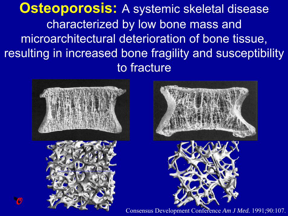
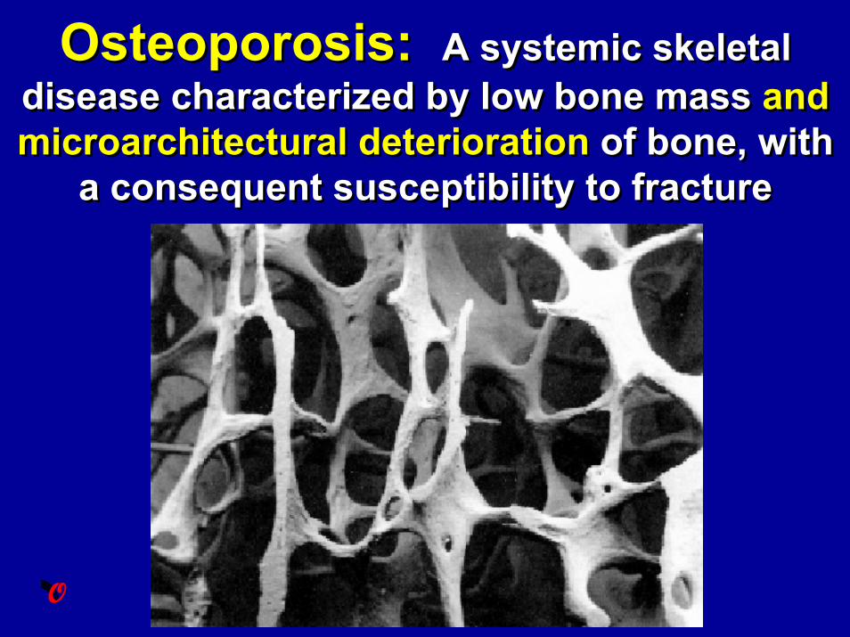
Osteoporosis: A systemic skeletal disease characterized by low bone mass and
microarchitectural deterioration of bone tissue, resulting in increased bone fragility and susceptibility
to fracture
Consensus Development Conference Am J Med. 1991;90:107.

NIH Consensus Panel. JAMA. 2001;285:785.
More Recent NIH Consensus Definition Includes “Bone Quality”• Osteoporosis is a skeletal disorder
characterized by compromised bone strength predisposing to an increased risk of fracture
• Bone strength reflects two main features: bone density
bone quality

Osteoporosis is Usually Multifactorial
• Low bone mass at skeletal maturity (~ age 30)• Advanced age• Low calcium and/or vitamin D intake• Use of prednisone (corticosteroids)• Smoking/High alcohol consumption• Lower body weight• Low physical activity• Loss of estrogen/testosterone


Bone Density Declines with Advancing Age
This decline leads to increased risk for osteoporotic fracture, most commonly of the spine/hip/wrist
Male
Age
Female

Men
Hip fracture risk in men reaches the samelevel as in women, but at an age ~6 years ol
With Bone Loss, Fracture Risk Increases in Both Men and Women
Cooper, JBMR 1992
Frac
ture
Ris
k
Vertebra
Hip
Women
Age (years)80706050

www.nof.org
HipVertebral(Morphometric)
700,000
Wrist0
250,000
500,000
750,000
300,000 300,000
250,000
Other
Only ~25%-30% of morphometric vertebral fractures are diagnosed
clinically
Clinically Apparent
Of the 6 million fractures per year in the US, approximately 1.5 million are
due to osteoporosis

Consequences of Osteoporotic Fracture
Pain; acute and/or chronicReduced ability to perform activities of daily living
• ~50% of people able to live independently after a hip fracture
Change in body habitus Respiratory compromiseDepression/anxiety/fear of fallingCost ~$13 billion annuallyDeath; ~20% one year mortality following hip fracture

Survival Rates After Fractures
Cooper C, Am J Epidemiol. 1993;137:1001.
%Survival
Time after fracture (years)
ExpectedObserved
100
80
60
40
20
01 2 3 4 5
Vertebral Fracture(relative survival = 0.81)
100
80
60
40
20
01 2 3 4 5
Hip Fracture(relative survival = 0.82)

How Common is Osteoporosis?
Women
Men ~33%
~50%

What Does Someone with Osteoporosis Look Like?
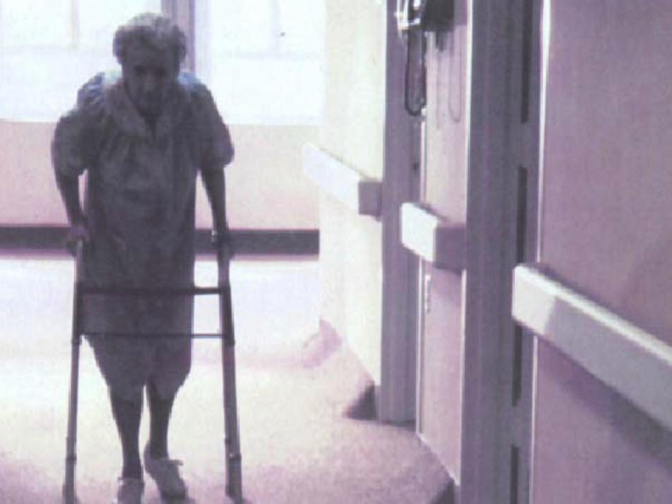


Osteoporosis is Not Simply “Getting Old”

Think of Osteoporosis in the Same Way That You For High
BP or High Cholesterol Disease Clinical measure
Outcome Hypertension BP CVA Hyperlipidemia Lipids MI Osteoporosis BMD FX
How Do You Know if Your Patient Has High
Cholesterol?
Measure it!

How Do You Know if Your Patient Has Low Bone
Density?MEASURE IT!Risk factors do not work to guess bone densityAdvanced ageFemale genderCaucasian raceThinFamily history of osteoporosisEtc, etc.

Risk Factors Do Not Allow Prediction of BMD
Risk Factors Do Not Allow Prediction of BMD
IMPACT Trial: ~7000 postmenopausal women had BMD measurement and risk factor assessment
48% of those with osteoporosis had no risk factors
53% of those with risk factors did not have osteoporosis
Watts, Arth Rheum 2001

“You can’t find a fever if you don’t check the
temperature.”G. Magnin, M.D.
When Should Bone Density be Measured?
When Should Bone Density be Measured?
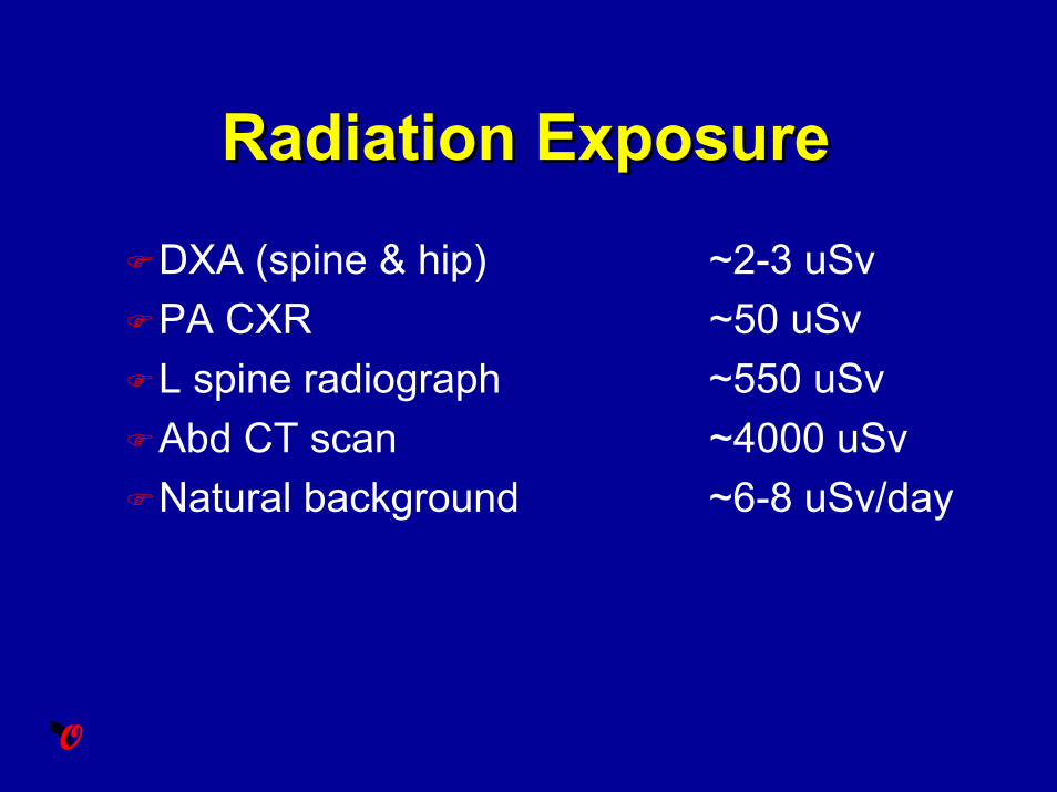
Radiation ExposureRadiation Exposure
DXA (spine & hip) ~2-3 uSvPA CXR ~50 uSvL spine radiograph ~550 uSvAbd CT scan ~4000 uSvNatural background ~6-8 uSv/day

All women aged 65 and olderPostmenopausal women under age 65 with risk factorsAll men aged 70 and olderAdults with a fragility fractureAdults with a disease or condition associated with low bone mass or bone lossAdults taking medications associated with low bone mass or bone lossAnyone being considered for pharmacologic therapy Anyone being treated, to monitor treatment effectAny not receiving therapy in whom evidence of bone loss would lead to treatment
ISCD Indications for BMD TestingISCD Indications for BMD Testing

Indications for BMD TestingCategory USPSTF NOF AACE ACR ACOG OSC ISCD
♀ ≥ 65 ♀ 60-64 with risk factors * ♀ < 60 with risk factors * Anyone with risk factors *
♂ ≥ 70
Monitoring Rx
• Risk factors vary according to organization• Some organizations only consider postmenopausal women
Bray, www.iscd.org

2500
2000
500
1000
1500
Age (years)50 60 70 80
Frac
ture
s/10
0,00
0pe
rson
-yea
rs
Vertebral
Hip
90
Fracture Risk Increases With Age
10
20
30 % w
ho have hadB
MD
measurem
entBut BMD Measurement Declines
Neuner, JAGS 2006Cooper JBMR, 1992

Average Life Expectancy; 2001 USFemale 79.5 years, Male 74.1 years
CDC/Natl Center for Health Statisticswww.cdc.gov/nchs/fastats/lifeexpec.htm
0
5
10
15
20
65 70 75 80 85 90Current Age (Years)
Male
Female

“Bone mineral density measurement should be obtained
routinely in all women over the age of 65 years and in men and
younger women who have had a fragility fracture. Compliance with this recommendation alone would be a great advance in comparison
with current practice.”Raisz, NEJM 2005

BMD Measurement is Not Being Done Following Fracture
Feldstein, Osteoporos Int, 2005
Retrospective cohort study NW
US HMO; 1171 men
Age > 65 yearswith any new
fracture
100
75
50
25
0Pos
t-fra
ctur
e A
sses
smen
t/Tre
atm
ent
2000 2001
BMD measured
New RxRx continued

BMD Measurement
Needs to Become More
“User Friendly”
BMD Measurement
Needs to Become More
“User Friendly”

Densitometers Measure BMD
But T-scores Are UsedFor Diagnosis

WHO Diagnostic CategoriesWHO Diagnostic Categories
Normal
Osteopenia
Osteoporosis
-1.0
-2.5
SevereOsteoporosis
These Criteria ApplyONLY to the L-spineProximal Femur and
1/3 Radius
WHO Technical Series, 1994

T-score Compares With Young Adult; Z-score with Age-Matched
T-score Compares With Young Adult; Z-score with Age-Matched
T-score = Patients BMD- Young Normal Mean BMDSD of Young Normal
T-score = Patients BMD- Young Normal Mean BMDSD of Young Normal
Z-score = Patients BMD- Age Matched Mean BMDSD of Age Matched
Z-score = Patients BMD- Age Matched Mean BMDSD of Age Matched

T-score Calculation Example• ASSUME• Mean YN BMD = 1.200 g/cm2
• SD of the YN population = 0.100 g/cm2
• Your patient’s BMD is 1.000 g/cm2
1.000 - 1.200 = -0.200-0.200/0.100 = -2.0

Why Use T-scores?BMD is not measured the same using different densitometersThere are differences in
• Technologies of x-ray generation• Edge detection approaches • Region of interest placement
Hologic BMD in g/cm2 ~10% lower than GE LunarT-scores allow use of same diagnostic criteria with instruments of different manufacturers
T-Score Used for Diagnosis
Z-Score Used for Children and Health Adults Under Age 50
Clinical Lore that a Z-score of ≤ -2.0 Indicates Need to Perform Evaluation for
Secondary Causes of Bone Loss

Spine and Hip DXA Are the Gold Standard
Spine and Hip DXA Are the Gold Standard

Apply the WHO Criteria for Diagnosis Using the Lowest T-score of the L1-4 Spine,
Total Femur or Femur Neck
Report an Overall Diagnosis, Not Site-Specific DiagnosesReport an Overall Diagnosis, Not Site-Specific Diagnoses

Diagnosis Using WHO:Use the Lowest T-score
Don’t Be Bothered by T-score Discordance
Diagnosis Using WHO:Use the Lowest T-score
Don’t Be Bothered by T-score Discordance
Ideally, BMD measurement at all sites would yield the same T-scoreThat this does NOT occur is referred to as skeletal discordance

Discordance Due to Spine DJD
L1-L4 BMD = 1.248 g/cm2
T-score = 0.6Total hip BMD = .725 g/cm2
T-score = -2.3

Use the L1-L4 T-score, However
L1 -3
L2 -2.3
L3 2.4
L4 2.0
L1-4 -0.5L1-2 T-score -2.5
Exclude vertebrae from analysis if
there is a > 1.0 T-score difference
between adjacent vertebral bodies

Need to Use at Least Two VertebraeDon’t “Cherry-pick” the Lowest Vertebral Body
Need to Use at Least Two VertebraeDon’t “Cherry-pick” the Lowest Vertebral Body

When the Spine BMD is Worthless, Measure the Forearm
L1-L4 T-score = 3.2 .3 T-score = -2.9
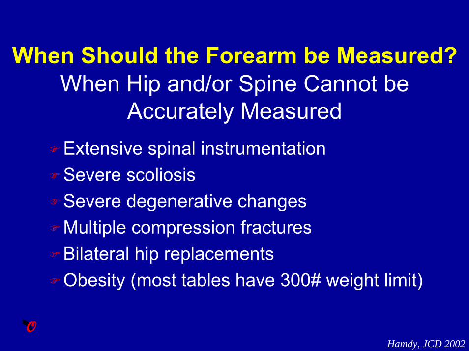
When Should the Forearm be Measured?When Hip and/or Spine Cannot be
Accurately MeasuredExtensive spinal instrumentationSevere scoliosisSevere degenerative changesMultiple compression fracturesBilateral hip replacementsObesity (most tables have 300# weight limit)
Hamdy, JCD 2002

WHO Technical Series, 1994
Isn’t the Forearm a “Peripheral” Site?Isn’t the Forearm a “Peripheral” Site?
“Osteoporosis is defined as a bone mass more than 2.5 SD below the
young adult reference mean at the spine, hip or mid-radius.”

The WHO Criteria Apply to the Femur Neck and Total Proximal Femur
Not Ward’s Area or the Greater Trochanter
“This patient has osteoporosis and given her young age
should receive teriparatide.”

It is Impossible to Tell if a Person has “Lost” Bone
Based on a Single Measurement
HALF OF US ARE BELOW AVERAGE
Don’t tell patients that their BMD is XX% of young normal
or that their bones are 95 years old (unless the patient is 95)
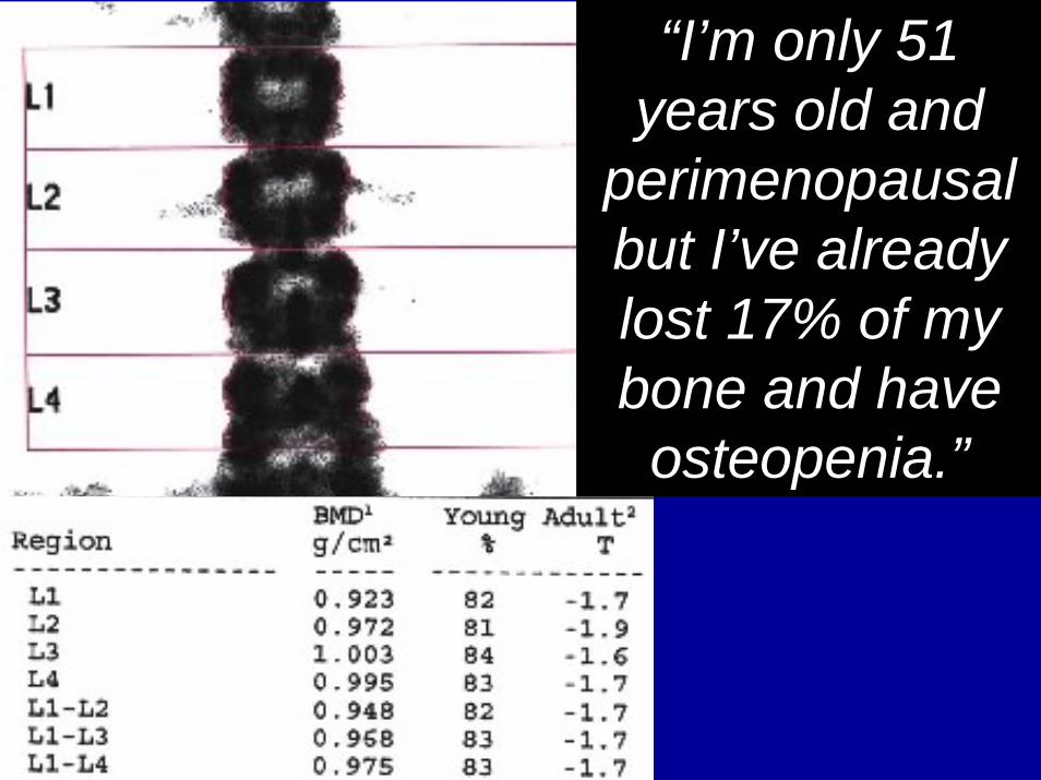
“I’m only 51 years old and
perimenopausalbut I’ve already lost 17% of my bone and have osteopenia.”
“Shortopenia” is Not a Disease!“Shortopenia” is Not a Disease!
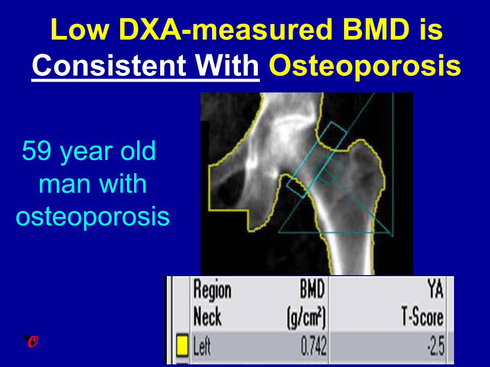
Low DXA-measured BMD is Consistent With Osteoporosis
59 year old man with
osteoporosis


59 Year-old Man with “Osteoporosis”
59 Year-old Man with “Osteoporosis”
Status post bariatric surgery• weight decrease ~125 pounds
Back injury while golfing – imaging negative for fracture but noted radiographic osteopeniaNormal testosterone, thyroid studiesCalcium: 8.3 mg/dl (8.5-10.4)iPTH: 304 pg/ml (7-53)24 hour urine calcium 25 mg25-OH vitamin D 7 ng/ml
Status post bariatric surgery• weight decrease ~125 pounds
Back injury while golfing – imaging negative for fracture but noted radiographic osteopeniaNormal testosterone, thyroid studiesCalcium: 8.3 mg/dl (8.5-10.4)iPTH: 304 pg/ml (7-53)24 hour urine calcium 25 mg25-OH vitamin D 7 ng/ml

Vitamin D Deficiency Osteomalacia
When Should Follow-up DXA be Performed?
“It depends”Not more frequently than yearlyInitiation of steroids is an exception (6 months)ISCD position; measure one year after initiation of Rx to document response (stability or increase)Medicare has defined monitoring interval as no more frequently than every 23 months

Stable BMD on Treatment is Success12 mo BMD change in ~3000 ALN treated patients
Hochberg, Arthritis Rheum 1999
< 0
0-3%> 3%
Spine BMD
7
6
5
4
3
2
1
0

This is Due to Anti-resorptiveInduced Reduction in “Stress
Risers”
What is a Real Change on Follow-up DXA?
Necessary to perform an in-vivo precision assessment• This is facility, technician and patient population dependent
At UW, the L1-L4 spine LSC is 0.040 grams/cm2 and 0.020 grams/cm2 at the mean total femur
• These values will vary between facilities and technologists• For example, the L1-L4 LSC at the Wm. S. Middleton VAMC is
0.049 grams/cm2
A “decrease” from .890 to .875 g/cm2 is no change

Use the BMD in grams/cm2
NOT the T-score When Performing Follow-up DXA
Know What Constitutes a Real Change in BMD at Your Facility
Need to Know Your LSC
If a Precision Assessment Has Not Been Done, Monitoring BMD is Not
Possible

“I’m on Drug X but my bone density is going down. Is there anything else that can be done?”
1/19/06L1-L4 BMD = .685 g/cm2
T-score = -4.1
12/11/04L1-L4 BMD = .705 g/cm2
T-score = -3.9
A 0.020 g/cm2
“difference”is not a real
change

Precision and AccuracyPrecision = agreement of serial measurements of the same thingAccuracy = ability to determine the “true”value

Use the excel-basedprecision
calculator at iscd.org

If Huge Change in BMD (~10%) Look For Technical Problems

Make Sure You Are Comparing Apples With Apples

8% Decline in BMD on Bisphosphonate RxBaseline Follow-up
L1-L4 BMD 0.629 g/cm2L1-L4 BMD 0.717 g/cm2

BMD Comparison Between Facilities
• It is not possible to quantitatively compare BMD or to calculate a least significant change between facilities without cross-calibration
Binkley, et. al., J Clin Densitom, 2006

Things NOT To Tell Your Patients About Their DXA
“Bone loss” without knowledge of prior bone density“Mild,” “moderate,” or “marked” osteopenia or osteoporosisSeparate diagnoses for different regions (e.g., osteopenia at the hip and osteoporosis at the spine)Expressions such as “You have the bones of an 80-year-old," if the patient is not 80 years oldThe change in BMD if it is not a significant change based on the precision error and LSC
Binkley, et. al., J Clin Densitom, 2006

Densitometric Vertebral Fracture Assessment (VFA) is Now
Available

VFA is a Quick, Convenient, Low Radiation Tool to Detect
Individuals with Unappreciated Vertebral Fractures
Only ~25% of Vertebral Fractures are Clinically
ApparentMay be Asymptomatic…….May be Asymptomatic…….
…… or Unrecognized…… or Unrecognized
Estimated that ~1% of Back Pain Episodes are Caused by
Vertebral Fracture
Estimated that ~1% of Back Pain Episodes are Caused by
Vertebral Fracture
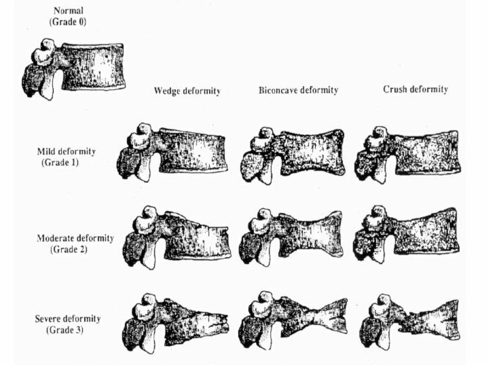

Moderate and
Severe Fracturesare Easy
to Identify
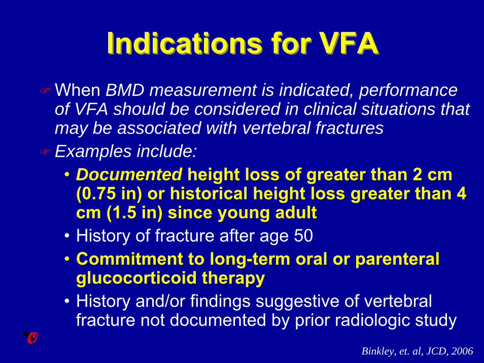
Indications for VFAIndications for VFA
Binkley, et. al, JCD, 2006
When BMD measurement is indicated, performance of VFA should be considered in clinical situations that may be associated with vertebral fracturesExamples include:• Documented height loss of greater than 2 cm
(0.75 in) or historical height loss greater than 4 cm (1.5 in) since young adult
• History of fracture after age 50 • Commitment to long-term oral or parenteral
glucocorticoid therapy• History and/or findings suggestive of vertebral
fracture not documented by prior radiologic study

Why Should We Care About Unappreciated Vertebral
Fracture?

Fracturepresence
andseveritypredicts future risk %
with
1 o
r mor
e ne
w fr
actu
res
38
24
none mild moderate severeFracture Severity at Baseline
40
30
20
10
0
11
4
Because They Identify People at Higher RiskBecause They Identify People at Higher Risk

Absolute Fracture Risk is Coming
A WHO Absolute Fracture Risk (Fracture Probability) Technical Document Will be
Published Soon
A WHO Absolute Fracture Risk (Fracture Probability) Technical Document Will be
Published Soon

Relative Risk Does Not Clearly Define Fracture Risk
“Your T-score is -2.0. You are at a four-fold increased risk for fracture.”“Your T-score is -2.0. You are at a
four-fold increased risk for fracture.”
Absolute fracture risk in the next 10 yearsIf femur neck T-score = -2.0
Absolute fracture risk in the next 10 yearsIf femur neck T-score = -2.0
50 year old: 9.2%50 year old: 9.2%
80 year old: 20.5%80 year old: 20.5%

Numerically More Fractures Occur in People Without OsteoporosisOnly 44% of women and 21% of men who sustain non-vertebral fractures have osteoporosis by BMD
Schuit SCE et al. Bone. 2004;34:195-202
5794 participants in the Rotterdam study;
Mean follow-up 6.8 yrsFN BMD at baseline
Osteoporosis
Osteopenia
Normal
Perc
ent
100
80
60
40
20
0All non-vert Fx
Men n = 145
Women n = 449

BMD Measurement Identifies Those at High Fracture Risk, But Alone, it is Not Sensitive Enough
By Basing Treatment Decisions Solely on T-score, We Are
Missing About Half of Women Who Are At Risk for Fracture
By Basing Treatment Decisions Solely on T-score, We Are
Missing About Half of Women Who Are At Risk for Fracture

The WHO Paradigm Will Add Clinical Factors to BMD
High Bone Mineral Density Low
Frac
ture
Ris
k
Age 50
Age 65
Age 80Notably age
Kanis, IOF 2006

% w
ith 1
or m
ore
new
frac
ture
s
38
24
none mild moderate severeFracture Severity at Baseline
40
30
20
10
0
11
4
And Prior FractureAnd Prior Fracture
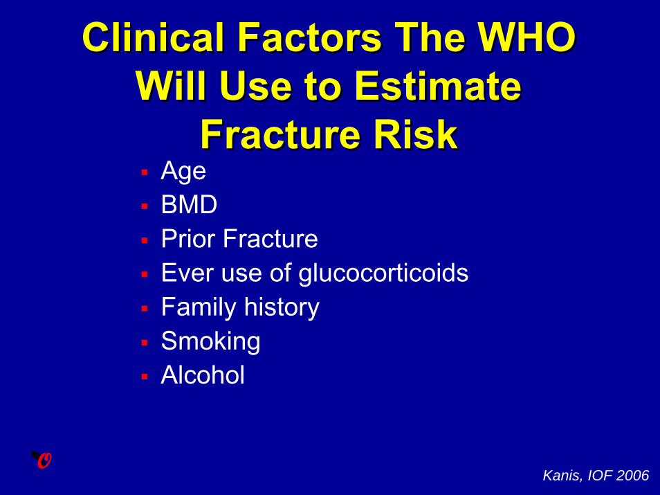
Clinical Factors The WHO Will Use to Estimate
Fracture Risk
Clinical Factors The WHO Will Use to Estimate
Fracture RiskAgeBMDPrior FractureEver use of glucocorticoidsFamily historySmokingAlcohol
Kanis, IOF 2006

Expect the WHO Absolute Risk Approach to Become Part of
the DXA Printout
“Based upon age, femur neck BMD and prior fracture, this persons 10-year risk of hip fracture
is 8% and of any fracture is 15%.Per the National Osteoporosis Foundation,
pharmacologic treatment is recommended when the 10-year fracture risk is above xx%.”

What About “Peripheral” BMD Measurements?
• Use of WHO criteria for the diagnosis of normal, osteopenic or osteoporotic BMD inappropriate
• Currently, if central DXA available; don’t make dx of osteoporosis based on peripheral measurement
• Cannot be used to monitor osteoporosis therapy

Conclusion: Application of a single T-score criteria (WHO)is not appropriate for different sites and technologies
0
1
-1
-2
-3
-430 40 50 60 70 80
Spine QCT
Heel QUS
Spine DXAHip DXA
T-S
core
90Age
Do the WHO Criteria Apply to “Peripheral” Measurements?

Can/Should the WHO Criteria Be Applied to
Populations Other than Postmenopausal Caucasian
Women?
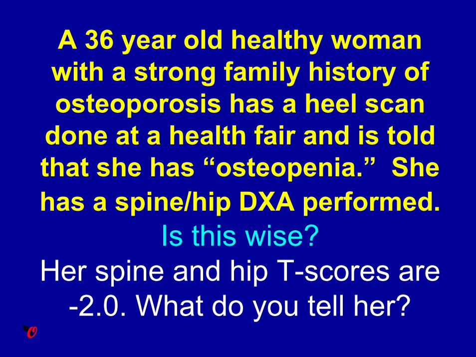
A 36 year old healthy woman with a strong family history of osteoporosis has a heel scan
done at a health fair and is told that she has “osteopenia.” She has a spine/hip DXA performed.
Is this wise?Her spine and hip T-scores are
-2.0. What do you tell her?

WHO Criteria Should Not be Applied to Healthy
Premenopausal Women• Z-scores rather than T-scores should be
used• Osteoporosis may be diagnosed if there is low
BMD with secondary causes (glucocorticoidtherapy, hypogonadism, hyperparathyroidism, etc) or with risk factors for fracture
• The diagnosis of osteoporosis in premenopausal women should not be made on densitometric criteria alone
Osteoporosis: A systemic skeletal disease characterized by low bone mass and microarchitectural deterioration of bone, with
a consequent susceptibility to fracture
Osteoporosis: A systemic skeletal disease characterized by low bone mass and microarchitectural deterioration of bone, with
a consequent susceptibility to fracture

The WHO Criteria Do Not Apply to Young Adults or Children
The WHO Criteria Do Not Apply to Young Adults or Children
Z-scores, not T-scores, are preferredThis is particularly important in childrenA Z-score of -2.0 or lower is defined as “below the expected range for age" and a Z-score above -2.0 is “within the expected range for age
ISCD Position Development Conference, 2005

A 16 Year-Old Healthy Girl is Referred with “Osteoporosis.”She Was Training for Cross-
country and Sustained a Tibial Stress-Fracture While Running.
Her Spine T-score is -2.0 and She Has Been Started on Alendronate.

Densitometric Diagnosis In Children
T-scores should not be used in childrenZ-scores should be used insteadSpine and total body are the preferred sites to measureThe diagnosis of osteoporosis in children should not be made on the basis of densitometriccriteria aloneTerminology such as “low bone density for chronologic age” may be used if the Z-score is <–2.0


The “Correct” Approach to T-score Race-Adjustment is Controversial and Politically
Charged. Even the Definition of “Race”
is Not Easy

ISCD Position Regarding Osteoporosis Diagnosis in Non-
Caucasians
Utilize a uniform Caucasian (universal) normative database
and a T-score of -2.5 for osteoporosis diagnosis

Estimated Male Lifetime Fracture Risk
• Age 50, Rochester; 13% Melton JBMR, 1992
• Age 50, Malmo; 22% Kanis Ost Int, 2000
• Age 60, Dubbo; 25% Nguyen Am J Epidemiol, 1996
• Age 50, Dubbo; 32% Nguyen, ASBMR 2005
In men age age 50 and older, T-scores should be used and osteoporosis diagnosed if the T-score is ≤ –2.5The diagnosis of osteoporosis in healthy men under age 50 should not be made on the basis of densitometric criteria alone
Osteoporosis Diagnosis in Men Osteoporosis Diagnosis in Men
ISCD Position Development Conference, 2005

Which Men Should Have Bone Mass Measurement?
WHO task-force (Genant, OI 1999) and ISCD
Radiographic osteopenia and/or vertebral deformityLoss of height or thoracic kyphosisPrior low trauma fractureConditions recognized to ⇑ risk for bone loss and Fx
• Hypogonadism/prostate Ca, glucocortocoid rx, hyperparathyroidism, etc
Screening at age 70

BMD measurement with central DXA at age is recommended [Grade A]
CMAJ, Nov 12, 2002
Measuring bone density in men and womafter the age of 65 is justifiable

In Some Studies, Spine BMD is Not Lower in Older Men
Melton, JBMR 1998

What Skeletal Sites to Measure in Men?596 men, clinical DXA scans, age 23-95, mean 65
If “routine” isspine & hipmiss 17%
45454545
25
27
45
38
43
5
T Š -2.5; men age � 70N = 149
Femur
L-spineRadius
Vallarta-Ast, JCD 2002

Patients With Osteoporotic Fractures Are Not Being Treated
• Feldstein AC et al. J Bone Joint Surg. 2004• Panneman MJM et al Osteoporos Int. 2004• Jachna CM et al. Osteoporos Int. 2003• Port L, et al Osteoporos Int. 2003 • Liel Y et al. Osteoporos Int. 2003• Solomon DH et al Am J Med. 2003 • Kiebzak et al. Arch Intern Med. 2002• Harrington JT et al Arthritis Rheum. 2002• Cuddihy MT et al Arch Intern Med. 2002• Khan SA et al Arch Intern Med. 2001• Bellantonio S et al J Am Geriatr Soc. 2001• Rothberg AD et al S Afr Med J. 2000
Feldstein, Osteoporos Int, 2005
100
75
50
25
0Pos
t-fra
ctur
e A
sses
smen
t/Tre
atm
ent
2000 2001
BMD measured
New RxRx continued
Retrospective cohort study NW US HMO; 1171 men > 65
years with any new fracture

“The responsibility to ensure appropriate osteoporosis screening
and treatment begins with any clinician… The status quo of
“missed opportunities” is unacceptable.
The buck stops with us.”Mazanec, Arch Intern Med May, 200
Osteoporosis Screening Time to Take Responsibility
Osteoporosis Screening Time to Take Responsibility

Osteoporotic Fractures Should be Thought of as “Bone Attacks”