otitis externa: why treatment fails and cases re-occurotitis... · otitis externa: why treatment...
TRANSCRIPT
Otitis externa: Why treatment fails and cases re-occur
Dr Rob Hilton BVSc(Hons) MANZCVS (Canine Medicine) Cert.VD MRCVS
www.skinvet.org Tele 0433-853560
Otitis externa: Why treatment fails and cases re-occur Dr Rob Hilton BVSc(Hons) MANZCVS (Canine Medicine) Cert.VD MRCVS
Clinical Anatomy – the basics
The ear canal consists of cartilages (both auricular and annular)
and the specialized skin that lines it. The dermis of the ear canal is separated from the
auricular cartilages by a subcutis that becomes thinner more distally in the canal.
The auditory canal contains sebaceous glands, producing a lipid-rich secretion and
ceruminal (modified apocrine “sweat”) glands, producing an aqueous secretion. Ear
wax, often referred to as “cerumen” is an emulsion of secretions of these glands
combined with sloughed keratinocytes.
The number of hair follicles varies between individuals and breeds and they decrease
in number in the horizontal canal. In some individuals, hair tufts may be found
immediately surrounding the attachment of the tympanic membrane.
Studies on the ear canal volume of the dog vary. The volume is related to body weight
and is of the order of 0.5-1ml
In order of most common, the organisms isolated from the vertical canal of normal
dogs ears consist of:
1. Yeast (Malassezia spp)
2. Coagulase negative staphylococci and micrococci
3. Staphylococcus pseudintermedius and other coagulase positive staphylococci
4. Corynebacterium spp
Gram –ve cocci (Enterococcus spp) and Gram -ve rods (Pasteurella and Proteus spp)
are found in less than 4% and 2% of ears respectively and are likely to represent
transient rather than normal flora.
Normal ear cytology
Data on what constitutes normal findings in ear swabs vary markedly between authors.
Based on 400x high power, Genel et al state:
• Mean Malassezia counts of > or = 5 in the dog and > or = 12 in the cat were
considered abnormal. The cat value is much higher than other reports and the
author prefers to regard numbers of Malassezia in the cat above 5 per HPF to
be suspicious.
• Mean bacterial (cocci) counts of > or = 25 in the dog and > or = 15 in the cat
were considered abnormal.
Otitis externa: Why treatment fails and cases re-occur
Dr Rob Hilton BVSc(Hons) MANZCVS (Canine Medicine) Cert.VD MRCVS
www.skinvet.org Tele 0433-853560
• When used to differentiate normal from inflamed external ear canals, these
figures provided a low sensitivity but a specificity of > or = 95% Tater et al report that “nucleated keratinocytes were occasionally observed in both
species, and should not be mistaken for a pathological process (parakeratotic
hyperkeratosis).”
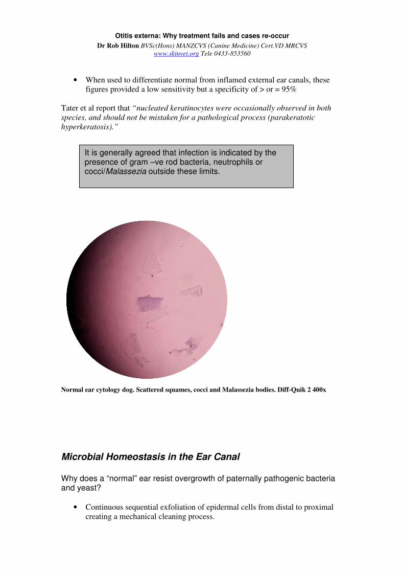
Normal ear cytology dog. Scattered squames, cocci and Malassezia bodies. Diff-Quik 2 400x
Microbial Homeostasis in the Ear Canal
Why does a “normal” ear resist overgrowth of paternally pathogenic bacteria and yeast?
• Continuous sequential exfoliation of epidermal cells from distal to proximal
creating a mechanical cleaning process.
It is generally agreed that infection is indicated by the presence of gram –ve rod bacteria, neutrophils or cocci/Malassezia outside these limits.
Otitis externa: Why treatment fails and cases re-occur
Dr Rob Hilton BVSc(Hons) MANZCVS (Canine Medicine) Cert.VD MRCVS
www.skinvet.org Tele 0433-853560
• Normal ear wax has antimicrobial effects on yeast and bacteria through
antimicrobial peptides and immunoglobulins in normal ceruminal gland
secretions and lipids providing an antimicrobial and water repellent role.
• Potential antimicrobial actions of the normal microflora
• A normal epidermis, more resistant to biofilm formation
The response of the ear canal to chronic otitis includes:
• Epidermal and follicular hyperplasia without changes in hair follicle numbers.
• Dermal inflammation and fibrosis
• Grossly dilated ceruminous glands, dominating the cerumen composition.
As chronic otitis proceeds:
• The ear canal becomes narrow from proliferative changes
• Self cleaning from sequential exfoliation is lost leading to:
o Reduced micro-organism clearance
o Debris and exudate build up
• Ear wax composition is abnormal
• Corrugated epidermis is supports biofilm production
KEY POINT
Once the cycle of infection
and loss of homeostasis is set
up, the ear disease will
progress, independent of the
primary cause (often
allergy) that initiated it.

Otitis externa: Why treatment fails and cases re-occur
Dr Rob Hilton BVSc(Hons) MANZCVS (Canine Medicine) Cert.VD MRCVS
www.skinvet.org Tele 0433-853560
Causes of otitis
Primary• Allergy
• Keratinization disorders
• Endocrinopathies
• Immune mediated disease
• Foreign bodies
• Ear mites/parasites
• Foreign bodies
• Tumours
Predisposing• Anatomic – pendulous,
narrow or hairy
• Humidity and moisture
• Inappropriate cleaning interventions
The otic cycle of disaster
Infection
Loss of self cleaning
Fibrosis
Ceruminal gland hyperplasia
•Primary cause •Predisposing factors
Altered otic environment
End stage earOtitis media

Otitis externa: Why treatment fails and cases re-occur
Dr Rob Hilton BVSc(Hons) MANZCVS (Canine Medicine) Cert.VD MRCVS
www.skinvet.org Tele 0433-853560
Understanding this process means that treatment needs to be in two phases:
1. Treat the infection until resolution is confirmed both visibly by otoscope and
cytologically. “Treat till you beat”
2. Institute a maintenance program (initially weekly) with monitoring in the
knowledge that a chronically damaged ear canal may NEVER regain full
homeostatic function.
It is highly probable that by taking an approach of dispensing aural medication
without planned follow-up will result in a re-flare soon after treatment is stopped,
usually more severe each time with a progression of infection type from
Yeast and cocci Staph, Strep, anaerobes Gram –ve rods,
In the author’s experience, most referral cases of otitis represent the same problem
that has existed for an extended period without ever being resolved.
Key Point
You can not manage an ear in less
than 3 revisits
1. Treat until visible and cytologic
cure
2. Maintain and monitor
Otitis externa: Why treatment fails and cases re-occur
Dr Rob Hilton BVSc(Hons) MANZCVS (Canine Medicine) Cert.VD MRCVS
www.skinvet.org Tele 0433-853560
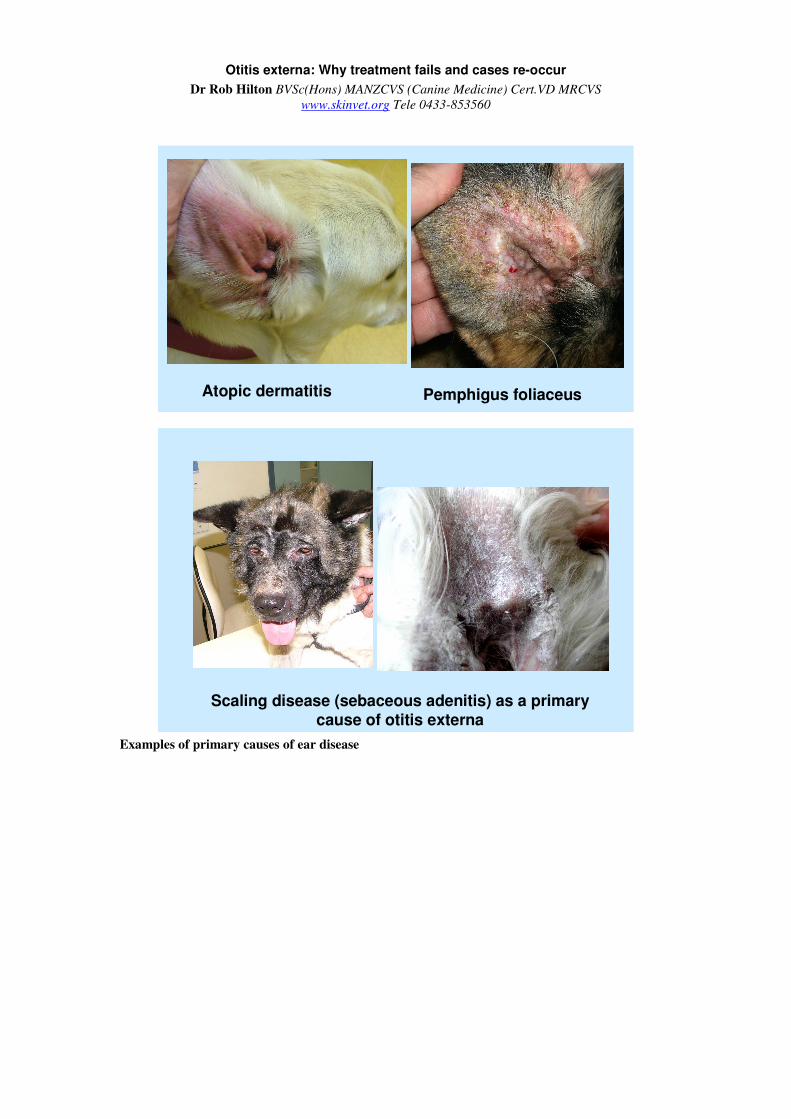
Atopic dermatitis Pemphigus foliaceus
Scaling disease (sebaceous adenitis) as a primary cause of otitis externa
Examples of primary causes of ear disease

Otitis externa: Why treatment fails and cases re-occur
Dr Rob Hilton BVSc(Hons) MANZCVS (Canine Medicine) Cert.VD MRCVS
www.skinvet.org Tele 0433-853560
Ruptured tympanic membrane, erosion, ulceration and exudate in a case of Pseudomonas otitis
Otitis externa: Why treatment fails and cases re-occur
Dr Rob Hilton BVSc(Hons) MANZCVS (Canine Medicine) Cert.VD MRCVS
www.skinvet.org Tele 0433-853560
Reasons why initial treatment may fail
Canal narrowing and end stage changes: Get the ear open!
Epidermal, ceruminal gland and follicular hyperplasia combined with dermal fibrosis
effectively close the ear canal. If the canal can not be made more open to effect
cleaning and treatment then the prognosis is much worse. In advanced cases, there
may be ossification of the annular cartilages and this carries a poor medical prognosis.
In many cases, it is not possible to otoscopically examine past the proximal vertical
canal and effective cleaning is not possible.
All efforts should be made to open the canal as much as possible and cleaning under
anaesthesia often needs to be delayed until this is achieved.
The author recommends an otic preparation containing a potent topical corticosteroid
which is active both superficially and in the deep dermis. To this end, high potency
corticosteroid formulations containing 0.1% mometasone or betamethasone valerate
are suitable. Common first line otic preparations containing 0.5-1% prednisolone are
NOT recommended these are of the order of 50-100 times less potent with respect to
corticosteroid activity at the concentrations used.
Prednisolone systemically is essential unless medically contraindicated. 1mg/kg daily
for 8-10 days then every other day for 10-14 days reduces inflammation and pain.
Oclacitinib (Apoquel®) is not as effective for reducing canal thickening.
The importance of cleaning. Inadequate cleaning = treatment failure
Serious ear disease needs to be seriously cleaned:
• To decrease the microbial load in the canal by mechanical and antiseptic
means
• To remove as much organic material as possible. Many antibiotics especially
aminoglycosides (gentamycin etc) and polymyxin are significantly less active
in the presence of exudates and organic material
• To check for and remove any distal canal cerumoliths. These concretions of
inspissated wax and debris are often found in the distal horizontal canal,
forming a plug on the tympanic membrane. If not removed, their behaviour is
to act as a foreign body, just like a grass seed
• To break up biofilm (see later)
• To inspect the tympanic membrane and assess if intact. A visually intact
tympanic membrane is not a 100% guarantee as to the fact that it is
functionally intact.
Otitis externa: Why treatment fails and cases re-occur
Dr Rob Hilton BVSc(Hons) MANZCVS (Canine Medicine) Cert.VD MRCVS
www.skinvet.org Tele 0433-853560
Tips on ear cleaning
1. If not already done so, collect samples for cytology and if necessary culture
(see later)
2. Delay full cleaning until canal has been medically opened (see before)
3. Plan time and cost wise on a full surgical procedure. To do an effective
clean on an ear may easily take 20-30 mins per ear. General anaesthesia is
usually required with placement of an endotracheal tube. If the tympanic
membrane is ruptured, it is possible fluid may pass down the Eustachian tube.
4. In the past, it was recommended to only use saline as a cleaning solution until
the tympanic membrane can be seen and evaluated as most (all?) past ear
cleaners are ototoxic. Otoflush® Dermcare contains Disodium edetate
(tris EDTA) and polyhexamethylene biguanidine hydrochloride (PHMB) in a
buffered solution. It is highly effective against all classes of bacteria and fungi.
The manufacturer advises that Otoflush is “not contraindicated in cases where
the tympanic membrane integrity is unknown or is ruptured.” The author uses
this product for ear cleaning as a matter of standard protocol.
The author’s technique
1. Instrumentation. a. Otoscope with fully charged batteries
b. IV extension set cut to length for flushing and aspirating
(feeding tubes may also be used),
c. Alligator forceps
d. A supply of cotton wool.
2. Clean the medial pinna and its folds first with cotton wool
3. Using Dermcare Otoflush®, repeated cycles of flush/suck until the
distal horizontal canal comes into view
4. Mopping. To view the distal canals, make a small ball of cotton wool
and pass down otoscope cone with alligator forceps to mop up cleaner
and remaining debris and inspect. Repeat flush/suck/mop as needed.
5. Check for a distal canal cerumenoliths. These can be very difficult
to break up by flushing. Picking with the alligator forceps allows the
cerumenolith to be broken up, removed in pieces and finally the
remnants removed by flush/suck/mop. Care and time is needed to
avoid causing haemorrhage or damaging the tympanic membrane
6. Assess the tympanic membrane. If the tympanic membrane is
clearly ruptured, collect middle ear exudate for comparative cytology
and flush the tympanic cavity.
7. Give systemic corticosteroids and instil an antimicrobial and corticosteroid formulation based on cytology findings and the status
of the tympanic membrane. Systemic antimicrobials are not required
unless there is otitis media or if there is extensive canal ulceration and
suspected bacterial invasion of the canal lining.

Otitis externa: Why treatment fails and cases re-occur
Dr Rob Hilton BVSc(Hons) MANZCVS (Canine Medicine) Cert.VD MRCVS
www.skinvet.org Tele 0433-853560
Biofilm – why logical treatment may fail
A biofilm is formed by micro-organisms sticking to each other and often adhering to
a surface. These adherent cells are frequently embedded within a self-produced matrix
of slime-like material consisting of proteins, DNA and polysaccharides. The
corrugated and abnormal surface produced by ear canal epidermal hyperplasia
supports biofilm adhesion
5 stages of a Pseudomonas aeruginosa biofilm development. Stage 1, initial
attachment; stage 2, irreversible attachment; stage 3, maturation I; stage 4,
maturation II; stage 5, dispersion
D. Davis - From: D. Monroe. "Looking for Chinks in the Armor of Bacterial Biofilms". PLoS Biology 5 (11, e307).
DOI:10.1371/journal.pbio.0050307.}, CC BY 2.5, https://commons.wikimedia.org/w/index.php?curid=3364284
When growing in biofilm, the microbes have a different physiology to the free-living
(planktonic) forms. Micro-organisms in a biofilm are semi-dormant at the base and
may be refractory to logical antibiotics and can develop increasing antimicrobial
resistance. Biofilms are recognised as important on teeth (tartar), on medical implants,
in industry and now in tissues such as airways, bone, urinary tract and ear canals.
Organisms of veterinary importance that are capable of ear biofilm formation include
Staphylococcus spp , Pseudomonas aeruginosa, Malassezia spp, Streptococcus spp
and a number of other opportunist pathogens.
Diagnosis of biofilm. At present we have no specific means of diagnosing the
presence of biofilm but it may be suspected if there is a persistent, possibly slimy,
exudate in the canal and that on cytology, the pathogen(s) are not being eliminated by
what would be logical therapy.

Otitis externa: Why treatment fails and cases re-occur
Dr Rob Hilton BVSc(Hons) MANZCVS (Canine Medicine) Cert.VD MRCVS
www.skinvet.org Tele 0433-853560
Disruption of ear biofilm. Antibiotics are often ineffective in breaking down biofilm.
Veterinary evidence is lacking but studies show the following agents have a disruptive
effect on biofilms:
• Triz-EDTA acts through ion chelation and, in addition , has a synergistic
action with antiseptics, antibiotics and proteases
• Silver sulphadiazine at higher concentrations has been shown in vitro to
disrupt biofilms
• Acetylcysteine has been shown in vitro and in vivo to be a potent disruptor of
biofilms. Solutions of 20mg/ml are bactericidal against many bacteria
including Pseudomonas aeruginosa. There is data to suggest acetylcysteine is
fungistatic against Candida spp; no data exists for Malassezia. In human
medicine, combination solutions of acetylcysteine and ciprofloxacin have been
found to be potentially synergistic. Anecdotal reports of good results exist for
2% acetylcysteine in combination with 7.5-15mg/ml enrofloxacin. Limited
data suggests that solutions <2% have an acceptable low(er) ototoxic potential.
2% acetylcysteine is increasing in use among veterinary dermatologists but
specific studies are still lacking.
• Mechanical mopping may assist (see flush/suck/mop technique above)
• In the future, we may see products ,suitable for otic use, containing silver
nano/micro-particles, proteolytic enzymes or other novel agents that disrupt
biofilms
Neutrophils and rod-like bacteria. Suggestive of Pseudomonas otitis Diff-Quik blue , 1000x

Otitis externa: Why treatment fails and cases re-occur
Dr Rob Hilton BVSc(Hons) MANZCVS (Canine Medicine) Cert.VD MRCVS
www.skinvet.org Tele 0433-853560
Choosing antimicrobials: Choices and pitfalls
Ear Cytology - How to tips 1. The author collects samples from both ears and by, convention, smears the left
ear towards the frosted side of the slide (or make R and L figures on the same
slide)
2. If Otodectes is suspected, collect a sample using copious quantities of oil
AFTER collecting the cytology sample.
3. Rapidly air dry
4. Stain with just Diff-Quik 2 (blue) by putting a drop on the slide and then
placing a cover slip. This will stain keratinocytes, bacteria, yeast and the
nuclei of leucocytes; what we are generally looking for. It will not highlight
fungal hyphae well. By using the fixative, organisms in a waxy base may wash
off. The red Diff-Quik stain is only of value for staining eosinophil granules.
5. Diff-Quik stain needs to be replaced regularly. Old stain fails to stain cells, has
“pseudobacteria” of stain precipitate and will grow its own culture of
contaminant bacteria. The author uses a dropper bottle of Diff Quik 2 that is
replaced weekly.
6. Use low power first to find good fields then go to suitable fields with 400x
magnification.
7. There is no reason to use oil immersion for ear cytology. In addition to mess
and spoiling slides, the field on 1000x is reduced by a factor of 5.25; 1000x
makes the evaluation of the slide as a whole more time consuming and does
not improve resolution. Ensure the microscope condenser is near the top to
maximise resolution and the light beam diaphragm is not closed down. If the
microscope does not allow for the differentiation of rods from cocci then it
should be serviced or replaced.
8. Streptococci, staphylococci and entrococci are effectively impossible to
differentiate on ear smears. Enterococci are Gram –ve cocci. The
morphological description of staphylococci forming grape like clusters and
streptococci forming chains is more useful in vitro than for ear cytology.
9. Enterobacteria (Pseudomonas, E. coli and Proteus ) form rods about 1.5-2x as
long as wide. Small gram positive rods are consistent with Corynebacterium
and are often seen in mixed cultures.
10. Pseudomonas aeruginosa is often seen as a solo infection and is often
associated with an outpouring of neutrophils. Classical green tinted pus
suggests Pseudomonas infection but a brown exudate does NOT rule it out.
11. Mixed infections with many diverse rods and cocci may be mixed anaerobes
12. Do not rely on colour of ear exudate without cytology. Brown “yeasty”
material may contain significant numbers of bacteria, including rods and
leukocytes
There is little evidence that culture and
sensitivity testing alters outcomes in most cases of
otitis externa.
Dermatologists are tending to do fewer cultures
and relying more on cytology to select and
monitor an initial course of topical medication
Otitis externa: Why treatment fails and cases re-occur
Dr Rob Hilton BVSc(Hons) MANZCVS (Canine Medicine) Cert.VD MRCVS
www.skinvet.org Tele 0433-853560
Choosing the medication
• Cultures are looking for growth inhibition at concentrations reached by
systemic dosing (µgm/ml). When topical medication is used, the doses the
bacteria are exposed to are mg/ml (of the order of 1000x higher). Hence
organisms reported as “resistant” on culture may indeed be sensitive
• Culture is important in cases of otitis media where systemic antibiotics are
utilized. It is important to obtain the sample from the middle ear cavity as
differing organisms and differing sensitivities may be isolated from different
levels of an infected ear canal
• The key is to do cytology before treatment and then look for clearance of the
organisms 10-14 days later. Even in severe cases, a 7 day cytology should still
show a major reduction in organism numbers if the medication choice,
formulation and dose are correct
• Streptococci, anaerobes and enterococci are inherently resistant to gentamycin
and enrofloxacin. Failure to clear cocci on cytology suggests the presence of
these organisms or a drug-resistant staphylococcus. Many Gram +ve cocci
will respond to miconazole/clotrimazole but not all. An empirical choice for
persistent cocci is either silver sulphadiazine or chloramphenicol topically (off
label use). Topical chloramphenicol (off label) is also often effective for
mixed anaerobe infections
• The use of a triz-EDTA flush before instillation of topical medication can
synergistically potentiate the antibacterials. Triz-EDTA alone will support
yeast and other fungal overgrowth. The addition of PHMB to the buffered
Triz-EDTA in the Otoflush® product provides a potent antibacterial and
antifungal action.
• Azole (clotrimazole, miconazole) resistance among Malassezia spp has been
identified. In the author’s experience more cases of treatment failure with
respect to Malassezia are related to cerumenoliths and inadequate cleaning
rather than resistance. Malassezia sensitivity testing is not routinely offered
by most laboratories but, fortunately, most azole-resistant cases will respond
to nystatin.
• It is important that adequate volumes of antimicrobial medication be instilled
and placed deep in the vertical canal at the junction of the horizontal canal.
Volumes required vary between 0.5 – 1 ml, depending on the canal volume.
To assist in proper placement and correct volume, the author supplies owners
with a syringe and a 1 – 1.5cm soft plastic canula made from a cut-down
intravenous extension set.
• Systemic treatment for 4 weeks + is indicated for otitis media. There is no
substitute for topical treatment. Systemic antibiotics are unlikely to reach

Otitis externa: Why treatment fails and cases re-occur
Dr Rob Hilton BVSc(Hons) MANZCVS (Canine Medicine) Cert.VD MRCVS
www.skinvet.org Tele 0433-853560
therapeutic levels in the ear canal unless there is erosion/ulceration with serum
exudation. Systemic ketoconazole is more of an adjunct treatment of otitis
external and should not be relied on for monotherapy.
• Ototoxicity of topical medications depends on BOTH the drug uses and the
base. There is data from clinical cases of dogs with otitis and tympanic
membrane rupture that Brainstem Auditory Evoked Response (BAER)
improved after repeated aqueous instillation of gentamycin in aqueous
solutions. There is no safety data on the use of oil or propylene glycol based
drops in the presence of a non-intact tympanum and clinical consensus is that
these bases are ototoxic and potentially enhance the ototoxicity of the
antimicrobials in the preparation.
o Least ototoxic: Enrofloxacin, silver sulphadiazine, 0.15%
chlorhexidine, ketoconazole, dexamethasone, Triz-EDTA-PHMB
o Increased ototoxic potential: gentamycin, chloramphenicol
o High ototoxic potential: Polymyxin-B, ticarcillin, amikacin,
tobramycin and ANY non-aqueous based medication
• Compounded or specially formulated ear medications are indicated when
o There is a failure of logical registered treatment applied in appropriate
volumes to a cleaned ear.
o Where there is a ruptured tympanic membrane and registered aqueous
ear preparations have failed or are deemed inappropriate.
o When high volume flushing is needed, for example to assist in treating
otitis media.
Cytological resolution post treatment.
Treat till you beat !
It is vital to monitor the patient every
10-14 day and continue topical
treatment until visible and cytological
resolution of the infection. Then
proceed into a maintenance program

Otitis externa: Why treatment fails and cases re-occur
Dr Rob Hilton BVSc(Hons) MANZCVS (Canine Medicine) Cert.VD MRCVS
www.skinvet.org Tele 0433-853560
Ointment base with scattered squames. Diff Quik 2 x 400
New Developments
Thermoreversible long acting gels, such as poloxamer based, have been used off label
at intervals of 1-2 weeks on an off-label compounded basis in dogs where daily
treatment is not possible. Their efficacy and use has been largely anecdotal.
Recently, a study has been published comparing the use of two veterinarian applied
treatments, 1 week apart, of a terbinafine-florfenicol-betamethasone acetate otic gel,
Osurnia, Elanco, compared to once daily with a veterinary licensed otic drop based
product along with twice weekly cleaning. The veterinarian-administered otic gel
“provided equivalent efficacy and higher quality of life score to dogs with otitis
externa and their owners, compared to an owner-administered topical otic therapy”.
Providing the tympanic membrane is intact, this novel therapy has benefits in those
dogs with ear infections susceptible to the constituent agents, particularly those dogs
that are difficult to medicate or owner compliance is a concern.
Surgical and other referral
Referral for advanced ear evaluation is indicated when middle ear involvement is
suspected and the state of the horizontal canal does not permit proper evaluation and
treatment. Flexible video otoscope is the optimal medical approach to evaluate and
flush a middle ear cavity
Signs of otitis media
• Pain, especially when pressure applied to bulla area
• Facial nerve paralysis (loss of blink reflex)
• Horner’s syndrome
• Circling, balance loss and nystagmus (otitis INTERNA)
• Discharge emanating from a ruptured tympanic membrane or less commonly a
bulging and possibly discoloured intact-appearing tympanic membrane.
CAUTION: With standard otoscopes, especially in a case with chronic
changes, the tympanic membrane can be difficult to accurately assess.
You’ve got to know when to hold them, know when to
fold them, know when to walk away, know when to run.
Kenny Rogers, “The Gambler”
Otitis externa: Why treatment fails and cases re-occur
Dr Rob Hilton BVSc(Hons) MANZCVS (Canine Medicine) Cert.VD MRCVS
www.skinvet.org Tele 0433-853560
Surgery
A full discussion of surgery techniques is beyond the scope of this article.
Some cases of otitis media required surgical drainage via a bulla osterotomy.
The Zepp lateral ear resection is of value in a limited number of cases. It should be
reserved for cases where most of the pathology is in the vertical canal, the owner is
aware ongoing cleaning will be needed but is likely to be easier and hearing is present.
It is contraindicated in cases with severe or end stage horizontal canal changes, otitis
media, when a “cure” is the desired outcome and when the animal will not permit
ongoing maintenance.
Total ear canal ablation with bulla osteotomy (TECA) is likely to produce resolution
of clinical signs with hearing loss. There are potential complications from the surgery
and should only be done by surgeons trained and experienced in the procedure. The
indications include:
• Severe ear pain that makes topical treatment impossible.
• Failure of at least two different courses of logical medical treatment
• Proliferative end stage changes in a canal that can not be opened
• Ossification of the aural cartilages carriers a poor medical prognosis
• Refractory otitis media where canal changes render a bulla osteotomy and
drainage unlikely to be effective
Maintaining the chronic ear
The author’s protocol: Flush once a week with a gentle ear cleaner that has good antimicrobial properties
30-60 minutes later, instil a corticosteroid steroid-containing medication
• Data exists that ears maintained with a steroid medication in addition to a
cleaner have a lower rate of re-infection than just with a cleaner alone.
• This reflects the experience and practice of many dermatologists
• The author’s experience is that alcohol-based corticosteroids, particularly
hydrocortisone aceponate spray, when used off label in the ear have
excessive drying action
After long standing infection and inflammation, as
described earlier, the ear canal has lost its self
cleaning function and possibly may never fully
regain it. Maintain and monitor is essential
Otitis externa: Why treatment fails and cases re-occur
Dr Rob Hilton BVSc(Hons) MANZCVS (Canine Medicine) Cert.VD MRCVS
www.skinvet.org Tele 0433-853560
• The author prefers to use a registered otic preparation containing
betamethasone valerate despite the fact that the preparation contains
antimicrobials. It is the authors opinion that the risk of overgrowth of
resistant pathogens is more theoretical than real, especially when an
antimicrobial ear flush is used. This is not to say there is no risk and
informed consent is needed.
The patient is reviewed at 6-8 weeks otoscopically and cytologically, 6 days after
cleaning.
If the response is very good-excellent, the maintenance interval can be extended
progressively from 10-14 days, with monitoring every 3 months.
The major risk factor is contact sensitivity developing to components of the flush or
steroid-containing otic preparation. While, uncommon it is occasionally seen and
needs to be recognised by the clinician. This risk, while small, needs to be balanced
against the fact that without maintenance, reinfection is almost certain.
Summary
Failure to control otitis externa can be related to the following factors:
• Not understanding the pathophysiology of chronic otitis
• Failure to control a manageable primary cause
• End stage changes
• Organic material, biological and other foreign material left in ear canal
• Biofilms
• Ineffective medication duration and volumes
• Otitis media
• True resistance
• Failure to monitor and maintain
References are available on request
The author wishes to express his gratitude to Dr. Anne Woolley for her proof reading
and corrections to this article.