oto443339 233. - sbccp - sociedade brasileira de … · techniques to increase the pretest...
TRANSCRIPT

http://oto.sagepub.com/Otolaryngology -- Head and Neck Surgery
http://oto.sagepub.com/content/147/2/233The online version of this article can be found at:
DOI: 10.1177/0194599812443339
2012 147: 233 originally published online 2 April 2012Otolaryngology -- Head and Neck SurgeryDeborah Rubens
Jonathan Walsh, Liwei An, Brad Mills, Zaegyoo Hah, Jacob Moalem, Matthew Miller, Ellen Giampoli, Kevin Parker andQuantitative Crawling Wave Sonoelastography of Benign and Malignant Thyroid Nodules
Published by:
http://www.sagepublications.com
On behalf of:
American Academy of Otolaryngology- Head and Neck Surgery
can be found at:Otolaryngology -- Head and Neck SurgeryAdditional services and information for
http://oto.sagepub.com/cgi/alertsEmail Alerts:
http://oto.sagepub.com/subscriptionsSubscriptions:
http://www.sagepub.com/journalsReprints.navReprints:
http://www.sagepub.com/journalsPermissions.navPermissions:
What is This?
- Apr 2, 2012OnlineFirst Version of Record
- Jul 31, 2012Version of Record >>
at SOCIEDADE BRASILEIRA DE CIRUR on October 26, 2012oto.sagepub.comDownloaded from
Original Research—Endocrine Surgery
Quantitative Crawling WaveSonoelastography of Benign andMalignant Thyroid Nodules
Otolaryngology–Head and Neck Surgery147(2) 233–238� American Academy ofOtolaryngology—Head and NeckSurgery Foundation 2012Reprints and permission:sagepub.com/journalsPermissions.navDOI: 10.1177/0194599812443339http://otojournal.org
Jonathan Walsh, MD1, Liwei An, PhD2, Brad Mills2,Zaegyoo Hah, PhD2, Jacob Moalem, MD3, Matthew Miller, MD1,Ellen Giampoli, MD4, Kevin Parker, PhD2, andDeborah Rubens, MD5
Sponsorships or competing interests that may be relevant to content are dis-
closed at the end of this article.
Abstract
Objective. The purpose of this study is to determine if crawlingwave elastography, a novel sonoelastography technique, can beused to provide quantitative measurements of thyroid tissueshear velocity (a measure of tissue stiffness) and distinguishbetween benign and malignant thyroid nodules.
Study Design. Diagnostic test assessment.
Setting. Academic university.
Subjects and Methods. Fresh thyroid specimens (n = 20) with44 regions of interest were imaged ex vivo with crawlingwave sonoelastography over a 9-month period in 2010 at asingle institution. Using the sonoelastography technique,shear velocity estimations and contrast-to-noise ratios werecalculated. The higher the shear velocity (SV) and contrast-to-noise ratio (CNR), the greater the tissue stiffness.Histological diagnosis was correlated with shear velocityand contrast-to-noise ratio values.
Results. Both the shear velocity and contrast-to-noise valuesof papillary thyroid carcinoma (n = 10, CNR = 5.29, SV =2.45 m/s) were significantly higher than benign nodules (n =22, CNR = –0.41, SV = 1.90 m/s). There is a maximum sensi-tivity and specificity of 100% and 90.9%, respectively, for dif-ferentiating papillary thyroid carcinoma from benign nodulesusing contrast-to-noise ratio values. There is a maximum sen-sitivity and specificity of 83.3% and 72.7%, respectively, for dif-ferentiating papillary thyroid carcinoma from benign nodulesusing shear velocity values. Insufficient samples were obtainedfor comparison with other histological types.
Conclusion. Crawling wave sonoelastography can providequantitative estimations of shear velocity, thereby depictingthe elastic properties of thyroid nodules. The shear velocityand contrast-to-noise ratio can differentiate between benignthyroid nodules and papillary thyroid carcinoma with highspecificity and sensitivity.
Keywords
elastography, sonoelastography, crawling wave sonoelasto-graphy, thyroid, thyroid nodules, ultrasound, benign, malig-nant, quantitative
Received September 2, 2011; revised November 28, 2011; accepted
March 6, 2012.
As more thyroid nodules are detected with the
increasing use of thyroid ultrasound, there is greater
need to improve the diagnostic specificity and sen-
sitivity for malignancy through noninvasive techniques.
With the use of ultrasound, it is estimated that 60% of
women aged 75 years will have thyroid nodules. In addition,
up to 35% of thyroid glands on autopsy contain papillary
thyroid carcinoma.1 Currently, features such as microcalcifi-
cations, increased Doppler flow, hypoechogenicity, and irre-
gular borders lack sufficient sensitivity and specificity to
differentiate thyroid carcinoma.2 The American Thyroid
Association (ATA) has recommended biopsy for most
nodules 1 cm or greater or any nodule 5 to 9 mm with sus-
picious sonographic features in a high-risk patient. This,
combined with the recommendation to biopsy up to 4
1Department of Otolaryngology, University of Rochester, Rochester, New
York, USA2Department of Electrical and Computer Engineering, University of
Rochester, Rochester, New York, USA3Department of Surgery, University of Rochester, Rochester, New York,
USA4Department of Pathology, University of Rochester, Rochester, New York,
USA5Department of Radiology, University of Rochester, Rochester, New York,
USA
This article was presented at the 2011 AAO-HNSF Annual Meeting & OTO
EXPO; September 11-14, 2011; San Francisco, California.
Corresponding Author:
Jonathan Walsh, MD, Department of Otolaryngology, University of
Rochester, 601 Elmwood Avenue, Box 629, Rochester, NY 14642, USA
Email: [email protected]
at SOCIEDADE BRASILEIRA DE CIRUR on October 26, 2012oto.sagepub.comDownloaded from

nodules per patient, highlights the need for noninvasive
techniques to increase the pretest probability in detecting
malignancy in fine-needle aspiration (FNA) biopsy.3
Sonoelastography is a general term for the use of ultra-
sound in imaging tissue elastic parameters. Various sonoe-
lastography methods have been explored, including strain
variation, velocity, compression, and vibration amplitude.4
Early vibrational elastography was pioneered by Lerner
et al.5 Since then, elastography has grown significantly as a
field and is being used in liver imaging and to differentiate
breast and prostate cancer.6,7
More recently, attention has been drawn to the utility of
elastography in thyroid nodules. Multiple studies have
demonstrated its effectiveness in differentiating benign and
malignant nodules in vivo.8-18 However, the majority are
qualitative elastographic images that depend on maintaining
a highly standardized technique and a subjective grading
system.19,20
Crawling wave (CrW) elastography is a method of shear
wave sonoelastography that provides quantitative estima-
tions of tissue elasticity properties. In particular, it provides
estimations of tissue shear velocity (m/s), which is propor-
tional to tissue stiffness. The higher the shear velocity, the
greater the stiffness of the tissue. The reliability and accu-
racy of this technique already have been demonstrated in
other tissues.7,21-23
The purpose of this study was to demonstrate feasibility
and utility of using a novel technique, CrW sonoelastogra-
phy, in thyroid tissue to differentiate between benign and
malignant nodules and to compare the quantitative measure-
ments with histopathology. We hypothesized that CrW
sonoelastography can produce reliable quantitative results in
ex vivo thyroid tissue and differentiate with adequate sensi-
tivity and specificity between benign and malignant
nodules.
Materials and Methods
The study protocol was approved by the Institutional
Review Board of the University of Rochester, and signed
consent was obtained for all tissue specimens. A priori
power analysis determined that a sample size of 25 to 35
nodules was needed to obtain 80% power to detect a dif-
ference between the 2 groups. Twenty unselected adult
patients (3 men, 17 women; mean age 52.9) undergoing
total or hemithyroidectomy at the University of Rochester
from January to December 2010 were recruited for study.
Children, revision surgery, recurrent disease, or prior
radiation patients were excluded. Fresh, unfixed whole and
hemithyroid specimens were suspended in an agar gel
phantom (Figure 1). In each case, regions of interest
(ROIs) for scanning were defined as a nodule with sur-
rounding homogeneous thyroid tissue on B-mode ultra-
sound. Normal tissue scans were also performed in lobes
without nodules and with homogeneous-appearing ultra-
sound images. Two B&K Minishakers with piston
vibration exciters (Model 2706; Bruel & Kjaer, Naerum,
Denmark) were applied as the vibration sources. A
dual-channel signal generator (Model AFG320; Tektronix,
Beaverton, Oregon) produced 2 monochrome low-
frequency signals that were slightly offset (typically less
than 0.5 Hz). These signals were passed through a 2-chan-
nel amplifier before being input to the minishaker vibra-
tion devices. A LOGIQ 9 scanner (General Electric
Medical Systems, Milwaukee, Wisconsin) modified for
sonoelastography was used with a M12L linear array probe
(5-13 MHz bandwidth) for real-time visualization of the
propagating crawling waves (ie, shear wave interference
pattern motion). Two-dimensional B-mode and sonoelasto-
graphic video images were recorded over a range of shear
wave frequencies from 80 to 280 Hz. The corresponding 2-
dimensional (2D) imaging plane was then marked with a
needle for later direct histopathologic correlation. Imaging
data were then analyzed with Matlab 7.6 (Mathworks, Inc,
Natick, Massachusetts) creating 2D shear velocity maps.
Shear velocity (SV) means and contrast-to-noise ratios
(CNRs) were determined for scanned nodules. CNR,
defined as (jSV(nodule) – SV(background)j)/s(background), is
commonly used in image analysis. Histologic slides corre-
sponding to the 2D plane of scanning were created and
photographed. Histologic findings of the needle-localized
Figure 1. Experimental setup. (a) Vibrator extensions, (b) pistonvibration exciters, (c) gel phantom with suspended specimen, (d)ultrasound transducer, (e) amplifier, and (f) function generator.
234 Otolaryngology–Head and Neck Surgery 147(2)
at SOCIEDADE BRASILEIRA DE CIRUR on October 26, 2012oto.sagepub.comDownloaded from
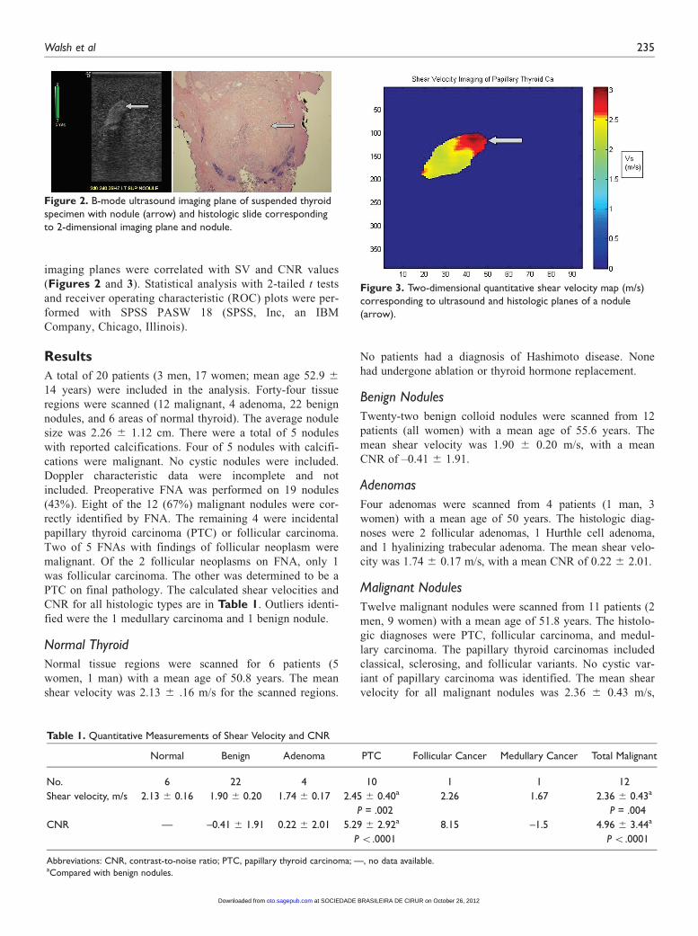
imaging planes were correlated with SV and CNR values
(Figures 2 and 3). Statistical analysis with 2-tailed t tests
and receiver operating characteristic (ROC) plots were per-
formed with SPSS PASW 18 (SPSS, Inc, an IBM
Company, Chicago, Illinois).
Results
A total of 20 patients (3 men, 17 women; mean age 52.9 6
14 years) were included in the analysis. Forty-four tissue
regions were scanned (12 malignant, 4 adenoma, 22 benign
nodules, and 6 areas of normal thyroid). The average nodule
size was 2.26 6 1.12 cm. There were a total of 5 nodules
with reported calcifications. Four of 5 nodules with calcifi-
cations were malignant. No cystic nodules were included.
Doppler characteristic data were incomplete and not
included. Preoperative FNA was performed on 19 nodules
(43%). Eight of the 12 (67%) malignant nodules were cor-
rectly identified by FNA. The remaining 4 were incidental
papillary thyroid carcinoma (PTC) or follicular carcinoma.
Two of 5 FNAs with findings of follicular neoplasm were
malignant. Of the 2 follicular neoplasms on FNA, only 1
was follicular carcinoma. The other was determined to be a
PTC on final pathology. The calculated shear velocities and
CNR for all histologic types are in Table 1. Outliers identi-
fied were the 1 medullary carcinoma and 1 benign nodule.
Normal Thyroid
Normal tissue regions were scanned for 6 patients (5
women, 1 man) with a mean age of 50.8 years. The mean
shear velocity was 2.13 6 .16 m/s for the scanned regions.
No patients had a diagnosis of Hashimoto disease. None
had undergone ablation or thyroid hormone replacement.
Benign Nodules
Twenty-two benign colloid nodules were scanned from 12
patients (all women) with a mean age of 55.6 years. The
mean shear velocity was 1.90 6 0.20 m/s, with a mean
CNR of –0.41 6 1.91.
Adenomas
Four adenomas were scanned from 4 patients (1 man, 3
women) with a mean age of 50 years. The histologic diag-
noses were 2 follicular adenomas, 1 Hurthle cell adenoma,
and 1 hyalinizing trabecular adenoma. The mean shear velo-
city was 1.74 6 0.17 m/s, with a mean CNR of 0.22 6 2.01.
Malignant Nodules
Twelve malignant nodules were scanned from 11 patients (2
men, 9 women) with a mean age of 51.8 years. The histolo-
gic diagnoses were PTC, follicular carcinoma, and medul-
lary carcinoma. The papillary thyroid carcinomas included
classical, sclerosing, and follicular variants. No cystic var-
iant of papillary carcinoma was identified. The mean shear
velocity for all malignant nodules was 2.36 6 0.43 m/s,
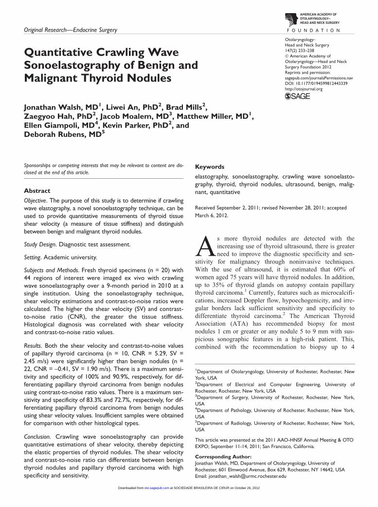
Figure 2. B-mode ultrasound imaging plane of suspended thyroidspecimen with nodule (arrow) and histologic slide correspondingto 2-dimensional imaging plane and nodule.
Table 1. Quantitative Measurements of Shear Velocity and CNR
Normal Benign Adenoma PTC Follicular Cancer Medullary Cancer Total Malignant
No. 6 22 4 10 1 1 12
Shear velocity, m/s 2.13 6 0.16 1.90 6 0.20 1.74 6 0.17 2.45 6 0.40a
P = .002
2.26 1.67 2.36 6 0.43a
P = .004
CNR — –0.41 6 1.91 0.22 6 2.01 5.29 6 2.92a
P \.0001
8.15 –1.5 4.96 6 3.44a
P \.0001
Abbreviations: CNR, contrast-to-noise ratio; PTC, papillary thyroid carcinoma; —, no data available.aCompared with benign nodules.
Figure 3. Two-dimensional quantitative shear velocity map (m/s)corresponding to ultrasound and histologic planes of a nodule(arrow).
Walsh et al 235
at SOCIEDADE BRASILEIRA DE CIRUR on October 26, 2012oto.sagepub.comDownloaded from
with a mean CNR of 4.96 6 3.44. The mean shear velocity
for PTC alone was 2.45 6 0.40 m/s, with a mean CNR for
PTC of 5.29 6 2.92.
Comparison
Comparison was between benign colloid nodules and malig-
nancy. Adenomas were excluded from the analysis due to
the insufficient numbers. Analysis demonstrated a signifi-
cant difference for both the mean shear velocities and CNR
between benign and malignant thyroid nodules (P = .004
and P \ .00012, respectively). When considering differen-
tiating between benign nodules and PTC, the difference was
even more significant for both shear velocity and CNR (P =
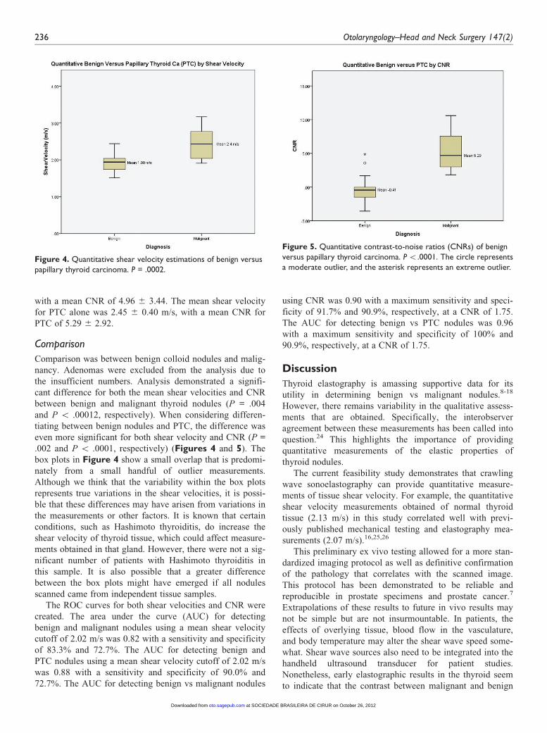
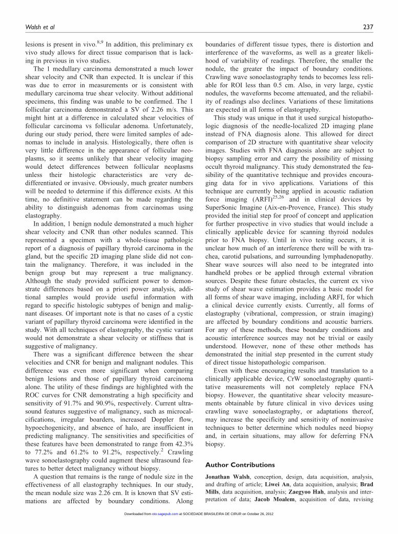
.002 and P \ .0001, respectively) (Figures 4 and 5). The
box plots in Figure 4 show a small overlap that is predomi-
nately from a small handful of outlier measurements.
Although we think that the variability within the box plots
represents true variations in the shear velocities, it is possi-
ble that these differences may have arisen from variations in
the measurements or other factors. It is known that certain
conditions, such as Hashimoto thyroiditis, do increase the
shear velocity of thyroid tissue, which could affect measure-
ments obtained in that gland. However, there were not a sig-
nificant number of patients with Hashimoto thyroiditis in
this sample. It is also possible that a greater difference
between the box plots might have emerged if all nodules
scanned came from independent tissue samples.
The ROC curves for both shear velocities and CNR were
created. The area under the curve (AUC) for detecting
benign and malignant nodules using a mean shear velocity
cutoff of 2.02 m/s was 0.82 with a sensitivity and specificity
of 83.3% and 72.7%. The AUC for detecting benign and
PTC nodules using a mean shear velocity cutoff of 2.02 m/s
was 0.88 with a sensitivity and specificity of 90.0% and
72.7%. The AUC for detecting benign vs malignant nodules
using CNR was 0.90 with a maximum sensitivity and speci-
ficity of 91.7% and 90.9%, respectively, at a CNR of 1.75.
The AUC for detecting benign vs PTC nodules was 0.96
with a maximum sensitivity and specificity of 100% and
90.9%, respectively, at a CNR of 1.75.
Discussion
Thyroid elastography is amassing supportive data for its
utility in determining benign vs malignant nodules.8-18
However, there remains variability in the qualitative assess-
ments that are obtained. Specifically, the interobserver
agreement between these measurements has been called into
question.24 This highlights the importance of providing
quantitative measurements of the elastic properties of
thyroid nodules.
The current feasibility study demonstrates that crawling
wave sonoelastography can provide quantitative measure-
ments of tissue shear velocity. For example, the quantitative
shear velocity measurements obtained of normal thyroid
tissue (2.13 m/s) in this study correlated well with previ-
ously published mechanical testing and elastography mea-
surements (2.07 m/s).16,25,26
This preliminary ex vivo testing allowed for a more stan-
dardized imaging protocol as well as definitive confirmation
of the pathology that correlates with the scanned image.
This protocol has been demonstrated to be reliable and
reproducible in prostate specimens and prostate cancer.7
Extrapolations of these results to future in vivo results may
not be simple but are not insurmountable. In patients, the
effects of overlying tissue, blood flow in the vasculature,
and body temperature may alter the shear wave speed some-
what. Shear wave sources also need to be integrated into the
handheld ultrasound transducer for patient studies.
Nonetheless, early elastographic results in the thyroid seem
to indicate that the contrast between malignant and benign
Figure 4. Quantitative shear velocity estimations of benign versuspapillary thyroid carcinoma. P = .0002.
Figure 5. Quantitative contrast-to-noise ratios (CNRs) of benignversus papillary thyroid carcinoma. P \.0001. The circle representsa moderate outlier, and the asterisk represents an extreme outlier.
236 Otolaryngology–Head and Neck Surgery 147(2)
at SOCIEDADE BRASILEIRA DE CIRUR on October 26, 2012oto.sagepub.comDownloaded from
lesions is present in vivo.8,9 In addition, this preliminary ex
vivo study allows for direct tissue comparison that is lack-
ing in previous in vivo studies.
The 1 medullary carcinoma demonstrated a much lower
shear velocity and CNR than expected. It is unclear if this
was due to error in measurements or is consistent with
medullary carcinoma true shear velocity. Without additional
specimens, this finding was unable to be confirmed. The 1
follicular carcinoma demonstrated a SV of 2.26 m/s. This
might hint at a difference in calculated shear velocities of
follicular carcinoma vs follicular adenoma. Unfortunately,
during our study period, there were limited samples of ade-
nomas to include in analysis. Histologically, there often is
very little difference in the appearance of follicular neo-
plasms, so it seems unlikely that shear velocity imaging
would detect differences between follicular neoplasms
unless their histologic characteristics are very de-
differentiated or invasive. Obviously, much greater numbers
will be needed to determine if this difference exists. At this
time, no definitive statement can be made regarding the
ability to distinguish adenomas from carcinomas using
elastography.
In addition, 1 benign nodule demonstrated a much higher
shear velocity and CNR than other nodules scanned. This
represented a specimen with a whole-tissue pathologic
report of a diagnosis of papillary thyroid carcinoma in the
gland, but the specific 2D imaging plane slide did not con-
tain the malignancy. Therefore, it was included in the
benign group but may represent a true malignancy.
Although the study provided sufficient power to demon-
strate differences based on a priori power analysis, addi-
tional samples would provide useful information with
regard to specific histologic subtypes of benign and malig-
nant diseases. Of important note is that no cases of a cystic
variant of papillary thyroid carcinoma were identified in the
study. With all techniques of elastography, the cystic variant
would not demonstrate a shear velocity or stiffness that is
suggestive of malignancy.
There was a significant difference between the shear
velocities and CNR for benign and malignant nodules. This
difference was even more significant when comparing
benign lesions and those of papillary thyroid carcinoma
alone. The utility of these findings are highlighted with the
ROC curves for CNR demonstrating a high specificity and
sensitivity of 91.7% and 90.9%, respectively. Current ultra-
sound features suggestive of malignancy, such as microcal-
cifications, irregular boarders, increased Doppler flow,
hypoechogenicity, and absence of halo, are insufficient in
predicting malignancy. The sensitivities and specificities of
these features have been demonstrated to range from 42.3%
to 77.2% and 61.2% to 91.2%, respectively.2 Crawling
wave sonoelastography could augment these ultrasound fea-
tures to better detect malignancy without biopsy.
A question that remains is the range of nodule size in the
effectiveness of all elastography techniques. In our study,
the mean nodule size was 2.26 cm. It is known that SV esti-
mations are affected by boundary conditions. Along
boundaries of different tissue types, there is distortion and
interference of the waveforms, as well as a greater likeli-
hood of variability of readings. Therefore, the smaller the
nodule, the greater the impact of boundary conditions.
Crawling wave sonoelastography tends to becomes less reli-
able for ROI less than 0.5 cm. Also, in very large, cystic
nodules, the waveforms become attenuated, and the reliabil-
ity of readings also declines. Variations of these limitations
are expected in all forms of elastography.
This study was unique in that it used surgical histopatho-
logic diagnosis of the needle-localized 2D imaging plane
instead of FNA diagnosis alone. This allowed for direct
comparison of 2D structure with quantitative shear velocity
images. Studies with FNA diagnosis alone are subject to
biopsy sampling error and carry the possibility of missing
occult thyroid malignancy. This study demonstrated the fea-
sibility of the quantitative technique and provides encoura-
ging data for in vivo applications. Variations of this
technique are currently being applied in acoustic radiation
force imaging (ARFI)25,26 and in clinical devices by
SuperSonic Imagine (Aix-en-Provence, France). This study
provided the initial step for proof of concept and application
for further prospective in vivo studies that would include a
clinically applicable device for scanning thyroid nodules
prior to FNA biopsy. Until in vivo testing occurs, it is
unclear how much of an interference there will be with tra-
chea, carotid pulsations, and surrounding lymphadenopathy.
Shear wave sources will also need to be integrated into
handheld probes or be applied through external vibration
sources. Despite these future obstacles, the current ex vivo
study of shear wave estimation provides a basic model for
all forms of shear wave imaging, including ARFI, for which
a clinical device currently exists. Currently, all forms of
elastography (vibrational, compression, or strain imaging)
are affected by boundary conditions and acoustic barriers.
For any of these methods, these boundary conditions and
acoustic interference sources may not be trivial or easily
understood. However, none of these other methods has
demonstrated the initial step presented in the current study
of direct tissue histopathologic comparison.
Even with these encouraging results and translation to a
clinically applicable device, CrW sonoelastography quanti-
tative measurements will not completely replace FNA
biopsy. However, the quantitative shear velocity measure-
ments obtainable by future clinical in vivo devices using
crawling wave sonoelastography, or adaptations thereof,
may increase the specificity and sensitivity of noninvasive
techniques to better determine which nodules need biopsy
and, in certain situations, may allow for deferring FNA
biopsy.
Author Contributions
Jonathan Walsh, conception, design, data acquisition, analysis,
and drafting of article; Liwei An, data acquisition, analysis; Brad
Mills, data acquisition, analysis; Zaegyoo Hah, analysis and inter-
pretation of data; Jacob Moalem, acquisition of data, revising
Walsh et al 237
at SOCIEDADE BRASILEIRA DE CIRUR on October 26, 2012oto.sagepub.comDownloaded from

article; Matthew Miller, acquisition of data, revising article; Ellen
Giampoli, acquisition of data; Kevin Parker, conception and
design, analysis, interpretation, revising article; Deborah Rubens,
conception and design, analysis, interpretation.
Disclosures
Competing interests: None.
Sponsorships: None.
Funding source: University of Rochester Department of
Otolaryngology.
References
1. Mazzaferri EL. Management of a solitary thyroid nodule.
N Engl J Med. 1993;328(8):553-559.
2. Sipos JA. Advances in ultrasound for the diagnosis and man-
agement of thyroid cancer. Thyroid. 2009;19(12):1363-1372.
3. Cooper DS, Doherty GM, Haugen BR, et al. Revised
American Thyroid Association management guidelines for
patients with thyroid nodules and differentiated thyroid cancer.
Thyroid. 2009;19(11):1167-1214.
4. Gao L, Parker KJ, Lerner RM, Levinson SF. Imaging of the
elastic properties of tissue—a review. Ultrasound Med Biol.
1996;22(8):959-977.
5. Lerner RM, Huang SR, Parker KJ. ‘‘Sonoelasticity’’ images
derived from ultrasound signals in mechanically vibrated tis-
sues. Ultrasound Med Biol. 1990;16(3):231-239.
6. Garra BS, Cespedes EI, Ophir J, et al. Elastography of breast
lesions: initial clinical results. Radiology. 1997;202(1):79-86.
7. Zhang M, Nigwekar P, Castaneda B, et al. Quantitative charac-
terization of viscoelastic properties of human prostate corre-
lated with histology. Ultrasound Med Biol. 2008;34(7):1033-
1042.
8. Asteria C, Giovanardi A, Pizzocaro A, et al. US-elastography
in the differential diagnosis of benign and malignant thyroid
nodules. Thyroid. 2008;18(5):523-531.
9. Bae U, Dighe M, Dubinsky T, Minoshima S, Shamdasani V,
Kim Y. Ultrasound thyroid elastography using carotid artery
pulsation: preliminary study. J Ultrasound Med. 2007;26(6):
797-805.
10. Dighe M, Bae U, Richardson ML, Dubinsky TJ, Minoshima S,
Kim Y. Differential diagnosis of thyroid nodules with US elas-
tography using carotid artery pulsation. Radiology. 2008;
248(2):662-669.
11. Dighe M, Kim J, Luo S, Kim Y. Utility of the ultrasound elas-
tographic systolic thyroid stiffness index in reducing fine-
needle aspirations. J Ultrasound Med. 2010;29(4):565-574.
12. Hong Y, Liu X, Li Z, Zhang X, Chen M, Luo Z. Real-time
ultrasound elastography in the differential diagnosis of benign
and malignant thyroid nodules. J Ultrasound Med. 2009;28(7):
861-867.
13. Kagoya R, Monobe H, Tojima H. Utility of elastography for
differential diagnosis of benign and malignant thyroid nodules.
Otolaryngol Head Neck Surg. 2010;143(2):230-234.
14. Luo S, Kim EH, Dighe M, Kim Y. Screening of thyroid nodules
by ultrasound elastography using diastolic strain variation. Conf
Proc IEEE Eng Med Biol Soc. 2009;2009:4420-4423.
15. Luo S, Kim EH, Dighe M, Kim Y. Thyroid nodule classifica-
tion using ultrasound elastography via linear discriminant anal-
ysis. Ultrasonics. 2011;51(4):425-431.
16. Lyshchik A, Higashi T, Asato R, et al. Thyroid gland tumor
diagnosis at US elastography. Radiology. 2005;237(1):202-
211.
17. Wang Y, Dan HJ, Dan HY, Li T, Hu B. Differential diagnosis
of small single solid thyroid nodules using real-time ultrasound
elastography. J Int Med Res. 2010;38(2):466-472.
18. Xing P, Wu L, Zhang C, Li S, Liu C, Wu C. Differentiation of
benign from malignant thyroid lesions: calculation of the
strain ratio on thyroid sonoelastography. J Ultrasound Med.
2011;30(5):663-669.
19. Itoh A, Ueno E, Tohno E, et al. Breast disease: clinical appli-
cation of US elastography for diagnosis. Radiology. 2006;
239(2):341-350.
20. Fleury Ede F, Fleury JC, Piato S, Roveda D Jr. New elasto-
graphic classification of breast lesions during and after com-
pression. Diagn Interv Radiol. 2009;15(2):96-103.
21. Hoyt K, Castaneda B, Parker KJ. Two-dimensional sonoelasto-
graphic shear velocity imaging. Ultrasound Med Biol. 2008;
34(2):276-288.
22. Wu Z, Hoyt K, Rubens DJ, Parker KJ. Sonoelastographic ima-
ging of interference patterns for estimation of shear velocity
distribution in biomaterials. J Acoust Soc Am. 2006;120(1):
535-545.
23. Zhang M, Castaneda B, Wu Z, et al. Congruence of imaging esti-
mators and mechanical measurements of viscoelastic properties
of soft tissues. Ultrasound Med Biol. 2007;33(10):1617-1631.
24. Park SH, Kim SJ, Kim EK, Kim MJ, Son EJ, Kwak JY.
Interobserver agreement in assessing the sonographic and elas-
tographic features of malignant thyroid nodules. AJR Am J
Roentgenol. 2009;193(5):W416-W423.
25. Friedrich-Rust M, Romenski O, Meyer G, et al. Acoustic
Radiation Force Impulse-Imaging for the evaluation of the
thyroid gland: a limited patient feasibility study. Ultrasonics.
2012;52(1):69-74.
26. Sporea I, Vlad M, Bota S, et al. Thyroid stiffness assessment
by acoustic radiation force impulse elastography (ARFI).
Ultraschall Med. 2011;32(3):281-285.
238 Otolaryngology–Head and Neck Surgery 147(2)
at SOCIEDADE BRASILEIRA DE CIRUR on October 26, 2012oto.sagepub.comDownloaded from