otolaryngology - ncapancapa.org/wp-content/uploads/2017/02/otolaryngology_pt.-1.pdf ·...
TRANSCRIPT

OTOLARYNGOLOGY
2017 PANRE Recertification Review Joshua F. Smith, MMS, PA-C Duke Head and Neck Surgery & Communication Sciences
Disclosure
I am the chair of the Professional Development Review Panel for the NCAPA and am a paid speaker.
I have no other financial disclosures or conflicts of interest.

Goals Review ENT Anatomy and Physiology
Discuss Common ENT Disorders
Signs and Symptoms
Physical Exam
Diagnostics
Treatment
Answer 100% of ENT PANRE questions correctly
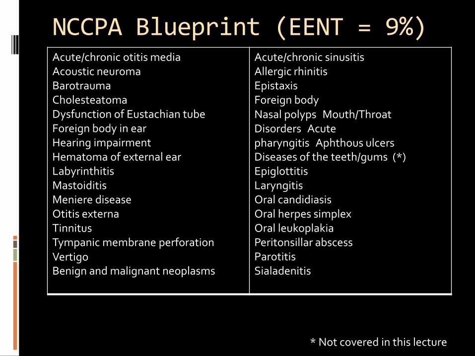
NCCPA Blueprint (EENT = 9%) Acute/chronic otitis media Acoustic neuroma Barotrauma Cholesteatoma Dysfunction of Eustachian tube Foreign body in ear Hearing impairment Hematoma of external ear Labyrinthitis Mastoiditis Meniere disease Otitis externa Tinnitus Tympanic membrane perforation Vertigo Benign and malignant neoplasms
Acute/chronic sinusitis Allergic rhinitis Epistaxis Foreign body Nasal polypsMouth/Throat DisordersAcute pharyngitisAphthous ulcers Diseases of the teeth/gums (*) Epiglottitis Laryngitis Oral candidiasis Oral herpes simplex Oral leukoplakia Peritonsillar abscess Parotitis Sialadenitis
* Not covered in this lecture
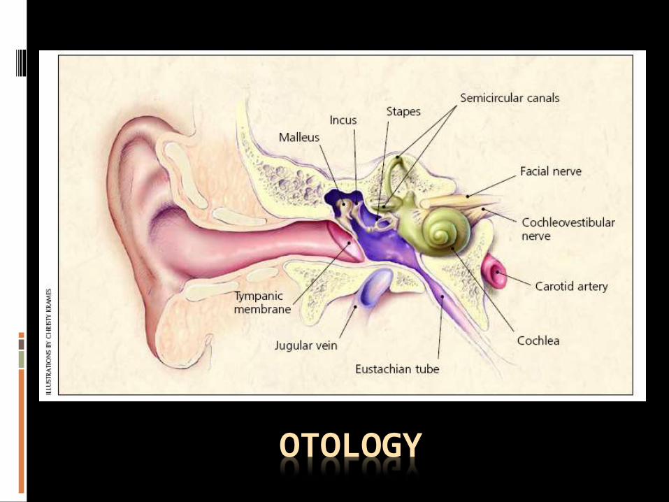
OTOLOGY
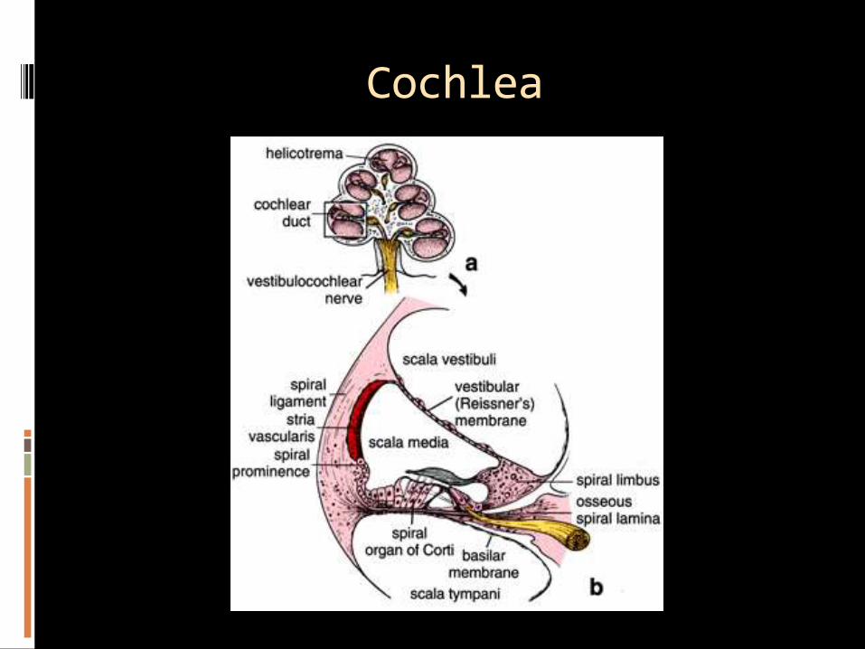
Cochlea
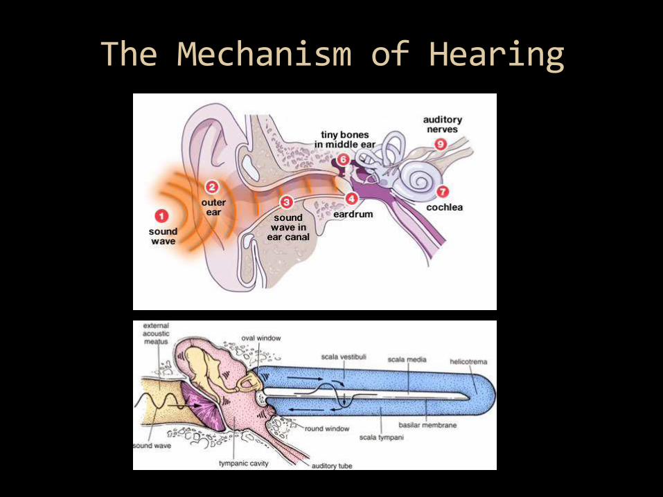
The Mechanism of Hearing
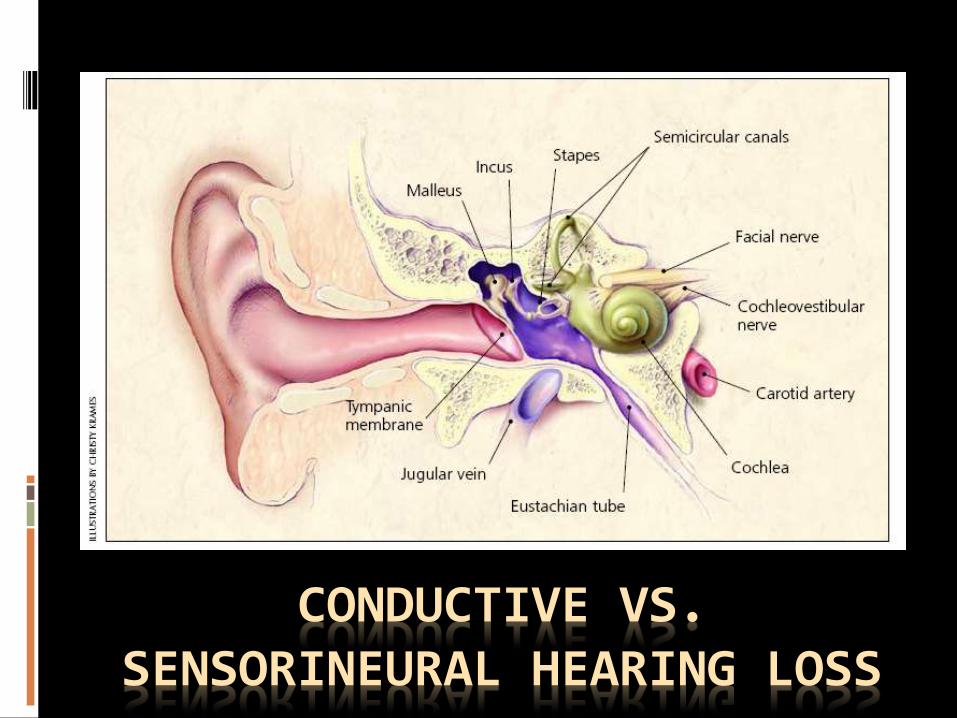
CONDUCTIVE VS. SENSORINEURAL HEARING LOSS
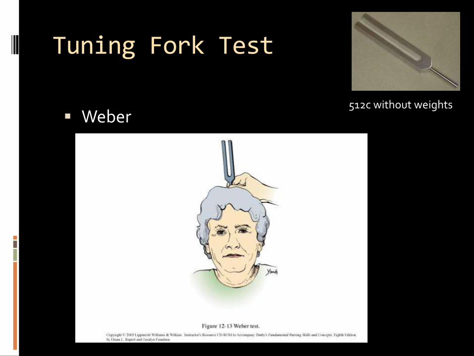
Tuning Fork Test
Weber 512c without weights

Tuning Fork Test
Tuning Fork Test- Rinne
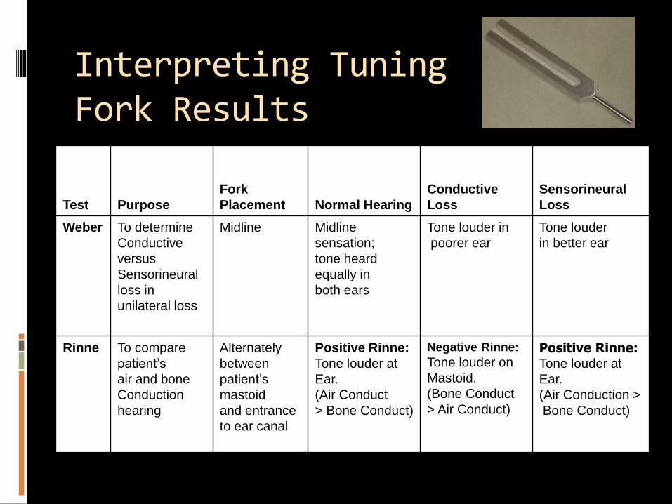
Interpreting Tuning Fork Results
Test Purpose
Fork
Placement Normal Hearing
Conductive
Loss
Sensorineural
Loss
Weber To determine
Conductive
versus
Sensorineural
loss in
unilateral loss
Midline Midline
sensation;
tone heard
equally in
both ears
Tone louder in
poorer ear
Tone louder
in better ear
Rinne To compare
patient’s
air and bone
Conduction
hearing
Alternately
between
patient’s
mastoid
and entrance
to ear canal
Positive Rinne:
Tone louder at
Ear.
(Air Conduct
> Bone Conduct)
Negative Rinne:
Tone louder on
Mastoid.
(Bone Conduct
> Air Conduct)
Positive Rinne: Tone louder at
Ear.
(Air Conduction >
Bone Conduct)
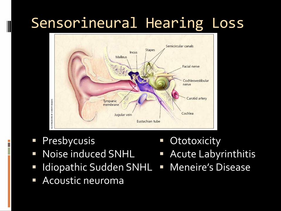
Presbycusis Noise induced SNHL Idiopathic Sudden SNHL Acoustic neuroma
Sensorineural Hearing Loss
Ototoxicity Acute Labyrinthitis Meneire’s Disease
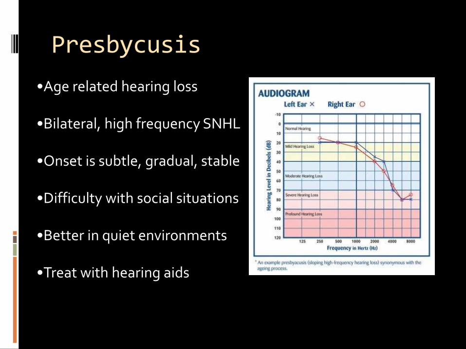
Presbycusis
•Age related hearing loss
•Bilateral, high frequency SNHL
•Onset is subtle, gradual, stable
•Difficulty with social situations
•Better in quiet environments
•Treat with hearing aids
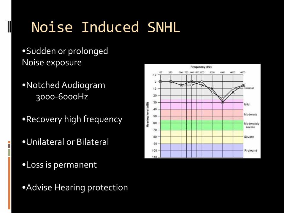
Noise Induced SNHL
•Sudden or prolonged Noise exposure •Notched Audiogram
3000-6000Hz •Recovery high frequency
•Unilateral or Bilateral
•Loss is permanent
•Advise Hearing protection
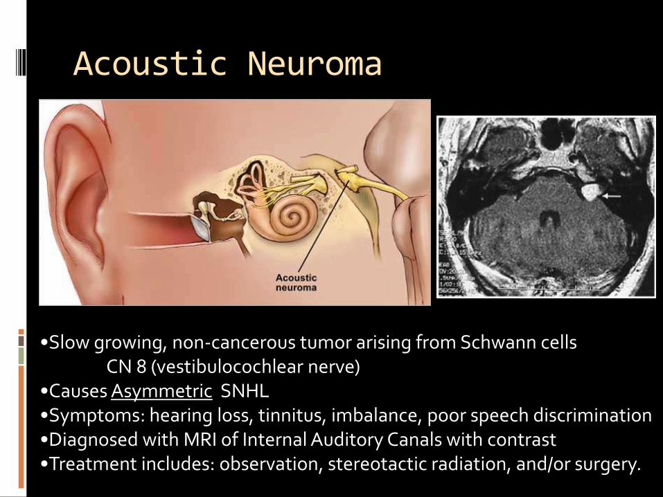
Acoustic Neuroma
•Slow growing, non-cancerous tumor arising from Schwann cells CN 8 (vestibulocochlear nerve) •Causes Asymmetric SNHL •Symptoms: hearing loss, tinnitus, imbalance, poor speech discrimination •Diagnosed with MRI of Internal Auditory Canals with contrast •Treatment includes: observation, stereotactic radiation, and/or surgery.
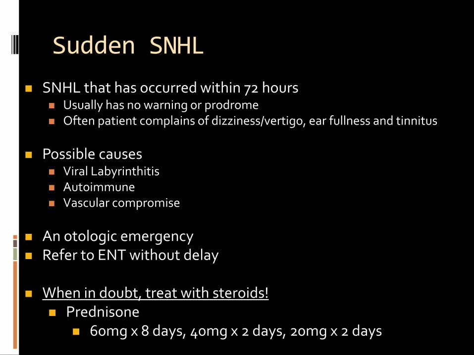
Sudden SNHL
SNHL that has occurred within 72 hours Usually has no warning or prodrome Often patient complains of dizziness/vertigo, ear fullness and tinnitus
Possible causes Viral Labyrinthitis Autoimmune Vascular compromise
An otologic emergency Refer to ENT without delay
When in doubt, treat with steroids!
Prednisone 60mg x 8 days, 40mg x 2 days, 20mg x 2 days
Sample PANRE question #1 Tina Tanner is a 62 year old female with gradual loss of hearing in her right ear for more than a year. Associated symptoms includes right sided tinnitus and imbalance. After performing a proper history and physical exam, you astutely decide to order an audiogram. The audiogram reveals an asymmetric sensorineural hearing loss on the right side. What is the next appropriate intervention? a. Order a contrasted MRI of the internal auditory canals b. Place the patient on a 12 day course of prednisone c. Order a non-contrasted CT of the temporal bone d. Start Meclizine 25mg TID for her imbalance e. Advise the patient to utilize a right-sided hearing aid
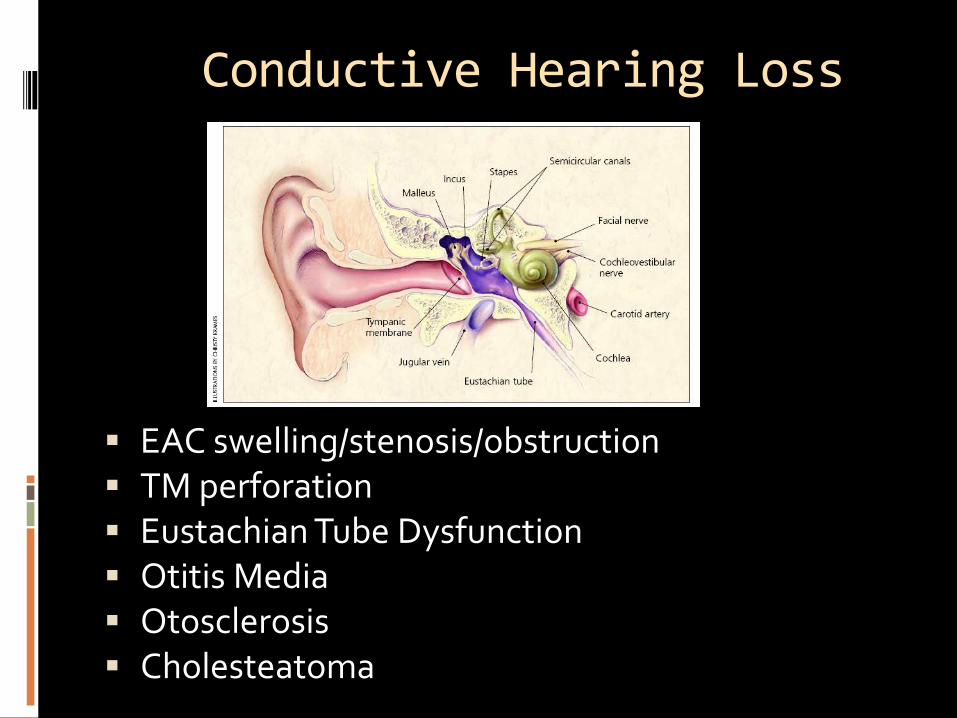
Conductive Hearing Loss
EAC swelling/stenosis/obstruction TM perforation Eustachian Tube Dysfunction Otitis Media Otosclerosis Cholesteatoma
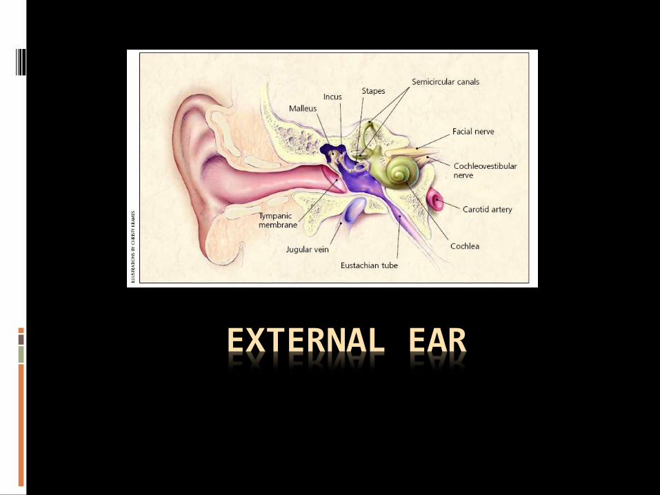
EXTERNAL EAR
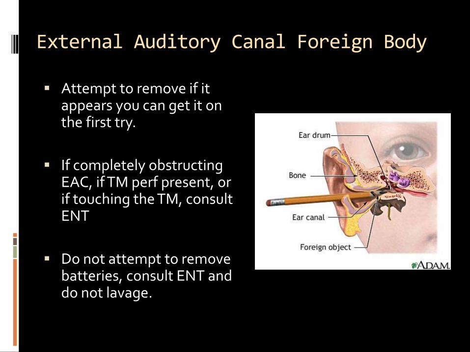
External Auditory Canal Foreign Body
Attempt to remove if it appears you can get it on the first try.
If completely obstructing EAC, if TM perf present, or if touching the TM, consult ENT
Do not attempt to remove batteries, consult ENT and do not lavage.
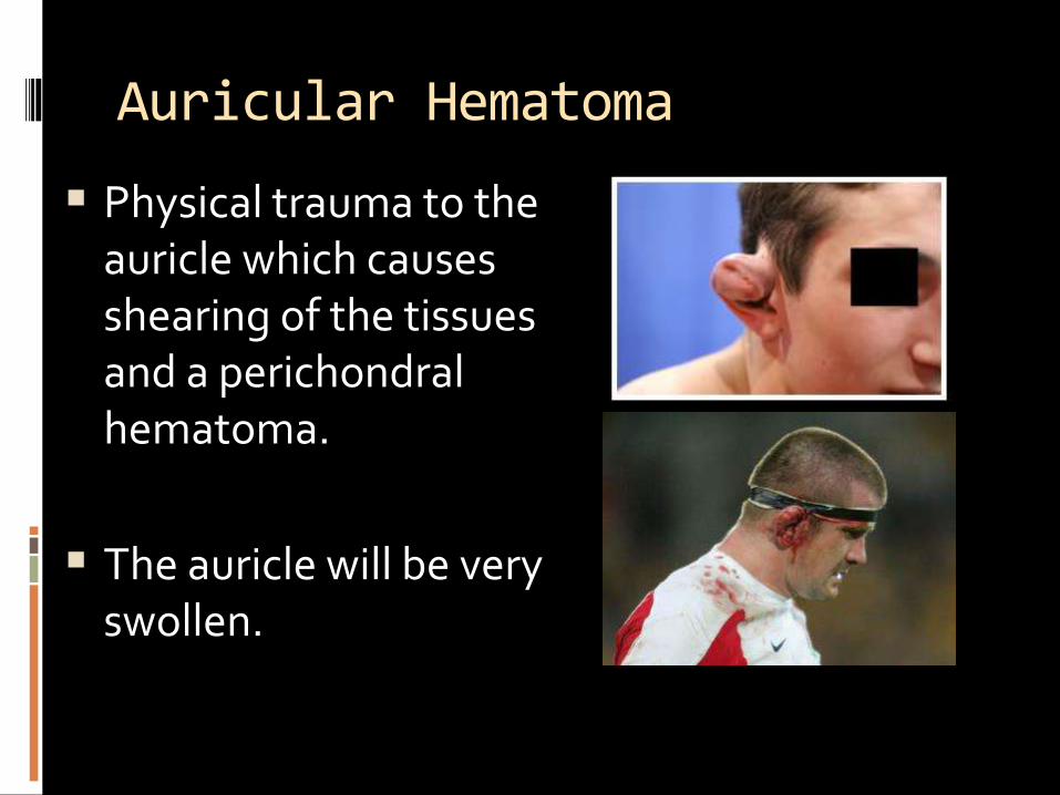
Auricular Hematoma
Physical trauma to the auricle which causes shearing of the tissues and a perichondral hematoma.
The auricle will be very swollen.
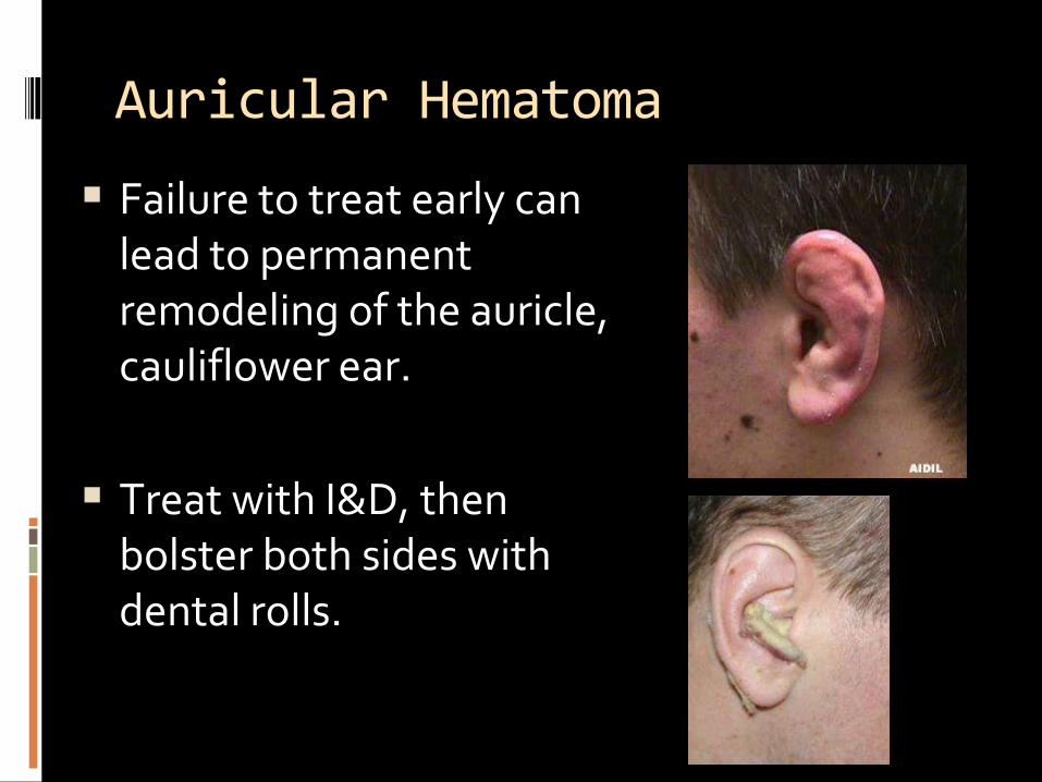
Auricular Hematoma
Failure to treat early can lead to permanent remodeling of the auricle, cauliflower ear.
Treat with I&D, then bolster both sides with dental rolls.
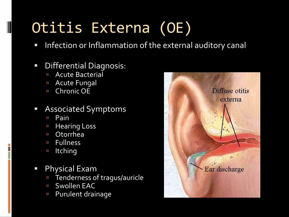
Otitis Externa (OE) Infection or Inflammation of the external auditory canal Differential Diagnosis:
Acute Bacterial Acute Fungal Chronic OE
Associated Symptoms Pain Hearing Loss Otorrhea Fullness Itching
Physical Exam Tenderness of tragus/auricle Swollen EAC Purulent drainage
Otitis Externa (OE)
Bacterial
Streptococcus
Staphylococcus
Pseudomonas
MRSA
Fungal (otomycosis)
Aspergillus
Candida
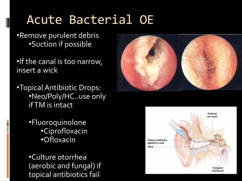
Acute Bacterial OE •Remove purulent debris
•Suction if possible
•If the canal is too narrow, insert a wick
•Topical Antibiotic Drops: •Neo/Poly/HC..use only if TM is intact
•Fluoroquinolone •Ciprofloxacin •Ofloxacin
•Culture otorrhea (aerobic and fungal) if topical antibiotics fail
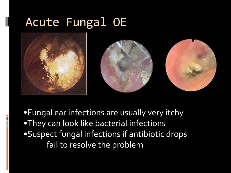
Acute Fungal OE
•Fungal ear infections are usually very itchy •They can look like bacterial infections •Suspect fungal infections if antibiotic drops fail to resolve the problem
Treatment of Fungal OE Remove Debris if possible
Topical treatment
Acetic acid ear drops
Antifungal drops (clotrimazole)
Powders CASH powder:
Chloramphenicol Amphotericin B Sulfamethoxazole Hydrocortisone
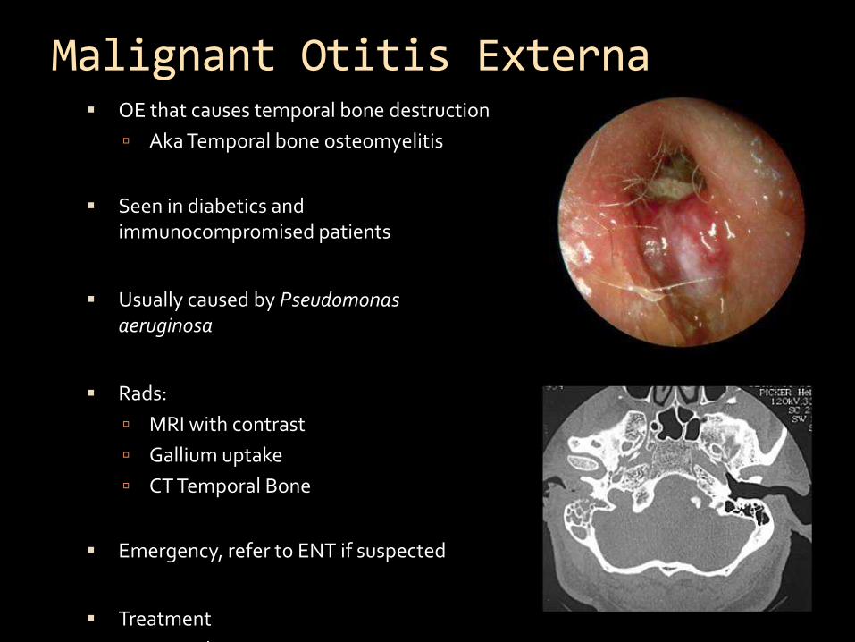
Malignant Otitis Externa OE that causes temporal bone destruction
Aka Temporal bone osteomyelitis
Seen in diabetics and immunocompromised patients
Usually caused by Pseudomonas aeruginosa
Rads:
MRI with contrast
Gallium uptake
CT Temporal Bone
Emergency, refer to ENT if suspected
Treatment
IV antibiotics
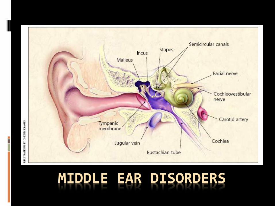
MIDDLE EAR DISORDERS

Eustachian Tube Dysfunction and tympanic membrane retraction Causes:
•Nasal Allergy
•URI
•Nasopharynx mass
•Anatomic
Signs:
•TM retraction
Symptoms:
•Hearing Loss
•Ear Fullness
•Popping/Crackling
•Improvement with Valsalva
Eustachian Tube Dysfunction
Treatment
If acute ETD, counsel patience and time
Nasal steroid spray
If ETD is chronic and hearing loss is present, bilateral myringotomy with tube placement
ETD and TM retraction- Risk of cholesteatoma

Otitis Media with Effusion Caused by:
•Chronic ETD
•Acute OM
•Barotrauma
•Nasopharynx Mass
Signs:
• Bubbles
•Amber coloration
•Air-Fluid Line
•Immobile TM to pneumatic
Symptoms:
•Conductive Hearing Loss
•Ear Fullness
Otitis Media with Effusion This is treated very similarly to TM
retraction.
Counseling is very important!
Hearing loss may be present for 3-4 months
Patience is key!
Nasal steroids
Myringotomy with tube placement if not better in 3-4 months.
Rule out Nasopharyngeal Mass/Tumor
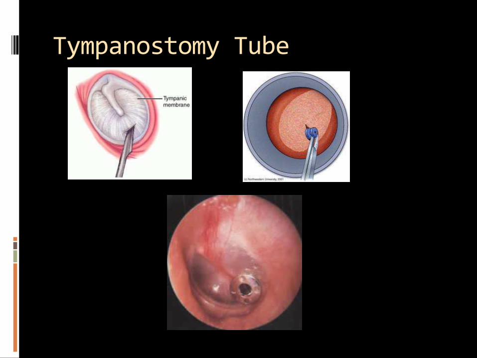
Tympanostomy Tube
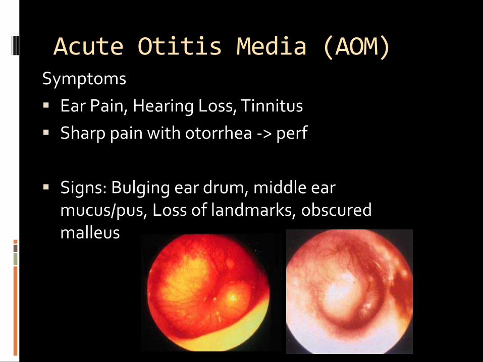
Acute Otitis Media (AOM) Symptoms
Ear Pain, Hearing Loss, Tinnitus
Sharp pain with otorrhea -> perf
Signs: Bulging ear drum, middle ear mucus/pus, Loss of landmarks, obscured malleus
Acute Otitis Media
Treatment should cover the most common pathogens:
Streptococcus pneumoniae
Moraxella catarhalis
Haemophilus influenza
Amoxicillin, Amox/clav, Fluoroquinolone, cephalosporins, SMX/TMP, doxycycline, and others.
Complications of AOM
Mastoiditis
Meningitis/Intracranial abscess
TM perforation
Hearing loss
Facial nerve paralysis
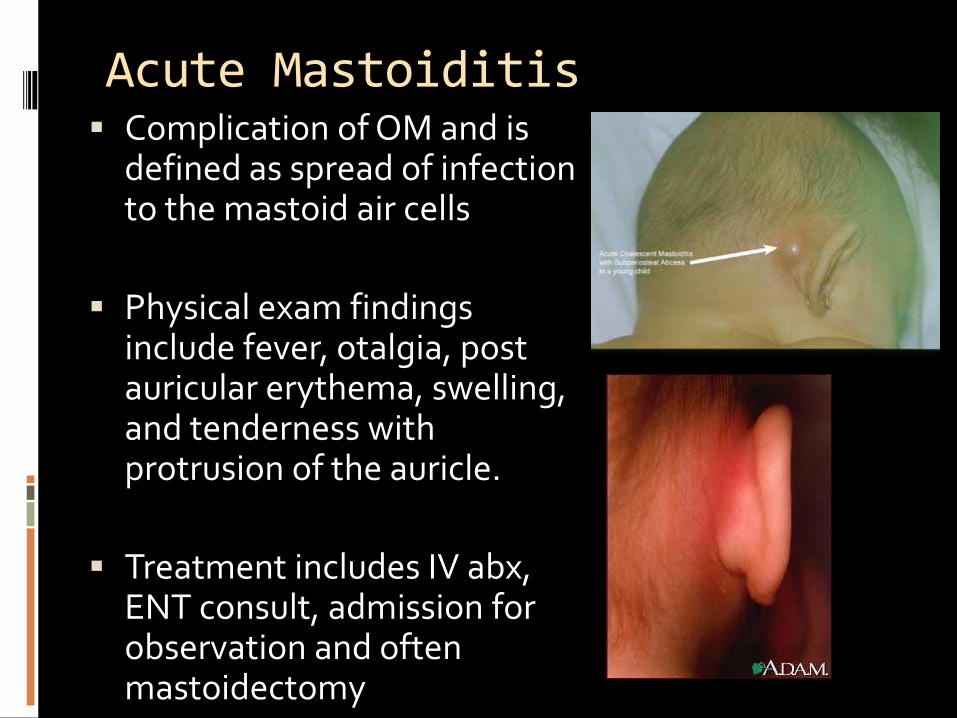
Acute Mastoiditis Complication of OM and is
defined as spread of infection to the mastoid air cells
Physical exam findings include fever, otalgia, post auricular erythema, swelling, and tenderness with protrusion of the auricle.
Treatment includes IV abx, ENT consult, admission for observation and often mastoidectomy
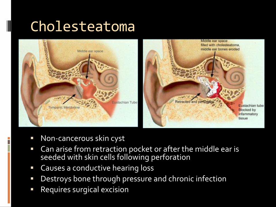
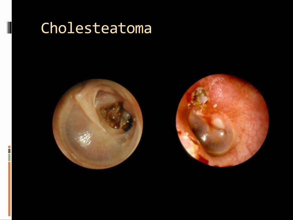
Cholesteatoma
Non-cancerous skin cyst
Can arise from retraction pocket or after the middle ear is seeded with skin cells following perforation
Causes a conductive hearing loss
Destroys bone through pressure and chronic infection
Requires surgical excision
Cholesteatoma

Tympanic Membrane Perforation
TM Perforation Symptoms
Hearing loss Tinnitus Otorrhea Ear pain if acute Bleeding
Treatment Watchful waiting Treat infections with topical drops (quinolones only) Tympanoplasty
Paper patch Temporal muscle fascia graft
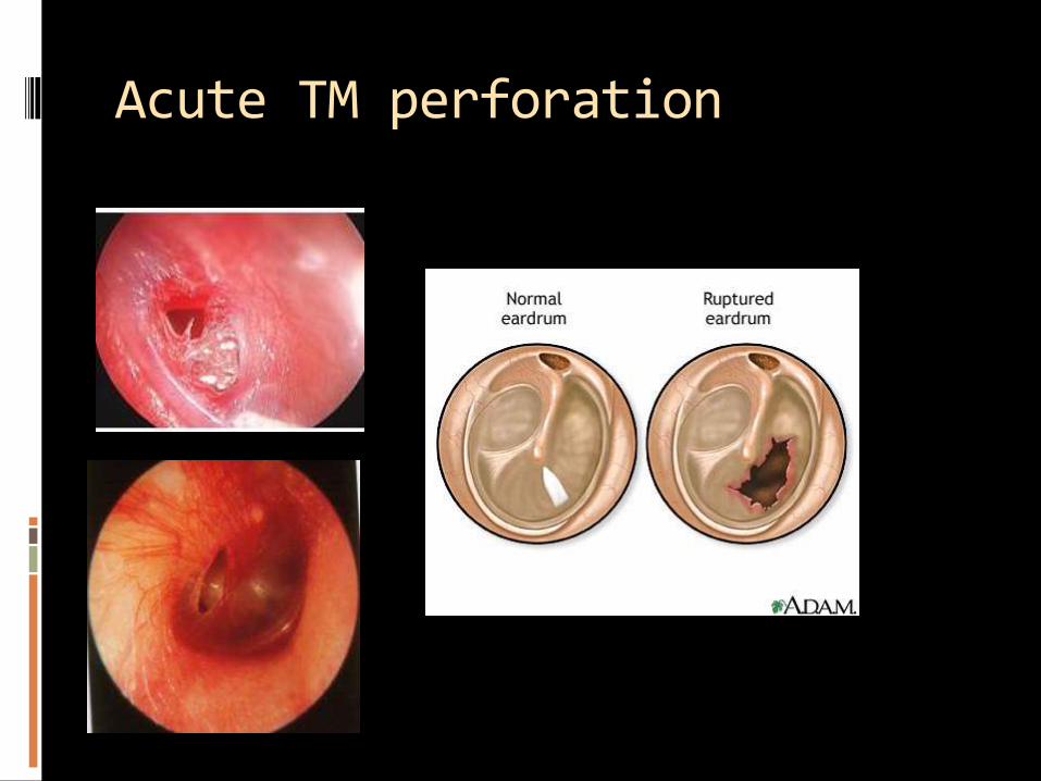
Acute TM perforation
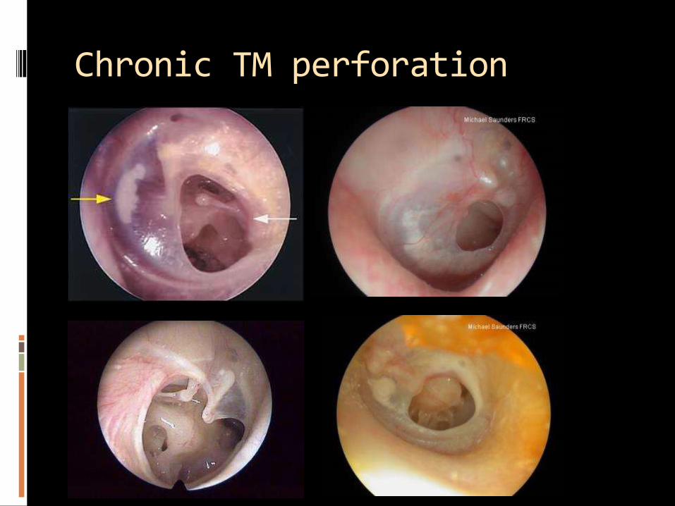
Chronic TM perforation
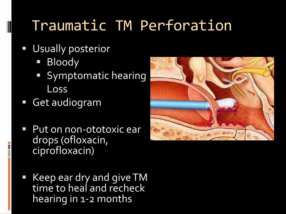
Traumatic TM Perforation Usually posterior Bloody Symptomatic hearing Loss
Get audiogram
Put on non-ototoxic ear drops (ofloxacin, ciprofloxacin)
Keep ear dry and give TM time to heal and recheck hearing in 1-2 months
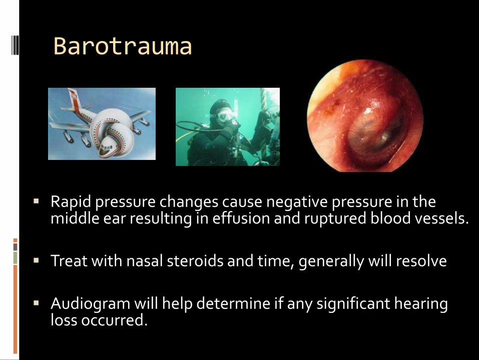
Barotrauma
Rapid pressure changes cause negative pressure in the middle ear resulting in effusion and ruptured blood vessels.
Treat with nasal steroids and time, generally will resolve
Audiogram will help determine if any significant hearing loss occurred.
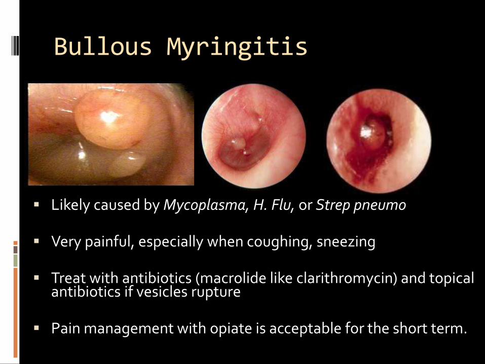
Bullous Myringitis
Likely caused by Mycoplasma, H. Flu, or Strep pneumo
Very painful, especially when coughing, sneezing
Treat with antibiotics (macrolide like clarithromycin) and topical antibiotics if vesicles rupture
Pain management with opiate is acceptable for the short term.

Otosclerosis
•Caused by fusion of the stapes footplate to the oval window
•Usually has family history
•Causes a conductive hearing loss
•Can be treated surgically with a stapedectomy
•Otherwise, can be treated with hearing aids
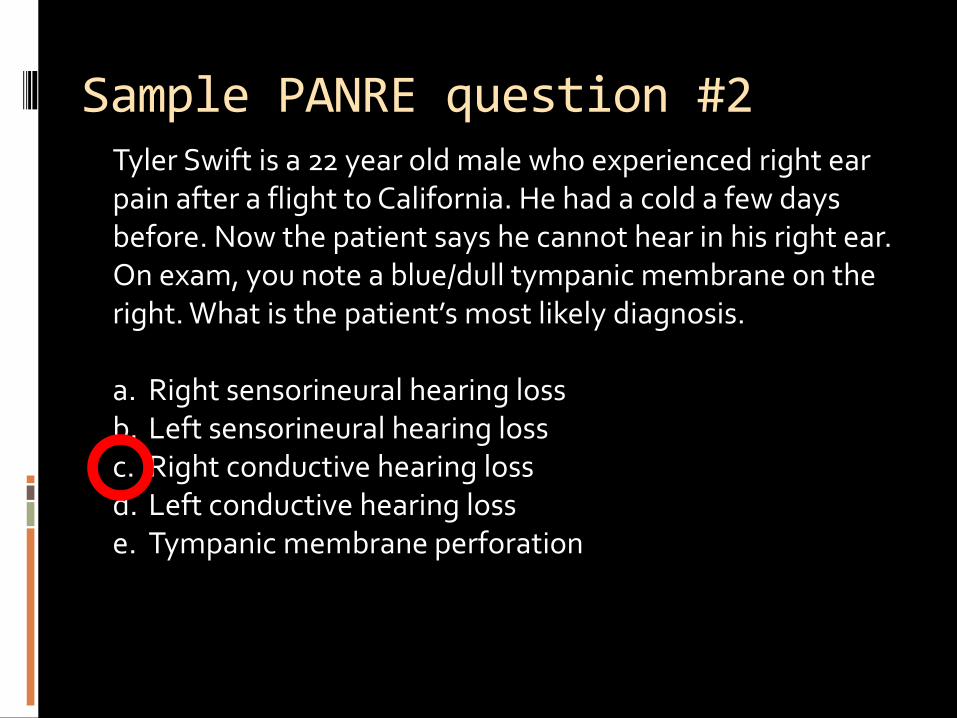
Sample PANRE question #2 Tyler Swift is a 22 year old male who experienced right ear pain after a flight to California. He had a cold a few days before. Now the patient says he cannot hear in his right ear. On exam, you note a blue/dull tympanic membrane on the right. What is the patient’s most likely diagnosis. a. Right sensorineural hearing loss b. Left sensorineural hearing loss c. Right conductive hearing loss d. Left conductive hearing loss e. Tympanic membrane perforation
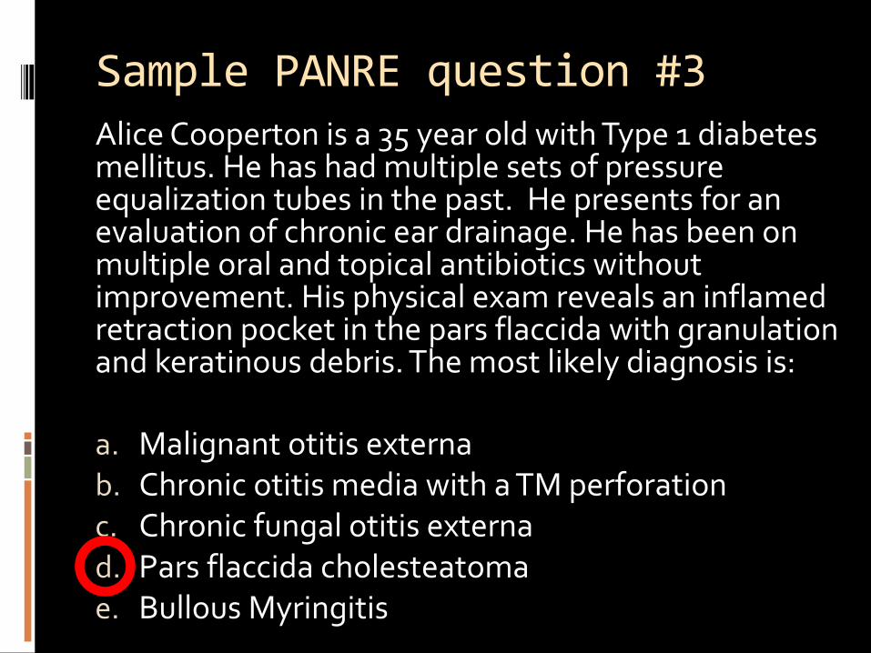
Sample PANRE question #3 Alice Cooperton is a 35 year old with Type 1 diabetes mellitus. He has had multiple sets of pressure equalization tubes in the past. He presents for an evaluation of chronic ear drainage. He has been on multiple oral and topical antibiotics without improvement. His physical exam reveals an inflamed retraction pocket in the pars flaccida with granulation and keratinous debris. The most likely diagnosis is:
a. Malignant otitis externa b. Chronic otitis media with a TM perforation c. Chronic fungal otitis externa d. Pars flaccida cholesteatoma e. Bullous Myringitis
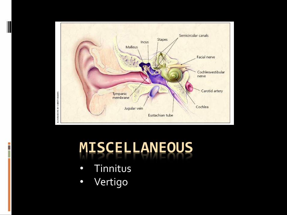
MISCELLANEOUS • Tinnitus • Vertigo
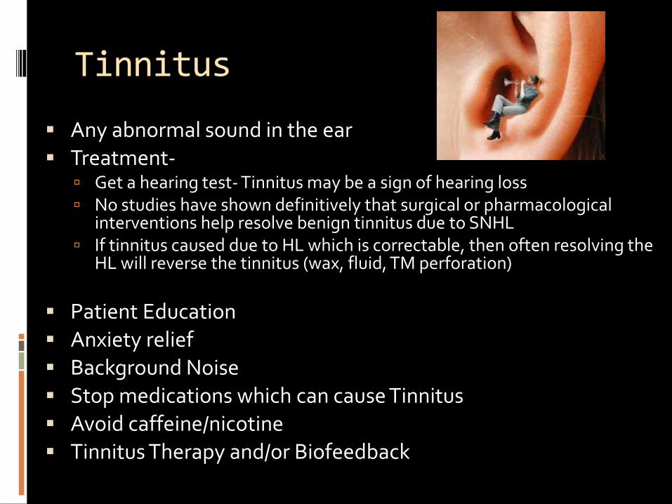
Tinnitus
Any abnormal sound in the ear
Treatment- Get a hearing test- Tinnitus may be a sign of hearing loss No studies have shown definitively that surgical or pharmacological
interventions help resolve benign tinnitus due to SNHL If tinnitus caused due to HL which is correctable, then often resolving the
HL will reverse the tinnitus (wax, fluid, TM perforation)
Patient Education
Anxiety relief Background Noise
Stop medications which can cause Tinnitus Avoid caffeine/nicotine
Tinnitus Therapy and/or Biofeedback
