otorhinolaryngology - · pdf fileotorhinolaryngology ... such as external otitis or an acute...
TRANSCRIPT
SEABEE OPERATIONAL MEDICAL & DENTAL GUIDE
OTORHINOLARYNGOLOGY
Return to Clinical Section Welcome Page
C O N T E N T S
INTRODUCTIONEXTERNAL EAREXTERNAL AUDITORY CANALOTIC PREPARATIONSMIDDLE EARCAUSES OF EAR PAIN (Table)INNER EARDISEASES OR CLINICAL SYNDROMES OF OTOLOGICAL ORIGINTHE DIZZY PATIENTRHINOLOGYMOUTH AND PHARYNXAUDIOLOGYBASIC HEARING TESTSOTORHINOLARYNGOLOGIC DRUGS OF CHOICE (Table)INTRODUCTION
Otorhinolaryngology (Ear, Nose and Throat or ENT) faces the same problems in the military thatare found in civilian medical practice, but the problems are compounded by (1) the exceptionalenvironmental conditions, and (2) the fact that some of the symptoms experienced may degradeperformance to the point that the safety of the Seabee is unable to function. The exceptionalenvironmental conditions include heat, cold, moisture, dryness, high ambient noise levels, andpoor hygiene conditions. These ENT conditions can elicit episodes of pain, vertigo,disequilibrium, and nausea. They may also introduce communication problems through temporaryor permanent impairment of auditory function. In addition, these conditions may be of suddenonset in apparently normal individuals, such as external otitis or an acute tonsillitis.
These lectures describe clinical ENT issues and audiology. Seabee physicians may findthemselves at long distances from large medical facilities when ENT problems arise, or they mayhave to care for patients until they can get an appointment at the nearest facility; therefore, thissection is intended to assist the physician with common clinical problems.
OTORHINOLARYNGOLOGY
SEABEE OPERATIONAL MEDICAL & DENTAL GUIDE
EXTERNAL EAR
Anatomy of the pinna• Helix: Crus, Ant. Incisure• Antihelix: Crura, Triangular Fossa• Concha: Cymba, Cavum• Scaphoid Fossa• Tragus• Antitragus• Intertragic Incisure• Post-Auricular Sulcus
Trauma• Lacerations§ Atraumatic cleaning with copious irrigation§ Suture with fine (6-O) monofilament nylonØ Primarily interrupted skin suturesØ Occasional through and through sutures
§ Antibiotics§ Pressure dressing
• Hematoma§ Utilize aseptic skin technique§ Needle aspiration or Incision and Drainage§ Pressure dressing
• Perichondritis§ Etiology: External Otitis or Trauma§ Antibiotics: coverage for Staph. and Pseudomonas§ Debridement of dead tissue
• Keloids§ Intralesional Cortisone
• Burns§ Gentle cleansing§ Sulfamylon cream/dressing
• COMEDONES or BLACKHEADS§ Nothing§ Skin prep with direct small ring extraction
Infections• Erysipelas§ Usually a Staphylococcal infection§ Staph. resistant PCN: Oxacillin, Augmentin
• Herpes Zoster OticusEXTERNAL AUDITORY CANAL
OTORHINOLARYNGOLOGY
SEABEE OPERATIONAL MEDICAL & DENTAL GUIDE
Anatomy• 25 mm to posterior/superior TM• 31 mm to anterior/inferior TM• Outer 1/3:§ Thicker skin§ Pilosebaceous glands§ Cerumen glands§ Incisure of Santorini
• Inner 2/3:§ thinner skin§ vertical and horizontal wrinkles§ no hair and minimal glands
Cerumen impaction• Water, alcohol, or acetic acid irrigations
Foreign bodies• Inert, hydroscopic, round or animal
Epithelial growths• Inclusions, benign or malignant growths
Infections• EXTERNA§ Acute§ Recurrent§ Chronic§ Eczematoid§ Seborrheic
• FURUNCLE§ Outer 1/3§ Usually Staphylococcal§ Often spreads to periauricular cellulitis§ scant pus§ hot compresses, oral antibiotics, I&D necrotic area
• INFECTIVE OTITIS - (Swimmers Ear)§ Debridement§ Intra-auricular wick§ Antibiotic drops
• OTOMYCOSIS§ Debridement§ Antifungal medications, (Domeboro's, Lotrimin)§ Keep ear dry
OTORHINOLARYNGOLOGY
SEABEE OPERATIONAL MEDICAL & DENTAL GUIDE
§ Acid-Alcohol drops• ECZEMATOID EXTERNA§ Allergic reaction§ Look for contact substances or ID reaction infection elsewhere§ Gentle cleaning§ Hypoallergenic medication§ Topical Cortisone preparations
• SEBORRHEIC EXTERNA§ Look for associated skin or scalp infections§ Treatment with anti-seborrheics, cortisone preparations, and skin emollients
OTIC PREPARATIONSThe most common organisms found in external otitis are Staphylococcus aureus, Pseudomonasaeruginosa, Proteus, Bacteroides fragilis, E. Coli, Klebsiella, and Enterobacter. Thoroughcleaning of the ear canal is the most important first step in the treatment of any type of externalotitis.
The next step is to choose a topical otic agent. For prevention as well as treatment, most oticpreparations contain some type of acid, such as a boric or acetic acid since most organismsflourish best when the canal is alkaline. Most of your "swimmer's ear" prophylactics are a mixtureof white vinegar and rubbing alcohol. Most otic preparations also contain antibiotics. Neomycinis active against Staphylococcus and Proteus, but the most common Pseudomonas strains areresistant. Polymyxin B and polymyxin E (Colimycin) are bactericidal to most gram negativeorganisms, notably Pseudomonas, but not proteus, Bacteroides fragilis (found in 13% of infectedcholesteatoma), gram positive organisms. Chloromycetin otic is a bacteriostatic preparationagainst all common pathogens; as it comes in an acid carrier, chloromycetin is often extremelypainful in the ear canal and or middle ear, so certain patients are given the powderedChloromycetin. "Chlor" is used when there is a neomycin-polymyxin failure in a chronic diseaseor when a reliable anaerobic culture suggests Bacteroides fragilis. Garamycin is available in anophthalmic solution to instill in ears; its spectrum includes Pseudomonas and most otherpathogens of chronic suppurative otitis except Bacteroides fragilis.
At this time there is no otic preparation containing specific fungicides, however topicalAmphotericin B (Fungizone) and Nystatin (Mycostatin) both being effective when used intreatment of Candidiasis (Monilia), can be adapted. Non-specific antiseptics effective againstAspergillus and Candida are 2% acetic acid and aluminum acetate solution (Otic Domeboro), 3%boric acid in 70% alcohol (Swimmer's Otic), or 25% m-cresyl acetate (Cresatin or Cresylate). Gentian Violet 2% in 95% alcohol, topical mercurials, or a boric acid and iodine mixture havebeen used in chronic mastoid cavity infection after the debris and secretions are removed, but theyare messy. Athlete's foot solutions and ointments can be used if there's no TM perforation.
Anti-inflammatory agents, most notably hydrocortisone, ar used to decrease mucosal andcutaneous edema in bacterial infections or for the treatment of psoriasis and seborrheic dermatitis.
OTORHINOLARYNGOLOGY
SEABEE OPERATIONAL MEDICAL & DENTAL GUIDE
For dermatitis, the steroid should be used alone or in a low sensitizing preparation like Orlex HCor VoSol HC.
Anesthetic agents such as Auralgan or Lidosporin, which contain benzocaine or lidocaine, mayprovide some relief of pain in otitis media and bullous myringitis and some relief from itching indermatitis. A few people become allergic to these medications.
There are ceruminolytic agents in use, the most popular agent being carbamide peroxide inglycerol (Debrox). Using this agent or simple 3% peroxide solution may take several days tosoften hard wax. Ceruminex, an agent that softens wax rapidly, has been cited as occasionallycausing a severe eczematoid reaction and should be limited to 15 to 30 minutes of skin contactand then thoroughly removed.
Remember also, the disadvantages of otic preparations: All drops produce debris requiringfurther cleaning after use. Most preparations are painful, due to the acid and alcohol base, whenused in the middle ear. The water based suspensions or ophthalmic preparations cause less painand reaction with the mucosa and are the best preparations applied in treatment of a perforatedtympanic membrane.
Use your otic preparations wisely. Clean the ear first, use only what is necessary, discontinue assoon as possible and remove remaining debris.
MIDDLE EAR
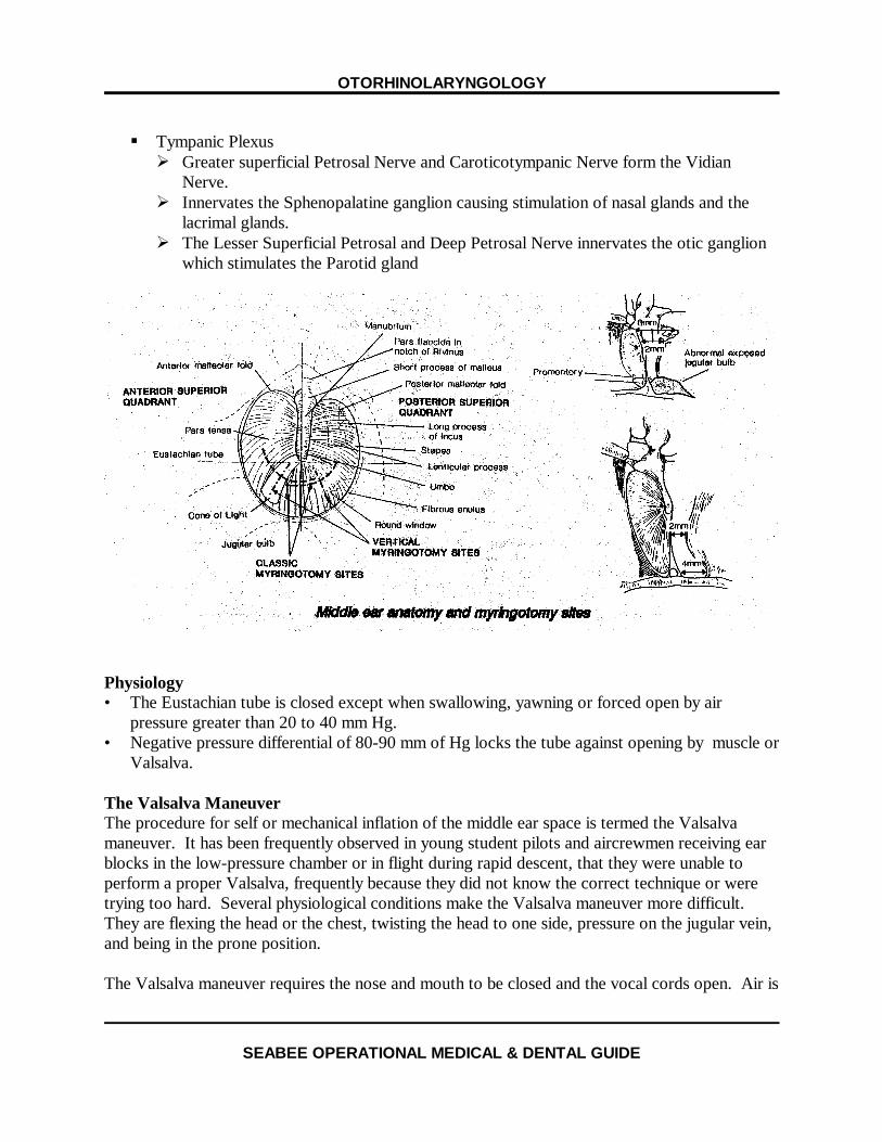
Anatomy• Tympanic Membrane-Landmarks§ Pars Flaccida - Shrapnell's membrane over the notch of Rivinus§ Pars TensaØ Manubrium and UmboØ Short processØ Light reflex
§Behind the membraneØ Chorda Tympani nerveØ IncusØ Stapedial TendonØ Round window nicheØ Promontory
§ Special featuresØ Migratory surface epithelium
• Tympanic cavity§ Epitympanum, Mesotympanum, and Hypotympanum§ Mastoid antrum and air cells are connected to the Tympanic Cavity through the Addus an
Antrum
OTORHINOLARYNGOLOGY
SEABEE OPERATIONAL MEDICAL & DENTAL GUIDE
§ Tympanic PlexusØ Greater superficial Petrosal Nerve and Caroticotympanic Nerve form the Vidian
Nerve.Ø Innervates the Sphenopalatine ganglion causing stimulation of nasal glands and the
lacrimal glands.Ø The Lesser Superficial Petrosal and Deep Petrosal Nerve innervates the otic ganglion
which stimulates the Parotid gland
Physiology• The Eustachian tube is closed except when swallowing, yawning or forced open by air
pressure greater than 20 to 40 mm Hg.• Negative pressure differential of 80-90 mm of Hg locks the tube against opening by muscle or
Valsalva.
The Valsalva ManeuverThe procedure for self or mechanical inflation of the middle ear space is termed the Valsalvamaneuver. It has been frequently observed in young student pilots and aircrewmen receiving earblocks in the low-pressure chamber or in flight during rapid descent, that they were unable toperform a proper Valsalva, frequently because they did not know the correct technique or weretrying too hard. Several physiological conditions make the Valsalva maneuver more difficult. They are flexing the head or the chest, twisting the head to one side, pressure on the jugular vein,and being in the prone position.
The Valsalva maneuver requires the nose and mouth to be closed and the vocal cords open. Air is
OTORHINOLARYNGOLOGY
SEABEE OPERATIONAL MEDICAL & DENTAL GUIDE
then forced into the nose and nasopharynx forcing open the Eustachian tube and increasing thepressure in the middle ear space. This can be observed as a bulging of the tympanic membrane,especially in the posterior superior quadrant.
The most frequently observed problems with the students were fear that they would damage orrupture their eardrums, closing the vocal cords when they build up pressure like in the M-1maneuver, and straining so hard that marked venous congestion in the head further preventsopening of the Eustachian tube.
Although it is possible to rupture the tympanic membrane when it is abnormally weak fromprevious disease, simple inflation done properly has little danger. Repeated overinflation doescarry some risk and is discussed under politzerization and round window rupture.
One of the best methods to prevent vocal cord closure is to instruct the patient or aircrewman toclose his nose with his fingers and then attempt to blow his fingers off his nose, causing the noseto bulge from the pressure. The buildup of pressure should be rapid and sustained no longer thanone to one and a half seconds to prevent the venous congestion that reduces the efficiency of theEustachian tube function.
Should the examiner fail to see any movement of the tympanic membrane when he is evaluatingthe patient for Valsalva, he should then look for the small, quick retraction movement of theToynbee maneuver, accomplished by closing the nose and swallowing. If a Toynbee is presentand the aircrewman feels pressure in his ears during Valsalva, has no sign of ear disease, and nohistory of problems with pressure changes, he usually can be qualified for aviation. The bestevaluation for candidates is, of course, the low-pressure chamber or an actual unpressurized flightwith rapid descent. Difficulty with pressure equalization during SCUBA diving is often a poorprognosis for aviation.
Acute otitis media• Etiology: eustachian tube dysfunction• Causes§ Anatomic§ UTI§ Allergic rhinitis§ Adenoiditis or nasopharyngitis§ Sinusitis
• Symptoms§ Fullness§ Pain§ Decreased hearing§ Rarely dizziness or TM perforation
• Treatment§ Antibiotics
OTORHINOLARYNGOLOGY
SEABEE OPERATIONAL MEDICAL & DENTAL GUIDE
Ø Amoxicillin x 7-10 daysØ Emycin/SulfaØ AugmentinØ TMP-SMXØ CeclorØ CeftinØ Suprax
§ DecongestantsØ SudafedØ Entex
§ Pain medication§ Hydration§ Treat any other associated conditions
Chronic or recurrent otitis media with effusion• Consider pressure equalization (PE) tube
Bullous myringitis• May be form of otitis media• Often seen after viral URI or mycoplasma pneumonia• Signs & symptoms§ Sudden onset of deep ear pain§ May have serosanguinous drainage§ Single of multiple bullae form on TM and adjacent bony wall
• Treatment§ Same as acute otitis media§ Rupture bulae with suction tip but do not do myringotomy§ Auralgan ear drops occasionally helpful - warm
Otitis medial complications• Mastoiditis§ Usually 1-2 weeks after acute illness§ Pain deep or behind ear§ Pus formation beneath temporalis muscle§ TreatmentØ IV Cefuroxime plus metronidazoleØ Chloromycetin
• Chronic perforation§ Frequent suction cleaning of drainage§ Avoid water in ear§ Cortisporin otic drops only if draining
OTORHINOLARYNGOLOGY
SEABEE OPERATIONAL MEDICAL & DENTAL GUIDE
Cholesteatoma• Squamous epithelium growing in the middle ear space• Usually from a pars flaccida perforation or retraction pocket or similar pathology in the
posterior superior quadrant of the pars tensa or a marginal perforation• 90% require surgical intervention and one year minimum follow-up• Should be on limited duty• Could lead to serious complications, a surgical emergency, or even death if left untreated or
improperly followed-up.
Traumatic perforation of the TM• Keep water out of ear• Suction cleaning only• No ear drops unless ear is infected and draining• Get baseline audiogram• Marginal stimulation with trichloroacetic acid (TCAA) 25%
Perforations usually heal within three weeks, but the patient must avoid any significant barometricpressure changes as the perforation nears closure, and at no time should water or other fluids beallowed in the ear. Keep The Ear Dry! Never use ear drops. unless a true infection with purulentdrainage develops and then use only the suspension preparations.
Chronic Perforation of the TMSmall, dry, central perforations may be closed by cauterizing the edge of the perforation withtrichloroacetic acid. It can be left open or one may elect to place a small patch made fromcigarette paper or other thin paper over the perforation. Usually the patch is moistened inantibiotic drops before application.
Large perforations with a dry middle ear may be closed by a tissue graft if the Eustachian tube isfunctioning. Testing of this function is fairly accurate by tympanography. Poor or absentEustachian tube function gives surgery a decreased chance for success. If the ossicles showfixation or if there is considerable scarring with adhesions, hearing might decrease somewhatfurther even though the perforation is closed, as a result of the poorer transmission of sound andthe cancellation effect of sound striking both windows at the same time. A perforation, per se,which allows for equalization of pressure between the middle ear and the atmosphere does notaffect flying. Sudden cold or hot air or water and loud noise may cause vertigo more easily in theperforated ear. Of course, water in a perforated ear usually leads to infection and drainage.
BarotraumaAerotitis media occurs rather frequently in the aviation community and is directly related to thefunction of the Eustachian tube in equalizing the pressure between the atmosphere and the middleear space. The tympanic end of the Eustachian tube is bony and usually open, whereas thepharyngeal end is cartilaginous, slit-like, and closed, acting like a one-way flutter valve. Openingof the Eustachian tube occurs with the contraction of the levator and tensor veli palatini muscles
OTORHINOLARYNGOLOGY
SEABEE OPERATIONAL MEDICAL & DENTAL GUIDE
during acts of chewing, swallowing, or yawning. As one ascends to altitude, the outside pressuredecreases, and the greater middle ear pressure forces open the "flutter valve," pharyngeal end ofthe Eustachian tube every 400 to 500 feet to about 35,000 feet, and then every 100 feetthereafter. During descent, the collapsed, closed, pharyngeal end of the Eustachian tube preventsair from entering the tube. The increasing relative negative pressure in the middle ear furtherholds the soft tissues together, and muscular (active) opening of the Eustachian tube must beaccomplished before the differential pressure reaches 80 or 90 mm Hg. Once this magnitude ofdifferential pressure is established, muscular action cannot overcome the suction effect on theclosed Eustachian tube, and the tube is said to be "locked." This relative negative pressure notonly retracts the tympanic membrane but pulls on the delicate mucosal lining, leading to effusionand hemorrhage. Pain may be severe, with nausea and occasionally vertigo. On rare occasionsrupture of the tympanic membrane has been seen, and some aircrewmen have developed shock orsyncope.
Otoscopic presentations vary greatly, but they can range from a retraction of the tympanicmembrane with the classic backward displacement of the malleus, a prominent short process, andanterior and posterior folds, to hyperemia or hemorrhages in the tympanic membrane. There mayalso be varying amounts of serous and bloody fluid visible behind the membrane.
Active treatment is directed toward equalization of pressure, relief of pain, and prevention ortreatment of infections in the ear, Eustachian tube, or nasopharynx. In an aircraft or low-pressurechamber, descent should be stopped, and, if possible, there should be a return to a higher altitudewhere equalization can be attempted using the Valsalva maneuver or Politzer method. Descentshould then be gradual, if possible. Middle ear inflation (politzerization) should be done especiallyif a negative pressure appears to remain on the ground and there is pain present. Caution shouldbe exercised if there is an upper respiratory infection present. Oral decongestants may be helpfuland are recommended, but the effect of antihistamines is questionable. In cases of thick effusionand poor Eustachian tube function or inability to Valsalva, daily or every other day politzerizationor tubal insufflation may be in order. Persistent serous fluid may be removed by needle aspiration,but thick mucoid or organized blood must be removed by myringotomy if it has not cleared aftertwo or three weeks of intensive therapy. Antibiotics are used only when infection is present in theupper respiratory region or develops during treatment.
OTORHINOLARYNGOLOGY
SEABEE OPERATIONAL MEDICAL & DENTAL GUIDE
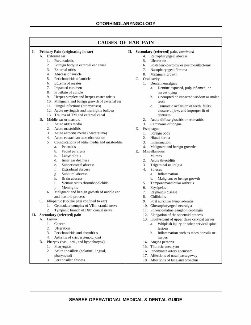
CAUSES OF EAR PAIN
I. Primary Pain (originating in ear)A. External ear
1. Furunculosis2. Foreign body in external ear canal3. External otitis4. Abscess of auricle5. Perichondritis of auricle6. Eczema of meatus7. Impacted cerumen8. Frostbite of auricle9. Herpes simplex and herpes zoster oticus10. Malignant and benign growth of external ear11. Fungal infections (otomycoses)12. Acute myringitis and myringitis bullosa13. Trauma of TM and external canal
B. Middle ear or mastoid1. Acute otitis media2. Acute mastoiditis3. Acute aerotitis media (barotrauma)4. Acute eustachian tube obstruction5. Complications of otitis media and mastoiditis
a. Petrositisb. Facial paralysisc. Labyrinthitisd. Inner ear deafnesse. Subperiosteal abscessf. Extradural abscessg. Subdural abscessh. Brain abscessi. Venous sinus thrombophlebitisj. Meningitis
6. Malignant and benign growth of middle earand mastoid process
C. Idiopathic (tic-like pain confined to ear)1. Geniculate complex of VIIth cranial nerve2. Tympanic branch of IXth cranial nerve
II. Secondary (referred) painA. Larynx
1. Cancer2. Ulceration3. Perichondritis and chondritis4. Arthritis of cricoarytenoid joint
B. Pharynx (nas-, oro-, and hypopharynx)1. Pharyngitis2. Acute tonsillitis (palatine, lingual,
pharyngeal)3. Peritonsillar abscess
II. Secondary (referred) pain, continued4. Retropharyngeal abscess5. Ulceration6. Postadenoidectomy or posttonsillectomy7. Nasopharyngeal fibroma8. Malignant growth
C. Oral cavity1. Dental neuralgias
a. Dentine exposed, pulp inflamed, ornerves dying
b. Unerupted or impacted wisdom or molarteeth
c. Traumatic occlusion of teeth, faultyclosure of jaw, and improper fit ofdentures
2. Acute diffuse glossitis or stomatitis3. Carcinoma of tongue
D. Esophagus1. Foreign body2. Hiatal hernia3. Inflammation4. Malignant and benign growths
E. Miscellaneous1. Mumps2. Acute thyroiditis3. Trigeminal neuralgia4. Sinuses
a. Inflammationb. Malignant or benign growth
5. Temporomandibular arthritis6. Erysipelas7. Raynaud's disease8. Chilblains9. Post auricular lymphadenitis10. Glossopharyngeal neuralgia11. Sphenopalatine ganglion cephalgia12. Elongation of the sphenoid process13. Involvement of upper three cervical nerves
a. Whiplash injury or other cervical spinelesions
b. Inflammation such as tabes dorsalis orherpes
14. Angina pectoris15. Thoracic aneurysm16. Innominate artery aneurysm17. Affections of nasal passageway18. Affections of lung and bronchus
OTORHINOLARYNGOLOGY
SEABEE OPERATIONAL MEDICAL & DENTAL GUIDE
PolitzerizationPolitzerization is the mechanical inflation of the middle ear usually required for treatment of acuteear and sinus blocks, chronic Eustachian tube dysfunction, or middle ear disease. To perform thisprocedure, one needs a source of pressure, either an air pump or rubber bag, with a one-wayvalve. For the air pump, it is most important to have variable control of the pressure and apressure gauge, if possible. Most pressure/vacuum units in the Navy have a pressure gaugecalibrated in pounds per square inch. If no gauge is present, the starting pressure should just besufficient to blow off a lightly applied finger. When a pressure gauge is available, initial attemptsshould be done with ten pounds per square inch or less. To seal and deliver the pressure into thenose, an olive tip of metal, hard rubber, or glass is the most efficient. This tip may be attached toan atomizer if smoke or mist is desired for diagnostic or therapeutic reasons. If the patient has avery thin tympanic membrane, lower pressure must be tried first. An explanation to the patient isimportant to assure cooperation and prevent sudden movements that could injure the nose.
The first attempt at politzerization should be done by inserting the olive tip into a nostril, getting agood seal but not striking the vestibule or septal walls. The opposite naris is occluded, and thepatient is instructed to repeat K-K-K-K-K, loudly and sharply, as a one second burst of air isdelivered. A characteristic soft palate flutter sound is heard if the procedure is performedcorrectly.
If no results are obtained with this technique, the patient is instructed to swallow, and as thethyroid notch raises up, air pressure is again applied in the nose. For people who have troublewith a dry swallow, a sip of water may be given. In the low-pressure chamber, this method ismost often used to get maximum opening of the Eustachian tube. It must be remembered thatwith the water technique, prolonged or high pressure might cause damage to the tympanicmembrane with even a remote possibility of damage to the round window membrane and innerear. As it is important to look at the patient's tympanic membranes before inflation, it is equally asimportant to observe them afterwards to determine the extent or success of the procedure.
INNER EAR
AnatomySituated medial to the middle ear entirely within the petrous portion of the temporal bone lies theinner ear. It is composed of dense, compact bone two to three millimeters thick, forming theosseous labyrinth. This is divided into semicircular canals, vestibule, and cochlea. Within thebony labyrinth is a membranous counterpart. The supporting fluid outside of the membranouslabyrinth is perilymph. It is somewhat similar to cerebrospinal fluid and is high in sodium content. The fluid inside the membranous labyrinth, endolymph, has a high potassium content.
OTORHINOLARYNGOLOGY
SEABEE OPERATIONAL MEDICAL & DENTAL GUIDE
The cochlea is a two and a half-turn coil about a central core called the modiolus, with the apexpointing anteriorly and laterally. There are three compartments. The first two, the scala vestibuliassociated with the oval window and the scala tympani associated with the round window, containperilymph and are joined at the apex of the cochlea through the helicotrema. The third or centralcompartment is the scala media or cochlear duct, containing endolymph. It contains the neuralend organ of hearing, the organ of Corti, which rests on the thick basilar membrane that separatesthis compartment from the scala tympani. The delicate Reissner's membrane separates the scalamedia from the scala vestibuli. The organ of Corti contains about 24,000 hair cells arrangedthroughout the cochlea as a single row of inner cells and from three to five rows of outer cells. Between them, they form a somewhat triangular tunnel of Corti that has its own slightly differentfluid, cortilymph. It is known that high frequency sounds stimulate the hair cells near thevestibule, and low frequency sounds stimulate those near the apex. The area of the promontoryor basilar turn of the cochlea is stimulated by frequencies in the range of 3000 to 5000 Hz; itappears to be the most vulnerable to acoustic trauma, probably from the shearing force in the fluidso near the stapes footplate and the beginning curve in the scala.
TraumaTemporal bone fractures are of two types for the most part of two types. The longitudinal ormiddle fossa fracture that parallels the long axis of the petrous pyramid is usually due to forcesapplied to the temporoparietal region. The middle ear is always damaged. The tympanicmembrane is torn and bleeds. The labyrinthine capsule is usually spared, as is the facial nerve. Longitudinal temporal bone fractures are four times more frequent than the transverse variety. The transverse or posterior fossa fractures usually result from forces applied to the occipital oroccipitomastoid region. There is essentially a fracture of the labyrinth that spares the middle ear. There may be hemotympanum, but rarely rupture of the tympanic membrane. Usually, there isboth cochlear and vestibular function loss, and the facial nerve is damaged in the internal auditorymeatus or horizontal portion. Only sterile ear instruments should be used for examination, anddry ear precautions must be taken.
Initial treatment should include cranial checks, prophylactic antibiotics, and a completeneurological evaluation. The patient should be moved to the care of a neurosurgeon/otologist assoon as condition permits. A baseline audiogram is valuable if the patient's condition permits.
OTORHINOLARYNGOLOGY
SEABEE OPERATIONAL MEDICAL & DENTAL GUIDE
Barotrauma In the past few years, an increasing number of cases of barotrauma to the inner ear have beenreported from the diving community, and several cases of proven rupture of the round windowmembrane have been reported or evaluated at the Naval Aerospace Medical Institute (NAMI). These have been associated with overly aggressive use of the Valsalva maneuver to clear what thepatient thought was an ear block. In reality, the problem was an over-inflated middle ear anddistended tympanic membrane, which gives a similar blocked feeling, but usually has no pain. When the round window membrane ruptures, there may be variable degrees of tinnitus andpersistent or positional vertigo, often with nausea and vomiting. Calorics are usually diminishedon the involved side, and a sensorineural hearing loss, often across the board, is present with poordiscrimination of words.
The key to successful treatment is early suspicion and diagnosis and immediate repair by theotologist. Most complete recoveries have had repairs within 48 hours. A quick, simple tuningfork test will separate nerve loss from a conductive loss.
NystagmusThe search for the presence or absence of spontaneous or positional nystagmus is an integral partof the otoneurological examination and the fitness for duty examination.
Nystagmus is called right or left according to the direction of the rapid eye movement or quickcomponent. When nystagmus is provoked only in the direction of the quick component, it istermed "first degree." When nystagmus is also noted in forward gaze, it is "second degree," andwith nystagmus present in all directions of gaze, it is "third degree." Nystagmus is furthercategorized as vertical, oblique (rare), horizontal, or rotatory. Proper evaluation calls forobservation of the eyes in the right, left, upward, downward, and primary positions. If the patientis asked to look too far on lateral gaze, a few flicks of nystagmus are frequently seen and are anormal phenomenon of accommodation. After the test for spontaneous nystagmus, tests forpositional nystagmus are carried out with the patient's eyes in the straight ahead position. Themethod most often used is that of Cawthorne, Dix, and Hallpike. The patient is rapidly placedsupine with the head hanging over the edge of the table, and the eyes are observed for 60 seconds. The patient is then raised up and then returned to the hyperextended position with the head inone direction, again for 60 seconds. The procedure is repeated in the opposite direction. Nystagmus, if present should be immediately recorded as to type, direction, amplitude, andintensity. The position should be held until the nystagmus subsides; however, if it persists longerthan 60 seconds, it is considered permanent. In older persons where vertebral artery occlusionmay be the cause of the nystagmus and vertigo, one must use caution and good judgement toassure that the patient is not left in this position too long.
OTORHINOLARYNGOLOGY
SEABEE OPERATIONAL MEDICAL & DENTAL GUIDE
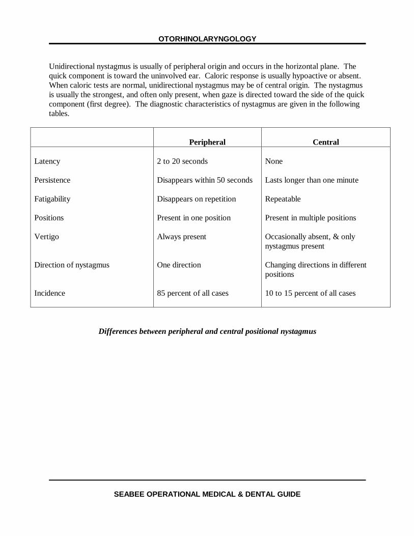
Unidirectional nystagmus is usually of peripheral origin and occurs in the horizontal plane. Thequick component is toward the uninvolved ear. Caloric response is usually hypoactive or absent. When caloric tests are normal, unidirectional nystagmus may be of central origin. The nystagmusis usually the strongest, and often only present, when gaze is directed toward the side of the quickcomponent (first degree). The diagnostic characteristics of nystagmus are given in the followingtables.
Peripheral Central
Latency
Persistence
Fatigability
Positions
Vertigo
Direction of nystagmus
Incidence
2 to 20 seconds
Disappears within 50 seconds
Disappears on repetition
Present in one position
Always present
One direction
85 percent of all cases
None
Lasts longer than one minute
Repeatable
Present in multiple positions
Occasionally absent, & onlynystagmus present
Changing directions in differentpositions
10 to 15 percent of all cases
Differences between peripheral and central positional nystagmus
OTORHINOLARYNGOLOGY
SEABEE OPERATIONAL MEDICAL & DENTAL GUIDE
Peripheral(Labyrinth, Vestibular
Nerve
Central (CNS)
Form
Frequency
Intensity
Direction of fast component
Duration
Dissociation between eyes
Vertigo
Cochlear signs
Autonomic nervous system signs
Past pointing and falling
Horizontal-rotatory
1/2-6 seconds
Decreasing intensity
Towards "stimulated" labyrinthor away from "destroyed"labyrinth
Minutes to weeks
None
Present
Frequently present
Definite
Direction of slow phase
Horizontal; vertical; diagonal;rotatory; multiple; retractorius;convergence; pendular; alternating
Any frequency, usually low orvariable at long intervals (weeks tomonths)
Constant
Towards side of CNS lesion
Weeks to months
Possible
Present or absent
Seldom present
Less definite or absent
Direction of fast phase
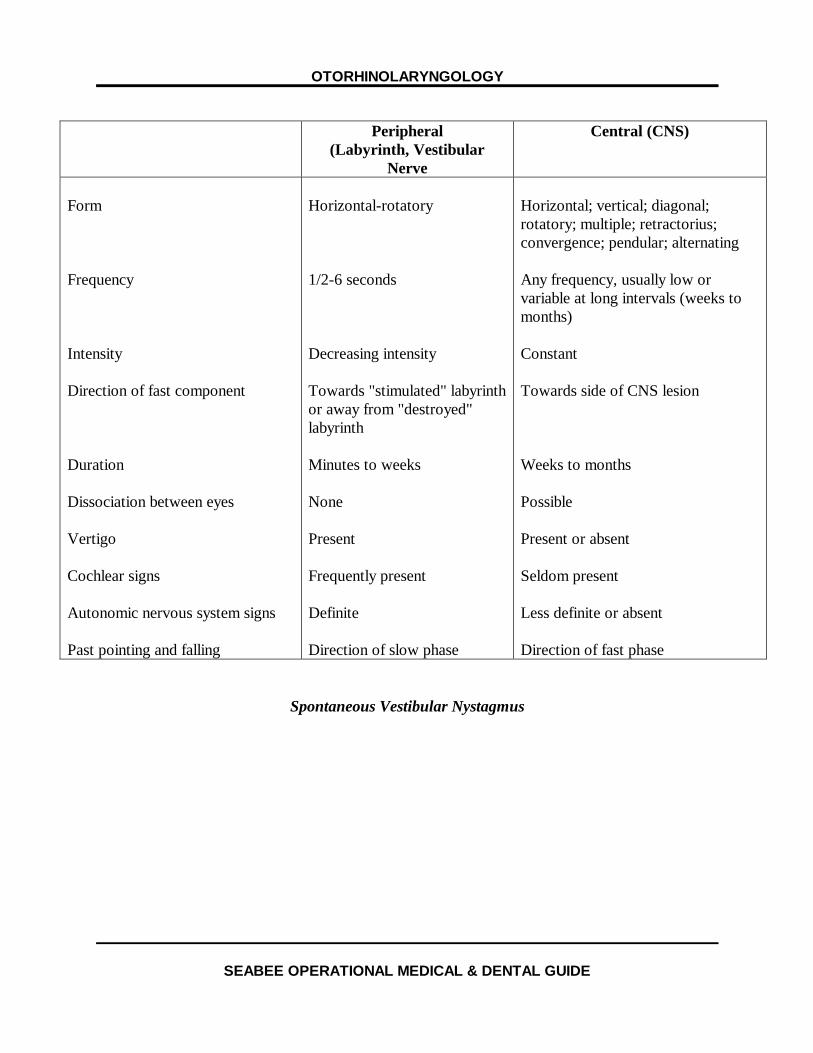
Spontaneous Vestibular Nystagmus
OTORHINOLARYNGOLOGY
SEABEE OPERATIONAL MEDICAL & DENTAL GUIDE
Peripheral(Labyrinth, Vestibular Nerve)
Central (CNS)
Hallucination of movement
Onset
Intensity
Duration
Influenced by head position
Nystagmus
Autonomic nervous systemsymptoms
Tinnitus
Deafness
Disturbances of consciousness
Other neurological signs
Definite
Usually paroxysmal
Usually severe
Minutes to weeks
Frequently
Present
Definite
Frequently present
Frequently present
Seldom present
Usually absent
Less definite
Seldom paroxysmal
Seldom severe
Weeks to months
Seldom
Present or absent
Less definite or absent
Seldom present
Seldom present
More frequently present
Frequently present
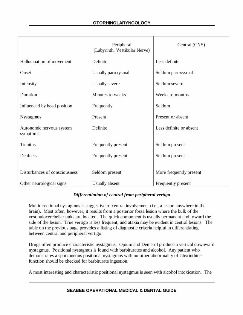
Differentiation of central from peripheral vertigo
Multidirectional nystagmus is suggestive of central involvement (i.e., a lesion anywhere in thebrain). Most often, however, it results from a posterior fossa lesion where the bulk of thevestibulocerebellar units are located. The quick component is usually permanent and toward theside of the lesion. True vertigo is less frequent, and ataxia may be evident in central lesions. Thetable on the previous page provides a listing of diagnostic criteria helpful in differentiatingbetween central and peripheral vertigo.
Drugs often produce characteristic nystagmus. Opium and Demerol produce a vertical downwardnystagmus. Positional nystagmus is found with barbiturates and alcohol. Any patient whodemonstrates a spontaneous positional nystagmus with no other abnormality of labyrinthinefunction should be checked for barbiturate ingestion.
A most interesting and characteristic positional nystagmus is seen with alcohol intoxication. The
OTORHINOLARYNGOLOGY
SEABEE OPERATIONAL MEDICAL & DENTAL GUIDE
nystagmus is typically in two phases and is often recorded as PAN (positional alcohol nystagmus)I and II. As little as 0.02 percent blood concentration may produce both phases. Phase I beginsabout 30 minutes after ingestion, as the blood alcohol peaks, and lasts approximately three and ahalf hours. The nystagmus is always in the direction of the gaze or toward the position of thehead (a right-beating nystagmus appears with right gaze, head turned to the right side or if theright of the patient's head is down in the lateral position). There is a gradual diminution after thepeak and an intermediate period of about 1.7 hours in which there is no nystagmus. Approximately 5 hours after the initial ingestion, PAN II begins; the nystagmus is in the oppositedirection of the gaze or lateral head position and persists for several hours after the blood alcohollevel has disappeared. PAN II nystagmus is greatest when the "hangover" symptoms are greatest.
DISEASES OR CLINICAL SYNDROMESOF OTOLOGICAL ORIGIN
The majority of cases of dizziness which the Medical Officer will see associated with disease orinjury of the inner ear or eighth cranial nerve are acute labyrinthitis, epidemic vertigo, vestibularneuronitis, Meniere's disease, acoustic neuroma, benign paroxysmal positional vertigo, andtrauma. These must be differentiated from the many causes of dizziness or vertigo (see table onfollowing page).
LabyrinthitisLabyrinthitis has many classifications, but, in general, it is serous, diffuse, destructive, or toxic. Serous and diffuse destructive labyrinthitis are associated with otitis media, cholesteatoma, or earsurgery. When the disease is of the serous type, the vestibular and cochlear functions aredepressed, with the vestibular symptoms usually preceding the cochlear depression by a few hoursto several days. There is usually spontaneous nystagmus to the opposite ear, nausea andvomiting, true vertigo, ataxia, past-pointing, and loss of hearing.
In patients with chronic ear disease, especially cholesteatoma, a fistula test should be performedby exerting pressure and then suction using a pneumo-otoscope. Production of nystagmus andvertigo indicates the presence of a labyrinthine fistula. An acute, initially severe, and sudden onsetof symptoms may be associated with the erosion into the labyrinth; however, in cholesteatoma,the lining or sac protects the labyrinth, and only quick head movements or pressure applied in thecanals cause vertigo in many cases. Patients who have had ear surgery or manipulation of thestapes may have all the usual findings, except nystagmus.
OTORHINOLARYNGOLOGY
SEABEE OPERATIONAL MEDICAL & DENTAL GUIDE
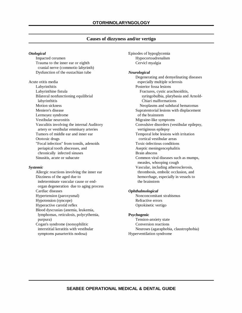
Causes of dizzyness and/or vertigo
OtologicalImpacted cerumenTrauma to the inner ear or eighth cranial nerve (commotio labyrinth)Dysfunction of the eustachian tube
Acute otitis mediaLabyrinthitisLabyrinthine fistulaBilateral nonfunctioning equilibrial labyrinthitisMotion sicknessMeniere's diseaseLermoyez syndromeVestibular neuronitisVasculitis involving the internal Auditory artery or vestibular emmisary arteriesTumors of middle ear and inner earOtotoxic drugs"Focal infection" from tonsils, adenoids periapical tooth abscesses, and chronically infected sinusesSinusitis, acute or subacute
SystemicAllergic reactions involving the inner earDizziness of the aged due to indeterminate vascular cause or end- organ degeneration due to aging processCardiac diseasesHypertension (paroxysmal)Hypotension (syncope)Hyperactive carotid reflexBlood dyscrasias (anemia, leukemia, lymphomas, reticulosis, polycythemia, purpura)Cogan's syndrome (nonsyphilitic interstitial keratitis with vestibular symptoms panarteritis nodosa)
Episodes of hypoglycemiaHypocortoadrenalismCervicl myalgia
NeurologicalDegenerating and demyelinating diseases especially multiple sclerosisPosterior fossa lesions Fractures, cystic arachnoiditis, syringobulbia, platybasia and Arnold- Chiari malformations Neoplasms and subdural hematomasSupratentorial lesions with displacement of the brainstemMigraine-like symptomsConvulsive disorders (vestibular epilepsy, vertiginous epilepsyTemporal lobe lesions with irritation cortical vestibular areasToxic-infectious conditionsAseptic meningencephalitisBrain abscessCommon viral diseases such as mumps, measles, whooping coughVascular, including atherosclerosis, thrombosis, embolic occlusion, and hemorrhage, especially in vessels to the brainstem
OphthalmologicalNonconcomitant strabismusRefractive errorsOptokinetic vertigo
PsychogenicTension-anxiety stateConversion reactionsNeuroses (agaraphobia, claustrophobia)
Hyperventilation syndrome
OTORHINOLARYNGOLOGY
SEABEE OPERATIONAL MEDICAL & DENTAL GUIDE
In isolated serous labyrinthitis, there is usually return of labyrinthine function over weeks ormonths. If any fistula is suspected or injury occurred in surgery, systemic antibiotics areindicated. With fistulas, there is often a permanent nerve-type hearing loss, and some patientshave chronic positional vertigo.
Suppurative labyrinthitis results in violent and sudden onset of vertigo, disturbed equilibrium,nystagmus, and vomiting. Cochlear and vestibular responses are lost. Complications such asmeningitis or brain abscess lead to toxic symptoms of headache, malaise, and fever. Vigoroustherapy with antibiotics and surgery must be instituted, and some small mortality can be expectedeven with treatment. For those who recover, there is usually no recovery of the cochlear orvestibular responses, and three to five weeks are required for compensation. Return to a flyingstatus is not recommended, except in the mildest cases. It is often impossible to be sure ofcomplete eradication of disease, and there is questionable compensation for loss of hearing andlabyrinthine function and occasional residual ataxia.
Toxic labyrinthitis is one of the most common types seen, and a great deal of disagreementremains about its classification. The etiology ranges from acute febrile diseases to toxic orchemical substances to idiopathic. The most common characteristic is whirling vertigo withgradual onset reaching a maximum in 24 to 48 hours, and at its height, there may be nausea andvomiting. There may be no cochlear or vestibular abnormalities in those cases associated with orfollowing acute febrile illness, but when associated with drugs, either system may be affected. Usually there is recovery from vertigo in three to six weeks.
Most commonly, toxic labyrinthitis is associated with pneumonia, cholecystitis, influenza, allergy,extreme fatigue, overindulgence in food or alcohol, and certain ototoxic drugs. Palliativetreatment with antivertiginous drugs and bed rest is helpful. The physician should always beaware of a missed or changing diagnosis with these patients. They should not be dismissed withthe "they always get well" attitude.
Epidemic VertigoAlthough to a great extent this disease may be of central origin, it is important to differentiate itfrom other vertiginous conditions, and this can often only be done by exclusion. Characteristically, symptoms are acute onset of severe dizziness, nausea, vomiting, a slight fever,headache, and asthenia, with a duration of several weeks to months. Recovery, however, is usual. There is usually an epidemic character to the disease, and it is associated with either an upperrespiratory infection or gastroenteritis. Caloric and audiological tests usually are normal, butspinal fluid may show some lymphocytic cells. Cases with gastrointestinal symptoms are morefrequent in mid-January, and those with upper respiratory symptoms occur in the autumn. Laboratory tests are of little value.
Treatment is supportive, with variable help from antivertiginous and antinausea drugs such asDramamine, Vontrol, Torecan, and Tigan. These patients should be able to return to flying withinone month after all symptoms have ceased.
OTORHINOLARYNGOLOGY
SEABEE OPERATIONAL MEDICAL & DENTAL GUIDE
Vestibular NeuronitisVestibular neuronitis is characterized by an attack of sudden, debilitating vertigo, nausea,vomiting, and spontaneous nystagmus. In most cases, there appears to be an antecedent orconcomitant infection in the upper respiratory tract, maxillary sinuses, or teeth. The cochlea isspared, but one or both of the labyrinths have abnormal calorics. Vestibular symptoms decreasesomewhat after a few hours, but they remain fairly severe for the first week, slowly decreasingover the next four to eight weeks. About 70 percent of these patients have permanent, decreasedcaloric function.
Management is directed toward supportive treatment of the symptoms and an aggressive workupto rule out other possible diagnoses. Vestibular neuronitis is a self-limiting disease, althoughreturn to work may require from three to twelve weeks. Generally, an aviator is permanentlygrounded for military flying because of the sudden debilitating nature of the attacks which can berecurrent even as long as four years after the initial attack.
Meniere's DiseaseAlthough much disagreement persists as to whether this is a disease or a symptom complex, andits etiology is still unknown, there is usually the classical triad of episodic vertigo, tinnitus, anddeafness. The average age of onset is 44 (Cawthorne & Hewlett, 1954), and it is predominantlyunilateral, with only about ten percent of the patients having bilateral involvement.
The onset of symptoms is insidious, usually with a sensation of dullness or fullness in the ear, andan initial fluctuation in hearing of 10 to 30 dB, usually in the low tones. The hearing improvessomewhat between attacks, but it continues to deteriorate as time goes on. There may beincreased sensitivity to sound, or music may sound distorted. Tinnitus, varying from a whistle toa roar, develops, followed by a turning or whirling vertigo that may lead to nausea, vomiting, andeven prostration. Any head movement aggravates the condition, with the vertigo lasting severalhours. Some patients can have fleeting attacks lasting several minutes, and still others haveattacks lasting a week or longer and may take months to regain normal equilibrium.
Besides the fluctuating hearing, spontaneous nystagmus, usually rotary and oftendirection-changing, and a direction-fixed, positional nystagmus are the most common findings. The caloric reaction is usually abnormal. Aside from the hearing loss, Meniere's patientsfrequently have recruitment and diplacusis, low threshold discomfort, and low discriminationscores. Tone decay and a Type II Bekesy are present. A fairly reliable diagnostic test is theglycerin test, where a patient ingests 1.5 gm/kg body weight of glycerol mixed with equal parts ofnormal saline and a few drops of lemon juice. Audiograms are taken immediately and at one, two,and three hours after ingestion. A positive test is said to be an improvement in hearing of 15 dBin any one frequency from 250 to 4000 Hz or 12 percent improvement in the discriminating score.
There is no effective, long-term treatment for Meniere's disease. For many years, some physicianshave controlled their patients with a neutral-ash, salt-free diet, supplemented with diuretics. Shea
OTORHINOLARYNGOLOGY
SEABEE OPERATIONAL MEDICAL & DENTAL GUIDE
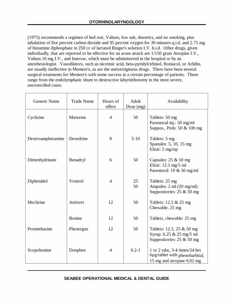
(1975) recommends a regimen of bed rest, Valium, low salt, diuretics, and no smoking, plusinhalation of five percent carbon dioxide and 95 percent oxygen for 30 minutes q.i.d. and 2.75 mgof histamine diphosphate in 250 cc of lactated Ringer's solution I.V. b.i.d. Other drugs, givenindividually, that are reported to be effective for an acute attack are 1/150 grain Atropine I.V.,Valium 10 mg I.V., and Innovar, which must be administered in the hospital or by ananesthesiologist. Vasodilators, such as nicotinic acid, beta-pyridylcirbinol, Roniacol, or Arlidin,are usually ineffective in Meniere's, as are the antivertiginous drugs. There have been severalsurgical treatments for Meniere's with some success in a certain percentage of patients. Theserange from the endolymphatic shunt to destructive labyrinthotomy in the most severe,uncontrolled cases.
Generic Name Trade Name Hours ofeffect
AdultDose (mg)
Availability
Cyclizine
Dextroamphetamine
Dimenhydrinate
Diphenidol
Meclizine
Promethazine
Scopolomine
Marezine
Dexedrine
Benadryl
Vontrol
Antivert
Bonine
Phenergan
Donphen
4
8
6
4
12
12
12
4
50
5-10
50
2550
50
50
50
0.2-1
Tablets: 50 mgParenteral inj.: 50 mg/mlSuppos., Peds: 50 & 100 mg
Tablets: 5 mgSpanules: 5, 10, 15 mgElixir: 5 mg/tsp
Capsules: 25 & 50 mgElixir: 12.5 mg/5 mlParenteral: 10 & 50 mg/ml
Tablets: 25 mgAmpules: 2 ml (20 mg/ml)Suppositories: 25 & 50 mg
Tablets: 12.5 & 25 mgChewable, 25 mg
Tablets, chewable: 25 mg
Tablets: 12.5, 25 & 50 mgSyrup: 6.25 & 25 mg/5 mlSuppositories: 25 & 50 mg
1 or 2 tabs, 3-4 times/24 hrs6µg/tablet with phenobarbital,15 mg and atropine 0,02 mg
OTORHINOLARYNGOLOGY
SEABEE OPERATIONAL MEDICAL & DENTAL GUIDE
Antivertiginous drugsAcoustic NeuromaAn acoustic neuroma is a fairly rare, extremely slow growing neoplasm that originates on thevestibular portion of the eighth cranial nerve in the internal auditory canal. It constitutes abouteight to ten percent of all brain tumors and is most common in the fourth and fifth decade of life. Early diagnosis, which offers the best chance for a surgical cure and the least morbidity andmortality, is often based on a strong suspicion. Symptoms, often difficult to pinpoint but mostoften present, are steady, unilateral tinnitus, hearing loss, and a feeling of unsteadiness. Somepatients have vague complaints of headache, local retroaural discomfort, and facial paresthesia orpain. A significant finding is speech discrimination much more severe than indicated by apure-tone hearing test.
Diagnostic evaluation should include a complete audiological examination of pure tone andspeech, stapedial reflex, acoustic reflex decay, and a Brainstem Evoked Response, Auditory(BERA). Stenver's and Town's X-rays are valuable for an initial screen, but computerized axialtomography or a posterior fossa myelogram are more often necessary. Typically, there is asensorineural-type hearing with poor speech discrimination that is inconsistent with the pure-tonetest, absence of recruitment or low small increment sensitivity index (SISI) scores, pronouncedtone decay, a type III or IV Bekesy tracing, reduced caloric response, widening of the internalauditory canal, decreased corneal sensitivity on the involved side, and decreased or absentstapedial reflex.
Suspected cases, which are not diagnostic, should be kept under the watchful eye of anotolaryngologist or neurologist and not dismissed or forgotten after the initial workup.
Benign Paroxysmal Positional VertigoBenign paroxysmal positional vertigo must be differentiated from Meniere's and eighth nervetumors. In general, onset of nystagmus and vertigo occur when the head moves to a certainposition. There usually is a latent period of several seconds, and the nystagmus fatigues withrepeated testing. Most cases have normal calorics and audiological examinations. Symptomsabate in about eight weeks, but they may recur or even last for years. There is no treatmentexcept avoidance of the position that creates the nystagmus and vertigo, as well as reassurance tothe patient. Pilots should be grounded until all symptoms have disappeared, and each case mustbe considered on an individual basis.
THE DIZZY PATIENT
Vertigo: An hallucination of movement• True vertigo§ A sensation of motion or turning§ Patient turning in relation to surroundings or surroundings turning around patient§ Usual origin is vestibular system
OTORHINOLARYNGOLOGY
SEABEE OPERATIONAL MEDICAL & DENTAL GUIDE
• Dizziness§ Less severe and less distinct§ Sensations of giddiness, faintness, confusion, blankness, unsteadiness, or lightheadedness§ A floating sensation
• Causes of dizziness and/or vertigo§ OtologicØ Impacted cerumenØ Trauma to inner ear of VIIIth cranial nerve (commotio labyrinthi)Ø Acute otitis mediaØ LabyrinthitisØ Labyrinthine fistulaØ Bilateral non-functioning equilibrial labyrinthsØ Motion sicknessØ Meniere's diseaseØ Lermoyez syndromeØ Vestibular neuronitisØ Vasculitis involving internal auditory artery or vestibular emissary veinsØ Tumors of middle and inner earØ Ototoxic drugsØ Focal infection from tonsils, adenoids, periapical tooth abscess, and chronically
infected sinusesØ Sinusitis, acute or subacute
§ NeurologicØ Degenerating and demyelinating diseases, especially multiple sclerosisØ Posterior fossa lesions¨ Fractures, cystic arachnoiditis, syringobulbia, platybasia, and Arnold-Chiari
malformations¨ Neoplasms and subdural hematomata
Ø Supratentorial lesions with brainstem displacementØ Migraine-like syndromesØ Convulsive disorders (vestibular epilepsy, vertiginous epilepsy)Ø Temporal lobe lesions with irritation of cortical vestibular areasØ Toxic-infectious conditions¨ Aseptic meningoencephalitis¨ Brain abscess¨ Common viral diseases such as mumps, measles, whooping cough
Ø Vascular; including thrombosis, atherosclerosis, embolic occlusion, and hemorrhage,especially in brainstem vessels
§ SystemicØ Allergic reactions involving inner earØ Dizziness of the aged due to indeterminate vascular cause or end-organ degenerationØ Cardiac diseases
OTORHINOLARYNGOLOGY
SEABEE OPERATIONAL MEDICAL & DENTAL GUIDE
Ø Hypertension (paroxysmal)Ø Hypotension (syncope)Ø Hyperactive carotid reflexØ Blood dyscrasias (anemia, leukemia, reticulosis, polycythemia, purpura)Ø Cogan's syndrome (nonsyphilitic interstitial keratitis with vestibulo-bulbochochlear
symptoms; panarteritis nodosa)Ø Episodes of hypoglycemiaØ HypocorticoadrenalismØ Cervical myalgia
§ OphthalmologicØ Non-concomitant strabismusØ Refractive errorsØ Opticokinetic vertigo
§ PsychogenicØ Tension/anxiety statesØ Conversion reactionØ Neuroses (agoraphobia, claustrophobia)Ø Hyperventilation syndrome
RHINOLOGY
Nasal and Sinus PhysiologyThe primary functions of the nose are filtration, warming, and humidification of air; it alsosubserves the sense of smell, and it is the origin and recipient of numerous reflex areas. Thesinuses have no primary function.
Air filtration is accomplished by the vibrissae in the anterior nares and by mucus. Most of themucous glands are in the nasal mucosa. The mucous blanket is moved by cilia toward thenasopharynx at the rate of five mm per minute. Although amazingly resistent to heat, cold, fumes,dust, and chemicals, the cilia are most vulnerable to drying from inspired dry air, such as centralheating or 100 percent oxygen.
Air flow during inspiration is directed over the turbinates to the roof of the nasal cavity and theninto the nasopharynx. The air is warmed by heat transfer from the mucous membranes. Duringexpiration, the air makes a loop before exiting the nose anteriorly, allowing for retention of themoisture in the air. The air flow volume is regulated by the changing size of the turbinates.
Bacterial Diseases of the NoseVestibulitis. An inflammation of the hair follicles in the nasal vestibule may cause chronic crustingand tenderness of the nasal tip or ala; it is often recurrent. Treatment consists of gentle cleaningof the nasal vestibule and the application of topical antibiotic ointment, usually containingNeomycin, two or three times daily. Ophthalmic ointments work well, but treatment must be
OTORHINOLARYNGOLOGY
SEABEE OPERATIONAL MEDICAL & DENTAL GUIDE
continued for two to three weeks after symptoms disappear to prevent recurrences.
Furunculosis. Furunculosis of the vestibule is also common and usually associated with digitaltrauma and nose blowing. A crack in the skin allows the entrance of strep or staph organisms. Most infections localize, but occasionally they may become a spreading cellulitis. Squeezing orincising the area is dangerous, as it may cause spread to the cavernous sinus. Pain and systemicsymptoms may be marked. Treatment consists of a "hands off" policy, adequate doses ofappropriate antibiotics, hot, moist packs, and good analgesics.
Rhinitis. Rhinitis can develop as a complication of an upper respiratory infection if symptoms lastlonger than seven to ten days. Thick yellow or greenish nasal drainage, fever, throat and ear pain,and productive cough suggest complications. The most common causes are excessive noseblowing (forces bacteria into the sinuses and Eustachian tube traumatizes the sinus orifices) andsevere coughing (strips the cilia from the bronchial lining)
Treatment should place emphasis on maintaining good nasal and sinus drainage, good tissuehydration, and rest; antibiotics are used for bacterial infections or complications. The penicillins,erythromycin, or the tetracyclines, in order of preference, handle most complications, but culturesshould be taken to help in resistant cases.
In general, pilots or flight personnel should not fly with a cold. Even a slight amount of nasalcongestion and tissue edema may be enough to interfere with pressure equalization of the sinusesand ears, leading to aerotitis, aerosinusitis, or barometric vertigo. The examiner should stronglyadvise against self-medication and frequently reiterate the many predictable, immeasurable factors,such as level of awareness and performance, that may be affected by disease or medication. Theexaminer must make individual judgements, depending on the aircraft, aircrewman's job, type offlight, and medication, when deciding to ground flight personnel. Before personnel are allowed toreturn to flight status, a careful examination of the ears, nose, and throat should be made. Symptoms are often gone several days before the tissues return to normal and before essentialfunctions return sufficiently to handle the many different and rapid environmental changesassociated with flying.
Diseases of the Nose and SinusesAllergic Rhinitis. Allergic rhinitis, a very unpredictable and difficult problem, may be acute orchronic, seasonal or perennial. Common symptoms are nasal obstruction, clear rhinorrhea,sneezing, itching of the eyes, soft palate, and nose, and occasional associated headache, mostlyfrontal. Some cases of allergic rhinitis are similar to a cold, but they usually last only one or twodays or over 10 days and are more frequent than viral upper respiratory infections.
Seasonal allergies are often caused by pollens from grasses, trees, or flowers and last two or threeweeks. If a specific allergen is found, desensitization is often effective. After an allergy shot, apilot is grounded for at least six hours. Perennial rhinitis can be quite variable with no pattern, orit may be nearly constant. Allergies may be caused by house dust, molds, dog dander, wool,
OTORHINOLARYNGOLOGY
SEABEE OPERATIONAL MEDICAL & DENTAL GUIDE
feathers, tobacco pollutants, or food. Avoidance, if possible, is the best method of control;however, desensitization may be effective for dusts and molds.
Examination of the nasal mucosa often reveals edema and pallor of the turbinates, especially theinferior turbinates and the anterior tips of the middle turbinates. The turbinates may be soengorged they appear purple. The posterior turbinate tips may protrude into the nasopharynx orbecome irregular and look like mulberries. Red or inflamed mucosa has also been noted,especially if the allergen is a pollutant.
A basic allergy workup should include the following:• History - present, childhood, family.• Nasal smear for eosinophiles.• Sinus X-rays.• Complete blood count.• Thyroid function test.• Total protein, IgE, and gamma globulin blood level.
Basic treatment measures are as follows:• Take antihistamines, with or without decongestants. Alternating the antihistamine every
two weeks is often effective. Consider topical nasal steroids or Nasalcrom spray.• Cover pillows and mattress with plastic.• Cover overstuffed furniture.• Eliminate wool from bedding.• Remove domestic animals from the house.• Air-condition the house.• Avoid milk and egg products; other foods can be eliminated, one at a time, a week apart.• Use nonallergenic cosmetics.
Severe allergy attacks may require a short course of systemic steroids for control. Milder casesthat create obstruction of the nasal airway and sinus orifices can often be helped by topicalsteroids in an aerosol form, such as Decadron or Beclomethasone.
Non-allergic Rhinitis. Non-allergic rhinitis, often included under the term of vasomotor rhinitis,has as the most common symptoms chronic, intermittent, often alternating nasal stuffiness orobstruction, and postnasal drip. In the course of treatment, it is important to rule out allergies, toexplain the physiology of the nose to the patient, and to prevent the overuse of nose drops orinhalers that may cause a rhinitis medicamentosa. Once rhinitis medicamentosa develops, it canonly be cured by complete abstinence from nose drops. In about three weeks, the normal reflexactivity should return. Septal deviations should be corrected if they are a factor in obstructions. Humidification of the house or bedroom, or the use of Proetz solution or ointments to preventdrying of the mucosa is often helpful. Thyroid function may be a factor in some cases; forborderline hypothyroid states, thyroid extract or Cytomel has been effective. Certain emotionalstates cause nasal symptoms, and they often respond when this problem has been explored or
OTORHINOLARYNGOLOGY
SEABEE OPERATIONAL MEDICAL & DENTAL GUIDE
treated. Rhinitis of pregnancy usually responds to no treatment except delivery. Certainantihypertensive and birth control pills may cause nasal congestion; decrease or change in thedrugs often improves or cures the problem.
Polyps and Polypoid Degeneration. When the nasal mucosa, and in some cases the sinus mucosa,reacts to allergies or inflammation, edema develops due to increased capillary permeability andtransudation of fluid into the cell and extracellular spaces.
Polyps and Polypoid Degeneration. When the nasal mucosa, and in some cases the sinus mucosa,reacts to allergies or inflammation, edema develops due to increased capillary permeability andtransudation of fluid into the cell and extracellular spaces.
Air conditioners may contain much dust and mold, causing more trouble for a person withallergies to these substances. Electrostatic filters may do a better job, but may produce ozonewhich is toxic. If the first outlet is eight to ten feet from the unit, it is usually safe. Humidification is good for the dry nasal mucosa but it also increases the growth of molds in thehouse.
The mucosa appears "waterlogged" or "intumescent." Over a period of time, with the help ofgravity, this tissue may elongate to form nasal polyps, especially in the region of the middlemeatus and maxillary sinus ostia. In some cases, the anterior tip of the middle turbinate may justremain edematous, and this condition is called polypoid degeneration, rather than a polyp. Thetissue may lose some of its cilia and is replaced with goblet cells.
Polyps and polypoid degeneration may obstruct the sinus ostia leading to acute and chronic sinusdisease or sinus blocks and, therefore, should be removed when obstructive. Small, or single,nonobstructive polyps need not be removed unless they enlarge. Occasionally, polyps are foundwithin the maxillary sinus; these polyps eventually move out of the sinus ostium and into thenasopharynx, where they expand in size. These polyps are called anterochoanal or choanalpolyps, and their removal requires a Caldwell-Luc antrostomy to remove the base and preventrecurrence. Polyps in the maxillary sinuses are disqualifying for aviation candidates, as is nasalpolyposis. A possible exception can be made for a single, small polyp on one side in anasymptomatic, non-allergic candidate. Recurrence of polyps after removal is common; this isespecially true when the disease remains in the ethmoid sinuses. In some cases, the use of shortcourses of broad spectrum and topical steroids, such as aerosol Decadron or Beclomethasone,may reduce the size of the polyps. A common dose schedule is two sprays in each nostril, twicedaily for one week, then one spray in each nostril twice daily for four days, finishing with onespray daily in each nostril for the remainder of the week or longer, if desired. The use of topicalsteroids may be irritating to the mucosa, and use beyond one month is not recommended.
EpistaxisThe majority of nosebleeds are caused by trauma and occur from the vascular plexus on the
OTORHINOLARYNGOLOGY
SEABEE OPERATIONAL MEDICAL & DENTAL GUIDE
anterior septum, known as Kiesselbach's plexus or Little's area. Common causes are air drying,violent sneezing or blowing the nose, and picking the nose. Severe bleeding, especially highanterior and posterior bleeding, occurs from the ethmoid artery, a branch of the internal carotid,and the sphenopalatine artery, a branch of the external carotid artery.
In general, treatment of simple anterior bleeding should first be direct pressure, for at least five orten minutes, against the anterior septum. Pledgets of cotton moistened in a vasoconstrictor, suchas one percent Neo-Synephrine, one percent epinephrine, or one to four percent cocaine, alongwith pressure, are even more effective; large clots should be gently suctioned away. If bleeding iscontrolled, the bleeding site may be cauterized with 25 to 50 percent trichloroacetic acid, fivepercent chromic acid, or silver nitrate in a 50 percent solution or on a stick applicator. Thesesolutions should be applied with a small, moist applicator under direct vision. Anterior bleedingsites not controlled by direct pressure or chemical cautery should be infiltrated with Xylocaine andepinephrine, using both the tissue wheal and the epinephrine effect for control. The site may thenbe cauterized by chemical or electrocautery; deep burns or cauterization of adjacent structures,such as the ala or vestibule, must be avoided. If the coagulated fluid and blood stick to the tip ofthe cautery and are pulled off with the coagulum, the bleeding may restart. In those cases wherebleeding cannot be controlled, one might attempt cautery with a suction tip electrode; if this fails,the nose can be packed with Vaseline and antibiotic ointment impregnated in half-inch selvagegauze. It is best to pack both sides to prevent loss of the pack by shifting of the septal cartilagefrom a one-sided pressure. The pack should be left in place for at least 24 hours, but usuallynever more than 72 hours. All raw or cauterized surfaces should be lightly covered with anantibiotic ointment, and a small piece of compressed Gelfoam over the anterior septum furtherprotects against air trauma. The ointment application should be repeated three or four times aday.
Posterior bleeding, usually in the older age group, is a serious condition, and, if coupled withhypertension, it requires aggressive medical and rhinological management. The patient should beadmitted to sick bay, sedated, and kept in a head-elevated position. After vasoconstrictor andtopical anesthetic application to both nasal passages, an attempt can be made to control theposterior bleeding by the use of a specifically designed postnasal balloon, or a common, 15 cc-sizeFoley catheter. The balloon is checked before insertion, then it is passed along the floor of thenose, and when it is in the lower nasopharynx, the balloon is filled with about 5 cc of water. It isthen drawn back up against the posterior choana and further filled to the point of tolerablediscomfort to the patient. Anterior packing is inserted bilaterally with fixation of the catheter tothe lip or against the packing, but never against the ala or septum to prevent pressure necrosis. The posterior pharynx is checked hourly for bleeding, and the hemoglobin and hematocrit aremonitored according to the amount of oozing or bleeding; blood typing and cross matching areadvisable. Blood coagulation studies are usually done, but it is unusual to see only nasal bleedingwith abnormality of the clotting mechanism. A patient with a posterior nasal pack or balloon isnever sent home. He should be closely monitored because of the possibility of a nasovagal reflexaction when the nasopharynx is packed, that might lead to apnea or hypoxia. Uncontrolledbleeding of the ethmoidal arteries requires ligation in the orbit or, as a last resort, an internal
OTORHINOLARYNGOLOGY
SEABEE OPERATIONAL MEDICAL & DENTAL GUIDE
carotid ligation. Uncontrolled sphenopalatine artery bleeding requires ligation through atransmaxillary sinus approach, or ligation of the external carotid in the neck.
Barotrauma to SinusesAerosinusitis results when equalization of pressure between the sinus cavities and the atmosphereis prevented by obstruction of their orifices. There are numerous causes, but heading the list arethe common cold and allergies. Other causes are anatomical defects, infection, and polyps.
As an aviator goes to altitude, the outside pressure decreases, and discomfort may be felt in theobstructed sinus. It is usually not severe, however, and most often air forces its way out past theobstruction. When the aviator descends, the pressure in the obstructed sinus remains less than thesurrounding pressure, creating a vacuum effect on the delicate, thin, mucosal lining and resultingin pain that is often severe. Some fluid may be drawn into the cavity, but the more seriouscomplication is pulling away of the mucoperiosteum, with formation of a hematoma. Sinus blocksoccur most often in the frontal sinus (70 percent), and the aviator must be grounded until thehematoma resolves, and the ostium is patent. This may require three weeks to three months. Forthis reason, anyone suspected of a sinus block should have sinus X-rays to determine the extent ofinjury and then should be followed at approximately three-week intervals, until clear.
Treatment of the acute block• Stop descent in aircraft or low-pressure chamber, if possible, and return to altitude for pain
relief.• If available, spray the nasal passage with a vasoconstrictor nasal spray (nose drops).• Do the Valsalva maneuver or use the Politzer method.• Make a slow descent equalizing pressure with the above maneuvers.• Place patient on antihistamine-decongestant or decongestant therapy.• Take screening Water and Caldwell sinus X-rays.• If an upper respiratory infection is present, treat with antibiotics.• Control severe pain with Codeine or Percodan.• If a frontal sinus hematoma is present, ground the aviator for at least three weeks. With no
apparent X-ray pathology, the aviator should be grounded for at least 72 hours, or until anynasal symptoms have been cleared. Recurrent trauma may result in mucocele formation,requiring a major surgical procedure and permanent gounding.
SinusitisThe majority of acute sinusitis cases follows an acute upper respiratory infection, like the commoncold, and they are often the result of improper nose blowing. Another cause, which may have amore rapid onset, is swimming or diving; occasionally, an upper molar tooth abscess breaks intothe maxillary antrum. The extent and persistence of the infection depends on two majorphysiological principles, ventilation and drainage; the treatment is directed toward theseprinciples. The most common bacterial causes are the Gram-positive cocci.
Acute suppurative sinusitis usually has symptoms of nasal congestion and pressure or pain over
OTORHINOLARYNGOLOGY
SEABEE OPERATIONAL MEDICAL & DENTAL GUIDE
the involved sinus. Toxicity is usually mild, except in cases of pansinusitis when the frontals orsphenoids are involved. Pus draining from the middle meatus or above the middle turbinate, painand pressure over a maxillary or frontal sinus, and decreased transillumination may be sufficient tomake a diagnosis. The X-ray is indispensable, however, in determining the extent of the disease,fluid levels, and response to medication, all of which may indicate the proper approach totreatment.
Maxillary sinusitis, usually has the least toxicity, but a persistent fluid level or pain after 48 hoursof adequate antibiotic therapy suggests the need for irrigation of the antrum, either through thecanine fossa or through the thin, bony wall of the inferior meatus. The maxillary sinus mucosa hasgreat reparative power; after removal of the pus by irrigation, it may clear within a few days. Ifthe antral infection is dental in origin, it is useless to attempt a cure without treatment of theoffending tooth.
Ethmoid sinusitis is probably the most common infection. Due to the proximity of the ethmoidsinuses to the frontal and maxillary sinuses, ethmoid sinusitis either causes or is associated withthe infections in those sinuses also. Ethmoid infections usually cause more inflammation andmucosal swelling. Pain may be near the root of the nose or frontal region. Edema of the lower lidis often present in children. Orbit involvement may result in painful eye movement due to aperiostitis about the pulley of the superior oblique muscle or, in the case of rupture into the orbit,proptosis.
Frontal sinusitis usually is associated with toxicity, frontal headache, often in mid-morning to lateafternoon, tenderness to percussion over the sinus, or pressure on the floor in the supraorbitalregion; swelling of the upper eyelid may be highly suggestive. Treatment should be vigorous toprevent osteomyelitis of the skull or fistulas that lead to complications, such as soft tissue or sinuscavity abscesses, meningitis, brain abscess, and even death.
Sphenoid sinusitis is uncommon, but it may result as a direct extension of infection in neighboringsinuses, nasal mucosa, or the nasopharynx. The symptoms are variable, but they may consist of adeep, boring, occipital or parietal headache with inability to concentrate, fever, malaise, andanorexia. Rupture or osteomyelitis from sphenoid infection leads to rapidly fatal meningitis orcavernous sinus thrombosis. Diagnosis can usually only be made by suspicion and X-rays, usingproper contrast in the lateral and submental vertex positions; fluid levels will only be seen if theX-rays are taken in the upright position. These patients require high doses of intravenousantibiotics and emergency surgical intervention.
Since the cardinal principle of treatment in sinus infections is ventilation and drainage, thefollowing treatment is suggested:• The nasal mucosa must be protected from drying. The patient must be kept hydrated, and, in
some cases, use of a humidifier or vaporizer may help.• An oral decongestant may be used alone or with an antihistamine. Antihistamines may make
secretions too thick or the mucosa too dry, so it is often helpful to use a mucous-thinning
OTORHINOLARYNGOLOGY
SEABEE OPERATIONAL MEDICAL & DENTAL GUIDE
medication, such as glyceryl-guaiacolate.• Antibiotics are given orally in adequate doses for at least seven days in most uncomplicated
cases, but in pansinusitis or cases of moderate to severe toxicity, and especially in frontal orsphenoid involvement, intravenous antibiotics are necessary. Most organisms are sensitive topenicillin or erythromycin, but it is strongly recommended that a culture be taken from theturbinates and the meatuses. Be sure not to touch the nasal vestibule and hairs, as these areasmay have different predominant organisms. The nasopharynx is another area from which toobtain a culture of prevalent sinus drainage.
• Bed rest, hydration, and adequate pain medication are important in patients with toxicity.• Antral irrigation, either through the natural ostium or the canine fossa or inferior meatus
puncture approach, is indicated for persistent pain or fluid levels after 48 hours of antibiotictherapy. Persistent swelling, and pain in the frontal sinus region, in spite of intense therapy,may signal the need for a frontal sinus drainage, usually done by trephine through the sinusfloor. A rubber or plastic tube drain is sutured in place to allow irrigation and drainage untilthe natural ostium drainage is reestablished.
• Daily mucosal shrinkage and gentle nasal suction cleaning may help promote drainage.Localheat is often helpful, not only as comfort to the patient, but to increase the vitality of themucosa.
• Persistent or subacute ethmoid disease may respond to Grossan nasal irrigation.• Acute sphenoid infection with toxic signs is an emergency life-threatening situation requiring
immediate hospitalization and surgery, so one should always be alert to this disease, by itselfor as a complication from the other sinuses.
Neglected sinus infections or subacute disease lead to chronic irreversible changes in the sinusmucosa. With chronic purulent drainage or sinus blockage, one usually as to resort to surgeryafter conservative treatment fails. For the maxillary sinus, an endoscopic opening of the naturalostia (ostiotomy) is most often used. Removal of the ethmoid cells is more difficult and is donewith an intranasal approach when polyps and persistent disease are present. Chronic sphenoiddisease is not only rare, but most difficult to diagnose, because X-rays may be inconclusive andsymptoms extremely variable. Chronic disease in the frontal sinus, be it osteomyelitis or mucoceleformation, dictates a major surgical procedure through either a bicoronal incision flap approach orthe osteoplastic eyebrow incision approach, with complete removal of the sinus mucosa andobliteration of the sinus, usually with fat. These surgical procedures and treatment may not resultin relief of nasal symptoms or remove the tendency toward recurrent infection. A frontal sinusobliteration is usually disqualifying for most types of aviation.
Antral cysts (frequently seen on the Waters X-ray as a smooth, rounded density in the loweraspect of the maxillary sinus) are benign, filled only with xanthochromic or clear fluid. Theyusually require no treatment, unless they fill the sinus, obstruct drainage, and become symptomatic
Maxillary Sinus Irrigation-Inferior Meatus Puncture• Anesthesia.§ Spray mucosa initially with a vasoconstrictor.
OTORHINOLARYNGOLOGY
SEABEE OPERATIONAL MEDICAL & DENTAL GUIDE
§ For local anesthesia use four or five percent topical cocaine and two percent Xylocainewith Epinephrine 1:1000 (dental carpule or equivalent).
§ Apply pledgets of cotton moistened with cocaine (never sloppy wet) in the inferior meatusand on the inferior turbinate. After initial application, cocaine on a wire applicator isplaced against the lateral wall of the inferior meatus about one inch or 1.5 to 2.5 cmbehind the anterior edge of the meatus for five minutes.
§ Insert a long (3 1/2 inch) needle into the inferior meatus until it strikes bone in the area ofthe intended puncture and infiltrate with local anesthetic.
• Equipment.§ Straight three and a half inch, 18 gauge spinal needle or equivalent trocar with stylet.§ Sterile saline to which a small amount of Neo-Synephrine may be added.§ One 30 to 50 cc syringe and one 5 cc syringe.§ Plastic or rubber extension tubing.§ Culture tube.
• Technique.§ With the patient in an upright position and the head against a firm headrest, the puncture
needle or trocar is inserted into the inferior meatus about two centimeters posterior to theedge of the inferior meatus and engaged in the thin bone of the lateral wall of this area.
§ The thumb is placed against the stylet and the needle is directed laterally in line with theouter canthus of the eye, using the fingers of the opposite hand to steady the needle. Pressure is slowly, but steadily, increased until the needle is felt to penetrate into the sinus.
§ The needle is pushed into the sinus until it strikes the lateral sinus wall and then withdrawnabout one centimeter. If a low-lying cyst is present, the needle is directed as far inferior aspossible just after penetration to puncture the cyst.
§ Direct observation of the drainage or aspiration with a small syringe may be diagnostic orproduce a pure specimen for culture.
§ The large syringe and extension tubing filled with normal saline are inserted into the needleand aspiration is attempted. Air bubble or exudate indicates the needle is in the properposition. No aspiration may mean the needle is in the mucosa, plugged, or not in the sinusproper.
§ Irrigation is carried out with the patient leaning forward over a large basin with his mouthopen, and gentle, but steady, pressure is applied to the syringe.
§ Instant, severe pain suggests the needle is in the mucosa; readjust the needle's position andrepeat. Intolerance to irrigation pressure dictates termination of the procedure andpossible attempt at natural ostia irrigation. A slow buildup of pressure and occasionallypain is expected with an obstructed ostia, but it is usually tolerable or relieved as the sinusis irrigated.
§ Irrigation should be carried out until the washing is clear or, in the case of a clearirrigation, until at least three full syringes have been used.
§ The final irrigation should be made with the sinus ostia dependent. Insufflation of air intothe sinus has been associated with air isolation and should not be performed.
OTORHINOLARYNGOLOGY
SEABEE OPERATIONAL MEDICAL & DENTAL GUIDE
§ The needle is withdrawn with a smooth rapid movement and the nasal passageimmediately inspected for retained pus or thick mucus. This material is aspired, being sureto include aspiration of the posterior floor and middle meatus.
Nasal FracturesNasal fractures are common injuries which can usually be handled in the clinic or sick bay by theexaminer. There are basically three types: (1) a simple fracture of the nasal bones, most oftenjust the tip, (2) lateral displacement of the nasal bones to one side, often as a green stick fractureon one side and impaction on the other, and (3) marked flattening of the nasal bridge withcomminution of the bones or an accordion fracture displacement of the septum. Diagnosis of adisplaced fracture can best be made by inspection, palpation, lateral X-ray of the nasal bones andcomparison of the patient to his or her ID card or recent photo.
Shortly after injury, when the airway is compromised, or before profuse swelling has occurred,reduction should be accomplished under local or general anesthesia. When the injury is veryrecent and the patient is still in a shock or "numb" like state, stable lateral displaced fractures canoften be reduced without anesthesia by simple, quick, firm, thumb pressure on the convex side ofthe nose. When soft tissue swelling is marked, distorting the true alignment of the nasal bones,one may elect to wait four or five days for the swelling to recede before reduction. A compoundfracture should be reduced within a few hours and then have a plastic-type laceration closure sincereduction maneuvers usually tear out delicate sutures. Antibiotic coverage is recommended toprevent complications.
• Anesthesia Technique for Reduction of Nasal Fractures.§ Shrink the nasal mucosa with one percent Neo-Synephrine.§ Use fresh cocaine from one to five percent, most common is four percent or plain two or
four percent Xylocaine.§ Moisten long, thin, cotton pledgets with cocaine, squeeze out the excess, and insert them
into the superior and middle aspects of the nasal passages. They should touch the septumand turbinate mucosa beneath the nasal bones where reduction instruments are inserted. The sphenopalatine and long nasopalatine nerves may be blocked by applying anestheticon a long applicator to the area of the sphenoid rostrum. The area can be reached byinserting the applicator back past the posterior tip of the middle turbinate. The ethmoidnerves may be blocked by applying anesthetic on an applicator inserted superiorly justexterior to the middle turbinate tip.
§ Local anesthesia, using two percent Xylocaine with epinephrine (dental syringe carpule), isobtained by inserting a long dental needle into the nasal vestibule just above the upperlateral cartilage at the limen nasi. Slide the needle is slid beneath the skin in thesubcutaneous tissue but external to the nasal bones to the desired locations.
§ Infiltration sites are the glabella for the superior trochlear nerve, the inner canthus regionfor the ethmoid and the infratrochlear nerves. A more lateral reinsertion of the needle tothe infraorbital notch will block the infraorbital nerve. Following an initial wheal, theneedle is slowly withdrawn while injecting a tract of anesthesia. Repeat on each side.
OTORHINOLARYNGOLOGY
SEABEE OPERATIONAL MEDICAL & DENTAL GUIDE
§ Local anesthesia of the superior septum is obtained by injection of the septum just beneaththe tip of the nasal bones and obtaining a "run" of the anesthesia beneath themucoperichondrium. For the entire septum several anterior injections are required withthe bevel toward the cartilage.
• Equipment for Nasal Fracture Reduction.§ Elevator - Most often used is the Sayer elevator. Others are flat scalpel handles or the
Salinger reduction instrument.§ Bayonet forceps.§ Gelfoam pledgets.§ Half-inch vaseline-impregnated gauze, minimum of two tubes.§ Antibiotic ointment.§ Rubber finger cot.§ Quarter-inch regular or plastic tape.§ Malleable metal nasal splint.
• Nasal Fracture Reduction Techniques.§ Septal Fractures OnlyØ Grasp the septum between two fingers, pull forward up, and side to side, using a
thumb or finger of the opposite hand to unbuckle a concavity.Ø The nose is then packed (beware of toxic shock syndrome) on both sides to maintain a
good alignment alone or against a stint.Ø Stints of dental wax or Teflon sheets can be used and held in position with through
and through septal sutures.§ Depressed Nasal TipØ Place a finger cot over the elevator and insert it in either nostril to just beneath the
fracture. Using the fingers of the opposite hand to move and guide the fragment, liftthe fragment with the elevator and slowly withdraw.
Ø Place compressed Gelfoam beneath the fracture site on both sides. Selvage gauzeanterior packing, external taping, and a metal splint offer the best results.
• Lateral Displaced or Comminuted Fracture.§ Measure the distance externally from the nostril to the glabella on the elevator. Insert the
elevator the measured length into the most open side of the nose.§ With a steady lift of the elevator, move the fracture further to the deviated side. Then
move the nasal bones across the midline an equal distance to the opposite side; return thefragments to the midline. Some bleeding is expected, but in the majority of cases,Gelfoam is all that is required for control, and it helps prevent adhesions that may occursuperiorly. External taping and metal protection aid in maintaining alignment.
• Taping and Splinting Techniques.§ Apply benzoin solution to the forehead, nose, and cheek areas.§ If the nose is packed, the initial tape should run from one side to the other parallel with the
dorsum across the packed nares. Do not pull tight, and allow for tissue swelling by
OTORHINOLARYNGOLOGY
SEABEE OPERATIONAL MEDICAL & DENTAL GUIDE
cutting or pinching the tape at the tip.§ Fixation of the nose is provided by an initial tape across the dorsum from cheek to cheek,
then a crisscross taping from the forehead to the cheek on both sides. This may beweaved in with the dorsal tapes. If the nose is packed, be sure all of the tip is coveredwith tape to prevent swelling.
§ A malleable aluminum splint is placed over the nasal taping and held in place by similarcrisscross taping.
§ For drainage, a folded two by two-inch pad taped across the lower lip allows the patient tobreathe and eat without interference.
• Errors in Nasal Fracture Treatment: According to DeWeese and Saunders, the followingcommon errors are associated with treatment of nasal fractures:§ The doctor attempts to set a nose that was also fractured years previously, but the patient
becomes aware of the old deformity only when the new trauma calls attention to it. (Check the patients ID card).
§ X-rays reveal no fracture when one is present, altering the doctor's clinical judgement. Inreality, X-rays are of little practical value in management of nasal fractures. However,they are of great value in management of fractures of the zygoma and infraorbital sinus.
§ The doctor regards easy to reduce fractures too seriously, and severe fractures too lightly,leading to unnecessary anesthesia or poor reduction because of limited anesthesia.
§ The doctor waits longer than five or six days to reduce; thereafter, reduction may bedifficult.
§ In addition, attempting to reduce a fracture in a grossly swollen nose may lead toinsufficient reduction or poor alignment.
Maxillary FracturesMaxillary fractures should always be suspected in direct trauma to the face when there ismalocclusion or restriction of mandibular movement, flattening of the side of the face, a "blackeye" which included ecchymosis and subconjunctival hemorrhage, anesthesia over the facesupplied by the infraorbital nerve, or the more serious sign of diplopia. X-rays are extremelyimportant in diagnosis, as well as postreduction evaluation. A full series should include theWaters, Caldwell, lateral, and submental vertex.
Zygomatic arch fractures can be elevated under local anesthesia through a temporalis fasciaapproach or a buccal mucosa approach. All other fractures require more extensive openreduction, often with wire fixation or prosthetic support and protection requiring the assistanceand training of an oral surgeon, otolaryngologist, or in the case of a true "blow out" fracture, anophthalmologist.THE MOUTH AND PHARYNX
This part of the ENT examination should be thorough and easy on the patient, but it is often mostdifficult and stressing, both for the physician and the patient. The following points and techniquesare recommended. The patient should always be as comfortable as possible and in an upright
OTORHINOLARYNGOLOGY
SEABEE OPERATIONAL MEDICAL & DENTAL GUIDE
position. Explanation and instructions to the patient before the procedure is started are absolutelynecessary. The physician should reassure the patient and refrain from using uncomfortable words,such as gag, and from putting the mirror down the throat, or pulling the tongue. The patientshould be encouraged to relax his tongue during the oral and pharyngeal examination and tobreath through his mouth. If there is concern about disease transmission, the physician can wear amask.
The correct technique for using a tongue depressor is to insert the blade into the mouth withouttouching the tongue and then to press straight down on the anterior two-thirds of the tongue. Except for hypopharyngoscopy, the patient should not stick out his tongue because this raises andfirms up the tongue, preventing good exposure of the tonsils and pharyngeal area. When warminga mirror over a flame, the physician should always test the back side of the mirror for properwarmth against his wrist or face so that the patient will not fear being burned. On introduction ofthe nasopharyngeal mirror, sizes zero, one, or two, it is helpful to slide the handle along thecorner of the mouth and touch the patient's face with the finger to steady the mirror. This alsohelps distract the patient's thoughts about gagging. The nasopharyngeal mirror may be slippedinto the nasopharynx alongside of the uvula and may even touch the tip, but touching the base ofthe tongue should be avoided. When holding the tongue for the laryngeal examination, the undersurface should be wrapped with cotton gauze to protect it from the sharp edges of the teeth. Thefingers can be steadied against the lower teeth and upper lip. If the patient sits up straight andbrings his head and chin forward, the larynx is more fully visible. Fingers against the patient's facesteady the mirror (three, four, five,) as it is introduced into the mouth, without touching thetongue, toward the uvula and soft palate. Often, the vocal cords can be seen without touching thesoft palate, but if necessary, contact should be positive and firm, with little or no movement aftercontact is made.
If a patient is unable to breathe through his mouth when requested, it may be necessary to havehim hold his nose closed. These examinations should last only 10 to 15 seconds because ofsalivation, anxiety, and discomfort. For patients with hyperactive gag reflexes, mild mucosalanesthetics such as Chloraseptic or Benadryl Elixir can be tried first. Stronger anesthetic agentssuch as Cetacaine, Hurricaine, four percent Xylocaine, or five percent cocaine may be necessary,but some are toxic and rapidly absorbed from the oral mucosa, so care must be exercised in theamount and rapidity with which they are applied. For extremely difficult cases, I.V. diazepam(Valium) of 2.5 to 5 mg over a 90 second period of administration, gives an excellent effect for 15to 20 minutes. Since apnea, caine reactions, or cardiac arrest are always a definite danger withthese drugs, resuscitative equipment should be at hand.
Common Oral DiseasesThrush. Since the advent of antibiotics, thrush, formerly seen chiefly in children, is now beingseen in adults when the normal flora is altered. The usually white mucosal lesions are scraped formicroscopic diagnosis of the characteristic yeast cells.
Treatment is Mycostatin suspension, 1 cc (10,000 units) swished in the mouth for a full 5 minutes
OTORHINOLARYNGOLOGY
SEABEE OPERATIONAL MEDICAL & DENTAL GUIDE
daily, for 7 or more days. Stop all other antibiotics. A one percent Gentian Violet solution mayalso be applied b.i.d. to the lesions, but the messy staining properties of this solution havedecreased its use.
Herpetic Lesions. Fever blisters and cold sores caused by the herpes simplex virus begin with avesicle that, unlike the aphthous ulcer, usually involves the gingiva; the vesicle breaks and formsan irregular ulcer. These lesions are most common after a febrile illness, trauma, actinic exposure,or stress.
Treatment is symptomatic with nonirritating mouthwashes and oral irrigations; mild anestheticointments and solutions may be helpful. Benzoin and Orabase may protect or dry the vesicles andulcers. Early application of Stoxil ointment or ether has been used, but the success rate isvariable, and there is some suspicion of RNA alteration to a carcinogenic state. Corticosteroidsare contraindicated.
Aphthous Stomatitis. Recurrent canker sores are found most often as multiple, well-delineatedshallow ulcers on the buccal and labial mucosa, tongue, soft palate (including tonsillar pillars), andpharynx; occasionally, there is only a single lesion. These yellow-gray, membrane-covered ulcersheal spontaneously in one to two weeks. There may be severe pain requiring topical and oralanesthetics for eating. Longer relief can often be obtained by cleaning the lesion off and applyingKenalog in Orabase, while it is still dry. This may be repeated three or four times daily. Somephysicians advocate the use of Tetracycline suspension, 250 mg/per tsp., held in the mouth for atleast two minutes and then swallowed, four times daily. Aphthous stomatitis should bedifferentiated from the herpetic gingival stomatitis by lack of bleb or vesicle formation orassociated systemic disease, before cortisone treatment is started.
Pharyngitis Sicca or Chronic Dry Throat. The pharynx is usually dry, smooth, and shiny, withsome yellow-green crusts. Treatment usually provides only temporary relief, but 50 percentpotassium iodide, 10 drops in milk t.i.d., SSKI, 6 drops in half a glass of water t.i.d., or paintingthe throat with Mandel's solution may be helpful. Occasionally, three to five grams of ammoniumchloride t.i.d. also helps, but fluid and electrolyte balance must be watched.
Pharyngeal Infection. It is sometimes difficult to determine if a pathogen is responsible for aninfection in the nose or throat, or which pathogen is responsible. Many organisms such asStreptococcus viridans, Neisseria, anaerobic streptococci, Staphylococcus albus, or yeast arealways present and termed normal flora. Although a culture, which takes 24 to 48 hours to grow,may be helpful in treatment and should be obtained, it should be remembered that staphylococcican be obtained from 60 to 80 percent of the population, and beta-streptococci are often isolatedfrom patients with a viral infection. Furthermore, pathogens may become established in the hostand remain for months without causing disease. This is referred to as a carrier state. Intreatment, the physician must make an intelligent "guess" about the etiology of the infection, usingthe most important clinical picture, a smear from the infected area for pus cells and predominantorganisms, and then correlate this information with the bacteriological findings. Always keep in
OTORHINOLARYNGOLOGY
SEABEE OPERATIONAL MEDICAL & DENTAL GUIDE
mind the possibility of gonococcal or chlamydial infections.
Acute Tonsillitis. Acute bacterial tonsillitis or pharyngitis is most often caused by beta-hemolyticstreptococci, Group A. It usually has a rather abrupt onset, with fever of 101oF+ and chills. Themucosa is grossly inflamed, with white or yellow exudate on the lymphoid follicles. If theexudative tonsillar tissue becomes necrotic, it is termed necrotizing tonsillitis.
The antibiotic treatment of choice is penicillin, most often given orally, 250 mg, q.i.d. An initialI.M. dose of 1.2 to 1.5 million units of procaine penicillin may be given to adults, to obtain a morerapid blood level. Therapy should be continued ten days. Alternatives are clindamycin for failuresor cephalexin, cefadroxil, and erythromycin.
With toxic symptoms, the patient should be on bed rest and forced fluids. Hot throat irrigationshourly or at least four times daily, coupled with analgesics, such as Empirin Compound #3,Ascodeen - 30, or Tylenol #3, are necessary for both comfort and a more rapid recovery.
Infection of the lingual tonsils at the base of the tongue, often not properly diagnosed without theaid of the laryngeal mirror, may cause considerable dysphagia. Besides the normal treatment fortonsillitis, the physician may need to add, by direct application, gargle or spray, soothingsubstances such as Chloraseptic solution, Mandel's paint, or a topical anesthetic, such as Dyclone,0.5 percent or 1 percent or Hurricaine 20%.
Nasopharyngitis. Occasionally, a physician may see a patient who appears toxic and febrile, withpressure or pain in the ears, a severe headache, or retrobulbar pain. Usually, the oropharynx issomewhat inflamed, and there is occasionally neck stiffness or edema of the uvula. Examinationof the nasopharynx with a mirror will make the diagnosis of nasopharyngitis with the discovery ofexudate in upper reaches of the nasopharynx. Treatment with I.M./I.V. antibiotics initially, plussupportive treatment, is advocated.
Viral Pharyngitis. Sore throat, lymphoid injection without exudate, general or posterior cervicaladenopathy, and malaise are the usual symptoms of a viral pharyngitis; a normal white bloodcount with increase in the lymphocytes is often the blood picture. Tonsillitis that has amembranous exudate, marked lymphoid hypertrophy, often a negative throat culture, and doesnot respond to penicillin, should be evaluated for infectious mononucleosis. Diagnostic testsinclude white blood count, differential, and a mononucleosis spot test.
In areas of frequent cases of gonorrhea, resistant or unusual cases of pharyngitis should becultured, specifically for Neisseria gonococci.
Thornwaldt's Disease. Physicians should be aware of a nasopharyngeal bursa or pouch thatsometimes forms in the midline of the adenoid tissue and, when it becomes infected, producesoccipital headaches and an irritating, purulent postnasal discharge; it can also be present afteradenoidectomy. Diagnosis is made by ruling out sinus disease and visualization of the draining
OTORHINOLARYNGOLOGY
SEABEE OPERATIONAL MEDICAL & DENTAL GUIDE
bursa with the nasopharyngeal mirror, or, more clearly, the nasopharyngoscope, or Yankhauerscope. Treatment requires either electrocoagulation or surgical removal of the cyst or pouch.
Peritonsillar Abscess. Known also as quinsy (sore throat), this abscess results when tonsillarinfection spreads or breaks through posteriorly into the potential areolar space between the tonsiland the superior constrictor of the pharynx. Formation of the abscess results in displacement ofthe tonsil toward the midline, anteriorly and downward, with displacement of the uvula to theopposite side; it also causes fullness or cellulitis of the soft palate. There is a variable degree oftrismus, pain referred to the neck or ear, variable adenopathy, and often the classic "potato"speech that results from the spasm or cellulitis involving the pharyngeal muscularity.
Treatment consists of high doses of systemic antibiotics for 10 to 14 days and incision anddrainage (I & D) of the abscess immediately, if fluctuant, or as soon as fluctuance develops. Ifspontaneous drainage is noted from a necrotic site, gentle suction and blunt, careful widening ofthe opening assists in providing adequate drainage. Hot saline throat irrigations every few hoursand adequate analgesics are necessary for the first few days. The I & D site should be reopened in24 hours. Occasionally, a second or third reopening may be necessary if considerable puscontinues to accumulate. Emergency tonsillectomies are performed by some physicians as atreatment for the abscess, but this should only be done by experienced hands because of thedanger of bleeding or sepsis. It is advised that an elective tonsillectomy be considered in about sixweeks, after the acute infection has subsided.
Incision and Drainage of Peritonsillar Abscesses.• Equipment§ Long handle, curved Kelly forceps with smooth blunt tips.§ Suction machine with tonsil and/or nasal suction tips.§ Long knife handle with #15 blade.§ Large metal basin.§ Culture tube.
• Anesthesia§ Premedication with I.M. Demerol & Vistaril or I.V. Valium is recommended.§ Topical Cetacaine, one percent cocaine, or four percent Xylocaine is helpful.§ Local infiltration at the incision site with dental two percent Xylocaine and epinephrine,
1:100,000, is often used, but some physicians are against infiltration into cellulitic tissue.
• Procedure§ The best site for incision is at the point of intersection of a vertical line from the last molar
tooth on the involved side and a horizontal line from the lower edge of the soft palate onthe opposite, uninvolved side. The incision should be made from lateral toward themidline, about 1.5 to 2 cm long, just through the mucosa.
§ The curved Kelly is introduced into the incision and spread, opening over the top, butnever through, the tonsil. The tip is at first directed straight in, then slightly downward
OTORHINOLARYNGOLOGY
SEABEE OPERATIONAL MEDICAL & DENTAL GUIDE
and somewhat lateral.§ When the abscess cavity is opened, there is a sudden, often forceful, release of thick pus,
for which both the physician and the patient should be prepared.§ With the patient leaning slightly forward, immediate, rapid, but gentle, suction is applied
to the draining pus and incision site.§ The incision must be opened sufficiently. Bleeding is usually slight and clots form in five
or ten minutes. A sterile nasal suction tip may be inserted into the incision site for betterevacuation of the pus, but strong suction should not be applied, as this may create severebleeding.
§ Hot saline irrigations, three or four times per day, are recommended. One or two liters ofsaline are used for each irrigation. The solution can be used directly from a commercialcontainer or mixed by the pharmacy. Murphy drip bottles, irrigation cans, or the solutionbottles connected to I.V. tubing are placed eight to ten feet high. A small oral irrigationtip or glass or plastic eyedropper can be used to deliver a forceful, narrow stream. Thesolution should be as hot as tolerable without burning the oral tissue.
§ An acceptable alternative to incision and drainage (I & D) is aspiration of the abscess byan 18 gauge needle attached to a 10 cc syringe.
LaryngologyThere are four major functions of the larynx: airway, sphincter, protection, and phonation. As anairway, the vocal cords are constantly regulating the required air flow needed by the lungs andmaintaining a proper resistance or back pressure.
When we strain or lift with our arms and chest muscles, the vocal cords close, trapping air in thechest cavity, fixing the chest wall, and allowing for maximum efficiency in the lift. This functioncomes into play for the cough and for the effort in defecation.
The larynx is said to be the "Watch Dog of the Lungs." Through the sensory branches of thesuperior laryngeal nerve, foreign bodies, abnormal mucus, pus, or fluids are prevented fromentering the trachea by the rapid closure of the cords, followed by coughing or by clearing of thethroat.
The vocal cords produce sound which is modified by the lips, teeth, tongue, and palate to formspeech or singing tones.
Diseases of the LarynxHoarseness. Hoarseness is defined as roughness or discordance in the quality of the voice. It isapparent that it is a symptom and not a disease process in itself. Often, the first and only dangersignal of serious disease, local or systemic, involves this area. Unfortunately, the degree ofhoarseness presents no clue to the type of illness or its prognosis. A thorough examination isnecessary in all cases to ascertain the exact cause and to prescribe the proper treatment. Generally, there are intrinsic lesions such as inflammation, benign or malignant neoplasms,allergies, and trauma. There may be disturbances in innervation, either central or peripheral.
OTORHINOLARYNGOLOGY
SEABEE OPERATIONAL MEDICAL & DENTAL GUIDE
Hoarseness may be a manifestation of system disease, such as TB, syphilis, muscular dystrophy,arthritis, or endocrine disorders, and finally, psychosomatic involvements must be consideredwhen all else has been eliminated. No more than two weeks should pass before a examination ismade of the vocal cords.
Acute Laryngitis. The chief symptoms of acute laryngitis are pain and hoarseness, and they maybe secondary to an upper respiratory infection, most often viral, or hemophilus in children orstreptococcus in adults. On laryngeal examination, the vocal cords and adjacent subglottic andarytenoid area are inflamed, and there may be various degrees of swelling.
In most cases, treatment of the primary illness with appropriate antibiotics, cough suppressants,steam inhalation, elimination of irritants, especially tobacco and alcohol, and voice rest, issufficient. Lozenges such as Cepacol, anesthetics, troche with benzocaine, or throat sprays suchas Laralgan, may be soothing. Laryngitis from vocal trauma and noxious gases is best treatedwith voice rest and humidification. Thermal burns or caustic injury may require, in addition to theother treatments, system steroids and tracheotomy.
Chronic Laryngitis. Chronic laryngitis includes many different conditions and implieslongstanding inflammatory changes in the mucosa, as might be expected from recurrent acuteepisodes, chronic improper use of voice (singers, speakers, and hucksters), and exposures toadverse conditions, such as dust and fumes. Smoking and alcohol have been shown to contribute,as well as TB, syphilis, and chronic sinusitis or bronchitis. Chronic laryngitis may take the form ofsmall, bilateral vocal nodules or large polyps at the junction of the anterior and middle third of thevocal cords. Other forms are hypertrophic or hemorrhagic changes on the cord or dry thickeningof the interarytenoid area. Vocal activities must be limited and rest encouraged. Surgicalmeasures will occasionally become necessary. Encourage the patient to keep well hydrated. Expectorant drugs, such as potassium iodide or ammonium chloride are advocated, as is the useof humidification. The Medical Officer should follow the patient closely by regular mirrorlaryngoscopy to assure early treatment should surgical pathology develop.
Salivary GlandsCalculi occur more frequently in the submaxillary duct and gland. A common sign may be painfulswelling of the glands when the patient eats. Localization of the calculus can often be made bybimanual palpation of the gland or duct, along the floor of the mouth, and a dental X-ray of thefloor of the mouth. If the calculus is in the duct, it can often be milked toward the papilla. Removal is facilitated, after local infiltration with Xylocaine, by cutting off the papilla to enlargethe orifice and then slitting along Wharton's duct. Calculi in the proximal duct or gland mayrequire excision of the gland if the obstruction cannot be relieved. Infection behind theobstruction usually responds to drainage but may require antibiotics as in sialadenitis.
Acute Sialadenitis. The parotid is more often affected than the submaxillary gland, usuallyresulting from retrograde extension of the mouth infection and dehydration, especially in theelderly. The duct should be milked and a culture taken; however, the most common organism is
OTORHINOLARYNGOLOGY
SEABEE OPERATIONAL MEDICAL & DENTAL GUIDE
often a penicillin-resistant, coagulase-positive staphylococcus. The empirical choice of antibioticswould be adequate doses of anti-staphylococcal penicillin, cephalothin, or clindamycin. Correction of the dehydration is essential. In severe resistant cases, I & D of the gland may belifesaving.
Chronic Sialectasis. Recurrent infections or, occasionally, congenital anomalies lead to stasis ofsecretions and chronic dilation of the ducts and alveoli, which can be diagnosed by sialography. Long-term therapy with tetracycline is often helpful, but unresolved symptoms may necessitateexcision of the affected gland.
Auriculotemporal Syndrome. After parotidectomy or injury to the gland, the patient mayexperience gustatory sweating, called Frey's Syndrome. Temporary relief might be obtained byScopolamine, but more lasting results may require a tympanic neurectomy of Jacobson's nerve andthe chorda tympani nerve. Treatment with an antiperspirant may suffice.
AUDIOLOGY
Conductive Hearing Loss• External Canal§ Obstruction - wax etc. producing total occlusion§ Collapse - false audiogram
• Tympanic Membrane§ Stiffness - thickened, scarred, or adhesions§ Flaccid - atrophic or atelectatic§ Perforations - usually large
• Ossicles§ Stiffness - adhesions, otosclerosis, or anatomic§ Disarticulation - without perforation usually 50-55 dB§ Otosclerosis: estimated at 10% of population with only 12% clinical. Ø Bilateral in 50%Ø Onset in 4-5 decade with females predominating 2-4:1Ø Genetic: DominantØ Hearing loss: Conductive, early low frequency tilt, later flat. May have neurosensory
loss with cochlear involvement.Ø Surgery: 97% successful A/B closure. Not recommended for pilots. Grounded up to
two years. Occasional dead ear.
Perceptive or Neurosensory Loss (Cochlear)• Acoustic Trauma§ Impulse - 105 dB or greaterØ usually immediate and often persistent tinnitus
OTORHINOLARYNGOLOGY
SEABEE OPERATIONAL MEDICAL & DENTAL GUIDE
Ø occasional vertigo§ Accumulative (Gunners or Aviators ears)Ø develop a notch in 2-4 KHz rangeØ tinnitus is variableØ usually associated with sound pressure of 85 dB for 8 hrs. For every 5 dB above,
decreases exposure time 50%Ø usually requires several years to develop and is irreversible
• Inner Ear Trauma§ Temporal Bone FracturesØ Longitudinal - C.N. 8/cochlear usually spared. Occasional C.N. 7 injury; most often
Hemotympanum, TM and canal lacerations and ossicular disarticulationØ Transverse - C.N. 7 and/or C.N. 8 injury. Middle ear may be clear
• Inner Ear Fistula (PLF)§ Mechanism - an explosive over pressure through ruptured TM of Eustachian Tube,
explosive pressure through cochlear aqueduct or internal auditory canal§ Symptoms - positional to persistent vertigo; fluctuating to profound Neurosensory hearing
loss§ Diagnosis - positive fistula test with tympanogram or pneumo-otoscopy, exploratory
tympanotomy.§ Treatment - keep head elevated and affected ear up, sedation and transport for
diagnosis/surgery ASAP; avoid straining or nose blowing
• Meniere's disease§ Symptoms - Triad of fluctuating sensorineural hearing loss, fluctuating tinnitus, and
attacks of true vertigoØ duration of vertigo may range from minutes to several hours, but rarely several daysØ periods of vertigo usually interspersed with periods of total remission that may be as
short as hours or as long as several yearsØ the variable hearing loss does not exhibit periods of remissionØ 85% of the cases are unilateral with hypofunction of the labyrinthsØ approximately 60% of cases have a final remission with variable residual tinnitus,
hearing loss and vestibular dysfunction§ Diagnosis - hearing loss is initially low tone sensorineural type with good discriminationØ with time the levels will decline across all frequencies.Ø discrimination will also deteriorateØ Glycerol test
§ Treatment - the unpredictable length of attacks and remissions, and the inability tomeasure the degree of hearing loss and vestibular dysfunction make Meniere's an enigmafor treatmentØ Medical therapy includes diuretics, salt restriction, vestibular suppressants,
vasodilators and occasionally vitamins.

OTORHINOLARYNGOLOGY
SEABEE OPERATIONAL MEDICAL & DENTAL GUIDE
Ø Surgical procedures are either destructive, conservative or preservative and have awide variation of success
§ Disposition - Personnel with Meniere's disease are not fit for military duty and require amedical board.
Sudden Deafness• Localized lesions in Temporal Bone§ Acoustic Neuroma§ CPA tumor§ PLF§ Aneurysm of A.I.C. artery
• Systemic diseases of the Temporal Bone§ Viral infection§ Accelerated coagulation§ Hyperviscosity of Polycythemia and Macroglobulinemia§ Arteriosclerosis secondary toØ AgingØ HypertensionØ DiabetesØ Hyperlipidemia
§ Collagen disease§ Multiple Sclerosis§ Syphilis
• Symptoms - rare§ usually unilateral and often associated with transient vertigo and persistent tinnitus
• Treatment - must be instituted within 48 hours to be most effective. The most effectivetreatment regimens are as follows:§ Method A:Ø Mandatory bed rest for 3 to 10 days (component of all regimens)Ø Atropine 0.75 mg I.M. (only within 4 hrs of onset)Ø Procaine 0.2% in 250 ml of D5/W I.V. bid x 3 daysØ Benadryl 50 mg P.O. qid x 3 weeksØ Arlidin 6 mg P.O. qid x 3 weeksØ Vitamin C 1 gm I.V. with Procaine
§ Method B:Ø Histamine vasodilation using 2.75 mg of histamine in 200 ml of D5/W I.V. at a rate to
cause flushing, but not cause headache or significant drop in blood pressure.Ø Dextran 10% 500 cc q 12 hours x 3 daysØ Nicotinic Acid 50 - 300 mg AC and HS x 3 weeks
§ Method C:Ø Systemic steroids, such as prednisone 60 mg daily x 7 days, tapered to zero over seven
days.
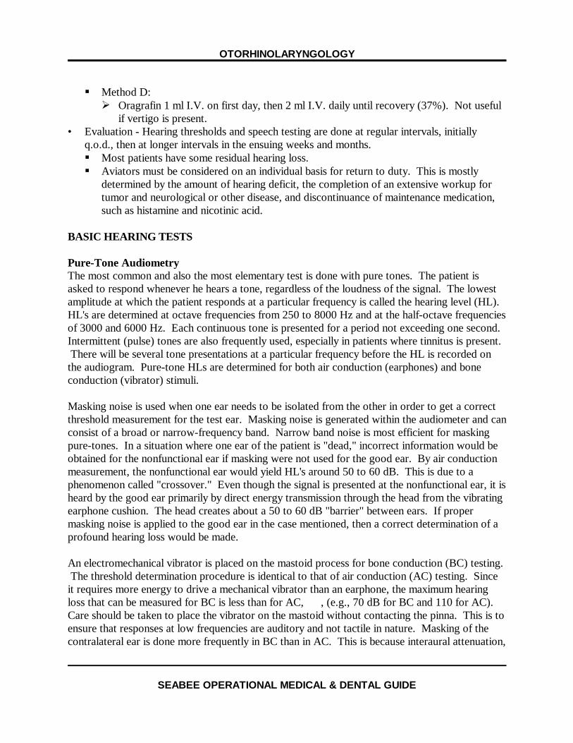
OTORHINOLARYNGOLOGY
SEABEE OPERATIONAL MEDICAL & DENTAL GUIDE
§ Method D:Ø Oragrafin 1 ml I.V. on first day, then 2 ml I.V. daily until recovery (37%). Not useful
if vertigo is present.• Evaluation - Hearing thresholds and speech testing are done at regular intervals, initially
q.o.d., then at longer intervals in the ensuing weeks and months.§ Most patients have some residual hearing loss.§ Aviators must be considered on an individual basis for return to duty. This is mostly
determined by the amount of hearing deficit, the completion of an extensive workup fortumor and neurological or other disease, and discontinuance of maintenance medication,such as histamine and nicotinic acid.
BASIC HEARING TESTS
Pure-Tone AudiometryThe most common and also the most elementary test is done with pure tones. The patient isasked to respond whenever he hears a tone, regardless of the loudness of the signal. The lowestamplitude at which the patient responds at a particular frequency is called the hearing level (HL). HL's are determined at octave frequencies from 250 to 8000 Hz and at the half-octave frequenciesof 3000 and 6000 Hz. Each continuous tone is presented for a period not exceeding one second. Intermittent (pulse) tones are also frequently used, especially in patients where tinnitus is present. There will be several tone presentations at a particular frequency before the HL is recorded onthe audiogram. Pure-tone HLs are determined for both air conduction (earphones) and boneconduction (vibrator) stimuli.
Masking noise is used when one ear needs to be isolated from the other in order to get a correctthreshold measurement for the test ear. Masking noise is generated within the audiometer and canconsist of a broad or narrow-frequency band. Narrow band noise is most efficient for maskingpure-tones. In a situation where one ear of the patient is "dead," incorrect information would beobtained for the nonfunctional ear if masking were not used for the good ear. By air conductionmeasurement, the nonfunctional ear would yield HL's around 50 to 60 dB. This is due to aphenomenon called "crossover." Even though the signal is presented at the nonfunctional ear, it isheard by the good ear primarily by direct energy transmission through the head from the vibratingearphone cushion. The head creates about a 50 to 60 dB "barrier" between ears. If propermasking noise is applied to the good ear in the case mentioned, then a correct determination of aprofound hearing loss would be made.
An electromechanical vibrator is placed on the mastoid process for bone conduction (BC) testing. The threshold determination procedure is identical to that of air conduction (AC) testing. Sinceit requires more energy to drive a mechanical vibrator than an earphone, the maximum hearingloss that can be measured for BC is less than for AC, , (e.g., 70 dB for BC and 110 for AC). Care should be taken to place the vibrator on the mastoid without contacting the pinna. This is toensure that responses at low frequencies are auditory and not tactile in nature. Masking of thecontralateral ear is done more frequently in BC than in AC. This is because interaural attenuation,
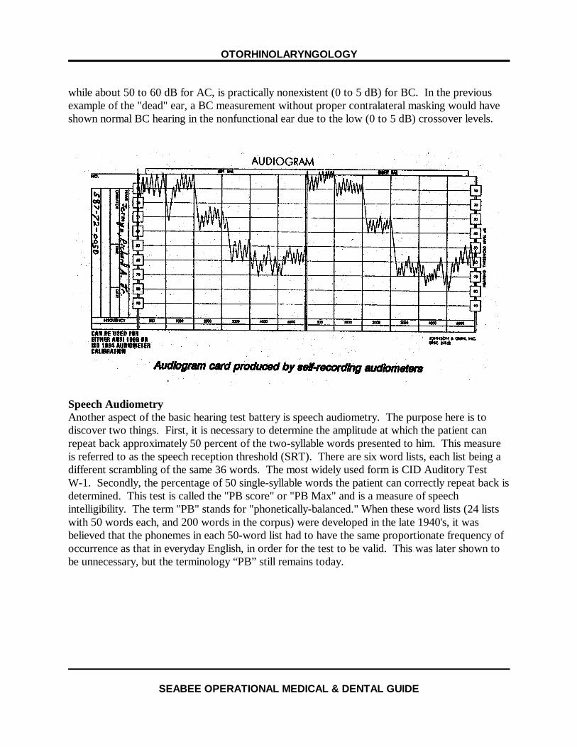
OTORHINOLARYNGOLOGY
SEABEE OPERATIONAL MEDICAL & DENTAL GUIDE
while about 50 to 60 dB for AC, is practically nonexistent (0 to 5 dB) for BC. In the previousexample of the "dead" ear, a BC measurement without proper contralateral masking would haveshown normal BC hearing in the nonfunctional ear due to the low (0 to 5 dB) crossover levels.
Speech AudiometryAnother aspect of the basic hearing test battery is speech audiometry. The purpose here is todiscover two things. First, it is necessary to determine the amplitude at which the patient canrepeat back approximately 50 percent of the two-syllable words presented to him. This measureis referred to as the speech reception threshold (SRT). There are six word lists, each list being adifferent scrambling of the same 36 words. The most widely used form is CID Auditory TestW-1. Secondly, the percentage of 50 single-syllable words the patient can correctly repeat back isdetermined. This test is called the "PB score" or "PB Max" and is a measure of speechintelligibility. The term "PB" stands for "phonetically-balanced." When these word lists (24 listswith 50 words each, and 200 words in the corpus) were developed in the late 1940's, it wasbelieved that the phonemes in each 50-word list had to have the same proportionate frequency ofoccurrence as that in everyday English, in order for the test to be valid. This was later shown tobe unnecessary, but the terminology “PB” still remains today.
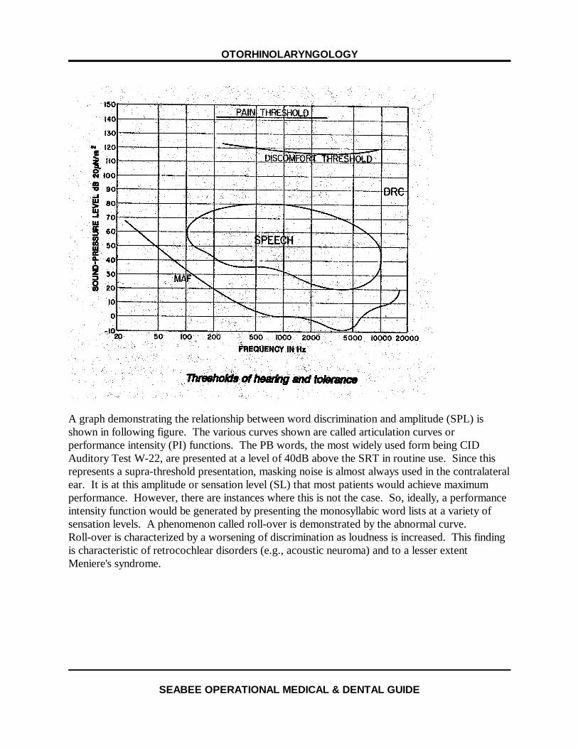
OTORHINOLARYNGOLOGY
SEABEE OPERATIONAL MEDICAL & DENTAL GUIDE
A graph demonstrating the relationship between word discrimination and amplitude (SPL) isshown in following figure. The various curves shown are called articulation curves orperformance intensity (PI) functions. The PB words, the most widely used form being CIDAuditory Test W-22, are presented at a level of 40dB above the SRT in routine use. Since thisrepresents a supra-threshold presentation, masking noise is almost always used in the contralateralear. It is at this amplitude or sensation level (SL) that most patients would achieve maximumperformance. However, there are instances where this is not the case. So, ideally, a performanceintensity function would be generated by presenting the monosyllabic word lists at a variety ofsensation levels. A phenomenon called roll-over is demonstrated by the abnormal curve. Roll-over is characterized by a worsening of discrimination as loudness is increased. This findingis characteristic of retrocochlear disorders (e.g., acoustic neuroma) and to a lesser extentMeniere's syndrome.
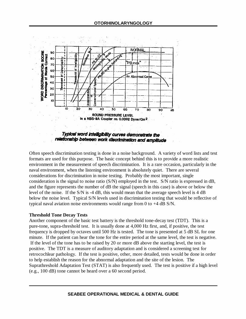
OTORHINOLARYNGOLOGY
SEABEE OPERATIONAL MEDICAL & DENTAL GUIDE
Often speech discrimination testing is done in a noise background. A variety of word lists and testformats are used for this purpose. The basic concept behind this is to provide a more realisticenvironment in the measurement of speech discrimination. It is a rare occasion, particularly in thenaval environment, when the listening environment is absolutely quiet. There are severalconsiderations for discrimination in noise testing. Probably the most important, singleconsideration is the signal to noise ratio (S/N) employed in the test. S/N ratio is expressed in dB,and the figure represents the number of dB the signal (speech in this case) is above or below thelevel of the noise. If the S/N is -4 dB, this would mean that the average speech level is 4 dBbelow the noise level. Typical S/N levels used in discrimination testing that would be reflective oftypical naval aviation noise environments would range from 0 to +4 dB S/N.
Threshold Tone Decay TestsAnother component of the basic test battery is the threshold tone-decay test (TDT). This is apure-tone, supra-threshold test. It is usually done at 4,000 Hz first, and, if positive, the testfrequency is dropped by octaves until 500 Hz is tested. The tone is presented at 5 dB SL for oneminute. If the patient can hear the tone for the entire period at the same level, the test is negative. If the level of the tone has to be raised by 20 or more dB above the starting level, the test ispositive. The TDT is a measure of auditory adaptation and is considered a screening test forretrocochlear pathology. If the test is positive, other, more detailed, tests would be done in orderto help establish the reason for the abnormal adaptation and the site of the lesion. TheSuprathreshold Adaptation Test (STAT) is also frequently used. The test is positive if a high level(e.g., 100 dB) tone cannot be heard over a 60 second period.
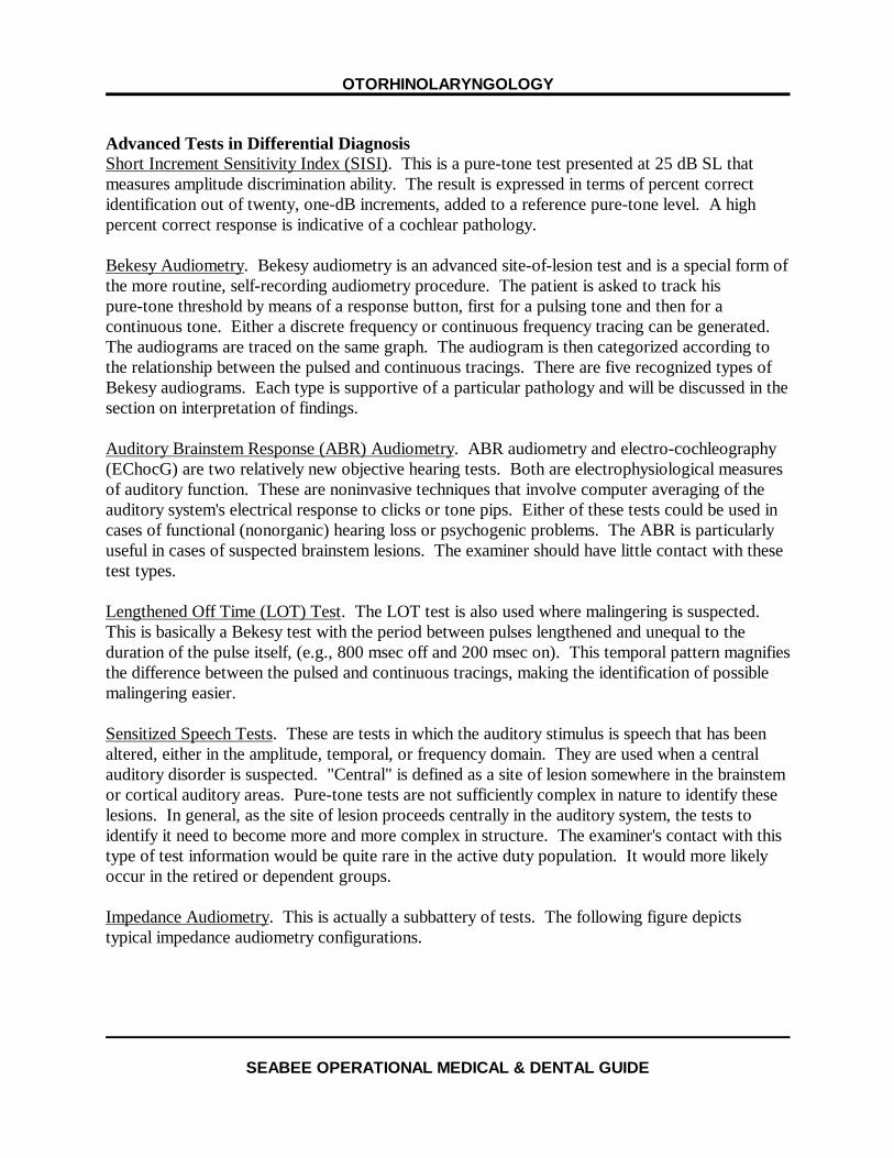
OTORHINOLARYNGOLOGY
SEABEE OPERATIONAL MEDICAL & DENTAL GUIDE
Advanced Tests in Differential DiagnosisShort Increment Sensitivity Index (SISI). This is a pure-tone test presented at 25 dB SL thatmeasures amplitude discrimination ability. The result is expressed in terms of percent correctidentification out of twenty, one-dB increments, added to a reference pure-tone level. A highpercent correct response is indicative of a cochlear pathology.
Bekesy Audiometry. Bekesy audiometry is an advanced site-of-lesion test and is a special form ofthe more routine, self-recording audiometry procedure. The patient is asked to track hispure-tone threshold by means of a response button, first for a pulsing tone and then for acontinuous tone. Either a discrete frequency or continuous frequency tracing can be generated.The audiograms are traced on the same graph. The audiogram is then categorized according tothe relationship between the pulsed and continuous tracings. There are five recognized types ofBekesy audiograms. Each type is supportive of a particular pathology and will be discussed in thesection on interpretation of findings.
Auditory Brainstem Response (ABR) Audiometry. ABR audiometry and electro-cochleography(EChocG) are two relatively new objective hearing tests. Both are electrophysiological measuresof auditory function. These are noninvasive techniques that involve computer averaging of theauditory system's electrical response to clicks or tone pips. Either of these tests could be used incases of functional (nonorganic) hearing loss or psychogenic problems. The ABR is particularlyuseful in cases of suspected brainstem lesions. The examiner should have little contact with thesetest types.
Lengthened Off Time (LOT) Test. The LOT test is also used where malingering is suspected. This is basically a Bekesy test with the period between pulses lengthened and unequal to theduration of the pulse itself, (e.g., 800 msec off and 200 msec on). This temporal pattern magnifiesthe difference between the pulsed and continuous tracings, making the identification of possiblemalingering easier.
Sensitized Speech Tests. These are tests in which the auditory stimulus is speech that has beenaltered, either in the amplitude, temporal, or frequency domain. They are used when a centralauditory disorder is suspected. "Central" is defined as a site of lesion somewhere in the brainstemor cortical auditory areas. Pure-tone tests are not sufficiently complex in nature to identify theselesions. In general, as the site of lesion proceeds centrally in the auditory system, the tests toidentify it need to become more and more complex in structure. The examiner's contact with thistype of test information would be quite rare in the active duty population. It would more likelyoccur in the retired or dependent groups.
Impedance Audiometry. This is actually a subbattery of tests. The following figure depictstypical impedance audiometry configurations.
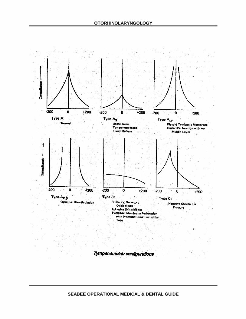
OTORHINOLARYNGOLOGY
SEABEE OPERATIONAL MEDICAL & DENTAL GUIDE
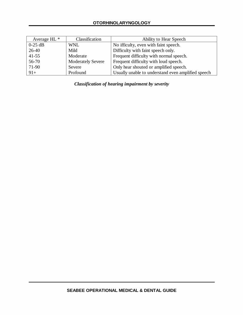
OTORHINOLARYNGOLOGY
SEABEE OPERATIONAL MEDICAL & DENTAL GUIDE
Average HL * Classification Ability to Hear Speech0-25 dB26-4041-5556-7071-9091+
WNLMildModerateModerately SevereSevereProfound
No ifficulty, even with faint speech.Difficulty with faint speech only.Frequent difficulty with normal speech.Frequent difficulty with loud speech.Only hear shouted or amplified speech.Usually unable to understand even amplified speech
Classification of hearing impairment by severity

OTORHINOLARYNGOLOGY
SEABEE OPERATIONAL MEDICAL & DENTAL GUIDE
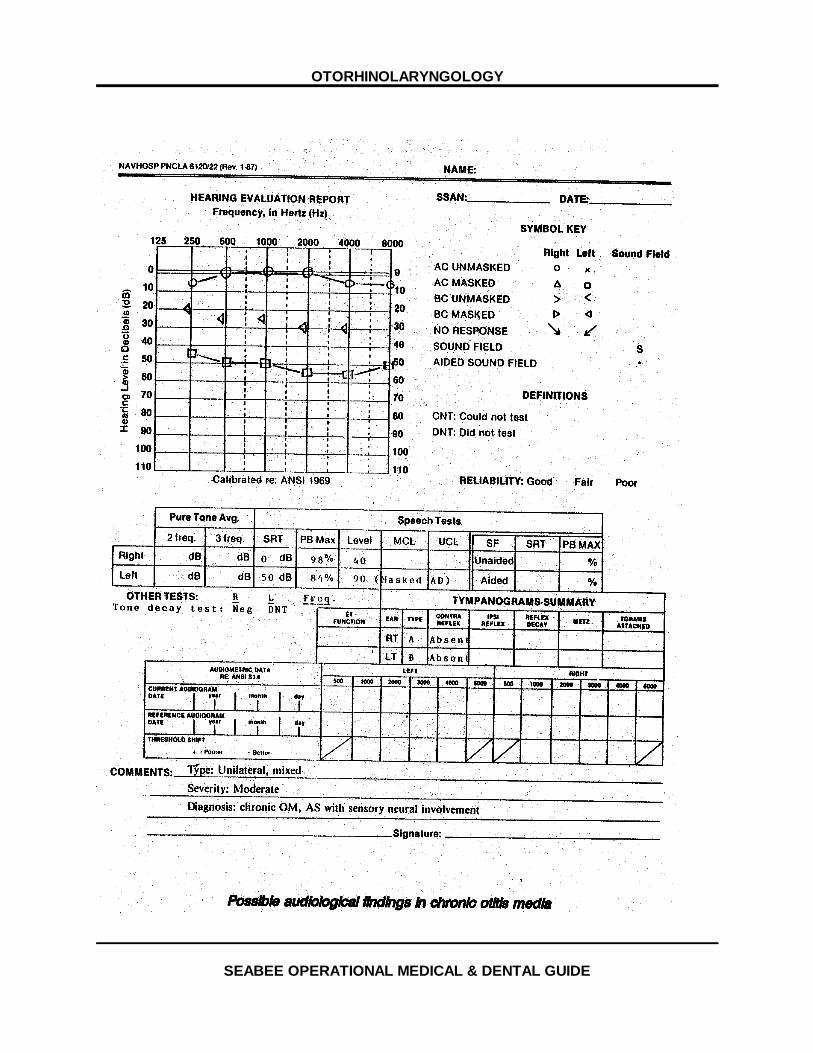
OTORHINOLARYNGOLOGY
SEABEE OPERATIONAL MEDICAL & DENTAL GUIDE
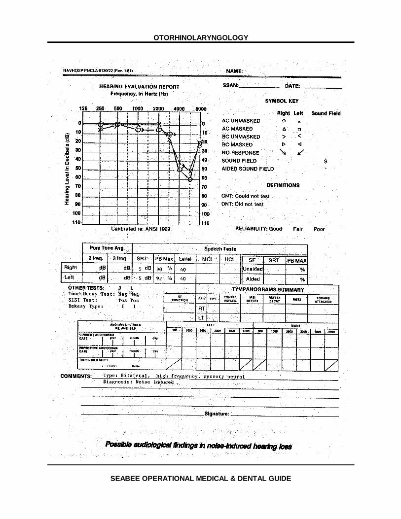
OTORHINOLARYNGOLOGY
SEABEE OPERATIONAL MEDICAL & DENTAL GUIDE
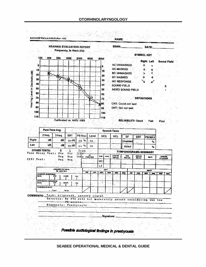
OTORHINOLARYNGOLOGY
SEABEE OPERATIONAL MEDICAL & DENTAL GUIDE
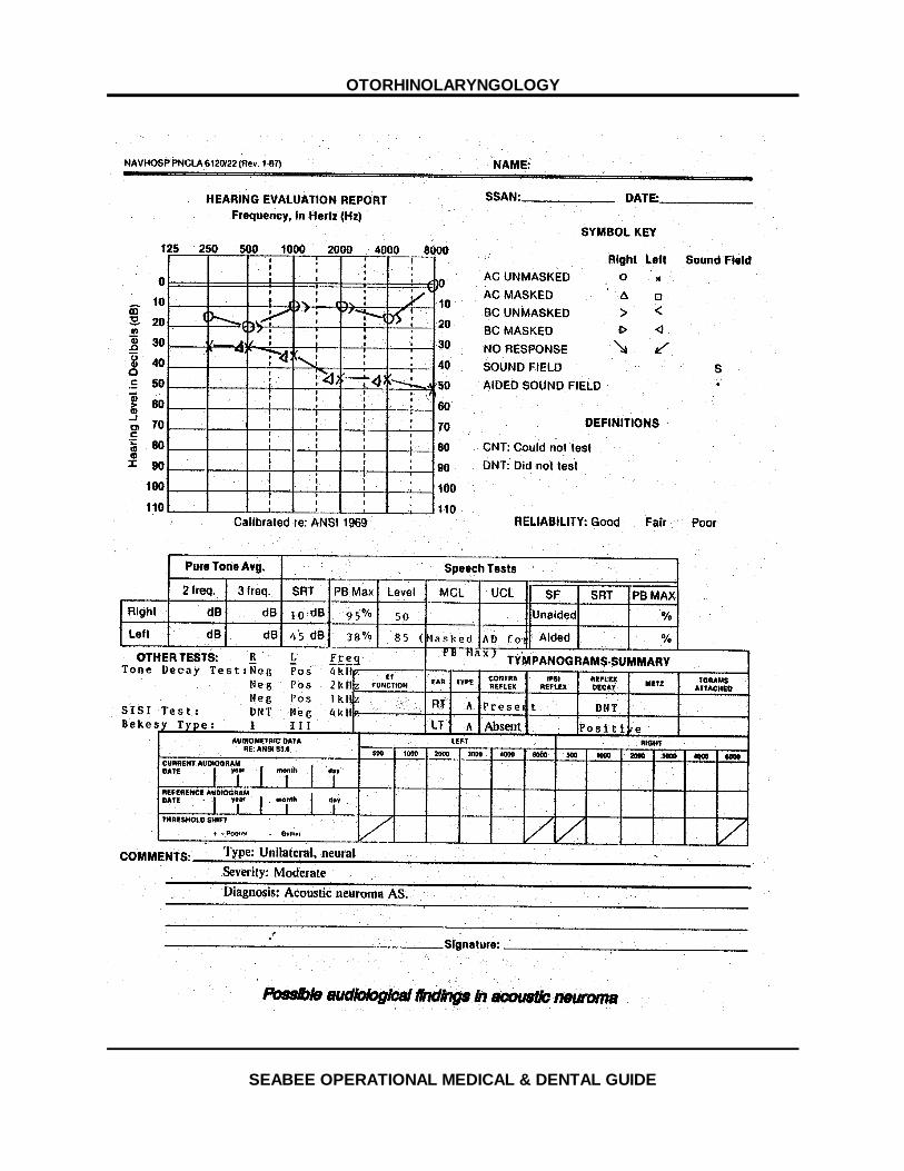
OTORHINOLARYNGOLOGY
SEABEE OPERATIONAL MEDICAL & DENTAL GUIDE
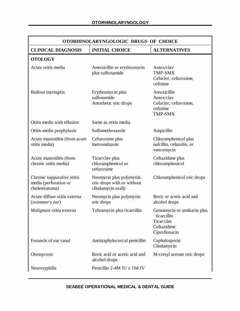
OTORHINOLARYNGOLOGY
SEABEE OPERATIONAL MEDICAL & DENTAL GUIDE
OTORHINOLARYNGOLOGIC DRUGS OF CHOICE
CLINICAL DIAGNOSIS INITIAL CHOICE ALTERNATIVES
OTOLOGY
Acute otitis media Amoxicillin or erythromycinplus sulfonamide
Amox/clavTMP-SMXCefaclor, cefuroxime,cefixime
Bullous myringitis Erythromycin plussulfonamideAnesthetic otic drops
AmoxicillinAmox/clavCefaclor, cefuroxime,cefiximeTMP-SMX
Otitis media with effusion Same as otitis media
Otitis media prophylaxis Sulfamethoxazole Ampicillin
Acute mastoiditis (from acuteotitis media)
Cefuroxime plusmetronidazole
Chloramphenicol plusnafcillin, cefazolin, orvancomycin
Acute mastoiditis (fromchronic otitis media)
Ticar/clav pluschloramphenicol orcefuroxime
Ceftazidime pluschloramphenicol
Chronic suppurative otitismedia (perforation orcholesteatoma)
Neomycin plus polymyxinotic drops with or withoutclindamycin orally
Chloramphenicol otic drops
Acute diffuse otitis externa(swimmer's ear)
Neomycin plus polymyxinotic drops
Boric or acetic acid andalcohol drops
Malignant otitis externa Tobramycin plus ticarcillin Gentamycin or amikacin plus ticarcillinTicar/clavCeftazidimeCiprofloxacin
Furuncle of ear canal Antistaphylococcal penicillin CephalosporinClindamycin
Otomycosis Boric acid or acetic acid andalcohol drops
M-cresyl acetate otic drops
Neurosyphilis Penicillin 2-4M IU x 10d IV
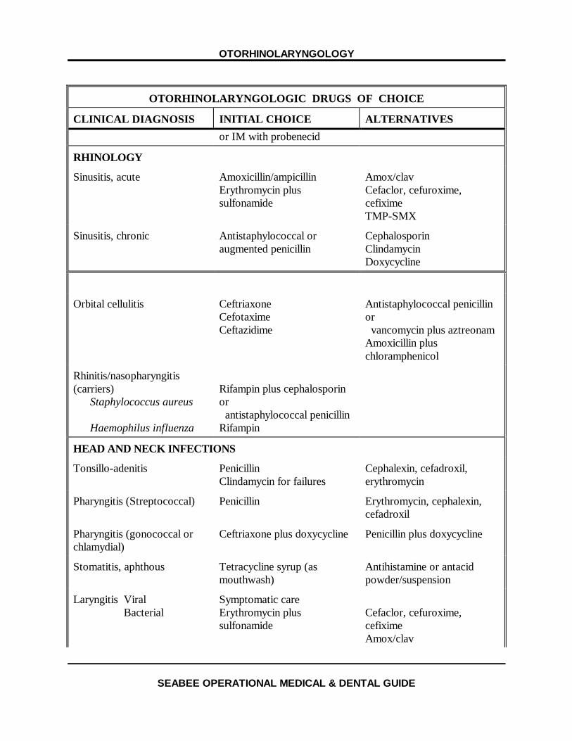
OTORHINOLARYNGOLOGY
SEABEE OPERATIONAL MEDICAL & DENTAL GUIDE
OTORHINOLARYNGOLOGIC DRUGS OF CHOICE
CLINICAL DIAGNOSIS INITIAL CHOICE ALTERNATIVESor IM with probenecid
RHINOLOGY
Sinusitis, acute Amoxicillin/ampicillinErythromycin plussulfonamide
Amox/clavCefaclor, cefuroxime,cefiximeTMP-SMX
Sinusitis, chronic Antistaphylococcal oraugmented penicillin
CephalosporinClindamycinDoxycycline
Orbital cellulitis CeftriaxoneCefotaximeCeftazidime
Antistaphylococcal penicillinor vancomycin plus aztreonamAmoxicillin pluschloramphenicol
Rhinitis/nasopharyngitis(carriers)
Staphylococcus aureus
Haemophilus influenza
Rifampin plus cephalosporinor antistaphylococcal penicillinRifampin
HEAD AND NECK INFECTIONS
Tonsillo-adenitis PenicillinClindamycin for failures
Cephalexin, cefadroxil,erythromycin
Pharyngitis (Streptococcal) Penicillin Erythromycin, cephalexin,cefadroxil
Pharyngitis (gonococcal orchlamydial)
Ceftriaxone plus doxycycline Penicillin plus doxycycline
Stomatitis, aphthous Tetracycline syrup (asmouthwash)
Antihistamine or antacidpowder/suspension
Laryngitis ViralBacterial
Symptomatic careErythromycin plussulfonamide
Cefaclor, cefuroxime,cefiximeAmox/clav
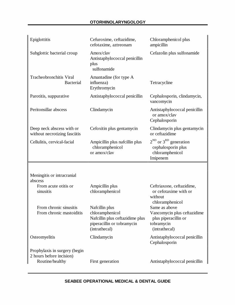
OTORHINOLARYNGOLOGY
SEABEE OPERATIONAL MEDICAL & DENTAL GUIDE
Epiglottitis Cefuroxime, ceftazidime,cefotaxime, aztreonam
Chloramphenicol plusampicillin
Subglottic bacterial croup Amox/clavAntistaphylococcal penicillinplus sulfonamide
Cefazolin plus sulfonamide
Tracheobronchitis ViralBacterial
Amantadine (for type Ainfluenza)Erythromycin
Tetracycline
Parotitis, suppurative Antistaphylococcal penicillin Cephalosporin, clindamycin,vancomycin
Peritonsillar abscess Clindamycin Antistaphylococcal penicillin or amox/clavCephalosporin
Deep neck abscess with orwithout necrotizing fasciitis
Cefoxitin plus gentamycin Clindamycin plus gentamycinor ceftazidime
Cellulitis, cervical-facial Ampicillin plus nafcillin plus chloramphenicolor amox/clav
2ND or 3RD generation cephalosporin plus chloramphenicolImipenem
Meningitis or intracranialabscess
From acute otitis orsinusitis
From chronic sinusitisFrom chronic mastoiditis
Ampicillin pluschloramphenicol
Nafcillin pluschloramphenicolNafcillin plus ceftazidime pluspiperacillin or tobramycin(intrathecal)
Ceftriaxone, ceftazidime, or cefotaxime with orwithout chloramphenicolSame as aboveVancomycin plus ceftazidime plus piperacillin ortobramycin (intrathecal)
Osteomyelitis Clindamycin Antistaphylococcal penicillinCephalosporin
Prophylaxis in surgery (begin2 hours before incision)
Routine/healthy First generation Antistaphylococcal penicillin
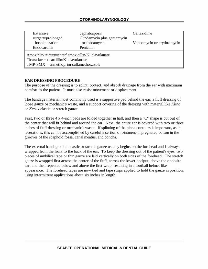
OTORHINOLARYNGOLOGY
SEABEE OPERATIONAL MEDICAL & DENTAL GUIDE
Extensivesurgery/prolonged hospitalizationEndocarditis
cephalosporinClindamycin plus gentamycin or tobramycinPenicillin
Ceftazidime
Vancomycin or erythromycin
Amox/clav = augmented amoxicillin/K+ clavulanateTicar/clav = ticarcillin/K+ clavulanateTMP-SMX = trimethoprim-sulfamethoxazole
EAR DRESSING PROCEDUREThe purpose of the dressing is to splint, protect, and absorb drainage from the ear with maximumcomfort to the patient. It must also resist movement or displacement.
The bandage material most commonly used is a supportive pad behind the ear, a fluff dressing ofloose gauze or mechanic's waste, and a support covering of the dressing with material like Klingor Kerlix elastic or stretch gauze.
First, two or three 4 x 4-inch pads are folded together in half, and then a "C" shape is cut out ofthe center that will fit behind and around the ear. Next, the entire ear is covered with two or threeinches of fluff dressing or mechanic's waste. If splinting of the pinna contours is important, as inlacerations, this can be accomplished by careful insertion of ointment-impregnated cotton in thegrooves of the scaphoid fossa, canal meatus, and concha.
The external bandage of an elastic or stretch gauze usually begins on the forehead and is alwayswrapped from the front to the back of the ear. To keep the dressing out of the patient's eyes, twopieces of umbilical tape or thin gauze are laid vertically on both sides of the forehead. The stretchgauze is wrapped first across the center of the fluff, across the lower occiput, above the oppositeear, and then repeated below and above the first wrap, resulting in a football helmet likeappearance. The forehead tapes are now tied and tape strips applied to hold the gauze in position,using intermittent applications about six inches in length.