outline - centurion university
TRANSCRIPT

Outline
o Introduction
o Formation of blood cells
o Types of leucocytes
o Cells of the innate immune system
o Phagocytosis and intracellular
killing
o Cells of the adaptive immune
system
o Cell-Mediated Cytotoxicity

Introduction
• Blood cells or leucocytes serve as sentinels and defenders
against infection.
• They move around the body via the lymphatic and blood
circulatory systems.
• Leucocytes are classified by morphology- number of nuclei
lobes and presence or absence of cytoplasmic granules.
• Leukocytes may be found as individual cells throughout the
body, as accumulations within lymphoid organs (e.g., spleen,
lymph nodes) and at sites of infection or inflammation.

Formation of blood cellsAll bloodborne cells originate in the bone marrow.
Pluripotent hematopoietic stem cell in the bonemarrow give rise to two major lineages; a myeloidlineage and a lymphoid lineage.
Cells of the myeloid lineage differentiate further intoplatelets, erythrocytes, eosinophils, basophils (andmast cells), neutrophils, monocytes/macrophages,and some dendritic cells.
Cells of the lymphoid lineage differentiate furtherinto T and B lymphocytes, NK cells, and some

Fig 1: Hematopoiesis
Types of leucocytes
White blood cells that have multilobed nuclei
and contain conspicuous cytoplasmic granules
are known as granulocytes .
Others with a single, unlobed nucleus and
cytoplasm that contains few or no granules are
known as agranular leukocytes.
Agranular leukocytes derive from lymphoid or
myeloid lineage precursors and account for
approximately 35% to 38% of the leukocytes in

Fig 2: Types of leukocytes.
Cells of the innate immune system
Myeloid Cells: First line of defense against
invading organisms in non-specific innate
immunity.
Neutrophils
Eosinophils
Basophils/Mast cells
Monocytes/Macrophages/Dendritic
Cells
Lymphoid Cells:

Neutrophils
Comprises approximately 60% of the peripheral blood
leukocytes, neutrophils are the most numerous
leukocyte population.–Neutrophils have multi lobed nuclei (2-5) and cytoplasmic granules that
stain with both acid and basic dyes.
– often called polymorphonuclear cells (PMN's).
The neutrophil's main role is in inflammation.
– First to arrive at inflammation site
– Leave blood/endothelium into tissue (extravasation).
Neutrophils are attracted into the tissue by chemotactic
factors stimulated by tissue damage
– complement proteins, clotting
proteins and T cell derived

In the tissues, neutrophils are active
phagocytes.
microorganisms via
or independent
They destroy ingested
oxygen-dependent
pathways.Produce myeloperoxidases to assist oxidated antimicrobial effects.
Produce lactoferrin and lysozyme as direct antimicrobial agents.
Produce leukotrienes and prostaglandins, products of the lipoxygenase
and cyclo-oxygenase pathways, to mediate vascular functions.
Deficiencies in pathways increase susceptibility
to infections.

Characteristics of Neutrophil
granulesPrimary granules Secondary granules
Azurophilic; young neutrophils Specific for mature neutrophils
Contain:
cationic proteins, lysozyme, defensins,
elastase and
Contain:
Lysozyme,
NADPH oxidase components and
myeloperoxidase Lactoferrin and B12-binding protein

Eosinophils
Eosinophils have bilobed nuclei and
cytoplasmic granules that stain with the
acid dye eosin (hence its name).
of circulatingConstitute 1%-3%
leucocytes.
Involved in asthma.
sometimesEosinophils are motile,
phagocytic, and are particularly
active in parasitic infection.

BasophilsBasophils have bilobed nuclei and cytoplasmicgranules that stain with the basic dye methyleneblue.
Found in low numbers in the blood (<1%). Act like mast cells.
They are nonphagocytic
Involved in allergic reactions (Type I hypersensitivity responses).
Have high affinity Fc receptors for IgE on their surface.
When an individual is exposed to an allergen, specific IgE is produced. This IgE binds to the surface of basophils.
Upon re-exposure to the allergen, the allergen binds to IgE on thesurface of basophils resulting in degranulation.
Cross-linking of the IgE causes the basophils to release pharmacologically active mediators (histamine, prostaglandins, leukotrienes)

Mast cells
Similar importance in allergic reactions as
basophils, but only found in tissues.
Contain granules with preformed
mediators to be released after stimulation– histamine, prostaglandins
– leukotrienes
Stimulation of mast cells occurs by the
anaphylatoxins (complement proteins C3a
and C5a) or by cross-linking of surface
immunoglobulin (IgE).
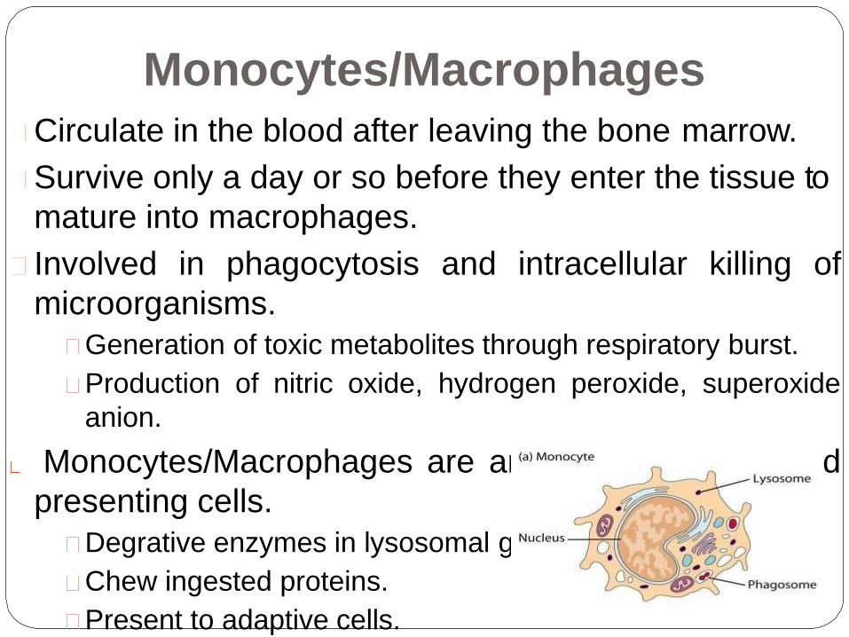
Monocytes/Macrophages
Circulate in the blood after leaving the bone marrow.
Survive only a day or so before they enter the tissue to
mature into macrophages.
Involved in phagocytosis and intracellular killing of
microorganisms.
Generation of toxic metabolites through respiratory burst.
Production of nitric oxide, hydrogen peroxide, superoxide
anion.
Monocytes/Macrophages are antigen processing and
presenting cells.
Degrative enzymes in lysosomal granules.
Chew ingested proteins.
Present to adaptive cells.
Macrophages
When monocytes enter the tissues and become
macrophages:
– Enlarge and increase production of intracellular
lysozymes
– Greater phagocytosis.
– Can live for years in tissue; highly motile.
Activation of these cells occurs by phagocytosis
of antigens, or in response to T cell derived
cytokines.
Activated macrophages recognize and remove
unwanted particulate matter including products

After activation, these cells secrete cytokines, chemokines,
lysozymes
response.
scavengers
and other factors to upregulate immune
In
can
chronic inflammation, macrophage
become “giant cells” or “foamy
macrophages”.
Fig: Electron micrograph of macropha

Dendritic cellsSpecialized phagocytic cells found in most tissues.
Arise both from the myeloid and lymphoid lineages.
Abundant at interfaces between the external and internal
environments (skin, lining of the gastrointestinal tract), where
they encounter invading pathogens.
Actively motile; continuously sample surroundings by endocytic
processes.
Dendritic cells are very efficient at activation of T cells.

Natural Killer cellsAlso known as large granular lymphocytes (LGLs)
Functionally cytotoxic representing an innate population that kill virally infected or tumor target cells.
Killing is nonspecific - they do not need to recognize foreign antigenspresented on the target cell.– NK cells do not have a specific cell receptor. Target recognition occurs by a Killer Inhibitory Receptor, KIR,which assess MHC I molecules on the target cell surface. MHC 1 molecule is lacking on infected and tumortargets.
Kill targets by releasing perforin which damages target cell membranes. Can alsoinduce apoptosis in the target cell.
NK cells is different from NK T cells.
-NKT cells has some of the attributes of T cell and NK cell.
Like T cells, NK1-T cells have T cell receptors
(TCRs).Unlike most T cells, the TCRs of NK1-T cells interact
with MHC-like molecules called CD1 rather than with
class I or class II MHC molecules. Like NK cells, theyhave
variable levels of CD16 and other receptors typical of NK
cells, and they can kill cells.

Phagocytosis and intracellularkilling
Phagocytosis is the engulfment and degradation of microbes andother particulate matter by cells such as macrophages/monocytes,dend ritic cells, and neutrophils .
Steps in phagocytosis-1) Detection of the foreign particle and movement of the phagocyte to the area
by chemotaxis.2) Attachment of the foreign particle to the phagocyte.
3) Engulfment or ingestion of the foreign particle into a vesicle called aphagosome.
4) Fusion with lysosome and formation of the phagolysosome.
5) Intracellular killing and digestion.6) In the case of macrophages, egestion and antigen presentation.
Phagocytes detect microbes by the presence of N-formylatedpeptides, activated complement proteins and the mediators ofinflammation.
Phagocytes attach to microbes using opsonins, such as IgG and the complement protein C3b. In the absence of opsonins, phagocytes can

Fig: Steps in phagocytosis
Pathways of intracellular killing
Lysosomes employ multiple mechanisms for
killing and degrading ingested microbe. These
include;Oxygen-independent killing
Oxygen-dependent-MPO independent killing
Oxygen-dependent-MPO dependent killing
Nitric Oxide mediated killing
In oxygen-independent killing, activated
phagocytes synthesize lysozyme and various
hydrolytic enzymes whose degradative
activities do not require oxygen.

Mediators of oxygen-independent
killing in phagolysosome
Effector Molecule
Cationic proteins (cathepsin)
Lysozyme
Lactoferrin
FunctionDamage to microbial
mHyedmroblryasneessmucopeptides in
the cell wall
Deprives pathogens of iron
Hydrolytic enzymes (proteases) Digests killed organisms
During phagocytosis there is an increase in glucose and oxygen
consumption which is referred to as the respiratory burst.
The consequence of the respiratory burst is that a number of
oxygen-containing compounds are produced which kill the
bacteria being phagocytosed. This is referred to as oxygen-
dependent intracellular killing.

Oxygen-dependent myeloperoxidase-
independent reactions

Oxygen-dependent myeloperoxidase-
dependent reactions
Detoxification reactions
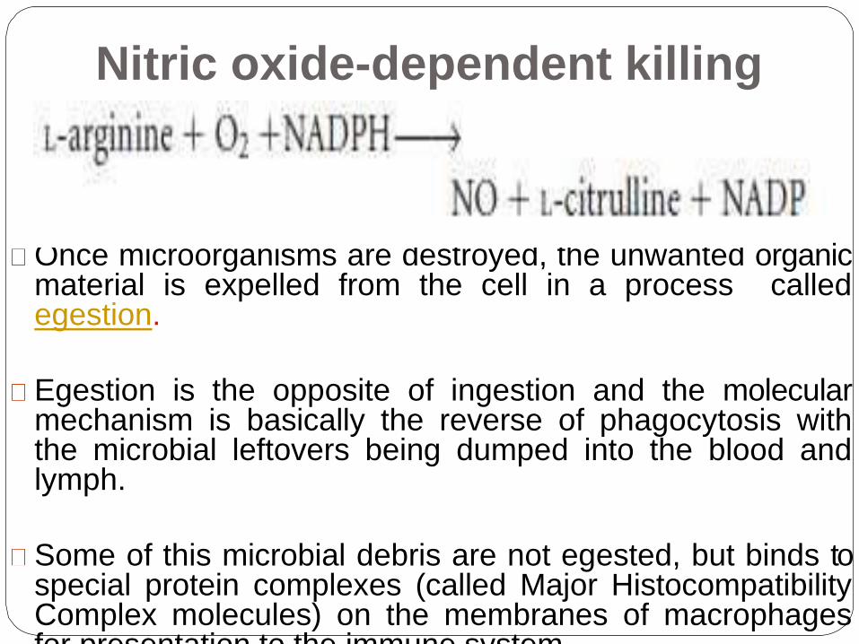
Nitric oxide-dependent killing
Once microorganisms are destroyed, the unwanted organicmaterial is expelled from the cell in a process calledegestion.
Egestion is the opposite of ingestion and the molecularmechanism is basically the reverse of phagocytosis withthe microbial leftovers being dumped into the blood andlymph.
Some of this microbial debris are not egested, but binds tospecial protein complexes (called Major HistocompatibilityComplex molecules) on the membranes of macrophagesfor presentation to the immune system.

Cells of the adaptive immune
systemAdaptive immune responses are mediated by a
group of
which T and
(T cells
include
and
recognize foreign
B cells)
material
leukocytes, the
B
that
or
specialized
lymphocytes,
lymphocytes
specifically
antigens.
All lymphocytes are derived from bone marrow
stem cells, but T cells then develop in the
thymus, while B cells develop in the bone

B Lymphocytes
Develop from stem cells in the bone marrow.
Produce antibodies with specificity for antigens and display
it on their surfaces to function as BCRs.
Integral in humoral immunity
Plasma cells = terminally differentiated B cells that secrete
immunoglobulins.
Memory cells- secondary immune response is swifter and
stronger.
Upon activation, a B cell can switch to produce a
different class of antibody, with the same antigen
into antibody secreting cells is antigen-
specificity.
Activation
dependent.

T-LymphocytesT lymphocytes develop in the thymus.
Regulate immune responses.
Integral in cell mediated immunity.
Critical in B cell-antibody production.
Mature T cells display either CD4 or CD8.Cells with a CD4 marker are called helper T cells (Th cells).
CD8 marker positive cells are cytotoxic T cells (Tc cells).
There are several different types of T cell, and they have a variety of functions :
Type 1 helper T cells or TH1 cells - interacts with mononuclear phagocytes and helps them destroy intracellular pathogens•
Type 2 helper T cells or TH2 cells; interacts with B cells and helps them to divide, differentiate, and make antibody•
Cytotoxic T lymphocytes (CTLs or TC cells). responsible for the destruction ofhost cells that have become infected by viruses or other intracellular pathogens.
Regulatory T cells or Tregs, help to control the development of immune responses, and limit reactions against self tissues.

Fig: B and T
Lymphocytes

Cell-Mediated CytotoxicityCytotoxicity describes the ways in which leukocytes recognize and destroy other cells.
Cell-mediated cytotoxicity is an essential defense against:intracellular pathogens, including viruses;
some bacteria;
some parasites.
Tumor cells, eukaryotic pathogens, and even cells of the bodymay also become the target of cytotoxic cells.
Several types of cell have cytotoxic activity including:cytotoxic T lymphocytes (CTLs);
natural killer (NK) cells
CTLs and NK cells use a variety of different mechanisms to kill their targets. These include:
direct cell–cell signaling via surface molecules; and
granule-associated killing


ImmunopathologyAutoimmune disease - When the immune system reacts
against ‘self’ components, for example rheumatoid arthritis
or pernicious anemia.
Immunodeficiency- If any elements of the immune system
are defective, the individual may not be able to fight
infections adequately.primary immundeficiencies are hereditary and start to manifest shortly
after birth; eg chronic granulomatous disease (CGD) and leukocyte
adhesion deficiency (LAD).
Secondary immunodeficiencies develop later in life, for example the
acquired immune deficiency syndrome (AIDS).
Hypersensitivity- Sometimes immune reactions are out of
all proportion to the damage that may be caused by a
pathogen. The immune system may also mount a reaction
to a harmless antigen, such as a food molecule causing

Table 1. Characteristic infections in primary
immunodeficiencies.

Conclusionsystem has evolved to protect us fromThe immune
pathogens.
Phagocytes and lymphocytes are key mediators ofimmunity. Phagocytes internalize pathogens and degradethem. Lymphocytes (B and T cells) have receptors thatrecognize specific molecular components of pathogensand have specialized functions. B cells make antibodies(effective against extracellular pathogens), cytotoxic Tlymphocytes (CTLs) kill virally infected cells, and helper Tcells coordinate the immune response by direct cell–cellinteractions and the release of cytokines.
The immune system may fail (immunopathology). This canbe a result of immunodeficiency, hypersensitivity, ordysregulation leading to autoimmune diseases.

References
Doan, T., Melvold, R., Viselli, S. and Waltenbaugh,C.(2013). Lippincott’s illustrated reviews
2ndImmunology. Philadelphia, Edn. LippincottWilliams & Wilkins. ISBN 978-1-4511-0937-5.
Kindt, T.J., Osborne, B.A. and Goldsby, R.A. (2006).Kuby Immunology, 6th edn. Oxford: WH Freeman.
Male, D., Brostoff, J., Roth, D.B. and Roitt, I.(2013).Immunology. China, 8th Edn. Elsevier. ISBN 978-0-702-04548-6.
Roitt, I.M. and Delves, P. (2011)Essential