outline i. triangles of neck ii. deep lecture powerpoint 2019.pdf · triangles of neck – for...
TRANSCRIPT
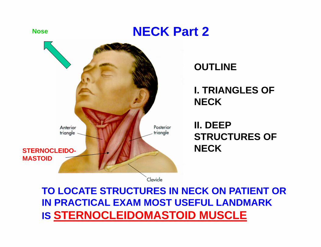
NECK Part 2
TO LOCATE STRUCTURES IN NECK ON PATIENT OR IN PRACTICAL EXAM MOST USEFUL LANDMARKIS STERNOCLEIDOMASTOID MUSCLE
OUTLINE
I. TRIANGLES OFNECK
II. DEEPSTRUCTURES OFNECKSTERNOCLEIDO-
MASTOID
Nose
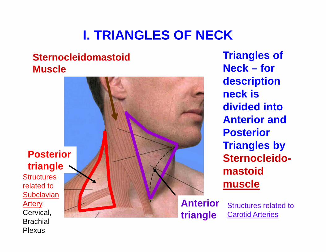
Triangles of Neck – for description neck is divided into Anterior and Posterior Triangles bySternocleido-mastoid muscle
Posterior triangle
Anterior triangle
SternocleidomastoidMuscle
I. TRIANGLES OF NECK
Structures related to Carotid Arteries
Structures related to Subclavian Artery, Cervical, Brachial Plexus

1. Boundaries
Superficial cover - Superficial fascia, Platysma and Investing Layer
Deep - (floor) Prevertebral layer of fascia
Inferior -Clavicle
A. POSTERIOR TRIANGLE
PosteriorTrapezius
Anterior -SternocleidomastoidMuscle

1) Lesser Occipitalbehind ear
4) Supraclavicular -lower neck and shoulder
2) Great Auricular -skin over parotid, inferior to ear
3) Transverse Cervical - anterior neck
NOSE
B. CONTENTS OF POSTERIOR TRIANGLE
External JugularVein - courseson surface of Sternocleidomastoid
Occipital Artery
CUTANEOUSNERVES
NOTE: SADLY, STUDENTS HAVENOT REMEMBERED THIS ON THE PRACTICAL EXAM

Scalenus Ant. M.
Phrenic n . ant to Ant. Scalene, Posterior to Sternocleido.BRACHIAL
PLEXUS
Scalenus Med. M.
ACCESSORY N. CN XI
Note: Accessory nerve - divides Posterior triangle into 'Carefree' -superior 'Careful' -inferior
Nose
POSTERIOR TRIANGLE -deeper, saw throughclavicle

Transverse cervical artery
Supra-scapular artery
Scalenus Ant. M.
Phrenic nerve
BRACHIALPLEXUS
Scalenus Med. M.
Subclavian artery
Subclavian vein
note: Subclavian vein is not in the posterior triangle
Nose
from Thyro-cervicalTrunk
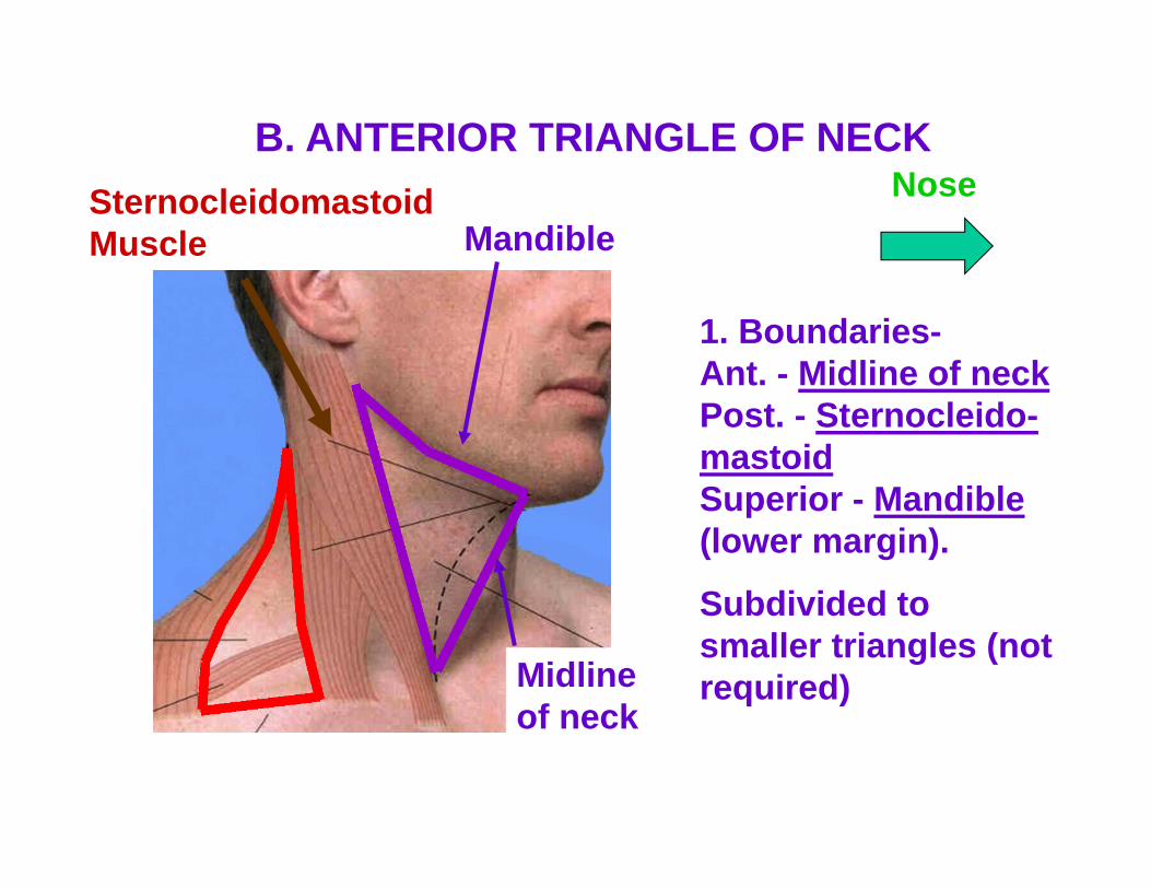
1. Boundaries-Ant. - Midline of neckPost. - Sternocleido-mastoidSuperior - Mandible (lower margin).
Subdivided to smaller triangles (not required)Midline
of neck
SternocleidomastoidMuscle
B. ANTERIOR TRIANGLE OF NECKNose
Mandible

In Carotid sheath:Int. and Common Carotid A., Int. Jug. V., Vagus N.
Follow to branches of Ext. carotid
- cut through Sternocleido-mastoid
Hypoglossal n. XII and Ansa Cervicalis
2. CONTENTS OF ANTERIOR TRIANGLE OF NECKNose
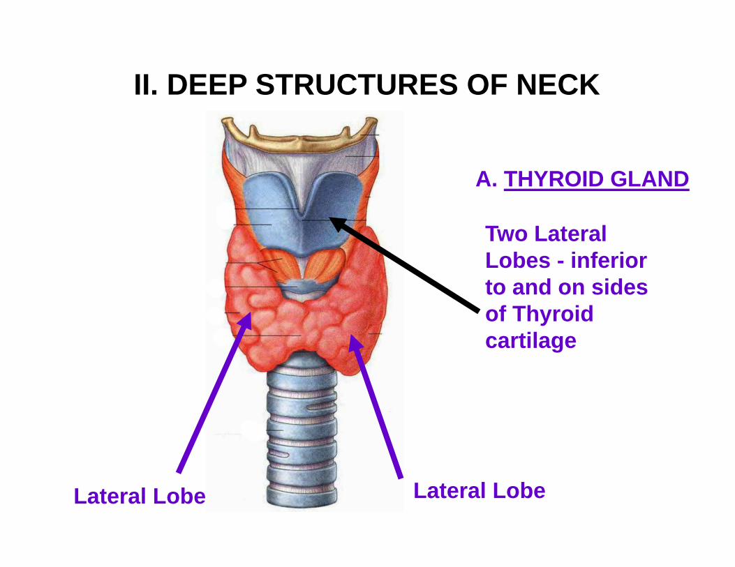
Two Lateral Lobes - inferior to and on sides of Thyroid cartilage
Lateral Lobe
A. THYROID GLAND
Lateral Lobe
II. DEEP STRUCTURES OF NECK
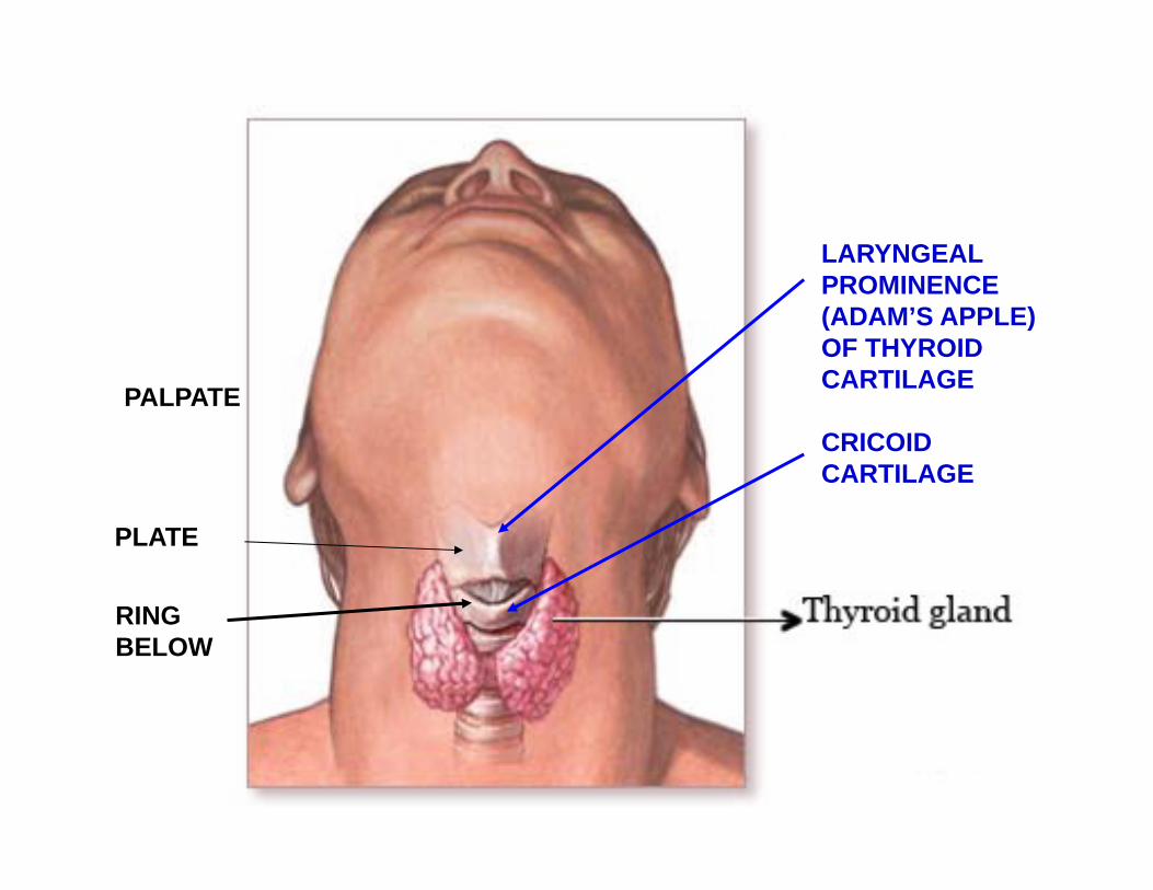
LARYNGEAL PROMINENCE (ADAM’S APPLE) OF THYROIDCARTILAGE
CRICOIDCARTILAGE
PLATE
RINGBELOW
PALPATE

THYROID GLAND
Right lateral lobe
Left lateral lobe
Isthmus -located below cricoid cartilage
Pyramidal lobe - when present often attached to hyoid bone by fibrous strand
Absence ofIsthmus
Normal variations common
*QUESTION: WHERE DOESTHE THYROID ARISE IN DEVELOPMENT?
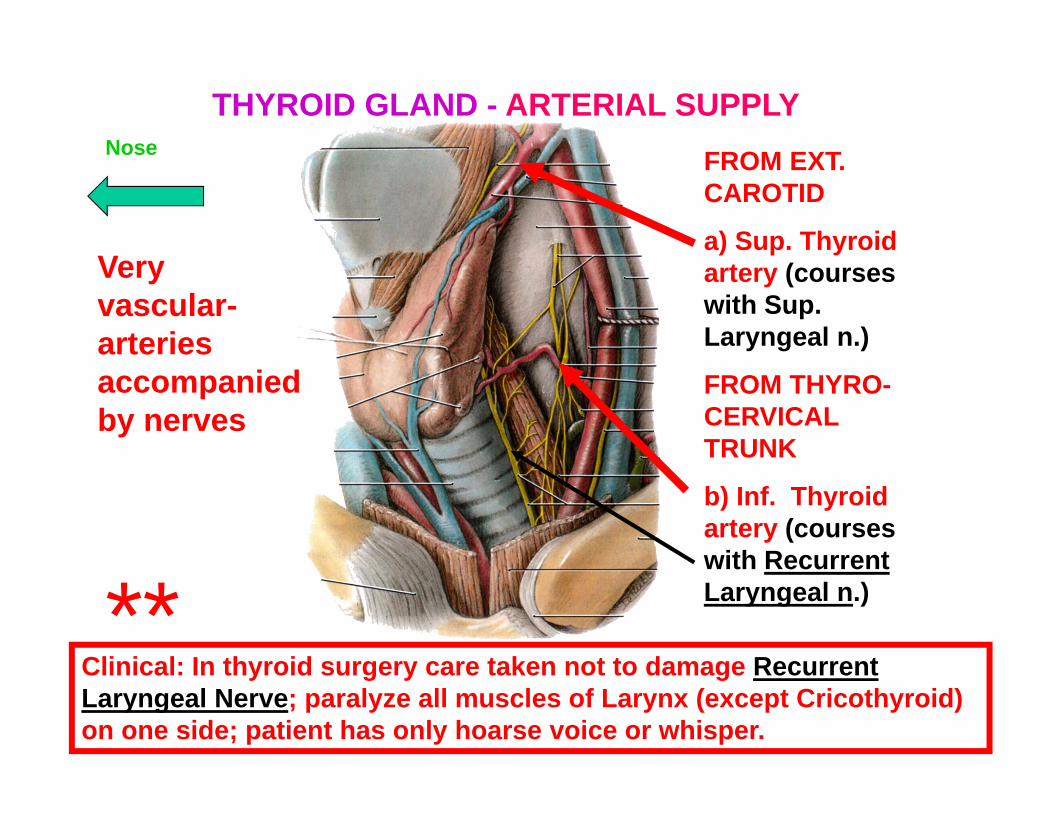
Very vascular-arteries accompanied by nerves
FROM EXT. CAROTID
a) Sup. Thyroid artery (courses with Sup. Laryngeal n.)
FROM THYRO-CERVICAL TRUNK
b) Inf. Thyroid artery (courses with Recurrent Laryngeal n.)
Clinical: In thyroid surgery care taken not to damage Recurrent Laryngeal Nerve; paralyze all muscles of Larynx (except Cricothyroid) on one side; patient has only hoarse voice or whisper.
THYROID GLAND - ARTERIAL SUPPLYNose
**

THYROID GLAND - LOTS OF VEINS
1) Superior Thyroid vein follows Superior Thyroid artery
2) Middle Thyroid vein - to Internal Jugular
CLINICAL NOTE: THERE CAN BE A LARGE VEIN IN FRONT OF (ANTERIOR TO) THE TRACHEA - IMPORTANT IN TRACHEOTOMY; BLEEDING AVOIDED BY CRICOTHYROTOMY
3) Inferior Thyroid vein(s) -drain to Left Brachio-cephalic Vein
Left BrachiocephalicVein
Int. Jugular Vein
**

PARATHYROID GLANDS
- 4 small bodies (2 on each side) located posterior to or within Thyroid gland
Superior parathyroid gland
Inferior parathyroid gland
ANT. VIEW POSTERIOR VIEW

Sympathetic trunk- deep to (not in) Carotid Sheath
Directly Anterior to vertebrae
DO NOT confuse with Vagus nerve X
SYMPATHETIC CHAIN
Note: Sympatheticsto most of headare from SuperiorCervical Ganglion
Nose