overview of valvular heart disease january 28, 2006 david r. richards, do, facc, fase midohio...
TRANSCRIPT

Overview of Valvular Heart
DiseaseJanuary 28, 2006
David R. Richards, DO, FACC, FASEMidOhio Cardiology and Vascular Consultants
Director, Heart Disease Management Program Riverside Hospital

Valve Disease: general concepts
• Etiology and natural history• Physical findings• Therapy
– types of surgical therapy– indications for surgery– indications for anticoagulation– antibiotic prophylaxis

Etiology of valve disease
• “Secondary” valve disease
• “Primary” valve disease

Etiology of valve disease
• “Secondary” valve disease– Hypertension– CAD– Cardiomyopathy
• “Primary” valve disease

Etiology of valve disease
• “Secondary” valve disease• “Primary” valve disease
– Calcific aortic stenosis– Rheumatic valve disease– Mitral prolapse / myxomatous mitral
disease– Primary aortic regurgitation– Infective endocarditis

Diseasesprimary
• degenerative• rheumatic• endocarditis• myxomatous• congenital
secondary• CAD / cardiomyopathy
Mechanisms
• Aortic stenosis• Mitral stenosis• Mitral regurg.• Aortic regurg. • Tricuspid regurg

Mechanisms of Valve Disease
Rheumatic Post-inflammatorythickening andcalcification
Mitral StenosisAortic StenosisMitral Regurg
Degenerative Age-relatedcalcification andfibrosis
Aortic StenosisMitral Regurg
Myxomatous Redundant andfloppy leaflets
Mitral RegurgAortic Regurg
Endocarditis Leafletdestruction
Mitral RegurgAortic Regurg

Valvular Emergencies
• Acute Endocarditis
• Papillary Muscle Rupture
• Flail Mitral Leaflet
• Prosthetic Valve Thrombosis / Dehiscence

65 y.o. female with MVR and acute CHF

65 y.o. female with MVR and acute CHF

65 y.o. female with MVR and acute CHF

S/P thrombolytic therapy

S/P bioprosthetic valve replacement

Valve disease: Diagnosis
• Physical exam suggests diagnosis• Transthoracic Echo (TTE) confirms
mechanism and severity of lesion• Transesophageal Echo (TEE) usually
reserved to:• plan surgery• confirm borderline diagnosis/severity

2 D Echocardiography

Transesophageal Echo (TEE)

S1 S2
systole diastole
MV closure
AV closure

S1 S2
Mild AS
Severe AS
Mitral regurg
MVP

S1 S2
Mitral Stenosis
Severe AR
Mild AR

Valve disease: Management
• Medical therapy ineffective– except: vasodilators for AR
• Surgical therapy curative• Surgery for symptoms or LV dysfunction• Surgical trends:
– minimally invasive surgery– valve repair– homograft use


Mechanical Prostheses

2D Echo: normal mechanical MV

Heterografts: Porcine

Aortic Homograft

TEE: aortic homograft

Mitral Annuloplasty Ring

Prosthetic Valves: selection
• Bioprosthetic
• Mechanical
• Homograft
• No Coumadin needed• Less thromboembolic
complications
• Lifelong cure
• No Coumadin needed• Potential lifelong
integrity
Lifespan 10-15 yrs.
Lifelong Coumadin1% annual comp. Rate
Limited availability? Late failureTechnically challenging
Pros Cons

Prosthetic Valves: selection
• Bioprosthetic
• Mechanical
• Homograft
• Elderly pts.(lifespan < 15 yrs.• Contraindication to Coumadin
• Elderly who already need Coumadin• All other patients
• Young patients with Aortic Valve disease

Prosthetic Valves:types of dysfunction
• Stenosis– degenerative– thrombosis
• Regurgitation– Paravalvular– Transvalvular
• Endocarditis• Mechanical Failure

Prosthetic Valve Endocarditis

Valve disease: Management
• Endocarditis prophylaxis
High-riskpatient
High-riskprocedure+ = prophylaxis

Endocarditis prophylaxis
High-riskpatient
High-riskprocedure+
•*Congenital disease•*Prior endocarditis •*Prosthetic valves •Acquired valve disease•MVP with MR
•Dental•GU•GI•Resp

Antibiotic Regimens
Oral, Dental, Upper Resp Procedures:• Amoxicillin 2.0 gm p.o.• Alternative:
– Clindamycin 600 mg p.o.– Cephalexin, Azithromycin
GU, GI Procedures:• Ampicillin and Gentamycin• Alternative: Vancomycin


Case 1
• 36 year old male presents with palpitations. No past history. No meds. Sibling has heart murmur.
• Exam: normal S1, S2. No murmur. Soft mid-systolic click.
• EKG: normal except for PACs.

Case 1: 2D echo
Findings: Posterior Leaflet ProlapseMild (1+) Regurgitation

Mitral Valve Prolapse
• A form of myxomatous valve disease• symptoms may be from:
– mitral regurgitation– hyperadrenergic state
• May progress to “surgical” MR• Often familial• Overdiagnosed clinically

Severe Posterior Leaflet Prolapse


Case 2
• 56 year old male with known heart murmur and MVP for 20 years. 3 days prior to admission, he had acute onset dyspnea and orthopnea.
• Exam: pulse 110. 3/6 holosystolic murmur at apex. Bilateral crackles.
• Labs: Troponin negative• EKG: sinus tachy• CXR: pulmonary edema

Case 2: TEE
Findings: Severe MV prolapseFlail Posterior LeafletSevere (4+) MR

Flail Mitral Valve Leaflet
• A complication of myxomatous valve disease: rupture of chordae tendinae
• Rarely from endocarditis, rheumatic, etc • Presents as severe MR with CHF• Accurately diagnosed with TEE • High untreated mortality• Accounts for 30 to 50 % of MV surgery• Highly amenable to valve repair
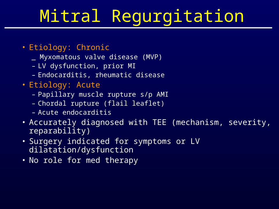
Mitral Regurgitation
• Etiology: Chronic_ Myxomatous valve disease (MVP)– LV dysfunction, prior MI– Endocarditis, rheumatic disease
• Etiology: Acute– Papillary muscle rupture s/p AMI– Chordal rupture (flail leaflet)– Acute endocarditis
• Accurately diagnosed with TEE (mechanism, severity, reparability)
• Surgery indicated for symptoms or LV dilatation/dysfunction
• No role for med therapy


Case 3
• 53 y.o. female with chronic dyspnea. Atrial fib for 12 years.
• Exam: – 4/6 blowing systolic murmur at apex
with harsh component at LSB– harsh diastolic rumbling murmur– reduced S2, loud opening snap– prominent JVD

Case 3: 2D echo
Findings: Rheumatic changes of MVSevere MS, Moderate ASModerate MR

Mitral Valve Stenosis
• A complication of acute rheumatic fever• Valve disease occurs 20 yrs after initial
acute illness• Presents as exertional dyspnea and murmur• Complications: A.Fib., emboli, refractory
pulmonary hypertension• Therapy: Commisurotomy or valve
replacement


Case 3b
• 72 y.o. female with dyspnea.• Exam:
– 2/4 systolic murmur– Normal S1 and S2

Case 3b

Case 3b
Normal Aortic Valve Calcific Aortic Stenosis

Aortic Stenosis
• Most common etiology is degenerative calcific disease (age < 50, bicuspid AV or rheumatic)
• Classic Triad: Chest Pain, Dyspnea, Syncope
• Reduced exercise capacity may be earliest symptom (use exercise test)
• Surgery indicated for– any symptoms– LV dilation or dysfunction (EF <50%, ESD > 50mm)– NOT for specific valve area


Case 4
• 35 y.o. male found to have heart murmur. No symptoms.
• Exam: – ejection click– 2/4 diastolic murmur

Case 4: 2D Echo
Findings: Moderate ARBicuspid AV Normal LV size and function

Aortic Regurgitation
• Most common etiology is degenerative (age < 50, bicuspid AV or rheumatic)
• Reduced exercise capacity may be earliest symptom (use exercise test)
• Surgery indicated for– any symptoms– LV dilation or dysfunction (EF <50%, ESD >
50mm)

Case 5
• 10 years later, patient develops acute fever, weakness. Patent reports severe dyspnea at rest.
• Exam: BP 80/50, HR 110, bilateral crackles, soft diastolic murmur, S4 gallop

Case 5: 2D Echo

Case 5: 2D Echo

Case 5: 2D Echo

Case 4
•Echo: bicuspid AV with vegetation, severe AR,dilated LV with EF 30%
•antibiotics, diuretics, & pressors areinitiated. The patient initially stabilizes, but within 24 hours develops recurrent hypotensionand respiratory failure.

25 y.o woman with fatigue
Findings: MV mass ? Myxoma

