oxidation of adenosine and inosine: the chemistry of 8-oxo
TRANSCRIPT
Bowling Green State University Bowling Green State University
ScholarWorks@BGSU ScholarWorks@BGSU
Chemistry Faculty Publications Chemistry
3-2013
Oxidation Of Adenosine And Inosine: The Chemistry Of Oxidation Of Adenosine And Inosine: The Chemistry Of
8-oxo-7,8-dihydropurines, Purine Iminoquinones, And Purine 8-oxo-7,8-dihydropurines, Purine Iminoquinones, And Purine
Quinones As Observed By Ultrafast Spectroscopy Quinones As Observed By Ultrafast Spectroscopy
Denis I. Nilov
Dmitry Y. Komarov
Maxim S. Panov
Kanykey E. Karabaeva
Andrey S. Mereshchenko
See next page for additional authors
Follow this and additional works at: https://scholarworks.bgsu.edu/chem_pub
Part of the Chemistry Commons
Repository Citation Repository Citation Nilov, Denis I.; Komarov, Dmitry Y.; Panov, Maxim S.; Karabaeva, Kanykey E.; Mereshchenko, Andrey S.; Tarnovsky, Alexander N.; and Wilson, R. Marshall, "Oxidation Of Adenosine And Inosine: The Chemistry Of 8-oxo-7,8-dihydropurines, Purine Iminoquinones, And Purine Quinones As Observed By Ultrafast Spectroscopy" (2013). Chemistry Faculty Publications. 120. https://scholarworks.bgsu.edu/chem_pub/120
This Article is brought to you for free and open access by the Chemistry at ScholarWorks@BGSU. It has been accepted for inclusion in Chemistry Faculty Publications by an authorized administrator of ScholarWorks@BGSU.

Author(s) Author(s) Denis I. Nilov, Dmitry Y. Komarov, Maxim S. Panov, Kanykey E. Karabaeva, Andrey S. Mereshchenko, Alexander N. Tarnovsky, and R. Marshall Wilson
This article is available at ScholarWorks@BGSU: https://scholarworks.bgsu.edu/chem_pub/120
Oxidation of Adenosine and Inosine: The Chemistry of 8‑Oxo-7,8-dihydropurines, Purine Iminoquinones, and Purine Quinones asObserved by Ultrafast SpectroscopyDenis I. Nilov, Dmitry Y. Komarov, Maxim S. Panov, Kanykey E. Karabaeva, Andrey S. Mereshchenko,Alexander N. Tarnovsky,* and R. Marshall Wilson*
Department of Chemistry and Center for Photochemical Sciences, Bowling Green State University, Bowling Green, Ohio 43403,United States
*S Supporting Information
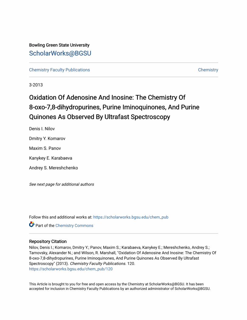
ABSTRACT: Oxidative damage to purine nucleic acid bases proceeds through quinoidal intermediates derived from theircorresponding 8-oxo-7,8-dihydropurine bases. Oxidation studies of 8-oxo-7,8-dihyroadenosine and 8-oxo-7,8-dihydroinosineindicate that these quinoidal species can produce stable cross-links with a wide variety of nucleophiles in the 2-positions of thepurines. An azide precursor for the adenosine iminoquinone has been synthesized and applied in ultrafast transient absorptionspectroscopic studies. Thus, the adenosine iminoquinone can be observed directly, and its susceptibility to nucleophilic attackwith various nucleophiles as well as the stability of the resulting cross-linked species have been evaluated. Finally, theseobservations indicate that this azide might be a very useful photoaffinity labeling agent, because the reactive intermediate,adenosine iminoquinone, is such a good mimic for the universal purine base adenosine.
■ INTRODUCTIONOxidatively damaged nucleic acids are now widely accepted asone of the principal sources of genetic damage includingpossible sources of carcinogenesis, mutation, aging, and celldeath.1−8 Purines are more susceptible to single-electronoxidative damage than are pyrimidines. The oxidationpotentials of unmodified bases are G (1.29 V), A (1.42 V), C(1.6 V), and T (1.7 V) versus NHE.9 Oxidation of purine basesinitially leads to 8-oxo-7,8-dihydroguanosine (8-oxoG or 8-oxodG (1)), 8-oxo-7,8-dihydroadenosine (8-oxoA or 8-oxodA(2)), and 8-oxo-7,8-dihydroinosine (8-oxoI, or 8-oxodI (3)),which can occur both within existing RNA/DNA strands, aswell as in nucleotide pools. As indicated by the aforementionedreduction potentials, guanosine is the most easily oxidized baseand has been studied in this regard extensively.10,11 In contrast,the oxidation of adenosine and inosine has been studied muchless extensively, possibly because their oxidation products are
not formed in such high yields, are more difficult to detect, andbecause 8-oxodA (2) is less mutagenic than 8-oxodG (1).12
These oxidized bases might lead to miscoding in DNAreplication and transcription processes,13,14 and may beassociated with aging.15 In addition, it has been noted that 8-oxo derivatives are formed more readily in RNA than in DNA,possibly because single-stranded RNA is more susceptible tooxidation than is duplex DNA or DNA protected bychromatin.16,17
Repair enzymes for replacing 8-oxodG (1) with undamagedguanosine are well-known.1,18 The comparable repair enzymesfor replacing 8-oxodA (2) with undamaged adenosine seem tobe much less well studied possibly because 8-oxodA (2) lesionsare much less mutagenic than 8-oxodG lesions, and are only
Received: August 7, 2012Published: January 22, 2013
Article
pubs.acs.org/JACS
© 2013 American Chemical Society 3423 dx.doi.org/10.1021/ja3068148 | J. Am. Chem. Soc. 2013, 135, 3423−3438

about 1/3 as common.19−22 In fact, there seems to be lesseffective repair of 8-oxodA:T lesions in certain cases.23,24 In asimilar vein, the course of further oxidation of 8-oxodG (1) hasbeen well studied and found to afford 4 and 5 (Nuc2 = OH orO in water), Scheme 1.25 Ultimately, the modified bases 4 and5 may hydrolyze to form the urea derivative, 6. These oxidationprocesses are usually attributed to attack on the purine nucleusby hydroxyl radicals and other reactive oxygen species (ROS)to form the 8-oxo species 1 or 2 and their further oxidation by avariety of mechanisms.1−8,12−14,16 However, in a separate lineof research, photolysis of 8-azidoadenosine has been found toproduce a diiminoquinone purine intermediate related to 7,O replaced by HN.26 This intermediate is readily attackedby a variety of nucleophiles at the 2-position to form productsrelated to 8, O replaced by HN. Nucleophiles examinedby transient absorption spectroscopy in this work includecysteine, diethylamine, sodium hydroxide, as well as amino acidesters and nucleic acid bases. A similar quinone intermediate,9NH2, is now thought to play a pivotal role in the oxidation of8-oxoG (1).27−31 These observations suggest that oxidation of8-oxopurines might follow similar oxidative pathways passingthrough quinoidal purines rather than via direct attack ofhydroxyl radical on the purine nucleus. Thus, 8-oxoA (2) mightalso be susceptible to oxidative substitution to form 8 via 7,which in turn might link the oxidation of adenosine with that ofguanosine, as outlined in Scheme 1, and arrive at the same finalproducts as ROS mechanisms. Perhaps more significantly,attack on 7 by nucleophiles that do not appreciably enhance thesusceptibility of 8 to further oxidation might lead to stableadducts giving rise to covalent cross-links between nucleic acidsand proteins or other biomolecules. For these reasons, we haveundertaken a study of adenosine oxidation using themechanism outlined in Scheme 1 as our working hypothesis.In this study, we have concentrated on the oxidation of the 8-oxopurines 8-oxoA (2) and 8-oxoI (3), because these molecules
might form relatively stable “cross-linked” adducts at the 2-position related to 8, in contrast to the much more extensivelystudied 8-oxoG (1), which forms more labile “cross-linked”adducts at the 5-position as indicated in Scheme 1.27−31
■ RESULTS AND DISCUSSIONChemical Oxidation Studies. Using the model for purine
oxidation outlined in Scheme 1, we have investigated theoxidation of 8-oxoA (2) with a variety of oxidizing agents. Thefull description of the following oxidation reactions along withthe spectroscopic characterization of the products is providedin the Supporting Information. Thus, treatment of 8-oxoA (2)with NBS in methanol alone at room temperature leads toformation of the methanol adduct 8OMe, R = 1′-ribosyl, Nuc1= OCH3, or in a methanolic imidazole solution, the imidazoleadduct 8IM, R = 1′-ribosyl, Nuc1 = imidazolyl as the majorproducts (Scheme 2). From these results, it is clear thatoxidation of 8-oxoA (2) produces an intermediate that issusceptible to nucleophilic attack at the purine 2-position, andthat virtually any nucleophile, even weak ones like methanoland water, vide infra, will add to the 2-position of thisintermediate. We attempted to observe this reactive inter-mediate by conducting the oxidation reactions in the cold, butwere unsuccessful. These transient studies are described in fulldetail in the Supporting Information. Apparently, these putativequinoidal intermediates are too reactive to survive into thesecond-time domain. Sigma adducts related to 10OMe havebeen observed previously and found to aromatize in themicrosecond- and millisecond-time domains.32
As might be expected, oxidation of 8-oxoA (2) oftenproduces nucleophile adducts that are very susceptible tofurther oxidation as outlined in Scheme 1. This subsequentoxidation is indicated by the formation of N 1′-ribosyl urea (6)observed in the NBS oxidation of 2, Scheme 2. In many cases ofDNA oxidation,14 2′-deoxy-1′-ribosyl urea has been observed,and N 1′-ribosyl urea is occasionally observed in small amountsin the oxidations conducted in this work. It was found that theinitial adducts in the 2-position are often more easily oxidizedthan the starting 8-oxoA (2). Thus, addition of aliphatic amines,such as diethyl amine and primary amine related to lysine,quench the intermediate 7 quite rapidly even in the cold.
Scheme 1
Journal of the American Chemical Society Article
dx.doi.org/10.1021/ja3068148 | J. Am. Chem. Soc. 2013, 135, 3423−34383424

Unfortunately, it has not been possible to isolate these amineadducts, possibly because they are highly susceptible to furtheroxidation. Related amine adducts formed in the photochemistryof 8-azidoadenosine are oxidized upon standing in air.33 Thus,we suspect that these susceptible adducts are oxidized anddestroyed more rapidly than they are formed or during theextensive HPLC procedures necessary for their isolation.An amine that does not significantly activate the purine
nucleus to further oxidation is imidazole. Thus, when the NBSoxidation of 8-oxoA (2) is conducted in a methanolic solutionof imidazole, the imidazole adduct 8IM (R = 1′-ribosyl, Nuc =imidazolyl) (Scheme 2) can be isolated. In many ways,imidazole is a unique amine nucleophile. The n-lone pair onnitrogen serves as a good nucleophile, but once attached to thepurine ring, the imidazole electron pair on the nucleophilicnitrogen becomes part of the imidazole aromatic sextet, andthus is not effectively donated to the purine ring where suchdonation would activate the purine ring to further oxidation.An alternative strategy for reducing overoxidation associated
with the addition of electron-rich nucleophiles to the purinering is to begin with a less electron-rich purine. The nucleic acid8-oxoI (3) would seem to be an ideal candidate for testing thisapproach, because the electron-donating 6-amino group inadenosine has been replaced by the electron-withdrawingcarbonyl group in inosine. The potential required to oxidize 8-oxoI (3) has been measured and found to be 1.2 V/NHE ascompared to about 0.92 V/NHE for 8-oxoA (2),34 and 0.74 V/NHE for 8-oxoG (1).35 The relationship between thesepotentials is supported by the observation that treatment of8-oxoI (3) with methanolic NBS, the same conditions that weresuccessful for the oxidation of 8-oxoA (2), does not lead to theformation of adducts. However, treatment of 3 with the morepowerful oxidizing agent sodium hypochlorite in aqueousmedia in the presence of imidazole leads to the formation of theimidazole adduct in the 2-position, 12IM, Scheme 3.Oxidative damage to nucleic acids is thought to be associated
with “reactive” oxygen species (ROS) such as hydroxyl radicalsand singlet oxygen.36−41 Therefore, the visible light irradiationof both 8-oxoA (2) and 8-oxoI (3) in the presence of oxygen,imidazole, and rose bengal or riboflavin was examined in aneffort to determine if singlet oxygen might affect their
transformation to 8IM and 12IM, respectively. These experi-ments produced only trace amounts of products. However,when Na2S2O8 was added to the reactions photosensitized withriboflavin and visible light, the imidazole adducts 8IM and12IM (11%) were formed, albeit in low yields. Thus, theriboflavin sensitized generation of SO4
•− is apparently a viablemethod for the oxidation of 2 and 3. Of particular note is theobservation that direct irradiation of 3 at 254 nm in thepresence of Na2S2O8 and imidazole significantly enhances theyield of 12IM; see Table 1. In Burrows’s studies of the
oxidation of 8-oxoG (1), Na2IrCl6 has been found to be theoxidizing agent of choice.42−44 This reagent proved to be one ofthe more effective in the oxidation of 8-oxoA (2) and 8-oxoI(3) as well; see Table 1.While the aforementioned observations provide good
circumstantial evidence for the intermediacy of the quinoidalintermediates 7 and 11, alternative mechanistic explanations forthese reactions are conceivable. Furthermore, simple oxidativemethods for formation of 7 and 11 suffer from the fact thatmany of products that might be formed through the addition ofvarious nucleophiles are themselves more easily oxidized thanare the starting 8-oxopurines, and thus cannot be isolated orreadily observed. Therefore, an alternative strategy for the
Scheme 2
Scheme 3
Table 1. Yields of Purine Oxidation Products
purineoxidized reaction conditions products and % yield
2 NBS, CH3OH 8OMe triacetate, 6-acetamido,6%a
2 NBS, CH3OH, imidazole 8IM tri-TBDMS, 19%b
2 NBS, H2O, imidazole 8IM, 23%2 Na2IrCl6·6H2O, H2O,
imidazole8IM, 21%
3 aq NaOCl, imidazole 12IM, 40.5%3 Na2S2O8, hν (254 nm),
imidazole12IM, 35%
3 Na2IrCl6·6H2O, H2O,imidazole
12IM, 69%
aProduct isolated following treatment of crude reaction mixture withacetic anhydride. bProduct isolated following treatment of crudereaction mixture with TBDMSCl.
Journal of the American Chemical Society Article
dx.doi.org/10.1021/ja3068148 | J. Am. Chem. Soc. 2013, 135, 3423−34383425

formation of iminoquinone 7 has been devised that providesmuch more information about the reactivity of 7 and at thesame time offers the possibility of isolation of products thatwould be susceptible to further oxidation. Thus, the nitrenederived from 6-azido-8-oxo-7,8-dihydropurine (13) shouldundergo rapid, possibly solvent mediated, proton reallocationas outlined in Scheme 4 to form iminoquinone 7, and theformation and reactivity of 7 should be much more easilystudied when generated under these spectroscopically favorablephotochemical conditions.
Toward this end, the triacetate and tribenzoate of azide 13,13Ac3, and 13Bz3 have been synthesized45−47 as described indetail in the Supporting Information. The preparation andirradiation of the acetylated and benzoylated derivatives greatlyfacilitates HPLC product analysis, but does not significantlyaffect the product distribution.Irradiation of 13Ac3 in methanolic solutions of imidazole
affords both reduction, 2Ac3, and the desired imidazolesubstitution product, 8IMAc3, as outlined in Scheme 5. Inaddition, the iminoquinone hydrolysis product 3Ac3 is formedas well. The imidazole adduct 8IM isolated from this azidephotochemistry was compared and found to be identical to theimidazole adduct isolated from the chemical oxidation of 8-oxoA (2), Scheme 2.Scheme 5 and Table 2 summarize the products formed in the
photolyses of azides 13Ac3 and 13Bz3 in methanolic solutions.Because methanol by itself is a poor nucleophile, thephotodecomposition of azide 13Bz3 in pure methanol leadsto little, if any, formation of adduct 8OMeAc3. However,photolysis of methanolic solutions of 13Ac3 and 13Bz3 in thepresence of the powerful nucleophile imidazole leads to theformation of appreciable yields of imidazole adduct 8IMAc3and 8IMBz3. The formation of 3Ac3 and 3Bz3 is apparently dueto the hydrolysis of the imino group in the iminoquinone 7Ac3and 7Bz3, respectively, followed by reduction of the quinone 11as outlined in Scheme 6.The photochemical behavior of 8-azidoadenosine, 14, is also
of considerable interest, because it is one of the most widelyapplied photoaffinity labeling agents used to cross-link manybiological molecules. Its preparative photochemistry has beenstudied in detail26 and found to proceed through thediiminoquinone 15. In this work, preparative irradiation ofazide 14 was carried out at 356 nm in water in the absence of
and in the presence of imidazole (0.1 M). The analysis of thesereaction mixtures indicated the formation of reduction product16, water addition product 17OH, over oxidation product 18,dimerization products of undetermined structure, and in thepresence of imidazole, imidazole adduct 17IM, Scheme 7 andTable 2.The imidazole adducts and their acetylated and benzoylated
derivatives have been obtained by several chemical andphotochemical methods. In addition to the imidazole adducts8IM and 17IM, the 8-oxoI imidazole adduct 12IM, Scheme 3,has been observed in the chemical oxidation of 8-oxoI (3).These observations, as well as similar observations for 8-oxoG,25,27−31 are consistent with the general mechanismoutlined in Scheme 1 in which the pivotal intermediates arequinoidal purines. The 8-oxoA and 8-oxoI adducts in the 2-position, 8 and 12, respectively, are distinguished from the 8-oxoG adduct in the 5-position, 19 in Scheme 1, in that thepurine nuclei remain intact following nucleophile attack, and,consequently, might form stable cross-links with many otherbiological molecules. It should be emphasized that in thesestudies of quinoidal purines, 7, 11, and 21, adducts in the 5-position were not identified. In addition, the further oxidationof adducts in the 2-position, such as 8IM and 12IM, has yet tobe studied in detail. The implication of the above observationswith imidazole is that histidine adducts might provide relativelystable protein cross-links, but this correlation remains to betested with the amino acid itself. While these and many relatedstudies will be necessary to evaluate the working hypothesisoutlined in Scheme 1, the availability of azide 13 has providedan ideal opportunity to probe the intermediacy of iminoqui-none 7 in the early stages of this reaction scheme.A variety of azidopurines have been used in photoaffinity
labeling studies,26 but 6-azido-8-oxo-7,8-dihydropurines (13)have never been applied in such studies. The azide 13 wouldseem to be the ideal tool for probing the fate/role ofiminoquinone 7 in biological systems leading to genemalfunction. One might argue that the 6-azido group of 13would alter the usual Watson−Crick hydrogen bonding ormodify the normal syn/anti base conformer distribution. Inaddition, it is well-known that azidopyrimidines undergoazide−tetrazine tautomerization such as that shown for azide13 in Scheme 8.45−47 However, azide 13 displays an intenseabsorption at 2150 cm−1. So its azide tautomer 13 is the major,if not the only, form present. Even so, both tautomers might beexpected to form the nitrene precursor of 7 upon irradiation at254 nm.48 Thus, 13 rapidly forms the iminoquinone 7 uponirradiation (<100 ps, Scheme 4), and 7 reacts with nucleophilesso slowly (μs to ms) that there is adequate time for it to probemultiple binding sites and react with nucleophiles in the mostsuitable of those sites. Thus, while one might question the
Scheme 4
Scheme 5
Journal of the American Chemical Society Article
dx.doi.org/10.1021/ja3068148 | J. Am. Chem. Soc. 2013, 135, 3423−34383426

viability of 13 as a mimic for the affinities of 8-oxoA (2), theiminoquinone 7 should be an ideal mimic for 2.Many 8-oxoA (2) and 8-oxoI (3) adducts are susceptible to
further oxidation, which ultimately leads to the destruction ofthe purine nucleus, and the release of any cross-linkedmolecules. While such further oxidation can be minimized bythe photochemical generation of the iminoquinone 7 fromazide 13, even this route suffers from adduct overoxidationpossibly as outlined in Scheme 9. This problem of oxidation ofcross-linked molecules requires further study and evaluation.Further oxidation of amine adducts of 8-azidoadenosine (14)such as the triamine 17NR2 is a major problem, becauase theseadducts are extremely susceptible to oxidation even byatmospheric oxygen, which leads to ephemeral cross-links andcomplex reaction mixtures. Therefore, further studies compar-ing the stabilities of the very electron-rich amine adducts suchas 17NR2 with the much less electron-rich amine adducts of 13,see Scheme 10, would seem to be most desirable. However, at
this time, it would seem safe to say that 8NR2 should be lesssusceptible to further oxidation than 17NR2.Finally, in this section, the chemical oxidation of 8-oxo-7,8-
dihydropurines has been investigated and shown to lead to
Table 2. Irradiation of Azides 13Ac3 and 13Bz3 in MeOH and 14 in Water
product distribution (%)a
azide solvent conditions, hν = 1 h/mmol nuc 2 3 8IM unidentifiedb
13Bz3 MeOH 254 nm MeOH 30 30 8OMec 405 mmol/L Bz3 Bz3
13Ac3 MeOH 254 nm imidazole 10 20 50 105 mmol/L Ac3 Ac3 Ac3
13Bz3 MeOH 356 nm imidazole 0 30 50 205 mmol/L Bz3 Bz3
13Ac3 MeOH 356 nm imidazole 20 60 205 mmol/L Ac3 Ac3
14 H2O 356 nm H2O 16 17OH 180.5% HOAc 5 mmol/L ∼7 ∼10 ∼20 ∼20
14 H2O 356 nm H2O 16 17OH 17IM 180.5% HOAc 5 mmol/L imidazole ∼20 ∼10 ∼10 ∼20 ∼20
aThe sugar esters tend to hydrolyze partially during the course of the reaction due to the basic alcoholic conditions of the imidazole reactions.bThese products seem to constitute a complex mixture of over oxidation or coupling products. cAdduct observed in only trace amounts.
Scheme 6
Scheme 7
Scheme 8 Scheme 9
Scheme 10
Journal of the American Chemical Society Article
dx.doi.org/10.1021/ja3068148 | J. Am. Chem. Soc. 2013, 135, 3423−34383427

nucleophile attack and substitution at the purine 2-position.The photochemistry of 6- and 8-azidopurines leads to similarproducts so long as the 2-position is unsubstituted in thestarting azide. Because this azide photochemistry provides anexcellent tool for the spectroscopic mechanistic study of theformation of quinoidal purines and their chemistry, we haveundertaken such a study and report the results in the followingsection.Transient Absorption Spectroscopic Studies. Because
the azide 14 provides an ideal precursor for the diiminoquinone15 (Scheme 11),26 the azide 13 would seem to provide an idealprecursor to the iminoquinone 7, and the azide 20 to theiminoquinone 21. We have studied the photochemistry of theseazides in aqueous media using ultrafast transient absorptionspectroscopy. The iminoquinone 7, derived from the oxidationof 8-oxo-7,8-dihydroadenosine, 2, and the photochemistry ofthe azide 13, is probably the most interesting of these, becauaseadenosine is the universal base found in DNA, RNA, and manycoenzymes. However, 8-azidoadenosine, 14, is a widely usedphotoaffinity labeling agent, which has been shown to be aprecursor of the diiminoquinone 15 involved in the cross-linking of this reagent to many biomolecules,49 and is includedin this study to contrast its ease of formation and the reactivitywith that of iminoquinones 7 and 21.
Photodecomposition of azide 14 following 270 nm excitationhas been studied previously by ultrafast transient absorptionspectroscopy with a 400 fs time resolution.26 In the currentwork, transient absorption, ΔA, spectra of 13, 14, and 20 weremeasured and studied following 300 nm excitation with a timeresolution of 200−300 fs (limited by solvent artifact, and, as aresult, solvent dependent, see Supporting Information). Thetransient spectra of 14 were found to be strongly pH dependentin the present work. At pH = 2.6 or lower, a band centered atλmax = 390 nm develops within ca. 500 fs, spectrally narrowsindicative of vibrational relaxation of the responsible productspecies (exponential time constants (τ) are wavelengthdependent ranging between ca. 0.5−10 ps), and remainsconstant for at least 1 ns (Figure 1A). In contrast, at pH = 9.2or higher, the formation of the 390 nm band was not observedin aqueous solution of azide 14. Instead, a new band centered atλmax = 465 nm develops within 300 fs, undergoes several-picosecond spectral sharpening, and then survives for at least 1ns (Figure 1C). Transient absorption spectra of azide 14,recorded at intermediate pH values such as pH = 3.7 (Figure1B), have a broad band centered at λmax = 440 nm. This band isbroader as compared to the bands at pH = 2.6 and 9.2 andseems to be a combination of the 390 and 465 nm bands.
Scheme 11
Figure 1. Ultrafast transient absorption spectra of azide 14 in water solution at pH = 2.6 (A), 3.7 (B), and 9.2 (C). Time delays between pump andprobe pulses are expressed in picoseconds and given in legends.
Journal of the American Chemical Society Article
dx.doi.org/10.1021/ja3068148 | J. Am. Chem. Soc. 2013, 135, 3423−34383428

Clearly two quite different intermediates are being generatedfrom azide 14 at different pH’s. Under acidic conditions, the 6-amino group of 14 will be protonated, 14H in Scheme 12. Thenitrene 22H will not be stabilized by the powerful electron-donating effect of the unprotonated amino group. Under theseconditions, rapid opening of the five-membered ring to theiminonitrile 23H might occur.26,50−53 In contrast, under basicconditions, the nitrene will be stabilized by the powerfulelectron-donating effect of this amino group and becomes thepowerful base, 22, which will lead to proton reallocation toform 15. The formation of 15 is complete within ca. 300 fs. Theresulting vibrationally hot diiminoquinone 15 then undergoesvibrational cooling manifested by spectral narrowing overseveral picoseconds, Figure 1C. Spectral assignments ofintermediates and products observed throughout the currentwork were assisted by calculating vertical excitation transitions(VETs) of these species at the TDDFT (B3LYP functional, 6-
31G(d,p) basis set) and CASPT2/ANO-RCC levels oftheory.54 The calculated absorption spectra (Scheme 12,Table 3) support the aforementioned assignments. Thus, thering-cleaved iminonitrile 23H is calculated to have a majorelectronic transition at 389 nm, Scheme 12, and this ring-opening might easily occur within 500 fs, Figure 1A. Incontrast, the diiminoquinone 15 is calculated to displayabsorption in the 430−450 nm region.26 The fast sub-300 fsproton reallocation necessary to transform 22 in to 15 inScheme 12 apparently takes place via some type of proton relaymechanism, which might be a coordinated process related tothat shown in Scheme 12. Calculations of energetics involved inthe transformation of 22 to 15 via such a proton relay process(a relaxed reaction coordinated scan involving two watermolecules) indicated rather high energy barriers on the order of18 kcal/mol for the proton-relay process shown in Scheme 12.Therefore, if this type of proton reallocation is to take place
Scheme 12
Table 3. Calculated Vertical Excitation Energies and Oscillator Strengths ( f) for Intermediates 15Me, 23HMe, and 22Me inMethanola
aAll calculations were performed in the gas phase.
Journal of the American Chemical Society Article
dx.doi.org/10.1021/ja3068148 | J. Am. Chem. Soc. 2013, 135, 3423−34383429

within the <300 fs window indicated, either proton tunnelingwill have to play a very significant role, or the energetics of thesystem must change appreciably upon solvation with more thantwo water molecules in the formation of the species absorbingat 465 nm.Because there are apparently two alternative nitrene decay
pathways, one via five-membered ring cleavage and the othervia diiminoquinone formation, there must be two convergentproduct forming pathways, because the same products areapparently formed from both types of intermediates. Two suchalternative pathways for nucleophile attack are outlined inScheme 13. This same dichotomy has been discussed in detailin previous papers,26,50−53 and little new can be added to thatdiscussion except to note that these two pathways are regulatedby the pH of the solution and other factors that alter theavailability of electron density on the nitrene nitrogen.One such factor is the acylation of the 6-amino group, which
has an effect similar to that of the protonation of this amino
group, and leads to five-membered ring fragmentation in both14Bz5 and 14Ac5, Figure 2. Thus, excitation of 14Bz5 in anonprotic solvent leads to transient absorption bands at λmax =395 nm within ca. 300 fs. This band relaxes into two bands at370 and 416 nm with τ = 2.5 ps (Figure 2A). In a similarfashion, irradiation of 14Ac5 in a nonprotic solvent affords aband centered λmax = 375 nm with τ = 1.5 ps that undergoeslittle or no further change (Figure 2B). These bands centered atλmax = 365−375 nm are assigned to corresponding nitrenes andthe mature bands in the 375−416 region to the correspondingring-opened iminonitriles. Apparently even acyl groups on the6-amino group are sufficiently electron withdrawing to favor thering-opening pathway, if alternative protonation pathways arenot available.In previous work with electron-rich nitrenes, we observed
that the rate of nitrene protonation/relaxation was related tothe structure of the proton source with the process in methanolbeing ca. 4 times faster than that in 2-propanol.32 This same
Scheme 13
Figure 2. Ultrafast transient absorption spectra of acylated derivatives of azide 14 in nonprotic solvents. Column A: 14Bz5 (1 mM) indichloromethane. Column B: 14Ac5 (1 mM) in methylcyclohexane. Time delays in picoseconds between pump and probe pluses are given in thelegends. The 395 and 375 nm absorption bands develop within the first 300 fs after the excitation pulse, and they decay either with formation of twoabsorption bands at 370 and 416 nm (τ = 2.5 ps, A) or very slowly on a several nanosecond time scale (B).
Journal of the American Chemical Society Article
dx.doi.org/10.1021/ja3068148 | J. Am. Chem. Soc. 2013, 135, 3423−34383430
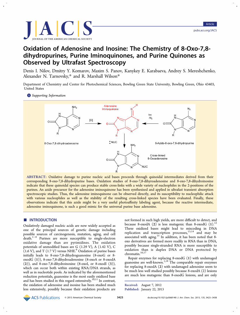
general trend is observed with azide 14, Figure 3. Thus, inmethanol, nitrene 22 (λmax = 460 nm) undergoes protonreallocation/relaxation, which is characterized by an exponen-tial rise-time constant of τ = 1.5 ps. While in 2-propanol, asignificantly longer reallocation/relaxation time constant isobserved, τ = 2.5 ps. Thus, proton transfer, Scheme 12,proceeds with about the same very rapid rate in water andmethanol, but with a slightly slower rate in 2-propanol. In all ofthese systems, this proton transfer ranks among the most rapidyet observed.55−58
The reactions of diiminoquinone 15 with various nucleo-philes were studied by nanosecond transient absorptionspectroscopy (Table 4). Thus, azide 14 was irradiated inwater at pH = 3.1, 6.5, and 11.5 (Figure 4), and in 0.1 Maqueous solutions of imidazole (Figure 5A), a modelcompound for histidine, and in 0.1 M sodium phenoxide(Figure 5B), a model compound for tyrosine. According tothese spectra, quenching of diiminoquinone 15 under neutralconditions, pH = 6.5, Figure 4B, is a relatively slow process(Schemes 7, 13, and 14), with quenching of diiminoquinone 15occurring in τ1 = ca. 100 μs, and subsequent aromatization ofthe corresponding C2-adduct proceeding with τ2 > 35 ms.However, these processes are accelerated under both acidic andbasic conditions to τ2 = ca. 20 ms at pH = 3.1 and τ2 = 4.2 ms atpH 11.5. The reaction of 15 with 0.1 M aqueous sodiumphenoxide takes place with τ2 = 3 ms (Figure 5B) and morerapidly with imidazole, τ2 = 900 μs (Figure 5A). Thesenucleophilic substitution reactions all proceed throughintermediates similar to 24Nuc in Scheme 13. The initial
diminioquinone 15 is only clearly visible in Figure 4B, the pH =6.5 spectrum, λmax = 450 nm at 32 μs. It is formed in thepicosecond time domain and slowly reacts with nucleophilesand rearomatizes within the microsecond−millisecond timedomain. It is this rearomatization process that is visible inFigure 4A and C and Figure 5A and B. It is interesting tocompare the behavior of 8-azidoadenosine, 14, with that of 8-azidoinosine, 20. The nitrene 25 generated from 20 is notconjugated to a powerful electron-donating group, andtherefore might not undergo proton reallocation to form theiminoquinone 21, Scheme 15. Thus, irradiation of 20 in water,pH = 1.7−7.5, alcohols, or aprotic solvents such as dichloro-methane all lead to the rapid development of a 380 nm bandcharacteristic of opening of the five-membered ring andformation of the iminonitrile 26, Scheme 15. Because noproton reallocation patterns could be observed under anyconditions examined, it would seem that irradiation of 20 leadsto opening of the five-membered ring to form 26 exclusively,Scheme 15. It is worthy of note that during theoreticalstructural optimization of nitrene 25 using either DFT or MP2calculations, the C8−N9 bond always broke to form theiminonitrile isomer 26. The most informative of these transientexperiments was that conducted in water at pH 7.5 (Figure6A). The very broad absorption at 450−600 nm is assigned tothe nitrene 25 that decays with the same time constant as thegrowth of the iminonitrile 26 band at 380 nm, τ = 7 ps. Incontrast, opening of the five-membered ring in methanol(Figure 6B) and dichloromethane (Figure 6C) is more rapidand apparently occurs in near-concert with nitrene formation,because there is only a slight narrowing of the 380 nm peak,which can be attributed to vibrational cooling of the openiminonitrile 26. Furthermore, the iminonitrile 26Ac3 displaysspectroscopic behavior similar to that of the diiminoquinone 15in the microsecond−millisecond time domains (Figures 7 and4, respectively). Thus, both 26Ac3 and 23H react with water ormethanol within 15−20 ms. However, it seems that theiminonitriles are better electrophiles than are the iminoqui-nones: iminonitrile 26Ac3 reacts much more rapidly with
Figure 3. Ultrafast transient absorption spectra of azide 14. Column A: 14, 1 mM solution in methanol. Column B: 14, 0.75 mM in 2-propanol.Time delays in picoseconds between pump and probe pluses are given in the legends. The major time constants describing the ΔA evolution shownare τ = 1.5 ps (A) and τ = 2.5 ps (B).
Journal of the American Chemical Society Article
dx.doi.org/10.1021/ja3068148 | J. Am. Chem. Soc. 2013, 135, 3423−34383431

imidazole, τ2 = 75 μs, than does the diiminoquinone 15, τ2 =900 μs (Figures 7B and 5A, respectively). It is worthy of notethat the absorption associated with the reaction of imidazolewith the iminonitrile, 26Ac3, Figure 7B, derived from 20Ac3 isunusually broad as compared to other transient absorptions
involving reactions of imidazole with iminoquinones 15 and 7(Figure 5A or 10B, respectively). This extremely broad bandmay be due to intermediates involved in the ring closure.Azide 13 is perhaps the most interesting azide studied in this
work, because it is a precursor for the natural iminoquinone 7,
Table 4. Time Constants τ1 and τ2 for the Reactions of Purine Intermediates 15, 23H, and 26 under Various Conditions
Figure 4. Nanosecond−millisecond transient absorption spectra of azide 14 in water at different pH values.
Figure 5. Nanosecond−millisecond transient absorption spectra of azide 14 in water in the presence of (A) imidazole (0.1 M) and (B) sodiumphenoxide (0.1 M).
Scheme 14
Journal of the American Chemical Society Article
dx.doi.org/10.1021/ja3068148 | J. Am. Chem. Soc. 2013, 135, 3423−34383432

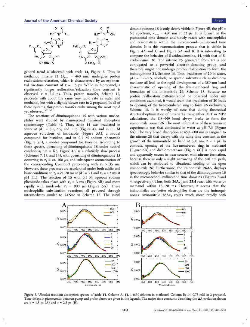
Scheme 16. Ring-opening does not seem to be a viable reactionpathway for nitrene 27. Instead, nitrene 27 is stabilized viaproton reallocation. There are at least two possible mechanismsfor proton reallocation as outlined in Scheme 16. The first ofthese is a direct 1,4-proton shift, and the second is a protonreallocation mediated by a water or an alcohol molecule. Thenitrene 27 is expected to be of a similar basicity as nitrene 25,and therefore not be particularly susceptible to protonation.Consequently, the opening of the six-membered ring of nitrene27 must be quite slow, if it occurs at all, for the slow protonreallocation to compete effectively. In fact, protonation seemsto be the only nitrene stabilization process active in this system.When azide 13Bz3 was photolyzed in alcohol solvents, Figure 8,bands at λmax = 375 and 550625 nm are initially formed andslowly shift to a new band centered at λmax = 405 nm. In allcases, the broad band in the 550−625 nm region decays withthe same time constant as the growth of the 405 nm band.Therefore, the 375 and 550−625 nm bands have been assignedto the nitrene 27Bz3 and the 405 nm band assigned to theiminoquinone 7Bz3 (Scheme 16 and Table 5). In methanol, theproton reallocation time constant is τ = 90 ps and in 2-propanolτ = 300 ps. These observations indicate that the solvent playsan active role in proton reallocation, and therefore the direct1,4-proton shift mechanism probably is not the only route forthe formation of the iminoquinone 7. This more rapid alcohol-
mediated route is further supported by femtosecond/pico-second flash photolysis data for azide 13Bz3 in aprotic solvents,Figure 9, in which the strong iminoquinone band of 7Bz3 atλmax = 405 nm is not observed. In both methylcyclohexane anddichloromethane, 13Bz3 affords bands at λmax = 345−350 nmand a broad band in the 550−625 nm region. In dichloro-methane, the short wavelength band tends to slowly shift to thered to form a new band at ca. 400 nm with a time constant of τ= 600 ps (Figure 9B), while the long wavelength band at 500−650 nm remains stable. These observations are consistent withthe calculated absorption spectrum for 27 in Table 5. The shortwavelength band centered at λmax = 345−350 nm inmethylcyclohexane is assigned to an initially formed singletnitrene 27s, which is stable for at least 1 ns, Figure 9A. Thesame short-time band is observed in dichloromethane, but anew band slowly grows with λmax = 410 nm, τ = 600 ps, Figure9B.The broad absorption in the region 550−625 nm changes
little, indicating that these changes might be associated withISC from the singlet nitrene to the triplet nitrene 27t (λmax =546 nm). This spectral evolution also might be interpreted asdue to formation of both iminoquinone 7 (λmax = 405 nm)produced by the unimolecular proton shift pathway in Scheme16 and the triplet nitrene 27t via ISC. Triplet nitrenes such as27t would be expected to react via abstraction of hydrogen
Scheme 15
Figure 6. Ultrafast transient absorption spectra of azide 20 (2 mM) solutions in water at pH = 7.5, τ = 7 ps (nitrene 25 decay at 450−600 nm andiminonitrile 26 growth at 380 nm) (A), methanol, τ < 300 fs (nitrene 25 formation and opening to iminonitrile 26) (B), and 20Ac3 indichloromethane, τ < 300 fs (nitrene 25Ac3 formation and opening to iminonitrile 26Ac3) (C). Time delays in picoseconds between pump andprobe pluses are given in the legends.
Figure 7. Microsecond−millisecond transient absorption spectra ofazide 20Ac3 in methanol (A) and in 0.1 M methanolic solution ofimidazole (B).
Journal of the American Chemical Society Article
dx.doi.org/10.1021/ja3068148 | J. Am. Chem. Soc. 2013, 135, 3423−34383433
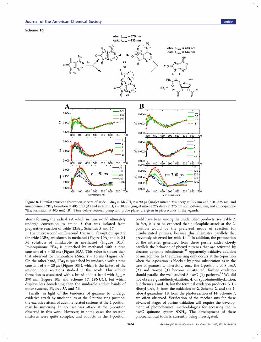
atoms forming the radical 29, which in turn would ultimatelyundergo conversion to amine 2 that was isolated frompreparative reaction of azide 13Bz3, Schemes 5 and 17.The microsecond−millisecond transient absorption spectra
for azide 13Bz3 are shown in methanol (Figure 10A) and in 0.1M solution of imidazole in methanol (Figure 10B).Iminoquinone 7Bz3 is quenched by methanol with a timeconstant of τ > 30 ms (Figure 10A). This value is slower thanthat observed for iminonitrile 26Ac3, τ = 15 ms (Figure 7A).On the other hand, 7Bz3 is quenched by imidazole with a timeconstant of τ = 20 μs (Figure 10B), which is the fastest of theiminoquinone reactions studied in this work. This adductformation is associated with a broad adduct band with λmax =390 nm (Figure 10B and Scheme 17, 28NUC), but whichdisplays less broadening than the imidazole adduct bands ofother systems, Figures 5A and 7B.Finally, in light of the tendency of guanine to undergo
oxidative attack by nucleophiles at the 5-purine ring position,the exclusive attack of adenine-related systems at the 2-positionmay be surprising. In no case was attack at the 5-positionobserved in this work. However, in some cases the reactionmixtures were quite complex, and adducts in the 5-position
could have been among the unidentified products; see Table 2.In fact, it is to be expected that nucleophile attack at the 2-position would be the preferred mode of reaction forunsubstituted purines, because this chemistry parallels thatpreviously observed for azide 14.26 In addition, the protonationof the nitrenes generated from these purine azides closelyparallels the behavior of phenyl nitrenes that are activated byelectron-donating substituents.32 Apparently oxidative additionof nucleophiles to the purine ring only occurs at the 5-positionwhen the 2-position is blocked by prior substitution as in thecase of guanosine. Therefore, once the 2-positions of 8-oxoA(2) and 8-oxoI (3) become substituted, further oxidationshould parallel the well-studied 8-oxoG (1) pathway.25 We didnot observe guanidinohydantioin, 4, or spiroiminodihydantion,5, Schemes 1 and 18, but the terminal oxidation products, N 1′-ribosyl urea, 6, from the oxidation of 2, Scheme 2, and the 1-ribosyl guanidine, 18, from the photoreaction of 14, Scheme 7,are often observed. Verification of the mechanisms for theseadvanced stages of purine oxidation will require the develop-ment of photochemical methodologies for accessing the 8-oxoG quinone system 9NH2. The development of thesephotochemical tools is currently being investigated.
Scheme 16
Figure 8. Ultrafast transient absorption spectra of azide 13Bz3 in MeOH, τ = 90 ps (singlet nitrene 27s decay at 375 nm and 550−625 nm, andiminoquinone 7Bz3 formation at 405 nm) (A) and in 2-PrOH, τ = 300 ps (singlet nitrene 27s decay at 375 nm and 550−625 nm, and iminoquinone7Bz3 formation at 405 nm) (B). Time delays between pump and probe pluses are given in picoseconds in the legends.
Journal of the American Chemical Society Article
dx.doi.org/10.1021/ja3068148 | J. Am. Chem. Soc. 2013, 135, 3423−34383434

■ CONCLUSIONSA working hypothesis for the oxidation of purines was outlinedin Scheme 1 and is refined in Scheme 18. The initial oxidationsteps to form the 8-oxo-7,8-dihydropurines 1, 2, and 3 are wellestablished and have been observed in many biological
systems.1,2 Burrows and Adam have invoked the quinoidalguanosine 9NH2 to explain the further oxidation of 1,27−31 andthe intermediacy of the related diiminoquinonone 15 has beenestablished in the photochemistry of purine azide 14.26 In thiswork, we have focused upon the possible role of the purine
Table 5. Calculated B3LYP/6-31G(d) Vertical Excitation Energies and Oscillator Strengths ( f) for Intermediates 27Me, 27t,7Me, 28OMe, and 28IMa
aAll calculations were performed in the gas phase.
Figure 9. Ultrafast transient absorption spectra of azide 13Bz3 in methylcyclohexane (A) and in dichloromethane (B). Time delays in picosecondsbetween pump and probe pluses are given in the legends. In (B), following the spectral narrowing and reshaping process on a time scale of severalpicoseconds, the spectral evolution displays a τ = 600 ps rise component.
Journal of the American Chemical Society Article
dx.doi.org/10.1021/ja3068148 | J. Am. Chem. Soc. 2013, 135, 3423−34383435

iminoquinone 7 and quinone 11 in the oxidation of adenosineand inosine, respectively. The observation of oxidativenucleophilic addition to the 2-position of 2 and 3 in thepresence of imidazole supports the intermediacy of 7 and 11,and the general hypothesis outlined in Scheme 18.
Furthermore, the intermediacy of 7 and its conversion to 8has been examined in detail using femtosecond and nano-second transient absorption spectroscopy. These observationsstrongly support the involvement of the iminoquinone 7 in thefurther oxidation of 2. Because 2 is derived from the oxidation
Scheme 17
Figure 10.Microsecond−millisecond transient absorption spectra of azide 13Bz3 in pure methanol, 28OMe (A), and in 1 M solution of imidazole inmethanol, 28IM (B).
Scheme 18
Journal of the American Chemical Society Article
dx.doi.org/10.1021/ja3068148 | J. Am. Chem. Soc. 2013, 135, 3423−34383436

of adenosine (Eox = 1.42 V/NHE), and has been widelyobserved in biological systems, one can assume that the furtheroxidation of 2 to 7 (Eox = 0.92 V/NHE) can proceed in livingsystems as well, and might lead to a wide variety of cross-linkingprocesses in biological systems.The literature has focused upon the further oxidation of 8-
oxo-7,8-dihydroguanosine (1) because it is more easily formedthan 8-oxo-7,8-dihydroadenosine (2). In fact, it has beenestimated that the formation of about 105 oxidative lesionsrelated to 1 and 2 are formed and repaired each day in theaverage rat cell.59 Furthermore, because the ratio of 1/2 isabout 3/1,21 the oxidation of adenosine might possibly be ofequal or greater significance than that of guanosine. The relativesignificance of these two types of oxidative lesions might wellbe biased by the fact that the adenosine lesions tend to formmuch more stable, less readily repaired, cross-links than doguanosine lesions.The observation that imidazole forms especially stable
adducts with oxidized adenosine deserves comment. Theamino acid histidine occurs in the active sites of numerousenzymes, many of which interact with DNA.60,61 Twoparticularly interesting cases are His-365 found in the activesite of Escherichia coli DNA topoisomerase I62 and the seryl-histidine unit found in the active sites of many hydrolyticenzymes such as the intein−extein junctions of homingendonucleases such as DNAase I.63 The interaction of theseenzymes that function in the routine maintenance of DNA withoxidatively damaged DNA would seem to be a most interestingarea for future investigation.While the work described in this Article significantly extends
our understanding of purine oxidation pathways by establishingthe viability of quinoidal purine intermediates, it does notaddress the questions of bridges between the various purinefamilies and the latter stages of purine oxidation to urea 6 andparabanic acid64,65 shown as dashed arrows in Scheme 18.Further work will be required to elucidate the possible oxidativepathways between 11 and 1, 8 and 9, and the ultimate N 1′-ribosyl urea 6.Finally, the azide 14 is one of the most widely used
photoaffinity labeling agents. The observations reported herefurther support the intermediacy of the diiminoquinone 15 asthe pivotal intermediate in this chemistry.26 Furthermore,comparison of the transient absorption spectroscopy of 14 withthat of 13 is consistent with previously studied nitrenechemistry where it was observed that singlet nitrenes inelectronic communication with electron-donating groupsbecome powerful bases and protonate rapidly.32 Thus, in thiswork, the activated basic nitrene 22 can be protonated withinthe first 300 fs, while the deactivated nitrene 27 requires 90−300 ps (Figure 8) for protonation. On the other hand, onceprotonated, iminoquinone 7 is a more powerful electrophilethan the diiminoquinone 15, and thus might well be a moreeffective cross-linking agent and PAL agent in many biologicalsystems. Work is continuing to further characterize thechemical reactivity of iminoquinone 7 in duplex nucleic acidenvironments.
■ ASSOCIATED CONTENT*S Supporting InformationCatalog of chemical structures with their structure numbers,and a description of the instrumentation used to obtain ultrafastfemto- and picosecond data. Syntheses of starting materials andcharacterization of synthetic intermediates, starting materials,
and photoproducts. This material is available free of charge viathe Internet at http://pubs.acs.org.
■ AUTHOR INFORMATIONCorresponding [email protected]; [email protected] authors declare no competing financial interest.
■ ACKNOWLEDGMENTSWe thank Dr. Larry Sallans of the Mass Spectrometry Facility atthe University of Cincinnati for providing us with detailed massspectrometric analyses. Financial support from the Ohio SuperComputer Center and the NSF (ANT, Career Award, CHE-0847707) and the R. Marshall and Antonia G. WilsonChemistry Fund is acknowledged. We express our appreciationto Prof. Felix N. Castellano and Valentina A. Prusakova fortheir help in acquisition of nanosecond spectroscopy data. Wewould like to thank Prof. Massimo Olivucci and Elena V.Laricheva for their help in theoretical calculations and Prof.Ksenija Glusac, Dr. Pavel Kucheryavy, and EkaterinaMirzakulova for their help in electrochemistry experiments.
■ REFERENCES(1) Wang, D.; Kreutzer, D. A.; Essigmann, J. M. Mutat. Res. 1998,400, 99−115.(2) Cadet, J.; Douki, T.; Gasparutto, D.; Ravanat, J.-L. Mutat. Res.2003, 531, 5−23.(3) Barnes, D. E.; Lindahl, T. Annu. Rev. Genet. 2004, 38, 445−476.(4) Nakabeppu, Y.; Sakumi, K.; Sakamoto, K.; Tsuchimoto, D.;Tsuzuki, T.; Nakatsu, Y. Biol. Chem. 2006, 387, 373−379.(5) Wilson, D. M.; Bohr, V. A. DNA Repair 2007, 6, 544−559.(6) Finkel, T.; Holbrook, N. J. Nature 2000, 408, 239−247.(7) Feig, D. I.; Loeb, L. A. J. Mol. Biol. 1994, 235, 33−41.(8) Wiseman, H.; Halliwell, B. Biochem. J. 1996, 313, 17−29.(9) Steenken, S.; Jovanovic, S. V. J. Am. Chem. Soc. 1997, 119, 617−618.(10) Solivio, M. J.; Joy, T. J.; Sallans, L.; Merino, E. J. J. Inorg.Biochem. 2010, 104, 1000−1005.(11) Solivio, M. J.; Nemera, D. B.; Sallans, L.; Merino, E. J. Chem. Res.Technol. 2012, 25, 326−336.(12) Tan, X.; Grollman, A. P.; Shibutani, S. Carcinogenesis 1999, 20,2287−2292.(13) von Sonntag, C. Free-Radical-Induced DNA Damage and ItsRepair: A Chemical Perspective; Springer-Verlag: New York, 2006; pp211−447.(14) Bjelland, S.; Seeberg, E. Mutat. Res. 2003, 531, 37−80.(15) Dirks, A. J.; Hofer, T.; Marzetti, E.; Pahor, M.; Leeuwenburgh,C. Aging Res. Rev. 2006, 5, 179−195.(16) Hofer, T.; Seo, A. Y.; Prudencio, M.; Leeuwenburgh, C. Biol.Chem. 2006, 387, 103−111.(17) Hofer, T.; Badouard, C.; Bajak, E.; Ravanat, J.-L.; Mattsson, A.;Cotgreave, I. A. Biol. Chem. 2005, 386, 333−337.(18) Dizdaroglu, M. Mutat. Res. 2003, 531, 109−126.(19) Michaels, M. L.; Tchou, J.; Grollman, A. P.; Miller, J. H.Biochemistry 1992, 31, 10964−10968.(20) Jaruga, P.; Dizdaroglu, M. Nucleic Acids Res. 1996, 24, 1389−1394.(21) Llano, J.; Eriksson, L. A. Phys. Chem. Chem. Phys. 2004, 6,4707−4713.(22) Ito, T.; Kuno, S.; Uchida, T.; Fujita, J.-i.; Nishimoto, S.-i. J. Phys.Chem. B 2009, 113, 389−394.(23) Jensen, A.; Calvayrac, G.; Karahalil, B.; Bohr, V. A.; Stevnsner,T. J. Biol. Chem. 2003, 278, 19541−19548.(24) Grin, I. R.; Dianov, G. L.; Zharkov, D. O. FEBS Lett. 2010, 584,1553−1557.
Journal of the American Chemical Society Article
dx.doi.org/10.1021/ja3068148 | J. Am. Chem. Soc. 2013, 135, 3423−34383437

(25) Perrier, S.; Hau, J.; Gasparutto, D.; Cadet, J.; Favier, A.; Ravanat,J.-L. J. Am. Chem. Soc. 2006, 128, 5703−5710.(26) Polshakov, D.; Rai, S.; Wilson, R. M.; Mack, E. T.; Vogel, M.;Krause, J. A.; Burdzinski, G.; Platz, M. S. Biochemistry 2005, 44,11241−11253.(27) Xu, X.; Fleming, A. M.; Muller, J. G.; Burrows, C. J. J. Am. Chem.Soc. 2008, 130, 10080−10081.(28) Johansen, M. E.; Muller, J. G.; Xu, X.; Burrows, C. J.Biochemistry 2005, 44, 5660−5671.(29) Hosford, M. E.; Muller, J. G.; Burrows, C. J. J. Am. Chem. Soc.2004, 126, 9540−9541.(30) Ye, Y.; Muller, J. G.; Luo, W.; Mayne, C. L.; Shallop, A. J.; Jones,R. A.; Burrows, C. J. J. Am. Chem. Soc. 2003, 125, 13926−13927.(31) Adam, W.; Arnold, M. A.; Nau, W. M.; Pischel, U.; Saha-Moller,C. R. J. Am. Chem. Soc. 2002, 124, 3893−3904.(32) Voskresenska, V.; Wilson, R. M.; Panov, M.; Tarnovsky, A. N.;Krause, J. A.; Vyas, S.; Winter, A. H.; Hadad, C. M. J. Am. Chem. Soc.2009, 131, 11535−11547.(33) Mack, E. T. University of Cincinnati, unpublished results.(34) Yanagawa, H.; Ogawa, Y.; Ueno, M. J. Biol. Chem. 1992, 267,13320−13326.(35) Steenken, S.; Jovanovic, S. V.; Bietti, M.; Bernhard, K. J. Am.Chem. Soc. 2000, 122, 2373−2374.(36) Adam, W.; Saha-Moller, C. R.; Schonberger, A.; Berger, M.;Cadet, J. J. Photochem. Photobiol. 1995, 62, 231−238.(37) Sheu, C.; Foote, C. S. J. Am. Chem. Soc. 1995, 117, 474−477.(38) Sheu, C.; Foote, C. S. J. Am. Chem. Soc. 1995, 117, 6439−6442.(39) Raoul, S.; Cadet, J. J. Am. Chem. Soc. 1996, 118, 1892−1898.(40) Adam, W.; Saha-Moller, C. R.; Schonberger, A. J. Am. Chem. Soc.1996, 118, 9233−9238.(41) Duarte, V.; Gasparutto, D.; Yamaguchi, L. F.; Ravanat, J.-L.;Martinez, G. R.; Medeiros, M. H. G.; Di Mascio, P.; Cadet, J. J. Am.Chem. Soc. 2000, 122, 12622−12628.(42) Luo, W.; Muller, J. G.; Rachlin, E. M.; Burrows, C. J. Chem. Res.Toxicol. 2001, 14, 927−938.(43) Duarte, V.; Muller, J. G.; Burrows, C. J. Nucleic Acids Res. 1999,27, 496−502.(44) Luo, W.; Muller, J. G.; Rachlin, E. M.; Burrows, C. J. Org. Lett.2000, 2, 613−616.(45) Temple, C., Jr.; Montgomery, J. A. J. Org. Chem. 1965, 30, 826−829.(46) Temple, C., Jr.; Coburn, W. C., Jr.; Thorpe, M. C.;Montgomery, J. A. J. Org. Chem. 1965, 30, 2395−2398.(47) Temple, C., Jr.; Thorpe, M. C.; Corburn, W. C., Jr.;Montgomery, J. A. J. Org. Chem. 1966, 31, 935−938.(48) Addicott, C.; Wong, M. W.; Wentrup, C. J. Org. Chem. 2002, 67,8538−8546.(49) Marburg, S.; Jorn, D.; Tolman, R. L. J. Heterocycl. Chem. 1982,19, 671−672.(50) Gadosy, T. A.; McClelland, R. A. J. Am. Chem. Soc. 1999, 121,1459−1465.(51) Dehaen, W.; Becher, J. Acta Chem. Scand. 1993, 47, 244−254.(52) Funicello, M.; Spagnolo, P.; Zanirato, P. Acta Chem. Scand.1993, 47, 231−243.(53) Spinelli, D.; Zanirato, P. J. Chem. Soc., Perkin Trans. 2 1993,1129−1133.(54) Assignment of spectral bands was supported by theoreticalcalculations using Gaussian 09 and MOLCAS 7.4 software: (a) Frisch,M. J.; Trucks, G. W.; Schlegel, H. B.; Scuseria, G. E.; Robb, M. A.;Cheeseman, J. R.; Scalmani, G.; Barone, V.; Mennucci, B.; Petersson,G. A.; Nakatsuji, H.; Caricato, M.; Li, X.; Hratchian, H. P.; Izmaylov,A. F.; Bloino, J.; Zheng, G.; Sonnenberg, J. L.; Hada, M.; Ehara, M.;Toyota, K.; Fukuda, R.; Hasegawa, J.; Ishida, M.; Nakajima, T.; Honda,Y.; Kitao, O.; Nakai, H.; Vreven, T.; Montgomery, J. A., Jr.; Peralta, J.E.; Ogliaro, F.; Bearpark, M.; Heyd, J. J.; Brothers, E.; Kudin, K. N.;Staroverov, V. N.; Kobayashi, R.; Normand, J.; Raghavachari, K.;Rendell, A.; Burant, J. C.; Iyengar, S. S.; Tomasi, J.; Cossi, M.; Rega,N.; Millam, N. J.; Klene, M.; Knox, J. E.; Cross, J. B.; Bakken, V.;Adamo, C.; Jaramillo, J.; Gomperts, R.; Stratmann, R. E.; Yazyev, O.;
Austin, A. J.; Cammi, R.; Pomelli, C.; Ochterski, J. W.; Martin, R. L.;Morokuma, K.; Zakrzewski, V. G.; Voth, G. A.; Salvador, P.;Dannenberg, J. J.; Dapprich, S.; Daniels, A. D.; Farkas, O.;Foresman, J. B.; Ortiz, J. V.; Cioslowski, J.; Fox, D. J. Gaussian 09,revision A.1; Gaussian, Inc.: Wallingford, CT, 2009. (b) MOLCAS7.4: Aquilante, F.; De Vico, L.; Ferre, N.; Ghigo, G.; Malmqvist, P.;Neogrady, P.; Pedersen, T. B.; Pitonak, M.; Reiher, M.; Roos, B. O.;Serrano-Andres, L.; Urban, M.; Veryazov, V.; Lindh, R. J. Comput.Chem. 2010, 31, 224−247.(55) Peon, J.; Polshakov, D.; Kohler, B. J. Am. Chem. Soc. 2002, 124,6428−6438.(56) Dix, E. J.; Goodman, J. L. J. Phys. Chem. 1994, 98, 12609−12612.(57) Pines, E.; Pines, D.; Barak, T.; Magnes, B. Z.; Tolbert, L. M.;Haubrich, J. E. Ber. Bunsen-Ges. 1998, 102, 511−517.(58) Lima, J. C.; Abreu, I.; Brouillard, R.; Macanita, A. L. Chem. Phys.Lett. 1998, 298, 189−195.(59) Park, E.-M.; Shigenaga, M. K.; Degan, P.; Korn, T. S.; Kitzler, J.W.; Wehr, C. M.; Kolachana, P.; Ames, B. N. Proc. Natl. Acad. Sci.U.S.A. 1992, 89, 3375−3379.(60) Luscombe, N. M.; Laskowski, R. A.; Thornton, J. M. NucleicAcids Res. 2001, 29, 2860−2874.(61) Leavens, F. M. V.; Churchill, C. D. M.; Wang, S.; Wetmore, S.D. J. Phys. Chem. B 2011, 115, 10990−11003.(62) Perry, K.; Mondragon, A. J. Biol. Chem. 2002, 277, 13237−13245.(63) Li, Y.; Hatfield, S.; Li, J.; McMills, M.; Zhao, Y.; Chen, X. Bioorg.Med. Chem. 2002, 10, 667−673.(64) Vialas, C.; Claparols, C.; Pratviel, G.; Meunier, B. J. Am. Chem.Soc. 2000, 122, 2157−2167.(65) Jena, N. R.; Mishra, P. C. Free Radical Biol. Med. 2012, 53, 81−94.
Journal of the American Chemical Society Article
dx.doi.org/10.1021/ja3068148 | J. Am. Chem. Soc. 2013, 135, 3423−34383438