oxidative stress controls the choice of alternative last exons via a
TRANSCRIPT
902–914 Nucleic Acids Research, 2017, Vol. 45, No. 2 Published online 2 September 2016doi: 10.1093/nar/gkw780
Oxidative stress controls the choice of alternative lastexons via a Brahma–BRCA1–CstF pathwayGabriele A. Fontana1,†, Aurora Rigamonti1,†, Silvia C. Lenzken1, Giuseppe Filosa1,Reinaldo Alvarez1, Raffaele Calogero3, Marco E. Bianchi2 and Silvia M.L. Barabino1,*
1Department of Biotechnology and Biosciences, University of Milan-Bicocca, Piazza della Scienza 2, 20126 Milan,Italy, 2Division of Genetics and Cell Biology, San Raffaele Scientific Institute and University, Via Olgettina 60, 20132Milan, Italy and 3Department of Biotechnology and Health Sciences, University of Torino, Via Nizza 52, I-10126Torino, Italy
Received October 27, 2015; Revised August 23, 2016; Accepted August 26, 2016
ABSTRACT
Alternative splicing of terminal exons increases tran-script and protein diversity. How physiological andpathological stimuli regulate the choice between al-ternative terminal exons is, however, largely un-known. Here, we show that Brahma (BRM), the AT-Pase subunit of the hSWI/SNF chromatin-remodelingcomplex interacts with BRCA1/BARD1, which ubiq-uitinates the 50 kDa subunit of the 3′ end process-ing factor CstF. This results in the inhibition of tran-script cleavage at the proximal poly(A) site and a shifttowards inclusion of the distal terminal exon. Uponoxidative stress, BRM is depleted, cleavage inhibi-tion is released, and inclusion of the proximal lastexon is favoored. Our findings elucidate a novel reg-ulatory mechanism, distinct from the modulation oftranscription elongation by BRM that controls alter-native splicing of internal exons.
INTRODUCTION
Pre-mRNA splicing and 3′ end processing are essential stepsfor the expression of the vast majority of metazoan genes.Recently, RNAseq analysis showed that not only alternativesplicing (AS) but also alternative polyadenylation (APA) ismore frequent and complex than previously anticipated (1–4). The choice of alternative poly(A) sites generates differ-ent 3′ UTRs that can affect translation, stability and local-ization of the mRNA. Alternative pre-mRNA processingchanges the length of the 3′ UTR during cell differentia-tion contributing to the regulation of gene expression (5,6).When coupled to the inclusion of an alternative last exon(ALE), alternative polyadenylation leads to the generationof mRNA variants that differ in their 3′ UTR and that mayencode proteins with different C-terminal regions.
Whereas the molecular details of pre-mRNA 3′ end pro-cessing are rather well known, how the choice of APAsites is regulated is only partially understood. The mature3′ ends of most eukaryotic mRNAs are generated by en-donucleolytic cleavage of the primary transcript followedby the addition of a poly(A) tail to the upstream cleavageproduct (1,7). Maturation of the 3′ end is executed by alarge multicomponent complex that is assembled in a co-operative manner on specific cis-acting sequence elementsin the pre-mRNA (8,9). In mammalian cells, the cleavageand polyadenylation specificity factor (CPSF) recognizesthe consensus hexanucleotide AAUAAA, whereas the cleav-age stimulation factor (CstF), a hexameric complex of sub-units of 77, 64 and 50 kDa (10,11), binds a more degen-erate GU- or U-rich element downstream of the poly(A)site. Both CPSF and CstF interact with RNA polymerase II(RNAPII) at the promoter and appear to remain associatedwith it during elongation (12,13). CstF50 and CstF77 inter-act specifically with the carboxy-terminal domain (CTD) ofRNAPII largest subunit. CstF50 was also shown to bindthe BARD1/BRCA1 ubiquitin ligase after DNA damage,resulting in the inhibition of 3′ end processing (14). Thisobservation and the fact that APA is modulated in devel-opment, differentiation and neuronal activation (reviewedin (9)) indicates that 3′ end processing can be regulated inresponse to physiological and pathological stimuli.
Oxidative stress is a widely occurring phenomenon in bi-ological systems, and is due to an imbalance between theintracellular production or influx of reactive oxygen species(ROS) and the availability of antioxidant compounds, suchas glutathione. Oxidative stress has been implicated in theetiology of many neurodegenerative disorders, includingAlzheimer’s and Parkinson’s diseases (15). At the cell level,oxidative stress elicits a wide spectrum of responses rang-ing from proliferation to growth arrest, senescence, or celldeath. The particular outcome reflects the balance between
*To whom correspondence should be addressed. Tel: +39 02 6448 3352; Fax: +39 02 6448 3569; Email: [email protected]†These authors contributed equally to the work as first authors.Present address: Gabriele A. Fontana, Friedrich Miescher Institute for Biomedical Research, Maulbeerstrasse 66, 4058 Basel, Switzerland.
C© The Author(s) 2016. Published by Oxford University Press on behalf of Nucleic Acids Research.This is an Open Access article distributed under the terms of the Creative Commons Attribution License (http://creativecommons.org/licenses/by-nc/4.0/), whichpermits non-commercial re-use, distribution, and reproduction in any medium, provided the original work is properly cited. For commercial re-use, please [email protected]
Downloaded from https://academic.oup.com/nar/article-abstract/45/2/902/2953299by gueston 17 February 2018
Nucleic Acids Research, 2017, Vol. 45, No. 2 903
a variety of intracellular stress signalling pathways that areactivated in response to the oxidative insult and that ulti-mately modulate gene expression.
We recently described that exposure of human neurob-lastoma SH-SY5Y cells to different sources of ROS leadsto genome-wide alternative splicing changes, modifyingthe relative proportion of alternatively spliced forms (16).Here, we show that oxidative stress specifically affects thechoice of ALEs increasing the production of transcriptsvariants terminating at a more proximal poly(A). Oxida-tive stress induces the transcriptional downregulation ofBrahma (BRM), one of the two alternative ATPase sub-units of the SWI/SNF complex. We find that in normalcondition BRM is enriched on the proximal ALE. In ad-dition we observe the accumulation of BARD1, a proteinthat forms a functional heterodimer with BRCA1, whichhas E3 ubiquitin-ligase activity and interacts with the 50kDa subunit of CstF inhibiting 3′ end processing (17). Con-sistent with these observations, we detect an ubiquitinatedpool of CstF50 and show that ubiquitination is mediated byBARD1/BRCA1. Taken together, our results suggest thatthe presence of BRM on the proximal exon leads to theBARD1/BRCA1-mediated ubiquitination of CstF50 andthe inhibition of 3′ end processing at the proximal poly(A).This in turn allows transcription to proceed to the distal ter-minal exon.
MATERIALS AND METHODS
Cell culture and transfections
Human neuroblastoma SH-SY5Y, SH-SY5Y/SOD1, SH-SY5Y/SOD1(G93A) and HEK293T cells were cultured inD-MEM High Glucose medium (Gibco, Invitrogen), 10%fetal bovine serum (FBS), 2.5 mM L-glutamine, 100 U/mlpenicillin, and 100 �g/ml streptomycin (Euroclone) at 37◦Cwith 5% CO2. Paraquat (Sigma-Aldrich) treatment of SH-SY5Y cells was carried out as described (16). Resveratrol(Sigma-Aldrich) was dissolved in DMSO, and cells weretreated with 10 �M resveratrol for 24 h. Control cells weretreated with vehicle. Adult dermal fibroblats (ATCC, PCS-201-012) were cultured D-MEM High Glucose medium(Gibco, Invitrogen), 10% fetal bovine serum (FBS), 5 mML-glutamine, 100 U/ml penicillin and 100 �g/ml strepto-mycin (Euroclone) at 37◦C with 5% CO2. Treatment with 2mM paraquat was performed for 48 h, while treatment with0.2 mM H2O2 (Sigma-Aldrich) was performed for 24 h.
Cells were transfected using polyethylenimine (PEI,Sigma-Aldrich) according to the manufacturer’s instruc-tions. Stably transfected cells were obtained by selectionwith 1 �g/ml puromycin (Sigma-Aldrich). For depletionof BRCA1, HEK293T cells were transfected twice (day 0and day 3) in six-well plates with 75 pmol of the corre-sponding Silencer® select siRNAs and Negative controlsiRNA #1 (Ambion). Transfection was carried out usingLipofectamine® RNAiMax reagent (Invitrogen) followingmanufacturer’s instructions. Cells were expanded and lysedat day 6.
For BRM-K755A overexpression, cells were electropo-rated with Amaxa® nucleofector® system (Lonza) usingAmaxa® Cell Line Nucleofector® Kit V according to themanufacturer’s instructions.
Intracellular ROS were measured with 2′,7′-dichlorodihydrofluorescein diacetate (H2DCF-DA;Sigma-Aldrich, D6883). Cells were exposed to H2DCF-DA(20 �M) during the last 30 min of culture, then collectedand washed with PBS. A blank sample (cells not exposedto H2DCF-DA) was also prepared. The H2DCF-DA flu-orescence was measured by flow cytometry after additionof propidium iodide (PI) to the samples. Only the cellularpopulation of PI impermeable cells was considered formeasuring the fluorescence intensity of H2DCF-DA (18).
Plasmids and antibodies
The cDNAs encoding the N-terminal flag-tagged humanBRM and N-terminal flag-tagged BRG1 proteins were agift from B. Emerson. These cDNAs were subcloned intothe SmaI–SalI (New England Biolabs) sites of the pAD5-CMV-Wpre-PGK-Puro expression vector (a kind gift fromS. Philipsen). The mutation K755A (19) was introduced us-ing a two-step mutagenic PCR procedure using Phusionpolymerase (Finnzymes). The interfering shRNA oligonu-cleotides targeting human SMARCA2 and SMARCA4transcripts were designed using the SiDesign Center (Dhar-macon). The shRNA primers were cloned into pSUPurovector. pSUPuro, and the pSUPuro ß2 T-Cell ReceptorBeta used as an unrelated control shRNA were gifts fromM.D. Ruepp. BARD1 and BRCA1 constructs were a giftof N. Chiba. The cDNA encoding CstF50 was subclonedfrom pcDNA3 HA-CstF50 (a gift from M.D. Ruepp)into a p3XFLAG-myc-CMV26 removing the myc tag. Thehistidine-tagged Ubiquitin was a gift of M.L. Guerrini. Allconstructs were verified by sequencing (BMR Genomics).All oligonucleotide sequences are listed in SupplementaryTable S2. The commercial antibodies used are listed in theSupplementary Table S1. Non-immune rabbit IgGs (Mil-lipore) were used as a control in the immunoprecipitationassays.
Luciferase assay
SH-SY5Y cells were transiently co-transfected with theindicated pGL2 luciferase reporter plasmids and withthe Renilla-encoding pRL-TK plasmid (Promega Inc.).Twenty-four hours after transfection, cells were lysed andluciferase activity quantified using the Dual Luciferase Re-porter kit (Promega Inc.) and a Berthold luminometer(Berthold Technologies). For the luciferase experiments,paraquat was added 3 h after transfection.
Bioinformatic analysis
Transcripts characterized by alternative splicingevents at their 3′ end were detected by R script-ing using Bioconductor 2.12 packages Genomi-cRanges, TxDb.Hsapiens.UCSC.hg19.knownGeneand HuExExonProbesetLocation (www.bioconductor.org). Transcripts were extracted fromTxDb.Hsapiens.UCSC.hg19.knownGene (80922 tran-scripts). After removal of transcripts lacking a link toEntrez Gene Identifier (20), 71 350 transcripts (26 5661exons), associated to 22 932 EG, were left for further
Downloaded from https://academic.oup.com/nar/article-abstract/45/2/902/2953299by gueston 17 February 2018

904 Nucleic Acids Research, 2017, Vol. 45, No. 2
analysis. Subsequently, we selected all genes associatedto the presence of alternative splicing even at the 3′ end(12 839 genes, 58 451 transcripts, 26 608 exons involvedin ALE). Affymetrix Human Exon 1.0 ST Array (HuEx-1 0-st) exon-level probe sets chromosomal locations wereextracted from HuExExonProbesetLocation (21). Onlyexon-level probesets associated to the Affymetrix coreannotation were considered (284805 exon-level probesets).These exon-level probesets mapped on 12839 genes (59986 UCSC transcripts, 230 112 exons). Out of the 230112 exons 22 983 were associated to ALE. Alternativelyspliced exon-level probesets were retrieved by Lenzkenet al. (16). Four hyndred six exon-level probesets mappingon 418 exons were associated to 262 genes (1191 UCSCtranscripts, 4844 exons). Within the 418 exons 89 exons (78UCSC genes) were involved in ALE.
RNA analysis
RNA preparation and RT-PCR reactions were performedas described (16). The sequences of the exon-specificprimers are listed in Supplementary Table S2. Assay con-ditions were optimized for each gene with respect to primerannealing temperatures, primer concentrations, and MgCl2concentrations. Quantification was performed with Bioan-alyzer 2100 (Agilent Technologies).
Chromatin immunoprecipitation and qPCR analysis
107 SH-SY5Y cells were cross-linked with 1% formaldehyde(Sigma-Aldrich), and quenched with the addition of 125mM glycine (Sigma-Aldrich). Cell pellets were lysed in LysisBuffer (50 mM HEPES–KOH pH 7.5, 140 mM NaCl, 1 mMEDTA pH 8, 1% Triton X-100, 0.1% sodium deoxycholate,0.1% SDS, protease inhibitors (Roche) for 1 hour on ice, andthen sonicated with a Branson 250 sonifier (Branson Inc.)to obtain chromatin fragments of ∼400 nt. Aliquots corre-sponding to 5 × 10⁶ cells were flash-frozen in liquid nitrogenand then maintained at −80◦C. For each immunoprecipi-tation, 1% of the total genomic DNA was saved as inputDNA. The chromatin solution was precleared at 4◦C for 1h with sepharose beads. Immunoprecipitation was carriedout using the Chromatin IP Assay Kit (Millipore), follow-ing manufacturer’s instructions. Bound material was elutedwith 500 �l of Elution Buffer (1% SDS, 100 mM NaHCO3)for 1 h at room temperature. Crosslinking was reversed at65◦C overnight, with the addition of 250 mM NaCl and2 �g/ml Proteinase K (Sigma). DNA was purified by phe-nol:choloform:isoamyl alcohol (Sigma-Aldrich) extractionand ethanol precipitation. For ChIP-ReChIP experiments,eluates from the first ChIP assays were diluted in the ChIPDilution buffer (Millipore) to reduce the SDS concentrationto 0.1% (w/v). Then, the second ChIP was performed. Im-munoprecipitated DNA was analyzed in triplicate by qPCRusing the SYBR Green method with Mesa Green qPCRmaster mix (Biosense) in an ABI PRISM 7500 (AppliedBiosystem). The primers used for the qPCRs are listed inthe Supplemental Table S2. Data were normalized by theFold Enrichment Method as follows: Relative Enrichment= 2 – (�Ct antibody- �Ct IgG).
Micrococcal nuclease assays
107 SH-SY5Y cells were synchronized in G0 by serumstarvation as assessed by FACS analysis (FACSCalibur,BD Biosciences). Cells were cross-linked with 1% (v/v)formaldehyde and quenched with the addition of 125 mMglycine. Cell pellets were lysed in Lysis Buffer (50 mMHEPES–KOH pH 7.5, 140 mM NaCl, 1% Triton X-100,0.1% sodium deoxycholate, 0.1% SDS, protease inhibitors(Roche). Nuclei were disrupted by five bursts of sonica-tion. Aliquots of 5 × 10⁶ cells were flash-frozen in liquidnitrogen and then maintained at −80◦C. One undigestedaliquot was used as control. Digestion was performed with20 U of Micrococcal Nuclease (Fermentas) in 1 mM finalCaCl2 and MgCl2 for 30 min at room temperature, thenstopped with 20 mM EDTA. Crosslinking was reversed at65◦C overnight, with the addition of 250 mM NaCl and 2�g/ml Proteinase K followed by phenol:choloform:isoamylalcohol extraction and ethanol precipitation. qPCR analy-sis was performed on 4 ng. The primers used for the qPCRsare listed in Supplemental Table S2. Each sample was an-alyzed in triplicate, and the average Ct was calculated foreach primer set. In order to determine the Relative Nucle-osome Occupancy associated with each primer set, the fol-lowing equation was used: Relative Nucleosome Occupancy= 10(Ct Untreated – Ct MNase).
Nuclear extracts preparation and co-immunoprecipitations
Nuclear protein extracts (NEs) were prepared using accord-ing to the Lamond protocol (www.Lamondlab.com). Forco-immunoprecipitations, 300 �g of NEs were first pre-cleared with protein A sepharose (GE) for 2 h at 4◦C andthen incubated with the indicated antibodies or with rab-bit IgG as a control. The antibodies were coupled to pro-tein A-Sepharose beads following manufacturer’s instruc-tions. Immunoprecipitations were carried out for 2 h at 4◦Con a rotating wheel. Beads were then washed three timeswith a solution composed of 10 mM Tris–HCl, 100 mMNaCl, 0.01% Nonidet P-40, and 0.04% bovine serum albu-min (BSA). The proteins bound to the beads were elutedby boiling the samples at 95◦C for 10 min. Aliquots wereanalyzed by SDS-PAGE and immunoblotting.
For ubiquitin immunoprecipitation, cells were lysed in1% SDS lysis buffer (50 mM Tris–HCl pH 7.4, 0.5 mMEDTA, 1% SDS) and further boiled for an additional 10min. Lysates were clarified by centrifugation at 16 000 g for10 min. Supernatant was diluted 10 times with a buffer com-posed of 50 mM Tris–HCl pH 7.4, 150 mM NaCl, 1% Tri-ton and complete Protease Inhibitor Cocktail (Roche). Im-munoprecipitation was performed with anti-Multi Ubiqui-tin mAb-Agarose (MBL) overnight at 4◦C. The precipitateswere washed three times and the samples were resolved on6% SDS-PAGE.
For Flag-tagged CstF50 immunoprecipitation, cells werelysed as for ubiquitin immunoprecipitation. Immunopre-cipitation was performed with ANTI-FLAG® M2 AffinityGel (Sigma) overnight at 4◦C. The precipitates were washedthree times and eluted in a rotating wheel for 1 h at 4◦C witha buffer containing 50 mM Tris–HCl pH 7.4, 150 mM NaCl,1% Triton, complete Protease Inhibitor Cocktail (Roche)
Downloaded from https://academic.oup.com/nar/article-abstract/45/2/902/2953299by gueston 17 February 2018
Nucleic Acids Research, 2017, Vol. 45, No. 2 905
and 3× flag-peptide (5 �g/ml). The samples were resolvedon 7% SDS-PAGE.
For myc-tagged BARD1 immunoprecipitation, cells werelysed in 50 mM Tris–HCl, pH 7.4, with 150 mM NaCl,1 mM EDTA and 1% Triton X-100. Lysates were incu-bated overnight at 4◦C with anti-myc antibody (71D10Cell Signalling). The complexes were coupled to protein A-Sepharose beads following manufacturer’s instructions. Im-munoprecipitations were carried out for 2 h at 4◦C on a ro-tating wheel. Beads were then washed three times with a so-lution containing 10 mM Tris–HCl, 100 mM NaCl, 0.05%Nonidet P-40, and 0.04% BSA. The proteins bound to thebeads were eluted by boiling the samples at 95◦C for 10 min.The sample were resolved on 7% SDS-PAGE.
Quantification of western blots was performed by usingNIH ImageJ software the Image Studio software (OdisseyFC-Licor) or Image Studio v1.1.
Statistical analysis
Statistical analysis of RT-PCR experiments was performedeither using a one-way ANOVA test followed by a post-hocTukey–Kramer multiple comparison test, or a two-tailed,paired t-test. Analysis of FACS experiments was carriedout using a one-way ANOVA test followed by a post-hocTukey–Kramer multiple comparison test. Analysis of west-ern blot data was performed using a two-tailed, paired t-test.
RESULTS
BRM is down-regulated in response to oxidative stress
Oxidative stress has long been implicated in neuronal celldeath that is associated with neurodegenerative disorders.For example, exposure of cells to paraquat (N,N’-dimethyl-4,4′-bipyridinium dichloride, PQ), a neurotoxic herbicide,increases the production of the superoxide anion (22), andhas been linked to the incidence of Parkinson’s disease.Moreover, the second most frequent cause of hereditaryAmyotrophic Lateral Sclerosis (ALS) are mutations in thecytoplasmic superoxide dismutase 1 (SOD1) protein andhave been shown to increases intracellular ROS levels (23).
We recently showed that treatment with PQ or expres-sion of the mutant SOD1(G93A) protein cause extensivechanges in both mRNA expression and alternative splicingin human neuroblastoma SH-SY5Y cells (16). Among themost down-regulated genes was SMARCA2, the gene en-coding BRM (Figure 1A and Supplementary Figure S1).SOD1(G93A) expression almost abrogated BRM expres-sion at the protein level, while expression of the hSWI/SNFalternative ATPase subunit BRG1 was not significantly af-fected (Figure 1B). Similarly, treatment of SH-SY5Y cellswith PQ strongly reduced BRM mRNA and protein levelswithout affecting BRG1 expression (Supplementary FigureS1). To test a possible causal link between oxidative stressand BRM expression, we exposed primary adult fibroblaststo different sources of ROS. Treatment with H2O2 (Figure1C) or with PQ (Supplementary Figure S1) effectively re-duced BRM expression.
We set out to identify regulatory elements in theSMARCA2 promoter that respond to oxidative stress. In-
spection of the −3444/+57 region identified features asso-ciated to promoter sequences (Supplementary Figure S2):peaks of evolutionary conservation, a CpG island, a puta-tive DNase hypersensitive region, and peaks of enrichmentof acetylation of histone 3 lysine 27 (H3KAc27). A highGC-content and the absence of a detectable TATA-box sug-gested that the promoter belongs to the class of GC pro-moters. We then cloned a 3.4 kb genomic fragment (fromposition –3344 to +57) into a promoter-less luciferase re-porter. This region conferred sensitivity to PQ (Supplemen-tary Figure S2) and showed reduced luciferase expressionwhen transfected in SH-SY5Y/SOD1(G93A) cells (Figure1D). Further 5′ deletion analysis pointed to the −146/+57fragment as critical for the oxidative stress response (Figure1D). Addition of resveratrol, a natural antioxidant, to SH-SY5Y/SOD1(G93A) cells reduced ROS production (Fig-ure 1E) and restored luciferase expression to both the long3344/+57 and the minimal -146/+57 promoter constructs(Figure 1F).
Overall these results indicate that SMARCA2 transcrip-tion and hence BRM expression are reduced by oxidativestress.
Selection of the proximal alternative last exon is favored inBRM-depleted cell
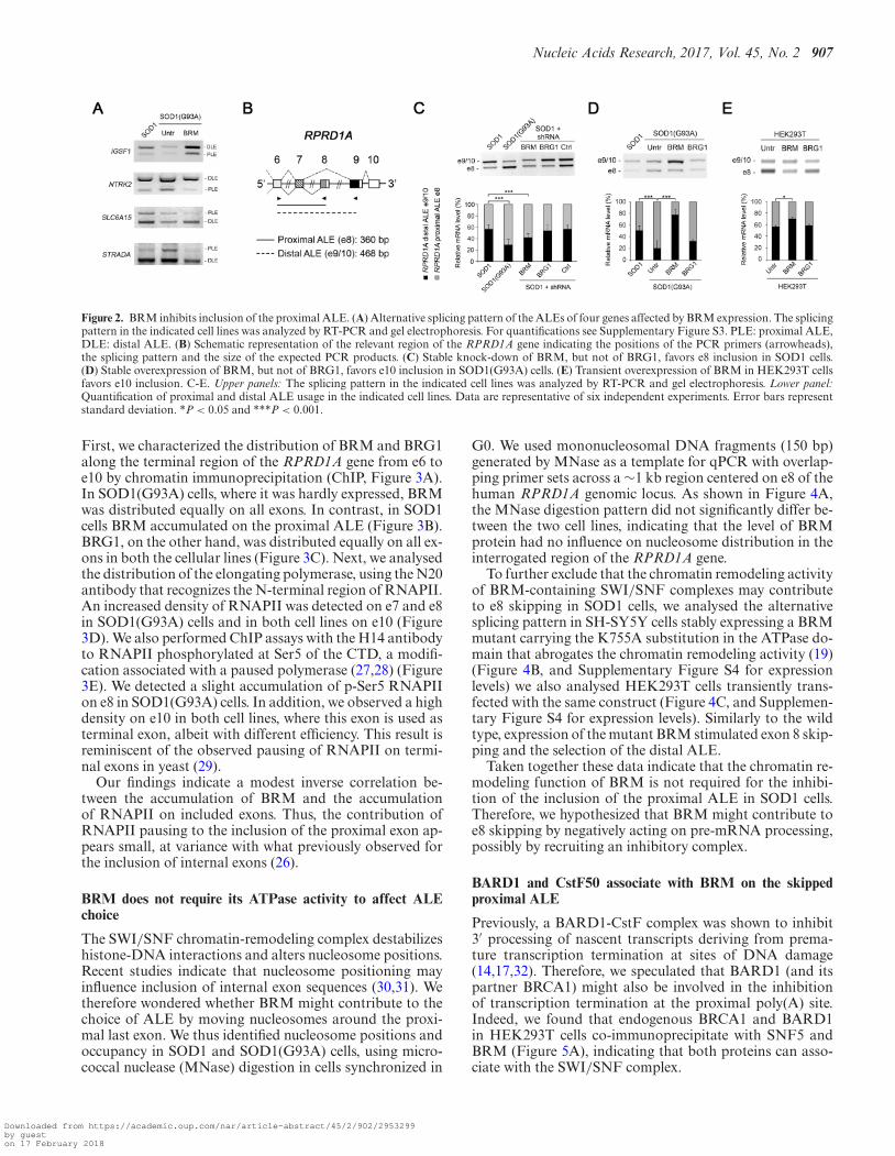
Using splicing-sensitive microarrays we had previously de-tected a large number of AS changes (262 genes, involving418 exons) in SH-SY5Y/SOD1(G93A) cells (16). Withinthis dataset, 89 exons (in 78 genes) appeared as ALEs. Wevalidated by PCR six of these genes: in five out of six genes,the distal ALE was favoured in the presence of BRM (i.e.in SH-SY5Y/SOD1 cells), whereas the proximal ALE waspreferred when BRM was expressed at low level as in SH-SY5Y/SOD1(G93A) cells (Figure 2A, and SupplementaryFigure S3).
We chose the RPRD1A gene for mechanistic analysis be-cause of the ease of PCR quantification of the ALE choice.As shown in Figure 2B, alternative splicing of the RPRD1Aprimary transcript generates two mRNA variants with dif-ferent terminal exons. When exon 8 (e8) is chosen as prox-imal ALE, a shorter mRNA is produced. In contrast, alonger mRNA isoform is generated when e8 is skipped andcleavage and polyadenylation occur in the distal ALE exon10 (e10). Exon e8 was preferentially skipped in SOD1 cells,where BRM is expressed, while it was preferentially in-cluded in SOD1(G93A) cells in which BRM is hardly de-tected (Figure 2C and D). An inverse correlation betweenBRM expression and e8 skipping was also found in SOD1cells where BRM expression was silenced with a specificshRNA (Figure 2C, and Supplementary Figure S4 for ex-pression levels). Moreover, when BRM was exogenously ex-pressed in the SOD1(G93A) background, e8 was mostlyskipped and the mRNA variant ending in e10 was favoured(Figure 2D, and Supplementary Figure S4 for expressionlevels). In contrast, neither silencing of BRG1 in SOD1 cellsnor its overexpression in SOD1(G93A) cells affected the ra-tio of the two mRNA isoforms (Figure 2C and D). Thetransient transfection of expression constructs for BRMand BRG1 in HEK293T cells further confirmed the BRM-dependent inhibition of e8 selection (Figure 2E and Supple-
Downloaded from https://academic.oup.com/nar/article-abstract/45/2/902/2953299by gueston 17 February 2018

906 Nucleic Acids Research, 2017, Vol. 45, No. 2
Figure 1. Oxidative stress impairs BRM expression. (A) Both PQ and mutant SOD1(G93A) expression reduce the level of the SMARCA2 transcript.qPCR validation of the gene-level microarray data of BRM expression in the indicated cell lines. Assays were performed in triplicate from three biologicalreplicates. (B) Left panel: representative western blot analysis of BRM and BRG1 in SH-SY5Y cells stably expressing wild type SOD1 or the mutantSOD1(G93A) protein. Right panel: quantification of BRM and BRG1 protein levels from three independent biological replicates. Error bars representstandard deviation. (C) Oxidative stress inhibits BRM expression in human fibroblasts. Representative western blot analysis of BRM and BRG1 in primaryfibroblasts treated with 0.2 mM H2O2 for 24 h. (D) Promoter activity of the 3.4 kb fragment containing the SMARCA2 regulatory region and its deletionconstructs in the indicated cell lines. Cells were transiently co-transfected with the indicated luciferase reporter plasmids and with the Renilla luciferase-encoding pRL-TK plasmid. Renilla luciferase activity was used to normalize the transfection efficiency. Results are expressed as fold induction relative tocontrols. Error bars represent standard deviations calculated on three independent experiments. Left panel: schematic representation of the SMARCA2-promoter luciferase reporter plasmids used in transient transfection assays. (E) Expression of mutant SOD1(G93A) protein induces oxidative stress. Cellswere incubated with 20 �M H2DCF-DA for 30 min at 37◦C and assayed by FACS as described under ‘Materials and Methods.’ Resveratrol was added18 h prior to H2DHCF-DA incubation. Left panel: FACS profiles. Black filled, SH-SY5Y/SOD1 cells; grey filled, SH-SY5Y/SOD1(G93A) cells; empty,resveratrol-treated SH-SY5Y/SOD1(G93A) cells. Right panel: quantification of H2DHCF-DA fluorescence. Statistical analysis was carried out using aone-way ANOVA test followed by a post-hoc Tukey–Kramer multiple comparison test. **P < 0.01. Error bars represent standard deviations calculated onthree independent experiments. (F) Resveratrol treatment restores promoter activity of the −3344/+57 fragment and of the −146/+57 minimal promoterin SH-SY5Y(SOD1) cells. Results are expressed as fold induction relative to vehicle-treated cells. Error bars represent standard deviations calculated onthree independent experiments.
mentary Figure S4 for expression levels). Similar results inthe different cell lines were obtained for the SLC6A15 gene(Supplementary Figure S3).
Overall, these results indicate that a high expression levelof BRM favours the skipping of the proximal ALE ofRPRD1A.
BRM accumulates on the skipped proximal ALE
A well-known example of selection of alternative poly(A)sites located in different terminal exons is provided by theimmunoglobulin mu (Ig�) gene. The regulation of this pro-cessing event was shown to depend on the different lev-
els of the 3′ end processing factor CstF during the mat-uration from B cell to a plasma cell (24,25). We there-fore tested the expression levels of the core cleavage andpolyadenylation factors in SH-SY5Y/SOD1 and in SH-SY5Y/SOD1(G93A) cells, but did not detect significant dif-ferences in the levels of CPSF, CstF and CF Im subunits(data not shown).
BRM was previously shown to accumulate on the vari-ant exon v5 of the CD44 gene and to promote its inclusionby modulating the elongation rate of RNAPII, and by inter-acting with the splicing factor Sam68 (26). We thus exploredthe possibility that BRM could contribute to the choice ofALE by regulating the elongation rate of the polymerase.
Downloaded from https://academic.oup.com/nar/article-abstract/45/2/902/2953299by gueston 17 February 2018
Nucleic Acids Research, 2017, Vol. 45, No. 2 907
Figure 2. BRM inhibits inclusion of the proximal ALE. (A) Alternative splicing pattern of the ALEs of four genes affected by BRM expression. The splicingpattern in the indicated cell lines was analyzed by RT-PCR and gel electrophoresis. For quantifications see Supplementary Figure S3. PLE: proximal ALE,DLE: distal ALE. (B) Schematic representation of the relevant region of the RPRD1A gene indicating the positions of the PCR primers (arrowheads),the splicing pattern and the size of the expected PCR products. (C) Stable knock-down of BRM, but not of BRG1, favors e8 inclusion in SOD1 cells.(D) Stable overexpression of BRM, but not of BRG1, favors e10 inclusion in SOD1(G93A) cells. (E) Transient overexpression of BRM in HEK293T cellsfavors e10 inclusion. C-E. Upper panels: The splicing pattern in the indicated cell lines was analyzed by RT-PCR and gel electrophoresis. Lower panel:Quantification of proximal and distal ALE usage in the indicated cell lines. Data are representative of six independent experiments. Error bars representstandard deviation. *P < 0.05 and ***P < 0.001.
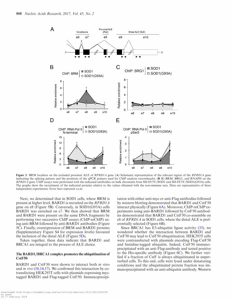
First, we characterized the distribution of BRM and BRG1along the terminal region of the RPRD1A gene from e6 toe10 by chromatin immunoprecipitation (ChIP, Figure 3A).In SOD1(G93A) cells, where it was hardly expressed, BRMwas distributed equally on all exons. In contrast, in SOD1cells BRM accumulated on the proximal ALE (Figure 3B).BRG1, on the other hand, was distributed equally on all ex-ons in both the cellular lines (Figure 3C). Next, we analysedthe distribution of the elongating polymerase, using the N20antibody that recognizes the N-terminal region of RNAPII.An increased density of RNAPII was detected on e7 and e8in SOD1(G93A) cells and in both cell lines on e10 (Figure3D). We also performed ChIP assays with the H14 antibodyto RNAPII phosphorylated at Ser5 of the CTD, a modifi-cation associated with a paused polymerase (27,28) (Figure3E). We detected a slight accumulation of p-Ser5 RNAPIIon e8 in SOD1(G93A) cells. In addition, we observed a highdensity on e10 in both cell lines, where this exon is used asterminal exon, albeit with different efficiency. This result isreminiscent of the observed pausing of RNAPII on termi-nal exons in yeast (29).
Our findings indicate a modest inverse correlation be-tween the accumulation of BRM and the accumulationof RNAPII on included exons. Thus, the contribution ofRNAPII pausing to the inclusion of the proximal exon ap-pears small, at variance with what previously observed forthe inclusion of internal exons (26).
BRM does not require its ATPase activity to affect ALEchoice
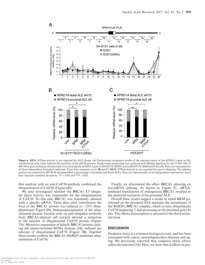
The SWI/SNF chromatin-remodeling complex destabilizeshistone-DNA interactions and alters nucleosome positions.Recent studies indicate that nucleosome positioning mayinfluence inclusion of internal exon sequences (30,31). Wetherefore wondered whether BRM might contribute to thechoice of ALE by moving nucleosomes around the proxi-mal last exon. We thus identified nucleosome positions andoccupancy in SOD1 and SOD1(G93A) cells, using micro-coccal nuclease (MNase) digestion in cells synchronized in
G0. We used mononucleosomal DNA fragments (150 bp)generated by MNase as a template for qPCR with overlap-ping primer sets across a ∼1 kb region centered on e8 of thehuman RPRD1A genomic locus. As shown in Figure 4A,the MNase digestion pattern did not significantly differ be-tween the two cell lines, indicating that the level of BRMprotein had no influence on nucleosome distribution in theinterrogated region of the RPRD1A gene.
To further exclude that the chromatin remodeling activityof BRM-containing SWI/SNF complexes may contributeto e8 skipping in SOD1 cells, we analysed the alternativesplicing pattern in SH-SY5Y cells stably expressing a BRMmutant carrying the K755A substitution in the ATPase do-main that abrogates the chromatin remodeling activity (19)(Figure 4B, and Supplementary Figure S4 for expressionlevels) we also analysed HEK293T cells transiently trans-fected with the same construct (Figure 4C, and Supplemen-tary Figure S4 for expression levels). Similarly to the wildtype, expression of the mutant BRM stimulated exon 8 skip-ping and the selection of the distal ALE.
Taken together these data indicate that the chromatin re-modeling function of BRM is not required for the inhibi-tion of the inclusion of the proximal ALE in SOD1 cells.Therefore, we hypothesized that BRM might contribute toe8 skipping by negatively acting on pre-mRNA processing,possibly by recruiting an inhibitory complex.
BARD1 and CstF50 associate with BRM on the skippedproximal ALE
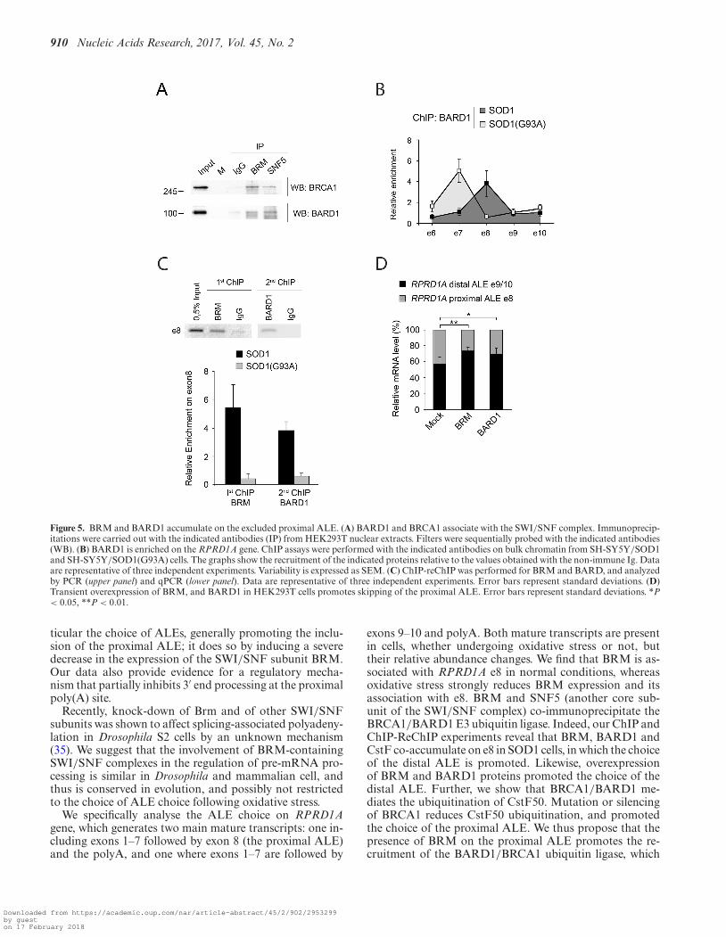
Previously, a BARD1-CstF complex was shown to inhibit3′ processing of nascent transcripts deriving from prema-ture transcription termination at sites of DNA damage(14,17,32). Therefore, we speculated that BARD1 (and itspartner BRCA1) might also be involved in the inhibitionof transcription termination at the proximal poly(A) site.Indeed, we found that endogenous BRCA1 and BARD1in HEK293T cells co-immunoprecipitate with SNF5 andBRM (Figure 5A), indicating that both proteins can asso-ciate with the SWI/SNF complex.
Downloaded from https://academic.oup.com/nar/article-abstract/45/2/902/2953299by gueston 17 February 2018
908 Nucleic Acids Research, 2017, Vol. 45, No. 2
Figure 3. BRM localizes on the excluded proximal ALE of RPRD1A gene. (A) Schematic representation of the relevant region of the RPRD1A geneindicating the splicing pattern and the positions of the qPCR primers used for ChIP analysis (arrowheads). (B–E) BRM, BRG1, and RNAPII on theRPRD1A gene. ChIP assays were performed with the indicated antibodies on bulk chromatin from SH-SY5Y/SOD1 and SH-SY5Y/SOD1(G93A) cells.The graphs show the recruitment of the indicated proteins relative to the values obtained with the non-immune sera. Data are representative of threeindependent experiments. Error bars represent s.e.m.
Next, we determined that in SOD1 cells, where BRM ispresent at higher level, BARD1 is recruited on the RPRD1Agene on e8 (Figure 5B). Conversely, in SOD1(G93A) cellsBARD1 was enriched on e7. We then showed that BRMand BARD1 were present on the same DNA fragments byperforming two successive ChIP assays (ChIP-reChIP) us-ing anti-BRM followed by anti-BARD1 antibodies (Figure5C). Finally, overexpression of BRM and BARD1 proteins(Supplementary Figure S4 for expression levels) favouredthe inclusion of the distal ALE (Figure 5D),
Taken together, these data indicate that BARD1 andBRCA1 are integral to the process of ALE choice.
The BARD1/BRCA1 complex promotes the ubiquitination ofCstF50
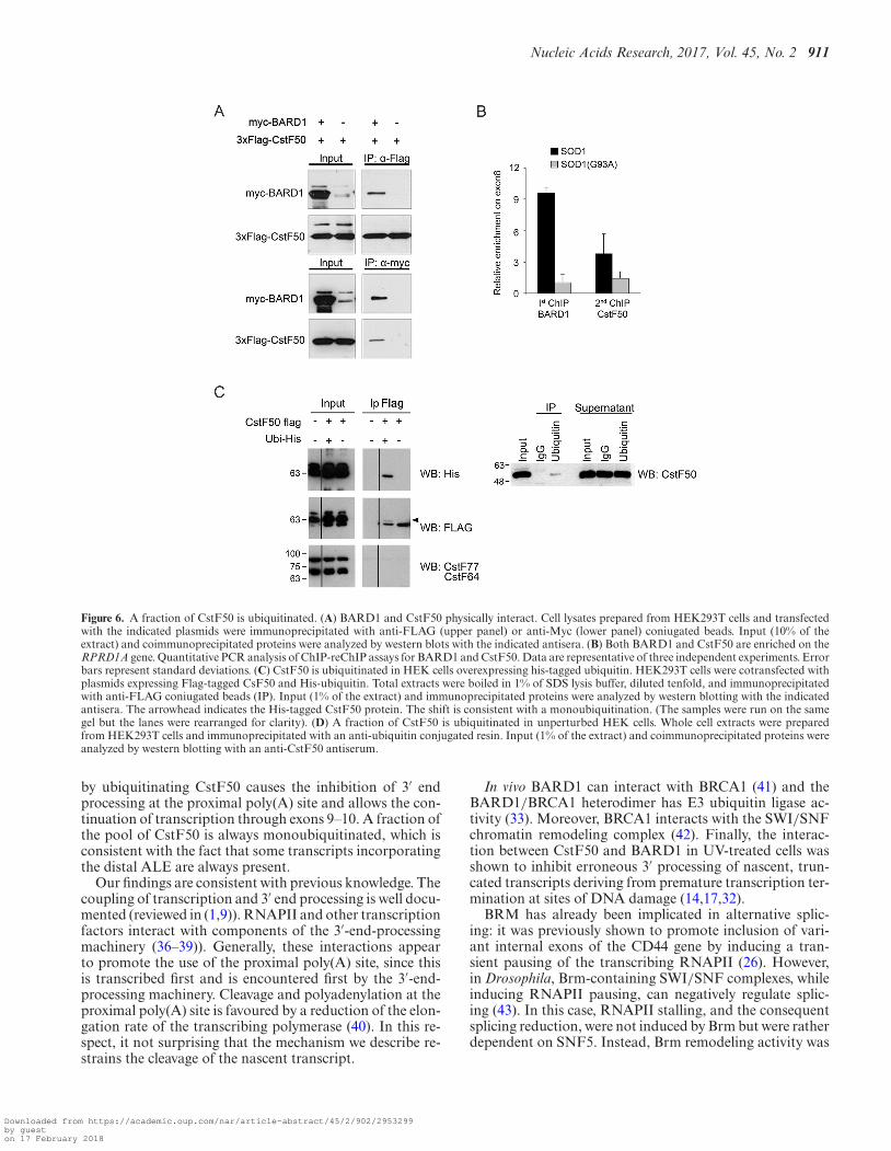
BARD1 and CstF50 were shown to interact both in vitroand in vivo (10,14,17). We confirmed this interaction by co-transfecting HEK293T cells with plasmids expressing myc-tagged BARD1 and Flag-tagged CstF50. Immunoprecipi-
tation with either anti-myc or anti-Flag antibodies followedby western blotting demonstrated that BARD1 and CstF50interact physically (Figure 6A). Moreover, ChIP-reChIP ex-periments using anti-BARD1 followed by CstF50 antibod-ies demonstrated that BARD1 and CstF50 co-assemble one8 of RPRD1A in SOD1 cells, where the distal ALE is pref-erentially selected (Figure 6B).
Since BRCA1 has E3-ubiquitin ligase activity (33), wewondered whether the interaction between BARD1 andCstF50 may lead to CstF50 ubiquitination. HEK293T cellswere contransfected with plasmids encoding Flag-CstF50and histidine-tagged ubiquitin. Indeed, CstF50 immuno-precipitated with an anti Flag-antibody and tested positiveto the His-specific antibody (Figure 6C). We further veri-fied if a fraction of CstF is always ubiquitinated in unper-turbed cells. To this end, cells were lysed under denaturingconditions and the ubiquitinated protein fraction was im-munoprecipitated with an anti-ubiquitin antibody. Western
Downloaded from https://academic.oup.com/nar/article-abstract/45/2/902/2953299by gueston 17 February 2018
Nucleic Acids Research, 2017, Vol. 45, No. 2 909
Figure 4. BRM ATPase activity is not required for ALE choice. (A) Nucleosome occupancy profile of the relevant region of the RPRD1A gene in G0-synchronized cells. Lines indicate the positions of the qPCR primers. Nucleosome positioning was analyzed with MNase digestion for the 33 605 100–33606 100 region of human chromosome 18 containing the RPRD1A gene in SH-SY5Y/SOD1 and in SH-SY5Y/SOD1(G93A) cells. Data are representativeof three independent biological replicates. Error bars represent s.e.m. (B and C) BRM ATPase activity is not required for exon 8 skipping. The splicingpattern was analyzed by RT-PCR and quantified as percentage of proximal and distal ALEs. Data are representative of six independent experiments. Errorbars represent standard deviations. *P < 0.05 and **P < 0.01.
blot analysis with an anti-CstF50 antibody confirmed theubiquitination of CstF50 (Figure 6D).
We next investigated whether the BRCA1 E3 ubiqui-tin ligase activity was responsible for the ubiquitinationof CstF50. To this end, BRCA1 was transiently silencedwith a specific siRNA. Three days after transfection thelevel of the BRCA1 protein was reduced to ∼25% (Sup-plementary Figure S4). Immunoprecipitation of the ubiq-uitinated protein fraction with an anti-ubiquitin antibodyfrom BRCA1-silenced cell extracts showed a reductionin the amount of ubiquitinated CstF50 protein (Figure7A). Moreover, expression of deltaN-BRCA1 protein, lack-ing the amino-terminal RING domain (34), reduced theamount of ubiquitinated CstF50 (Figure 7B). Togetherthese results confirm the BRCA1/BARD1-mediated ubiq-uitination of CstF50.
Finally, we determined the effect BRCA1 silencing onpre-mRNA splicing. As shown in Figure 7C, siRNA-mediated knockdown of endogenous BRCA1 resulted inthe preferred inclusion of the proximal ALE.
Overall these results suggest a model in which BRM po-sitioned on the proximal ALE mediates the recruitment ofthe BARD1/BRCA1 complex, which in turn ubiquitinatesCstF50 impairing 3′ end processing at the proximal poly(A)site. This allows transcription to proceed to the distal termi-nal exon.
DISCUSSION
Oxidative stress is a common biological event, and has beenassociated with cancer, neurodegenerative diseases and ag-ing. We previously reported that oxidative stress affectssplice site selection (16). Here, we show that it affects in par-
Downloaded from https://academic.oup.com/nar/article-abstract/45/2/902/2953299by gueston 17 February 2018
910 Nucleic Acids Research, 2017, Vol. 45, No. 2
Figure 5. BRM and BARD1 accumulate on the excluded proximal ALE. (A) BARD1 and BRCA1 associate with the SWI/SNF complex. Immunoprecip-itations were carried out with the indicated antibodies (IP) from HEK293T nuclear extracts. Filters were sequentially probed with the indicated antibodies(WB). (B) BARD1 is enriched on the RPRD1A gene. ChIP assays were performed with the indicated antibodies on bulk chromatin from SH-SY5Y/SOD1and SH-SY5Y/SOD1(G93A) cells. The graphs show the recruitment of the indicated proteins relative to the values obtained with the non-immune Ig. Dataare representative of three independent experiments. Variability is expressed as SEM. (C) ChIP-reChIP was performed for BRM and BARD, and analyzedby PCR (upper panel) and qPCR (lower panel). Data are representative of three independent experiments. Error bars represent standard deviations. (D)Transient overexpression of BRM, and BARD1 in HEK293T cells promotes skipping of the proximal ALE. Error bars represent standard deviations. *P< 0.05, **P < 0.01.
ticular the choice of ALEs, generally promoting the inclu-sion of the proximal ALE; it does so by inducing a severedecrease in the expression of the SWI/SNF subunit BRM.Our data also provide evidence for a regulatory mecha-nism that partially inhibits 3′ end processing at the proximalpoly(A) site.
Recently, knock-down of Brm and of other SWI/SNFsubunits was shown to affect splicing-associated polyadeny-lation in Drosophila S2 cells by an unknown mechanism(35). We suggest that the involvement of BRM-containingSWI/SNF complexes in the regulation of pre-mRNA pro-cessing is similar in Drosophila and mammalian cell, andthus is conserved in evolution, and possibly not restrictedto the choice of ALE choice following oxidative stress.
We specifically analyse the ALE choice on RPRD1Agene, which generates two main mature transcripts: one in-cluding exons 1–7 followed by exon 8 (the proximal ALE)and the polyA, and one where exons 1–7 are followed by
exons 9–10 and polyA. Both mature transcripts are presentin cells, whether undergoing oxidative stress or not, buttheir relative abundance changes. We find that BRM is as-sociated with RPRD1A e8 in normal conditions, whereasoxidative stress strongly reduces BRM expression and itsassociation with e8. BRM and SNF5 (another core sub-unit of the SWI/SNF complex) co-immunoprecipitate theBRCA1/BARD1 E3 ubiquitin ligase. Indeed, our ChIP andChIP-ReChIP experiments reveal that BRM, BARD1 andCstF co-accumulate on e8 in SOD1 cells, in which the choiceof the distal ALE is promoted. Likewise, overexpressionof BRM and BARD1 proteins promoted the choice of thedistal ALE. Further, we show that BRCA1/BARD1 me-diates the ubiquitination of CstF50. Mutation or silencingof BRCA1 reduces CstF50 ubiquitination, and promotedthe choice of the proximal ALE. We thus propose that thepresence of BRM on the proximal ALE promotes the re-cruitment of the BARD1/BRCA1 ubiquitin ligase, which
Downloaded from https://academic.oup.com/nar/article-abstract/45/2/902/2953299by gueston 17 February 2018
Nucleic Acids Research, 2017, Vol. 45, No. 2 911
Figure 6. A fraction of CstF50 is ubiquitinated. (A) BARD1 and CstF50 physically interact. Cell lysates prepared from HEK293T cells and transfectedwith the indicated plasmids were immunoprecipitated with anti-FLAG (upper panel) or anti-Myc (lower panel) coniugated beads. Input (10% of theextract) and coimmunoprecipitated proteins were analyzed by western blots with the indicated antisera. (B) Both BARD1 and CstF50 are enriched on theRPRD1A gene. Quantitative PCR analysis of ChIP-reChIP assays for BARD1 and CstF50. Data are representative of three independent experiments. Errorbars represent standard deviations. (C) CstF50 is ubiquitinated in HEK cells overexpressing his-tagged ubiquitin. HEK293T cells were cotransfected withplasmids expressing Flag-tagged CsF50 and His-ubiquitin. Total extracts were boiled in 1% of SDS lysis buffer, diluted tenfold, and immunoprecipitatedwith anti-FLAG coniugated beads (IP). Input (1% of the extract) and immunoprecipitated proteins were analyzed by western blotting with the indicatedantisera. The arrowhead indicates the His-tagged CstF50 protein. The shift is consistent with a monoubiquitination. (The samples were run on the samegel but the lanes were rearranged for clarity). (D) A fraction of CstF50 is ubiquitinated in unperturbed HEK cells. Whole cell extracts were preparedfrom HEK293T cells and immunoprecipitated with an anti-ubiquitin conjugated resin. Input (1% of the extract) and coimmunoprecipitated proteins wereanalyzed by western blotting with an anti-CstF50 antiserum.
by ubiquitinating CstF50 causes the inhibition of 3′ endprocessing at the proximal poly(A) site and allows the con-tinuation of transcription through exons 9–10. A fraction ofthe pool of CstF50 is always monoubiquitinated, which isconsistent with the fact that some transcripts incorporatingthe distal ALE are always present.
Our findings are consistent with previous knowledge. Thecoupling of transcription and 3′ end processing is well docu-mented (reviewed in (1,9)). RNAPII and other transcriptionfactors interact with components of the 3′-end-processingmachinery (36–39)). Generally, these interactions appearto promote the use of the proximal poly(A) site, since thisis transcribed first and is encountered first by the 3′-end-processing machinery. Cleavage and polyadenylation at theproximal poly(A) site is favoured by a reduction of the elon-gation rate of the transcribing polymerase (40). In this re-spect, it not surprising that the mechanism we describe re-strains the cleavage of the nascent transcript.
In vivo BARD1 can interact with BRCA1 (41) and theBARD1/BRCA1 heterodimer has E3 ubiquitin ligase ac-tivity (33). Moreover, BRCA1 interacts with the SWI/SNFchromatin remodeling complex (42). Finally, the interac-tion between CstF50 and BARD1 in UV-treated cells wasshown to inhibit erroneous 3′ processing of nascent, trun-cated transcripts deriving from premature transcription ter-mination at sites of DNA damage (14,17,32).
BRM has already been implicated in alternative splic-ing: it was previously shown to promote inclusion of vari-ant internal exons of the CD44 gene by inducing a tran-sient pausing of the transcribing RNAPII (26). However,in Drosophila, Brm-containing SWI/SNF complexes, whileinducing RNAPII pausing, can negatively regulate splic-ing (43). In this case, RNAPII stalling, and the consequentsplicing reduction, were not induced by Brm but were ratherdependent on SNF5. Instead, Brm remodeling activity was
Downloaded from https://academic.oup.com/nar/article-abstract/45/2/902/2953299by gueston 17 February 2018

912 Nucleic Acids Research, 2017, Vol. 45, No. 2
Figure 7. Ubiquitination of CstF50 is catalyzed by BRCA1. (A) BRCA1 knockdown impairs CstF50 ubiquitination. Whole cell extracts from HEK293Tcells transfected with control or BRCA1 siRNAs were immunoprecipitated with a control or an anti-ubiquitin resin. Western blots were analysed withthe indicated antibodies. �-catenin was used as control for BRCA1-independent ubiquitination and is highlighted by an arrow. The asterisk indicatesnon-specific bands. Upper panel: Quantification of CstF50 ubiquitination upon BRCA1 silencing. Quantification was performed on three independentimmunoprecipitation experiments. Immunoprecipitated CstF50 protein was normalized on �-catenin. Error bars represent standard deviations. Lowerpanel: representative western blot. (B) Overexpression of a catalytically inactive mutant BRCA1 protein impairs CstF50 ubiquitination. HEK293T cellswere cotransfected with plasmids expressing CstF50, BARD1 and either wild type BRCA1 or the BRCA1-�N mutant. Ubiquitinated proteins wereimmunoprecipitated with an anti-ubiquitin resin as described in ‘Materials and Methods’ and analysed by western blotting with the indicated antisera.Upper panel: Quantification was performed on three independent immunoprecipitation experiments. Immunoprecipitated CstF50 protein was normalizedon IgG bands that are indicated in the Ponceau Staining. Error bars represent standard deviations. *P < 0.05. Lower panel: representative western blot. Thearrow indicates the bands corresponding to the IgGs. (C) BRCA1 knockdown favors inclusion of the distal ALE. Quantification of the amount of proximaland distal ALE usage in the indicated cell lines. Data are representative of three independent experiments. Error bars represent standard deviation. ***P< 0.001.
shown to be necessary for the subsequent chromatin remod-eling following the release of the stalled polymerase (43).
In line with the report by Zraly et al. (43), we found thatBRM negatively affects the inclusion of the proximal ALE.This effect does not require its ATPase activity. Moreover,we did not detect differences in the distribution of nucleo-somes on the proximal ALE. These findings suggest that thechoice of ALE is not correlated with the nucleosome remod-eling activity of BRM. We also observed that RNAPII is notgreatly enriched on the skipped proximal ALE. Instead, weobserved an inverse correlation between the accumulationof BRM and RNAPII particularly on the distal ALE, wherethere was a distinct enrichment of RNAPII but BRM wasvirtually absent. Our data indicate that in the case of thechoice of alternative terminal exons, the modulation of the
elongation rate of RNAPII is not the main role of BRM,and rather implicate a different molecular mechanism.
The choice of alternative poly(A) sites located in differ-ent terminal exons occurs in conjunction with splicing, andsplicing factors are known to influence 3′ processing (re-viewed in (44)). Thus, the mechanism we describe is cer-tainly not the unique determinant of the choice betweenproximal and distal terminal exons. Moreover, we cannotexclude that BRM and BRCA1/BARD1 may also affectbinding of splicing factors to the terminal exon. Futurework will determine how the interplay between chromatinremodeling complexes, splicing and polyadenylation factorseventually determines which terminal exon will be includedin the mature transcript.
Downloaded from https://academic.oup.com/nar/article-abstract/45/2/902/2953299by gueston 17 February 2018

Nucleic Acids Research, 2017, Vol. 45, No. 2 913
SUPPLEMENTARY DATA
Supplementary Data are available at NAR Online.
ACKNOWLEDGEMENTS
We gratefully acknowledge B.M. Emerson, I. Irminger-Finger, T. Banerjee, M.L. Guerrini, J.D. Parvin, M. Pilyu-gin, M.-D. Ruepp, O. Muhlemann, N. Chiba and W. Kellerfor reagents. We are grateful to S. Polo for help with ubiq-uitination analysis and to M. Lupi for technical assistance.
FUNDING
MIUR-PRIN [20083R593R 003]; Swiss National ScienceFoundation Sinergia [CRSII3 136222 to G.F.]. Funding foropen access charge: University of Milan, Fondo ricerca diAteneo.Conflict of interest statement. None declared.
REFERENCES1. Di Giammartino,D.C., Nishida,K. and Manley,J.L. (2011)
Mechanisms and consequences of alternative polyadenylation. Mol.Cell, 43, 853–866.
2. Wang,E.T., Sandberg,R., Luo,S., Khrebtukova,I., Zhang,L.,Mayr,C., Kingsmore,S.F., Schroth,G.P. and Burge,C.B. (2008)Alternative isoform regulation in human tissue transcriptomes.Nature, 456, 470–476.
3. Shi,Y. (2012) Alternative polyadenylation: new insights from globalanalyses. RNA (New York, N.Y.), 18, 2105–2117.
4. Pickrell,J.K., Marioni,J.C., Pai,A.A., Degner,J.F., Engelhardt,B.E.,Nkadori,E., Veyrieras,J.B., Stephens,M., Gilad,Y. and Pritchard,J.K.(2010) Understanding mechanisms underlying human geneexpression variation with RNA sequencing. Nature, 464, 768–772.
5. Sandberg,R., Neilson,J.R., Sarma,A., Sharp,P.A. and Burge,C.B.(2008) Proliferating cells express mRNAs with shortened 3′untranslated regions and fewer microRNA target sites. Science, 320,1643–1647.
6. Ji,Z., Lee,J.Y., Pan,Z., Jiang,B. and Tian,B. (2009) Progressivelengthening of 3′ untranslated regions of mRNAs by alternativepolyadenylation during mouse embryonic development. Proc. Natl.Acad. Sci. U.S.A., 106, 7028–7033.
7. Proudfoot,N.J. (2011) Ending the message: poly(A) signals then andnow. Genes Dev., 25, 1770–1782.
8. Danckwardt,S., Hentze,M.W. and Kulozik,A.E. (2008) 3′ end mRNAprocessing: molecular mechanisms and implications for health anddisease. EMBO J., 27, 482–498.
9. Elkon,R., Ugalde,A.P. and Agami,R. (2013) Alternative cleavage andpolyadenylation: extent, regulation and function. Nat. Rev. Genet.,14, 496–506.
10. Edwards,R.A., Lee,M.S., Tsutakawa,S.E., Williams,R.S., Nazeer,I.,Kleiman,F.E., Tainer,J.A. and Glover,J.N. (2008) The BARD1C-terminal domain structure and interactions with polyadenylationfactor CstF-50. Biochemistry, 47, 11446–11456.
11. Bai,Y., Auperin,T.C., Chou,C.Y., Chang,G.G., Manley,J.L. andTong,L. (2007) Crystal structure of murine CstF-77: dimericassociation and implications for polyadenylation of mRNAprecursors. Mol. Cell, 25, 863–875.
12. Dantonel,J.C., Murthy,K.G., Manley,J.L. and Tora,L. (1997)Transcription factor TFIID recruits factor CPSF for formation of 3′end of mRNA. Nature, 389, 399–402.
13. McCracken,S., Fong,N., Yankulov,K., Ballantyne,S., Pan,G.,Greenblatt,J., Patterson,S.D., Wickens,M. and Bentley,D.L. (1997)The C-terminal domain of RNA polymerase II couples mRNAprocessing to transcription. Nature, 385, 357–361.
14. Kleiman,F.E. and Manley,J.L. (2001) The BARD1-CstF-50interaction links mRNA 3′ end formation to DNA damage andtumor suppression. Cell, 104, 743–753.
15. Lin,M.T. and Beal,M.F. (2006) Mitochondrial dysfunction andoxidative stress in neurodegenerative diseases. Nature, 443, 787–795.
16. Lenzken,S.C., Romeo,V., Zolezzi,F., Cordero,F., Lamorte,G.,Bonanno,D., Biancolini,D., Cozzolino,M., Pesaresi,M.G.,Maracchioni,A. et al. (2011) Mutant SOD1 and mitochondrialdamage alter expression and splicing of genes controllingneuritogenesis in models of neurodegeneration. Hum. Mutat., 32,168–182.
17. Kleiman,F.E. and Manley,J.L. (1999) Functional interaction ofBRCA1-associated BARD1 with polyadenylation factor CstF-50.Science, 285, 1576–1579.
18. Rizzardini,M., Mangolini,A., Lupi,M., Ubezio,P., Bendotti,C. andCantoni,L. (2005) Low levels of ALS-linked Cu/Zn superoxidedismutase increase the production of reactive oxygen species andcause mitochondrial damage and death in motor neuron-like cells. J.Neurol. Sci., 232, 95–103.
19. Richmond,E. and Peterson,C.L. (1996) Functional analysis of theDNA-stimulated ATPase domain of yeast SWI2/SNF2. NucleicAcids Res., 24, 3685–3692.
20. Maglott,D., Ostell,J., Pruitt,K.D. and Tatusova,T. (2011) EntrezGene: gene-centered information at NCBI. Nucleic Acids Res., 39,D52–D57.
21. Sanges,R., Cordero,F. and Calogero,R.A. (2007) oneChannelGUI: agraphical interface to Bioconductor tools, designed for life scientistswho are not familiar with R language. Bioinformatics (Oxford,England), 23, 3406–3408.
22. Cocheme,H.M. and Murphy,M.P. (2008) Complex I is the major siteof mitochondrial superoxide production by paraquat. J. Biol. Chem.,283, 1786–1798.
23. Ciriolo,M.R., De Martino,A., Lafavia,E., Rossi,L., Carri,M.T. andRotilio,G. (2000) Cu,Zn-superoxide dismutase-dependent apoptosisinduced by nitric oxide in neuronal cells. J. Biol. Chem., 275,5065–5072.
24. Takagaki,Y., Seipelt,R.L., Peterson,M.L. and Manley,J.L. (1996) Thepolyadenylation factor CstF-64 regulates alternative processing ofIgM heavy chain pre-mRNA during B cell differentiation. Cell, 87,941–952.
25. Takagaki,Y. and Manley,J.L. (1998) Levels of polyadenylation factorCstF-64 control IgM heavy chain mRNA accumulation and otherevents associated with B cell differentiation. Mol. Cell, 2, 761–771.
26. Batsche,E., Yaniv,M. and Muchardt,C. (2006) The human SWI/SNFsubunit Brm is a regulator of alternative splicing. Nat. Struct. Mol.Biol., 13, 22–29.
27. Morris,D.P., Michelotti,G.A. and Schwinn,D.A. (2005) Evidence thatphosphorylation of the RNA polymerase II carboxyl-terminal repeatsis similar in yeast and humans. J. Biol. Chem., 280, 31368–31377.
28. Boehm,A.K., Saunders,A., Werner,J. and Lis,J.T. (2003)Transcription factor and polymerase recruitment, modification, andmovement on dhsp70 in vivo in the minutes following heat shock.Mol. Cell Biol, 23, 7628–7637.
29. Carrillo Oesterreich,F., Preibisch,S. and Neugebauer,K.M. (2010)Global analysis of nascent RNA reveals transcriptional pausing interminal exons. Mol. Cell, 40, 571–581.
30. Tilgner,H., Nikolaou,C., Althammer,S., Sammeth,M., Beato,M.,Valcarcel,J. and Guigo,R. (2009) Nucleosome positioning as adeterminant of exon recognition. Nat. Struct. Mol. Biol., 16,996–1001.
31. Schwartz,S., Meshorer,E. and Ast,G. (2009) Chromatin organizationmarks exon-intron structure. Nat. Struct. Mol. Biol., 16, 990–995.
32. Mirkin,N., Fonseca,D., Mohammed,S., Cevher,M.A., Manley,J.L.and Kleiman,F.E. (2008) The 3′ processing factor CstF functions inthe DNA repair response. Nucleic Acids Res., 36, 1792–1804.
33. Mallery,D.L., Vandenberg,C.J. and Hiom,K. (2002) Activation of theE3 ligase function of the BRCA1/BARD1 complex by polyubiquitinchains. EMBO J., 21, 6755–6762.
34. Chiba,N. and Parvin,J.D. (2002) The BRCA1 and BARD1association with the RNA polymerase II holoenzyme. Cancer Res.,62, 4222–4228.
35. Waldholm,J., Wang,Z., Brodin,D., Tyagi,A., Yu,S., Theopold,U.,Farrants,A.K. and Visa,N. (2011) SWI/SNF regulates the alternativeprocessing of a specific subset of pre-mRNAs in Drosophilamelanogaster. BMC Mol. Biol., 12, 46.
36. Shi,Y., Di Giammartino,D.C., Taylor,D., Sarkeshik,A., Rice,W.J.,Yates,J.R. 3rd, Frank,J. and Manley,J.L. (2009) Moleculararchitecture of the human pre-mRNA 3′ processing complex. Mol.Cell, 33, 365–376.
Downloaded from https://academic.oup.com/nar/article-abstract/45/2/902/2953299by gueston 17 February 2018
914 Nucleic Acids Research, 2017, Vol. 45, No. 2
37. Nagaike,T., Logan,C., Hotta,I., Rozenblatt-Rosen,O., Meyerson,M.and Manley,J.L. (2011) Transcriptional activators enhancepolyadenylation of mRNA precursors. Mol. Cell, 41, 409–418.
38. Rozenblatt-Rosen,O., Nagaike,T., Francis,J.M., Kaneko,S.,Glatt,K.A., Hughes,C.M., LaFramboise,T., Manley,J.L. andMeyerson,M. (2009) The tumor suppressor Cdc73 functionallyassociates with CPSF and CstF 3′ mRNA processing factors. Proc.Natl. Acad. Sci. U.S.A., 106, 755–760.
39. Katahira,J., Okuzaki,D., Inoue,H., Yoneda,Y., Maehara,K. andOhkawa,Y. (2013) Human TREX component Thoc5 affectsalternative polyadenylation site choice by recruiting mammaliancleavage factor I. Nucleic Acids Res., 14, 7060–7072.
40. Pinto,P.A., Henriques,T., Freitas,M.O., Martins,T., Domingues,R.G.,Wyrzykowska,P.S., Coelho,P.A., Carmo,A.M., Sunkel,C.E.,Proudfoot,N.J. et al. (2011) RNA polymerase II kinetics in polopolyadenylation signal selection. EMBO J., 30, 2431–2444.
41. Wu,L.C., Wang,Z.W., Tsan,J.T., Spillman,M.A., Phung,A., Xu,X.L.,Yang,M.C., Hwang,L.Y., Bowcock,A.M. and Baer,R. (1996)Identification of a RING protein that can interact in vivo with theBRCA1 gene product. Nat. Genet., 14, 430–440.
42. Bochar,D.A., Wang,L., Beniya,H., Kinev,A., Xue,Y., Lane,W.S.,Wang,W., Kashanchi,F. and Shiekhattar,R. (2000) BRCA1 isassociated with a human SWI/SNF-related complex: linkingchromatin remodeling to breast cancer. Cell, 102, 257–265.
43. Zraly,C.B. and Dingwall,A.K. (2012) The chromatin remodeling andmRNA splicing functions of the Brahma (SWI/SNF) complex aremediated by the SNR1/SNF5 regulatory subunit. Nucleic Acids Res.,40, 5975–5987.
44. Millevoi,S. and Vagner,S. (2010) Molecular mechanisms ofeukaryotic pre-mRNA 3′ end processing regulation. Nucleic AcidsRes., 38, 2757–2774.
Downloaded from https://academic.oup.com/nar/article-abstract/45/2/902/2953299by gueston 17 February 2018