p21 (waf1/cip1) and fasl gene activation via sp1 and nf … (gibco, grand island, ny ... cells were...
TRANSCRIPT
The International Journal of Biochemistry & Cell Biology 37 (2005) 784–796
p21 (Waf1/Cip1) and FasL gene activation via Sp1 and NF�Bis required for leukemia cell survival but not for cell death
induced by diverse stimuli
J. Savickienea,∗, G. Treigytea, K.-E. Magnussonb, R. Navakauskienea
a Department of Developmental Biology, Institute of Biochemistry, LT-08662 Vilnius, Lithuaniab Division of Medical Microbiology, Department of Molecular and Clinical Medicine,
Linkoping University, SE-58185 Linkoping, Sweden
Received 23 June 2004; received in revised form 14 September 2004; accepted 7 October 2004
Abstract
The molecular mechanisms of the cellular response to different apoptotic effectors are only partially understood. Herein,the role of transcription factors, Sp1 and NF�B in differentiation-related and etoposide-induced apoptosis was examined in anumber of human leukemia cell lines (HL-60, NB4, HEL, THP-1, K562). This was investigated with respect to the recruitmentof one cell-cycle regulating gene, p21 and one cell death gene, FasL. Using electrophoretic mobility shift assay (EMSA), weconsistently observed Sp1 and NF�B binding activity to the promoter of either gene during cell differentiation and the decreasea ontrast,S is effectw one, thusr osisu d leukemiac rt thei nes.©
K
olnuclease;
n),r
1
ssociated with apoptosis upon long-term treatment with differentiation inducers in HL-60, NB4 and HEL cells. By cp1 and NF�B binding capacities were lost in all myeloid cell lines undergoing etoposide-induced fast apoptosis. Thas eliminated by the broad-spectrum caspase inhibitor, benzyloxycarbonyl-valinyl-alaninyl-aspartyl fluoromethylket
estoring transcription factors’ binding activity. However, sustained NF�B binding to the FasL promoter was noticed in apoptndergoing HEL cells treated by etoposide. Our results suggest that p21 and FasL gene activation is required for myeloiell survival or maturation but not for cell death via Sp1 and NF�B as regulators of these genes. The findings also suppodea of a common mechanism for cellular responses to different apoptotic effectors in malignant hematopoietic cell li
2004 Elsevier Ltd. All rights reserved.
eywords:Apoptosis; Differentiation; Leukemia; Transcription factors; Etoposide
Abbreviations:DMSO, dimethylsulfoxide; ATP, adenosine-5′-triphosphate; FasL, Fas ligand; NF�B, nuclear factor kappa B; PMA, phorb12-myristate 13-acetate; PMSF, phenylmethylsulfonylfluoride; RA, all-trans retinoic acid; RAR, retinoic acid receptor; RNase, riboTRAIL, TNF-related apoptosis-inducing ligand; z-VAD.fmk, benzyloxycarbonyl-valinyl-alaninyl-aspartyl fluoromethylketone
∗ Corresponding author. Tel.: +370 5 272 9187; fax: +370 5 272 9196.E-mail addresses:[email protected] (J. Savickiene), [email protected] (G. Treigyte), [email protected] (K.-E. Magnusso
[email protected] (R. Navakauskiene).
357-2725/$ – see front matter © 2004 Elsevier Ltd. All rights reserved.doi:10.1016/j.biocel.2004.10.002

J. Savickiene et al. / The International Journal of Biochemistry & Cell Biology 37 (2005) 784–796 785
1. Introduction
A balance between the signals supporting cell sur-vival and inducing cell death regulates cell homeosta-sis, which is abrogated in malignant transformed cells.One of the approaches to cancer therapy is the preven-tion of uncontrolled growth and selective induction ofmalignant cell death. This may be achieved either withdifferentiation inducers or cytotoxic drugs. Retinoidsare promising agents for leukemia therapy due to anti-cancer properties by modulating cell proliferation, dif-ferentiation and apoptosis (Altucci & Gronemeyer,2001; Collins, 1987). The topoisomerase II inhibitor,etoposide is an important drug in the treatment ofleukemias and efficient inducer of apoptosis (Froelich-Ammon & Osheroff, 1995). However, the molecularmechanisms of cellular response to different apoptoticeffectors, such as retinoic acid (RA) or etoposide, maydiffer.
Malignant cells usually respond to drug-inducedstress either by cell cycle arrest and/or by undergo-ing apoptotic cell death. The response depends on theactivation of p53 or the induction of certain transcrip-tion factors (Harwood et al., 2000; Nelson & Kastan,1994; Piret & Piette, 1996). The Fas receptor (CD95),a 48 kDa cell surface glycoprotein, is regarded as amajor mediator of apoptosis. Ligation of Fas receptorwith anti-Fas antibody or its specific ligand (FasL) in-duces a death signal in many cell types including thoseof haematopoietic origin (Dirks et al., 1997; Kim, Lee,Ha peso eenr ensi-t yticl ,In ceda .,1 estt nt ofF y,S ;V eF con-t ac-t ;K &
Khachigian, 2001). Topoisomerase II poisons can in-duce apoptosis via activation of NF�B or AP-1 and Fassignaling (Kasibhata et al., 1998; Piret & Piette, 1996).Sp1 involvement in apoptosis regulation via extracel-lular Fas/FasL engagement in smooth muscle cells hasbeen recently demonstrated (Kavurma et al., 2001).However, the regulatory role of Sp1 in the process ofapoptosis in many cell types is presently unknown.
The p21 (Waf1/Cip1) protein induces growth arrestby blocking cyclin-dependent kinases (CDK) or theactivity of proliferating cell nuclear antigen (PCNA),which influences cell proliferation, differentiation andapoptosis (Gartel & Tyner, 1999; Harper, Adami, Wei,Keymarsi, & Elledge, 1993; Harper & Elledge, 1996).Exit from the cell cycle is a prerequisite for terminaldifferentiation, and treatment of myeloid cells with in-ducers of differentiation, such as PMA, okadaic acid orRA, enhances p21 gene transcription (Casini & Pelicci,1999; El-Deiry et al., 1994; Harper et al., 1993; Jianget al., 1994). In some systems, p21 either protects cellsfrom apoptosis (Gorospe et al., 1997; Polyak et al.,1996) or promotes apoptosis (Gartel & Tyner, 1999).Agents that cause DNA damage, such as UV radiationor certain carcinogens, activate tumor suppressor p53-dependent p21 gene transcription (Dulic et al., 1994;El-Deiry et al., 1994). p53-independent expression ofp21 occurs during terminal differentiation in a numbercell types (Jiang et al., 1994; Macleod et al., 1995).The majority of these modulators affect p21 gene ac-tivity via Sp family proteins, which bind to regulatoryeT c-t andS test ,2p ites( eda e byb inr 02a &T
f-t ands esp elB(
ong, & Park, 2000; Salih et al., 2002). Little is knownbout the regulation of Fas signaling in various tyf myeloid leukemia’s. Conflicting data have also beported concerning the expression of Fas and sivity to Fas-induced apoptosis in acute promyeloceukemia (APL) cell lines (Kikuchi et al., 1996; Ohashiwase, & Nagumo, 2000; Salih et al., 2002). The sig-ificance of Fas expression in chemotherapy-indupoptosis has been questioned as well (Eischen et al997; Gamen et al., 1997), and some reports sugg
hat cytotoxic drug-induced apoptosis is independeas (Siitonen et al., 2000; Siitonen, Mantymaa, Sallavolainen, & Koistinen, 2000;Tolomeo et al., 1998illunger et al., 1997). The promoter region of thasL gene has recently been cloned and found to
ain binding sites for a number of transcription fors including NF�B, AP-1, Sp1 (Harwood et al., 2000asibhata et al., 1998; Kavurma, Santiago, Bonfoco,
lements in the proximal p21 gene promoter (Gartel &yner, 1999). The Sp family of structurally and funionally related transcription factors (Sp1, Sp2, Sp3p4) recognizes GC-rich or GT-rich DNA and regula
he promoters of several genes (Bouwman & Philipsen002; Lania, Majello, & De Luca, 1997). The human21 (Waf1/Cip1) gene contains six Sp1 binding sGartel & Tyner, 1999). The ubiquitously expressnd closely related Sp1 and Sp3 regulate p21 geninding with different affinities as has been shownat liver (Koutsodontis, Moustakas, & Kardassis, 20)nd in Caco-2 cells (Gartel, Goufman, Najmabadi,yner, 2000).
The NF�B family of transcription factors is oen proposed as a key regulator of differentiationurvival. In mammals, this protein family includ50/p105, p52/p100, p65 (RelA), c-Rel and RGlosh, May, & Kopp, 1998). NF�B is activated by

786 J. Savickiene et al. / The International Journal of Biochemistry & Cell Biology 37 (2005) 784–796
multiple physiological or environmental stresses (Pahl,1999). In the nucleus, NF�B dimers bind to a set ofrelated 10 bp DNA sites, collectively called�B sites,thereby regulating the expression of many genes. How-ever, interestingly, NF�B can exert both pro- and anti-apoptotic effects in different cells types (Barkett &Gilmore, 1999).
In this study, we have explored the regulatory roleof Sp1 and NF�B during differentiation-associated andetoposide-induced apoptotic processes in leukemia celllines with different p53 and Fas receptor status.
2. Materials and methods
2.1. Materials
RA, PMA, NBT, RNase A, proteinase K, ethidiumbromide, hemin chloride and etoposide were purchasedfrom Sigma (St. Louis, MO), and z-VAD.fmk fromPromega Corp. (Madison, WI). The stock solutions ofRA (500�M) in ethanol and hemin (25 mg/ml) in 1.4 Mammonium hydroxide were stored at−20◦C.
2.2. Cell culture and cell lines
The human leukemia cell lines (promyelocyticHL-60, acute promyelocytic NB4, chronic myeloidK562, erythroleukemia HEL and acute monocyticTHP-1) were maintained in RPMI 1640 medium sup-p ines -m wna edf
2
4c T)r r-i s-t t,M
2
f
PBS. Cells were stained with 0.01% acridine or-ange/0.01% ethidium bromide (AO/EtBr) mixture (1:1,v/v), 6�l per 100�l cell suspension (Mercille &Massie, 1994). Apoptotic bodies containing cells werecounted using a fluorescence microscope by scoring atleast 300 cells.
2.5. Isolation and electrophoresis of fragmentedDNA
Cells were harvested, washed with PBS and pel-leted by centrifugation. Cell pellets were then treatedfor 10 s with lysis buffer (1% Nonidet P-40 in 20 mMEDTA, 50 mM Tris–HCl, pH 7.5), 10�l per 106 cells.After centrifugation for 5 min at 1600×g, supernatantswere collected and the extraction was repeated with thesame amount of lysis buffer. Fragmented DNA fractionwas prepared accordingHerman et al., 1994. The DNAfragments were analysed by 1.5% agarose gel elec-trophoresis and visualised by UV illumination. DNAmarker of 123 bp was used as a molecular weight stan-dard (Sigma).
2.6. Preparation of nuclear extracts
Cells (5× 10⁶ to 5× 10⁷) were harvested and pel-leted at 500×g for 6 min, then washed twice in icecold PBS and lysed in Nuclei EZ lysis buffer (Sigma)for 5 min on ice. After centrifugation at 500×g for5 min at 4◦C, nuclei were washed in the same coldb leiw s-p zena ticm sisoE l)ci for1f tra-t rec-o
2
ides( d-
lemented with 10% heat-inactivated fetal boverum, 100 U/ml penicillin and 100�g/ml streptoycin (Gibco, Grand Island, NY). Cells were grot 37◦C in a humidified 5% CO2 atmosphere and us
or assays during exponential phase of growth.
.3. Assessment of cell differentiation
Granulocytic differentiation of HL-60 and NBells was determined by nitroblue tetrazolium (NBeduction (Collins, 1987). Hemoglobin production dung erythroid differentiation of HEL cells was eablished by benzidine staining (Jeannesson, Ginoanfait, & Jardillier, 1984).
.4. Morphologic assessment of apoptotic cells
After any incubation, cells were pelleted at 500×gor 5 min and resuspended (5× 10⁶ cells/ml) in 100�l
uffer, vortexed briefly and set on ice for 5 min. Nucere pelleted at 500×g for 5 min, then completely suended in Nuclei EZ storage buffer (Sigma) and frot−70◦C. Nuclear protein extracts for electrophoreobility shift assay (EMSA) were prepared by lyf nuclei in buffer (20 mM Tris–HCl, pH 8.0, 200�MDTA, 2 mM EGTA, 20% glycerol, 400 mM NaContaining 3 mM DTT, 1 mM PMSF and 1× proteasenhibitor cocktail P2714 (Sigma). After incubationh on ice, the extracts were centrifuged at 18,000×g
or 20 min, and used immediately. Protein concenions were determined by RCDC protein assay asmmended by the manufacturer (Bio-Rad).
.7. Oligonucleotides
Te probes used were synthetic oligonucleotMWG-Biotech AG, Sweden) representing bin
J. Savickiene et al. / The International Journal of Biochemistry & Cell Biology 37 (2005) 784–796 787
ing sites: (5′-AGTTGAGGGGACTTTCCCAGGC-3′)consensus NF�B; (5′-AAGCCTGG GCAACATAGA-AAGTCCCCATCTGTACAAAAA-3′) NF�B fromthe FasL promoter; (5′-GCCTGGGCCCCGGGAGG-GCGGTCCCGGGCGGCGC-3′) Sp1 (elements 1 and2) from the p21 promoter; (5′-ATCAGAAAATTG-TGGGCGGAAACTTCCAGG-3′) Sp1 from the FasLpromoter; (5′-TTTGGGGCTCGAGGTCCCG-3′) mu-tated Sp1 (element 1) from the p21 promoter.
2.8. Electrophoretic mobility shift assay
Complementary oligonucleotides were annealedand labeled at their 5′ ends using [�-32P-ATP] (Amer-sham, UK) and T4 polynucleotide kinase (MBI Fer-mentas Inc., Vilnius, Lithuania). Standard DNA reac-tions were performed with 12–15�g nuclear extractsin a 20�l of reaction buffer (10 mM HEPES, pH 7.9,3 mM MgCl2, 0.1 mM EDTA, 40 mM NaCl, 10% glyc-erol) containing 2�g BSA, 1�g poly(dI-dC), 1 pM la-beled oligonucleotide for 30 min at room temperature.When desired, unlabeled competitor oligonucleotidewas added to protein extracts at 50- or 100-fold molarexcess for 15 min preincubation. DNA-protein com-plexes were resolved on 6% polyacrylamide gel con-taining 1× Tris–borate buffer. After electrophoresis,gels were dried and then exposed to X-ray films.
3. Results
3t
thep ors,w eenn e-m tifs(c ctsw tiono m-p Sp1a eins( 1p lare and
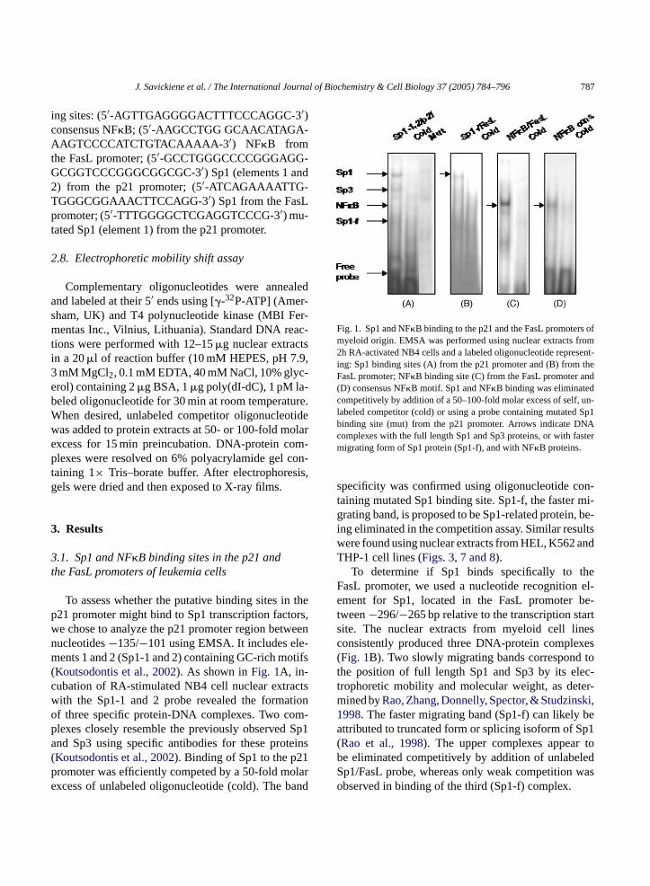
Fig. 1. Sp1 and NF�B binding to the p21 and the FasL promoters ofmyeloid origin. EMSA was performed using nuclear extracts from2h RA-activated NB4 cells and a labeled oligonucleotide represent-ing: Sp1 binding sites (A) from the p21 promoter and (B) from theFasL promoter; NF�B binding site (C) from the FasL promoter and(D) consensus NF�B motif. Sp1 and NF�B binding was eliminatedcompetitively by addition of a 50–100-fold molar excess of self, un-labeled competitor (cold) or using a probe containing mutated Sp1binding site (mut) from the p21 promoter. Arrows indicate DNAcomplexes with the full length Sp1 and Sp3 proteins, or with fastermigrating form of Sp1 protein (Sp1-f), and with NF�B proteins.
specificity was confirmed using oligonucleotide con-taining mutated Sp1 binding site. Sp1-f, the faster mi-grating band, is proposed to be Sp1-related protein, be-ing eliminated in the competition assay. Similar resultswere found using nuclear extracts from HEL, K562 andTHP-1 cell lines (Figs. 3, 7 and 8).
To determine if Sp1 binds specifically to theFasL promoter, we used a nucleotide recognition el-ement for Sp1, located in the FasL promoter be-tween−296/−265 bp relative to the transcription startsite. The nuclear extracts from myeloid cell linesconsistently produced three DNA-protein complexes(Fig. 1B). Two slowly migrating bands correspond tothe position of full length Sp1 and Sp3 by its elec-trophoretic mobility and molecular weight, as deter-mined byRao, Zhang, Donnelly, Spector, & Studzinski,1998. The faster migrating band (Sp1-f) can likely beattributed to truncated form or splicing isoform of Sp1(Rao et al., 1998). The upper complexes appear tobe eliminated competitively by addition of unlabeledSp1/FasL probe, whereas only weak competition wasobserved in binding of the third (Sp1-f) complex.
.1. Sp1 and NF�B binding sites in the p21 andhe FasL promoters of leukemia cells
To assess whether the putative binding sites in21 promoter might bind to Sp1 transcription facte chose to analyze the p21 promoter region betwucleotides−135/−101 using EMSA. It includes elents 1 and 2 (Sp1-1 and 2) containing GC-rich mo
Koutsodontis et al., 2002). As shown inFig. 1A, in-ubation of RA-stimulated NB4 cell nuclear extraith the Sp1-1 and 2 probe revealed the formaf three specific protein-DNA complexes. Two colexes closely resemble the previously observednd Sp3 using specific antibodies for these protKoutsodontis et al., 2002). Binding of Sp1 to the p2romoter was efficiently competed by a 50-fold moxcess of unlabeled oligonucleotide (cold). The b
788 J. Savickiene et al. / The International Journal of Biochemistry & Cell Biology 37 (2005) 784–796
To test the role of NF�B, we used a nucleotide cor-responding to a putative atypical NF�B binding se-quence from the FasL promoter (Israel et al., 1989). Asshown inFig. 1C, nuclear extracts from leukemic cellsformed one DNA–protein complex with this probe,as for the probe possessing a consensus NF�B motif(Fig. 1D). The nuclear proteins may represent a com-plex of p50 and p65 subunits of NF�B according to itselectrophoretic mobility. The binding was abolished bycorresponding unlabeled oligonucleotides.
3.2. Sp1 and NF�B binding activity to the p21 andNF�B promoters during leukemia celldifferentiation and leading apoptosis
To begin investigating a possible role of Sp1 andNF�B in programmed cell death of terminally differ-entiated cells, we compared differentiation and subse-quent apoptosis in different leukemia cell lines: HL-60,NB4 and HEL. The promyelocytic leukemia cells NB4and HL-60 differ in some characteristics: NB4 cell linehas at(15;17) translocation associated with the APL,whereas HL-60 cells lack this chromosomal transloca-tion and p53 (Collins, 1987). Both cell lines showedgranulocytic differentiation after treatment with 1�MRA. HL-60 cells were stained positively with NBTon day 5 (about 60%), while NB4 cells reached thislevel on day 3 (Fig. 2C). The staining of the cells withEtBr/AO revealed induction of apoptosis, increasingfrom 3–5% (spontaneous) to more than 40% in HL-6 y 5( d at andt ,w e inu hS mi-g andR asm rad-u p1b nlyid duali o-t ter.T niedb
ing nuclear extracts from proliferating and differenti-ating HL-60 cells, we did not find any interaction offull length Sp1 with the p21 or the FasL promoters(Fig. 2A). By contrast, Sp1-f binding to the FasL pro-moter was detected in control and RA-induced HL-60cells at the commitment stage only. In RA-treated bothcell lines, similar effects were observed for the associ-ation of NF�B to consensus motif and to the FasL pro-moter (Fig. 2B). Thus, the down-regulation of Sp1 andNF�B binding activities was associated with apoptosisfollowing long-term RA-treatment, most likely withoutthe involvement of the Fas/FasL pathway.
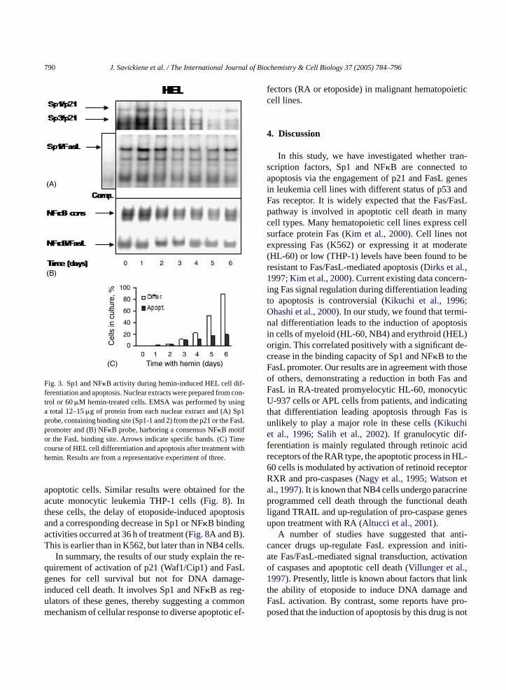
To examine whether this is a general phenomenonfor leukemic cell differentiation, we performed EMSAusing extracts from human erythroleukemia HEL cellsinduced to differentiate to erythrocytes by 60�Mhemin. A typical HEL cell population contained, on day6, about 80% of differentiated cells and 20% of cellsundergoing apoptosis (Fig. 3C). Furthermore, EMSAdemonstrated a gradual decrease in Sp1 and NF�Bbinding to the p21 and FasL promoters during cell dif-ferentiation (Fig. 3A and B). The mobility of complexeswas the same as in other cell lines but bands corre-sponding to these complexes were prominent on day 6.This could reflect a lower proportion of cells undergo-ing apoptosis at the same time-point, as compared tomore apoptosis sensitive HL-60 and NB4 cells.
Thus, our observations demonstrate a positive roleof Sp1 and NF�B in the differentiation process and anegative one in apoptosis of terminally differentiatedc
3te
m-a e-l e-n ide( gc sLp sticd -p ible,c ibitorz nw tion
0 and around 30% in NB4 cell population on daFig. 2C). EMSA revealed that RA treatment causeime-dependent decrease in Sp1 binding to the p21he FasL promoters in NB4 cells (Fig. 2A). Two bandshich competed with unlabeled probe, were visiblntreated NB4 cells (Fig. 2A) representing full lengtp1 and Sp3. The formation of the third, fasterrating complex (Sp1-f) was noticed in untreatedA-treated NB4 cells. However, Sp1-s complex wore prominent after day 1 of RA-treatment and gally decreased during NB4 cell differentiation. Sinding to the FasL promoter slightly decreased o
n the cells undergoing apoptosis (Fig. 2A). NB4 cellifferentiation process was associated with a gra
ncrease in NF�B binding activity to consensus mif and with sustained affinity for the FasL promohe binding reduction to these sites was accompay the increase in apoptosis on day 5 (Fig. 2B). Us-
ells of myeloid or erythroid origin.
.3. The loss of Sp1 and NF�B binding activity tohe p21 and the FasL promoters duringtoposide-induced leukemia cell apoptosis
To further characterize the influence of DNA dage on NF�B and Sp1 activity, we treated promy
ocytic leukemia HL-60 cells having a null-p53 photype, or p53-carrying NB4 cells with etopos68�M) for different times. In NB4 cells, the druaused Sp1 and NF�B binding to the p21 and the Faromoters during 3 h of treatment, as well as draecrease at 6–18 h (Fig. 4A and B). To inhibit casases in intact cells, we employed the irreversell-permeable, the broad-spectrum caspase inh-VAD.fmk (25�M). The time of the inhibitor additioas chosen to yield maximum changes in transcrip

J. Savickiene et al. / The International Journal of Biochemistry & Cell Biology 37 (2005) 784–796 789
Fig. 2. Sp1 and NF�B binding activity during granulocytic differentiation and apoptosis. Nuclear extracts were prepared from control or 1�MRA-treated (A) NB4 and (B) HL-60 cells. EMSA was performed using a total 12–15�g protein from each nuclear extract and Sp1 probe,containing binding site (Sp1-1 and 2) from the p21 or the FasL promoter, and NF�B probe harboring a consensus NF�B motif, or the FasLbinding site. Arrows indicate specific bands, the decrease of which is shown by competition (comp.) analysis. (C) Time course of HL-60 andNB4 cell differentiation and apoptosis after treatment with RA. Results are from one representative experiment of three.
factors activity between 6 and 18 h of etoposide treat-ment. As expected, z-VAD.fmk produced a marked in-hibitory effect and restored transcription factors bind-ing activity seen at 6–18 h (Fig. 4A and B) confirm-ing caspase-dependent events. Identical results wereobtained with extracts from etoposide-treated HL-60cells (Fig. 5A and B). There was a strong relationshipbetween the changes in transcription factors bindingactivity and the number of apoptotic cells (Fig. 5D),the appearance of internucleosomal fragmentation at6–18 h and the absence of the ladders using z-VAD.fmk(Fig. 5C).
Similar results with a decrease in Sp1 binding tothe p21 promoter or NF�B to consensus motif were
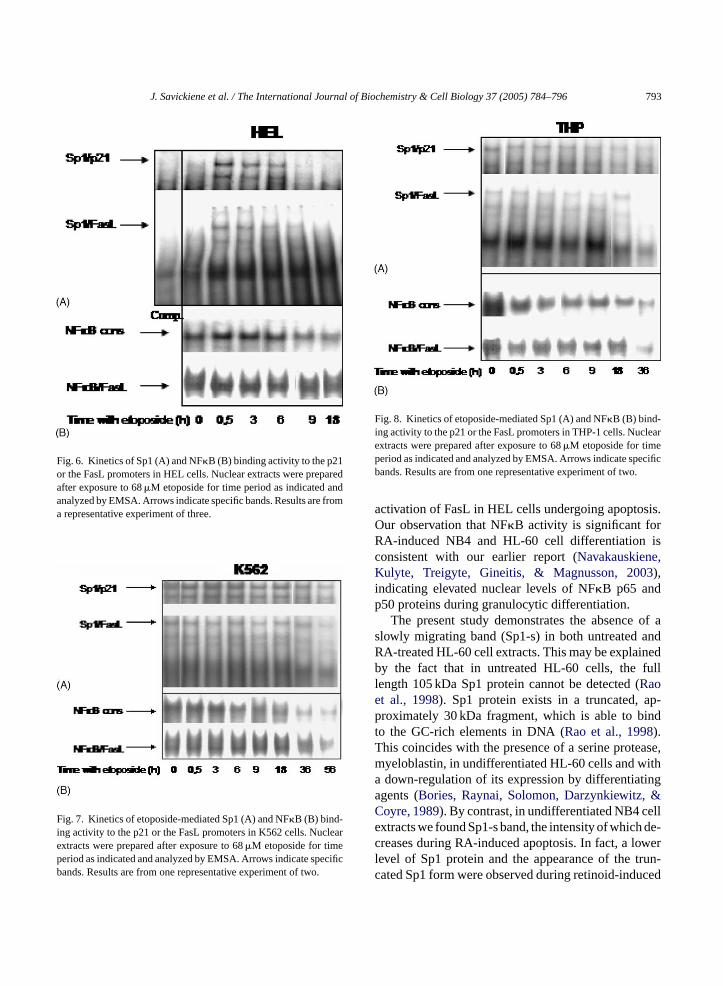
obtained using etoposide-treated HEL cell extracts(Fig. 6). However, in these cells NF�B binding ca-pacity to the FasL promoter was detected after 18 hexposure to the drug (Fig. 6B), suggesting that, in HELcells, NF�B might participate in DNA damage-inducedFasL-mediated apoptosis.
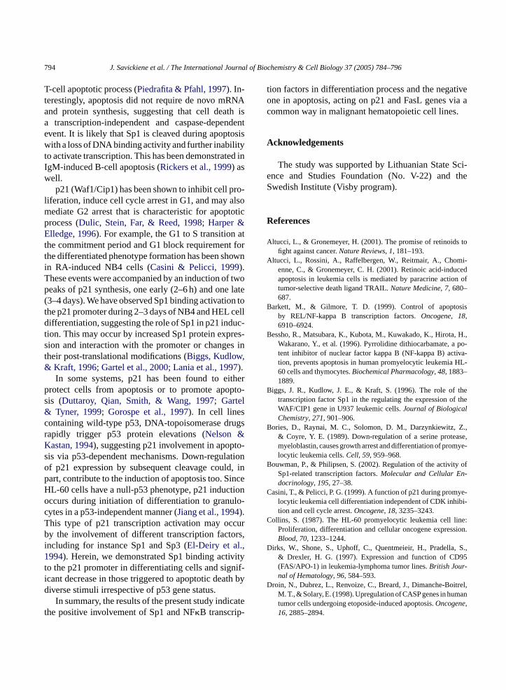
K562 cell line is normally resistant to the inductionof apoptosis by a number of agents (McGahon et al.,1994). We thus investigated whether etoposide wasable to trigger cell death in K562 cells via NF�B orSp1. Fig. 7 demonstrates an inducible formation ofNF�B binding complexes with consensus motif andthe FasL promoter within 18 h. There was a decreaseat 56 h, likely to be due to an augmented number of
790 J. Savickiene et al. / The International Journal of Biochemistry & Cell Biology 37 (2005) 784–796
Fig. 3. Sp1 and NF�B activity during hemin-induced HEL cell dif-ferentiation and apoptosis. Nuclear extracts were prepared from con-trol or 60�M hemin-treated cells. EMSA was performed by usinga total 12–15�g of protein from each nuclear extract and (A) Sp1probe, containing binding site (Sp1-1 and 2) from the p21 or the FasLpromoter and (B) NF�B probe, harboring a consensus NF�B motifor the FasL binding site. Arrows indicate specific bands. (C) Timecourse of HEL cell differentiation and apoptosis after treatment withhemin. Results are from a representative experiment of three.
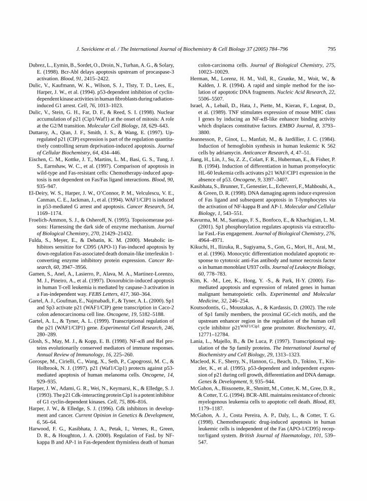
apoptotic cells. Similar results were obtained for theacute monocytic leukemia THP-1 cells (Fig. 8). Inthese cells, the delay of etoposide-induced apoptosisand a corresponding decrease in Sp1 or NF�B bindingactivities occurred at 36 h of treatment (Fig. 8A and B).This is earlier than in K562, but later than in NB4 cells.
In summary, the results of our study explain the re-quirement of activation of p21 (Waf1/Cip1) and FasLgenes for cell survival but not for DNA damage-induced cell death. It involves Sp1 and NF�B as reg-ulators of these genes, thereby suggesting a commonmechanism of cellular response to diverse apoptotic ef-
fectors (RA or etoposide) in malignant hematopoieticcell lines.
4. Discussion
In this study, we have investigated whether tran-scription factors, Sp1 and NF�B are connected toapoptosis via the engagement of p21 and FasL genesin leukemia cell lines with different status of p53 andFas receptor. It is widely expected that the Fas/FasLpathway is involved in apoptotic cell death in manycell types. Many hematopoietic cell lines express cellsurface protein Fas (Kim et al., 2000). Cell lines notexpressing Fas (K562) or expressing it at moderate(HL-60) or low (THP-1) levels have been found to beresistant to Fas/FasL-mediated apoptosis (Dirks et al.,1997; Kim et al., 2000). Current existing data concern-ing Fas signal regulation during differentiation leadingto apoptosis is controversial (Kikuchi et al., 1996;Ohashi et al., 2000). In our study, we found that termi-nal differentiation leads to the induction of apoptosisin cells of myeloid (HL-60, NB4) and erythroid (HEL)origin. This correlated positively with a significant de-crease in the binding capacity of Sp1 and NF�B to theFasL promoter. Our results are in agreement with thoseof others, demonstrating a reduction in both Fas andFasL in RA-treated promyelocytic HL-60, monocyticU-937 cells or APL cells from patients, and indicatingthat differentiation leading apoptosis through Fas isuef cidr HL-6 torR eta inep eathl nesu
nti-c initi-a ationo1 inkt andF pro-p not
nlikely to play a major role in these cells (Kikuchit al., 1996; Salih et al., 2002). If granulocytic dif-
erentiation is mainly regulated through retinoic aeceptors of the RAR type, the apoptotic process in0 cells is modulated by activation of retinoid recepXR and pro-caspases (Nagy et al., 1995; Watsonl., 1997). It is known that NB4 cells undergo paracrrogrammed cell death through the functional d
igand TRAIL and up-regulation of pro-caspase gepon treatment with RA (Altucci et al., 2001).
A number of studies have suggested that aancer drugs up-regulate FasL expression andte Fas/FasL-mediated signal transduction, activf caspases and apoptotic cell death (Villunger et al.,997). Presently, little is known about factors that l
he ability of etoposide to induce DNA damageasL activation. By contrast, some reports haveosed that the induction of apoptosis by this drug is

J. Savickiene et al. / The International Journal of Biochemistry & Cell Biology 37 (2005) 784–796 791
Fig. 4. The loss of Sp1 (A) and NF�B (B) binding activity during etoposide-induced apoptosis and the recovery by caspase inhibitor z-VAD.fmkin NB4 cells. Nuclear extracts were prepared after exposure to 68�M etoposide in the absence or the presence of 25�M z-VAD.fmk for timeperiod as indicated and analyzed by EMSA. Arrows indicate specific bands. Results are from a representative experiment of two.
mediated via the Fas pathway in lymphoid and myeloidcells (McGahon, Costa Pereira, Daly, & Cotter, 1998;Siitonen et al., 2000). Our data shows that etoposide-mediated apoptosis is likely to be independent of thecell Fas status. At first, we have observed that untreatedcell lines, non-expressing (K562) and low-expressing(THP-1) Fas have relatively high Sp1 and NF�B bind-ing activities to the FasL promoter. Later, decreases inthese activities are associated with delayed apoptosis inthose cell lines. Thus, our observations are consistentwith the idea that the recruitment of Fas/FasL systemis not necessary for etoposide-induced apoptosis. Thedeath process of etoposide-sensitive cells, such as HL-60 and U937, has been shown to up-regulate Casp-2and Casp-3 genes associated with enhanced synthesisof related procaspases (Droin et al., 1998). The per-manent peptide z-VAD.fmk, which is homologues tosequences targeted by ICE-like caspases, efficientlysuppresses Casp gene expression (Droin et al., 1998),thereby preventing apoptotic DNA fragmentation.
K562 cell resistance to the induction of apoptosis isattributed to the activity of p210bcr-abl tyrosine kinaseencoded by the Bcr-Abl fusion gene and is associatedwith a delayed activation of procaspase-3 and targetprotein cleavage in response to DNA damage (Dubrez
et al., 1998). However, multiple Fas-resistant cell lines(such as K562) remain sensitive to chemotherapy-induced apoptosis (Eischen et al., 1997). Interestingly,Fas resistance could be overcome by cycloheximidein some Fas-resistant or etoposide-resistant cell lines(Kim et al., 2000; Siitonen et al., 2000), suggest-ing the presence of short-lived apoptosis-inhibitoryproteins (FLIP, RIP, XIAP, cIAP2) (Fulda, Meyer, &Debatin, 2000; Willems et al., 2000) that can accountfor the deficient functioning of the Fas system in thosecells. Moreover, etoposide-induced, Fas-independentand Fas-mediated apoptosis lead to the activation ofcaspase-3 in many cell lines and the late stages ofapoptosis after such treatments proceed through a com-mon caspase pathway (Eischen et al., 1997; Zhuang &Cohen, 1998).
Topoisomerase poisons, including etoposide, acti-vate NF�B and induce apoptosis (Bessho et al., 1996;Piret & Piette, 1996). In our study, NF�B bindingactivity to consensus motif and to the FasL promoteroccurred during early hours of etoposide treatment.The decrease was associated with cell death in myeloidcell lines. No decrease in NF�B binding to the FasLpromoter in HEL cells was observed during 18 h ofetoposide treatment, suggesting a long lasting NF�B

792 J. Savickiene et al. / The International Journal of Biochemistry & Cell Biology 37 (2005) 784–796
Fig. 5. The loss of Sp1 (A) and NF�B (B) binding activity during etoposide-induced apoptosis and the recovery by caspase inhibitor z-VAD.fmkin HL-60 cells. Nuclear extracts were prepared after exposure to 68�M etoposide in the absence or the presence of 25�M z-VAD.fmk for timeperiod as indicated and analyzed by EMSA. Arrows indicate specific bands.(C) Effect of caspase inhibitor z-VAD.fmk on etoposide-inducedDNA fragmentation of HL-60 cells. Etoposide-induced cells were treated without or with z-VAD.fmk for 6 h. Cells were lysed and DNA extractedfor electrophoresis in 1.5% agarose gel. (D) Time course of etoposide-induced apoptoside in HL-60 cells. Results are from one representativeexperiment of two.
J. Savickiene et al. / The International Journal of Biochemistry & Cell Biology 37 (2005) 784–796 793
Fig. 6. Kinetics of Sp1 (A) and NF�B (B) binding activity to the p21or the FasL promoters in HEL cells. Nuclear extracts were preparedafter exposure to 68�M etoposide for time period as indicated andanalyzed by EMSA. Arrows indicate specific bands. Results are froma representative experiment of three.
Fig. 7. Kinetics of etoposide-mediated Sp1 (A) and NF�B (B) bind-ing activity to the p21 or the FasL promoters in K562 cells. Nuclearextracts were prepared after exposure to 68�M etoposide for timeperiod as indicated and analyzed by EMSA. Arrows indicate specificbands. Results are from one representative experiment of two.
Fig. 8. Kinetics of etoposide-mediated Sp1 (A) and NF�B (B) bind-ing activity to the p21 or the FasL promoters in THP-1 cells. Nuclearextracts were prepared after exposure to 68�M etoposide for timeperiod as indicated and analyzed by EMSA. Arrows indicate specificbands. Results are from one representative experiment of two.
activation of FasL in HEL cells undergoing apoptosis.Our observation that NF�B activity is significant forRA-induced NB4 and HL-60 cell differentiation isconsistent with our earlier report (Navakauskiene,Kulyte, Treigyte, Gineitis, & Magnusson, 2003),indicating elevated nuclear levels of NF�B p65 andp50 proteins during granulocytic differentiation.
The present study demonstrates the absence of aslowly migrating band (Sp1-s) in both untreated andRA-treated HL-60 cell extracts. This may be explainedby the fact that in untreated HL-60 cells, the fulllength 105 kDa Sp1 protein cannot be detected (Raoet al., 1998). Sp1 protein exists in a truncated, ap-proximately 30 kDa fragment, which is able to bindto the GC-rich elements in DNA (Rao et al., 1998).This coincides with the presence of a serine protease,myeloblastin, in undifferentiated HL-60 cells and witha down-regulation of its expression by differentiatingagents (Bories, Raynai, Solomon, Darzynkiewitz, &Coyre, 1989). By contrast, in undifferentiated NB4 cellextracts we found Sp1-s band, the intensity of which de-creases during RA-induced apoptosis. In fact, a lowerlevel of Sp1 protein and the appearance of the trun-cated Sp1 form were observed during retinoid-induced
794 J. Savickiene et al. / The International Journal of Biochemistry & Cell Biology 37 (2005) 784–796
T-cell apoptotic process (Piedrafita & Pfahl, 1997). In-terestingly, apoptosis did not require de novo mRNAand protein synthesis, suggesting that cell death isa transcription-independent and caspase-dependentevent. It is likely that Sp1 is cleaved during apoptosiswith a loss of DNA binding activity and further inabilityto activate transcription. This has been demonstrated inIgM-induced B-cell apoptosis (Rickers et al., 1999) aswell.
p21 (Waf1/Cip1) has been shown to inhibit cell pro-liferation, induce cell cycle arrest in G1, and may alsomediate G2 arrest that is characteristic for apoptoticprocess (Dulic, Stein, Far, & Reed, 1998; Harper &Elledge, 1996). For example, the G1 to S transition atthe commitment period and G1 block requirement forthe differentiated phenotype formation has been shownin RA-induced NB4 cells (Casini & Pelicci, 1999).These events were accompanied by an induction of twopeaks of p21 synthesis, one early (2–6 h) and one late(3–4 days). We have observed Sp1 binding activation tothe p21 promoter during 2–3 days of NB4 and HEL celldifferentiation, suggesting the role of Sp1 in p21 induc-tion. This may occur by increased Sp1 protein expres-sion and interaction with the promoter or changes intheir post-translational modifications (Biggs, Kudlow,& Kraft, 1996; Gartel et al., 2000; Lania et al., 1997).
In some systems, p21 has been found to eitherprotect cells from apoptosis or to promote apopto-sis (Duttaroy, Qian, Smith, & Wang, 1997; Gartel& Tyner, 1999; Gorospe et al., 1997). In cell linesc ugsrK to-s tiono d, inp inceH tiono lo-cT curb rs,i1 vityt nif-i th byd
catet
tion factors in differentiation process and the negativeone in apoptosis, acting on p21 and FasL genes via acommon way in malignant hematopoietic cell lines.
Acknowledgements
The study was supported by Lithuanian State Sci-ence and Studies Foundation (No. V-22) and theSwedish Institute (Visby program).
References
Altucci, L., & Gronemeyer, H. (2001). The promise of retinoids tofight against cancer.Nature Reviews, 1, 181–193.
Altucci, L., Rossini, A., Raffelbergen, W., Reitmair, A., Chomi-enne, C., & Gronemeyer, C. H. (2001). Retinoic acid-inducedapoptosis in leukemia cells is mediated by paracrine action oftumor-selective death ligand TRAIL.Nature Medicine, 7, 680–687.
Barkett, M., & Gilmore, T. D. (1999). Control of apoptosisby REL/NF-kappa B transcription factors.Oncogene, 18,6910–6924.
Bessho, R., Matsubara, K., Kubota, M., Kuwakado, K., Hirota, H.,Wakarano, Y., et al. (1996). Pyrrolidine dithiocarbamate, a po-tent inhibitor of nuclear factor kappa B (NF-kappa B) activa-tion, prevents apoptosis in human promyelocytic leukemia HL-60 cells and thymocytes.Biochemical Pharmacology, 48, 1883–1889.
Biggs, J. R., Kudlow, J. E., & Kraft, S. (1996). The role of thetranscription factor Sp1 in the regulating the expression of theWAF/CIP1 gene in U937 leukemic cells.Journal of Biological
B Z.,ase,mye-
B y of-
C ye-bi-
C ne:sion.
D , S.,95
D itrel,an
ontaining wild-type p53, DNA-topoisomerase drapidly trigger p53 protein elevations (Nelson &astan, 1994), suggesting p21 involvement in apopis via p53-dependent mechanisms. Down-regulaf p21 expression by subsequent cleavage coulart, contribute to the induction of apoptosis too. SL-60 cells have a null-p53 phenotype, p21 inducccurs during initiation of differentiation to granuytes in a p53-independent manner (Jiang et al., 1994).his type of p21 transcription activation may ocy the involvement of different transcription facto
ncluding for instance Sp1 and Sp3 (El-Deiry et al.,994). Herein, we demonstrated Sp1 binding acti
o the p21 promoter in differentiating cells and sigcant decrease in those triggered to apoptotic deaiverse stimuli irrespective of p53 gene status.
In summary, the results of the present study indihe positive involvement of Sp1 and NF�B transcrip-
Chemistry, 271, 901–906.ories, D., Raynai, M. C., Solomon, D. M., Darzynkiewitz,
& Coyre, Y. E. (1989). Down-regulation of a serine protemyeloblastin, causes growth arrest and differentiation of prolocytic leukemia cells.Cell, 59, 959–968.
ouwman, P., & Philipsen, S. (2002). Regulation of the activitSp1-related transcription factors.Molecular and Cellular Endocrinology, 195, 27–38.
asini, T., & Pelicci, P. G. (1999). A function of p21 during promlocytic leukemia cell differentiation independent of CDK inhition and cell cycle arrest.Oncogene, 18, 3235–3243.
ollins, S. (1987). The HL-60 promyelocytic leukemia cell liProliferation, differentiation and cellular oncogene expresBlood, 70, 1233–1244.
irks, W., Shone, S., Uphoff, C., Quentmeieir, H., Pradella& Drexler, H. G. (1997). Expression and function of CD(FAS/APO-1) in leukemia-lymphoma tumor lines.British Jour-nal of Hematology, 96, 584–593.
roin, N., Dubrez, L., Renvoize, C., Breard, J., Dimanche-BoM. T., & Solary, E. (1998). Upregulation of CASP genes in humtumor cells undergoing etoposide-induced apoptosis.Oncogene,16, 2885–2894.
J. Savickiene et al. / The International Journal of Biochemistry & Cell Biology 37 (2005) 784–796 795
Dubrez, L., Eymin, B., Sordet, O., Droin, N., Turhan, A. G., & Solary,E. (1998). Bcr-Abl delays apoptosis upstream of procaspase-3activation.Blood, 91, 2415–2422.
Dulic, V., Kaufmann, W. K., Wilson, S. J., Tlsty, T. D., Lees, E.,Harper, J. W., et al. (1994). p53-dependent inhibition of cyclin-dependent kinase activities in human fibroblasts during radiation-induced G1 arrest.Cell, 76, 1013–1023.
Dulic, V., Stein, G. H., Far, D. F., & Reed, S. I. (1998). Nuclearaccumulation of p21 (Cip1/Waf1) at the onset of mitosis: A roleat the G2/M transition.Molecular Cell Biology, 18, 629–643.
Duttaroy, A., Qian, J. F., Smith, J. S., & Wang, E. (1997). Up-regulated p21 (CIP) expression is part of the regulation quantita-tively controlling serum deprivation-induced apoptosis.Journalof Cellular Biochemistry, 64, 434–446.
Eischen, C. M., Kottke, J. T., Martins, L. M., Basi, G. S., Tung, J.S., Earnshaw, W. C., et al. (1997). Comparison of apoptosis inwild-type and Fas-resistant cells: Chemotherapy-induced apop-tosis is not dependent on Fas/Fas ligand interactions.Blood, 90,935–947.
El-Deiry, W. S., Harper, J. W., O’Connor, P. M., Velculescu, V. E.,Canman, C. E., Jackman, J., et al. (1994). WAF1/CIP1 is inducedin p53-mediated G arrest and apoptosis.Cancer Research, 54,1169–1174.
Froelich-Ammon, S. J., & Osheroff, N. (1995). Topoisomerase poi-sons: Harnessing the dark side of enzyme mechanism.Journalof Biological Chemistry, 270, 21429–21432.
Fulda, S., Meyer, E., & Debatin, K. M. (2000). Metabolic in-hibitors sensitize for CD95 (APO-1) Fas-induced apoptosis bydown-regulation Fas-associated death domain-like interleukin 1-converting enzyme inhibitory protein expression.Cancer Re-search, 60, 3947–3956.
Gamen, S., Anel, A., Lasierro, P., Alava, M. A., Martinez-Lorenzo,M. J., Pineiro, A., et al. (1997). Doxorubicin-induced apoptosisin human T-cell leukemia is mediated by caspase-3 activation ina Fas-independent way.FEBS Letters, 417, 360–364.
G Sp1co-2
G of
G -nses.
G ., &53-
H . J.itor
H lop-t
H reen,F-
uman
colon-carcinoma cells.Journal of Biological Chemistry, 275,10023–10029.
Herman, M., Lorenz, H. M., Voll, R., Grunke, M., Woit, W., &Kalden, J. R. (1994). A rapid and simple method for the iso-lation of apoptotic DNA fragments.Nucleic Acid Research, 22,5506–5507.
Israel, A., Lebail, D., Hata, J., Piette, M., Kieran, F., Logeat, D.,et al. (1989). TNF stimulates expression of mouse MHC classI genes by inducing an NF-�B-like enhancer binding activitywhich displaces constitutive factors.EMBO Journal, 8, 3793–3800.
Jeannesson, P., Ginot, L., Manfait, M., & Jardillier, J. C. (1984).Induction of hemoglobin synthesis in human leukemic K 562cells by adriamycin.Anticancer Research, 4, 47–51.
Jiang, H., Lin, J., Su, Z. Z., Colart, F. R., Huberman, E., & Fisher, P.B. (1994). Induction of differentiation in human promyelocyticHL-60 leukemia cells activates p21 WAF/CIP1 expression in theabsence of p53.Oncogene, 9, 3397–3407.
Kasibhata, S., Brunner, T., Genestier, L., Echeverri, F., Mahboubi, A.,& Green, D. R. (1998). DNA damaging agents induce expressionof Fas ligand and subsequent apoptosis in T-lymphocytes viathe activation of NF-kappa B and AP-1.Molecular and CellularBiology, 1, 543–551.
Kavurma, M. M., Santiago, F. S., Bonfoco, E., & Khachigian, L. M.(2001). Sp1 phosphorylation regulates apoptosis via extracellu-lar FasL-Fas engagement.Journal of Biological Chemistry, 276,4964–4971.
Kikuchi, H., Ilizuka, R., Sugiyama, S., Gon, G., Mori, H., Arai, M.,et al. (1996). Monocytic differentiation modulated apoptotic re-sponse to cytotoxic anti-Fas antibody and tumor necrosis factor� in human monoblast U937 cells.Journal of Leukocyte Biology,60, 778–783.
Kim, K. -M., Lee, K., Hong, Y. -S., & Park, H-Y. (2000). Fas-mediated apoptosis and expression of related genes in humanmalignant hematopoietic cells.Experimental and Molecular
K rolethecell
L eg-f
M Kin-pres-ge.
M . R.,nic
M G.mancep-
artel, A. J., Goufman, E., Najmabadi, F., & Tyner, A. L. (2000).and Sp3 activate p21 (WAF1/CIP) gene transcription in Cacolon adenocarcinoma cell line.Oncogene, 19, 5182–5188.
artel, A. L., & Tyner, A. L. (1999). Transcriptional regulationthe p21 (WAF1/CIP1) gene.Experimental Cell Research, 246,280–289.
losh, S., May, M. J., & Kopp, E. B. (1998). NF-�B and Rel proteins evolutionarily conserved mediators of immune respoAnnual Review of Immunology, 16, 225–260.
orospe, M., Cirielli, C., Wang, X., Seth, P., Capogrossi, M. CHolbrook, N. J. (1997). p21 (Waf1/Cip1) protects against pmediated apoptosis of human melanoma cells.Oncogene, 14,929–935.
arper, J. W., Adami, G. R., Wei, N., Keymarsi, K., & Elledge, S(1993). The p21 Cdk-interacting protein Cip1 is a potent inhibof G1 cyclin-dependent kinases.Cell, 75, 806–816.
arper, J. W., & Elledge, S. J. (1996). Cdk inhibitors in devement and cancer.Current Opinion in Genetics& Developmen,6, 56–64.
arwood, F. G., Kasibhata, J. A., Petak, I., Vernes, R., GD. R., & Houghton, J. A. (2000). Regulation of FasL by Nkappa B and AP-1 in Fas-dependent thyminless death of h
Medicine, 32, 246–254.outsodontis, G., Moustakas, A., & Kardassis, D. (2002). The
of Sp1 family members, the proximal GC-rich motifs, andupstream enhancer region in the regulation of the humancycle inhibitor p21WAF1/Cip1 gene promoter.Biochemistry, 41,12771–12784.
ania, L., Majello, B., & De Luca, P. (1997). Transcriptional rulation of the Sp family proteins.The International Journal oBiochemistry and Cell Biology, 29, 1313–1323.
acleod, K. F., Sherry, N., Hannon, G., Beach, D., Tokino, T.,zler, K., et al. (1995). p53-dependent and independent exsion of p21 during cell growth, differentiation and DNA damaGenes& Development, 9, 935–944.
cGahon, A., Bissonette, R., Shmittt, M., Cotter, K. M., Gree, D& Cotter, T. G. (1994). BCR-ABL maintains resistance of chromyelogenous leukemia cells to apoptotic cell death.Blood, 83,1179–1187.
cGahon, A. J., Costa Pereira, A. P., Daly, L., & Cotter, T.(1998). Chemotherapeutic drug-induced apoptosis in huleukemic cells is independent of the Fas (APO-1/CD95) retor/ligand system.British Journal of Haematology, 101, 539–547.
796 J. Savickiene et al. / The International Journal of Biochemistry & Cell Biology 37 (2005) 784–796
Mercille, S., & Massie, B. (1994). Induction of apoptosis in nutrient-deprived cultures of hybridoma and myeloma cells.Biotechnol-ogy and Bioengineering, 44, 1140–1154.
Nagy, L., Thomazy, V. A., Shipley, G. L., Fesus, L., Lamph, W.,Heyman, R. A., et al. (1995). Activation of retinoid X recep-tors in HL-60 cell lines.Molecular and Cellular Biology, 15,3540–3551.
Navakauskiene, R., Kulyte, A., Treigyte, G., Gineitis, A., & Mag-nusson, K. -E. (2003). Translocation of transcription regulatorsinto the nucleus during granulocytic commitment of HL-60 cells.Biochemistry& Cell Biology, 81, 285–295.
Nelson, W. G., & Kastan, M. B. (1994). DNA strand breaks: TheDNA template alterations that trigger p53-dependent DNA dam-age response pathways.Molecular and Cellular Biology, 14,1815–1823.
Ohashi, M., Iwase, M., & Nagumo, M. (2000). Changes in suscepti-bility to Fas-mediated apoptosis during differentiation of HL-60cells.Journal of Leukocyte Biology, 67, 374–380.
Pahl, H. L. (1999). Activators and target genes of Rel/NF-kappa Btranscription factors.Oncogene, 18, 6853–6866.
Piedrafita, F. J., & Pfahl, M. (1997). Retinoid-induced apoptosisand Sp1 cleavage occur independently of transcription and re-quire caspase activation.Molecular and Cellular Biology, 17,6348–6358.
Piret, B., & Piette, J. (1996). Topoisomerase poisons activate thetranscription factor NB-�B in ACH-2 and CEM cells.NucleicAcids Research, 24, 4242–4248.
Rao, J., Zhang, F., Donnelly, R. J., Spector, N. L., & Studzinski, G.P. (1998). Truncation of Sp1 transcription factor by myeloblastinin undifferentiated HL-60 cells.Journal of Cellular Physiology,175, 121–128.
Rickers, A., Peters, N., Badock, V., Beyaert, R., Vandenabeele, P.,Dorken, B., et al. (1999). Cleavage of transcription factor Sp1 by
caspases during anti-IgM-induced B-cell apoptosis.EuropeanJournal of Biochemistry, 261, 269–274.
Salih, H. R., Starling, G. C., Brandl, S., Pelka-Fleisher, R., Hafer-lach, T., Hiddemann, W., et al. (2002). Differentiation of promye-locytic leukemia: Alterations in Fas (CD95/Apo-1) and Fas lig-and (CD178) expression.British Journal of Hematology, 117,76–85.
Siitonen, T., Mantymaa, P., Sally, M., Savolainen, E. R., & Koistinen,P. (2000). Etoposide-induced apoptosis is not associated withthe Fas pathway in acute myeloblastic leukemia cells.LeukemiaResearch, 24, 282–288.
Tolomeo, M., Dusonchet, L., Meli, M., Grimaudo, S., D’Alessandro,N., Papoff, G., et al. (1998). The CD95/CD95 ligand system isnot the major effector in anticancer drug-mediated apoptosis.CellDeath and Differentiation, 5, 735–742.
Villunger, A., Egle, A., Kos, M., Hartmann, B. L., Geley, S.,Kofler, R., et al. (1997). Drug-induced apoptosis is associ-ated with enhanced Fas (Apo-1/CD95) signaling in human T-acute lymphatic leukemia cells.Cancer Research, 57, 3331–3334.
Watson, R. W., Rotstein, O. D., Parodo, J., Bitar, R., Hackan, D.,& Marshall, J. C. (1997). Granulocytic differentiation of HL-60 cells results in spontaneous apoptosis mediated by increasedcaspase activation.FEBS Letters, 412, 603–609.
Willems, F., Amraoni, Z., Vanderheyde, N., Verhasselt, V., Ah-soy, E., Scaffigi, C., et al. (2000). Expression of c-FLIP (L)and resistance to CD95-mediated apoptosis of monocyte-deriveddendritic cells: Inhibition by bisindolylmaleimide.Blood, 95,3478–3482.
Zhuang, J., & Cohen, G. M. (1998). Release of mitochondrial cy-tochrome c is upstream of caspase activation in chemical-inducedapoptosis in human monocytic tumour cells.Toxicology Letters,102/103, 121–129.