p21 with a phenylalanine 28 + leucine mutation reacts normally
TRANSCRIPT

THE JOURNAL OF BIOLOGICAL CHEMISTRY (c) 1991 by The American Society for Biochemistry and Molecular Biology, Inc.
Vol. 266, No . 26, Issue of ' September 15, PP. 17700-17706, 1991 Printed in U. S. A.
p21 with a Phenylalanine 28 + Leucine Mutation Reacts Normally with the GTPase Activating Protein GAP but Nevertheless Has Transforming Properties*
(Received for publication, November 29, 1990)
Jochen Reinstein$, Ilme SchlichtingS, Matthias Frech, Roger S . Goody, and Alfred Wittinghofer From the Mar-Planck-Institut fur Medizinische Forschung, Abteilung Biophysik, Jahnstrasse 29, W-6900 Heidelberg, Federal Republic of Gremany
The H-rus gene product p21H has been mutated at Phe-28, which makes a hydrophobic interaction with the guanine base of bound GDP/GTP. The mutation Phe-28 -., Leu drastically increases nucleotide disso- ciation rates without affecting association rates. This is due to a perturbed binding of base, 0- and 8-phos- phate, and Mg2+, as evidenced from 31P NMR and flu- orescence measurements. The region around the y- phosphate appears normal. The affinity of Mg2+ for both the di- and the triphosphate conformation of the mutant was also measured by fluorescence. The asso- ciation constant is 3.5 X lo7 M" for the Gpp(NH)p complex, 500 times higher than for the GDP form. The mutation does not change appreciably the intrinsic or the GTPase activating protein (GAP)-stimulated GTPase. The mutated protein induces neurite differ- entiation however when pressure-loaded into PC12 cells, which is equivalent to transformation of NIH 3T3 cells. This shows that p21(F28L) is converted to the GDP bound form by GAP but is transforming be- cause the high dissociation rate for nucleotides leads to a protein predominantly in the active GTP bound form.
p21 is the product of the N-, K-, and H-ras genes and is believed to be involved in a growth-promoting signal trans- duction process (for a review, see Barbacid (1987); McCormick (1989a)). Like many guanine nucleotide-binding proteins it has an inactive GDP bound and an active GTP bound con- formation. The three-dimensional structures of the GDP bound and the GTP bound form have been elucidated (Tong et al., 1989; Pai et al., 1989; Milburn et al., 1990; Pai et al., 1990; Schlichting et al., 1990a). In the GTP bound confor- mation p21 reacts with the GTPase activating protein, GAP,' which is believed to be either the negative regulator of p21 activity or the actual downstream target molecule that is involved in mediating the signal of p21 (Trahey and Mc- Cormick, 1987; Vogel et al., 1988; McCormick, 1989b; Hall, 1990).
Mutations of p21 have been found in many human tumors
* The costs of publication of this article were defrayed in part by the payment of page charges. This article must therefore be hereby marked "aduertisement" in accordance with 18 U.S.C. Section 1734 solely to indicate this fact.
This paper is dedicated to Prof. H. Zahn for his 75th birthday. $ Present address: Rosenstiel Center, Brandeis University, Wal-
tham, MA 02254. ' The abbreviations used are: GAP, GTPase activating protein;
mant, N-methylanthraniloyl; Gpp(NH)p, guanyl-5'-yl imidodiphos- phate; EF-Tu, elongation factor Tu; Hepes, 4-(2-hydroxyethyl)-l- piperazineethanesulfonic acid.
(Barbacid, 1987; Bos, 1989). They have transforming proper- ties in rodent fibroblasts or can induce neurite outgrowth in the pheochromocytoma cell line PC12. Mutations isolated in vivo are in positions 12/13 or 61 of the polypeptide chain. Transforming mutations generated i n vitro have been de- scribed for many other positions, and the three-dimensional structure shows that all these residues are involved in protein- nucleotide interaction (Pai et al., 1990). I t is believed that oncogenic mutants are transforming, because their GTPase is not increased by GAP and they are thus constantly in their active GTP conformation. Indeed it has been shown for some of these mutations that they physically interact with GAP but that their GTPase is not activated (Adari et al., 1988; Cales et al., 1988; Gibbs et al., 1988; Sigal et al., 1988; Vogel et al., 1988). Here we describe in detail the biochemical, structural, and biological properties of a novel mutant of p21 (Phe-28 .--, Leu) whose GTPase activity is stimulated by GAP but is nevertheless transforming.
MATERIALS AND METHODS
Protein Purification-Protein purification was performed essen- tially as described previously (Tucker et al., 1986); The final purity of the proteins was >95%, as judged from sodium dodecyl sulfate- polyacrylamide gel electrophoresis. Proteins contained 1 mol of gua- nine nucleotide bound per mole of protein. Protein concentrations were determined with the Bradford assay using bovine serum albumin as a standard (Bradford, 1976), while [8-'H]GDP binding activity was determined by the filter binding assay (Tucker et al., 1986). Standard buffer was always, unless stated otherwise, 64 mM Tris-HC1, pH 7.6, 1 mM dithiothreitol, 10 mM MgCl,, and 1 mM sodium azide.
GTPase Actiuity-GTPase activity measurements were performed essentially as described previously (John et al., 1988). Briefly, p21. GDP (2 p ~ ) was preincubated with [T-'"P]GTP (40 p ~ ) for 30 min at room temperature in a total volume of 1 ml in 1 mM EDTA, 64 mM Tris-HC1, pH 7.6, 1 mM dithiothreitol, 1 mM sodium azide. The concentration of MgCl, was brought to 10 mM and the temperature to 37 "C. 50-pl portions were removed a t defined time intervals and the production of ["'PIPi was measured as described.
Kinetics of Nucleotide Dissociation and Association-The rate of dissociation of [8-:'H]GDP from the p21.[8-'H]GDP complex was measured as described previously (John et al., 1988). Nitrocellulose filters from Schleicher & Schull (BA 85, pore size = 0.45 pm) were used.
For the measurement of association rates a p21(F28L)-guanosine complex was prepared as described (John et al., 1990) and frozen in aliquots a t -70 "C. 0.5-1.0 p~ protein was reacted with an excess N - methylanthraniloyl-GDP (mantGDP) under various conditions in a stopped-flow apparatus (HighTech Scientific, Salisbury, United Kingdom). Excitation of fluorescence of mantGDP was a t 366 nm, and detection was through a filter with a cutoff a t 445 nm. Data were collected using an analog-digital connector in an Apple I1 computer or in a Nicolet 2090 digital storage oscilloscope. For the competition experiments between wild type p21 and pZl(F28L) the latter (0.5 p M ) was saturated with 1 p~ mantGDP in the presence of EDTA, and after several minutes 2 p~ p21c was added. The slow fluorescence
17700

p21 with Phe-28 + Leu Mutation 17701
increase was due to the slow dissociation of mantGDP from p21(F28L) and its fast association with ~ 2 1 ~ .
For the measurement of the binding affinity of Mg2' to p21- nucleotide complexes, the p21(F28L)-guanosine complex was first reacted with fluorescent nucleotides. A typical experiment used 0.6 p~ p21(F28L), 0.8 PM mantGpp(NH)p, 10 p~ MgC12, 1 mM dithio- erythritol in 30 mM Tris-HC1, pH 7.5 a t 25 "C. This solution was titrated with M$"EDTA. The data were treated as for the binding of a fluorescent substrate to protein in the presence of inhibitor (see Reinstein et al. (1990), Equation 12), using a dissociation constant of 1.7 PM for the EDTA"< interaction at pH 7.5.
For the dissociation rate constants of M e , a solution of 1 p M p21(F28L) with 0.38 pM mant-nucleotide in 30 mM Tris, pH 7.5, 1 mM dithioerythritol a t 25 "C was reacted with EDTA in a stopped- flow apparatus. The EDTA concentration was varied until it produced no further increase of the maximal M$+ dissociation rate. Kinetic data were analyzed by nonlinear regression, normally using a single exponential function, with the program Enzfitter (Elsevier Biosoft). Static and slow time scale dynamic fluorescent measurements were peformed on an SLM 8000 spectrophotometer.
Determination of Melting Temperature by Circular Dichroism-A fluorescence cuvette (0.4 cm width) was filled with 1.5 ml of p21- GDP solution (0.1 mg/ml) in 10 mM potassium phosphate buffer, pH 7.5. Circular dichroism was recorded with a Jobin-Yvon Dichrograph mark I11 at a fixed wavelength of 220 nm. The temperature increase per time was 40 "C/h using a Haake F3 water bath. The temperature was measured directly within the cuvette. To determine the melting temperature the following equation taken from Hecht et al. (1984), based on the Van't Hoff relation, was used.
6T = OD + (6, - 6D)/(1 + f )
where f = exp(H/RT(T/T, - l ) ) , 6T is ellipticity at temperature T, ft,, is ellipticity of denatured protein, 6N is ellipticity of native protein, and T, is melting temperature.
Interaction with GAP-Complexes between p21 and [Y-~'P]GTP were formed by incubating the protein with the radioactive nucleotide in the presence of 1 mM EDTA and 64 mM Tris-HCI, pH 7.6, 1 mM dithioerythritol. Excess nucleotide was removed by gel filtration on a small G-25 commercial column (NAP-5, Pharmacia LKB Biotech- nology Inc.). The protein was eluted from this column with the GAP reaction buffer: 20 mM Hepes-NaOH, pH 7.6, 1 mM dithioerythritol. The GTPase reaction was started by addition of 2 mM MgC1, and the appropriate amount of recombinant human GAP (0.75 nM). The GTPase reaction was measured as described (Gibbs et al., 1988; Vogel et al., 1988; Frech et al., 1990) by following the decrease of the concentration of [y-32P]GTP bound to p21 by filtration of the reaction mixture through nitrocellulose filters (0.45 pm). The initial rates were determined from the decrease of p21-GTP with time. Different rates obtained with different concentrations of substrate were analyzed for Vmax and K , using the program Enzfitter.
Biological Activity of p21 in PC12 Celk-The pheochromocytoma cell line PC12 was grown under standard conditions with Dulbecco's modified Eagle's medium supplemented with 10% horse serum, 5% fetal calf serum, and antibiotics (Greene and Tischler, 1982). Approx- imately 5 X lo4 cells were collected by centrifugation in an Eppendorf tube. 40 11 of a protein solution containing 3 mg/ml p21 in standard buffer were added to the cells. The protein was introduced into the cytoplasm by pushing the cells through a yellow pipette tip held a t the bottom of the tube. This creates enough pressure to slightly rupture the cell membrane so that the protein can enter the cell (Borasio et al., 1989). The cells were incubated again in standard medium in a Petri dish, and neurite outgrowth was then monitored under the microscope approximately 50-70 h after loading the cells.
NMR Spectroscom-For NMR the protein samples as isolated (in the GDP bound form) were dialyzed against potassium phosphate or standard (Tris) buffer, freeze-dried, and redissolved in D20. The last two steps were repeated for proton NMR samples. For phosphor NMR measurement of the p21.Gpp(NH)p complexes the protein bound GDP was exchanged for Gpp(NH)p as described recently (John et al., 1990). Samples for proton NMR measurements contained -45 mM potassium phosphate, 45 mM MgCl,, 6 mM dithioerythritol, 4 mM NaN3, and 3 mM p21. The 31P NMR samples contained 50 mM Tris buffer, 10 mM MgC12, 10-20 mM dithioerythritol and 1 mM p21 in the GDP and Gpp(NH)p form, 1 mM p21(F28L).GDP, and 0.3 mM p21(F28L) .Gpp(NH)p.
NMR experiments were performed on a commercial Bruker AM 500 spectrometer working with a proton resonance frequency of 500
MHz and a phosphorus resonance frequency of 202 MHz. For the proton NMR experiments 0.5-ml aliquots of sample in 5-mm sample tubes and a 5-mm probe were used, whereas 2.2-ml aliquots of sample in 10-mm sample tubes and a 10-mm probe were used for "P spec- troscopy. The proton spectra are referenced to internal sodium 2,2- dimethyl-2-silapentane-5-sulfonate, the phosphorus spectra to exter- nal 85% H3P04. Sample temperature was kept a t 303 K with a stream of dry air, which was temperature-regulated with a standard Bruker VTlOOO unit. The residual HDO resonance was suppressed by per- manent (except acquisition) selective irradiation with the HDO fre- quency. Quadrature detection was used in all experiments. All two- dimensional experiments were performed in the phase sensitive mode with the time-proportional incrementation technique (Marion and Wuthrich, 1983). For COSY and NOESY spectra usually 128 tran- sients of 4 K data points were collected for each of 512 increments with a relaxation delay of 1.1 s between successive transients. The NOESY spectra were recorded with a mixing time of 0.15 s which was randomly varied by 15%. A sweep width of 4545.45 Hz was used in both dimensions. Prior to Fourier transformation apodization was carried out in both dimensions using an unshifted sine-bell filter for the double-quantum-filtered COSY spectra and a ~/32-shifted sine- bell filter for the NOESY spectra. The digital resolution was 8.9 Hz/ point in tl after zero filling. Standard procedures and commercially available software on an Aspect 3000 computer were used throughout.
RESULTS
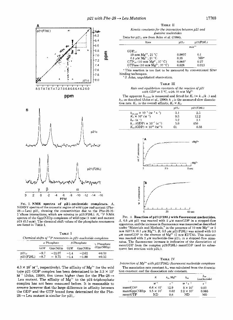
NMR Spectra-The role of Phe-28 in the binding of nu- cleotide in the three-dimensional structure of p21 complexed to the GTP analogue Gpp(NH)p as derived from x-ray crys- tallography is shown in Fig. 1. It can be seen that the guanine base is sandwiched between the side chains of Lys-117 and Phe-28, both making hydrophobic interactions (Pai et aL, 1989; Milburn et al., 1990; Pai et al., 1990). We have shown earlier by proton nuclear Overhauser enhancement NMR spectroscopy measurements that the a:omatic protons of a phenylalanine residue are within 5.0 A of the H-1' of the ribose of the nucleotide (Schlichting et al., 1988). By site- directed mutation of Phe-28 to Leu we could show that in solution Phe-28 is making a hydrophobic interaction with the guanine base (Schlichting et al., 1990b).
Fig. 2 shows the aromatic portion of the two-dimensional NOESY spectrum of p21(F28L) in comparison with wild type p21. The two-dimensional NMR spectrum again proves that the Phe-28 side chain resonances are missing in p2l(F28L). This confirms that in solution the aromatic protons of Phe- 28 of wild type p21 are within 0.5 nm of the C-l'-H protons. The C-l'-H protons of the ribose make no other observable nuclear Overhauser enhancement to a proton on the protein, which means that no other proton is within 0.5 nm of C-1'- H. We showed earlier that in p21(F28L) the H-8 proton of the guanine base and the C-l'-H proton of the ribose are shifted by 0.23 and 0.18 ppm, respectively, as compared with wild type p21, where they are 8.08 and 6.08 ppm, respectively (Schlichting et al., 1990b). This indicates that the environ- ment of the guanine base changes in replacing Phe-28 by Leu.
p21 binds guanine nucleotides with high affinity ( K , in the order of 10'" to lo1' M")(Feuerstein et al., 1987a; 1987b; Neal et al., 1988 John et at., 1990) and with high specificity. The main determinants of interaction are around the @-phosphate and the base. The 31P NMR spectrum of GDP bound to p21(F28L) has shown that the chemical shift of the P-phos- phate is unperturbed as compared with wild type, whereas the a-phosphorus atom experiences a downfield shift of 1 ppm (Schlichting et al., 1990b). This and the proton NMR data suggest that the mutation Phe-28 + Leu affects the binding of base and ribose and, weakly, of a-phosphate, but that the @-phosphate stays firmly bound in the phosphate binding loop (residues 10-17). It still experiences the unusual 4 ppm shift between free and bound GDP, which is typical for many other phosphoryl transfer proteins (Klaus et al., 1986).

17702 p21 with Phe-28 + Leu Mutation
FIG. 1. The guanine base binding site in the structure of the triphos- phate complex of p21. The model of p21(F28L) (in purple) has been built with FRODO assuming no structural change between wild type and mutant protein. The extra side chain of Phe-28 of wild type p21 is shown in yellow. The residues important for the binding of the base are shown with numbers: Lys-117, Asp-119, LYS-147.
The Phe-28 -+ Leu mutation also has an effect on the 31P resonances of the complex with Gpp(NH)p. Fig 2B shows the '"P NMR of the p21-triphosphate complexes. The list of the chemical shift values shows that the a-phosphate, but also the @-phosphate resonances are shifted compared with wild type protein (Table I), the shift of the a-resonance being more dramatic. Only the magnetic environment of the y-phosphate is unperturbed. Together with the 31P NMR data of the GDP complex (Table I) these data also show that the effect of the mutation is different for the GDP and the GTP conformation.
Nucleotide Binding Characteristics-The Phe-28 -+ Leu mutation changes the binding of guanine nucleotides drasti- cally as seen from the dissociation rate constants of p21(F28L). Compared with wild type protein (John et al., 1988) the GDP dissociation rate is increased 140-fold at 21 "C (Table 11) and 110-fold at 5 "C (Table 11), both in the presence of excess M e ions. This shows a difference in the tempera- ture dependence of the rate constants, which is larger for p21(F28L) (37-fold, as compared with 23-fold for ~ 2 1 ~ ) . The GTP dissociation rate of p21(F28L) in the presence of M C is increased 54-fold at 37 "C as compared with wild type p21. The dissociation rate constants in the absence of M C are too fast to be measured for p21(F28L) with normal nitrocel- lulose filtration techniques. The GTPase rate is decreased only 2-fold. This together with the NMR data indicates that in the p21(F28L)-triphosphate complex the binding of the y- phosphates is unperturbed, at least less than the binding of base, ribose, and a,@-phosphate.
It has been shown before that a fluorescent analogue of GDP/GTP can be used to measure the kinetics of association and dissociation of guanine nucleotides and p21 (John et al., 1989; 1990; Neal et al., 1990). As described for wild type p21 the binding of mantGDP to p21(F28L) mutant in the presence of Mg2+ produces a large fluorescence increase that can be used to measure the association and dissociation kinetics. The apparent rate constant k,. ,app, for the reaction of the mutant protein with mantGDP is not linearly dependent on the nucleotide concentration. This means that the binding is a two-step reaction with a rapid equilibrium preceding a slow isomerization reaction, as proposed for wild type p21 previ- ously (John et al., 1990).
p21 + GDP + p21.GDP + p21*GDP* k+ I k-1
k-1 k-,
Using GDP as the competing ligand we can derive the kinetic constants for the reaction between p21(F28L) and GDP, which are listed in Table 111. We can conclude that the mutant protein also binds GDP in a two-step binding reaction with kinetics for k l , kV1, and kp similar to wild type. The only major difference between wild type and mutant protein is the in- creased dissociation rate constant (k-J of GDP from the p21(F28L). GDP complex, and this is mostly responsible for the 110-fold difference in affinity at 5 "C. This result could also mean that the slow isomerization of the binding reaction is not the rearrangement of the guanine binding pocket, since this, as shown above, should be perturbed in the mutant. Mg"+ Binding-Fig. 3A shows that mixing p21(F28L) with
mantGDP in the absence of M$+ does not produce any change in fluorescence, while in the presence of Mg', there is a large increase (-180%). This was surprising, since wild type p21 shows an almost equal fluorescence increase with or without metal ion (John et al., 1990). To determine whether mantGDP really binds to p21(F28L) without Mg2' we added nucleotide- free wild type p21 to the mixture of mantGDP and mutant protein in the absence of Mg2+ (Fig. 3B). Since the association of wild type p21 and mantGDP is fast (3 s" at the concentra- tion used), the observed slow increase in fluorescence with a rate constant of 0.13 s" means that mantGDP is tightly bound to p21(F28L) and that the slow increase monitors the dissociation of mantGDP from this complex.
Since for this mutant binding of mantGDP in the presence but not in the absence of M g ' produces a high fluorescence change, it was possible to conveniently monitor the affinity of Mg'+ to various p21(F28L)-nucleotide complexes. So far, the affinity of Mg+ to the p21. GDP complex has been deter- mined only indirectly by measuring the decrease of the dis- sociation rate of GDP from this complex with increasing M e concentration (John, 1990). Adding M g ' to p21(F28L). mantGDP produces a large fluorescence increase. Using this we can titrate the p21(F28L). mantGDP complex and measure a binding constant of 6.6 f 4 X 10' M" (Table IV) for this reaction. For the complex of p21(F28L) with GTP or Gpp(NH)p we find a much higher affinity (3.5 X lo7 M" and
p21 with Phe-28 + Leu Mutation 17703
TABLE I1 Kinetic constants for the interaction between p21 and
guanine nucleotides Data for o21r are from John et al. (1988).
A
I I I I I I I I I I I 8.0 7.8 7.6 7.4 7.2 7.0 6.8 6.6 6.4 6.2 6.0
Rate P21c pZl(F28L)
min" GDP,,
10 mM Mg'+, 21 "C 0.0007 0.1 0.1 p M M$+, 21 "C 0.35 ND"
GTP,,,, (10 mM Mg', 37 "C) 0.0056 0.27 GTPase (10 mM M e , 37 "C) 0.028 0.013
"This reaction is too fast to be measured by conventional filter binding techniques.
J . John, unpublished observation.
TABLE 111 Rate and equilibrium constants of the reaction of p21
with GDP at 5 "C, with 10 mM Mg'+ The apparent k,n,,l,l, is measured and fitted for K , (= k+' /k - , ) and
k+, as described (John et al., 1990); k 2 is the measured slow dissocia- tion rate. KO,, is the overall affinity, K 1 X K2.
p21r pZl(F28L)
PPm
0 2.5 5 sec
3 2 0 -2 -4 -6 -8 -10 -12 -14 -16 PPM
FIG. 2. NMR spectra of p21-nucleotide complexes. A, NOESY spectra of the aromatic region of wild type and mutant (Phe- 28+ Leu) p21, showing the connectivities due to the Phe-28-H- l'rihose interactions, which are missing in p21(F28L). B, '"P NMR spectra of the Gpp(NH)p complexes of wild type (1 mM) and mutant p21 (0.3 mM). The chemical shift values of the phosphate resonances are listed in Table I.
0 25 50 sec
FIG. 3. Reaction of p21(F28L) with fluorescent nucleotides. A , 0.5 p~ p21 was reacted with 2 p~ mantGDP in a stopped-flow apparatus, and the increase in fluorescence was measured as described under "Materials and Methods," in the presence of 10 mM Mg" or 2 mM EDTA (0.1 FM M e ) . B, 0.5 p M p21(F28L) was mixed with 0.5 p M mantGDP in the absence of M$+ (2 mM EDTA). This mixture was reacted with 2 VM nucleotide-free p 2 1 ~ in a stopped-flow appa- ratus. The fluorescence increase is indicative of the dissociation of mantGDP from the complex p21(F28L).mantGDP (and its suhse- quent fast reaction with ~ 2 1 ~ ) .
TABLE I Chemical shi/ts of resonances in p21-nucleotide complexes
p21r -9.7 -11.07 -1.4 -2.62 +0.33 p21(F28L) -8.7 - 9.75 -1.4 -1.88 +0.33
TABLE IV Interaction of M$+ withp2l(F28L)-fluorescent nucleotide complexes
The association rate constant k,, was calculated from the dissocia- tion constant and the dissociation rate constant.
4.3 x lo7 M-', respectively). The affinity of Mg2+ for the wild type p21. GDP complex has been determined to be 3.3 X lo5 M" (John, 1990), five times higher than for the Phe-28- Leu mutant. The affinity of Mg'+ to the p21-triphosphate
complex has not been measured before. It is reasonable to assume however that thz large difference in affinity between the GDP and the GTP bound form determined for the Phe- 28 - Leu mutant is similar for p21,.
"1 S -1 "l S - I S"
mantGDP 6.6 X lo4 12.9 8 X lo5 0.03 mantGpp(NH)p 3.5 X lo7 0.62 2.2 X lo7 0.066 mantGTP ND 0.8 ND ND

17704 p21 with Phe-28 + Leu Mutation
The fluorescence change due to Mg'+ binding also allowed us to measure the Mg" dissociation rate constants. They were 0.62 and 0.8 s" for the Gpp(NH)p and GTP complex, respec- tively, and 12.9 s" for the p21.GDP complex, as shown in Table IV. From the equilibrium constants and the dissociation rate constants we can also calculate the association rate constants for the reaction as shown. The data in Table IV show that the 650-fold higher affinity of M$+ for the p21- triphosphate complex over the diphosphate complex is due almost equally to a faster association and a slower dissociation rate. The data in Table IV also show that the dissociation of Mg"+ is much faster than the dissociation of nucleotide and therefore independent from it.
Temperature Stability-we have shown earlier that the nucleotide-free p21 is very unstable (half-life of 2 h a t 25 "C; John et al. (1990)) and that the binding of GDP/GTP stabi- lizes the protein. To find out whether the decreased affinity constants between nucleotide and p21(F28L) also decrease the stability of the protein it was of interest to measure the thermal stabilities of the mutant and the wild type protein. The thermal stabilities of the two proteins in the GDP form were measured by circular dichroism in phosphate buffer. Fig. 4 shows that p 2 1 ~ has a melting temperature of 55 "C. p21(F28L) has a slightly decreased temperature stability with T,,, of 51.7 "C. This suggests that tight binding of GDP is indeed necessary for thermal stability. On the other hand the melting temperature is not low enough to explain the result of the increased dissociation rate constants as due to insta- bility of the protein. Furthermore '"P NMR and nucleotide binding measurements of the p21(F28L).GDP complex did not show any melting of the structure at 37 "C.
Biological Actioity-It has been postulated that any p21 mutant that has an increased GDP dissociation rate and/or fails to be returned to the inactive state by either the intrinsic or the GAP mediated GTPase is a transforming Ras protein. The pheochromocytoma cell line PC12 can be induced to differentiate by oncogenic forms of p21 (Bar-Sagi and Fer- amisco, 1985). We introduced p21(F28L) into these cells by pressure loading as described before (Borasio et al., 1989). Fig. 5B shows that p21(F28L) also differentiates PC12 cells. Since it has been shown that the ability to induce differentiation in PC12 cells and the ability to transform NIH 3T3 cells, al- though seemingly opposite effects, are always correlated (Sas- sone-Corsi et al., 1989) we can conclude that most likely the p21(F28L) is an oncogenic mutant of p21.
Interaction with GAP-It was of interest whether this on- cogenic behavior of the mutant was due to the failure of the protein to interact with GAP or whether the increased disso- ciation rates alone would be sufficient to explain the behavior of the protein. We tested p21(F28L) and wild type p21 with full-length human GAP. This was expressed with baculovirus
300 310 320 330 340 350 Temperature (0 K)
FIG. 4. Circular dichroism temperature stability curves of wild type and mutant p21.GDP complexes. Change of ellipticity was recorded as a function of temperature. Protein concentrations were 0.1 mg/ml in 10 mM phosphate buffer, pH 7.45. The resulting melting temperatures are 55 "C for p21, and 51.7 "C for p21(F28L).
A 111
B
FIG. 5. Effect of p21(F281A) on PC12 cells. The rat pheochro- mocytoma cell line PC12 was pressure-loaded with wild type or mutant (Phe-28- Leu) protein as described under "Materials and Methods" and by Borasio et ai. (1989). The cells were incubated for 60 h in standard medium in a Petri dish and photographed under phase contrast conditions. A, wild type p21 B p21(F28L). No cells had neurites with p2lC; with p21(F28L) 5-10% of cells looked as in panel R.
in sf9 insect cells and purified to 90% homogeneity using a protocol devised by McCormick et al. (Halenbeck et al., 1990). We find that the mutant and the wild type protein are almost equally activated by GAP. For a detailed analysis we measured the interaction at a fixed GAP concentration with varying concentration of p21(F28L) as shown in Fig. 6. Treating GAP as the enzyme and p21 as the substrate, we determine a kcat of 813 min" and a KIM of 11 p ~ . The corresponding values for wild type p21 are 1260 min" and 8 ~ L M (John, 1990).' This means that the difference in behavior toward GAP is rather minor. Using the same conditions for the GAP assay we do not find any stimulation of oncogenic mutants like p21(G12R) or p21(Q61L).:'
DISCUSSION
It has been shown by several investigators that mutations of residues involved in binding the guanine base in p21 reduce drastically the binding of guanine nucleotides. These muta- tions included Thr-144 + Ile and Asp-119 + Asn (Feig et al., 1986), Asp-119 + Ala (Sigal et al., 1986), Lys-146 + Val (Feig and Cooper, 1988a), Asn-116 += Ile, Asn-116 + His, Lys-
P. Gideon, M. Frech, A. Lantwein, and A. Wittinghofer, submitted for publication.
:' M. Frech, unpublished observation.

p21 with Phe-28 +. Leu Mutation 17705
0 20 40
[p21(F28L) . GTP] ( p M )
0 20 40
[ ~ 2 1 . GTP1 (PM)
FIG. 6. Interaction between GAP and p 2 1 ~ ( B ) or pZl(F28L) ( A ) . A constant concentration of purified human GAP expressed in sf9 cells with baculovirus was reacted with increasing amounts of p21. [r-"2P]GTP in standard GAP reaction buffer (see "Materials and Methods"). The initial rate of GTP hydrolysis, meas- ured as loss of counts due to "Pi release on nitrocellulose filters, was plotted against p21 concentration. The data were analyzed treating p21 as substrate and GAP as enzyme using nonlinear fitting proce- dures as described under "Materials and Methods." A , mutant p21 using 0.75 nM GAP; B, wild type p21 using 5 nM GAP. Data for wild type p21 are from John (1990). Half-maximal rate and the corre- sponding p21. GTP concentration (K,) are indicated.
117 + Arg, Lys-117 + Glu, Asp-119 --.) Glu, Asp-119 + His, Asp-119 +Ala (Der et al., 1988) and Lys-117 + Gln (Clanton et al., 1986). Those results are somewhat at variance with the results on the mutants Asn-116- Lys and Asn-l16+Tyr, which have been reported not to bind guanine nucleotides (Clanton et al., 1986). However, no detailed biochemical in- vestigation has been performed with any of the mutants.
The mutation described here which modifies the hydropho- bic interaction between two aromatic groups, the guanine base and Phe-28, reduces the affinity for GDP (at 5 "C) 110- fold, and GTP similarly. Assuming that the much smaller side chain of Leu-28 does not contribute to the interaction with the base and that the structure of p21 is not perturbed, we can calculate that the contribution of the aromatic side chain to the binding energy is 2.6 kcal mol" and even larger at higher temperatures. Interestingly the mutation affects only the dissociation rate of the second slow isomerization step. This seems to be a general phenomenon, since we have found other mutants with the same characteristics. Although the binding affinities of the above mentioned mutants have not been determined properly, it appears from the published dissociation rate or relative affinity data that all the mutations around the guanine base binding site reduce the binding constant of guanine nucleotides mainly by affecting the off- rate.
The three-dimensional structure of trypsinized EF-Tu. GDP is easily superimposed on the structure of p21- G P ~ ( N H ) ~ . ~ With an additional alignment of the important Thr-35 of p21 with Thr-61 of EF-Tu one finds that there is no phenylalanine in EF-Tu corresponding to Phe-28. Instead
W. Kabsch and E. F. Pai, unpublished observations.
the three-dimensional structure shows a leucine (Leu-175) side chain, located opposite to Lys-136, making a hydrophobic interaction with the guanine base of GDP and itself held in place by Lys-176 of the Ser-Ala-Lys motif, which is conserved in many guanine nucleotide-binding proteins. It is of interest that EF-Tu has a much lower affinity for guanine nucleotides, in the order of lo9 M" at 0 "C (Fasano et al., 1978). The apparent association rate constant is 2.6 X lo5 M-' s-', very similar to that of p21, whereas the dissociation rate constant is 2.3 X s-', much lower than k-, of wild type p21 and similar to that of the mutant pZl(F28L). It appears that the "missing" phenylalanine of EF-Tu is mostly responsible for the lower affinity of EF-Tu for GDP and that the lower affinity is again determined only by the dissociation rate.
Our NMR and temperature stability measurements show that the overall structure of the Phe + Leu mutation is not drastically altered. However the "P and proton NMR data show that loosening the interaction of the base with its hydrophobic environment, formed by Phe-28 and Lys-117, does affect the binding site around the CY- and 0-phosphate in the triphosphate conformation of p21, as observed by the shift of the CY- and (3-phsphate resonances. However the structure around the y-phosphate seems to be unperturbed. This is also evident from the fact that the intrinsic and the GAP-catalyzed GTPase, which involves the nucleophilic attack of water on the y-phosphate with general base catalysis (Feuerstein et al., 1989; Pai et al., 1990), are barely affected by the mutation.
Recently exchange factors such as sCDC25 (Crechet et al., 1990)) or the mammalian exchange factor (Wolfman and Macara, 1990; Downward et al., 1990) have been described biochemically. These proteins accelerate the dissociation rate constant by an as yet unknown mechanism. Since all the mutations that modify the interaction between base and the protein as shown here and in earlier work and also mutations that effect the interaction between Mg2+ and the protein (John et al., 1990; Feig and Cooper, 1988b) lead to increased dissociation rates, it would be interesting to see how the exchange factors modify the kinetics of interaction between nucleotide and protein.
The affinity of M g + for p21. GDP is in the order of lo5 M" and for the p21-triphosphate complex it is higher by a factor of -500. The high resolution x-ray structure of p21. Gpp(NH)p has shown that Mg2+ has six ligands, two from the phosphate groups of Gpp(NH)p, two from the protein (Ser- 17 and Thr-35) and two from water (Pai et al., 1990). In the structure of the p21. GDP complex, four water ligands have been placed into the first coordination sphere of Mg2+, to- gether with the oxygen from Ser-17 and @-phosphate (Tong et al., 1991). In support of this Smithers et al. (1990) have found by EPR spectroscopy that four water ligands are com- plexed to the metal ion in the p21. GDP complex. The high affinity of Mg2+ to the p21-triphosphate complex (KG = 3.5 X lo7 M-') also shows that the metal ion can only be completely removed with high amounts of EDTA (Kc = 6 X IO5 M-', at pH 7.5).
It has been predicted that the ratio of the GDP dissociation rate constant over the GTPase activity (kOff/kcat) determines the nucleotide state of p21 in the cell (McCormick et al., 1988). Since the interaction of p21 with GAP is only reduced 65% by the Phe-28+ Leu mutation, the increase of the dissociation rate by a factor of more than 100-fold means, all things beeing equal, that the steady state concentration of p21 in the triphosphate state should also be higher by ap- proximately 100-fold. This provides the necessary evidence for the hypothesis put forward earlier (Walter et al., 1986) that base binding mutants are transforming because of their

17706 p21 with Phe-28 +Leu Mutation
fast recycling to the GTP bound state and because in the cell the concentration of GTP is much higher than that of GDP (Trahey and McCormick, 1987). I t was recently reported that in quiescent 3T3 cells with high expression of p21H, 0.5% of p21 is in the triphosphate complex (Satoh et al., 1990). If this estimate is applicable also to a normal, nonoverproducing cell, as seems likely (McCormick et al., 1988), we would estimate that more than 50% of p21(F28L) should be in the triphos- phate concentration. In the oncogenic mutant p21(G12V) the situation is reversed; the GDP dissociation constant is only weakly affected (reduced -3-fold), whereas the GTPase activ- ity in the presence of GAP is a t least 300-fold (Trahey and McCormick, 1987) lower as compared with wild type p21. p21(G12V) is thus mostly in the triphosphate state when microinjected into oocytes (Trahey and McCormick, 1987), but for a different reason than p21(F28L).
The functional role of GAP in signal transduction is still a matter of intensive research and the debate centers around the question of whether GAP is the negative regulator that keeps the concentration of active p21 down or whether it is the downstream target molecule (McCormick, 1989; Hall, 1990) Our results do not favor either argument. But since no mutation including Phe-28 + Leu has yet been found which is transforming and does not bind to GAP we would favor the notion that GAP is the actual effector molecule whose inter- action with p21 either alone or together with other proteins is the actual transmitter of the growth-promoting signal.
Acknowledgments-We thank F. McCormick for the si9 insect cells and K. C. Holmes for continuous support. We thank B. F. Clark and J . Nyborg for the EF-Tu coordinates.
REFERENCES
Adari, H., Lowy, D. R., Willumsen, B. M., Der, C. J., and McCormick,
Barbacid, M. (1987) Annu. Reu. Biochem. 56, 779-827 Bar-Sagi, D., and Feramisco, J . R. (1985) Cell 42,841-848 Borasio, G. D., John, J., Wittinghofer, A,, Barde, Y.-A., Sendtner,
Bos, J . L. (1989) Cancer Res. 49, 4682-4689 Bradford, M. M. (1976) Anal. Biochem. 72, 248-254 Cales, C., Hancock, J. F., and Hall, A. (1988) Nature 332, 548-551 Clanton, D. J., Hattori, S., and Shih, T. Y. (1986) Proc. Natl. Acad.
Sci. U. S. A. 83,5076-5080 Crechet, J.-B., Poullet, P., Mistou, M.-Y., Parmeggiani, A., Camonis,
J., Boy-Marcotte, E., Damak, F., and Jacquet, M. (1990) Science
Der, C. J, Weissmann, B., and MacDonald, M. J . (1988) Oncogene 3,
Downward, J., Riehl, R., Wu, L., and Weinberg, R. A. (1990) Proc.
Fasano, O., Bruns, W., Crechet, J.-B., Sander, G., and Parmeggiani,
Feig, L. A., and Cooper, G. M. (1988a) Mol. Cell. Biol. 8, 2472-2478 Feig, L. A,, and Cooper, G. M. (198813) Mol. Cell. Biol. 8, 3235-3243 Feig, L. A., Pan, B.-T., Roberts, T . M., and Cooper, G. M. (1986)
Proc. Natl. Acad. Sci. U. S. A. 83, 4607-4611 Feuerstein, J., Kalbitzer, H. R., John, J., Goody, R. S., and Witting-
hofer, A. (1987a) Eur. J. Biochem. 162, 49-55 Feuerstein, J., Goody, R. S., and Wittinghofer, A. (1987b) J. Biol.
Chem. 262,8455-8458 Feuerstein, J., Goody, R. S., and Webb, M. R. (1989) J. Biol. Chem.
264, 6188-6190 Frech, M., John, J., Pizon, V., Chardin, P., Tavitian, A., Clark, R.,
McCormick, F., and Wittinghofer, A. (1990) Science 249, 169-171 Gibbs, J. B., Schaber, M. D., Allard, J., Sigal, I. S., and Scolnick, E.
F. (1988) Science 240,518-521
M., and Heumann, R. (1989) Neuron 2,1087-1096
248,866-868
105-112
Natl. Acad. Sci. U. S. A. 87, 5998-6002
A. (1978) Eur. J. Biochem. 89, 557-565
M. (1988) Proc. Natl. Acad. Sci. U. S. A. 85,5026-5030
414
K. (1990) J. Biol. Chem. 265, 21922-21928
Greene, L. A., and Tischler, A. T. (1982) Adu. Cell. Neurobiol. 3, 373-
Halenbeck, R., Crosier, W. J., Clark, R., McCormick, F., and Koths,
Hall, A. (1990) Cell 61, 921-923 Hecht, M. H., Sturtevant, J. S., and Sauer, R. T. (1984) Proc. Natl.
Acad. Sci. U. S. A. 81, 5685-5689 John, J. (1990) Structure-Function Relationship of the ras Oncogene
Product p21. Ph.D. thesis, Universitat Heidelberg John, J., Frech, M., and Wittinghofer, A. (1988) J. Biol. Chem. 263,
11792-11799 John, J., Frech, M., Feuerstein, J., Goody, R. S., and Wittinghofer,
A. (1989) in Guanine-Nucleotide-Binding Proteins (Bosch, L., Kraal, B., and Parmeggiani, A., eds) pp. 209-214, Plenum Publish- ing Corp., New York
John, J., Sohmen, R., Feuerstein, J . , Linke, R., Wittinghofer, A,, and Goody, R. S. (1990) Biochemistry 29, 6058-6065
Klaus, W., Schlichting, I., Goody, R. S., Wittinghofer, A., Rosch, P., and Holmes, K. C. (1986) Biol. Chem. Hoppe-Seyler 367, 781-786
Marion, D., and Wuthrich, K. (1983) Biochem. Biophys. Res. Com- mun. 113,967-974
McCormick, F. (1989a) in Oncogenes and the Molecular Origins of Cancer (Weinberg, R. A., ed) pp. 125-145, Cold Spring Harbor Laboratory Press, Cold Spring Harbor, NY
McCormick, F. (1989b) Cell 56, 5-8 McCormick, F., Adari, H., Trahey, M., Halenbeck, R., Koths, K.,
Martin, G. A,, Crosier, W. J., Watt, K., Rubinfeld, B., and Wong, G. (1988) Cold Spring Harbor Symp. Quant. Biol. 53,849-853
Milburn, M. V., Tong, L., DeVos, A. M., Brunger, A., Yamaizumi, Z.,
Neal, S. E., Eccleston, J . F., Hall, A., and Webb, M. R. (1988) J. Biol. Nishimura, S., and Kim, S.-H. (1990) Science 247,939-945
Chem. 263, 19718-19722 Neal, S. E., Eccleston, J. F., and Webb, M. R. (1990) Proc. Natl.
Acad. Sci. U. S. A. 87, 3562-3565 Pai, E. F., Kabsch, W., Krengel, U., Holmes, K. C., John, J., and
Wittinghofer, A. (1989) Nature 341, 209-214 Pai, E. F., Krengel, U., Petsko, G. A., Goody, R. S., Kabsch, W., and
Wittinghofer, A. (1990) EMBO J. 9, 2351-2359 Reinstein, J., Vetter, I. R., Schlichting, I., Rosch, P. Wittinghofer, A.,
and Goody, R. S. (1990) Biochemistry 29, 7440-7450 Sassone-Corsi, P., Cer, C. J., and Verma, I. M. (1989) Mol. Cell. Biol.
9,3174-3183 Satoh, T., Endo, M., Nakafuku, M., Nakamura, S., and Kaziro, Y.
(1990) Proc. Natl. Acad. Sci. U. S. A. 87, 5993-5997 Schlichting, I., Wittinghofer, A., and Rosch, P. (1988) Biochem.
Biophys. Res. Commun. 150,444-448 Schlichting, I., Almo, S. C., Rapp, G., Wilson, K., Petratos, K.,
Lentfer, A., Wittinghofer, A., Kabsch, W., Pai, E. F., Petsko, G. A., and Goody, R. S. (1990a) Nature 345,309-315
Schlichting, I., John, J., Frech, M., Chardin, P., Wittinghofer, A, Zimmermann, H., and Rosch, P. (1990b) Biochemistry 29, 504- 511
Sigal, I. S., Gibbs, J. B., D’Alonzo, J. S., Temeles, G. L., Wolanski, B. S., Socher, S. H., and Scolnick, E. M. (1986) Proc. Natl. Acad.
Sigal, I. S., Marshall, M. S., Schaber, M. D., Vogel, U. S., Scolnick, E. M., and Gibbs, J . B. (1988) Cold Spring Harbor Symp. Quant. Biol. 53,863-869
Smithers, G. W., Poe, M., Latwesen, D. G., and Reed, G. H. (1990) Arch. Biochem. Biophys. 280, 416-420
Tong, L., Milburn, M. V., and Kim, S.-H. (1989) Science 245, 244 Tong, L., deVos, A. M., Milburn, M. V., and Kim, S. H. (1991) J.
Trahey, M., and McCormick, F. (1987) Science 238,542-545 Tucker, J. Sczakiel, G., Feuerstein, J., John, J., Goody, R. S., and
Wittinghofer, A. (1986) EMBO J. 5, 1351-1358 Vogel, U., Dixon, R. A. F., Schaber, M. D., Diehl, R. E., Marshall, M.
S., Scolnick, E. M., Sigal, I. S., and Gibbs, J. B. (1988) Nature
Walter, M., Clark, S. G., and Levinson, A. D. (1986) Science 233,
Wolfman, A., and Macara, I. G. (1990) Science 248,67-69
Sci. (U. S. A,) 83,952-956
Mol. Biol. 217, 503-516
335,90-93
649-652