paclitaxel and trail synergize to kill paclitaxel...
TRANSCRIPT
Abstract. Background: Small cell lung cancer (SCLC) isthe most aggressive form of lung cancer with poor diseaseoutcome. The chemotherapeutic agent paclitaxel (PA) iscommonly used as a second-line treatment in SCLC, butresponse rates are low. Materials and Methods: 86M1 SCLCcells were treated in the presence or absence of paclitaxeland TRAIL or the combination for 24 hours. Western blotanalysis was utilized to examine protein expression, cellsurface protein expression and membrane integrity wereelucidated by flow cytometry, and immunofluorescencemicroscopy was used to demonstrate translocation ofproteins to the cell nucleus. Results: Human 86M1 SCLCcells were found to be resistant to PA killing in vitro. Thisresistance is mediated by up-regulation of pro-survival
protein BCL-xl. However, PA also increases surfaceexpression of death receptors 4 and 5 (DR4 and DR5,respectively). The death receptors’ ligand increased SCLCkilling by PA through an apparent caspase-independent routeinvolving activation/translocation of AIF. Conclusion: Theaddition of TRAIL to PA can potentiate apoptosis in arelatively PA-resistant SCLC line (specifically 86M1 cells).More importantly, we are the first to report an active methodof resistance to paclitaxel in SCLC via BCL-xl up-regulation.
Small cell lung cancer (SCLC) is an aggressive form of lungcancer. Although SCLC is a highly chemosensitive disease,outcome is generally poor and the 5-year survival rate is <10%(1). Diagnosis of extensive stage (ES) comprisesapproximately two-thirds of new SCLC cases, and the mediansurvival of these patients is only 2-4 months if untreated, withsurvival increasing to 6-8 months with chemotherapy. Thisdisease is very responsive to first-line chemotherapy withresponse rates of greater than 50% routinely observed.However, these responses are often short-lived and diseaserecurrence in the ES patient population is frequent. Patientswith relapsed disease, or patients who fail to respond tochemotherapy generally succumb to their disease within a fewmonths (1). Treatment of patients with relapsed SCLC isespecially challenging if the disease is platinum-resistant,when disease progression occurs within 3 months ofcompletion of a platinum containing regimen. In thesepatients, median survival ranges from 3.7 to 4.7 months (2-7).In SCLC, paclitaxel is primarily considered a second-linetherapy after the failure of platinum-based treatment regimens(8). Several modes of action of paclitaxel have been described.The drug is most well known as a microtubule stabilizer.Specifically, paclitaxel binds to tubulin and interferes withspindle formation in mitosis, ultimately arresting cells in G1and G2/M phases of the cell cycle, leading to cell death (9-13). In addition to stabilizing microtubules paclitaxel may actto sequester free tubulin, effectively depleting the cell’s supplyof tubulin (14). Beyond these effects on microtubules, morerecent research has indicated that paclitaxel also induces
3193
Abbreviations: 7AAD: 7-Aminoactinomycin D; AIF: apoptosisinducing factor; BAK: Bcl-2 homologous antagonist/killer; BAX:Bcl-2–associated X; BCL-2: B-cell leukemia/lymphoma 2; BCL-xl:B-cell lymphoma-extra large; BIM: Bcl-2 interacting mediator ofcell death; CHAPS: 3-[(3-cholamidopropyl)dimethylammonio]-1-propanesulfonate; CTL: cytotoxic T lymphocyte; DR4: deathreceptor 4; DR5: death receptor 5; DTT: dithiothreitol; ES: extensivestage; FADD: Fas-associated protein with death domain; FAS L:FAS ligand; FBS: fetal bovine serum; FITC: fluoresceinisothiocyanate; FLICE: FADD-like interleukin-1 beta-convertingenzyme (caspase 8); GAPDH: glyceraldehyde 3-phosphatedehydrogenase; LDH: lactase dehydrogenase; MCL-1: inducedmyeloid leukemia cell differentiation protein; MHC: majorhistocompatibility complex; OD: optical density; PARP: poly (ADP-ribose polymerase); PMSF: phenylmethanesulphonylfluoride;PUMA: p53 upregulated modulator or apoptosis; PVDF:polyvinylidene fluoride; SCLC: small cell lung cancer; SDS: sodiumdodecyl sulfate; TNF: tumor necrosis factor; TRAIL: tumor necrosisfactor related apoptosis inducing ligand; XIAP: X-linked inhibitorof apoptosis protein.
Correspondence to: Scott J. Antonia, Immunology Program, MoffittCancer Center, 12902 Magnolia Drive, Tampa, FL 33612, U.S.A.E-mail: [email protected]
Key Words: Small cell lung cancer, TRAIL, paclitaxel, chemotherapy.
ANTICANCER RESEARCH 31: 3193-3204 (2011)
Paclitaxel and TRAIL Synergize to Kill Paclitaxel-resistantSmall Cell Lung Cancer Cells through a
Caspase-independent Mechanism Mediated through AIFTERRI B. HUNTER, NEIL J. MANIMALA, KIMBERLY A. LUDDY, TRACY CATLIN and SCOTT J. ANTONIA
Immunology Program, Moffitt Cancer Center, 12902 Magnolia Drive, Tampa, FL 33612, U.S.A.
0250-7005/2011 $2.00+.40
programmed cell death in cancer cells by binding to the pro-survival protein Bcl-2, blocking its function (15, 16). A varietyof pharmacoimmunologic effects have also been attributed topaclitaxel (17-19).
There are two major pathways of apoptotic cell death. Onepathway involves changes in mitochondrial membranepotential and the translocation of proteins from themitochondria into the cytoplasm, including translocation ofcytochrome c, ultimately triggering the activation of caspasesand other proteins, subsequently leading to apoptosis (20,21). BCL family proteins play key roles in the mitochondriaand are central to regulating mitochondrial membraneintegrity. The family consists of both pro-survival proteins,such as the well characterized BCL-2 and BCL-xl (22), aswell as pro-apoptotic proteins including BAX and BAK (23).The other major apoptotic pathway is primarily dependenton caspase-8 activation driven by the binding of deathreceptors on the cell surface by death ligands. Two wellcharacterized death receptors (DR) are DR4 and DR5 whichare both activated by tumor necrosis factor related apoptosisinducing ligand (TRAIL). TRAIL is present on activatedcytotoxic T lymphocytes (CTL), but not on unstimulatedperipheral blood T-cells (24). Binding of DR4/5 by TRAILresults in the recruitment of Fas-Associated protein withDeath Domain (FADD) and FADD-like interleukin-1 beta-converting enzyme (FLICE) to the intracellular portion of theDR. The resulting complex can then cleave and activatecaspase-8, leading to apoptosis through mitochondrialdependent (BID cleavage) or mitochondrial independent(caspase-3 cleavage) pathways (25-28). It has been reportedthat paclitaxel exerts effects on both of the aforementionedpathways (29-34). Additionally, paclitaxel has been shown toregulate the expression of DR4 and DR5 (35-37). However,much of this remains controversial and there is disagreementin the literature relating to the exact mechanism of paclitaxelinduced cancer cell apoptosis. This discord is primarily dueto the variation in the concentration of paclitaxel used in thestudies – some studies use biologically relevant doses ofpaclitaxel (25-150 nM), whereas other studies describeresults obtained from cells treated with 10 to 20-fold theaforementioned doses (38).
In the study described herein we sought to elucidatemechanisms by which SCLC are resistant to PA as well asassessing a potential treatment strategy to overcome thesemechanisms.
Materials and Methods
Cell propagation. 86M1 SCLC cells (39, 40) were maintained incomplete RPMI (RPMI-1640 media, supplemented with 10% fetalbovice serum, 100 U/ml penicillin, 100 μg/ml streptomycin, and 2mM L-glutamine and penicillin-streptomycin; Invitrogen, Carlsbad,CA, USA). Cells were enumerated, and live cells counted, using ahemacytometer and trypan blue exclusion.
Reagents. Paclitaxel was obtained as a 7 mM stock from CardinalHealth (Dublin, OH, USA)supplied through the Moffitt Pharmacy.Recombinant human TRAIL was obtained from R&D Systems(Minneapolis, MN, USA).
Cell death assay. Sensitivity of 86M1 SCLC to paclitaxel wasdetermined by lactase dehydrogenase (LDH) release using theCytoTox 96 Non-Radioactive Cytotoxicity Assay (Promega,Madison, WI, USA). Briefly, SCLC cells were culture for 48 hourswith or without different concentrations of paclitaxel ranging from0.1 to 1000 nM. Following drug exposure, the plates were gentlycentrifuged and 50 μl of the culture supernatant was added to a new96 well flat bottom plate. Kit substrate, 50 μl, was added and theplate was incubated for 30 minutes. Stop solution was then added,and the samples were gently mixed. Absorbance at 490 nm wasrecorded. Media background controls were also performed andsubtracted from all of the recorded OD values. Specific cell lysiswas quantitated using the following formula: (sample release minusspontaneous release) divided by (maximum release minusspontaneous release) ×100.
Death receptor expression. Surface expression of DR4 and DR5 wasanalyzed by flow cytometric analysis. Cells were treated with orwithout 100 nM paclitaxel for 20 hours. Cell culture suspensionswere then harvested and cell washed in phosphate-buffered saline(PBS) and stained with phycoerythrin (PE)-conjugated anti-DR4 or-DR5 antibodies (EBiosciences, San Diego, CA, USA) diluted 1:50in PBS. The cells were stained at room temperature for 20 minutes.The cells were then washed twice in PBS. A viability dye (7AAD)was then added. Live cells were gated as 7AAD-negative and thispopulation was used to evaluate DR expression.
Acridine orange/ethidium bromide staining. Apoptosis wasvisualized by chromatin condensation. Cells were treated with orwithout 100 nM paclitaxel for 20 hours. Where indicated, cells werethen treated with 50 ng/ml TRAIL (R&D Systems) for the final 1-4 hours of culture, following which the cells were harvested. Thecells were then incubated with 100 μg/ml acridine orange (Roche,San Francisco, CA, USA), a DNA fluorochrome which is able tocross the intact plasma membrane, for 5 minutes as previouslydescribed (41). Since these were suspended cells, the cells were thenplaced on a slide and coverslipped. Chromatin morphology wasanalyzed using an Olympus inverted fluorescence microscope andQ Capture Pro Software (Olympus, Center Valley, PA, USA).
Annexin V staining. After treatment with paclitaxel and/or TRAIL,cells were harvested and washed in cold PBS and then resuspended ata density of 1-10×106 cells per ml in 1X Annexin V binding buffer(BD Biosciences, Franklin Lakes, NJ, USA). 100 μl of the cells werelabelled with 10 μl Annexin V-Pacific Blue conjugate. The cells wereincubated for 15-20 minutes in the dark, after which 400 μl 1XAnnexin binding buffer was added to each tube. Cells wereimmediately analyzed using an LSRII cytometer (BD Biosciences).Annexin staining was analyzed using Flow Jo software (Tree Star Inc.,Ashland, OR, USA). Apoptosis was quantified by the percentage ofpositive cells falling under the defined positive curve.
Cell fractionation. Mitochondrial and cytosolic cell fractions wereobtained using the Mitchondria/Cytosol Fractionation kit fromBioVision (Mountain View, CA, USA). Fractionation was carried
ANTICANCER RESEARCH 31: 3193-3204 (2011)
3194
out according to the manufacturer’s instructions. An antibody raisedagainst a mitochondrial-specific protein, Cox IV (Cell SignalTechnologies, Danvers, MA, USA), was detected by Western blotto validate fractionation and as a loading control for mitochondrialfraction protein loading. Proteins were subjected to Western blotanalysis as described below.
Western blot analysis. Following treatment with or without 100 nMpaclitaxel for 20 hours, and, where indicated, 50 ng/ml of TRAIL forthe final 1-4 hours of culture, cells were harvested, washed, andresuspended in 1X CHAPS buffer (Cell Signal Technologies)supplemented with 5 mM dithiothreitol (DTT) (Cell SignalTechnologies) and 1 mM phenylmethylsulfonyl fluoride (PMSF)(Sigma, St. Louis, MI, USA). Protein concentration was determinedusing the BioRad (Hercules, CA, USA) protein assay and the lysateswere diluted to equal concentrations. 3X or 10X sodium dodecylsulfate (SDS)-loading buffer (BioRad) was added to a finalconcentration of 1X and samples were boiled for 5 minutes. Theproteins were resolved on 4-20 or 8-16% gradient pre-cast SDS-Hepespolyacrilamide gels (Pierce/Fisher, Pittsburgh, Pennsylvania, USA) andthen transferred to polyvinylidine fluoride (PVDF) membrane(Millipore, Billerica, MA, USA). Membranes were blocked with Tris-buffered saline containing 5% non-fat milk (Carnation, Glendale, CA,USA) and 0.5% Tween (Fisher, Pittsburgh, PA, USA). For phospho-protein blotting, the 5% non-fat milk was replaced with 5% bovineserum albumin (BSA) (Sigma). The membranes were incubated inprimary antibodies over night at 4˚C (antibodies indicated in figuresand figure legends). The membranes were then washed and incubatedfor 1 hour in horseradish peroxidase-conjugated secondary goat anti-rabbit or rabbit anti-mouse antibodies (Millipore). Proteins weredetected using ECL plus (Amersham) and hyperfilm (Amersham). Allantibodies were purchase from Cell Signal Technologies, except anti-AKT and anti-pAKT which were purchased from Santa CruzBiotechnology Inc. (Santa Cruz, CA, USA).
Flow cytometric analysis of mitochondrial membrane potential andcaspase activation. Mitochondrial membrane potential and caspaseactivation were determined by flow cytometry using the Dual SensorMitoCasp kit (Cell Technology Inc., Santa Monica, CA, USA). Thecells were acquired using a FACSCalibur cytometer (BDBiosciences) and staining intensity was analyzed using Flow Josoftware (Tree Star Inc.).
AIF fluorescence microscopy. SCLC cells were treated as indicatedand then harvested. The cells were permeablized and fixed using Fixand Perm (Calbiochem, San Diego, CA, USA). The cells were thenincubated with fluorescein isothiocyanate (FITC)-conjugated anti-AIF antibody (Abcam, Cambridge, MA, USA) and washed in PBS.The cells were then incubated with 4’,6-diamidino-2-phenylindole(DAPI) and allowed to settle on a slide under a coverslip.Fluorescence microscopy was performed using a Zeiss LSM 510Laser Scanning Confocal Microscope (Zeiss, Thornwood, NY,USA). Images and co-localization were evaluated using Image ProPlus 6.2 (Media Cybernetics, Silver Spring, MD, USA).
Results
86M1 SCLC cells are resistant to killing by paclitaxel. Wedetermined the effect of paclitaxel monotherapy on SCLCcell line 86M1. The cells were not sensitive to biologically
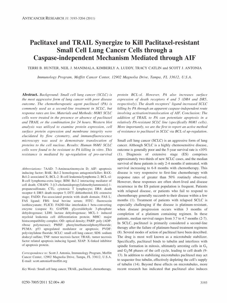
relevant doses (100-200 nM) of paclitaxel in vitro (Figure1A). In fact, the lethal dose, 50% (LD50) was not achievedwith 5-10 fold times this dose. For all remainingexperiments, a biologically relevant concentration of 100 nM(which resulted in the killing of approximately one quarterof the cells) was used.
DR expression on SCLC is increased following culture withpaclitaxel. Paclitaxel has been shown to induce apoptosis ofa cancer cells, however, the exact mechanism of this activityhas not been fully elucidated, although paclitaxel was firstdescribed as disrupting microtubules and inhibiting mitosis.With a recent report suggesting that chemotherapy canpromote the CTL-dependent induction of tumor cellapoptosis (42), it was a logical next step to determine ifpaclitaxel enhances the expression of DR that CTL bind toinitiate apoptosis, namely DR4 and DR5. Flow cytometrywas employed to show that DR4 and DR5 expression isupregulated on the surface of SCLC cells following 20 hoursof treatment with 100 nM paclitaxel (Figure 1B).
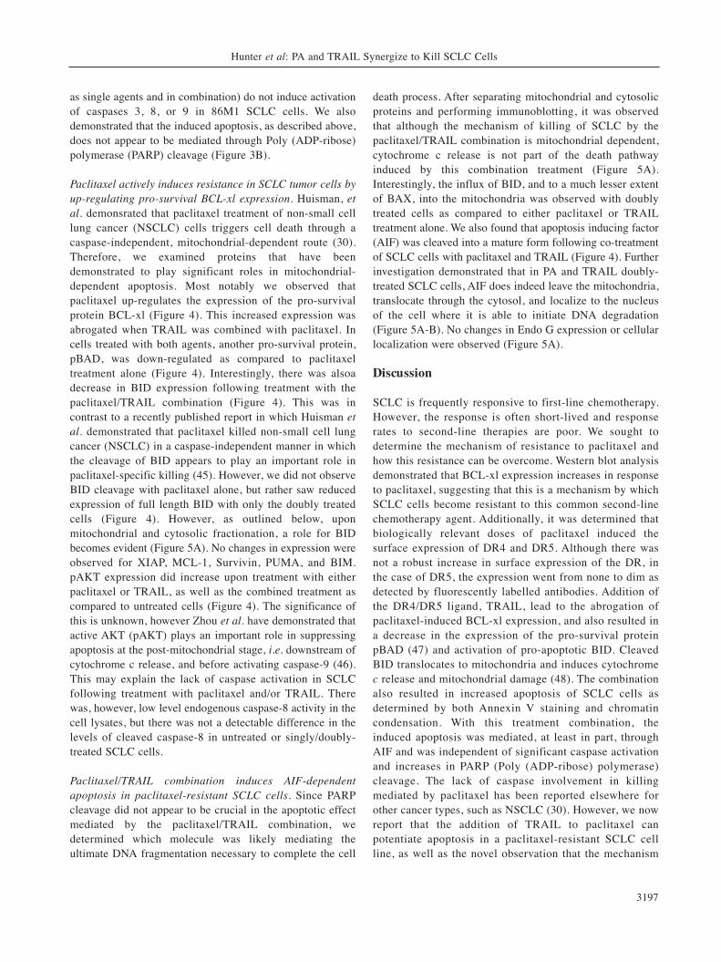
PA and TRAIL synergize to kill SCLC. CD8+ T-cells arethought to be the principle mechanism by which the immunesystem recognizes and kills tumor cells, and the goal of alarge proportion of proposed immunotherapies for cancer arebased on promoting anti-tumor T-cell responses (43). Thetwo major modes by which T-cells kill tumor cells arethrough the release of granzymes that enter the cells and setoff a cascade of caspase activation and by the ligation of DRon the target cells. The most characterized death receptorligands used by cytotoxic T-cells to induce apoptosis oftarget cells are FAS ligand (FAS L), tumor necrosis factor(TNF), and TRAIL (44). Since the receptors for TRAIL(DR4 and DR5) were upregulated upon treatment withpaclitaxel (Figure 1B), we next determined if paclitaxelwould sensitize SCLC to killing via TRAIL. Cells werecultured alone or with paclitaxel for 20 hours. Following thisincubation, the cells were then treated with TRAIL for 1-4hours. We then evaluated apoptosis in two differentexperiments. Annexin staining was used to identifymembrane instability (exposure of phosphatidyl serine).Paclitaxel alone induced only modest apoptosis, howeverwhen SCLC cells were treated with paclitaxel and TRAIL incombination, the number of apoptotic cells dramaticallyincreased (Figure 2A). Concordant with the Annexin stainingresults, the visualization of chromatin condensation was alsoseen only in doubly treated cells. Either treatment alone onlyresulted in a minimal number of cells displaying condensedchromatin upon microscopic examination (Figure 2B).
Paclitaxel/TRAIL combination induces apoptosis of SCLCthrough an apparent caspase-3 independent mechanism. Theroles of mitochondria and caspases in the paclitaxel and TRAIL
Hunter et al: PA and TRAIL Synergize to Kill SCLC Cells
3195
initiated apoptosis was examined using a MitoCasp assay kit,as well as examining caspase activation. Mitochondrialmembrane potential and caspase activation was determinedusing flow cytometry. Cells were treated with a fluorescentlylabeled cationic dye to detect membrane potential, or lossthereof. In healthy cells, the cationic dye is accumulated by themitochondria in proportion to the membrane potential. Inapoptotic cells, where the mitochondrial membrane potential iscompromised, the cationic dye does not accumulate in themitochondria and these cells exhibit a lower fluorescencesignal. Cells were also treated with carboxyfluorescein (FAM)labeled fluoromethyl ketone (FMK)-peptide inhibitors of
caspases that specifically bind active caspases. Using this assaywe found that there was a loss of membrane potential in cellstreated with paclitaxel, TRAIL and paclitaxel with TRAIL.There is no binding of the FAM-FMK molecules, indicating nocaspase activation (Figure 3A). The loss of membrane potentialin the paclitaxel and TRAIL singly treated cells was curious,as use of the two agents combined resulted in increasedapoptosis as compared to either agent alone.
To further confirm these results and understand themechanism involved in the increased apoptosis induced bythe paclitaxel/TRAIL combination, Western blot analysis wasperformed. We confirmed that paclitaxel and TRAIL (both
ANTICANCER RESEARCH 31: 3193-3204 (2011)
3196
Figure 1. Effect of paclitaxel on SCLC 86M1cells. A: SCLC cells were cultured with the indicated concentration of paclitaxel for 24 hours. Followingthe incubation, specific lysis was assessed by LDH release. Percentage specific lysis was calculated as: (sample release minus spontaneous release)divided by (maximum release minus spontaneous release) ×100. LD50 was not achieved even at 1000 nM paclitaxel. B: Death receptor 4 and 5expression is up-regulated on SCLC 86M1 cells following treatment with paclitaxel. Cells were treated with paclitaxel (100 nM) for 20 hours. Cellswere harvested and labelled with PE-conjugated anti-DR4 or -DR5 antibodies. Live cells were gated using exclusion of 7AAD labelling. The lightgrey histogram shows untreated cells and the black histogram corresponds to SCLC treated with paclitaxel.
as single agents and in combination) do not induce activationof caspases 3, 8, or 9 in 86M1 SCLC cells. We alsodemonstrated that the induced apoptosis, as described above,does not appear to be mediated through Poly (ADP-ribose)polymerase (PARP) cleavage (Figure 3B).
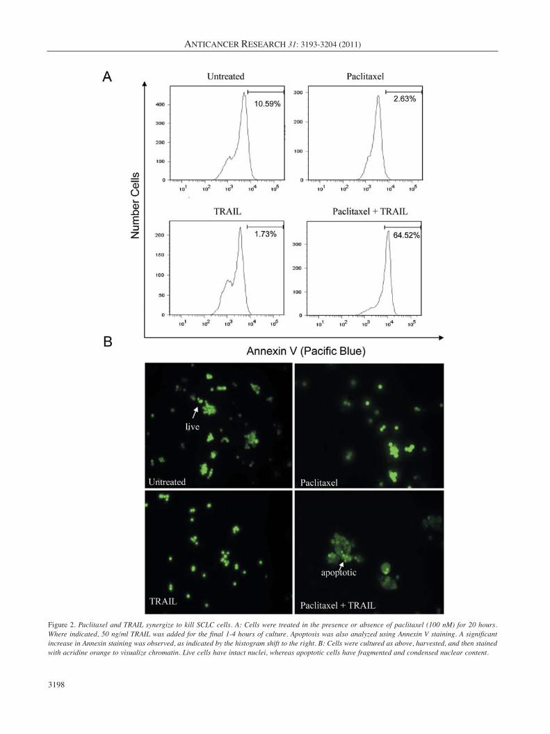
Paclitaxel actively induces resistance in SCLC tumor cells byup-regulating pro-survival BCL-xl expression. Huisman, etal. demonsrated that paclitaxel treatment of non-small celllung cancer (NSCLC) cells triggers cell death through acaspase-independent, mitochondrial-dependent route (30).Therefore, we examined proteins that have beendemonstrated to play significant roles in mitochondrial-dependent apoptosis. Most notably we observed thatpaclitaxel up-regulates the expression of the pro-survivalprotein BCL-xl (Figure 4). This increased expression wasabrogated when TRAIL was combined with paclitaxel. Incells treated with both agents, another pro-survival protein,pBAD, was down-regulated as compared to paclitaxeltreatment alone (Figure 4). Interestingly, there was alsoadecrease in BID expression following treatment with thepaclitaxel/TRAIL combination (Figure 4). This was incontrast to a recently published report in which Huisman etal. demonstrated that paclitaxel killed non-small cell lungcancer (NSCLC) in a caspase-independent manner in whichthe cleavage of BID appears to play an important role inpaclitaxel-specific killing (45). However, we did not observeBID cleavage with paclitaxel alone, but rather saw reducedexpression of full length BID with only the doubly treatedcells (Figure 4). However, as outlined below, uponmitochondrial and cytosolic fractionation, a role for BIDbecomes evident (Figure 5A). No changes in expression wereobserved for XIAP, MCL-1, Survivin, PUMA, and BIM.pAKT expression did increase upon treatment with eitherpaclitaxel or TRAIL, as well as the combined treatment ascompared to untreated cells (Figure 4). The significance ofthis is unknown, however Zhou et al. have demonstrated thatactive AKT (pAKT) plays an important role in suppressingapoptosis at the post-mitochondrial stage, i.e. downstream ofcytochrome c release, and before activating caspase-9 (46).This may explain the lack of caspase activation in SCLCfollowing treatment with paclitaxel and/or TRAIL. Therewas, however, low level endogenous caspase-8 activity in thecell lysates, but there was not a detectable difference in thelevels of cleaved caspase-8 in untreated or singly/doubly-treated SCLC cells.
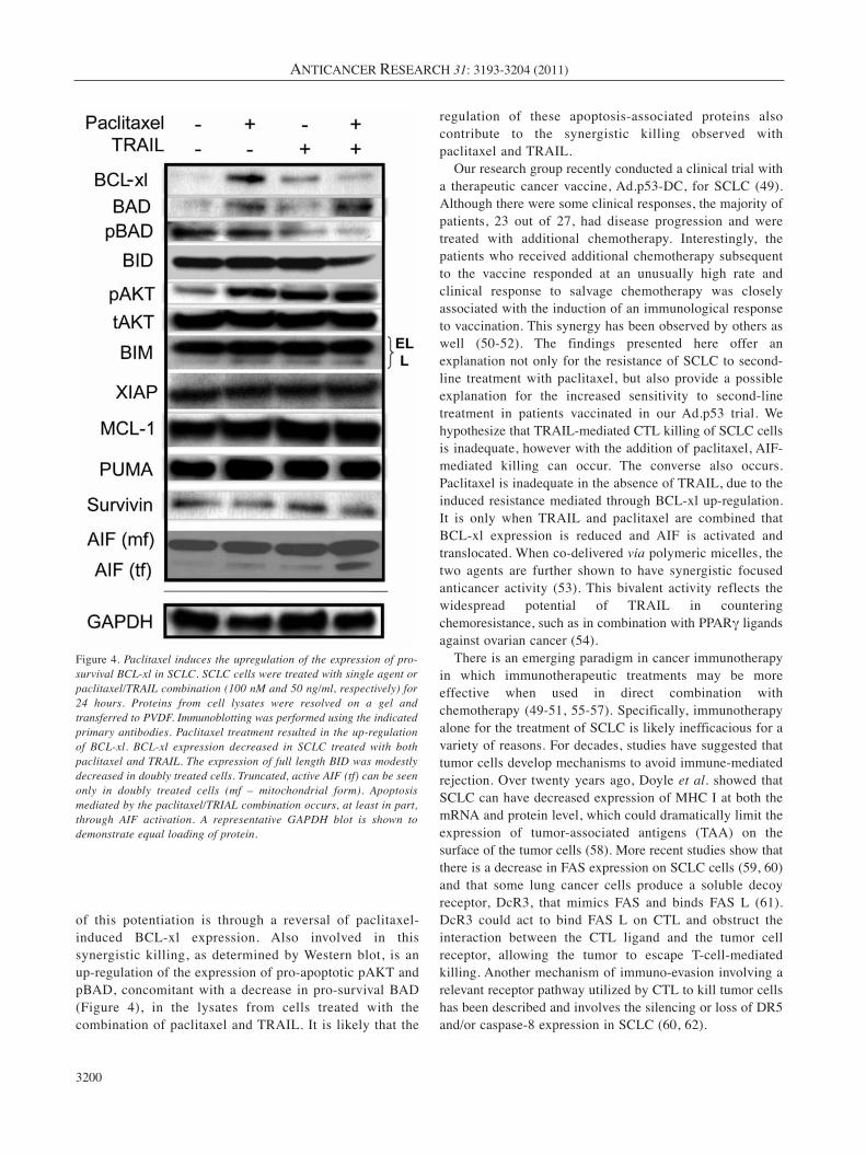
Paclitaxel/TRAIL combination induces AIF-dependentapoptosis in paclitaxel-resistant SCLC cells. Since PARPcleavage did not appear to be crucial in the apoptotic effectmediated by the paclitaxel/TRAIL combination, wedetermined which molecule was likely mediating theultimate DNA fragmentation necessary to complete the cell
death process. After separating mitochondrial and cytosolicproteins and performing immunoblotting, it was observedthat although the mechanism of killing of SCLC by thepaclitaxel/TRAIL combination is mitochondrial dependent,cytochrome c release is not part of the death pathwayinduced by this combination treatment (Figure 5A).Interestingly, the influx of BID, and to a much lesser extentof BAX, into the mitochondria was observed with doublytreated cells as compared to either paclitaxel or TRAILtreatment alone. We also found that apoptosis inducing factor(AIF) was cleaved into a mature form following co-treatmentof SCLC cells with paclitaxel and TRAIL (Figure 4). Furtherinvestigation demonstrated that in PA and TRAIL doubly-treated SCLC cells, AIF does indeed leave the mitochondria,translocate through the cytosol, and localize to the nucleusof the cell where it is able to initiate DNA degradation(Figure 5A-B). No changes in Endo G expression or cellularlocalization were observed (Figure 5A).
Discussion
SCLC is frequently responsive to first-line chemotherapy.However, the response is often short-lived and responserates to second-line therapies are poor. We sought todetermine the mechanism of resistance to paclitaxel andhow this resistance can be overcome. Western blot analysisdemonstrated that BCL-xl expression increases in responseto paclitaxel, suggesting that this is a mechanism by whichSCLC cells become resistant to this common second-linechemotherapy agent. Additionally, it was determined thatbiologically relevant doses of paclitaxel induced thesurface expression of DR4 and DR5. Although there wasnot a robust increase in surface expression of the DR, inthe case of DR5, the expression went from none to dim asdetected by fluorescently labelled antibodies. Addition ofthe DR4/DR5 ligand, TRAIL, lead to the abrogation ofpaclitaxel-induced BCL-xl expression, and also resulted ina decrease in the expression of the pro-survival proteinpBAD (47) and activation of pro-apoptotic BID. CleavedBID translocates to mitochondria and induces cytochromec release and mitochondrial damage (48). The combinationalso resulted in increased apoptosis of SCLC cells asdetermined by both Annexin V staining and chromatincondensation. With this treatment combination, theinduced apoptosis was mediated, at least in part, throughAIF and was independent of significant caspase activationand increases in PARP (Poly (ADP-ribose) polymerase)cleavage. The lack of caspase involvement in killingmediated by paclitaxel has been reported elsewhere forother cancer types, such as NSCLC (30). However, we nowreport that the addition of TRAIL to paclitaxel canpotentiate apoptosis in a paclitaxel-resistant SCLC cellline, as well as the novel observation that the mechanism
Hunter et al: PA and TRAIL Synergize to Kill SCLC Cells
3197
ANTICANCER RESEARCH 31: 3193-3204 (2011)
3198
Figure 2. Paclitaxel and TRAIL synergize to kill SCLC cells. A: Cells were treated in the presence or absence of paclitaxel (100 nM) for 20 hours.Where indicated, 50 ng/ml TRAIL was added for the final 1-4 hours of culture. Apoptosis was also analyzed using Annexin V staining. A significantincrease in Annexin staining was observed, as indicated by the histogram shift to the right. B: Cells were cultured as above, harvested, and then stainedwith acridine orange to visualize chromatin. Live cells have intact nuclei, whereas apoptotic cells have fragmented and condensed nuclear content.

Hunter et al: PA and TRAIL Synergize to Kill SCLC Cells
3199
Figure 3. SCLC apoptosis induced by paclitaxel/TRAIL. SCLC apoptosis induced by paclitaxel/TRAIL combination is concomitant with loss ofmitochondrial membrane potential and independent of caspase activation. A: Cells were treated with or without paclitaxel (100 nM) for 20 hours.Where indicated, 50 ng/ml TRAIL was added for the final 1-4 hours of culture. Cells were harvested and stained using MitoCasp reagents. Followingpaclitaxel, TRAIL, or combined treatment there was a loss ofmitochondrial membrane potential. However, active caspases were not identified. Jurkatcells treated with cycloheximide and TRAIL were used as a positive control (data not shown). B: Western blot analysis was performed to verify theflow cytometric anlaysis of caspase activation: 100 μg of protein was loaded per well of the SCLC lysates, 50 μg of control Jurkat lysate(cycloheximide and TRAIL treated) was loaded. SCLC cells have lower levels of caspase-3 and -9 expression than do JurkaT-cells and no changesin cleaved caspases were observed in extracts from SCLC cells left untreated or treated with either paclitaxel, TRAIL, or the combined treatment.A representative GAPDH blot is shown to demonstrate equal protein loading.
of this potentiation is through a reversal of paclitaxel-induced BCL-xl expression. Also involved in thissynergistic killing, as determined by Western blot, is anup-regulation of the expression of pro-apoptotic pAKT andpBAD, concomitant with a decrease in pro-survival BAD(Figure 4), in the lysates from cells treated with thecombination of paclitaxel and TRAIL. It is likely that the
regulation of these apoptosis-associated proteins alsocontribute to the synergistic killing observed withpaclitaxel and TRAIL.
Our research group recently conducted a clinical trial witha therapeutic cancer vaccine, Ad.p53-DC, for SCLC (49).Although there were some clinical responses, the majority ofpatients, 23 out of 27, had disease progression and weretreated with additional chemotherapy. Interestingly, thepatients who received additional chemotherapy subsequentto the vaccine responded at an unusually high rate andclinical response to salvage chemotherapy was closelyassociated with the induction of an immunological responseto vaccination. This synergy has been observed by others aswell (50-52). The findings presented here offer anexplanation not only for the resistance of SCLC to second-line treatment with paclitaxel, but also provide a possibleexplanation for the increased sensitivity to second-linetreatment in patients vaccinated in our Ad.p53 trial. Wehypothesize that TRAIL-mediated CTL killing of SCLC cellsis inadequate, however with the addition of paclitaxel, AIF-mediated killing can occur. The converse also occurs.Paclitaxel is inadequate in the absence of TRAIL, due to theinduced resistance mediated through BCL-xl up-regulation.It is only when TRAIL and paclitaxel are combined thatBCL-xl expression is reduced and AIF is activated andtranslocated. When co-delivered via polymeric micelles, thetwo agents are further shown to have synergistic focusedanticancer activity (53). This bivalent activity reflects thewidespread potential of TRAIL in counteringchemoresistance, such as in combination with PPARγ ligandsagainst ovarian cancer (54).
There is an emerging paradigm in cancer immunotherapyin which immunotherapeutic treatments may be moreeffective when used in direct combination withchemotherapy (49-51, 55-57). Specifically, immunotherapyalone for the treatment of SCLC is likely inefficacious for avariety of reasons. For decades, studies have suggested thattumor cells develop mechanisms to avoid immune-mediatedrejection. Over twenty years ago, Doyle et al. showed thatSCLC can have decreased expression of MHC I at both themRNA and protein level, which could dramatically limit theexpression of tumor-associated antigens (TAA) on thesurface of the tumor cells (58). More recent studies show thatthere is a decrease in FAS expression on SCLC cells (59, 60)and that some lung cancer cells produce a soluble decoyreceptor, DcR3, that mimics FAS and binds FAS L (61).DcR3 could act to bind FAS L on CTL and obstruct theinteraction between the CTL ligand and the tumor cellreceptor, allowing the tumor to escape T-cell-mediatedkilling. Another mechanism of immuno-evasion involving arelevant receptor pathway utilized by CTL to kill tumor cellshas been described and involves the silencing or loss of DR5and/or caspase-8 expression in SCLC (60, 62).
ANTICANCER RESEARCH 31: 3193-3204 (2011)
3200
Figure 4. Paclitaxel induces the upregulation of the expression of pro-survival BCL-xl in SCLC. SCLC cells were treated with single agent orpaclitaxel/TRAIL combination (100 nM and 50 ng/ml, respectively) for24 hours. Proteins from cell lysates were resolved on a gel andtransferred to PVDF. Immunoblotting was performed using the indicatedprimary antibodies. Paclitaxel treatment resulted in the up-regulationof BCL-xl. BCL-xl expression decreased in SCLC treated with bothpaclitaxel and TRAIL. The expression of full length BID was modestlydecreased in doubly treated cells. Truncated, active AIF (tf) can be seenonly in doubly treated cells (mf – mitochondrial form). Apoptosismediated by the paclitaxel/TRIAL combination occurs, at least in part,through AIF activation. A representative GAPDH blot is shown todemonstrate equal loading of protein.
Additionally, it has recently been shown that there is an up-regulation of BCL-2 in SCLC and that this increasedexpression potentiates chemotherapy resistance in SCLC celllines (63), possibly by blocking TRAIL-induced apoptosis(64). Interestingly, each of these described mechanisms of
tumor immuno-evasion relate directly to pathways utilized byCTL to recognize and induce apoptosis of tumor cells.Emerging literature, in concordance with our data representedherein, points to a need to consider combining chemotherapywith immunotherapy. Understanding the mode of actions and
Hunter et al: PA and TRAIL Synergize to Kill SCLC Cells
3201
Figure 5. Paclitaxel and TRAIL synergize to kill SCLC cells. Killing of SCLC cells by paclitaxel with TRAIL is independent of cytochrome c releaseand appears to be mediated through the translocation and ultimate nuclear localization of AIF. A: Mitochondrial and cytosolic fractions of SCLCtreated as indicated were resolved on polyacrylamide gels, transferred and then subjected to immunoblotting with the indicated antibodies. Mostnotably, pro-apoptotic BID was demonstrated to leave the cytosol and enter the mitochondria, cytochrome c release from the mitochondria intocytosol was not observed, and AIF translocated into the cytosol following treatment with paclitaxel and TRAIL. GAPDH and CoxIV were used asloading controls for the cytosolic and mitochondrial fractions, respectively. B. AIF was also determined to localize in the nucleus of doubly-treatedcells. Briefly, treated cells were stained with FITC- labeled AIF and DAPI. In untreated as well as singly treated cells (data not shown) AIF did notco-localize in the nucleus, however in doubly treated cells, AIF was found in the nucleus.
the pharmaco-immunologic effects of chemotherapeutic drugswill allow for thoughtful development of combinationtreatment strategies, specifically with immunotherapeuticregimens, to treat cancer.
Competing Interests
The Authors declare that they have no competing interests.
Authors’ Contribution
TBH participated in conception of the experiments and projectdesign, performed cell death assays, cell fractionation, andmicroscopy, and drafted the manuscript. NM, KL and TC performedWestern blot analyses. SJA assisted in conception of the project anddesign of the experiments and assisted in drafting of the manuscript.All authors have read and approved the final manuscript.
Acknowledgements
We thank and acknowledge the Flow Cytometry and the AnalyticalMicroscopy core facilities at the Moffitt Cancer Center. This workwas supported by a Flight Attendant Medical Research Insitute(FAMRI) Young Clinical Scientist Award (TBH).
References
1 Schiller JH, Adak S, Cella D, DeVore RF, 3rd and Johnson DH:Topotecan versus observation after cisplatin plus etoposide inextensive-stage small-cell lung cancer: E7593 – a phase III trialof the Eastern Cooperative Oncology Group. J Clin Oncol 19(8):2114-2122, 2001.
2 Masters GA, Declerck L, Blanke C et al: Phase II trial ofgemcitabine in refractory or relapsed small-cell lung cancer:Eastern Cooperative Oncology Group Trial 1597. J Clin Oncol21(8): 1550-1555, 2003.
3 Pujol JL, Daures JP, Riviere A et al: Etoposide plus cisplatin withor without the combination of 4’-epidoxorubicin pluscyclophosphamide in treatment of extensive small-cell lungcancer: a French Federation of Cancer Institutes multicenter phaseIII randomized study. J Natl Cancer Inst 93(4): 300-308, 2001.
4 Urban T, Chastang C, Lebas FX et al: The addition of cisplatinto cyclophosphamide-doxorubicin-etoposide combinationchemotherapy in the treatment of patients with small cell lungcarcinoma: A randomized study of 457 patients. "PetitesCellules" Group. Cancer 86(11): 2238-2245, 1999.
5 Eckardt JR: Emerging role of weekly topotecan in recurrentsmall cell lung cancer. Oncologist 9(Suppl 6): 25-32, 2004.
6 Sandler AB: Irinotecan in small-cell lung cancer: the USexperience. Oncology (Williston Park) 15(1 Suppl 1): 11-12, 2001.
7 Ardizzoni A, Manegold C, Debruyne C et al: Europeanorganization for research and treatment of cancer (EORTC)08957 phase II study of topotecan in combination with cisplatinas second-line treatment of refractory and sensitive small celllung cancer. Clin Cancer Res 9(1): 143-150, 2003.
8 Eckardt JR: Second-line treatment of small-cell lung cancer. Thecase for systemic chemotherapy. Oncology (Williston Park)17(2): 181-188, 91; discussion 91-2, passim, 2003.
9 Amos LA and Lowe J: How Taxol stabilises microtubulestructure. Chem Biol 6(3): R65-69, 1999.
10 Crown J and O’Leary M: The taxanes: an update. Lancet355(9210): 1176-1178, 2000.
11 Vikhanskaya F, Vignati S, Beccaglia P et al: Inactivation of p53in a human ovarian cancer cell line increases the sensitivity topaclitaxel by inducing G2/M arrest and apoptosis. Exp Cell Res241(1): 96-101, 1998.
12 Yoo YD, Park JK, Choi JY et al: CDK4 down-regulation inducedby paclitaxel is associated with G1 arrest in gastric cancer cells.Clin Cancer Res 4(12): 3063-3068, 1998.
13 Jordan MA, Toso RJ, Thrower D and Wilson L: Mechanism ofmitotic block and inhibition of cell proliferation by taxol at lowconcentrations. Proc Natl Acad Sci USA 90(20): 9552-9556,1993.
14 Foss M, Wilcox BWL, Alsop GB and Zhang D: Taxol CrystalsCan Masquerade as Stabilized Microtubules. PLoS ONE 3(1):e1476, 2008.
15 Rodi DJ, Janes RW, Sanganee HJ, Holton RA, Wallace BA andMakowski L: Screening of a library of phage-displayed peptidesidentifies human bcl-2 as a taxol-binding protein. J Mol Biol285(1): 197-203, 1999.
16 Rodi DJ and Makowski L: Similarity between the sequences oftaxol-selected peptides and the disordered loop of the anti-apoptotic protein, Bcl-2. Pac Symp Biocomput, pp. 532-541,1999.
17 Cao L, Sun D, Cruz T, Moscarello MA, Ludwin SK and WhitakerJN: Inhibition of experimental allergic encephalomyelitis in theLewis rat by paclitaxel. J Neuroimmunol 108(1-2): 103-111, 2000.
18 Tsavaris N, Kosmas C, Vadiaka M, Kanelopoulos P andBoulamatsis D: Immune changes in patients with advancedbreast cancer undergoing chemotherapy with taxanes. Br JCancer 87(1): 21-27, 2002.
19 Javeed A, Ashraf M, Ghafoor A and Mukhtar MM: Paclitaxeland immune system. Eur J Pharm Sci 2009.
20 Earnshaw WC, Martins LM and Kaufmann SH: Mammaliancaspases: structure, activation, substrates, and functions duringapoptosis. Annu Rev Biochem 68: 383-424, 1999.
21 Lamarca V and Scorrano L: When separation means death:killing through the mitochondria, but starting from theendoplasmic reticulum. EMBO J 28(12): 1681-1683, 2009.
22 Adams JM and Cory S: The Bcl-2 protein family: arbiters of cellsurvival. Science 281(5381): 1322-1326, 1998.
23 Antignani A and Youle RJ: How do Bax and Bak lead topermeabilization of the outer mitochondrial membrane? CurrOpin Cell Biol 18(6): 685-689, 2006.
24 Kayagaki N, Yamaguchi N, Nakayama M, Eto H, Okumura Kand Yagita H: Type I Interferons (IFNs) Regulate TumorNecrosis Factor-related Apoptosis-inducing Ligand (TRAIL)Expression on Human T-cells: A Novel Mechanism for theAntitumor Effects of Type I IFNs. J Exp Med 189(9): 1451-1460, 1999.
25 Carrington PE, Sandu C, Wei Y et al: The structure of FADDand its mode of interaction with procaspase-8. Mol Cell 22(5):599-610, 2006.
26 Kaufmann M, Bozic D, Briand C et al: Identification of a basicsurface area of the FADD death effector domain critical forapoptotic signaling. FEBS Lett 527(1-3): 250-254, 2002.
27 Boldin MP, Varfolomeev EE, Pancer Z, Mett IL, Camonis JHand Wallach D: A novel protein that interacts with the death
ANTICANCER RESEARCH 31: 3193-3204 (2011)
3202
domain of Fas/APO1 contains a sequence motif related to thedeath domain. J Biol Chem 270(14): 7795-7798, 1995.
28 Peter ME: The TRAIL DISCussion: It is FADD and caspase-8!Cell Death Differ 7(9): 759-760, 2000.
29 Huang Y, Sheikh MS, Fornace AJ Jr. and Holbrook NJ: Serineprotease inhibitor TPCK prevents Taxol-induced cell death andblocks c-Raf-1 and Bcl-2 phosphorylation in human breastcarcinoma cells. Oncogene 18(23): 3431-3439, 1999.
30 Huisman C, Ferreira CG, Broker LE et al: Paclitaxel triggers celldeath primarily via caspase-independent routes in the non-smallcell lung cancer cell line NCI-H460. Clin Cancer Res 8(2): 596-606, 2002.
31 Ofir R, Seidman R, Rabinski T et al: Taxol-induced apoptosis inhuman SKOV3 ovarian and MCF7 breast carcinoma cells iscaspase-3 and caspase-9 independent. Cell Death Differ 9(6):636-642, 2002.
32 Park SJ, Wu CH, Gordon JD, Zhong X, Emami A and Safa AR:Taxol induces caspase-10-dependent apoptosis. J Biol Chem279(49): 51057-51067, 2004.
33 Wieder T, Essmann F, Prokop A et al: Activation of caspase-8 indrug-induced apoptosis of B-lymphoid cells is independent ofCD95/Fas receptor-ligand interaction and occurs downstream ofcaspase-3. Blood 97(5): 1378-1387, 2001.
34 Blagosklonny MV, Schulte T, Nguyen P, Trepel J and NeckersLM: Taxol-induced apoptosis and phosphorylation of Bcl-2protein involves c-Raf-1 and represents a novel c-Raf-1 signaltransduction pathway. Cancer Res 56(8): 1851-1854, 1996.
35 Berndtsson M, Konishi Y, Bonni A et al: Phosphorylation ofBAD at Ser-128 during mitosis and paclitaxel-induced apoptosis.FEBS Letters 579(14): 3090, 2005.
36 Li R, Moudgil T, Ross HJ and Hu HM: Apoptosis of non-small-cell lung cancer cell lines after paclitaxel treatment involves theBH3-only proapoptotic protein Bim. Cell Death Differ 12(3):292-303, 2005.
37 Nimmanapalli R, Perkins CL, Orlando M, O’Bryan E, Nguyen Dand Bhalla KN: Pretreatment with Paclitaxel Enhances Apo-2Ligand/Tumor Necrosis Factor-related Apoptosis-inducingLigand-induced Apoptosis of Prostate Cancer Cells by InducingDeath Receptors 4 and 5 Protein Levels. Cancer Res 61(2): 759-763, 2001.
38 Blagosklonny MV and Fojo T: Molecular effects of paclitaxel:myths and reality (a critical review). Int J Cancer 83(2): 151-156, 1999.
39 Bepler G, Jaques G, Koehler A, Gropp C and Havemann K:Markers and characteristics of human SCLC cell lines.Neuroendocrine markers, classical tumor markers, andchromosomal characteristics of permanent human small celllung cancer cell lines. J Cancer Res Clin Oncol 113(3): 253-259, 1987.
40 Bepler G, Jaques G, Neumann K, Aumuller G, Gropp C andHavemann K: Establishment, growth properties, andmorphological characteristics of permanent human small celllung cancer cell lines. J Cancer Res Clin Oncol 113(1): 31-40,1987.
41 Ribble D, Goldstein N, Norris D and Shellman Y: A simpletechnique for quantifying apoptosis in 96-well plates. BMCBiotechnology 5(1): 12, 2005.
42 Casares N, Pequignot MO, Tesniere A et al: Caspase-dependentimmunogenicity of doxorubicin-induced tumor cell death. J ExpMed 202(12): 1691-701, 2005.
43 Rosenberg SA: Progress in human tumour immunology andimmunotherapy. Nature 411(6835): 380-384, 2001.
44 Park S-M, Schickel R and Peter ME: Nonapoptotic functions ofFADD-binding death receptors and their signaling molecules.Current Opinion in Cell Biology 17(6): 610, 2005.
45 Huisman C, Ferreira CG, Broker LE et al: Paclitaxel TriggersCell Death Primarily via Caspase-independent Routes in theNon-Small Cell Lung Cancer Cell Line NCI-H460. Clin CancerRes 8(2): 596-606, 2002.
46 Zhou H, Li X-M, Meinkoth J and Pittman RN: Akt RegulatesCell Survival and Apoptosis at a Postmitochondrial Level. J CellBiol 151(3): 483-494, 2000.
47 Zha J, Harada H, Yang E, Jockel J and Korsmeyer SJ: Serinephosphorylation of death agonist BAD in response to survivalfactor results in binding to 14-3-3 not BCL-X(L). Cell 87(4):619-628, 1996.
48 Luo X, Budihardjo I, Zou H, Slaughter C and Wang X: Bid, aBcl2 interacting protein, mediates cytochrome c release frommitochondria in response to activation of cell surface deathreceptors. Cell 94(4): 481-490, 1998.
49 Antonia SJ, Mirza N, Fricke I et al: Combination of p53 cancervaccine with chemotherapy in patients with extensive stage smallcell lung cancer. Clin Cancer Res 12(3 Pt 1): 878-887, 2006.
50 Wheeler CJ, Das A, Liu G, Yu JS and Black KL: Clinicalresponsiveness of glioblastoma multiforme to chemotherapyafter vaccination. Clin Cancer Res 10(16): 5316-5326, 2004.
51 Gribben JG, Ryan DP, Boyajian R et al: Unexpected associationbetween induction of immunity to the universal tumor antigenCYP1B1 and response to next therapy. Clin Cancer Res 11(12):4430-4436, 2005.
52 Arlen PM, Gulley JL, Parker C et al: A randomized phase IIstudy of concurrent docetaxel plus vaccine versus vaccine alonein metastatic androgen-independent prostate cancer. Clin CancerRes 12(4): 1260-1269, 2006.
53 Lee AL WY, Pervaiz S, Fan W and Yang YY: Synergisticanticancer effects achieved by co-delivery of TRAIL andpaclitaxel using cationic polymeric micelles. Macromol Biosci11(2): 296-307, 2011.
54 Bräutigam K B-WJ, Bauerschlag DO, von Kaisenberg CS, JonatW, Maass N, Arnold N and Meinhold-Heerlein I: Combinedtreatment with TRAIL and PPARγ ligands overcomeschemoresistance of ovarian cancer cell lines. J Cancer Res ClinOncol pp. 1-12, 2010.
55 Lake RA and Robinson BW: Immunotherapy and chemotherapy– a practical partnership. Nat Rev Cancer 5(5): 397-405, 2005.
56 Gabrilovich DI: Combination of chemotherapy andimmunotherapy for cancer: a paradigm revisited. Lancet Oncol8(1): 2-3, 2007.
57 Andersen MH, Sorensen RB, Schrama D, Svane IM, Becker JCand Thor Straten P: Cancer treatment: the combination ofvaccination with other therapies. Cancer Immunol Immunother57(11): 1735-1743, 2008.
58 Doyle A, Martin WJ, Funa K et al: Markedly decreasedexpression of class I histocompatibility antigens, protein, andmRNA in human small-cell lung cancer. J Exp Med 161(5):1135-1151, 1985.
59 Viard-Leveugle I, Veyrenc S, French LE, Brambilla C andBrambilla E: Frequent loss of Fas expression and function inhuman lung tumours with overexpression of FasL in small celllung carcinoma. J Pathol 201(2): 268-277, 2003.
Hunter et al: PA and TRAIL Synergize to Kill SCLC Cells
3203

60 Shivapurkar N, Reddy J, Matta H et al: Loss of expression ofdeath-inducing signaling complex (DISC) components in lungcancer cell lines and the influence of MYC amplification.Oncogene 21(55): 8510-8514, 2002.
61 Pitti RM, Marsters SA, Lawrence DA et al: Genomicamplification of a decoy receptor for FAS Ligand in lung andcolon cancer. Nature 396(6712): 699-703, 1998.
62 Hopkins-Donaldson S, Ziegler A, Kurtz S et al: Silencing ofdeath receptor and caspase-8 expression in small cell lungcarcinoma cell lines and tumors by DNA methylation. CellDeath Differ 10(3): 356-364, 2003.
63 Sartorius UA and Krammer PH: Up-regulation of Bcl-2 is involvedin the mediation of chemotherapy resistance in human small celllung cancer cell lines. Int J Cancer 97(5): 584-592, 2002.
64 Sun S-Y, Yue P, Zhou J-Y et al: Overexpression of Bcl2 BlocksTNF-Related Apoptosis-Inducing Ligand (TRAIL)-InducedApoptosis in Human Lung Cancer Cells. Biochemical andBiophysical Research Communications 280(3): 788, 2001.
Received March 31, 2011Revised August 4, 2011
Accepted August 5, 2011
ANTICANCER RESEARCH 31: 3193-3204 (2011)
3204