paediatric interstitial lung disease - bpold child erm chapter.pdf · chapter 17 paediatric...
TRANSCRIPT

CHAPTER 17
Paediatric interstitial lung disease
A. Bush, A.G. Nicholson
Imperial College and Royal Brompton Hospital, London, UK.
Correspondence: A. Bush, Dept of Paediatric Respiratory Medicine, Royal Brompton Hospital, SydneyStreet, London, SW3 6NP, UK. E-mail: [email protected]
Interstitial lung disease (ILD) in children (chILD) is very different in many aspects tothe adult disease. First, chILD is rare, estimated at 0.36 per 100,000, compared with 60–80 per 100,000 for ILD in adults [1]. Secondly, the spectrum of conditions, in particularin infancy, is much wider than in adults. The conditions encompass growth anddevelopmental issues, as well as immunological problems. The consequence is thatpaediatricians are even less advanced than adult physicians when it comes to makingdiagnoses by radiology and bronchoalveolar lavage (BAL), and this, combined with therarity of the conditions, means that there have been no randomised controlled trials oftreatment. Thus, chILD is very much work in progress. However, chILD is a reallyimportant topic for adult chest physicians; some of the paediatric diseases may in factpresent in adult life, and if diagnostic awareness is not heightened, patients maydisappear into a dustbin category such as usual interstitial pneumonia (UIP).Furthermore, rare genetic abnormalities may lead to an understanding of modifiergenes important in adult ILD. In this regard, it is a pity that recent guidelines saw fit toignore chILD altogether [2]. This chapter will first review recent advances in theclassification of ILD in children, and then discuss presentation, diagnosis anddifferential diagnosis, as well as what little is known about treatment options.
Classification of ILD in children
There are two published classifications [3, 4], and a third is still only reported as anabstract [5]. The definitive classification in the 0–2-yr age range is from North America[4], and this is recommended for adoption. The European Respiratory Society (ERS)Task Force also contained data in the young age group, but mainly focused on 0–18 yrs[3], and the abstract from North America [5] is in children aged 2–16 yrs; this showed avery different spectrum of chILD compared with infants. The full publication is eagerlyawaited at the time of writing.
ILD in infants aged 0–2 yrs
The antenatal period and the first 2 yrs of life are crucial in long-term lung health, andthere is a clinical logic as well as data to suggest considering this time period separatelyfrom the 2–16-yr age range, although the cut-off is not completely clear. For example,surfactant protein gene disorders, particularly Sp-B and ABCA3, commonly present inthe newborn period, but may present later in childhood or even in adult life as well (seebelow).
Eur Respir Mon, 2009, 46, 319–354. Printed in UK - all rights reserved. Copyright ERS Journals Ltd 2009; European Respiratory Monograph;ISSN 1025-448x.
319
This early time period is characterised by rapid growth of the airways, and particularlyof the alveolar-capillary membrane, the maturing of the immune system, and encounterswith new infectious, allergic and chemical challenges. The exact nature of the growthfactors that drive the growth and maturation of the lung are ill understood, but areprobably unique to this early time period. The immune system normally shows a changefrom the pregnancy-associated T-helper cell (Th) type 2 to a neonatal Th1 bias [6], and theinfant has to switch from reliance on maternal humoral immunity during pregnancy to thedevelopment of immune responses and immune memory functions. Novel infective andallergic proteins are encountered, and acid reflux is common; pollution, including tobaccosmoke exposure, will also impinge on the newborn respiratory system. The importance ofa developmental perspective is shown by the study of kindreds with Sp-C deficiency [7]. Inadult life, this is manifest by a pattern of UIP, but the same gene defect presenting ininfancy causes a cellular nonspecific interstitial pneumonia (NSIP). One could speculatethat other apparently exclusively paediatric conditions, such as pulmonary interstitialglycogenosis (PIG) and neuroendocrine cell hyperplasia of infancy (NEHI), may in factrepresent the response of the immature lung to insults that in adult life might cause a verydifferent pattern of ILD.
The North American chILD group have recently proposed dividing ILD in the 0–2-yrage group into eight categories (see below) [4]. The classification was based on 187biopsies (of which 22 were unclassifiable) from 11 institutions over a 5-yr period. Thestrengths of the classification include the large number of cases reported and theindependent pathological verification of the diagnoses. Ongoing issues include that ittakes no account of what are the (admittedly rare) diseases that may not come to biopsy,for example idiopathic pulmonary haemosiderosis (IPH); and the need to validate theclassification in a second population [8]. The classification might also be criticised asalmost too broad, because it also encompasses diseases with a major airway component,such as obliterative bronchiolitis, and conditions in which there is usually no diagnosticdoubt, such as bronchopulmonary dysplasia; perhaps ‘‘diffuse distal lung disease’’ mightbe a better term, but the term ‘‘chILD’’ is in fact probably here to stay. As in theclassification of adult ILD, where organising pneumonia (a predominantly alveolarfilling disorder) is included primarily as it enters the differential diagnosis of ILD, agreater number of non-interstitial disorders are included in the chILD classification, asthese entities enter into the pre-operative differential diagnosis due to the lowersensitivity of investigative procedures, such as high-resolution computed tomography(HRCT), in children. Table 1 is a summary of the classification; each section is discussedin more detail below. Diagnoses made in a partially overlapping age group reported inthe ERS Task Force (table 2) [3] included infection with Pneumocystis, Epstein–Barrvirus and respiratory syncytial virus; desquamative interstitial pneumonia (DIP),lymphoid interstitial pneumonia (LIP), NSIP and unclassified fibrosis; and some ILDscaused by an associated disease, such as alveolar proteinosis (unspecified), systemiclupus erythematosus, histiocytosis and aspiration. The ERS Task Force represented alarge survey, but there was no independent validation of the pathological diagnoses, andit would seem that molecular studies were infrequently performed.
Category one: diffuse developmental disorders. The first two categories, ‘‘diffusedevelopmental disorders’’ and ‘‘growth abnormalities reflecting deficient alveolarisa-tion’’ must surely be overlapping, since in practice growth and development are hard toseparate [8]. They are, however, considered separately in the chILD group classification,and hence in this chapter. Diffuse developmental disorders are believed to be due todefects in one of the primary molecular mechanisms of the lung (and/or pulmonaryvascular development, presumably on a molecular basis); they include acinar dysplasia,
A. BUSH AND A.G. NICHOLSON
320

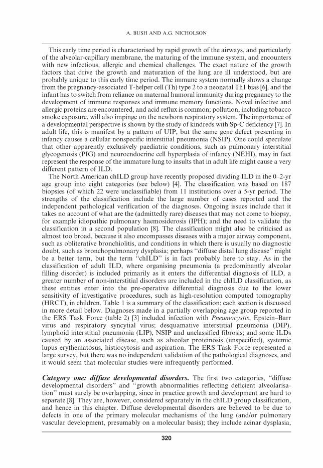
congenital alveolar dysplasia, and alveolar-capillary dysplasia with misalignment of thepulmonary veins (ACDMPV). Acinar dysplasia in pure form is characterised by lunggrowth arrest in the pseudoglandular or early canalicular phase and congenital alveolardysplasia by growth arrest in the late canalicular or early saccular phase. However, arecent paper has stressed that overlap conditions are common [9]. The constellation ofmalposition of pulmonary veins adjacent to small pulmonary arteries, medialhypertrophy of pulmonary arteries and arterioles, and reduced capillary density withlobular maldevelopment was considered diagnostic for ACDMPV (fig. 1). DEUTSCH etal. [4] had biopsies in term infants who presented at birth with therapy-unresponsive
Table 1. – Classification of interstitial lung disease (ILD) in children aged 0–2 yrs
Category Illustrative diseases
1 Diffuse developmental disorders (n511) Acinar dysplasia (n51)Congenital alveolar-capillary dysplasia (n52)Alveolar-capillary dysplasia with misalignment ofthe pulmonary veins (n58)
2 Growth abnormalities reflecting deficientalveolarisation (n546)
Pulmonary hypoplasia (n57)Chronic neonatal lung disease (bronchopulmonarydysplasia) (n520)
Related to chromosomal disorders (n515)Related to congenital heart disease (n54)
3 Specific conditions of undefined aetiology(n524)
Pulmonary interstitial glycogenosis (n518)Neuroendocrine cell hyperplasia of infancy (n56)
4 Surfactant dysfunction disorders (n518) Sp-B gene mutations (n50)Sp-C gene mutations (n57)ABCA3 gene mutations (n56)Histology consistent with surfactant protein disorderbut none detected (n55 in total):Pulmonary alveolar proteinosis (n52)Chronic pneumonitis of infancy (n51)Desquamative interstitial pneumonia (n51)Nonspecific interstitial pneumonia (n51)
5 Disorders of the normal host, presumedimmune intact (n523)
Infectious and post-infectious (n517)Environmental agents (n52 in total):Hypersensitivity pneumonitis (n52)Toxic inhalation (n50)
Aspiration syndromes (n53)Eosinophilic pneumonia (n51)
6 Disorders resulting from systemic diseaseprocesses (n56)
Collagen vascular disease (n54)Storage disease (n51)Sarcoidosis (n50)Langerhans’ cell histiocytosis (n50)Malignant infiltrates (n51)
7 Disorders of the immunocompromised host(n528)
Opportunistic infections (n520)Iatrogenic (n53)Related to transplant and rejection (n50)Diffuse alveolar damage, unknown aetiology (n55)
8 Disorders masquerading as ILD (n59) Arterial hypertensive vasculopathy (n58)Venous engorgement secondary to heart disease (n51)Veno-occlusive disease (n50)Lymphatic disorders (n50)
n5165 interpretable biopsies in total. Data taken from [4].
PAEDIATRIC INTERSTITIAL LUNG DISEASE
321
hypoxia and persistent pulmonary hypertension. One child was transplanted, the restwere dead within a month. MELLY et al. [9] reported a larger group, in which there werefour survivors. Histological features stressed by this group included the likely presenceof PIG cells in 17 out of 21 cases, and the great variety of the degree of misalignment,with higher capillary apposition and density being predictive of a better prognosis.Associated abnormalities, including Down syndrome, were common. Finally, there is asingle case report which describes complete resolution of severe pulmonary hypertensionon sildenafil in a baby who appears to fall into this diagnostic group [10].
Category two: growth abnormalities reflecting deficient alveolarisation. Abnormalalveolar development that is largely secondary is the hallmark of this group. Thisincludes pulmonary hypoplasia due to a small fetal thorax, reduced amniotic fluidvolume, diminished or absent fetal breathing movements, reduced pulmonary bloodflow, abdominal wall defects and chromosomal abnormalities. Post-natally, chroniclung disease of prematurity is in this group. It is arguable whether it is useful to includethese patients in a discussion of ILD, and in most (particularly chronic lung disease ofprematurity), lung biopsy would rightly not be contemplated. The exception might be inchildren with congenital heart disease [11], but the question would be related to theoperability of the abnormality, rather than a lung diagnosis. Histologically, there isvariable lobular simplification with alveolar enlargement, often most prominentsubpleurally. In nearly half, PIG cells were noted (often previously overlooked), andhypertensive pulmonary vasculopathy was common.
Table 2. – Classification of interstitial lung disease based on the European Respiratory Society Task Force [3]
Category Commonest age yrs Diagnoses made
Infection (n519; 14.5%) 3–12 (n510) AdenovirusMycoplasmaPneumocystis
Epstein–Barr virusRespiratory syncytial virus
Influenza A
Associated disease (n551; 38.9%) 6–12 (n517) Hypersensitivity pneumonitisAspiration syndromes
SarcoidosisAlveolar proteinosis
Bronchiolitis obliteransGraft versus host disease
‘‘Chronic disease’’Metabolic disorder
Systemic lupus erythematosusHistiocytosis
GranulomatosisHaemosiderosis
Rheumatoid arthritisVascular disordersLymphatic disorders
Idiopathic (n546; 35.1%) 6–12 (n513) UnclassifiedDesquamative interstitial pneumonia
Usual interstitial pneumoniaNonspecific interstitial pneumoniaLymphoid interstitial pneumonia
Unclassifiable (n514; 10.6%) 6–12 (n55)
A. BUSH AND A.G. NICHOLSON
322

Category three: specific conditions of undefined aetiology. These two conditions(NEHI and PIG) appear to be found purely in infancy; whether they are specificconditions, or related nonspecifically to disordered lung development, is unclear.
Neuroendocrine cell hyperplasia of infancy. The human airway epithelium containshighly specialised pulmonary neuroendocrine cells (PNEC), either alone or asinnervated neuroepithelial bodies. The ‘‘PNEC system’’ comprises both neural andendocrine cell phenotypes, the functions of which include the synthesis and release ofamine (serotonin) and a variety of neuropeptides (such as bombesin) [12]. Bombesincells peak in mid-gestation, and then reduce to the normal adult low levels by term [13].Thus it is possible to hypothesise that NEHI may represent a failure of the normalregression of these cells. The function of the PNEC system in the lung is unknown.Complex roles have been proposed, including modulation of fetal lung growth anddifferentiation and airway oxygen sensors involved in neonatal adaptation at birth.Post-natally, they may provide a lung stem cell niche that is important in airwayepithelial regeneration [14]. Thus, PNEC are a normal part of the lung, and notnecessarily pathological cells. Characteristically, NEHI presents in the first year of life(mean age 3.8 months in the largest published series) with tachypnoea and respiratorydistress, in a relatively well infant [15]. Rare cases of a NEHI-like syndrome have beendescribed in older children, in one case in association with emphysema for which therewas no underlying cause such as a1-antitrypsin deficiency [16]. Cough and wheeze arenot prominent in NEHI [15]. There is a male predominance. Presentation requiringintubation at birth has not been described [4]. Crackles are often heard. The chestradiograph (CXR) typically mimics post-viral infection airway changes. HRCT showspatchy ground-glass opacification, typically centrally and in the right middle lobe andlingula, with air trapping elsewhere. Experienced radiologists may feel sufficientlyconfident to diagnose NEHI on these appearances alone [17], but most paediatricianswould want to proceed to lung biopsy. The pathology is of apparently almost normallung tissue on haematoxylin and eosin staining, but occasionally there may be increased
Fig. 1. – A case of alveolar capillary dysplasia shows poorly developed alveolar walls, within which there is lowcapillary density and poor apposition to the epithelium. Towards the centre, an intra-acinar pulmonary artery showsmarked medial hypertrophy.
PAEDIATRIC INTERSTITIAL LUNG DISEASE
323
airway macrophages, mild smooth muscle hyperplasia, and epithelial clear cells. Thepathological hallmark of NEHI is increased numbers of bombesin-positive airway cells.These are also seen in healthy controls, but the upper limit of normal is 5% of theepithelial area [15]. KL-6 has been proposed to be a useful biomarker distinguishingNEHI from surfactant protein disorders; children with NEHI have normal levels ofKL-6, whereas these are elevated well above the normal range in surfactant proteindisorders, including ABCA3 defects [18]. There is no treatment other than oxygen if thechild is hypoxic, and the long-term outlook is relatively good, with no deaths recorded,and long-term pulmonary function at worst showing mild airflow obstruction. However,up to two-thirds of the children remained symptomatic at follow-up. Thus, parents ofinfants diagnosed with NEHI can probably be reassured as to the prognosis.
Scientifically, the description of bombesin-containing cells is straightforward, but theinterpretation is not. First, the connection between NEHI and chronic idiopathicbronchiolitis of infancy [19] and diffuse idiopathic neuroendocrine cell hyperplasia(DIPNECH) [20] is not clear. Secondly, PNEC have been reported as being increased inmany other conditions, including sudden infant death syndrome [21], bronchopulmon-ary dysplasia [22] and Wilson–Mikity disease [23]. Finally, neuroendocrine cells arenormally seen in the developing lung, and are indeed crucial in normal developmentalprocesses such as branching morphogenesis [12]. It is not clear to us that NEHI is truly aseparate entity, or overlaps with other conditions; or whether the bombesin-containingcells have any pathophysiological significance or are markers of some unknownunderlying problem.
Given these concerns, and given the rarity of NEHI, we continue to recommendinvasive diagnosis where possible, and always that such infants are carefully followedup. The exception to the support for an invasive diagnosis would be a well, thrivinginfant who has only trivial and stable oxygen dependency, and has a typical appearanceon HRCT with nothing to suggest an alternative diagnosis.
However, it should be noted that we still know very little about NEHI, and even thelargest papers are little more than extended case series. The possibility remains thatsome cases of NEHI may evolve into a chronic constrictive bronchiolitis-like picture inlater childhood. We have seen at least one case, diagnosed only on HRCT because thechild was well but tachypnoeic and a biopsy was refused by the family, who appears tohave followed just this course, with later onset of oxygen dependency. Furthermore,although the overall prognosis is usually good, we have seen biopsy-proven NEHIrelapse and require a further period of oxygen dependency. Finally, whether there is anyrelationship between NEHI and the adult condition of adult DIPNECH is not known.
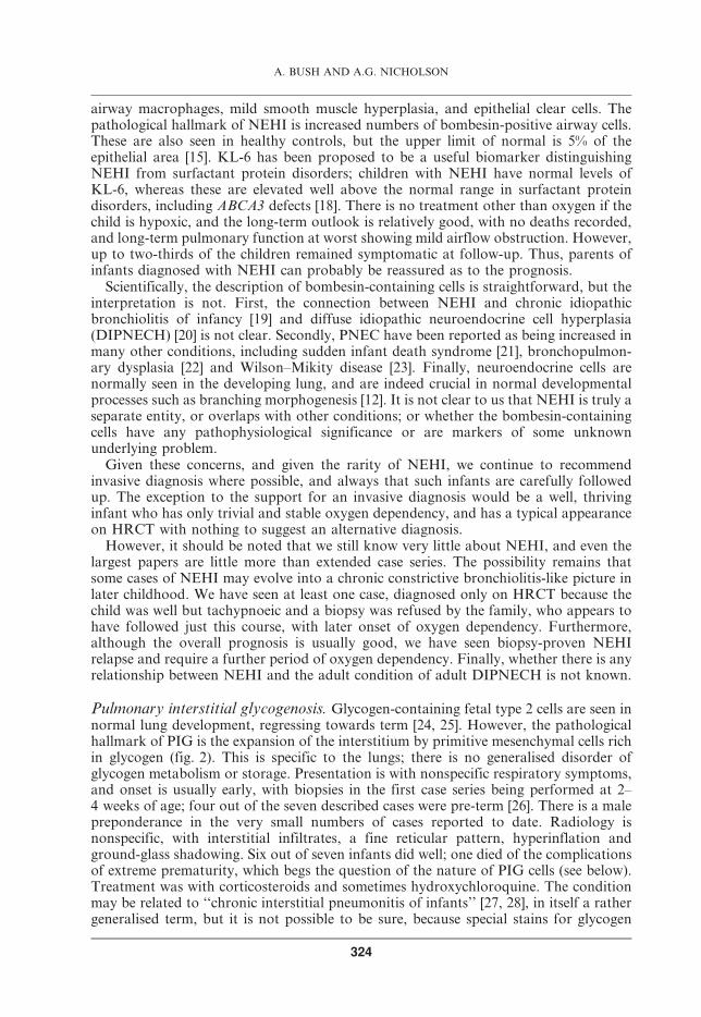
Pulmonary interstitial glycogenosis. Glycogen-containing fetal type 2 cells are seen innormal lung development, regressing towards term [24, 25]. However, the pathologicalhallmark of PIG is the expansion of the interstitium by primitive mesenchymal cells richin glycogen (fig. 2). This is specific to the lungs; there is no generalised disorder ofglycogen metabolism or storage. Presentation is with nonspecific respiratory symptoms,and onset is usually early, with biopsies in the first case series being performed at 2–4 weeks of age; four out of the seven described cases were pre-term [26]. There is a malepreponderance in the very small numbers of cases reported to date. Radiology isnonspecific, with interstitial infiltrates, a fine reticular pattern, hyperinflation andground-glass shadowing. Six out of seven infants did well; one died of the complicationsof extreme prematurity, which begs the question of the nature of PIG cells (see below).Treatment was with corticosteroids and sometimes hydroxychloroquine. The conditionmay be related to ‘‘chronic interstitial pneumonitis of infants’’ [27, 28], in itself a rathergeneralised term, but it is not possible to be sure, because special stains for glycogen
A. BUSH AND A.G. NICHOLSON
324

were not performed in these cases. This underscores the need for protocol-drivenhandling of surgical lung biopsies (see below).
An intriguing report described PIG in pre-term twins [29], with a favourable outcomeassociated with systemic steroid therapy. This report begs the question as to whether 1)there is an undescribed genetic component to PIG, or 2) PIG is part of the spectrum ofchronic lung disease of prematurity, or overlaps with it. However, it should be noted thatnine out of the 16 cases in the published literature were in term infants [4, 26, 29, 30].
As with NEHI, it is pertinent to question the specificity of glycogen-containing cells,and to what extent they are merely a marker for some other process, or indeed whetherPIG is a separate entity. First, glycogen-containing cells are seen at some stages of lungdevelopment [24, 25]. They are found in the early stages of lung development in cellslining the alveolar septa, becoming less prominent with advancing gestational age. Itmust be stressed that in PIG, alveolar lining cells are spared, and it is the mesenchymewhich is affected. Glycogen stores are seen in association with lamellar bodies in fetaltype 2 cells, suggesting a role in surfactant synthesis. Secondly, PIG cells have beenreported in association with congenital lobar emphysema, but not in sufficient quantitiesto cause ILD [31]. There is still much to be learned about the role of these cells and thespectrum of PIG; as with NEHI, even the largest papers are mere extended case series.
Category four: surfactant dysfunction disorders. These illustrate an importantprinciple which may find wide application in genetic disorders. Of the four surfactantproteins known (Sp-A, -B, -C and -D), Sp-A and -D are not surface active, and aremembers of the collectin family, along with mannose-binding lectin. Mutations in Sp-Band Sp-C have been shown to cause ILD (see below). The intracellular processing of theseproteins is of great complexity. ABCA3 encodes for a protein that is not itself surfaceactive but is involved in the processing of pulmonary surfactant [32]. Deficiency producesILD closely resembling Sp-B or Sp-C deficiency (see below). Some of the other conditionsdescribed below, which mimic surfactant protein disorders, may in fact be caused bydefects in other surfactant protein processing genes. It is also interesting to speculate that
Fig. 2. – A case of pulmonary interstitial glycogenosis shows the alveolar interstitium to be expanded bycytologically bland clear cells of uncertain histogenesis.
PAEDIATRIC INTERSTITIAL LUNG DISEASE
325

other apparently single-gene disorders may be caused by gene defects encoding processingproteins. Thus, cystic fibrosis (CF) has been described with apparently no mutation in theCF gene locus [33]. However, CF transmembrane conductance regulator (CFTR)interacts with numerous other proteins [34], and one could speculate that mutations insome or all of these could produce a CF-like disease. Given the complex post-translationalprocessing of the surfactant protein gene products, this may be a relevant mechanism inchILD. It has been suggested as being important in Hermansky–Pudlak syndrome (HPS;see below), which is characterised pathologically by abnormal lamellar bodies, amongother features. It should be noted that, although a family history of ILD should always besought, 50% of patients with one of the three genetic diseases described below have diseaseoccurring de novo. It is likely that the significance of these gene defects and polymorphismsis underappreciated in adult ILD [35].
Sp-B gene mutations. Sp-B deficiency is an autosomal recessive, loss of functionmutation. It is a rare condition, with estimated prevalence being one in a million livebirths [36]. The gene is located on chromosome 2, comprises approximately 10,000 basepairs (bp) in 11 exons (of which only the first 10 are translated), and encodes a 381-amino-acid pre-protein. 23 amino acids are removed co-translationally to produce pro-Sp-B, which then undergoes complex processing to produce the mature protein.Production is primarily by the type 2 cells [37]. The most common mutation is a 2-bpinsertion in codon 121 (121ins2), which accounts for about two-thirds of mutant alleles;more than 30 others have been described [37, 38]. The classical presentation is withrelentlessly progressive respiratory distress, mimicking hyaline membrane disease in thepre-term, with no response to treatment and death within months, unless lungtransplantation can be offered. The pathology is often but not invariably a pulmonaryalveolar proteinosis (PAP)-like picture (fig. 3), but there may be more of a type 2 cellhyperplasia. These infants may also have secondary abnormalities in Sp-C processing,(pro-Sp-C to Sp-C), with poorly organised lamellar bodies [39–41].
Fig. 3. – A case of alveolar proteinosis shows alveolar spaces filled by acellular eosinophilic proteinaceous debris,within which cholesterol clefts can be seen.
A. BUSH AND A.G. NICHOLSON
326
Although the classical disease is lethal in infancy, rare partial deficiencies have beendescribed, with prolonged survival [42, 43]. Furthermore, it is hypothesised thatheterozygosity for Sp-B deficiency, or some Sp-B single nucleotide polymorphisms, mayconfer an increased risk of acute lung injury and oxygen toxicity. Further work is neededto determine whether Sp-B may be a modifier gene for adult respiratory distresssyndrome or chronic respiratory diseases, including inorganic dust exposure [44–47].
Sp-C gene mutations. Sp-C deficiency is an autosomal dominant condition caused by again-of-function mutation (i.e. the disease is produced not by loss of function of thenormal protein, but by an abnormal new function in the mutated protein) [48]. Sporadicdisease has also been reported, about equally frequently with the inherited condition.The Sp-C gene is located on the short arm of chromosome 8, and is transcribed to a 900-bp mRNA, which after post-translational processing yields one of either a 191 or 197amino acid protein. At least 35 mutations have been described [49]. Several have beenshown to reside in the COOH-terminal domain, ay100-amino-acid region known asBRICHOS (group A mutations) [50]. These mutations result in endoplasmic stress dueto accumulation of misfolded protein and ultimately to cell apoptosis via a CASPASE 3and CASPASE 4 pathway, among other intracellular metabolic problems. Group Bmutations are clustered in exon 3, and these result in cytosolic accumulation; the exactmechanism of toxicity has not been determined. A (single) group C mutation has beendescribed in the cytosolic nontransmembrane NH2-domain; this mutant protein fails totraffic to the Golgi. The dominant negative effect of the abnormal Sp-C (failure oftranslation of the normal allele to leads to some normal Sp-C) is attributed to effects onthe trafficking and processing of the abnormal gene product [49].
In addition to being surface active, Sp-C may have other functions, including themodulation of inflammation. It binds to lipopolysaccharide, inhibiting its interactionswith macrophages and CD14. The role of these functions in Sp-C deficiency disease isunclear [49].
The clinical phenotype is extremely variable, and studies of several generations infamilies detect presentation with the same Sp-C mutation in the newborn period withrelentlessly progressive respiratory distress, and onset of UIP in late middle age [7]. Ithas been suggested that ABCA3 mutations may be modifier genes, in part accountingfor the varying clinical features of Sp-C mutations. ABCA3 mutations from foursymptomatic infants with the same Sp-C mutation, I73T, were studied. These infantswere part of a series of 55 children with chILD secondary to Sp-C deficiency. Three outof the four infants were also heterozygous for an ABCA3 mutation inherited from theparent who did not carry I73T (E292V, n52; L212M, n51). This suggests that thecombination was predictive of early onset of lung disease in Sp-C deficiency [51]. Innewborns, the histology may suggest chronic pneumonitis of infancy (CPI; see below),NSIP or DIP [49]. Spontaneous and prolonged remission of the childhood disease hasbeen described. Virtually any histological pattern of ILD can be caused by Sp-Cdeficiency [49]. ILD characterised by absence of mature Sp-C, but no Sp-C genemutations, has been described, and was presumably due to a mutation in a criticalenzyme in the processing pathway [52]. There is no known treatment; corticosteroidsand hydroxychloroquine have been used, but data are at the level of anecdote.
ABCA3 gene mutations. ABCA3 deficiency is an autosomal recessive condition ofunknown prevalence. This very large gene is located on chromosome 16, and contains60,000 bp in 33 exons (the first three of which are not translated) that encode a 1,704-amino-acid protein [53, 54]. The disease is thought to be due to loss of functionmutations. ABCA3 is part of a family of genes, some of which are associated with
PAEDIATRIC INTERSTITIAL LUNG DISEASE
327
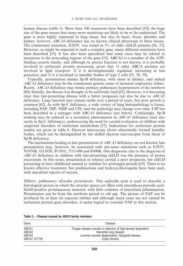
human disease (table 3). More than 100 mutations have been described [55]; the hugesize of the gene means that many more mutations are likely to be so far undetected. Thegene is most highly expressed in lung tissue, but also in heart, brain, platelets andkidney; however, ABCA3 deficiency has no known clinical phenotype in these tissues.The commonest mutation, E292V, was found in 5% of older chILD patients [56, 57].However, as might be expected in such a complex gene, many different mutations havebeen described [55]. It has also been speculated that some cases may be related tomutations in the noncoding regions of the gene [55]. ABCA3 is a member of the ATP-binding cassette family, and although its precise function is not known, it is probablyinvolved in surfactant protein processing, given that 1) other ABCA proteins areinvolved in lipid transport; 2) it is developmentally regulated, increasing in lategestation; and 3) it is localised to lamellar bodies of type 2 cells [55, 58, 59].
Typically, presentation mimics Sp-B deficiency, with onset in infancy, and indeedABCA3 deficiency may be the commonest genetic cause of neonatal respiratory failure.Rarely, ABCA3 deficiency may mimic primary pulmonary hypertension of the newborn[60]. Initially, the disease was thought to be uniformly fatal [61]. However, it is becomingclear that late-presenting disease with a better prognosis can also be due to ABCA3deficiency. Lung function may remain stable over a period of years, but poor growth iscommon [62]. As with Sp-C deficiency, a wide variety of lung histopathology is found,including PAP, DIP, NSIP and CPI, and the pathology may change over time; UIP hasbeen described in a teenager with ABCA3 deficiency (see below). Confusingly, Sp-Bstaining may be reduced as a secondary phenomenon in ABCA3 deficiency (and alsorarely in Sp-C deficiency), underscoring the need for careful evaluation of children withsuspected disorders of surfactant metabolism [57]. Indications for surfactant proteinstudies are given in table 4. Electron microscopy shows abnormally formed lamellarbodies, which can be distinguished by the skilled electron microscopist from those ofSp-B deficiency.
The mechanisms leading to late presentation of ABCA3 deficiency are not known; latepresentation may, however, be associated with mis-sense mutations such as E292V,N1076K, G1302E, P1301L, T1114M and E690K. One diagnostic clue to the diagnosis ofABCA3 deficiency in children with late-presenting chILD was the presence of pectusexcavatum. In this series, presentation in infancy carried a poor prognosis, but chILDpresenting in later childhood seemed to stabilise for prolonged periods [63]. There is noknown effective treatment, but prednisolone and hydroxychloroquine have been used,with anecdotal reports of success.
Others: pulmonary alveolar proteinosis. This umbrella term is used to describe ahistological picture in which the alveolar spaces are filled with amorphous periodic acid-Schiff-positive proteinaceous material, with little evidence of interstitial inflammation.Presentation can be from the newborn period to old age. The picture of PAP can beproduced by at least six separate entities and although many cases are not caused bysurfactant protein gene disorders, it seems logical to consider PAP in this section.
Table 3. – Disease caused by ABCA family members
Gene Disease
ABCA1 Tangier disease (results in reduction of high-density lipoprotein)ABCA3 Interstitial lung diseaseABCA4 Juvenile macular degeneration, Stargardt diseaseABCA7 (CFTR) Cystic fibrosis
A. BUSH AND A.G. NICHOLSON
328

As aforementioned, surfactant protein abnormalities caused by Sp-B, Sp-C andABCA3 mutations may lead to many histological appearances, but in particular to PAP,especially when presentation is in the newborn period.
Granulocyte-macrophage colony-stimulating factor (GM-CSF) receptor abnormal-ities can also lead to PAP. Clearance of surfactant by alveolar macrophages requires afunctional GM-CSF receptor. Mutations in the b- [62, 64] and more recently the a-chainof the GM-CSF receptor have been shown to be a rare cause of PAP [65, 66].
GM-CSF auto-antibody disease is a form of PAP more classically seen in adults [67],but paediatric cases have been described [68]. Diagnosis is confirmed by finding auto-antibodies to GM-CSF in serum or BAL. Initially, this variant of PAP was successfullytreated with serial large-volume lung lavage [69], but increasingly it is now treated withinhaled or systemic GM-CSF [70, 71].
PAP has been rarely described in association with immunodeficiency (agammaglo-bulinaemia) [72]. The exact mechanisms are poorly defined; coincidence cannot beexcluded.
Macrophage blockade can also potentially lead to PAP, and is seen exclusively inadults [73]. Causes include haematological malignancy and inorganic dust inhalation.
PAP may be a manifestation of the metabolic diseases lysinuric protein intolerance orNiemann–Pick disease (the latter only rarely).
Lysinuric protein intolerance is an autosomal recessive condition with multisystemmanifestations, including vomiting, diarrhoea, failure to thrive, hepatosplenomegaly,mental retardation, chronic renal disease and altered immune responses. It is caused bymutations in the solute carrier family 7A member 7 (SLC7A7) gene [74]. The diseasemay present solely with pulmonary manifestations, which may be of an acute life-threatening ILD, or more typically, a PAP-like picture. Successful treatment of the latterby whole lung lavage has been reported [75]. Unfortunately, PAP secondary to lysinuricprotein intolerance recurs after lung transplantation [76].
Niemann–Pick disease is a rare, autosomal recessive lipid storage disease, which ischaracterised by sphingomyelin deposition in a number of tissues due to deficiency ofthe lysosomal enzyme acid sphingomyelinase. It is a systemic disease, but is consideredhere as it enters the differential diagnosis of PAP. There are six known types (A–F) [77].The prevalence of lung disease in this condition is difficult to determine in the absence ofany big series. Types B and C2 are typically associated with restrictive lung disease inolder patients, and in particular, HRCT reveals abnormalities, typically thickenedinterlobular septa, intralobular lines and ground-glass shadowing, in almost all type Bpatients; these do not correlate well with pulmonary function abnormalities [78].Nodules, some of which may be calcified, are also reported. Typically, patients presentwith endogenous lipid pneumonia, with foamy macrophages primarily filling alveolarspaces, but also involving the interstitium [79–83]. An unusual feature in type B diseaseis the presence of lipid accumulation within ciliated respiratory epithelial cells (fig. 4).
Table 4. – Indications for surfactant protein studies
Severe unexplained newborn respiratory distressAny diffuse lung disease on HRCT, particularly if there is a family history of ILDHistopathology reported as showing PAP, NSIP, DIP, UIP or CPIAcute ILD (diffuse alveolar damage) with no obvious aetiologyAbnormal lamellar bodies on electron microscopy in ILDWell adult with family history of ILD or chILD, after genetic counselling
HRCT: high-resolution computed tomography; ILD: interstitial lung disease; PAP: pulmonary alveolarproteinosis; NSIP: nonspecific interstitial pneumonia; DIP: desquamative interstitial pneumonia; UIP: usualinterstitial pneumonia; CPI: chronic pneumonitis of infancy; chILD: ILD in children.
PAEDIATRIC INTERSTITIAL LUNG DISEASE
329
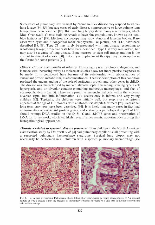
Some cases of pulmonary involvement by Niemann–Pick disease may respond to whole-lung lavage [84, 85], but rare cases of early disease, nonresponsive to large-volume lunglavage, have been described [86]. BAL and lung biopsy show foamy macrophages, whichMay–Grunewald–Giemsa staining reveals to have blue granulations, known as the ‘‘sea-blue histiocytes’’ [87]. Electron microscopy may show abnormal lamellar bodies. Rarecases with cysts and a congenital lobar emphysema-like picture, not ILD, have beendescribed [88, 89]. Type C1 may rarely be associated with lung disease responding towhole-lung lavage; bronchial casts have been described. Type E is very rare indeed, butmay also be a cause of lung disease. Bone marrow or stem cell transplantation is thecurrent treatment of choice [90], but enzyme replacement therapy may be an option inthe future for some patients [91].
Others: chronic pneumonitis of infancy. This category is a histological diagnosis, andis made with increasing rarity as molecular studies allow for more precise diagnoses tobe made. It is considered here because of its relationship with abnormalities ofsurfactant protein metabolism, as aforementioned. The first description of this conditionpredated the understanding of the role of surfactant protein and other genes in chILD.The disease was characterised by marked alveolar septal thickening, striking type 2 cellhyperplasia and an alveolar exudate containing numerous macrophages and foci ofeosinophilic debris (fig. 5). There were primitive mesenchymal cells within the widenedalveolar septa, but little inflammation. CPI occurs only in infants and very youngchildren [92]. Typically, the children were initially well, but respiratory symptomsappeared at the age of 1–9 months, with a fatal course despite treatment [93]. Occasionallong-term survivors have been described [94]. It is likely that many cases in fact hadabnormalities of surfactant protein genes, and certainly a pathological report of CPIshould prompt DNA studies on the Sp-B, -C and ABCA3 genes and preservation ofDNA for future work, which will likely reveal further genetic abnormalities causing thishistopathological appearance.
Disorders related to systemic disease processes. Four children in the North Americanclassification study by DEUTSCH et al. [4] had pulmonary capillaritis, all presenting witha suspected pulmonary haemorrhage syndrome. Surgical lung biopsy may notnecessarily be performed in all children with suspected pulmonary haemorrhage (see
a) b)
Fig. 4. – a) A case of Niemann–Pick disease shows filling of alveolar spaces by foamy macrophages. b) An unusualfeature of type B disease is that the presence of fine intracytoplasmic vacuolation is also seen in the ciliated epithelialcells within airways.
A. BUSH AND A.G. NICHOLSON
330

below and table 5), but should certainly be considered in those whose disease appearsrefractory to therapy.
Category five: disorders of the normal host, presumed immune intact.Many of theseare airway diseases, such as post-adenoviral obliterative bronchiolitis, which few wouldconsider an ILD, and would usually be diagnosed on HRCT without recourse to biopsy.Hypersensitivity pneumonia at all ages may also commonly be diagnosed on serology(positive precipitins, as described below) and HRCT appearances, again without biopsy[95–97]; at this age, likely allergen sources are avian, either pets, pigeon antigen from theclothes of an adult pigeon-fancier who lives with the child, or feathers in the bedding.Treatment is with allergen avoidance and prednisolone. Aspiration syndromes mayeasily be confused with surfactant protein disorders, or may be a comorbidity. It is wise,if any doubt exists, to perform both surfactant protein studies and a work-up foraspiration (pH study, exclude H-type fistula (congenital direct connection between thetrachea and oesophagus, with no oesophageal atresia) and laryngeal cleft, the latterrequiring a rigid bronchoscopy, speech and language assessment including video-fluoroscopy) in children presenting with ILD in whom either diagnosis is beingconsidered.
Fig. 5. – A case of chronic pneumonitis of infancy shows the alveolar interstitium to be markedly expanded by plumpfibroblasts mixed with a relatively mild nonspecific chronic inflammatory cell infiltrate. There is marked type 2 cellhyperplasia and also proteinaceous debris within alveolar spaces as a minor component.
Table 5. – Causes of diffuse alveolar haemorrhage with pulmonary capillaritis
Wegener’s granulomatosisSystemic necrotising vasculitis (microscopic polyarteritis)Mixed connective tissue diseaseBehcet’s syndrome (may also have large vessel bleeding)Henoch–Schonlein purpuraGoodpasture’s syndromeNephritis with or without immune complex disease
PAEDIATRIC INTERSTITIAL LUNG DISEASE
331

Category six: disorders resulting from systemic disease processes. All but two rarecases (sialidosis, previous myelomonocytic proliferative disorder) were related topulmonary capillaritis. Pulmonary haemorrhagic syndromes are described in detailbelow.
Category seven: disorders of the immunocompromised host. ILD may be the firstpresenting feature of an immunodeficiency, or occur in a known immunocompromisedchild. In the former case, histological findings may be of a lymphoproliferative state onthe follicular bronchiolitis (FB)–LIP spectrum (described in more detail below), whichshould always prompt an immune work-up including HIV testing and testing for anopportunistic infection. In the known immunocompromised child, the differentialdiagnosis is wide, and includes many conditions that most would not consider as ILD.Possibilities would include opportunistic infection, drug- or radiation-induced lungdamage, transplant complications (lung rejection, graft versus host disease), pulmonaryoedema secondary to iatrogenic cardiac dysfunction, pulmonary haemorrhage, andrecurrence of an original malignant condition such as leukaemia. Space precludes afurther discussion of these issues in this chapter.
Category eight: disorders masquerading as ILD. These include pulmonaryhypertensive changes, either arterial or venous, secondary to heart disease [98], andthe spectrum of lymphatic disorders. Lung biopsy should be avoided in the latter ifpossible, because the procedure may be complicated by prolonged lymphatic effusionsthat may be difficult to deal with. Pulmonary lymphangiectasia, once thought to be auniformly fatal condition, is compatible with prolonged survival [99]. Subtypes includediffuse isolated pulmonary; as part of generalised lymphangiectasia; secondary tocardiac disease, for example in Williams syndrome [100]; and late-presenting, localisedforms [101].
Unclassifiable biopsies. Even with this detailed review, 22 biopsies were unclassifiable,largely due to inadequate tissue sampling or the child having end-stage disease. Theimportant lessons are only to refer children for lung biopsy to really experiencedsurgeons; and, if the child presents with end-stage disease, consider carefully whether abiopsy is 1) safe, and 2) likely to yield important information.
ILD in infants aged 2–16 yrs
The major classification paper came from an ERS Task Force that spans the entireage range. This survey did not rely solely on biopsy, and rightly excludedimmunodeficiency, but did not feature an independent review of the biopsies [3]. 187centres were contacted, and 38 returned questionnaires on 155 cases, of which 24 wereexcluded. Of the 131 cases at all ages, idiopathic interstitial pneumonia was diagnosed in46 (35%), chronic infection in 19 (14.5%) and associated diseases in 51 (39%), of whichthe commonest were hypersensitivity pneumonitis and aspiration syndromes. Thefindings are summarised in table 2.
DETERDING et al. [5] in the USA have published, as an abstract, data from 101biopsies in the 2–18-yr age range, which are still too preliminary to use in clinicalpractice; the categories were very different from the 0–2-yr age range paper, and confirmthe European findings; cases included ILD that was immune mediated (34%); infectionand post-infective (16%); and only 8% were ‘‘infant’’ disorders; however, late-presentingsurfactant deficiency disorders should always be remembered.
A. BUSH AND A.G. NICHOLSON
332

Auto-immune diseases must be considered in this age group. Wegener’s granuloma-tosis in children is characterised by combinations of glomerulonephritis and upper andlower airway disease [102]. Most patients are cytoplasmic antineutrophil cytoplasmicantibody (cANCA) positive, with a raised erythrocyte sedimentation rate. In the largestseries in the literature, upper airway disease (sinusitis and epistaxis) was the third mostcommon presenting feature [102]. More than half had pulmonary involvement atpresentation, and positive pressure ventilation was not uncommonly required. Lungdisease included haemorrhage and nodules, but nonhaemorrhagic air space disease wasalso reported. Multiple pulmonary emboli secondary to a thrombotic tendency havebeen described. Lung biopsy findings included vasculitis without granulomas, chronicnecro-inflammatory changes with giant cells, and metastatic calcification andhaemosiderosis. Space precludes detailed discussion of the pulmonary manifestationsof other childhood connective tissue disorders, such as juvenile chronic rheumatoid,systemic lupus erythematosus, and scleroderma [103], but these are summarised intable 6. Presentation of these conditions with isolated lung disease is rare, but has beendescribed. Pulmonary sarcoidosis is very rare in childhood [104]; there are noparticularly different features compared with adult disease. In a large recent series,CXRs were normal in 10%, there was bilateral hilar lymphadenopathy in 71% (stage 1),8% had parenchymal involvement (stage 2) and only one patient had stage 3 disease[105]. Irreversible fibrosis was not seen in this series, but we have rarely seen severe, end-stage fibrosis in children with sarcoidosis.
Important conditions with a specific histopathological pattern.DIP and NSIP. The pathological diagnostic criteria are similar in adults and children.NSIP may be cellular or fibrotic in children. These histological diagnoses should triggera search for surfactant protein abnormality. Otherwise, it is unusual to find a specificunderlying cause in childhood.
Cryptogenic organising pneumonia. This has been described in children as an isolatedphenomenon, or in association with asthma [106], infections [107], drug reactions [108],chemotherapy [109, 110], bone marrow transplantation [111], autoimmune disorders [112,113] and even CF [114]. The prognosis is usually excellent with corticosteroid treatment.
Acute interstitial pneumonia. This is rare as an idiopathic illness in childhood. Somecases given this label in the past turned out subsequently to have surfactant proteinmutations [115].
Table 6. – Pulmonary manifestations of paediatric rheumatological conditions
Condition Manifestations
Systemic lupus erythematosus Pleural disease, diffuse alveolar damage and chronic chILD, pulmonaryhaemorrhage, distal airway disease, diaphragmatic weakness
Dermatomyositis Obliterative bronchiolitis, pulmonary vascular disease, pulmonary fibrosis
Scleroderma Pulmonary fibrosis, typically a pattern of nonspecific interstitial pneumonia,pulmonary hypertension, extra-pulmonary restriction due to thickened truncal skin;
aspiration due to oesophageal dysmotility is an important comorbidity
Juvenile chronic rheumatoid Pulmonary haemorrhage, pulmonary fibrosis (variable patterns, often withassociated lymphoid hyperplasia)
All Drug toxicity, and opportunistic infection if immunosuppressive therapy is used
chILD: interstitial lung disease in children.
PAEDIATRIC INTERSTITIAL LUNG DISEASE
333

UIP. True UIP is virtually unheard of in children. Although there are reports of UIP inmore than 100 children, the diagnostic pathological feature, fibroblastic foci at theleading edge of the fibrotic process, has not been described. UIP has recently beendescribed in an adolescent who had ABCA3 deficiency [116], so this diagnosis shouldalso prompt at least consideration of surfactant protein studies.
In summary, a systematic classification has yet to be developed for the 2–16-yr agegroup. It is suggested that a pragmatic approach, summarised in table 7, is takenpending the publication of further data. The main feature distinguishing children fromadults is the almost complete absence of UIP in children. The reasons for this areunclear.
Table 7. – Pragmatic classification of interstitial lung disease in children (chILD) in the 2–16-yr age range
Category of chILD Examples
chILD of known aetiology Infective or post-infective disordersEnvironmental inhalants (toxic substances, antigenic dusts)
Drug-induced disordersRadiation-induced disorders
Neoplastic diseasesLymphoproliferative disorders
Genetic disordersNeurocutaneous syndromes
Degenerative disorders
chILD associated with other conditions Collagen vascular diseasePulmonary vasculitis syndromesLangerhans’ cell histiocytosis
Liver diseaseBowel disease (e.g. granulomatous lungdisease associated with Crohn’s disease)Renal disease (e.g. pulmonary fibrosisassociated with chronic renal failure)
Organ failure (cardiac, renal)AmyloidosisSarcoidosis
Graft versus host diseaseARDS (recovering phase)
Hyper-eosinophilic syndromesPulmonary veno-occlusive disease
Primary pulmonary disorders DIPLIP/FB spectrum
NSIPAlveolar haemorrhagic syndromes
Eosinophilic syndromesObliterative bronchiolitisSome PAP syndromes
Langerhans’ cell histiocytosis (adult type)Pulmonary vascular disordersPulmonary lymphatic disordersPulmonary alveolar microlithiasis
ARDS: acute respiratory distress syndrome; DIP: desquamative interstitial pneumonia; LIP: lymphoid interstitialpneumonia; FB: follicular bronchiolitis; NSIP: nonspecific interstitial pneumonia; PAP: pulmonary alveolarproteinosis.
A. BUSH AND A.G. NICHOLSON
334
Other groups of conditions which are diagnostic considerations inchildren with ILD
Genetic causes of ILD
There are a number of genetic disorders other than the surfactant proteinabnormalities described in detail above, which may cause ILD. Some may also presentin adult life. They are summarised in table 8. They are mostly very rare, and theprincipal interest is the insight they give into mechanisms of disease.
Specific conditions: pulmonary alveolar microlithiasis. This condition is anautosomal recessive condition characterised by the slow formation of microliths in theintra-alveolar spaces (fig. 6). It is found in Turkish people in particular. Although onsetis usually in adult life, paediatric cases have been described [129]. The clinical coursemay be very indolent. Recently, a candidate gene, encoding the type 2b sodiumphosphate co-transporter SLC34A2, has been proposed [130]. There is no knowntreatment; hydroxyapatite, which inhibits ectopic calcification, is not useful. Onepersonal case treated with lobar large-volume lavage showed no benefit. However, therecent gene discovery suggests that strategies targeting phosphate metabolism may be ofmore interest.
Specific conditions: Hermansky–Pudlak syndrome. Although rare in most areas(prevalence approximately one in 1,000,000), in some parts of the world (e.g. PuertoRico) the disease is common, attaining a frequency of one in 1,800. HPS is thought to bea manifestation of defective formation or trafficking of intracellular vesicles [131, 132].There are eight different HPS genes, some of which lead to a relatively mild phenotype.All exhibit some degree of hypopigmentation and a bleeding diathesis. Pulmonaryfibrosis occurs in types 1, 2 and 4 (80% of HPS-1 and -4 patients; mild lung disease hasbeen reported in some of the four cases of HPS-2 in the literature), usually in adults aged.40 yrs; it is not seen in types 3, 5 and 6, and there is insufficient information for types 7and 8. Clues to this condition in the biopsy include vacuolated cells, giant lamellarbodies and increased autofluorescence. Anecdotally, pirfenidone may be beneficial inthe fibrotic forms [133].
Table 8. – Genetic disorders presenting as interstitial lung disease
Genetic disorder Ref.#
Sp-B, Sp-C, ABCA3 deficiencyGM-CSF receptor abnormalitiesStorage disorders, e.g. Gaucher’s disease, Niemann–Pick diseaseHermansky–Pudlak syndromeLysinuric protein intoleranceGM1 gangliosidosis [117]Fabry’s disease [118]Neurofibromatosis [119]FOXP3 deficiency [120]Pulmonary alveolar microlithiasis (mutations in the SLC34A2 gene)STRA6 deficiency [121, 122]Telomerase gene mutations (TERC, TERT) [123–126]‘‘Brain-lung-thyroid syndrome’’ [127, 128]
GM-CSF: granulocyte-macrophage colony-stimulating factor. #: conditions not referenced are discussed in moredetail in the text.
PAEDIATRIC INTERSTITIAL LUNG DISEASE
335

Pulmonary haemorrhagic syndromes
These may present acutely, mimicking acute respiratory distress syndrome with severehypoxaemia and widespread alveolar filling, or with chronic haemoptysis and anaemia. Inyoung children, presentation may be with iron deficiency anaemia rather thanhaemoptysis, and indeed the anaemia may respond to iron therapy [134]. The broaddiagnostic categories to exclude are as follows: 1) secondary haemorrhage, e.g. pulmonaryvenous hypertension; 2) localised anatomical abnormalities, such as bronchiectasis orpulmonary arteriovenous malformation; 3) other bleeding syndromes without capillaritis;and 4) pulmonary capillaritis, which may be associated with systemic disease, as has beendescribed above. Neonatal pulmonary haemorrhage is not a presentation of chILD.Whenthese entities have been ruled out, the diagnosis of exclusion is IPH. The originaldescriptions suggested it was related to immunoglobulin (Ig)D anti-milk antibodies, andthat a milk exclusion diet would be beneficial. Nowadays, these antibodies are rarely ifever measured, and milk exclusion diets are not thought to be helpful. We have never seena case of anti-cow’s-milk-protein IgD antibody-positive Heiner’s syndrome (cow’s milkprotein hypersensitivity), nor met anyone who has.
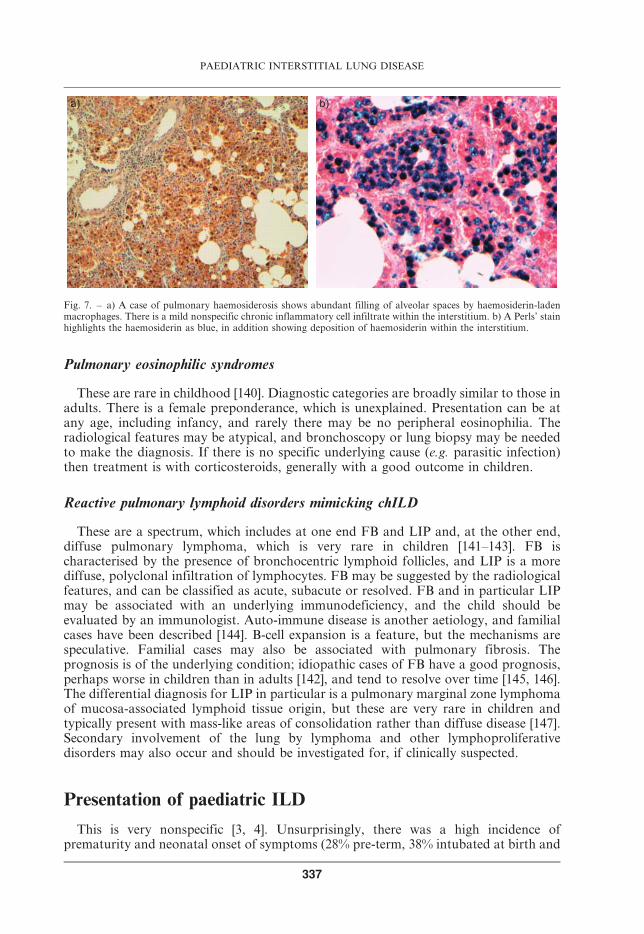
Diffuse pulmonary haemorrhage can be suspected on HRCT and confirmed by theBAL and/or biopsy finding of haemosiderin-laden macrophages (fig. 7). It must bestressed that this is not specific for IPH. In uncomplicated cases, surgical lung biopsy isnot required, but if capillaritis is suspected [135], or there is a poor response totreatment, then surgical lung biopsy should be considered.
Treatment of IPH is anecdote driven. Most would use pulsed intravenous methylprednisolone in a dose such as 500 mg?m-2 on three successive days, followed by monthlypulses for 6 months, with maintenance oral corticosteroids and hydroxychloroquine. Ifthis approach fails, then a number of cytotoxic agents and other therapies have beensuggested [136–139]. The prognosis must be guarded, but prolonged remission has beendescribed, and probably treatment should be weaned if there has been no overt bleedingfor 2–3 yrs and there are no haemosiderin-laden macrophages on surveillance BAL.
Fig. 6. – A case of alveolar microlithiasis shows abundant calcific spherites lying both within alveolar spaces and inthe interstitium. Many of these have shattered during processing.
A. BUSH AND A.G. NICHOLSON
336
Pulmonary eosinophilic syndromes
These are rare in childhood [140]. Diagnostic categories are broadly similar to those inadults. There is a female preponderance, which is unexplained. Presentation can be atany age, including infancy, and rarely there may be no peripheral eosinophilia. Theradiological features may be atypical, and bronchoscopy or lung biopsy may be neededto make the diagnosis. If there is no specific underlying cause (e.g. parasitic infection)then treatment is with corticosteroids, generally with a good outcome in children.
Reactive pulmonary lymphoid disorders mimicking chILD
These are a spectrum, which includes at one end FB and LIP and, at the other end,diffuse pulmonary lymphoma, which is very rare in children [141–143]. FB ischaracterised by the presence of bronchocentric lymphoid follicles, and LIP is a morediffuse, polyclonal infiltration of lymphocytes. FB may be suggested by the radiologicalfeatures, and can be classified as acute, subacute or resolved. FB and in particular LIPmay be associated with an underlying immunodeficiency, and the child should beevaluated by an immunologist. Auto-immune disease is another aetiology, and familialcases have been described [144]. B-cell expansion is a feature, but the mechanisms arespeculative. Familial cases may also be associated with pulmonary fibrosis. Theprognosis is of the underlying condition; idiopathic cases of FB have a good prognosis,perhaps worse in children than in adults [142], and tend to resolve over time [145, 146].The differential diagnosis for LIP in particular is a pulmonary marginal zone lymphomaof mucosa-associated lymphoid tissue origin, but these are very rare in children andtypically present with mass-like areas of consolidation rather than diffuse disease [147].Secondary involvement of the lung by lymphoma and other lymphoproliferativedisorders may also occur and should be investigated for, if clinically suspected.
Presentation of paediatric ILD
This is very nonspecific [3, 4]. Unsurprisingly, there was a high incidence ofprematurity and neonatal onset of symptoms (28% pre-term, 38% intubated at birth and
a) b)
Fig. 7. – a) A case of pulmonary haemosiderosis shows abundant filling of alveolar spaces by haemosiderin-ladenmacrophages. There is a mild nonspecific chronic inflammatory cell infiltrate within the interstitium. b) A Perls’ stainhighlights the haemosiderin as blue, in addition showing deposition of haemosiderin within the interstitium.
PAEDIATRIC INTERSTITIAL LUNG DISEASE
337
57% needing oxygen). 58% were male, and 34% had a family history of lung disease. 30%were biopsied by 3 months of age, 52% by 6 months, and 72% in the first year of life.Presenting features (descending order of frequency, more than one permitted per child)were hypoxaemia, tachypnoea, subcostal recession and other signs of respiratorydistress, gastro-oesophageal reflux, pulmonary hypertension, failure to thrive, crackles,cough and wheeze (surprisinglyy20%). 25% had no abnormal auscultatory findings.The network has defined ‘‘chILD syndrome’’ [148] to try to refine referrals for moredetailed work-up for ILD. This requires at least three of the following criteria in theabsence of any other aetiology as the primary cause: 1) symptoms of impairedrespiratory function, 2) hypoxaemia, 3) diffuse infiltrates, 4) presence of adventitioussounds (crackles), and 5) abnormal lung function. It should be noted that, although thisis a good guide to the presence of ILD, it cannot replace clinical judgement, and over-reliance on the index may lead to false negatives and positives. There is less informationto guide referral in older children.
Differential diagnosis of paediatric ILD
This has been partially covered above, and a full list of all possible diseases that maymimic ILD would encompass most of the spectrum of paediatric respirology. From theprevious section, it is clear that the presentation is very nonspecific. The CXR rarelyleads to diagnostic certainty, and further imaging is required. The most important thingfor the paediatrician faced with a child referred with a presumed airway disease is toconsider whether it could in fact be an ILD. A diffuse airway disease with distal airwayobstruction may lead to patchy air trapping and cause diagnostic confusion. If proximalairway disease (bronchiectasis) is also present, this helps to exclude an ILD, but chILDmay be complicated by traction bronchiectasis. Also, the CXR may be normal, and theILD only revealed by a computed tomography (CT) scan. Clearly, not all children withany form of airway disease merit CT scanning: diagnostic alertness and clinical skill andacumen are essential.
Diagnostic work-up of the child with suspected ILD
Overview
The child with ILD undergoes a staged work-up (table 9). Not all stages are essential;if there is a clear cut diagnosis of NEHI, for example, assessment of aspiration may beomitted.
Role of HRCT
A diagnostic work-up should be performed in a staged and focused manner. The firststep is to confirm that the child does have an ILD, almost invariably with HRCT, if thishas not already been performed. In addition to confirming the presence of ILD, HRCTmay allow specific diagnoses to be made, which include hypersensitivity pneumonitis,adult-type Langerhans’ cell histiocytosis, pulmonary haemorrhage and idiopathicalveolar microlithiasis [149, 150]. Some would include NEHI on this list. Increasingly,CT is performed with suspended respiration after bag and mask ventilation of a sedatedchild, or under a full general anaesthetic in those too young to breath-hold to order[151]. There has been no formal comparison of scan techniques to determine whether
A. BUSH AND A.G. NICHOLSON
338
suspended respiration improves rate of diagnosis. In cooperative patients, inspiratoryand expiratory scans can be obtained to look for air trapping. The same can be achievedby scanning in alternate side-lying in uncooperative children. A recent manuscriptreviewed 59 CT scans and reported that PAP was most frequently correctly diagnosed(n518), but it was only the first-choice diagnosis in fewer than half of the cases [150]. Allsingle cases of what was called ‘‘pulmonary fibrosis with calcification’’, lymphangiecta-sia and Langerhans’ cell histiocytosis were correctly diagnosed. Thus, for most chILDpatients, CT is not a definitive diagnostic investigation. Other roles of HRCT wouldinclude guiding the best site of any biopsy, and (perhaps) in follow-up. Finally, it is to behoped that by increasing the numbers of paired scans and definitive diagnoses (usuallyby surgical lung biopsy) we will be able to diminish the number of biopsies performed inthe future. At the present time, obtaining a reliable, confident and specific diagnosis onHRCT must be considered exceptional.
Assessment of disease severity
Once the presence of ILD has been confirmed, the next step is to define the severity ofthe disease, and subsequently work through programmed investigations to reach adefinitive diagnosis, before hopefully finding a treatment. A five-point severity score hasbeen proposed based on symptoms, level of arterial saturation and the presence orotherwise of pulmonary hypertension (table 10) [115]. This requires assessment ofoxygenation by an overnight pulse oximetry as a minimum, and the performance of anechocardiogram. The score does not include pulmonary function testing, but this shouldalways be performed in the older child at least.
Echocardiography. This test is used to noninvasively measure pulmonary arterypressure, a routine test in all cases of chILD. The test may also be diagnostic; cardiacmimics of ILD are excluded, e.g. left to right shunting causing pulmonary oedema.
Pulmonary function testing. Older children with ILD will often have the characteristicrestrictive physiology, with low lung volumes, reduced forced expiratory volume in 1 s(FEV1) and forced vital capacity (FVC), with a normal or increased FEV1/FVC ratio.These can be very useful in confirming the diagnosis and monitoring therapy. Anelevated diffusing capacity of the lung for carbon monoxide (DL,CO) in the setting of
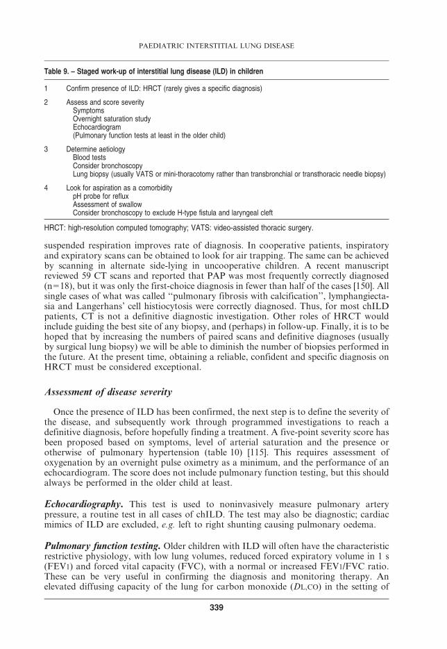
Table 9. – Staged work-up of interstitial lung disease (ILD) in children
1 Confirm presence of ILD: HRCT (rarely gives a specific diagnosis)
2 Assess and score severitySymptomsOvernight saturation studyEchocardiogram(Pulmonary function tests at least in the older child)
3 Determine aetiologyBlood testsConsider bronchoscopyLung biopsy (usually VATS or mini-thoracotomy rather than transbronchial or transthoracic needle biopsy)
4 Look for aspiration as a comorbiditypH probe for refluxAssessment of swallowConsider bronchoscopy to exclude H-type fistula and laryngeal cleft
HRCT: high-resolution computed tomography; VATS: video-assisted thoracic surgery.
PAEDIATRIC INTERSTITIAL LUNG DISEASE
339

ILD suggests pulmonary haemorrhage or pulmonary venous hypertension; a low DL,CO
is very nonspecific. Infant and pre-school pulmonary function is a rapidly expandingfield, but experience in ILD is substantially less in this age group and tests should beinterpreted with caution. Currently, we would consider them a research technique in thisage group.
Determination of aetiology
The planning of investigations, and their timing, will depend on the clinical pictureand the level of sickness of the child. Ideally, testing should precede blind trials oftreatment, but if the child is very sick on a ventilator, this may be thought inappropriate.In most cases, the first step will be the performance of a panel of blood tests to try todetermine the cause noninvasively [152]. Possible tests are summarised in table 11; aselective approach is advisable. Depending on the degree of clinical urgency, it may beappropriate to await the results before any further testing; a positive surfactant proteingene result may obviate the need for any further investigation. The next decision iswhether to perform fibreoptic bronchoscopy (FOB) or proceed directly to a lung biopsy.
The role of bronchoscopy. This requires relatively heavy sedation or, more usually, ageneral anaesthetic [153], and is only indicated if it is thought likely that the results willpreclude the need for a lung biopsy. If opportunistic infection is thought likely, thenFOB and BAL are the next choice investigation [154]. If this is negative, then theevidence is that it is better to proceed directly to a lung biopsy rather than waste timeperforming further BALs. Pulmonary haemorrhage can be confirmed by the presence ofhaemosiderin-laden macrophages in BAL [155, 156], but the test does not distinguishbetween primary and secondary causes, nor allow the diagnosis of pulmonarycapillaritis, which may require different treatment (see below). Other chILD diagnosesthat may be made on BAL include Niemann–Pick disease [87], Langerhans’ cellhistiocytosis [157, 158] and PAP [159]. There is insufficient paediatric experience torecommend BAL cytology as a means of definitive diagnosis of other chILDs.Transbronchial biopsy has only a limited role, exclusive of course in the management oflung transplant rejection. The samples obtained are very small, and, unless the suspectedILD has very specific and focal features that are uniformly distributed within the lung[160], such as pulmonary alveolar microlithiasis or metastatic thyroid cancer, thesamples are usually not adequate for the pathologist to make a diagnosis. Furthermore,morbidity from the procedure (bleeding, pneumothorax) is not trivial [161].
The timing and role of lung biopsy. Some teams would advocate a blind trial of oralcorticosteroids, and only biopsy children who do not respond. We would not supportthis, although we have to acknowledge the lack of an evidence base. First, with modernsurgical techniques, the morbidity of a lung biopsy is small [162]. Secondly, many ILDs
Table 10. – Illness severity score used in interstitial lung disease in children
Score Symptoms Hypoxaemia ,90% Pulmonaryhypertension
Sleep or exercise Rest
1 No No No No2 Yes No No No3 Yes Yes No No4 Yes Yes Yes No5 Yes Yes Yes Yes
A. BUSH AND A.G. NICHOLSON
340

are not steroid responsive, and indeed, if there is an occult undiagnosed infection,steroids may actually be harmful. Thirdly, the morbidity of high-dose corticosteroidsmay be considerable, and this includes complications of surgery if biopsy is undertakenafter a high-dose steroid trial. Fourthly, there are specific treatments for particular ILDs(see below), and these will not be offered if the diagnosis is not made. Fifthly, someconditions may have a genetic basis, and if a specific diagnosis is not made, the familymay miss out on crucial information. A final and subsidiary issue, more important to thegeneral population than the individual, is that our ignorance of these conditions isprofound, and only by finding out as much as we can about each case will we makeprogress. Thus, our recommendation is for a lung biopsy to be performed ahead of blindtrials of treatment unless the child is too sick, or a specific diagnosis has been made byother techniques.
Techniques of lung biopsy. These are percutaneous, CT-guided needle biopsy [163],video-assisted thoracic surgery (VATS) or via a mini-thoracotomy. We have nohesitation in discarding percutaneous biopsy [164]. There is a risk of bleeding andpneumothorax, a patchy abnormality may be missed, and the child needs a generalanaesthetic anyway. A surgical biopsy is the method of choice. We recommend thatideally this should be preceded by a BAL, best performed with a flexible bronchoscopeto get a good wedge position before lavage. This will maximise clinical information,making diagnosis of occult infection more probable; possibly indicating occult refluxfrom the quantification of lipid-laden macrophages, or, more specifically, by measuringBAL pepsin [165]; and BAL will be useful as a research tool to correlate BAL cytologywith the clinical picture and histology, hopefully in the future minimising the number of
Table 11. – Blood work to be considered in the work-up of interstitial lung disease (ILD) in children
Test Disease Comment
Serum KL-6 NEHI, surfactant protein deficiency Normal levels in NEHI, raised insurfactant protein deficiency
Sp-B, Sp-C, ABCA3 genes Surfactant protein deficiency Indicated in most children with ILD,unless there are extra-pulmonary
features or another obvious diagnosis
Angiotensin-converting enzyme Sarcoidosis Especially if extra-pulmonary features
Antineutrophil cytoplasmicantibodies
Wegener’s granuloma, othervasculitides
Especially if upper airway disease,renal disease or pulmonary
haemorrhage
Avian and Micropolysporafaeni precipitins
Hypersensitivity pneumonitis CT scan may be suggestive ofthis diagnosis
Viral and mycoplasma serology Obliterative bronchiolitis Not a true ILD, but may beconfused on CT
Immune work-up including HIV Lymphoproliferative syndromes,including follicular bronchiolitis
Also perform if ILD in fact provesto be an opportunistic infection
Auto-antibody studies Systemic lupus, rheumatoid diseases,scleroderma and other collagen
vascular disease
Especially if extra-pulmonaryfeatures and renal disease
GM-CSF studies (serum auto-antibody, receptor genetic studies)
Some of the variants of pulmonaryalveolar proteinosis
Adult type with response to GM-CSFhas been described in children
Note that not all tests need to be performed in all cases. NEHI: neuroendocrine cell hyperplasia of infancy; CT:computed tomography; GM-CSF: granulocyte-macrophage colony-stimulating factor.
PAEDIATRIC INTERSTITIAL LUNG DISEASE
341
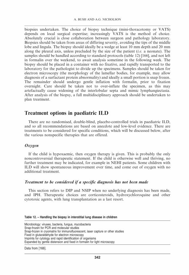
biopsies undertaken. The choice of biopsy technique (mini-thoracotomy or VATS)depends on local surgical expertise; increasingly VATS is the method of choice.Absolutely crucial is close collaboration between surgeon and pathology laboratory.Biopsies should be taken from areas of differing severity, avoiding the tips of the middlelobe and lingula. The biopsy should ideally be a wedge at least 10 mm depth and 20 mmalong the pleural axis, unless precluded by the size of the patient (i.e. a neonate). Thesamples should be handled according to standard protocols (table 12) [166], and not leftin formalin over the weekend, to await analysis sometime in the following week. Thebiopsy should be placed in a container with no fixative, and rapidly transported to thelaboratory for the pathologist to divide up the specimens. Samples should be taken forelectron microscopy (the morphology of the lamellar bodies, for example, may allowdiagnosis of a surfactant protein abnormality) and ideally a small portion is snap frozen.The remainder should undergo gentle inflation with formalin, prior to fixationovernight. Care should be taken not to over-inflate the specimen, as this mayartefactually cause widening of the interlobular septa and mimic lymphangiectasia.After analysis of the biopsy, a full multidisciplinary approach should be undertaken toplan treatment.
Treatment options in paediatric ILD
There are no randomised, double-blind, placebo-controlled trials in paediatric ILD,and so all recommendations are based on anecdote and low-level evidence. There aretreatments to be considered for specific conditions, which will be discussed below, afterthe various nonspecific therapies that are offered.
Oxygen
If the child is hypoxaemic, then oxygen therapy is given. This is probably the onlynoncontroversial therapeutic statement. If the child is otherwise well and thriving, nofurther treatment may be indicated, for example in NEHI patients. Some children withILD will show spontaneous improvement over time, and come out of oxygen with noadditional treatment.
Treatment to be considered if a specific diagnosis has not been made
This section refers to DIP and NSIP when no underlying diagnosis has been made,and IPH. Therapeutic choices are corticosteroids, hydroxychloroquine and othercytotoxic agents, with lung transplantation as a last resort.
Table 12. – Handling the biopsy in interstitial lung disease in children
Microbiology: viruses, bacteria, fungus, mycobacteriaSnap-frozen for PCR and molecular studiesSnap-frozen in cryomatrix for immunofluorescent, laser capture or other studiesFixed in glutaraldehyde for electron microscopyImprints for cytology and rapid identification of organismsExpanded by gentle distension and fixed in formalin for light microscopy
Data from [166].
A. BUSH AND A.G. NICHOLSON
342

Corticosteroid therapy. Depending on severity, this is given orally or as pulses. Pulsesmay anecdotally be less toxic [167]. The dose and timing are empirical (i.e. based onguesswork). We use methyl prednisolone, 500 mg?m-2 daily for three successive days,followed by single monthly pulses at the same dose for 6 months. Ideally, oral prednisoloneis avoided between pulses, but this may not be possible; our start dose would be 0.5 mg?kg-1
prednisolone on alternate days. If oral prednisolone is given from the outset instead ofpulses, a reasonable starting dose is 2 mg?kg-1?day-1, tapering according to response. Thereare anecdotal reports of the use of inhaled corticosteroids as maintenance, but the evidencethat these are deposited sufficiently distally and in an effective dose is scanty, and we do notrecommend them.
The hardest therapeutic decision may be to determine when there is no furtherresponse to steroids, and the time has come to taper the dose to avoid substantial steroidmorbidity. Our non-evidence-based policy would be to try three more pulses of methylprednisolone, and, if there is no improvement, assume that the limit of steroid usefulnesshas been reached. This is important, because fruitlessly prolonging steroid therapy,leading to osteoporosis, may lead to the child being turned down for lungtransplantation.
Hydroxychloroquine. This anti-malarial agent has a number of immunological effectsthat are possibly beneficial in ILD [168, 169]. There is evidence from case series that itmay be helpful, and it is very safe. There are reports of deafness complicating its use inIPH [170], and our own practice is to refer for an ophthalmic check at the start oftreatment. Nevertheless, our current practice is to add it to steroids in paediatric ILD,and maintain hydroxychloroquine therapy as an aid to steroid tapering.
Other cytotoxic agents. Evidence is even more anecdotal. There are isolated casereports and small case series advocating azathioprine, methotrexate, cyclosporin andplasmapheresis when steroids have failed. Our own experience with these agents isalmost universally dismal. A recent case series has suggested that 6-mercaptopurine maybe helpful in IPH [136]; the cynic would state that no medication can be considered to bewholly useless until it has been tried in this condition.
Lung transplantation. A small number of children with ILD have been successfullytransplanted, more commonly older children. Both cadaver and living related donationmay be considered. Other than in Langerhans’ cell histiocytosis (see below), the risk ofthe disease returning in the transplanted lung is minimal.
Treatment of specific conditions
The increasing availability of specific therapies is an important reason for pursuing aspecific diagnosis. Only a few examples are given, which serve to illustrate that there is moreto chILD therapy than ‘‘steroids for everyone’’. These therapies have potential benefit, butthe high fiscal cost and the potential for very severe side-effects militate against theirindiscriminate application. It is likely that more disease-specific therapies will becomeavailable in the future, making diagnostic precision evenmore important. Furthermore, theera of mutation-specific therapies is dawning, for example with PTC1241 (ataluren; PTCTherapeutics, Inc., South Plainfield, NJ, USA), a treatment for genetic diseases caused by apremature stop codon, in which the agent overrides the premature but not the normal stopsignal [171]. Whether this may apply to some of the genetic conditions described earlier inthis chapter is unknown; and the lesson that abnormal Sp-C may be toxic serves as awarning that overcoming a premature stop codon may not always be beneficial.
PAEDIATRIC INTERSTITIAL LUNG DISEASE
343

Hypersensitivity pneumonitis. Although prednisolone is an important treatment,identifying and removing the allergen is of fundamental importance if a good outcome isto be obtained.
Wegener’s granulomatosis and neutrophilic pulmonary capillaritis. Pulsed cyclo-phosphamide treatment should be considered. For refractory cases, there has beeninterest in using anti-B-cell strategies, employing the anti-CD20 monoclonal antibody,rituximab [172]. The potential toxicity of these therapies precludes their blindapplication.
Anti-tumour necrosis factor strategies for sarcoidosis and other conditions. Thesoluble tumour necrosis factor (TNF)-a receptor, etanercept, has been used on ananecdotal basis for refractory paediatric sarcoidosis [173], in combination withmethotrexate. Other causes of ILD that have been successfully treated with etanerceptinclude polyarteritis nodosa and other rare vasculitic diseases [174]. If etanercept fails,the anti-TNF-a monoclonal antibody, infliximab, may be worth trying. The largestdataset is in adult patients.
Pulmonary alveolar proteinosis. Treatment depends on the underlying cause, butlarge-volume lavage [69, 175–177] and inhaled or subcutaneous GM-CSF have beenused successfully [68, 70, 177]. Rituximab has been trialled in refractory cases [178].
Langerhans’ cell histiocytosis. Although Langerhans’ cell histiocytosis is usually amultisystem disease in children (fig. 8), predominantly pulmonary disease has beendescribed. Cytotoxic therapy supervised by an oncologist is the treatment of choice forboth forms of the condition. Many different combinations of therapies have been used.Passive and active tobacco smoking must be discouraged.
Monitoring treatment and progress
In older children who can perform pulmonary function tests, these are the techniqueof choice. In younger children, we routinely employ overnight pulse oximetrymonitoring. CXRs are probably of little use. Repeat HRCT, possibly using a limited
a) b)
Fig. 8. – a) A case of systemic histiocytosis shows a localised area of Langerhans’ cell accumulation around abronchiole, with admixed lymphocytes and eosinophils. b) Staining for S-100 highlights the presence of abundantLangerhans’ cells.
A. BUSH AND A.G. NICHOLSON
344

number of cuts, is another option, but should be used with caution to minimise radiationexposure. In most conditions, there is no role for repeat surveillance bronchoscopy. Theexception may be the pulmonary haemorrhagic syndromes, where after a period ofremission, it may be appropriate to re-bronchoscope the child before stoppingtreatment, to ensure there is no occult, ongoing bleeding. Repeat lung biopsy wouldbe indicated only under the most exceptional circumstances.
Making progress: the way forward
There is a crying need for focused, randomised, controlled therapeutic trials in wellcharacterised cohorts of patients. We also need to develop a greater understanding ofthese diseases. Paediatric ILD is a rare diagnosis, and no one centre will see enoughcases to run such trials. Thus, the only way forward is multinational collaboration. Thefirst pre-requisite for this is protocol-driven assessment of all these patients, specificallyensuring that information on presentation, imaging and investigations is collated in auniform manner. There needs to be a panel of radiologists who will assess the imaging,and pathologists to assess lung biopsies. The US chILD Research Network has been agreat example of how to do this, but even in their classification paper they had to admitthat the imaging was so patchy that no correlations with pathology were possible. Inparallel, we need hypothesis-generating studies, so that we can understand thepathophysiology of carefully characterised patients and develop novel therapies. Bothnovel and standard treatments must be tested in large cohorts using a randomised,double-blind, placebo-controlled methodology, with the confidence that all participat-ing centres are characterising the patients in the same, meticulously careful way. Thealternative, too bad to contemplate, is continued, anecdote-based therapy for these sick,vulnerable children.
PAEDIATRIC INTERSTITIAL LUNG DISEASE
345
Summary
Interstitial lung disease in children (chILD) is rare, and shows age-related differencesin nature. In children aged 0–2 yrs, it is classified into diffuse developmentaldisorders; growth abnormalities reflecting deficient alveolarisation; specific disordersof undefined aetiology (pulmonary interstitial glycogenosis, neuroendocrine cellhyperplasia of infancy); surfactant dysfunction disorders; disorders of the normalhost, presumed immune intact; disorders resulting from systemic processes; disordersof the immunocompromised host; and disorders masquerading as chILD. In olderchildren (aged 2–16 yrs) the classification is less well worked out: immune-mediated,often systemic disease, infective and post-infective disease is more common. There area large number of rare genetic conditions, which may also present in adult life; thesame genetic disease may cause many different histological appearances, which maybe age related.Presentation is usually nonspecific, and a systematic protocol of investigation isnecessary. The presence of chILD is usually confirmed by high-resolution computedtomography, and the severity is determined by physiological tests. Although lessinvasive testing, such as surfactant protein genetics, may be diagnostic, most will needto proceed to a surgical lung biopsy for definitive diagnosis. Precise diagnosis isimportant, because of the genetic implications for some families and also becauseincreasingly there are specific cytokine-targeted therapies, e.g. rituximab andetanercept, which are being used for specific indications.There are no randomised controlled trials of treatment in chILD. Oxygen therapy forthe hypoxaemic, while spontaneous recovery occurs, may be all that is necessary, butmany will need steroid therapy (pulse methyl prednisolone or oral prednisolone),which may be combined with hydroxychloroquine. There is even less evidence for theuse of cytotoxic therapies such as methotrexate, azathioprine and cyclosporin.International collaborations, with protocol-driven evaluation of chILD, are urgentlyrequired if evidence-based treatment is to be determined.
Keywords: Neuroendocrine cell, pulmonary haemorrhage, sarcoid, storage disorder,surfactant, Wegener’s granulomatosis.
Statement of interestNone declared.
References
1. Coultas DB, Zumwalt RE, Black WC, et al. The epidemiology of interstitial lung diseases. Am J
Respir Crit Care Med 1994; 150: 967–972.
2. Bradley B, Branley HM, Egan JJ, et al. Interstitial lung disease guideline: the British Thoracic
Society in collaboration with the Thoracic Society of Australia and New Zealand and the Irish
Thoracic Society. Thorax 2008; 63: Suppl. 5, v1–v58.
3. Clement A, Allen J, Corrin B, et al. Task force on chronic interstitial lung disease in
immunocompetent children. Eur Respir J 2004; 24: 686–697.
4. Deutsch GH, Young LR, Deterding RR, et al. Diffuse lung disease in young children: application
of a novel classification scheme. Am J Respir Crit Care Med 2007; 176: 1120–1128.
A. BUSH AND A.G. NICHOLSON
346

5. Deterding R, Young L, Dishop M, et al. Diffuse lung disease in older children: report of the
ChILD network review. Am J Respir Crit Care Med 2007; 175: A148.
6. Holt PG, Jones CA. The development of the immune system during pregnancy and early life.
Allergy 2000; 55: 688–697.
7. Thomas AQ, Lane K, Phillips J 3rd, et al. Heterozygosity for surfactant protein C gene mutation
associated with usual interstitial pneumonitis and cellular nonspecific interstitial pneumonitis in
one kindred. Am J Respir Crit Care Med 2002; 165: 1322–1328.
8. Nicholson AG, Bush A. Classification of diffuse lung disease in infants: the reality of groups. Am
J Respir Crit Care Med 2007; 176: 1060–1061.
9. Melly L, Sebire NJ, Malone M, et al. Capillary apposition and density in the diagnosis of alveolar
capillary dysplasia. Histopathology 2008; 53: 450–457.
10. Chaudhari M, Vogel M, Wright C, et al. Sildenafil in neonatal pulmonary hypertension due to
impaired alveolarisation and plexiform pulmonary arteriopathy. Arch Dis Child Fetal Neonatal Ed
2005; 90: F527–F528.
11. Hoffman JI, Rudolph AM, Heymann MA. Pulmonary vascular disease with congenital heart
lesions: pathologic features and causes. Circulation 1981; 64: 873–877.
12. Cutz E, Yeger H, Pan J. Pulmonary neuroendocrine cell system in pediatric lung disease – recent
advances. Pediatr Dev Pathol 2007; 10: 419–435.
13. Sunday ME, Kaplan LM, Motoyama E, et al. Biology of disease: gastrin-releasing peptide
(mammalian bombesin) gene expression in health and disease. Lab Invest 1988; 59: 5–24.
14. Weichselbaum M, Sparrow MP, Hamilton EJ, et al. A confocal microscopic study of solitary
pulmonary neuroendocrine cells in human airway epithelium. Respir Res 2005; 6: 115.
15. Deterding R, Pye C, Fan LL, et al. Persistent tachypnea of infancy is associated with
neuroendocrine cell hyperplasia. Pediatr Pulmonol 2005; 40: 157–165.
16. Alshehri M, Cutz E, Banzhoff A, et al. Hyperplasia of pulmonary neuroendocrine cells in a case
of childhood pulmonary emphysema. Chest 1997; 112: 553–556.
17. Brody AS, Crotty EJ. Neuroendocrine cell hyperplasia of infancy (NEHI). Pediatr Radiol 2006;
36: 1328.
18. Doan ML, Elidemir O, Dishop MK, et al. Serum KL-6 differentiates neuroendocrine
cell hyperplasia of infancy from the inborn errors of surfactant metabolism. Thorax 2009; 64:
677–681.
19. Hull J, Chow CW, Robertson CF. Chronic idiopathic bronchiolitis of infancy. Arch Dis Child
1997; 77: 512–515.
20. Aguayo SM, Miller YE, Waldron JA Jr, et al. Brief report: idiopathic diffuse hyperplasia of
pulmonary neuroendocrine cells and airways disease. N Engl J Med 1992; 327: 1285–1288.
21. Boenisch T, Farmilo AJ, Stead RH. Handbook of Immunochemical Staining Methods.
Carpintera, Dako Corp, 1989.
22. Johnson DE, Anderson WR, Burke BA. Pulmonary neuroendocrine cells in pediatric lung
disease: alterations in airway structure in infants with bronchopulmonary dysplasia. Anat Rec
1993; 236: 115–119.
23. Gillan JE, Cutz E. Abnormal pulmonary bombesin immunoreactive cells in Wilson-Mikity
syndrome (pulmonary dysmaturity) and bronchopulmonary dysplasia. Pediatr Pathol 1993; 13:
165–180.
24. Meyerholz DK, De Graaff JA, Gallup JM, et al. Depletion of alveolar glycogen corresponds with
immunohistochemical development of CD208 antigen expression in perinatal lamb lung. J
Histochem Cytochem 2006; 54: 1247–1253.
25. Ridsdale R, Post M. Surfactant lipid synthesis and lamellar body formation in glycogen-laden
type II cells. Am J Physiol Lung Cell Mol Physiol 2004; 287: L743–L751.
26. Canakis AM, Cutz E, Manson D, et al. Pulmonary interstitial glycogenosis: a new variant of
neonatal interstitial lung disease. Am J Respir Crit Care Med 2002; 165: 1557–1565.
27. Schroeder SA, Shannon DC, Mark EJ. Cellular interstitial pneumonitis in infants: a
clinicopathologic study. Chest 1992; 101: 1065–1069.
PAEDIATRIC INTERSTITIAL LUNG DISEASE
347

28. Scully RE, Mark EJ, McNeely WF, et al. Case records of the Massachusetts General Hospital:
weekly clinicopathological exercises. N Engl J Med 1999; 341: 2075–2083.
29. Onland W, Molenaar JJ, Leguit RJ, et al. Pulmonary interstitial glycogenosis in identical twins.
Pediatr Pulmonol 2005; 40: 362–366.
30. Smets K, Dhaene K, Schelstraete P, et al. Neonatal pulmonary interstitial glycogen accumulation
disorder. Eur J Pediatr 2004; 163: 408–409.
31. Kepron C, Cutz E, Viero S. Congenital lobar emphysema with pulmonary interstitial
glycogenosis: variant of emphysema or localised glycogenosis. Mod Pathol 2008; 21: 224.
32. Mulugeta S, Gray JM, Notarfrancesco KL, et al. Identification of LBM180, a lamellar body
limiting membrane protein of alveolar type II cells, as the ABC transporter protein ABCA3. J Biol
Chem 2002; 277: 22147–22155.
33. Groman JD, Meyer ME, Wilmott RW, et al. Variant cystic fibrosis phenotypes in the absence of
CFTR mutations. N Engl J Med 2002; 347: 401–407.
34. Wang X, Venable J, LaPointe P, et al. Hsp90 cochaperone Aha1 downregulation rescues
misfolding of CFTR in cystic fibrosis. Cell 2006; 127: 803–815.
35. Lee H, Ryu J, Wittmer H, et al. Familial idiopathic pulmonary fibrosis: clinical features and
outcomes. Chest 2005; 127: 2034–2041.
36. Cole FS, Hamvas A, Rubinstein P, et al. Population-based estimates of surfactant protein B
deficiency. Pediatrics 2000; 105: 538–541.
37. Hamvas A, Cole FS, Nogee LM. Genetic disorders of surfactant proteins. Neonatology 2007; 91:
311–317.
38. Hamvas A, Trusgnich M, Brice H, et al. Population-based screening for rare mutations: high
throughput DNA extraction and amplification from Guthrie cards. Pediatr Res 2001; 50: 666–668.
39. deMello DE, Heyman S, Phelps DS, et al. Ultrastructure of lung in surfactant protein B
deficiency. Am J Respir Cell Mol Biol 1994; 11: 230–239.
40. Stahlman MT, Gray MP, Falcomieri MW, et al. Lamellar body formation in normal and
surfactant-protein-B deficient fetal mice. Lab Invest 2000; 80: 395–403.
41. Li J, Ikegama M, Na CL, et al. N-terminally extended surfactant protein (SP) C isolated from
SP-B-deficient children has reduced surface activity and inhibited liopolysaccharide binding.
Biochemistry 2004; 43: 3891–3898.
42. Ballard PL, Nogee LM, Beers MF, et al. Partial deficiency of surfactant protein B in an infant
with chronic lung disease. Pediatrics 1995; 96: 1046–1052.
43. Dunbar AE 3rd, Wert SE, Ikegama M, et al. Prolonged survival in hereditary surfactant protein B
(SP-B) deficiency associated with a novel splicing mutation. Pediatr Res 2000; 48: 275–282.
44. Hataja R, Ramet M, Marttila R, et al. Surfactant proteins A and B as interactive genetic
determinants of neonatal respiratory distress syndrome. Hum Mol Genet 2000; 9: 2751–2761.
45. Lin Z, Pearson C, Chinnchilli V, et al. Polymorphisms of human SP-1, SP-B and SP-D genes.
Association of SP-B Thr131Le with ARDS. Clin Genet 2000; 58: 181–191.
46. Selman M, Lin HM, Montano M, et al. Surfactant protein A and B genetic variants predispose to
idiopathic pulmonary fibrosis. Hum Genet 2003; 113: 542–550.
47. Floros J, Thomas NJ, Liu W, et al. Family-based association tests suggest linkage between surfactant
protein B (SP-B) (and flanking region) and respiratory distress syndrome (RDS): SP-B haplotypes and
alleles from SP-B-linked loci are risk factors for RDS. Pediatr Res 2006; 59: 616–621.
48. Mulugeta S, Nguyen V, Russo SJ, et al. A surfactant protein C precursor protein BRICHOS
domain mutation causes endoplasmic reticulum stress, proteasome dysfunction, and caspase 3
activation. Am J Respir Cell Mol Biol 2005; 32: 521–530.
49. Beers MF, Mulugeta S. Surfactant protein C biosynthesis and its emerging role in conformational
lung disease. Annu Rev Physiol 2005; 67: 663–696.
50. Mulugeta S, Maguire JA, Newitt JL, et al. Misfolded BRICHOS SP-C mutant proteins induce
apoptosis via caspase-4- and cytochrome c-related mechanisms. Am J Physiol Lung Cell Mol
Physiol 2007; 293: 720–799.
A. BUSH AND A.G. NICHOLSON
348

51. Bullard JE, Nogee LM. Heterozygosity for ABCA3 mutations modifies the severity of lung disease
associated with a surfactant protein C gene (SFTPC) mutation. Pediatr Res 2007; 62: 176–179.
52. Amin RS, Wert SE, Baughman RP, et al. Surfactant protein deficiency in familial interstitial lung
disease. J Pediatr 2001; 139: 85–92.
53. Shulenin S, Nogee LM, Annilo T, et al. ABCA3 gene mutations in newborns with fatal surfactant
deficiency. N Engl J Med 2004; 350: 1296–1303.
54. Bullard JE, Wert SE, Nogee LM. ABCA3 deficiency: neonatal respiratory failure and interstitial
lung disease. Semin Perinatol 2006; 30: 327–334.
55. Nogee LM. Genetics of pediatric interstitial lung disease. Curr Opin Pediatr 2006; 18: 287–292.
56. Garmany TH, Bullard JE, Wissman KA, et al. The E292V missense mutation in the ABCA3 gene
increases risk for pneumothorax in newborns with respiratory distress. PAS 2005; 57: 2008.
57. Bullard JE, Wert SE, Whitsett JA, et al. ABCA3 mutations associated with pediatric interstitial
lung disease. Am J Respir Crit Care Med 2005; 172: 1026–1031.
58. Stahlman MT, Besnard V, Wert SE, et al. Expression of ABCA3 in developing lung and other
tissues. J Histochem Cytochem 2007; 55: 71–83.
59. Martis PC, Whitsett JA, Xu Y, et al. C/EBPa is required for lung maturation at birth.
Development 2006; 133: 1155–1164.
60. Kunig AM, Parker TA, Nogee LM, et al. ABCA3 deficiency presenting as persistent pulmonary
hypertension of the newborn. J Pediatr 2007; 151: 322–324.
61. Whitsett JA. Hereditary disorders of surfactant homeostasis cause acute and chronic lung disease
in infancy. Thorax 2008; 63: 295–296.
62. Dirksen U, Nishinakamura R, Groneck P, et al. Human pulmonary alveolar proteinosis
associated with a defect in GM-CSH/IL-3/IL-5 receptor common b chain expression. J Clin Invest
1997; 100: 2211–2217.
63. Doan ML, Guillerman RP, Dishop MK, et al. Clinical, radiological and pathological features of
ABCA3 mutations in children. Thorax 2008; 63: 366–373.
64. Bewig B, Wang XD, Kirsten D, et al. GM-CSF and GM-CSF bc receptor in adult patients with
pulmonary alveolar proteinosis. Eur Respir J 2000; 15: 350–357.
65. Suzuki T, Sakagami T, Rubin BK, et al. Familial pulmonary alveolar proteinosis caused by
mutations in CSF2RA. J Exp Med 2008; 205: 2703–2710.
66. Martinez-Moczygemba M, Doan ML, Elidemir O, et al. Pulmonary alveolar proteinosis caused
by deletion of the GM-CSFRa gene in the X chromosome pseudoautosomal region 1. J Exp Med
2008; 205: 2711–2716.
67. Inoue Y, Trapnell BC, Tazawa R, et al.Characteristics of a large cohort of patients with autoimmune
pulmonary alveolar proteinosis in Japan. Am J Respir Crit Care Med 2008; 177: 752–762.
68. Price A, Manson D, Cutz E, et al. Pulmonary alveolar proteinosis associated with anti-GM-CSF
antibodies in a child: successful treatment with inhaledGM-CSF.Pediatr Pulmonol 2006; 41: 367–370.
69. Beccaria M, Luisetti M, Rodi G, et al. Long-term durable benefit after whole lung lavage in
pulmonary alveolar proteinosis. Eur Respir J 2004; 23: 526–531.
70. Kavuru MS, Sullivan EJ, Piccin R, et al. Exogenous granulocyte-macrophage colony-stimulating
factor administration for pulmonary alveolar proteinosis. Am J Respir Crit Care Med 2000; 161:
1143–1148.
71. Robinson TE, Trapnell BC, Goris ML, et al. Quantitative analysis of longitudinal response to
aerosolized granulocyte-macrophage colony-stimulating factor in two adolescents with auto-
immune pulmonary alveolar proteinosis. Chest 2009; 135: 842–848.
72. Patiroglu T, Akiyildiz, Patiroglu TE, et al. Recurrent pulmonary alveolar proteinosis secondary
to agammaglobulinemia. Pediatr Pulmonol 2008; 43: 710–713.
73. Trapnell BC, Whitsett JA, Nakata K. Pulmonary alveolar proteinosis. N Engl J Med 2003; 349:
2527–2539.
74. Sperandeo MP, Andria G, Sebastio G. Lysinuric protein intolerance: update and extended
mutation analysis of the SLC7A7 gene. Hum Mutat 2008; 29: 14–21.
PAEDIATRIC INTERSTITIAL LUNG DISEASE
349

75. Ceruti M, Rodi G, Stella GM, et al. Successful whole lung lavage in pulmonary alveolar proteinosis
secondary to lysinuric protein intolerance: a case report. Orphanet J Rare Dis 2007; 2: 14.
76. Santamaria F, Brancaccio G, Parenti G, et al. Recurrent fatal pulmonary alveolar proteinosis
after heart–lung transplantation in a child with lysinuric protein intolerance. J Pediatr 2004; 145:
268–272.
77. Minai OA, Sullivan EJ, Stoller JK. Pulmonary involvement in Niemann–Pick disease: case report
and literature review. Respir Med 2000; 94: 1241–1251.
78. Mendelson DS, Wasserstein MP, Desnick RJ. Type B Niemann–Pick disease: findings at chest
radiography, thin-section CT, and pulmonary function testing. Radiology 2006; 238: 339–345.
79. Millat G, Chikh K, Naureckiene S, et al. Niemann–Pick disease type C: spectrum of HE1
mutations and genotype/phenotype correlations in the NPC2 group. Am J Hum Genet 2001; 69:
1013–1021.
80. Elleder M, Houstkova H, Zeman J, et al. Pulmonary storage with emphysema as a sign of
Niemann–Pick type C2 disease (second complementation group). Report of a case. Virchows Arch
2001; 439: 206–211.
81. Guillemot N, Troadec C, de Villemeur TB, et al. Lung disease in Niemann–Pick disease. Pediatr
Pulmonol 2007; 42: 1207–1214.
82. Bjurulf B, Spetalen S, Erichson A, et al. Niemann–Pick disease type C2 presenting as fatal
pulmonary alveolar proteinosis: morphological findings in lung and nervous tissue. Med Sci
Monit 2008; 14: CS71–CS75.
83. McGovern MM, Wasserstein MP, Giugliani R, et al. A prospective, cross-sectional survey study
of the natural history of Niemann–Pick disease type B. Pediatrics 2008; 122: e341–e349.
84. Nicholson AG, Wells AU, Hooper J, et al. Successful treatment of endogenous lipoid pneumonia
due to Niemann–Pick type B disease with whole-lung lavage. Am J Respir Crit Care Med 2002;
165: 128–131.
85. Palmeri S, Tarugi P, Sicurelli F, et al. Lung involvement in Niemann–Pick disease type C1:
improvement with bronchoalveolar lavage. Neurol Sci 2005; 26: 171–173.
86. Uyan ZS, Karadag B, Ersu R, et al. Early pulmonary involvement in Niemann–Pick type B
disease: lung lavage is not useful. Pediatr Pulmonol 2005; 40: 169–172.
87. Falco F, Bembi B, Cavazza A, et al. Pulmonary involvement in an adult female affected by type B
Niemann–Pick disease. Sarcoidosis Vasc Diffuse Lung Dis 2005; 22: 229–233.
88. Baldi BG, Santana AN, Takagaki TY, et al. Lung cyst: an unusual manifestation of Niemann–
Pick disease. Respirology 2009; 14: 134–136.
89. Arda IS, Gencoglu A, Coskun M, et al. A very unusual presentation of Niemann–Pick disease
type B in an infant: similar findings to congenital lobar emphysema. Eur J Pediatr Surg 2005; 15:
283–286.
90. Shah AJ, Kapoor N, Crooks GM, et al. Successful hematopoietic stem cell transplantation for
Niemann–Pick disease type B. Pediatrics 2005; 116: 1022–1025.
91. Schuchman EH. The pathogenesis and treatment of acid sphingomyelinase-deficient Niemann–
Pick disease. J Inherit Metab Dis 2007; 30: 654–663.
92. Katzenstein AL, Gordon LP, Oliphant M, et al. Chronic pneumonitis of infancy. A unique form
of interstitial lung disease occurring in early childhood. Am J Surg Pathol 1995; 19: 439–447.
93. Kavantzas N, Theocharis S, Agapitos E, et al. Chronic pneumonitis of infancy. An autopsy study
of 12 cases. Clin Exp Pathol 1999; 47: 96–100.
94. Kim JM, Kwon SY, Kim ES, et al. A good outcome for a case of chronic pneumonitis of infancy.
Yonsei Med J 2007; 48: 865–867.
95. Venkatesh P, Wild L. Hypersensitivity pneumonitis in children: clinical features, diagnosis, and
treatment. Pediatr Drugs 2005; 7: 235–244.
96. Fan LL. Hypersensitivity pneumonitis in children. Curr Opin Pediatr 2002; 4: 23–26.
97. Koh DM, Hansell DM. Computed tomography of diffuse interstitial lung disease in children. Clin
Radiol 2000; 55: 659–667.
A. BUSH AND A.G. NICHOLSON
350
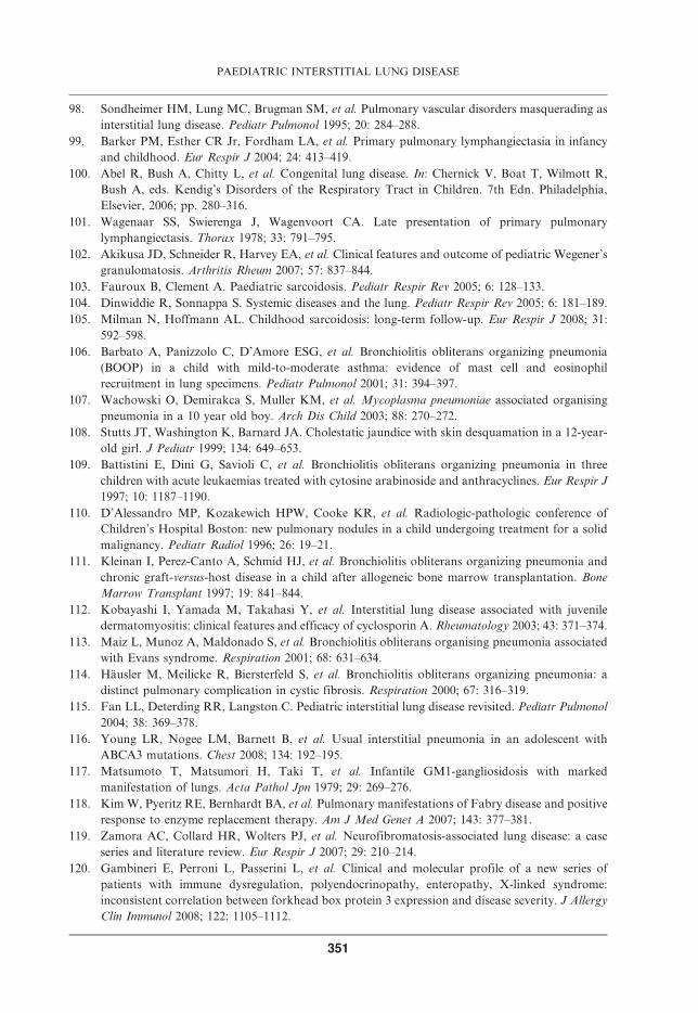
98. Sondheimer HM, Lung MC, Brugman SM, et al. Pulmonary vascular disorders masquerading as
interstitial lung disease. Pediatr Pulmonol 1995; 20: 284–288.
99. Barker PM, Esther CR Jr, Fordham LA, et al. Primary pulmonary lymphangiectasia in infancy
and childhood. Eur Respir J 2004; 24: 413–419.
100. Abel R, Bush A, Chitty L, et al. Congenital lung disease. In: Chernick V, Boat T, Wilmott R,
Bush A, eds. Kendig’s Disorders of the Respiratory Tract in Children. 7th Edn. Philadelphia,
Elsevier, 2006; pp. 280–316.
101. Wagenaar SS, Swierenga J, Wagenvoort CA. Late presentation of primary pulmonary
lymphangiectasis. Thorax 1978; 33: 791–795.
102. Akikusa JD, Schneider R, Harvey EA, et al. Clinical features and outcome of pediatric Wegener’s
granulomatosis. Arthritis Rheum 2007; 57: 837–844.
103. Fauroux B, Clement A. Paediatric sarcoidosis. Pediatr Respir Rev 2005; 6: 128–133.
104. Dinwiddie R, Sonnappa S. Systemic diseases and the lung. Pediatr Respir Rev 2005; 6: 181–189.
105. Milman N, Hoffmann AL. Childhood sarcoidosis: long-term follow-up. Eur Respir J 2008; 31:
592–598.
106. Barbato A, Panizzolo C, D’Amore ESG, et al. Bronchiolitis obliterans organizing pneumonia
(BOOP) in a child with mild-to-moderate asthma: evidence of mast cell and eosinophil
recruitment in lung specimens. Pediatr Pulmonol 2001; 31: 394–397.
107. Wachowski O, Demirakca S, Muller KM, et al. Mycoplasma pneumoniae associated organising
pneumonia in a 10 year old boy. Arch Dis Child 2003; 88: 270–272.
108. Stutts JT, Washington K, Barnard JA. Cholestatic jaundice with skin desquamation in a 12-year-
old girl. J Pediatr 1999; 134: 649–653.
109. Battistini E, Dini G, Savioli C, et al. Bronchiolitis obliterans organizing pneumonia in three
children with acute leukaemias treated with cytosine arabinoside and anthracyclines. Eur Respir J
1997; 10: 1187–1190.
110. D’Alessandro MP, Kozakewich HPW, Cooke KR, et al. Radiologic-pathologic conference of
Children’s Hospital Boston: new pulmonary nodules in a child undergoing treatment for a solid
malignancy. Pediatr Radiol 1996; 26: 19–21.
111. Kleinan I, Perez-Canto A, Schmid HJ, et al. Bronchiolitis obliterans organizing pneumonia and
chronic graft-versus-host disease in a child after allogeneic bone marrow transplantation. Bone
Marrow Transplant 1997; 19: 841–844.
112. Kobayashi I, Yamada M, Takahasi Y, et al. Interstitial lung disease associated with juvenile
dermatomyositis: clinical features and efficacy of cyclosporin A. Rheumatology 2003; 43: 371–374.
113. Maiz L, Munoz A, Maldonado S, et al. Bronchiolitis obliterans organising pneumonia associated
with Evans syndrome. Respiration 2001; 68: 631–634.
114. Hausler M, Meilicke R, Biersterfeld S, et al. Bronchiolitis obliterans organizing pneumonia: a
distinct pulmonary complication in cystic fibrosis. Respiration 2000; 67: 316–319.
115. Fan LL, Deterding RR, Langston C. Pediatric interstitial lung disease revisited. Pediatr Pulmonol
2004; 38: 369–378.
116. Young LR, Nogee LM, Barnett B, et al. Usual interstitial pneumonia in an adolescent with
ABCA3 mutations. Chest 2008; 134: 192–195.
117. Matsumoto T, Matsumori H, Taki T, et al. Infantile GM1-gangliosidosis with marked
manifestation of lungs. Acta Pathol Jpn 1979; 29: 269–276.
118. Kim W, Pyeritz RE, Bernhardt BA, et al. Pulmonary manifestations of Fabry disease and positive
response to enzyme replacement therapy. Am J Med Genet A 2007; 143: 377–381.
119. Zamora AC, Collard HR, Wolters PJ, et al. Neurofibromatosis-associated lung disease: a case
series and literature review. Eur Respir J 2007; 29: 210–214.
120. Gambineri E, Perroni L, Passerini L, et al. Clinical and molecular profile of a new series of
patients with immune dysregulation, polyendocrinopathy, enteropathy, X-linked syndrome:
inconsistent correlation between forkhead box protein 3 expression and disease severity. J Allergy
Clin Immunol 2008; 122: 1105–1112.
PAEDIATRIC INTERSTITIAL LUNG DISEASE
351
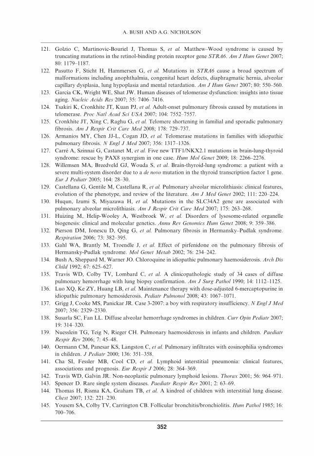
121. Golzio C, Martinovic-Bouriel J, Thomas S, et al. Matthew–Wood syndrome is caused by
truncating mutations in the retinol-binding protein receptor gene STRA6. Am J Hum Genet 2007;
80: 1179–1187.
122. Pasutto F, Sticht H, Hammersen G, et al. Mutations in STRA6 cause a broad spectrum of
malformations including anophthalmia, congenital heart defects, diaphragmatic hernia, alveolar
capillary dysplasia, lung hypoplasia and mental retardation. Am J Hum Genet 2007; 80: 550–560.
123. Garcia CK, Wright WE, Shat JW. Human diseases of telomerase dysfunction: insights into tissue
aging. Nucleic Acids Res 2007; 35: 7406–7416.
124. Tsakiri K, Cronkhite JT, Kuan PJ, et al. Adult-onset pulmonary fibrosis caused by mutations in
telomerase. Proc Natl Acad Sci USA 2007; 104: 7552–7557.
125. Cronkhite JT, Xing C, Raghu G, et al. Telomere shortening in familial and sporadic pulmonary
fibrosis. Am J Respir Crit Care Med 2008; 178: 729–737.
126. Armanios MY, Chen JJ-L, Cogan JD, et al. Telomerase mutations in families with idiopathic
pulmonary fibrosis. N Engl J Med 2007; 356: 1317–1326.
127. Carre A, Szinnai G, Castanet M, et al. Five new TTF1/NKX2.1 mutations in brain-lung-thyroid
syndrome: rescue by PAX8 synergism in one case. Hum Mol Genet 2009; 18: 2266–2276.
128. Willemsen MA, Breedveld GJ, Wouda S, et al. Brain-thyroid-lung syndrome: a patient with a
severe multi-system disorder due to a de novo mutation in the thyroid transcription factor 1 gene.
Eur J Pediatr 2005; 164: 28–30.
129. Castellana G, Gentile M, Castellana R, et al. Pulmonary alveolar microlithiasis: clinical features,
evolution of the phenotype, and review of the literature. Am J Med Genet 2002; 111: 220–224.
130. Huqun, Izumi S, Miyazawa H, et al. Mutations in the SLC34A2 gene are associated with
pulmonary alveolar microlithiasis. Am J Respir Crit Care Med 2007; 175: 263–268.
131. Huizing M, Helip-Wooley A, Westbroek W, et al. Disorders of lysosome-related organelle
biogenesis: clinical and molecular genetics. Annu Rev Genomics Hum Genet 2008; 9: 359–386.
132. Pierson DM, Ionescu D, Qing G, et al. Pulmonary fibrosis in Hermansky–Pudlak syndrome.
Respiration 2006; 73: 382–395.
133. Gahl WA, Brantly M, Troendle J, et al. Effect of pirfenidone on the pulmonary fibrosis of
Hermansky-Pudlak syndrome. Mol Genet Metab 2002; 76: 234–242.
134. Bush A, Sheppard M, Warner JO. Chloroquine in idiopathic pulmonary haemosiderosis. Arch Dis
Child 1992; 67: 625–627.
135. Travis WD, Colby TV, Lombard C, et al. A clinicopathologic study of 34 cases of diffuse
pulmonary hemorrhage with lung biopsy confirmation. Am J Surg Pathol 1990; 14: 1112–1125.
136. Luo XQ, Ke ZY, Huang LB, et al. Maintenance therapy with dose-adjusted 6-mercaptopurine in
idiopathic pulmonary hemosiderosis. Pediatr Pulmonol 2008; 43: 1067–1071.
137. Grigg J, Cooke MS, Panickar JR. Case 3-2007: a boy with respiratory insufficiency. N Engl J Med
2007; 356: 2329–2330.
138. Susarla SC, Fan LL. Diffuse alveolar hemorrhage syndromes in children. Curr Opin Pediatr 2007;
19: 314–320.
139. Nuesslein TG, Teig N, Rieger CH. Pulmonary haemosiderosis in infants and children. Paediatr
Respir Rev 2006; 7: 45–48.
140. Oermann CM, Panesar KS, Langston C, et al. Pulmonary infiltrates with eosinophilia syndromes
in children. J Pediatr 2000; 136: 351–358.
141. Cha SI, Fessler MB, Cool CD, et al. Lymphoid interstitial pneumonia: clinical features,
associations and prognosis. Eur Respir J 2006; 28: 364–369.
142. Travis WD, Galvin JR. Non-neoplastic pulmonary lymphoid lesions. Thorax 2001; 56: 964–971.
143. Spencer D. Rare single system diseases. Paediatr Respir Rev 2001; 2: 63–69.
144. Thomas H, Risma KA, Graham TB, et al. A kindred of children with interstitial lung disease.
Chest 2007; 132: 221–230.
145. Yousem SA, Colby TV, Carrington CB. Follicular bronchitis/bronchiolitis. Hum Pathol 1985; 16:
700–706.
A. BUSH AND A.G. NICHOLSON
352

146. Kinane BT, Mansell AL, Zwerdling RG, et al. Follicular bronchiolitis in the pediatric population.
Chest 1993; 104: 1183–1186.
147. Aghamohammadi A, Parvaneh N, Tirgari F, et al. Lymphoma of mucosa-associated lymphoid
tissue in common variable immunodeficiency. Leuk Lymphoma 2006; 47: 343–346.
148. Deterding R, Fan LL. Surfactant dysfunction mutations in children’s interstitial lung disease and
beyond. Am J Respir Crit Care Med 2005; 172: 940–941.
149. Copley SJ, Coren M, Nicholson AG, et al. Diagnostic accuracy of thin-section CT and chest
radiography of pediatric interstitial lung disease. AJR 2000; 174: 549–554.
150. Vrielynck S, Mamou-Mani T, Emond S, et al. Diagnostic value of high-resolution CT in the
evaluation of chronic infiltrative lung disease in children.AJR Am J Roentgenol 2008; 191: 914–920.
151. Long FR, Castile RG. Technique and clinical applications of full-inflation and end-exhalation
controlled-ventilation chest CT in infants and young children. Pediatr Radiol 2001; 31: 413–422.
152. Midulla F, de Blic J, Barbato A, et al. Flexible endoscopy of paediatric airways. Eur Respir J
2003; 22: 698–708.
153. Fan LL, Kozinetz CA, Deterding RR, et al. Evaluation of a diagnostic approach to pediatric
interstitial lung disease. Pediatrics 1998; 101: 82–85.
154. Riedler J, Grigg J, Robertson CF. Role of bronchoalveolar lavage in children with lung disease.
Eur Respir J 1995; 8: 1725–1730.
155. Salih ZN, Akhter A, Akhter J. Specificity and sensitivity of hemosiderin-laden macrophages in
routine bronchoalveolar lavage in children. Arch Pathol Lab Med 2006; 130: 1684–1686.
156. Perez-Arellano JL, Losa Garcıa JE, Garcıa Macıas MC, et al. Hemosiderin-laden macrophages in
bronchoalveolar lavage fluid. Acta Cytol 1992; 36: 26–30.
157. Refabert L, Rambaud C, Mamou-Mani T, et al. Cd1a-positive cells in bronchoalveolar lavage
samples from children with Langerhans cell histiocytosis. J Pediatr 1996; 129: 913–915.
158. Takizawa Y, Taniuchi N, Ghazizadeh M, et al. Bronchoalveolar lavage fluid analysis provides
diagnostic information on pulmonary Langerhans cell histiocytosis. J Nippon Med Sch 2009; 76:
84–92.
159. Maygarden SJ, Iacocca MV, Funkhouser WK, et al. Pulmonary alveolar proteinosis: a spectrum
of cytologic, histochemical, and ultrastructural findings in bronchoalveolar lavage fluid. Diagn
Cytopathol 2001; 24: 389–395.
160. Wallis C, Whitehead B, Malone M, et al. Pulmonary alveolar microlithiasis in childhood:
diagnosis by transbronchial biopsy. Pediatr Pulmonol 1996; 21: 62–64.
161. Whitehead B, Scott JP, Helms P, et al. Technique and use of transbronchial biopsy in children and
adolescents. Pediatr Pulmonol 1992; 12: 240–246.
162. Coren ME, Nicholson AG, Goldstraw P, et al. Open lung biopsy for diffuse interstitial lung
disease in children. Eur Respir J 1999; 14: 817–821.
163. Spencer DA, Alton HM, Raafat F, et al. Combined percutaneous lung biopsy and high-resolution
computed tomography in the diagnosis and management of lung disease in children. Pediatr
Pulmonol 1996; 22: 111–116.
164. Bush A. Diagnosis of interstitial lung disease. Pediatr Pulmonol 1996; 22: 81–82.
165. Farrell S, McMaster C, Gibson D, et al. Pepsin in bronchoalveolar lavage fluid: a specific and
sensitive method of diagnosing gastro-oesophageal reflux-related pulmonary aspiration. J Pediatr
Surg 2006; 41: 289–293.
166. Langston C, Patterson K, Dishop MK, et al. A protocol for the handling of tissue obtained by
operative lung biopsy: recommendations of the chILD pathology co-operative group. Pediatr Dev
Pathol 2006; 9: 173–180.
167. Kerem E, Bentur L, England S, et al. Sequential pulmonary function measurements during
treatment of infantile chronic interstitial pneumonitis. J Pediatr 1990; 116: 61–67.
168. van der Heijden JW, Dijkmans BA, Scheper RJ, et al. Drug insight: resistance to methotrexate
and other disease-modifying antirheumatic drugs – from bench to bedside. Nat Clin Pract
Rheumatol 2007; 3: 26–34.
PAEDIATRIC INTERSTITIAL LUNG DISEASE
353

169. Kalia S, Dutz JP. New concepts in antimalarial use and mode of action in dermatology. Dermatol
Ther 2007; 20: 160–174.
170. Coutinho MB, Duarte I. Hydroxychloroquine ototoxicity in a child with idiopathic pulmonary
haemosiderosis. Int J Pediatr Otorhinolaryngol 2002; 62: 53–57.
171. Kerem E, Hirawat S, Armoni S, et al. Effectiveness of PTC124 treatment of cystic fibrosis caused
by nonsense mutations: a prospective phase II trial. Lancet 2008; 372: 719–727.
172. Patel AM, Lehman TJ. Rituximab for severe refractory pediatric Wegener granulomatosis. J Clin
Rheumatol 2008; 14: 278–280.
173. Denys BG, Bogaerts Y, Coenegrachts KL, et al. Steroid-resistant sarcoidosis: is antagonism of
TNF-a the answer? Clin Sci (Lond) 2007; 112: 281–289.
174. Brik R, Gepstein V, Shahar E. Tumor necrosis factor blockade in the management of children
with orphan diseases. Clin Rheumatol 2007; 26: 1783–1785.
175. DuBois RM, McAllister WA, Branthwaite MA. Alveolar proteinosis: diagnosis and treatment
over a 10-year period. Thorax 1983; 381: 360–363.
176. Dogru D, Yalcin E, Aslan AT, et al. Successful unilateral partial lung lavage in a child with
pulmonary alveolar proteinosis. J Clin Anesth 2009; 21: 127–130.
177. Yamamoto H, Yamaguchi E, Agata H, et al. A combination therapy of whole lung lavage and
GM-CSF inhalation in pulmonary alveolar proteinosis. Pediatr Pulmonol 2008; 43: 828–830.
178. Borie R, Debray MP, Laine C, et al. Rituximab therapy in autoimmune pulmonary alveolar
proteinosis. Eur Respir J 2009; 33: 1503–1506.
A. BUSH AND A.G. NICHOLSON
354