paediatrics - at a glance
DESCRIPTION
Lawrence Miall Mary Rudolf Malcolm Levene Blackwell Science Paediatrics at a Glance Charlie, Mollie, Rosie Aaron, Rebecca Alysa, Katie, Ilana, Hannah, David and all those children who enlightened and enlivened us during our working lives. This book is dedicated to our childrenTRANSCRIPT
Paediatrics at a Glance
Lawrence MiallMary Rudolf
Malcolm Levene
Blackwell Science

Paediatrics at a Glance

This book is dedicated to our children
Charlie, Mollie, RosieAaron, RebeccaAlysa, Katie, Ilana, Hannah, Davidand all those children who enlightened and enlivened us during our working lives.

Paediatrics at a Glance
LAWRENCE MIALLMB BS, BSc, MMedSc, MRCP, FRCPCHConsultant Neonatologist and Honorary Senior LecturerNeonatal Intensive Care UnitSt James’s University HospitalLeeds
MARY RUDOLFMB BS BSc DCH FRCPCH FAAPConsultant Paeditrician in Community Child HealthLeeds Community Children’s ServicesBelmont HouseLeeds
MALCOLM LEVENEMD FRCP FRCPCH FMedScProfessor of PaediatricsSchool of MedicineLeeds General InfirmaryLeeds
BlackwellScience

© 2003 by Blackwell Science Ltda Blackwell Publishing companyBlackwell Science, Inc., 350 Main Street, Malden, Massachusetts 02148-5018, USABlackwell Science Ltd, Osney Mead, Oxford OX2 0EL, UKBlackwell Science Asia Pty Ltd, 550 Swanston Street, Carlton, Victoria 3053, AustraliaBlackwell Wissenschafts Verlag, Kurfürstendamm 57, 10707 Berlin, Germany
The right of the Authors to be identified as the Authors of this Work has been asserted in accordance with the Copyright, Designs and Patents Act 1988.
All rights reserved. No part of this publication may be reproduced, stored in a retrieval system, or transmitted, in any form or by any means, electronic, mechanical, photocopying, recording or otherwise, except as permitted by the UK Copyright, Designs and Patents Act 1988, without the prior permission of the publisher.
First published 2003
Library of Congress Cataloging-in-Publication Data
Miall, Lawrence.Paediatrics at a glance/Lawrence Miall, Mary Rudolf, Malcolm Levene.
p. ; cm.—(At a glance)Includes index.
ISBN 0-632-05643-61. Pediatrics—Handbooks, manuals, etc.
[DNLM: 1. Pediatrics—Handbooks. WS 39 M618p 2002] I. Rudolf, Mary. II. Levene, Malcolm I.III. Title. IV. At a glance series (Oxford, England)
RJ48 .M535 2002618 .92—dc21
2002009515
ISBN 0-632-05643-6
A catalogue record for this title is available from the British Library
Set in 9/11.5 Times by SNP Best-set Typesetter Ltd., Hong KongPrinted and bound in United Kingdom by Ashford Colour Press, Gosport
Commissioning Editor: Fiona GoodgameManaging Editor: Geraldine JeffersProduction Editor: Karen MooreProduction Controller: Kate Charman
For further information on Blackwell Science, visit our website:www.blackwell-science.com

32 Swollen joints 7733 Swellings in the neck 7834 Swellings in the groin and scrotum 7935 Pyrexia of unknown origin and serious
recurrent infections 8036 Rashes—types of skin lesions 8237 Rashes—acute rashes 8338 Rashes—chronic skin problems 8639 Rashes—discrete skin lesions 8840 Rashes—nappy rashes and itchy lesions 89
Part 6 Problems presenting through child health surveillance
41 Short stature and poor growth 9042 Failure to thrive (weight faltering) 9243 Heart murmurs 9444 Anaemia and pallor 9645 Neglect and abuse 9846 The child with developmental delay 100
Part 7 The newborn infant47 The newborn baby 10248 Congenital abnormalities 10449 Prematurity 10650 Neonatal jaundice 10851 Congenital heart disease 110
Part 8 Chronic illness in childhood52 Asthma 11253 Diabetes 11454 Cystic fibrosis 11655 Juvenile chronic arthritis 11756 Childhood cancer 118
Part 9 The child with a disability57 The child with a disability 12058 The child with visual and hearing impairment 12159 The child with cerebral palsy 12260 Epilepsy 12461 Learning disability 126
Index 129
A colour plate section follows at the end of the book.
5
Contents
Preface 6List of Abbreviations 7
Part 1 Evaluation of the child1 The paediatric consultation 102 Systems examination 123 Understanding investigations I 184 Understanding investigations II 20
Part 2 The developing child5 Growth and puberty 226 Development and developmental assessment 257 Infant nutrition 288 Common problems for parents 309 Adolescent issues 32
Part 3 The child in the community10 The child health service 3411 Child care and school 3612 Immunization and the diseases they protect against 3813 Screening and surveillance tests 39
Part 4 The acutely ill child14 The acutely ill child 4015 The unconscious child 4416 The fitting child 4617 The febrile child 4818 Acute diarrhoea and dehydration 5019 Vomiting 5220 The chesty child 5421 Stridor 5622 Acute abdominal pain 5823 Accidents and burns 6024 Poisoning 61
Part 5 Common symptoms25 Chronic diarrhoea 6226 Recurrent abdominal pain 6427 Constipation 6628 Urinary symptoms 6829 Headache 7230 Fits, faints and funny turns 7431 Leg pain and limp 76

6
Preface
AcknowledgementsVarious Figures are taken from: Rudolf, M.C.J. & Levene, M.I. (1999)Paediatrics and Child Health. Blackwell Science, Oxford.
5 Growth and pubertyFigure 5.1: Child Growth Foundation.Figure 5.3: Heffner, L.J. (2001) Human Reproduction at a Glance, pp.32 & 34. Blackwell Science, Oxford.
36 Rashes; types of skin lesionsFigure 36 (papules): Courtesy of Dr Katherine Thompson.Figure 36 (macule): Courtesy of Mollie Miall.
37 Acute rashesFigure 37 (chicken pox): Bannister, B.A., Begg, N.T. & Gillespie, S.H.(2000) Infectious Disease, p. 236. Blackwell Science, Oxford.
51 Congenital heart diseaseFigure 51: British Heart Foundation.
He knew the cause of every maladye,Were it of hoot or cold or moiste or drye,And where engendred and of what humour:He was a verray parfit praktisour.
Geoffrey Chaucer c.1340–1400A Doctor of Medicine, From Prologue to The CanterburyTales
Chaucer outlined with some clarity the qualities that a doctor of medi-cine requires, and emphasized that knowledge about the causes of mal-adies was required to come to competent diagnosis. We have structuredPaediatrics at a Glance around children’s common symptoms and mal-adies, and the likely causes for them. We have also attempted to distilfor the student not only the knowledge base they require but in additionthe competencies they must acquire in order to become ‘verray parfitpraktisours’ when working with children and their parents.
The world has changed since Chaucer’s time, and it is now widelyacknowledged that the medical curriculum suffers from ‘informationoverload’. We have made great efforts to adhere to the General MedicalCouncil’s recommendations in Tomorrow’s Doctors, and have onlyincluded the core knowledge that we consider is required by doctors intraining. We have in addition placed great emphasis on the evaluation ofthe child as he or she presents.
The focus of the book is similar to its parent book Paediatrics andChild Health. In both we have attempted to provide a working approach
to paediatric problems and child health as they present in primary, com-munity and secondary care. We have now taken the familiar At a Glanceformat and have visually presented each common symptom and led thestudent through the causes and key components of the evaluation so thata competent diagnosis can be made. Chapters are also devoted to pro-viding the reader with an understanding of children’s development andtheir place in society with additional chapters on nutrition, childcare,education and community services.
Although this book is principally intended for medical students, itmay well provide appropriate reading for nurses and other allied pro-fessionals who would like to deepen their understanding of children andpaediatric management. It is particularly likely to appeal to those whotake a visual approach to learning.
Hippocrates wrote in his Aphorisms for Physicians, ‘Life is short, science is long, opportunity is elusive, experience is dangerous,judgement is difficult’. We have produced this concise volume in thehope that it will help students cope with these hurdles to medical train-ing, and facilitate the development of clinical acumen in their work withchildren.
Lawrence MiallMary Rudolf
Malcolm LeveneJuly 2002

List of abbreviations
IDDM insulin-dependent diabetes mellitus Ig immunoglobulinIM intramuscular INR international normalized ratioIRT immunoreactive trypsinITP idiopathic thrombocytopenic purpuraIUGR intrauterine growth retardationIV intravenousIVC inferior vena cavaIVF in vitro fertilization IVH intraventricular haemorrhageIVU intravenous urogramJCA juvenile chronic arthritisJVP jugular venous pulseLMN lower motor neuroneLP lumbar punctureMCH mean cell haemoglobinMCUG micturating cystourethrogramMCV mean cell volumeMDI metered dose inhaler MLD mild learning difficultyMRI magnetic resonance imaging NEC necrotizing enterocolitis NHL non-Hodgkin’s lymphomaNICU Neonatal Intensive Care Unit NPA nasopharyngeal aspirateNSAID non-steroidal anti-inflammatory drugOAE otoautistic emissions OFC occipito frontal circumferencePCO2 partial pressure of carbon dioxidePCP Pneumocystis carinii pneumonia PCR polymerase chain reactionPCV packed cell volumePDA patent ductus arteriosusPEFR peak expiratory flow rate PMH past medical historyPT prothrombin timePTT partial thromboplastin timePUO pyrexia of unknown originPVL periventricular leucomalacia RAST radioallergosorbent testRDS respiratory distress syndrome RNIB Royal National Institute for the Blind ROP retinopathy of prematurity RSV respiratory syncitial virus SCBU Special Care Baby Unit SGA small for gestational age SIADH syndrome of inappropriate antidiuretic hormone
secretionSIDS sudden infant death syndrome SLD severe learning difficultySSPE subacute sclerosing encephalitis STD sexually transmitted disease
7
ACTH adrenocorticotrophic hormoneADD attention deficit disorder AIDS acquired immunodeficiency syndromeALL acute lymphoblastic leukaemia ALTE acute life-threatening event AML acute myeloid leukaemiaANA antinuclear antibodyAPTT activated partial thromboplatin timeASD atrial septal defect ASO antistreptolysin O titreA-V ateriovenousAVPU alert, verbal, painful, unresponsiveAVSD atrioventricular septal defectAXR abdominal X-rayAZT zidovudine (azidothymidine)BCG bacille Calmette–GuérinBP blood pressureBSER brainstem evoked responses CDH congenital dislocation of the hipCFTR cystic fibrosis transmembrane regulatorCHD congenital heart disease CMV cytomegalovirus CNS central nervous systemCPAP continuous positive airway pressure CPR cardiopulmonary resuscitationCRP C reactive proteinCSF cerebrospinal fluidCT computerized tomographyCXR chest X-rayDIC disseminated intravascular coagulationDKA diabetic ketoacidocisDMD Duchenne muscular dystrophyDMSA dimercaptosuccinic acidDTPA diethylenetriamine penta-acetateEB Epstein–BarrECG electrocardiogramEEG electroencephalogramENT ear, nose and throat ESR erythrocyte sedimentation rateFBC full blood countFDP fibrin degradation productFTT failure to thrive GCS Glasgow coma scaleGOR gastro-oesophageal refluxGP General PractitionerG6PD glucose-6-phosphate dehydrogenase HbF fetal haemoglobinHbS sickle-cell haemoglobinHIV human immunodeficiency virusHSP Henoch–Schönlein purpuraHUS haemolytic uraemic syndromeIBD inflammatory bowel diseaseICP intracranial pressure

8
T4 thyroxineTB tuberculosisTGA transposition of the great arteries TSH thyroid stimulating hormoneU&E urea and electrolytesUMN upper motor neuroneURTI upper respiratory tract infection
UTI urinary tract infectionVACTERL Vertebral anomalies, Anal atresia, Cardiac anomalies,
Tracheo-oEsophageal fistula, Renal anomalies, Limbdefects
VER visual evoked response VSD ventricular septal defectWCC white cell count


10 Evaluation of the child
1 The paediatric consultation
The doctor–patient relationship
The consultation• Introduce yourself to the child and their parents. They may be anxious so try to put them at ease• Use the child's name and talk in an age- appropriate manner• Explain what is going to happen• Use a child-friendly atmosphere, with toys available• Arrange the seating in a non-threatening way that makes you seem approachable• At the end, thank the child and parents and explain what will happen next
Observations• While taking the history, try to observe the child and parents• How do they relate to each other?• Do the parents seem anxious or depressed?• Will the child separate from the parent?• Does the child play and interact normally?• Is the child distractible or excessively hyperactive?
Ethical issuesA number of difficult ethical issues arise in treating infants and children. These include:• Deciding whether to provide intensive care to infants born so premature that they are at the threshold of viability (i.e. <24 weeks gestation)• Deciding whether to continue intensive therapy in an infant or child who has sustained an irreversible severe brain injury and who would be expected to have an extremely poor quality of life• Deciding whether to use bone marrow cells from one sibling to treat another sibling• Making a judgement as to when children are in such danger that they should be removed from the parents and taken into care for protection• Deciding whether to give life-saving treatment, such as a heart transplant, against the apparent wishes of a young child who may not understand all the implications of refusing such treatment• Respecting the confidentiality of a competent teenager who does not want her parents to know that she is being prescribed the oral contraceptive pill
Consent• Children have rights as individuals• Consent for the consultation and examination is usually obtained from the parents• Older children who are competent may consent to examination and treatment without their parents, but cannot refuse treatment against their parents' wishes• A child is defined in law as anyone under the age of 18 years
Paediatric medicine is unique in that the way in which we interact withour patients is very dependent on their age and level of understanding.When seeing a child over a period of time this interaction will evolvegradually from a relationship predominantly with the parents to onewith the child as an individual making their own decisions.
Paediatrics covers all aspects of medicine relating to children. As thechildren grow, so the nature of their medical needs changes, until theymatch those of an adult. The younger the child the greater the differencein physiology and anatomy from an adult, and so the greater the rangeof health-related issues to be considered. Paediatrics is not just about
diagnosing and treating childhood diseases, but also about maintainingnormal health and development and preventing illness. This requires anunderstanding and appreciation of child health and normal develop-ment so that we can put the illness into context, and treat both the illnessand the child.
The relationship in a paediatric consultation needs to be with both thechild and the carers, usually the parents. Whilst obtaining informationfrom the carer it is vitally important to establish and build a relationshipwith the child. This relationship changes rapidly with age—a newbornbaby will be totally reliant on the parent to represent them, whilst a
young child will have their own views and opinions, which need to berecognized. The older child needs to start taking responsibility for theirhealth, and should be fully involved in the consultation. This ability to interact with children as individuals, and with their parents and families at the same time, is one of the great skills and challenges ofchild health.
History takingTaking a good history is a vital skill. The history can often lead to thediagnosis without needing to perform extensive examination or investigations. The history can be taken from a parent, a carer or fromthe child. Record who gave the history and in what context. A typicalhistory should include:• Presenting complaint—record the main problems in the family’sown words as they describe them.• History of presenting complaint—try to get an exact chronologyfrom the time the child was last completely well.
Allow the family to describe events themselves; use questions todirect them and probe for specific information. Try to use open ques-tions—‘tell me about the cough’ rather than ‘is the cough worse in themornings?’ Use direct questions to try to confirm or refute possiblediagnoses.• Past medical history—in young children and infants this should startfrom the pregnancy, and include details of the delivery and neonatalperiod, including any feeding or breathing problems. Ask about all ill-nesses and hospital attendances, including accidents.• Ask about immunizations and foreign travel.• Developmental history—ask about milestones and school perfor-mance. Are there any areas of concern?• Family and social history—who is in the family and who lives athome? Ask about consanguinity as first-cousin marriages increase therisk of genetic disorders. Ask if there are any illnesses that run in thefamily. Does anyone have special needs and have there been any deathsin childhood?• Take a social history—which school or nursery does the childattend? Ask about jobs, smoking, pets and try to get a feel for the finan-cial situation at home. The social context of illness is very important inpaediatrics.• What drugs is the child taking and are there any allergies?• Complete the systems enquiry—screening questions for symptomswithin systems other than the presenting system.• Ask if there is anything else that the family thinks should be discussed.• At the end, try to come up with a problem list, which allows furthermanagement to be planned and targeted.
Approaching the examination• Make friends with the child to gain their cooperation. Try to be con-fident yet non-threatening. It may be best to examine a non-threateningpart of the body first before undressing the child, or to do a mock examination on their teddy bear.
• Try to get down to the child’s level—kneel on the floor or sit on thebed. Look at the child as you examine them. Use a style and languagethat is appropriate to their age—‘I’m going to feel your tummy’ is goodfor a small child but not an adolescent!• Explain what you are going to do, but be careful of saying ‘can Ilisten to your chest’ as they may refuse!• Babies are best examined on a couch with the parent nearby; toddlersmay need to be examined on the parent’s lap. Older children and adolescents should always be examined with a chaperone—usually aparent but if the child prefers, a nurse. Allow as much privacy as possible when dressing and undressing the child.• Sometimes you may need to be opportunistic and perform whatexamination you can, when you can. Always leave unpleasant thingsuntil the end—for example, looking in the throat and ears can oftencause distress.• In order to perform a proper examination the child will need to beundressed but this is often best done by the parent and only the regionthat is being examined needs to be undressed at any one time. Allowthem to get dressed before moving on to the next region.• Hygiene is important, both for the patient and to prevent the spread ofinfection to yourself and other patients. Always sterilize or dispose ofequipment, such as tongue depressors or auroscope tips, that has been incontact with secretions.• Much information can be gained by careful observation of the child—this can be done whilst talking to the parents or taking thehistory. Does the child look well, ill, or severely unwell? Is the childwell nourished? Are behaviour and responsiveness normal—is the childbright and alert, irritable or lethargic? Is the child clean and well caredfor?• Is there any evidence of cyanosis or pallor? Does the child lookshocked (mottled skin, cool peripheries) or dehydrated (sunken eyes,dry mouth)? Is there evidence of respiratory distress? What is the levelof consciousness?• Assess the child’s growth—height and weight should be plotted oncentile charts. Head circumference should be measured in infants and inthose where there is neuro-developmental concern.
The examination of individual systems is discussed in detail on thefollowing pages.
The paediatric consultation 11
KEY POINTS• The consultation is with the child and the carers and both must be involved.• History taking is a crucial skill.• Language and approach need to be adapted to the age of the child and theunderstanding of the family.• Consent should be obtained for examination, which must be conducted in achild-friendly manner.• Observation is often more important than hands-on examination whenassessing a child.
12 Evaluation of the child
2 Systems examination
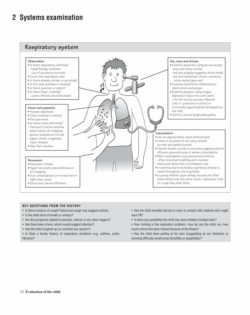
Respiratory system
Observation• Is there respiratory distress? -nasal flaring, recession -use of accessory muscles• Count the respiratory rate• Is there wheeze, stridor or grunting?• Is the child restless or drowsy?• Is there cyanosis or pallor?• Is there finger clubbing? - cystic fibrosis, bronchiectasis
Ear, nose and throat• Examine eardrums using an auroscope -grey and shiny: normal -red and bulging: suggests otitis media -dull and retracted: chronic secretory otitis media (glue-ear)• Examine nostrils for inflammation, obstruction and polyps• Examine pharynx using tongue depressor (leave this until last!) -Are the tonsils acutely inflamed (red +/- pustules or ulcers) or chronically hypertrophied (enlarged but not red)• Feel for cervical lymphadenopathy
Auscultation • Use an appropriately sized stethoscope!• Listen in all areas for air entry, breath sounds and added sounds• Absent breath sounds in one area suggests pleural effusion, pneumothorax or dense consolidation• With consolidation (e.g. pneumonia) there is often bronchial breathing with crackles heard just above the consolidated lung• In asthma and bronchiolitis expiratory wheeze is heard throughout the lung fields• In young children upper airway sounds are often transmitted over the whole chest. Asking the child to cough may clear them
Chest wall palpation• Assess expansion• Check trachea is central• Feel apex beat• Is there chest deformity? -Harrison's sulcus: asthma -barrel chest: air-trapping -pectus excavatum: normal -pigeon chest: congenital heart disease• May 'feel' crackles
Percussion• Resonant: normal• Hyper-resonant: pneumothorax or air-trapping• Dull: consolidation (or normal liver in right lower zone)• Stony dull: pleural effusions
KEY QUESTIONS FROM THE HISTORY• Is there a history of cough? Nocturnal cough may suggest asthma.• Is the child short of breath or wheezy?• Are the symptoms related to exercise, cold air or any other triggers?• Has there been a fever, which would suggest infection?• Has the child coughed up (or vomited) any sputum?• Is there a family history of respiratory problems (e.g. asthma, cystic fibrosis)?
• Has the child travelled abroad or been in contact with relatives who mighthave TB?• Is there any possibility the child may have inhaled a foreign body?• How limiting is the respiratory problem—how far can the child run, howmuch school has been missed because of the illness?• Has the child been pulling at his ears (suggesting an ear infection) orshowing difficulty swallowing (tonsillitis or epiglottitis)?

Systems examination 13
• Has the child ever complained of palpitations or of their heart racing?• Has anyone ever noticed a heart murmur in the past? (Physiological flowmurmurs may only be present at times of illness or after exercise.)• Is there a family history of congenital heart disease?• If the child has a heart defect, have they been taking prophylactic antibioticsfor dental or other invasive treatment? (Consider particularly for valve dis-orders and ventricular septal defects.)
KEY QUESTIONS FROM THE HISTORY• Has the child ever been cyanosed?• Has the child been breathless or tired (may suggest cardiac failure)?• Has the child been pale and sweaty (may suggest cardiac failure)?• Ask about the pattern of feeding in babies, as breathlessness may inhibitfeeding.• Review the child’s growth—is there evidence of failure to thrive?• Has there been any unexplained collapse, such as fainting?
Cardiovascular system
Observation• Is there central cyanosis? Peripheral cyanosis can be normal in young babies and those with cold peripheries• If the child is breathless, pale or sweating this may indicate heart failure• Is there finger clubbing? - cyanotic heart disease• Is there failure to thrive? -suggests heart failure
Palpation• Feel apex beat (position and character), reflects left ventricular function• Feel for right ventricular heave over sternum• Feel for thrills (palpable murmurs)• Hepatomegaly suggests heart failure. Peripheral oedema and raised JVP are rarely seen in children
Circulation• Measure blood pressure with age- appropriate cuff, which should cover 2/3 of the upper arm• Check capillary refill time; if more than 2 s, consider shock
Auscultation• On the basis of the child's age, pulse, colour and signs of failure try to think what heart lesion may be likely, then confirm this by auscultation• Listen for murmurs over the valve areas and the back (see p. 94). Diastolic murmurs are always pathological• Listen to the heart sounds: are they normal, increased (pulmonary hypertension), fixed and split (ASD) or are there added sounds (gallop rhythm in heart failure or ejection click in aortic stenosis)?
Pulse• Rate: fast, slow or normal?
Age Normal pulse (years) (beats/min)
<1 110–160 2-5 95–140 5-12 80–120 >12 60–100
• Rhythm: regular or irregular? Occasional ventricular ectopic beats are normal in children• Volume: full or thready (shock)• Character: collapsing pulse is most commonly due to patent arterial duct. Slow rising pulse suggests left ventricular outflow tract obstruction• Always check femoral pulses in infants—coarctation of the aorta leads to reduced or delayed femoral pulses
1 2
Systolic murmur
1

14 Evaluation of the child
KEY QUESTIONS FROM THE HISTORY• Review the child’s diet. Ask in detail what the child eats. ‘Take me througheverything you ate yesterday.’• Is the quantity of calories sufficient and is the diet well balanced and appropriate for the child’s age?• Ask about the pattern of weight gain. The parent-held record (red book) can provide invaluable information about previous height and weight measurements.• Does the child have a good appetite?• Has there been any vomiting?• In babies ask about posseting (small vomits of milk) and regurgitation ofmilk into the mouth, which may suggest gastro-oesophageal reflux.
Palpation• Use warm hands and ask whether the abdomen is tender before you begin• Is there distension, ascites or tenderness?• Palpate the liver: 1–2 cm is normal in infants. Is it smooth and soft or hard and craggy?• Feel for the spleen, using bimanual palpation. Turning the child onto the right side may help• Feel for renal enlargement• Palpate for other masses and check for constipation (usually a mass in the left iliac fossa)
Percussion• Percuss for ascites (shifting dullness) and to check for gaseous distension
Rectal examination• This is very rarely indicated, but examine the anus for fissures or trauma
Genitalia• Check for undescended testes, hydroceles and hernias. Retractile testes are normal• In girls examine the external genitalia if there are urinary symptoms
Auscultation• Listen for normal bowel sounds. 'Tinkling' suggests obstruction
Observation• Make sure the child is relaxed—small children can be examined on a parent's lap; older children should lie on a couch• Jaundice: look at the sclerae and observe the urine and stool colour (dark urine and pale stools suggests obstructive jaundice)• Check conjunctivae for anaemia• Oedema: check over tibia and sacrum. Peri- orbital oedema may be the first thing noticed by parents• Skin: look for spider naevi—suggests liver disease• Wasted buttocks: suggests weight loss and is characteristic of coeliac disease• Measure the mid upper arm circumference
Abdominal system and nutritional status(See also Chapters 3 and 22)
• Has there been any diarrhoea? Always assess what the parents mean bydiarrhoea—frequent or loose stools or both?• Has the child been constipated? Straining, pain on defaecation, poorappetite and a bloated feeling may suggest this is a problem.• Have there been any urinary symptoms such as frequency, dysuria or enure-sis?• Has the child got any abdominal pain? Ask about the site and nature of thepain.• Is there a relevant family history (e.g. coeliac disease, inflammatory boweldisease, constipation)?

Systems examination 15
KEY QUESTIONS FROM THE HISTORY• Has there been any developmental concerns—quickly review major milestones?• Has there been any concern about hearing or vision?• Did the child pass the hearing screening check (currently at 7 months)?• Has the child ever had a convulsion or unexplained collapse?• Is there a relevant family history (ask specifically about blindness, deafness,learning difficulties and genetic disorders such as muscular dystrophy)?
Neurological assessment
Observation• Abnormal movements: choreoathetoid 'writhing' movements, jerks in myoclonic epilepsy and infantile spasms• Gait—this can provide important clues: -stiffness: suggests UMN lesion -waddling: spastic diplegia, Duchenne muscular dystrophy(DMD) or congenital dislocation of hips -weakness on standing, e.g. Gower sign in DMD -broad based gait: ataxia• Muscle bulk/wasting• Posture: look for evidence of contractures
Cranial nerves• Examine as in adults
Coordination• Finger–nose test and heel–shin test, and observe gait. Very important if considering CNS tumours as cerebellar signs are common
Reflexes• Assess at knee, ankle, biceps, triceps and supinator tendons• Clonus may be seen in UMN lesions• Plantar reflex is upwards until 8 months of age, then downwards
Tone• Hypotonia suggests LMN lesion• Spasticity suggests UMN lesion and is seen in cerebral palsy, especially in thigh abductors and calf muscles (may cause toe walking)
Power• Describe in upper and lower limbs, against resistance
Young children cannot cooperate with a formal neurological examination so observation becomes more important: watch what the child is doing while you play with them• How does the infant move spontaneously? Reduced movement suggests muscle weakness• What position are they lying in? A severely hypotonic baby adopts a 'frog's leg' position (see below)• Palpate anterior fontanelle to assess intracranial pressure• Assess tone by posture and handling: a very floppy hypotonic baby tends to slip through your hands like a rag doll. Put your hand under the abdomen and lift the baby up in the ventral position: a hypotonic infant will droop over your hand. Pull the baby to sit by holding the baby's arms: observe the degree of head lag. Hypertonia is suggested by resistance to passive extension of the limbs and by scissoring (crossing-over) of the lower limbs when the infant is lifted up (see below)• Primitive reflexes are present at birth. Persistence beyond normal period suggests a UMN lesion Moro reflex Symmetrical abduction and then adduction of the arms when the baby's head is dropped back quickly into your hand (see below). Disappears by 4 months Palmar grasp Touching the palm causes the baby to grip an object. Disappears by 2 months Asymmetric tonic neck reflex The arm is extended on the side the baby is facing while the opposite arm is flexed(see below). Disappears by 6 months
Asymmetric tonic neck reflex
Moro reflexScissoring of thelower limbs
'Frog's leg' position
Neurological examination in infants
• Has there been any change in school performance or personality?• Has the child been clumsy or had a change in gait?• Has there been any headache or vomiting (may suggest raised intracranialpressure)?• Ask about function—how is the child limited by their condition, if at all?• Briefly review the social situation—does the family receive any relevant benefits, e.g. disability living allowance? Are there mobility problems?

16 Evaluation of the child
KEY QUESTIONS FROM THE HISTORY• Have the parents been concerned about the child’s vision?• Has anyone ever noticed a squint?• Is the child able to see clearly (for example, the board at school)?• Is there any relevant family history (e.g. retinitis pigmentosa, congenitalcataracts)?
Observation of eyes• Look at the iris, sclera and pupil• Check pupils are equal and react to light,
both directly and indirectly• Look for red reflex to exclude cataract,
especially in the newborn• Look at reflection of light on the cornea—
is it symmetrical or is one eye squinting? (see box opposite)
• Look at the inner epicanthic folds—if very prominent they may cause a pseudosquint
Assessment of a squint• Any squint in an infant beyond the age of 6
weeks needs referral to an ophthalmologist. A squinting eye that is left untreated may cause amblyopia (cortical blindness) on that side
• Some 'latent' squints are present only when the eye is tired; the history is important
• Check the corneal light reflex at different angles of gaze
• Check ocular movements—is there a fixed angle between the eyes or a paralytic squint, where the squint increases with eye movement?
• Check visual acuity• Perform fundoscopy and red reflex• Perform the cover test by asking the child to
fix on an object. Cover the 'good' eye and watch the squinting eye flick to fix on the object. Latent squints may also become apparent when that eye is covered
Fundoscopy• An essential but difficult skill—practise on
every child you see!• Look at the optic disc and retina, the red
reflex and the lens
Visual acuity• Does the child fix and follow an object
through 180 degrees?• Can they see small objects (e.g. hundreds
and thousands, small rolling Stycar balls)• Older children can perform a modified
Snellen chart with objects
Ocular movements and visual fields• Test full range of movements, looking for
paralytic muscle or nerve lesions• Look for and describe any nystagmus• Check visual fields by using a 'wiggling'
finger
Normal symmetrical light reflex
Pseudosquint due to prominent innerepicanthic folds
Left convergent squint—note assymetric light reflex
When the good eye is covered the squintingeye straightens (fixates)
The visual system
• Has the child been complaining of headaches, which may suggest poorvisual acuity?• Has the child seen an optician recently?• Are there any risk factors for visual problems, such as extreme prematurity,diabetes mellitus or other neurological concerns?

Systems examination 17
KEY QUESTIONS FROM THE HISTORY• Has the child had any joint pain or swelling?• Is the child able to walk and exercise normally?• Have the parents noticed any change in gait or clumsiness?• Has there been any unexplained fever (may suggest autoimmune disordersor septic arthritis)?
Musculoskeletal systemIndividual joint problems are discussed in Chapters 32 and 55.
Observation• Observe joints for swelling, redness or deformity• Observe muscle bulk above and below the joint• Observe the function: what is the gait, can the child do up buttons or hold a pencil?• Is there any obvious scoliosis?
Scoliosis• Observe the child standing: are the shoulders level?• Ask the child to touch their toes-scoliosis causes bulging of the ribs on one side. This is the most sensitive way to check for a scoliosis• Postural scoliosis is common in teenagers
Range of movements• Assess the limit of active movements, then move the child's limb to assess passive movements. Observe the face for signs of pain, and stop before this occurs• Check all the large joints in flexion, extension, rotation, abduction and adduction• It is particularly important to check that the hip joints fully abduct in newborns and in children with cerebral palsy in order to exclude hip dislocation. (see Chapter 48)
Palpation• Feel the temperature of the skin over the joint, and feel for joint tenderness• Feel for an effusion: in the knee milk fluid down and feel a bulge in the medial aspect. If the effusion is large the patella can be rocked in and out, causing a fluid bulge above it
• What is the level of function like—can the child manage fiddly tasks such asdoing up buttons?• Have the parents noticed any rashes (may suggest rheumatoid disease (seep. 117) or Henoch–Schönlein purpura (see p. 85))?

18 Evaluation of the child
3 Understanding investigations I
HaematologyHaemoglobin• High at birth (18 g/dl), falling to lowest point at 2 months (range 9.5–14.5 g/dl). Stabilizes by 6 months• Low haemoglobin indicates anaemia. Further investigation will pinpoint the cause (see below)
Blood film Normal valuesHaemoglobinHaematocritWhite cell countReticulocytesPlateletsMCVMCHESR
Mean cell volume (MCV)• Measures the size of RBCs• Microcytic anaemia (MCV <76 fl) is usually due to iron deficiency, thalassaemia trait or lead poisoning• Macrocytosis may reflect folate deficiency Low Hb
measure MCV
Low MCV(microcytic)
Normal MCV(normocytic)
Lowferritin
Abnormalelectrophoresis
Highreticulocyte count
Iron deficiencyanaemia
Thalassaemiatrait
Recent bloodloss
Haemolysis
Lowreticulocyte count
Normalbilirubin
Target cellsHigh bilirubin
Chronicillness
Mean cell haemoglobin (MCH)• Reflects the amount of haemoglobin in each red cell. Is usually low (hypochromic) in iron deficiency
White blood cells• Leucocytosis usually reflects infection—neutrophilia and 'left shift' (i.e. immature neutrophils) implies bacterial infection. Lymphocytosis is commoner in viral infections, atypical bacterial infection and whooping cough• Neutropenia (neutrophils < 1.0 x 10⁹/l) can occur in severe infection or due to immunosuppression. There is a high risk of spontaneous infection• Leukaemia: There is usually a very high (or occasionally low) WCC with blast cells seen. Bone marrow aspirate is required (see Chapter 56)
Clotting• Prothrombin time (PT) compared with a control is used to calculate the INR: normal is 1.0. Principally assesses extrinsic pathway. Prolonged in Vitamin K deficiency, liver disease and disseminated intravascular coagulation (DIC)• Activated partial thromboplastin time (APTT) reflects the intrinsic pathway. Prolonged in heparin excess, DIC and haemophilia A• Fibrin degredation products (FDPs) are increased in DIC• Bleeding time: literally the time a wound bleeds for. Prolonged in von-Willebrand's disease and thrombocytopenia
Platelets• High platelet count usually reflects bleeding or inflammation (e.g. Kawasaki's disease)• Low platelet count is commonly seen with idiopathic thrombocytopenic purpura (ITP) when there is a risk of spontaneous bruising and bleeding. In the newborn it may be low due to maternal IgG-mediated immune thrombocytopenia
11 – 14 g/dl 30 – 45% 6 – 15 x 109/l 0 – 2% 150 – 450 x 109/l 76 – 88 fl 24 – 30 pg 10 – 20 mm/h
Flow diagram to show the investigation of anaemia
Investigations should only be requested to confirm a clinical diagnosis,or if indicated after taking a thorough history and examination. Some-times investigations are performed to rule out more serious but lesslikely conditions. Blindly performing investigations as a ‘fishing’ exer-
cise in the hope of throwing up an abnormality is usually counter-productive, often leading to increased anxiety and further investigationswhen unexpected results are obtained. These pages describe how tointerpret some of the common investigations performed in paediatrics.

Understanding investigations I 19
Normal arterial blood gas valuespH PCO2 PO2 HCO3–
Determining the type of blood gas abnormality
7.35 – 7.42 4.0 – 5.5 kPa 11 – 14 kPa (children) 8 – 10 (neonatal period) 17 – 27 mmol/l
pH PCO2 PO2 HCO3–
Metabolic acidosis Low N/low* N LowRespiratory acidosis Low High N/low N/high*Metabolic alkalosis High N/high* N HighRespiratory alkalosis High Low N/high N/low*
*Refers to the compensated state
Compensation can occur by the kidneys, which can vary the amount of bicarbonate excreted. A persistent respiratory acidosis due to chronic lung disease willlead to retention of bicarbonate ions to buffer the acid produced by CO2 retention. Hence, a compensated respiratory acidosis will have a low–normal pH, a high PCO2 and a very high bicarbonate level
Ventilation
Henderson–Hasselbach equation
Renal adaptation
Metabolic acidosis • Severe gastroenteritis• Neonatal asphyxia (build-up of lactic acid)• Shock • Diabetic ketoacidosis• Inborn errors of metabolism• Loss of bicarbonate (renal tubular acidosis)
Respiratory acidosis• Respiratory failure and underventilation
Metabolic alkalosis• Usually due to vomiting, e.g. pyloric stenosis
Respiratory alkalosis• Hyperventilation (e.g. anxiety)• Salicylate poisoning: causes initial hyperventilation and then metabolic acidosis due to acid load
CO2 + H2O H+ + HCO3–H2CO3
Normal rangesSodium 135 – 145 mmol/lPotassium 3.5 – 5.0 mmol/lChloride 96 – 110 mmol/lBicarbonate 17 – 27 mmol/lCreatinine 20 – 80mmol/lUrea 2.5 – 6.5 mmol/lGlucose 3.0 – 6.0 mmol/lAlkaline 150 – 1000 (infants)phosphatase 250 – 800 (child)
Causes of abnormal sodium balance Hypernatraemia (Na+ >145 mmo/l)• Dehydration - fluid deprivation or diarrhoea• Excessive sodium intake -inappropriate formula feed preparation -deliberate salt poisoning (very rare)Hyponatraemia (Na <135 mmol/l)• Sodium loss -diarrhoea (especially if replacement fluids hypotonic) -renal loss (renal failure) -cystic fibrosis (loss in sweat)• Water excess -excessive intravenous fluid administration -SIADH (inappropriate antidiuretic hormone secretion)
Characteristic patterns of serum electrolyte abnormalitysometimes suggest particular diagnoses:• Pyloric stenosis: There is often a metabolic alkalosis, a low chloride and potassium concentration (due to repeated vomiting and loss of stomach acid) and a low sodium concentration• Diabetic ketoacidosis: There is a metabolic acidosis with a very low bicarbonate, a high potassium, high urea and creatinine and a very high glucose concentration • Gastroenteritis: Urea concentration is high, but the sodium may be either high or low depending on the sodium content of the diarrhoea, and on the type of rehydration fluid that has been administered
Interpretation of blood gases
Electrolytes and clinical chemistry
The acidity of the blood is measured by pH. A high pH refers to an alkalosis and a low pH to an acidosis. Once the blood becomes profoundly acidotic (pH<7.0), normal cellular function becomes impossible. There are metabolic and respiratory causes of both acidosis and alkalosis (see below). The pattern of blood gas abnormality (particularly the pH and PCO2) can be used to determine the type of abnormality.
The normal values are shown below. Alterations in sodium usually reflect alterations in the level of hydration and total body water content. A sudden fall in sodium can cause fitting. High potassium levels can cause serious cardiac arrhythmias and need to be controlled rapidly. High potassium levels are commonly seen in acute renal failure, or may be artefactual due to haemolysis if venepuncture was difficult. Therapies to reduce a high potassium level include salbutamol, insulin and dextrose (to drive potassium into the cells) and calcium resonium.

20 Evaluation of the child
4 Understanding investigations II
As respiratory disorders are so common in paediatric practice, it is very important to be able to accurately interpret chest radiographs. Ifthere is uncertainty the film should be discussed with an experiencedradiologist.• Identify the patient name, date and orientation (left and right).• Check the penetration—the vertebrae should just be visible behind theheart shadow.• Check that the alignment is central by looking at the head of the clavicles and the shape of the ribs on each side.• Comment on any foreign objects such as central lines.
Features to look for on a chest radiograph
Loss of heart border
CXR of right middle and upper lobe pneumonia
R L A P
Lateral X-ray showing right upper and middle lobe pneumonia
Diaphragmaticborder maintained
Middle lobeconsolidation
Heartshadow Diaphragm
Oblique fissure
Upper lobe consolidation
Upper lobeconsolidation
• Examine the bony structures, looking for fractures, asymmetry andabnormalities (e.g. hemivertebrae). Rib fractures are best seen byplacing the X-ray on its side.• Check both diaphragms and costo-phrenic angles are clear. The rightdiaphragm is higher than the left because of the liver. Check there is noair beneath the diaphragm (indicates intestinal perforation).• Look at the cardiac outline. At its widest it should be less than half thewidth of the ribcage (cardiothoracic ratio <0.5).• Look at the mediastinum—note that in infants the thymus gland cangive a ‘sail’-like shadow just above the heart.

Understanding investigations II 21
Lumbar puncture
or if there is a low platelet count or coagulopathy. A fine spinal needlewith a stylet is passed between the vertebral spines into the cere-brospinal fluid (CSF) space. A few drops of CSF are then collected formicroscopy, for culture and for analysis of protein and glucose concen-trations. Samples can also be sent for polymerase chain reaction (PCR)analysis to look for evidence of meningococcal or herpes infection ifmeningitis or encephalitis is suspected. Normal CSF is usually ‘crystalclear’. If it is cloudy, this suggests infection. Fresh blood which clearsusually indicates a traumatic tap, but a massive intracranial haemor-rhage must be considered if the CSF remains blood-stained. Old bloodfrom a previous haemorrhage gives a yellow ‘xanthochromic’ appear-ance. A manometer can be used to measure the CSF pressure, thoughthis is not routinely performed.
Urinalysis
Fresh urine should be collected into a sterile container from a mid-stream sample if possible. Urine bags placed over the genitalia may beused in infants, but beware of contamination.• Observe the urine—is it cloudy (suggests infection) or clear?• What is the colour—pink or red suggests haematuria from the lowerurinary tract? Brown ‘cola’ coloured urine suggests renal haematuria.• Smell the urine for ketones and for the fishy smell of infection.Unusual smelling urine may suggest an inborn error of metabolism.• Dipstick test the urine using commercial dipsticks. This may revealprotein (suggests infection, renal damage or nephrotic syndrome),glucose (present in diabetes), ketones (in diabetic ketoacidocis, DKA)or nitrites (suggestive of infection). These sticks are very sensitive tothe presence of blood, and may detect haematuria even if the urine looksclear.• Finally, examine the urine under the microscope for white cells, redcells, casts and the presence of organisms. If suspicious of infectionsend a sample for culture. A pure growth of >105 colony-forming unitsof a single organism and >50 white cells/mm3 confirms infection (see p. 69).
Analysis of CSF.
Normal Bacterial meningitis Viral meningitis
Appearance Crystal clear Turbid, organisms seen ClearWhite cells <5/mm3 ≠ ≠ ≠ (polymorphs) ≠ (lymphocytes)Protein 0.15–0.4 g/l ≠≠ NormalGlucose >50% blood Ø Normal
KEY POINTS• Before ordering an investigation consider how the result might alter themanagement.• Try to focus investigations on the differential diagnosis based on clinicalassessment.• Sometimes investigations can be used to quickly rule out important orserious diagnoses.• If a test is performed you must chase up the result.
• Check lung expansion—if there is air trapping the lung fields willcover more than nine ribs posteriorly, and the heart will look long andthin.• Examine the lung fields looking for signs of consolidation, vascularmarkings, abnormal masses or foreign bodies. Check that the lungmarkings extend right to the edge of the lung—if not, consider a pneu-mothorax (dark) or a pleural effusion (opaque). Consolidation may bepatchy or dense lobar consolidation. A lateral X-ray may be required todetermine exactly which lobe is affected. A rule of thumb is that con-solidation in the right middle lobe causes loss of the right heart bordershadow and right lower lobe consolidation causes loss of the rightdiaphragmatic shadow. Always look at the area ‘behind’ the heartshadow for infection in the lingula. If the mediastinum is pulled towardsan area of opacity consider collapse rather than consolidation as thepathology.
Lumbar puncture and CSF analysis
Lumbar puncture is usually performed to diagnose or exclude meningi-tis. It should not be performed if there is evidence of raised intracranialpressure, if the child is haemodynamically unstable (e.g. septic shock)

22 The developing child
5 Growth and puberty
GrowthAccurate measurement of growth is a vital part of the assessment of children. In order to interpret a child's growth, measurements must be plotted on a growth chart. If there is concern about growth, the rate of growth must be assessed by measuring the child on two occasions at least 4–6 months apart.
Height• Use a properly calibrated standing frame• The child should be measured barefoot with knees straight and feet flat on the floor• Stretch the child gently and read the measurement
Principles of plotting• The child's measurement should be marked with a dot (not a cross or circle) • Correct for prematurity up to the age of 2 years• Height should follow one centile between 2 years and puberty• Plateauing of growth and weight, or height less than 0.4% merits an evaluation of the child (see Chapter 41).• Infants may normally cross centiles in the first year or two, but consider whether failure to thrive is a problem (see Chapter 42).• A child's final height is expected to fall midway between the parents' centile positions
Plot on a growth chart.In the UK the 1990 UK Growth References are used: • Nine equidistant centile lines are marked • For weight, the centiles are splayed and the population is skewed towards being overweight • The 50th centile is the median for the population• A measurement on the 98th centile means only 2% of the population are taller or heavier than the child• A measurement on the 2nd centile means that only 2% of the population are lighter or shorter than the child
Length• The child should be measured lying down until 2 years of age • Measuring the length of infants requires skill• Use proper equipment and two people to hold the child
Weight • Scales must be calibrated accurately• Babies should be weighed naked (no nappy!)• Older children should be weighed in underwear only
Head circumference • Use flexible non-stretchable tape• Obtain three successive measurements and take the largest to be the occipito frontal circumference (OFC)
GROWTH CHART
The
y-ax
is u
ses
kg fo
r wei
ght
and
cm fo
r OFC
and
hei
ght/
leng
th
The x-axis may be divided in to months or decimal age© Child Growth Foundation

Growth and puberty 23
Examples of growth charts
Head
0 1 yr
0 1 yr 0 0 18 yr18 yr
0 1 yr 0 1 yr
Length
Weight
Head
LengthHeight
Height
Weight
Head
Length
Weight
Head
Length
Weight
EDD
Prematurity
Hydrocephalus Turner's syndrome Growth hormone deficiency
Coeliac disease Intrauterine growthretardation
Premature baby• The baby was born at 30 weeks gestation and is now 26 weeks old• Corrected age is 16 weeks
Coeliac disease• Note fall-off in weight at time of weaning when wheat was introduced• The fall-off in length occurs later
Intrauterine growth retardation (IUGR)• Low birth-weight baby• Many IUGR babies show catch-up but this baby clearly has not, and may have reduced growth potential• The IUGR probably started early in pregnancy because OFC and length are also affected
Hydrocephalus• The head circumference is crossing centile lines upwards• A normal but large head would grow above but parallel to the centile lines
Turner's syndrome• Poor growth• Absence of pubertal growth spurt• The child should have been referred for growth- promoting treatment when young
Growth hormone deficiency• Note the fall-off in height• GH deficiency is rare• It can be congenital, but as growth has plateaued at the age of 6 years, pituitary deficiency due to a brain tumour must be considered

24 The developing child
Genital development
Boys
Breast development
Pubic hair growth
Pubic hair growth
Stage 1
Stage 2
Stage 2 Stage 3 Stage 4 Stage 5
Stage 3 Stage 4 Stage 5
Stage 1 Stage 2 Stage 3 Stage 4 Stage 5
Stage 2 Stage 3 Stage 4 Stage 5
Girls
Puberty
The first signs of puberty are usually testicular enlargement in boys, and breast budding in girls. Puberty is precocious if it starts before the age of 8.5 years in girls and 9.5 years in boys. Puberty is delayed if onset is after 13 years in girls and 14 years in boys. A growth spurt occurs early in puberty for girls, but at the end of puberty for boys. Menarche occurs at the end of puberty. Delay is defined as no periods by 16 years of age.
Puberty is evaluated by clinical examination of the genitalia, breasts and secondary sexual characteristics. The scale used is known as Tanner staging.
Principles of puberty

Development and developmental assessment 25
6 Development and developmental assessment
Gross motor development
Fine motor development
Sitting6 weeksCurved back, needs support from adult
6–7 monthsSits with self-propping
9 monthsGets intositting positionalone
Prone positionBirthGenerally flexed posture
6 weeksPelvis flatter
4 monthsLifts head and shoulders with weight on forearms
6 monthsArms extendedsupporting chestoff couch
Pull to sit
Grasping and reaching
Building bricks Manipulation
4 monthsHolds a rattle and shakes purposefully
5 monthsReaches for object
6 monthsTransfers object from hand to hand
7 monthsFingerfeeds
5 monthsWhole handgrasp
Pencil skills
18 monthsScribbles with a pencil
3 yearsDraws a circle
4 yearsDraws a cross
5 yearsDraws a triangle
9 monthsImmature pincer grasp
10 monthsPoints at bead
12 monthsMature pincergrasp
15 monthsBuilds a towerof two cubes
18 monthsBuilds a tower of threeto four cubes
12 monthsGives bricks to examiner
Standing and walking6 monthsStands withsupport
10 monthsPulls to standingand standsholding on
12 monthsStands, and walks withone hand held
15 monthWalks indepen-dently and stoops to pick up objects
BirthCompletehead lag
6 weeksHead controldeveloping
4 monthsNo head lag

26 The developing child
Speech and language development
Social development
Speech3 monthsVocalizes
8 monthsDouble babble
12 monthsTwo or three words with meaning
18 months10 words
24 monthsLinking two words
3 yearsFull sentences,talks incessantly
ooh, aah dadamama
Mummy
NoTa
Teddy
Teddy goes to sleepTeddy's tired
Good night Teddy
Bed DogBottle
Daddy Bikky
baba
Daddy gone
6 weeksSmiles responsively
16 weeksLaughing out loud
7 monthsStranger anxiety
9 monthsPeek a boo,waves bye bye
15 monthsDrinks from a cup
18 monthsSpoon-feeding self
About 21/2 years(very variable)Toilet trained by day
3 yearsDresses self(except buttons)

Development and developmental assessment 27
KEY POINTS• Delay in one area is often not of concern and maybe familial.• Delay in all areas is a cause for concern (see Chapter 46, global develop-mental delay).• Do not forget to correct prematurity.• See p. 100 for causes of delayed development.
Milestones that are essential to remember.
Age Milestone
4–6 weeks Smiles responsively6–7 months Sits unsupported9 months Gets to a sitting position10 months Pincer grasp12 months Walks unsupported
Two or three words18 months Tower of three or four cubes24 months Two to three word sentences
At any age Maternal concernRegression in previously acquired skills
At 10 weeks No smiling
At 6 months Persistent primitive reflexesPersistent squintHand preferenceLittle interest in people, toys, noises
At 10–12 months No sittingNo double syllable babbleNo pincer grasp
At 18 months Not walking independentlyLess than 6 wordsPersistent mouthing and drooling
At 21/2 years No 2–3-word sentences
At 4 years Unintelligible speech
Developmental warning signs.
Development and developmental assessmentParents are always interested in their child’s developmental progressand are usually concerned if any aspect is delayed. It is an importantindicator of a child’s wellbeing, and delay or abnormal developmentmay indicate serious limitations for later life. Advanced development oflanguage and fine motor skills may be a sign of intelligence.
An assessment of developmental progress is important at all clinicalencounters with children. You firstly need to know the normal progres-sion of development in the early years, and then develop your skills inassessing babies and children of different ages.
How to perform a developmental assessment• Young children often will not co-operate so make the most of observing them informally. You may have to rely heavily on parentalreport, especially for language skills.• Be systematic and evaluate the four developmental areas in turn–gross motor, fine motor/adaptive, speech and language, social and alsoassess hearing and vision.• It is hard to remember all the milestones, so make sure you learn theessential milestones given below. Then ensure you know how skillsprogress. You can always check the age at which skills are acquiredafter you have finished your evaluation.• Remember that you need to correct for prematurity until the child istwo years old.• Present the tasks one at a time and try to have as few distractions forthe child as possible.• The most useful equipment to have is bricks and a crayon.
Developmental warning signsThere is a wide variation in the age at which milestones are met. It istherefore important to be aware when it is abnormal if a child has not yetacquired certain skills.

28 The developing child
7 Infant nutrition
Formula milk feeds Formula milks are based on cow's milk, but are carefully adjusted to meet the basic nutritional requirements of growing infants. The fat component is generally replaced with polyunsaturated vegetable oils to provide the correct essential fatty acids. Minerals, vitamins and trace elements are then added. Milks with more casein are produced for 'hungrier' babies aged 4–6 months, and 'follow-on' milks with more iron for babies over 6 months. Formula milk is usually made up from a dry powder, by adding one level measure of powder to each 30 ml (1 fl.oz) of cooled boiled water. Great care must be taken to sterilize the bottles and teats carefully to avoid introducing infection. The milk is then re-warmed prior to feeding. This should not be done in a microwave as pockets of milk may be heated to dangerous levels.
1. 2. 3. 4. 5. 6.Sterilize the feeding bottle
Add the appropriate volume of cooledboiled water to thebottle
Add 1 level scoop ofmilk powder to each30 ml of water
Shake bottle well Keep in fridge until ready to feed
Rewarm the feed to room temperature or body temperature prior to feeding
Breast-feeding
Ways to encourage successful breast-feeding• Introduce concept of breast-feeding to both parents antenatally• Put the baby to the breast immediately after delivery• Allow the baby to feed on demand, especially in the early days• Avoid offering any formula feeds• Ensure mother receives good nutrition and plenty of rest• Provide skilled breast-feeding advisors to help mother through any initial problems with breast-feeding• Ensure correct 'latching on' with the baby's mouth wide open and good positioning
Advantages of breast-feeding• Perfect balance of milk constituents• Little risk of bacterial contamination• Anti-infective properties (IgA, macrophages, etc.)• Ideal food for brain growth and development• Convenient• No expense of purchasing milk• Psychologically satisfying• Reduces risk of atopic disordersTheoretical problems with breast-feeding (rare)• Can initially be exhausting for the mother• Can transmit infection (e.g. HIV, although in developing countries the best advice is still to exclusively breast-feed)• Some drugs can be excreted in breast milk (e.g. warfarin)
Lactation• At birth, prolactin levels rise sharply and this is further stimulated by the infant sucking at the breast. Prolactin determines milk production from the breast alveoli, and is increased by the frequency, duration and intensity of sucking• The actual flow of milk is aided by the 'let- down' reflex. Rooting at the nipple causes afferent pathways to stimulate the post- erior pituitary to secrete oxytocin, which stimulates the smooth muscle around the alveolar ducts to express the milk from the breast. The let-down reflex can be stim- ulated by hearing the baby cry or by contact with the baby, and can be inhibited by stress or embarrassment
• The majority of the milk is taken from the breast in the first 5 min and this may be followed by non-nutritive sucking
Weaning • 0–4 months: breast or formula milk only• 4–6 months: puréed or liquidized foods• 6–9 months: finger foods, juice in a cup• 9–12 months: three meals a day, with family• >1 year: cow's milk in a beaker or cup; adult-type food chopped up
Infant stimulation(emotion)
Posteriorpituitary
Nipplestimulation
Let-downreflex
↑Milkproduction
Prolactin
Oxytocin

Nutrition from birth to school ageMilk provides all the nutrients needed by newborn infants for the first4–6 months of life. Breast milk is the ideal milk for human babies, butformula milk may be needed as an alternative in some cases. Thenewborn infant has high calorie and fluid requirements and to achieveoptimal growth requires approximately 150 ml/kg/day fluid and 110 kcal/kg/day (462 kJ/kg/day). About 40% of this energy comes fromcarbohydrate (mostly lactose) and 50% from fat. Milk also containsprotein in the form of casein, lactalbumin and lactoferrin. Colostrum isthe thin yellow milk produced in the first few days which is high inimmunoglobulins.
Infants also require adequate amount of minerals such as calcium andphosphate, as well as vitamins and trace elements. Breast milk is defi-cient in Vitamin K, and so all newborn infants are given vitamin K atbirth to prevent haemorrhagic disease of the newborn. Weaning ontosolids usually starts around 4 months, and infants should not have cowsmilk until they are over a year.
Technique of breast-feedingMothers should be encouraged to put their babies to the breast soonafter delivery. Little milk is produced but the suckling stimulates lacta-tion. Colostrum is produced in the first days which is rich in energy andanti-infective agents. It is important that the baby is taught to ‘latch-on’to the breast properly with a widely open mouth so that the areolar andnot just the nipple is within the babies mouth. The majority of the milkis taken by the baby in the first 5 min. Time after this is spent in non-nutritive suckling. Mothers can feel their breast ‘emptying’. Babiesshould not be pulled off the breast, but the suck released by inserting aclean finger at the side of the babies mouth. Each feed should start on the alternate breast. In the first few days the breasts may becomepainfully engorged with milk and the nipples sore, especially if thebaby’s position is not optimal. Mothers need a lot of encouragement andadvice to get through this time. It is important to try to avoid alternatingbreast and formula feeds. Formula feeds should only be introduced ifbreast-feeding is contraindicated or has failed completely. It is notappropriate to ‘top-up’ with formula or use bottles to give the mother arest. This may help in the short term but usually leads to production ofmilk tailing off and breast-feeding failing altogether.
WeaningMost healthy infants do not require weaning until 4–6 months of age,although some premature infants may want solids earlier. Generallycereals, rusks or rice-based mixtures are introduced first, mixed withexpressed breast milk or formula milk. This semi-solid mixture can begiven by a spoon before milk feeds. Puréed fruit or vegetables are alsosuitable. Modern baby cereals are gluten free which may be associatedwith a fall in the incidence of coeliac disease (see Chapter 25). As thechild grows older the feeds can become more solid and are given as
three meals a day. From 6 to 9 months they will enjoy finger-feedingthemselves and can chew on rusks or toast. From about 9 months theycan generally eat a mashed or cut-up version of adult food. Undilutedfull-fat pasteurized cows milk can be given from 1 year of age. Earlierintroduction of cows milk or persistence of exlusive breast-feeding canlead to iron deficiency. Vitamin supplements may be needed from 6months in breast-fed babies, until they are on a full mixed diet.
Nutrition in the preschool yearsAs a toddler the child becomes more adept at holding a spoon and canfeed independently, and can drink from a beaker or cup. Milk is nolonger the main source of nutrients, although the child should still drinka pint a day. Whole-fat milk should be used until age 5 to provide plentyof calories. Awell-balanced diet should include food from the four maingroups:• Meat, fish, poultry and eggs.• Dairy products (milk, cheese, yoghurt).• Fruit and vegetables.• Cereals, grains, potatoes and rice.
In order to avoid dental caries it is important to avoid very frequentsnacking on sugary foods or drinks—three meals and two snacks is recommended, although this may be adapted to the individual child.Iron-deficiency anaemia is common at this age, due to high require-ments for growth and poor dietary intake, especially in the ‘faddyeater’. Vitamin C present in orange juice can enhance iron absorptionfrom the gut.
Nutrition in the school-age childAt school, children have to learn to eat food out of the family setting.They usually have a midday meal, and fruit or milk may be provided atbreak times. The principles of healthy eating should be maintained,although peer pressure to eat crisps or sugary snacks is high. Schoolshave an educational role to play in encouraging healthy eating and ahealthy lifestyle. During adolescence there is a greater energy require-ment to allow for increased growth. This may coincide with a lifestylethat leads to snacking and missing meals, or to restrictive dieting orover-consumption of fast food. Obesity and eating disorders often havetheir onset around this time.
Infant nutrition 29
KEY POINTS• Breast milk provides ideal nutrition for babies.• The optimum time for weaning is 4–5 months.• Formula feeds need to be made up carefully to avoid infection.• ‘Doorstep’ cows milk should not be used until 1 year.• Full-fat milk is recommended until 5 years of age.• Toddlers need to be allowed to explore food and develop independent eatinghabits.

30 The developing child
8 Common problems for parents
History Ask what is troubling the parents most—is it the child or other stresses in their lives, such as tiredness, problems at work or marital problems? What are the triggers for difficult or unwanted behaviour? Does it occur when the child is hungry or tired, or at any particular time of day? Colic tends to occur in the evenings; tantrums may be commoner if the child is tired Does the behaviour happen consistently in all settings or is it specific to one place, e.g. the toddler may behave well at nursery but show difficult behaviour at home? Does the behaviour differ with each parent? How do the parents deal with the behaviour—do they get angry or aggressive, are they consistent, do they use bribery or do they give in to the toddler eventually? What strategies have the parents already tried to deal with the situation? Is there any serious risk of harm? Some behaviour, such as encopresis or deliberate self-harm, may reflect serious emotional upset. Most toddlers who are faddy eaters are growing well and do not suffer any long-term nutritional problems
Management In most cases the parents can be reassured that the behaviour is very common, often normal and that with time and common sense it can be controlled With tantrums it can be helpful to use the ABC approach: A What antecedents were there? What happened to trigger the episode? B What was the behaviour? Could it be modified, diverted or stopped? C What were the consequences of the behaviour? Was the child told off, shouted at or given a cuddle? Generally, it is best to reward good behaviour (catch the child being good) and ignore bad behaviour. Star charts can be very useful: the child gets a star for good behaviour (staying in bed, etc.) and then a reward after several stars Parents must try hard not to be angry or aggressive as this may reinforce the bad behaviour in the child
Examination Not usually required If the parents are concerned about sudden-onset or severe crying in a baby, it is important to exclude serious infection such as meningitis or urinary tract infection, intussusception, hernias and otitis media Babies with colic are usually less than 3 months old, go red in the face with a tense abdomen and draw up their legs. The episodes start abruptly and end with the passage of flatus or faeces
Eating problems in toddlers• Food refusal• Faddy eating—only eating one or two types of food• Overeating• Battles over eating and mealtimes• Snacking• Excessive drinking of juice
Unwanted habits• Thumb sucking• Nail biting• Masturbation• Head banging• Hair pulling• Bedwetting• Encopresis (passing faeces in inappropriate places)
Aggressive behaviour• Temper tantrums• Hitting and biting other children• Destroying toys • Destroying furniture• Commoner in boys and in larger families• May reflect aggression within family• Requires calm, consistent approach• Avoid countering with aggression• Use time-out and star charts
The crying baby• Wet or dirty nappy• Too hot or too cold• Hungry• Wind• Colic• Environmental stress• Reflux oesophagitis• TeethingIf sudden severe crying, consider:• Any acute illness• Otitis media• Intussusception• Strangulated inguinal hernia
Temper tantrums• Normal, peak at 18–36 months• Screaming• Hitting• Biting• Breath-holding attacks (see p. 74)Strategies that may help• Avoid precipitants such as hunger and tiredness• Divert the tantrum by distraction• Stay calm to teach control• Reward good behaviour• Try to ignore bad behaviour until calm• Use time-out
Sleeping problems• Difficulty getting to sleep• Waking during the night• Sleeping in parents bed• Nightmares and night terrors
What you need from your evaluation

Common emotional and behavioural problemsSuch problems are seen so often that many would regard them asnormal, although in a small minority the behaviour can be so disruptiveas to cause major family upset. The general practitioner or paediatricianshould be comfortable giving basic psychological advice on behaviourmanagement to help parents through what can be a stressful, exasperat-ing and exhausting phase of their child’s development.
Crying babies and colicCrying is usually periodic and related to discomfort, stress or tempera-ment. However, it may indicate a serious problem, particularly if ofsudden onset. In most instances it is just a case of ensuring that the baby is well fed, warm but not too hot, has a clean nappy, comfortableclothes and a calm and peaceful environment. A persistently cryingbaby can be very stressful for inexperienced parents. It is important thatthey recognize when they are no longer coping and are offered support.
Infantile colic is a term used to describe periodic crying affectinginfants in the first 3 months of life. The crying is paroxysmal, and maybe associated with hunger, swallowed air or discomfort from over-feeding. It often occurs in the evenings. Crying can last for severalhours, with a flushed face, distended and tense abdomen and drawn-uplegs. In between attacks the child is happy and well. It is important toconsider more serious pathology such as intussusception or infection.Colic is managed by giving advice on feeding, winding after feeds andby carrying the baby. Colic is not a reason to stop breast-feeding,although the mother going onto a cows milk-free diet can occasionallyhelp. Various remedies are available but there is little evidence for their effectiveness. Infantile colic usually resolves spontaneously by 3months.
Feeding problemsOnce weaned, infants need to gradually move from being fed with aspoon to finger feeding and feeding themselves. This is a messy time,but the infant needs to be allowed to explore their food and not be eitherforce-fed or reprimanded for making a mess.
Toddler eating habits can be unpredictable—eating huge amounts atone meal and sometimes hardly anything at the next. At this age, meal-times can easily become a battle and it is important that they are keptrelaxed and the child is not pressurized into eating. Small helpings thatthe child can complete work best, and second helpings can be given.Eating together as a family encourages the child to eat in a socialcontext. Feeding at mealtimes should not become a long protractedbattle!
Sleeping problemsBabies and children differ in the amount of sleep they need and parentsvary in how they tolerate their child waking at night. In most casessleeping ‘difficulties’ are really just habits that have developed by lackof establishing a clear bedtime routine. Difficulty sleeping may alsoreflect conflict in the family or anxieties, for example about startingschool or fear of dying. Successfully tackling sleeping problemsrequires determination, support and reassurance.• Refusal to settle at night. Difficulty settling may develop if babiesare only ever put to bed once they are asleep. A clear bedtime routine isimportant for older children—for example a bath, a story and a drink.• Waking during the night. This often causes a lot of stress as theparents become exhausted. It is important to reassure the child, then putthem back to bed quietly. Sometimes a technique of ‘controlled crying’
can be helpful—the child is left to cry for a few minutes, then reassuredand left again, this time for longer. Taking the child into the parental bedis understandable, but usually stores up problems for later when it is dif-ficult to break the habit.• Nightmares. The child wakes as the result of a bad dream, quicklybecomes lucid and can usually remember the content. The child shouldbe reassured and returned to sleep. If particularly severe or persistent,nightmares may reflect stresses and may need psychological help.• Night terrors. Night terrors occur in preschool years. The childwakes up confused, disorientated and frightened and may not recognizetheir parent. They take several minutes to become orientated and thedream content cannot be recalled. These episodes should not be con-fused with epilepsy. They are short-lived and just require reassurance,especially for the parents.
Temper tantrumsThese are very common in the third year of life (the ‘terrible two’s’) andare part of the child learning the boundaries of acceptable behaviour andparental control. They can, however, be extremely challenging, espe-cially when they occur in public!
The key to dealing with toddler tantrums is to try to avoid getting intothe situation in the first place. This does not mean giving in to the child’severy demand, but not letting the child get over tired or hungry, settingclear boundaries and enforcing these in a calm, consistent and con-trolled manner. There will still be times when tantrums occur—wheresafe to do so they are best ignored until the child calms down. If thisfails then ‘time-out’ can be a useful technique. The child is taken to asafe quiet environment, such as a bedroom, and left for a few minutesuntil calm. This is usually very effective as it removes the attention thechild desires, and allows the parents time to control their own anger.
Unwanted or aggressive behaviourYoung children often have aggressive outbursts which may involvebiting, hitting or scratching other children. These require consistentfirm management, with use of time-out and star charts for good behav-iour. It is important not to respond with more aggression, as this sendsconflicting messages to the child. If aggressive behaviour is persistent itis important to explore other tensions or disturbances within the family.At school age, the school will often need to be involved in a behaviourmanagement programme.
Unwanted behaviours such as thumb-sucking, hair-pulling, nail-biting and masturbation are also common in young children. The major-ity can be ignored and will resolve with time. Masturbation can usuallybe prevented by distracting the child or dressing them in clothes whichmake it more difficult. The older child should not be reprimanded butinformed that it is not acceptable in public.
Common problems for parents 31
KEY POINTS• Emotional and behavioural problems are extremely common, to the point ofbeing part of normal child development.• Parents need to be encouraged that they can manage most behaviour with aclear strategy.• A calm, confident and consistent approach to the child’s behaviour is recommended.• Parents should reward good behaviour and try to minimize attention givento undesirable behaviour.
32 The developing child
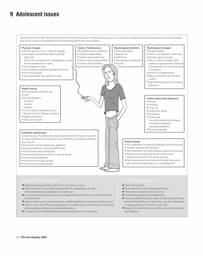
9 Adolescent issues
Approach to the adolescent Adolescence is generally a time of life when illness is rare Partly because of this, healthcare facilities for adolescents are poor, often falling between paediatric and adult care The low rate of contact with doctors means health promotion must be delivered to the adolescent Adolescents may be concerned about confidentiality when seeing their family doctor Drop-in clinics can offer immediate advice on health issues, counselling for emotional and personal problems and contraceptive advice The way in which health professionals treat adolescents is important
How to treat adolescents Take time to listen Show respect for their emerging maturity Allow them to express their concerns Avoid making judgmental statements Assure confidentiality (but make it clear there are times when confidentiality must be broken, e.g. after disclosure of ongoing abuse or if others are at risk) Respect the need for privacy—offer to see them without their parents
Physical changes• Growth spurt occurs—may feel 'gangly'• Secondary sex characteristics develop: -pubic hair -facial hair and testicular enlargement in boys -breast enlargement in girls• Voice deepens in boys• Girls undergo menarche and become fertile• Acne may develop• Gynaecomastia may develop in boys
Health issues• Contraception and safe sex• Acne• Eating disorders -anorexia -bulimia -obesity• Chronic illness (diabetes, cystic fibrosis, Crohn's disease, asthma)• Health promotion• Issues of consent
'Tasks' of adolescence• Establish sense of identity• Achieve independence• Achieve sexual maturity• Take on adult responsibility• Develop adult thinking
Psychological problems• Eating disorders• Depression • Self-harm • Overdosing on medicines• Suicide
Psychological changes• Develop insight• Able to use abstract reasoning• Develop logical thought• Able to reason morally, often leading to questioning of parents and awareness of social injustice in the world• Search for independence• May be emotional turmoil and conflict• Experimentation and risk-taking behaviour
Health destructive behaviour• Alcohol• Smoking• Drug use• Substance abuse• Accidents• Unsafe sex -sexually transmitted disease -unwanted pregnancy -teenage pregnancy• Excessive dieting
Social change• Still dependent on parents financially and for housing• Greater freedom and flexibility• Self-motivation and self-discipline expected by school• Sexual interest and activity increases; most experience some form of sexual activity• Face leaving school and moving to higher education, work, financial independence or unemployment
Vulnerable adolescentsCertain groups of adolescents are at particular risk of a poor outcomethrough adolescence and may also have difficulty accessing healthcare.They include:• those with chronic illness (e.g. diabetes)• physical disability or learning difficulties• the homeless and unemployed• victims of physical, emotional or sexual abuse• those who are pregnant• some ethnic minority groups• those from disrupted homes
Adolescence is the time between childhood and full maturity and is when growing-up occurs. It is a time of great physical, psychological and social change, and can be a time of considerable stress for adolescents and their parents.

Destructive health behaviour• Alcohol. For many, drinking is a regular part of their teenagelifestyle: 77% of boys and 66% of girls aged 15 drink 6–10 units ofalcohol per week.• Drugs and substance abuse. An increasing proportion of teenagersuse drugs. Cannabis and Ecstasy are commonly used and about 5% of teenagers experiment with hard drugs (cocaine, heroin, ampheta-mines). Solvent abuse is also common, especially amongst disadvan-taged teenagers. Signs of drug use may include: mood changes, loss ofappetite, loss of interest in schoolwork and leisure interests, drowsinessor sleeplessness, furtive behaviour, stealing, unusual smells on clothing.• Smoking. While the incidence of adult cigarette smoking is decreas-ing, teenage smoking is increasing, especially amongst teenage girls.Among 11- to 15-year-olds 10% smoke regularly. Those who smoke aremore likely to try other drugs and to drink alcohol. There is an increasedrisk of bronchitis, emphysema, lung cancer and cardiovascular diseasein those who smoke from an early age.• Accidents. Road traffic accidents (pedestrian, car and motorbike) arethe leading cause of death in this age group. Alcohol and failure to wearseat belts and crash helmets increase the risks. Sports injuries anddrowning are also common at this age.• Unsafe sex. 35% of girls and 45% of boys have experienced sex bythe age of 16. Most do not use contraception initially. They are at risk ofsexually transmitted disease, including human immunodeficiency virus(HIV), and pregnancy. Human papilloma virus is now thought to be amajor risk factor for cervical carcinoma.
Sexual health issues• Menstrual complaints
• Amenorrhoea is often physiological as periods may be very irregu-lar or scanty for months after the onset of menarche. Stress associatedwith moving schools or exams can disrupt periods and those undergoing intense athletic training may develop amenorrhoea due todisruption of the hypothalamic–pituitary axis. Pregnancy shouldalways be considered as a cause.• Menorrhagia (heavy periods) is common in the year after menarchedue to anovulatory cycles. • Dysmenorrhoea (painful menstrual cramps) is a common symptomand a common cause of missing school. Treatment includesprostaglandin synthetase inhibitors (e.g. mefanamic acid) or the com-bined oral contraceptive pill, which also offers contraception.
• Teenage pregnancy. 40% of sexually active teenagers become pregnant within 2 years of initiating intercourse. Britain has the highest teenage pregnancy rate in Europe. Many teenage girls get pregnant deliberately, and there are increased risks for the mother (pre-eclampsia, preterm labour, postnatal depression) and for the baby (higher infant mortality, sudden infant death syndrome (SIDS),accidental and non-accidental injury and low birthweight).• Abortion. One-third of teenage pregnancies end in abortion, which is
still a common form of contraception in this age group. There is a needfor careful counselling to prevent long-term psychological trauma. The‘morning-after pill’ is available and can be taken up to 72 h after unpro-tected intercourse.• Contraception. More adolescents are becoming sexually active atyounger ages, and less than half use any contraception at the time of thefirst intercourse. Motivation, information and ready access to con-traception are all required if teenage pregnancy is to be prevented.Condoms are cheap and prevent spread of sexually transmitted diseaseincluding HIV. They do have a failure rate and require some motivationto be used. The oral contraceptive pill is the most reliable method iftaken correctly but requires premeditation. There is a small risk ofthrombotic events, especially in cigarette smokers. Intrauterine devicesare not usually offered to nulliparous women and carry an increased riskof pelvic infection.
Eating disordersEating disorders are commoner in girls than boys, and often start asinnocent dieting behaviour. The age of onset is decreasing with 20%occurring before 13 years of age. Eating disorders are characterized byan intense fear of becoming fat and a distorted body image, so that evenextremely wasted individuals feel they are fat. There may be preoccu-pation with food and bizarre eating behaviours.• Anorexia nervosa—extreme dieting with restriction of fat and carbo-hydrate intake is used to control weight. There may also be excessivephysical activity. One definition of anorexia is the loss of more than20% of weight-for-height. Features include emaciation, amenorrhoea,constipation, hair loss and lanugo hair. Bradycardia, hypothermia andhypotension develop with extreme malnutrition. The mortality rate foranorexia can be up to 10% so it should be taken seriously. Managementinvolves refeeding up to the desired weight, either as an out-patient orby supervised eating in an in-patient facility. Occasionally nasogastricfeeding may be required. Psychotherapy and behavioural modificationtechniques are then used to try to maintain the desired weight. Anti-depressants should be used if there is coexisting depression. The overallprognosis is good, although about a quarter develop relapsing episodesof anorexia and 5% commit suicide, often in adult life.• Bulimia—bouts of binge eating, followed by purging with laxativesor by inducing vomiting. Weight is usually normal or increased.Oesophagitis, parotid swelling and staining of the teeth are all signs ofchronic vomiting. Psychotherapy and behavioural management can tryto encourage a normal self-image and more normal eating behaviour.
Adolescent issues 33
KEY POINTS• Adolescence is a time of great physical, psychological and social change.• Adolescents often show health-destructive and risk-taking behaviour.• Eating disorders are not uncommon and should be recognized.• Health workers need to find novel ways of engaging with adolescents, espe-cially particularly vulnerable groups.

34 The child in the community
10 The child health service
Who is involved?
School nursesare specially trained to work with children at school. They are responsible for identifying children with medical needs, facilitating their care at school, liaising between professionals and supplying medical inform- ation to school staff. They review every child at school entry
General practitionersnow have responsibility for theroutine aspects of the pre-school child health programme, i.e. routine examinations and immunizations (although community paediatricians may still run baby clinics in disadvantaged areas)
Health visitorsare nurses specially trained in childcare and development, and areresponsible for most of the child health surveillance and promotion programme for preschool children. They work with general practitioners, and run child health clinics, visit at home and provide support, particularly forfamiliesidentified as being in need or at risk
Parentshave a central role in enhancing their children's health and should be seen as partners in child healthsurveillance
Community paediatriciansspecialize in working in thecommunity. Some arespecialists in child protection,disability or audiology
What does the child health service provide?Guidance in areas of child health
Health promotion and education
Prevention of disease by immunization
Measurement and recording of physical growth
Monitoring of developmental progress
Detection of abnormalities
Identification of children in need
What records are kept?Parent-held child health records (the red book)Parents are issued with a child health record at the time of their child's birth. It records child health surveillance checks, growth, parental observations, primary care and dental and hospital visits, and health education and advice
Professional recordsProfessionals keep their own records of contact. Computer-based systems are increasingly used, particularly for child health surveillance
Special registersMany districts keep registers of children with special needs or chronic illness. These are useful for providing parents with information about services, keepingtrack of referral and review, anticipating needs and auditing the service. Parental permission is required before placing a child on a register
Child protection registerSocial services departments keep a register of children who have been abusedor neglected so that professionals can readily determine if a child or others inthe family are known to be at risk

Health education and promotionYoung families are growing up more isolated, without support ofextended families. The child health programme provides informationand advice to parents, and promotes parenting skills.
Baby care Advice is given about issues such as clothing, bathing, handling andpositioning the baby. Information is also given about normal develop-ment, what to expect from the child, how to promote learning and howto recognize developmental difficulties. Guidance is given for commonmedical problems, and how to manage them.
NutritionAddressing nutritional issues forms a major part of a health visitor’swork. It includes promotion of breast-feeding, advice about weaning,dealing with eating difficulties commonly encountered in toddlers, andeducation about healthy diets for the entire family.
Behavioural problemsBehavioural concerns around crying, sleep and temper tantrums areuniversal. Advice and support in the early stages can avoid them developing into major problems.
Dental careInformation should be provided about dental hygiene, the use of fluoride, and regular dental check-ups.
Passive smokingChildren exposed to passive smoking are at greatly increased risk forrespiratory disorders. Avoidance of exposing children to smoke at homeis an important health promotion issue.
AccidentsAccidents are the commonest cause of mortality in childhood and animportant cause of morbidity. Most accidents occur in the home, so edu-cation of parents can have an important impact on their prevention.Areas which should be addressed include:• Use of car seats and belts.• Road safety and cycle helmets.• Gates on stairs, guards on windows.• Caution in the kitchen.• Installation of smoke detectors.• Fire guards, flame proof clothing.• Covering electric sockets, avoid trailing flexes on kettles and irons.• Never leaving young children alone in the bath.• Keep medicines and poisons out of reach with locks on cupboards.• Keep small toys away from toddlers,and no nuts before age 5 years.
ObesityObesity is becoming epidemic in older children and there is seriousconcern about the impact this will have on their health as adults. Thereis also occasionally concern that excessive weight gain has a ‘medicalcause’. Medical causes such as hypothyroidism, Cushing’s disease andhypothalamic syndromes are extremely rare and always affect (reduce)a child’s growth in height. If an overweight or obese child’s height gainis normal then no investigations need be done. Attempts should be made
to ensure the child has a healthy balanced diet and an increase in physi-cal activity. Reversing obesity is hard and so preventive strategies areimportant in the childhood population.
Health promotion in schoolSchool provides an invaluable opportunity to educate the young abouthealthy living, and hopefully the school years are a time when adjust-ments in lifestyle can be made more easily than later on in life. Issues ofparticular importance which are addressed are:• Nutrition.• Physical activity.• Reducing risk factors for obesity.• Drugs and alcohol abuse.• Contraception and safe sex.• Sexually transmitted disease.• Smoking.• Healthy relationships.• Parenting skills.
Child protectionIt is the duty of professionals (and indeed anyone) to report concernsregarding the possibility that a child is the victim of neglect, non-accidental injury or emotional or sexual abuse. The role of the childhealth service includes:• Reporting suspected victims of abuse and neglect.• Following children at risk for abuse and neglect—health visitors areparticularly well placed for this.• Providing guidance to reduce the risk of abuse.• Liaison with social services.• Monitoring children in care and on the Child Protection Register (seealso Chapter 45).
Child protection procedures for an abused childIf there is any suspicion that a child has suffered abuse or neglect, thechild is referred immediately to a paediatrician experienced in childprotection work. If it is concluded that the child has been abused or is atrisk of abuse the social services department is immediately informed.
If the child is deemed to be in danger, or further assessment isrequired, he or she is admitted to a place of safety, usually a hospitalward or social services institution until a fuller enquiry can be made. Anemergency care order can be obtained from court if the family resistsadmission or investigation.
The social work team usually takes the lead in planning the strategyfor management. Initial policy is worked out at a case conference, at-tended by all professionals involved, and the parents. Many children areallowed home, initially under supervision and with appropriate support.Occasionally it is necessary to take the child away from the parents.This is generally a hard decision and requires a court order. The childmay be placed with another member of the family, in foster care, or inthe case of an older child in a group home.
For the child returned home, support must be provided. This may bein the form of placement in a social service day nursery, or voluntaryand self help groups may be available to help the parents overcome theirdifficulties. Social services departments keep a record, the Child Pro-tection Register, of children who have been abused or neglected, so thatprofessionals can readily determine if a child or others in the family areknown to be at risk.
The child health service 35
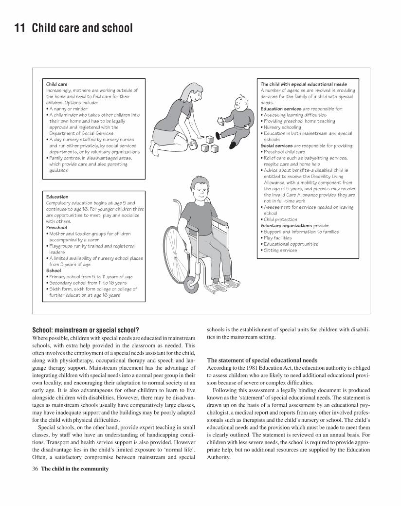
36 The child in the community
11 Child care and school
School: mainstream or special school?Where possible, children with special needs are educated in mainstreamschools, with extra help provided in the classroom as needed. This often involves the employment of a special needs assistant for the child,along with physiotherapy, occupational therapy and speech and lan-guage therapy support. Mainstream placement has the advantage ofintegrating children with special needs into a normal peer group in theirown locality, and encouraging their adaptation to normal society at anearly age. It is also advantageous for other children to learn to livealongside children with disabilities. However, there may be disadvan-tages as mainstream schools usually have comparatively large classes,may have inadequate support and the buildings may be poorly adaptedfor the child with physical difficulties.
Special schools, on the other hand, provide expert teaching in smallclasses, by staff who have an understanding of handicapping condi-tions. Transport and health service support is also provided. Howeverthe disadvantage lies in the child’s limited exposure to ‘normal life’.Often, a satisfactory compromise between mainstream and special
schools is the establishment of special units for children with disabili-ties in the mainstream setting.
The statement of special educational needsAccording to the 1981 Education Act, the education authority is obligedto assess children who are likely to need additional educational provi-sion because of severe or complex difficulties.
Following this assessment a legally binding document is producedknown as the ‘statement’ of special educational needs. The statement isdrawn up on the basis of a formal assessment by an educational psy-chologist, a medical report and reports from any other involved profes-sionals such as therapists and the child’s nursery or school. The child’seducational needs and the provision which must be made to meet themis clearly outlined. The statement is reviewed on an annual basis. Forchildren with less severe needs, the school is required to provide appro-priate help, but no additional resources are supplied by the EducationAuthority.
Child careIncreasingly, mothers are working outside of the home and need to find care for their children. Options include:• A nanny or minder• A childminder who takes other children into their own home and has to be legally approved and registered with the Department of Social Services• A day nursery staffed by nursery nurses and run either privately, by social services departments, or by voluntary organizations• Family centres, in disadvantaged areas, which provide care and also parenting guidance
EducationCompulsory education begins at age 5 and continues to age 16. For younger children there are opportunities to meet, play and socialize with others.Preschool• Mother and toddler groups for children accompanied by a carer• Playgroups run by trained and registered leaders• A limited availability of nursery school places from 3 years of ageSchool• Primary school from 5 to 11 years of age• Secondary school from 11 to 16 years• Sixth form, sixth form college or college of further education at age 16 years
The child with special educational needsA number of agencies are involved in providing services for the family of a child with special needs.Education services are responsible for:• Assessing learning difficulties• Providing preschool home teaching• Nursery schooling• Education in both mainstream and special schoolsSocial services are responsible for providing:• Preschool child care• Relief care such as babysitting services, respite care and home help• Advice about benefits-a disabled child is entitled to receive the Disability Living Allowance, with a mobility component from the age of 5 years, and parents may receive the Invalid Care Allowance provided they are not in full-time work• Assessment for services needed on leaving school• Child protectionVoluntary organizations provide:• Support and information to families• Play facilities• Educational opportunities• Sitting services

Common problems at schoolHyperactivityHyperactivity is characterized by poor ability to attend to a task, motoroveractivity, and impulsivity. Boys tend to suffer more than girls, andthere is often a family history. It is commoner in children with develop-mental delay, and those who were temperamentally difficult babies. It isalso seen in children who have never been given limits or taught todevelop self control, and can occur as a reaction to tensions and prob-lems in the home.
Hyperactive children are restless, impulsive and excitable, and fail tofocus on any activity for long. They tend to have little sense of danger,and require great vigilance. They often have difficulties on startingschool. Management includes routine and regularity in everyday life,with firm boundaries set for behaviour and consistency in discipline. Onstarting school, the support of the teacher is essential in helping withadjustment.
Attention deficit disorder Attention deficit disorder (ADD) refers to a difficulty in generallyfocusing on tasks or activities. It may or may not occur with hyperac-tivity and is commoner in boys. The child with ADD is fidgety, has a difficult time remaining in his or her seat at school, is easily distractedand impulsive, has difficulty following instructions, talks excessivelyand flits from one activity to another. Daydreaming occurs when hyper-activity is not a feature.
Management includes a regular daily routine with simple clear rules,and firm limits enforced fairly and sympathetically. Overstimulationand overfatigue need to be avoided, and a structured school programmeis required with good home communication. There is increasing evidence that central nervous system (CNS) stimulants such asmethylphenidate are effective in improving concentration. Dietsrestricting artificial colourings or flavourings remain controversial, butdo not help the majority of these children.
Aggressive behaviourAggressive behaviour can lead to bullying in school and delinquencybeyond. If extreme, the psychiatric term, conduct disorder, is applied.Counteracting aggression with more aggression is unproductive, andconsistency is required in the management. Both time-out and starcharts are positive methods. Staff at school must be involved in themanagement, in order to address academic or social problems, and toinstitute behaviour modification.
Teasing and bullyingTen per cent of children report being bullied once per week and 7% ofchildren are identified as bullies. Bullying tends to be more common inprimary schools, and varies from school to school. The child may react by becoming withdrawn, or aggressive or develop psychosomaticsymptoms, and it should be considered as a cause of distress whenevera child is disturbed, or refusing to go to school. In schools where bully-ing is a problem, a whole school approach is most effective, where boththe victims and the bullies are helped. The individual child needs helpin handling the situation and increasing self-esteem.
Non-attendance at schoolMost absences from school occur as a result of illnesses, which areusually minor, but may be prolonged through parental anxiety. In some
circumstances, children may be kept at home to help as carers or atwork. Two situations where the doctor may become involved are schoolrefusal and truancy. In school refusal everyone knows where the childis, but in the latter the child’s whereabouts are unknown during schoolhours. School refusal may be due to separation anxiety (common onfirst starting school) or school phobia, which is usually triggered by distressing events, such as problems with peers or teachers. Truancy iscommonest in high school. Persistent truancy is associated with gener-ally antisocial behaviour, poor academic achievement and unsettledfamily background.
Management of both must involve close collaboration between theparents and the teachers. In most cases of school refusal the child shouldbe returned to school as quickly as possible. Truancy is harder to tackle,and the education welfare officer should become involved if the truancyis persistent.
School failureReasons for school failure include the following.
Educational• Limited intellect.• ADD.• Hearing or visual deficit.• Dyslexia.• Dyspraxia.
Social• Problems at home.• Peer problems.• Absence from school.
School failure is associated with low self-esteem, behavioural difficulties and psychosomatic disorders, and has profound effects onachievement in adult life and chances of employment. It is important toaddress causes such as dyslexia, attention deficits and visual or hearingimpairments that reduce the child’s potential to learn, and can lead tofrustration and other negative psychological reactions.
DyslexiaDyslexia is the commonest type of specific learning difficulty. Thedyslexic child is unable to effectively process the information requiredin order to read. The result is a reading ability below that expected forthe child’s level of intelligence. Dyslexia must be differentiated fromslow reading due to limited intellect or inadequate teaching. It is com-moner in boys and there is often a family history.
There may be a history of delay in learning to talk, and spelling is alsoaffected. If unrecognized the child is likely to fail at school, and com-monly responds by withdrawing or exhibiting disruptive behaviour.The diagnosis must be confirmed on testing by an educational psy-chologist, and individual help is required to overcome the difficulties.
Dyspraxia (clumsiness)Fine motor incoordination leads to untidy writing, gross motor incoor-dination and difficulty with sports. The academic and social difficultiesthat ensue can cause considerable unhappiness and behaviour problemsif it is not recognized and dealt with helpfully. An occupational therapistcan assist in devising a programme to help overcome the difficulties andbuild self-confidence.
Child care and school 37

38 The child in the community
12 Immunization and the diseases they protect against
National immunization scheduleInfantBirth2 months
3 months4 months12-15 months
BCG for high-risk babiesDTP (diphtheria, tetanus, pertussis), polioHIB (Haemophilus influenzae B), meningococcus CDTP, polio, HIB, meningococcus CDTP, polio, HIB, meningococcus CMMR (measles, mumps, rubella)
Preschool3-5 years DT, acellular pertussis, polio
booster MMR
Secondary school11-14 years13-18 years
BCG if Heaf test negativeDiphtheria, tetanus booster Polio booster
Diphtheria• Throat infection with pharyngeal exudate• Membrane formation and upper airway obstruction• Exotoxin may cause myocarditis and neuritis with paralysis
Inactivated toxin (toxoid), IM
Meningococcus C• Acute purulent meningitis• Purpuric rash and septicaemic shock• Complications: high mortality, organ failure, tissue necrosis, deafness, paresis
Conjugated polysaccharide antigen, IM
Measles• Maculopapular rash, fever, coryza, cough and conjunctivitis• Complications: encephalitis with neurological damage and high mortality
Live attenuated virus grown on chick tissue, IM Contraindicated in immunodeficient children or those with severe egg allergy
Mumps• Fever• Enlargement of parotid glands• Complications: aseptic meningitis, sensorineural deafness and orchitis in adults Live attenuated virus grown on chick tissue, IM Contraindicated in immunodeficient children or those with severe egg allergy
Rubella• Mild illness with rash and fever• Effects on fetus: cataracts, deafness and congenital heart disease
Live attenuated virus, IM Contraindicated in pregnancy
Pertussis (whooping cough)• Prolonged URTI with paroxysms of coughing, followed by a whoop with vomiting• Complications: bronchopneumonia, convulsions, apnoea and bronchiectasis• Diagnosis confirmed by nasopharyngeal culture• Erythromycin shortens the illness if given early Killed organism, IM. May cause local pain, swelling, irritability and pyrexia. Only contraindications are a previous severe reaction or progressive neurological disease
Haemophilus meningitis• Was the main cause of meningitis in young children• Severe neurological sequelae: profound deafness, cerebral palsy, epilepsy, death Polysaccharide capsule of killed organism conjugated with protein, IM. Only effective against type B infection
Polio• Mild fever, may progress to meningitis• Anterior horn cell damage leads to paralysis, pain and tenderness• May lead to respiratory failure and bulbar paralysis• Residual paralysis is common in survivors A mix of 3 live attenuated strains of poliovirus orally, which establishes gut and systemic immunity. Contraindicated in the immunodeficient child
Tetanus• Enters body through open wounds• Neurotoxin causes progressive painful muscle spasms• Respiratory muscle involvement causes asphyxia and death
Inactivated toxin (toxoid), IM For a dirty wound, give booster if >10 years since last immunization If non-immunized, give tetanus immunoglobulin and a full course of inactivated toxoid
Tuberculosis (TB)• TB affects lungs, meninges, bones and joints• Active TB requires treatment over many months• Most infected children are identified as contacts of infected adults• Difficult diagnosis relies on demonstration of tuberculin sensitivity using Heaf or Mantoux skin testing
BCG, a live attenuated strain given intradermally. A papule forms and often ulcerates, healing in 6-8 weeks with a scar. Given to tuberculin-negative teenagers, and at birth to babies from high-risk families (all Asian babies in many parts of Britain)
KEY POINTSImmunizations should not be given to a child who:• Is younger than indicated in the schedule.• Is acutely unwell with fever.• Has had a serious reaction to a previous dose of the same vaccine.• Live attenuated vaccines (e.g. polio, measles, mumps, rubella, BCG (bacille
Calmette–Guérin)) should not be given to children with immune deficiencystates (e.g. cytotoxic therapy or high dose steroids).• Repeat immunizations should not be given sooner than indicated in theschedule.• If a child misses an immunization it should be given later. There is no needto restart the course.

Screening and surveillance tests 39
13 Screening and surveillance tests
Condition
Congenital hypothyroidismIf treated early with thyroid hormone, the child grows and developsnormally. Untreated it results in cretinism with severe learning disability (mental retardation)
Phenylketonuria (PKU)PKU causes severe learning disability (mental retardation). A low phenylalanine diet prevents the build-up of metabolites which cause brain damage Congenital cataractsEarly treatment prevents permanent visual impairment
Congenital dislocation of the hips (p. 105) Early orthopaedic treatment is effective in preventing limp and painful disability from dislocated, subluxed or dysplastic hips. A harness or splint is used initially. If this fails surgery is required Cryptorchidism (p. 79)Undescended testes are at risk for infertility and malignancy. Surgery should be performed before the age of 2 years Growth (Chapters 41 and 42) Poor growth can result from organic and inorganic causes
Development (Chapter 46) Delay in one or more areas of development may be indicative oflearning difficulties or a neurological problem
Congenital heart disease (Chapter 51)If congenital defects are missed, irreversible cardiopulmonary changes or infective endocarditis may result Hearing impairment (p. 121) If hearing deficits are not identified before language is acquired, permanent language impairment can result Vision and hearing at schoolMyopia is very common. Hearing impairment identified at this ageis due to secretory otitis media as most sensorineural deafnessis detected earlier
Age
Neonate
Neonate after 72 h of age
Neonates, 6 weeks
Neonates,10 days, 6 weeks
Neonates, 6 weeks
Weight at routinehealth visitor contact;length/height at 18 months, 3.5 yearsand school entry
Test at 6–8 weeks,7–9 months, 18–24 months, 3–4 years
Neonates, 6–8 weeks, preschool
7–9 months
School entry,8, 11 and 14 years
The test
Analysis of thyroid (T4) or thyroid stimulating hormone (TSH) from blood on filter paper obtained by heel prick. If positive, urgent referral to a paediatric endocrinologistis required The Guthrie test is carried out on the same sample as thyroid testing. The baby must be on full milk feeds for 3 days before testing. Referral to a metabolic clinic for dietaryadvice and long-term follow-up is required When looking through an ophthalmoscope, if white light instead of red is reflected from the retina, it suggests a cataract or other intraocular pathology. Immediate referral to an ophthalmologist is required The Ortolani and Barlow procedures are carried out for babiesup to the age of 3 months (see p. 105). Limited hip abduction, shortening of the leg and limp (once walking) are sought beyond3 months. Ultrasound is useful to confirm the diagnosis Examine the baby with warm hands. If on repeat examination the testes are still impalpable, refer to a paediatric surgeon
If weight or growth faltering is identified, a paediatric evaluation is required
All developmental areas are tested and referral to a paediatrician is made if delay or disorder is found
Identification of a heart murmur is the commonestpresentation The distraction hearing test is still commonly used. Neonatal screening by otoacoustic emission is being introduced
The Snellen chart or a letter-matching card for younger childrenis used. Referral to an optician is required if the child can onlyread letter size 6/12. Colour vision defects are identifiedusing screening cards at age 11. Sweep audiometry tests the child's ability to hear sounds at a set level across the main speech frequencies. Failure requires an ENT and audiological evaluation
KEY POINTSScreening is the identification of unrecognized disease or defects by using tests.It sorts out apparently well children who may have a problem from those who donot, but is not intended to be diagnostic. Cost must be considered and balancedagainst that of treatment if the problem presents later, and of medical care as awhole.
Conditions suitable for screening should:1 Be identifiable at a latent or early symptomatic stage.2 Be treatable.3 Early treatment should influence the course or prognosis.

40 The acutely ill child
14 The acutely ill child
Fluid loss • Blood loss• Gastroenteritis• Burns
Fluid maldistribution • Septic shock• Cardiac disease• Anaphylaxis
Respiratory distress • Foreign body• Croup• Asthma
Respiratory depression • Convulsions• Raised ICP• Poisoning
CIRCULATORY FAILURE (SHOCK)
RESPIRATORY FAILURE
CARDIAC ARREST
Children may become critically ill very rapidly, and their survival depends on prompt recognition of the severity of their illness, appropriate life support andrapid treatment. In children the cause of cardiac arrest is almost always due to respiratory or circulatory failure rather than a primary heart problem. It is therefore important to recognize the early signs of respiratory and circulatory failure and to correct these before cardiac arrest occurs.
Recognition of acute illness
Circulatory failure (shock)
Clinical features of shock• Tachycardia• Thready pulse• Delayed capillary refill• Mottled, pale skin• Cool extremities• Hypotension (late sign)• Tachypnoea• Restlessness• Reduced urine output• Metabolic acidosis Signs that suggest cause• Fever in sepsis• Purpuric rash (meningococcus)• Hepatomegaly (heart failure)• Focus of infection
Management• High flow oxygen, respiratory support• Intravenous fluid bolus• Consider inotropic support• Antibiotics for septic shock• Epinephrine for anaphylaxis
Septic shock:• meningococcal septicaemia• toxic shock syndrome
Blood loss
Fluid loss: dehydration
Fluid loss: • diabetic ketoacidosis
Fluid loss: burns
Heart failure
Anaphylaxis
Shock is used to describe a state of inadequate tissue perfusion due to an acute failure of circulation. The body responds by redistributing blood tovital organs such as the brain and the heart, at the expense of the skin, muscles and bowel. Children in shock look pale and have poor skin perfusion. Blood pressure is maintained in children by peripheral vasoconstriction, so that hypotension is a very late sign of shock.

Respiratory failureRespiratory failure is defined as inadequate respiration to maintainnormal arterial oxygen and carbon dioxide concentrations. Respiratoryfailure is obvious if the child is apnoeic or deeply cyanosed, but it isimportant to be able to detect impending respiratory failure and to inter-vene quickly.
Acute upper airway obstructionAcute upper airway obstruction is a medical emergency as death mayresult if it is not effectively treated immediately. It can be due to infec-tion (epiglottitis, croup) or inhalation of a foreign body (especiallycommon in toddlers who put small objects in their mouths). Presenta-tion is with acute sudden onset of choking, coughing and cyanosis, fol-lowed by collapse. There may be an inspiratory stridor and markedintercostal recession. If epiglottitis is suspected the doctor must notexamine the child’s throat and investigations should be delayed untilthe airway is protected or the obstruction relieved.
Choking should be managed by opening the airway, removing anyvisible obstruction or if this is impossible performing back-blows or theHeimlich manoeuvre to expel the obstruction from the airway. TheHeimlich manoeuvre should not be attempted in infants due to the riskof trauma to the liver and spleen; instead alternate back blows withchest thrusts with the child held in a head-down position. If these mea-sures are unsuccessful artificial ventilation will be required and anemergency tracheostomy or crico-thyroidotomy may be necessary tobypass the obstruction.
The acutely ill child 41
Causes of respiratory failure
Upper airway obstruction• Foreign body• Epiglottitis• Croup
Lower airway• Asthma• Bronchiolitis• Pneumonia• Cystic fibrosis• Neonatal lung disease
Clinical features• Shortness of breath• Tachypnoea• Cyanosis• Nasal flaring• Grunting • Intercostal recession• Restlessness or confusion Symptoms and signs of the underlying disease• Expiratory wheeze• Inspiratory stridor• Crackles• Neurological weakness
Investigations• Oxygen saturation• Arterial blood gases• Chest X-ray
Management• Assess severity by examination, blood gases and oxygen saturation monitor• Give high flow oxygen• Intubate and ventilate if rising pCO2. (The decision to ventilate is based on clinical criteria, not just blood gases)• Treat the underlying cause: antibiotics (infection), bronchodilators and steroids (asthma), remove foreign body
Neurological• Head injury• Meningitis• Raised ICP• Muscle weakness
Drug ingestion
Severe cardiac failure
The Heimlich manoeuvre

42 The acutely ill child
Cardiorespiratory arrestCardiac arrest is the endpoint of severe respiratory or cardiac failurethat has not been adequately treated. The causes are outlined above.Cardiorespiratory arrest outside hospital requires rapid basic lifesupport until skilled help arrives. In hospital cardiorespiratory arrestshould be managed by a team of skilled personnel. As most cardiores-piratory arrests in children are secondary to hypoxia rather than due tocardiac disease it is crucial to achieve adequate oxygenation using highflow oxygen and artificial respiration, and to circulate this oxygen byproviding appropriate cardiac massage.
Establishing an airway and artificial ventilationThe airway should be opened by lifting the chin and tilting the headback to a ‘sniffing the air’ position. In infants the head should be in theneutral position. If there is a possibility of cervical spine injury then theairway should be opened by the ‘jaw-thrust’method. The airway shouldthen be cleared by removing any vomit or secretions with suction. Arti-ficial ventilation can be given by mouth to mouth or in infants by mouthto mouth and nose. After five rescue breaths check for a central pulseand commence cardiac massage.
External cardiac massageIn infants cardiac massage can be achieved by encircling the chest withboth hands and compressing the sternum with the thumbs, one finger-
breadth below the nipples. In young children the heel of one hand isused one fingerbreadth above the xiphisternum and in older childrentwo hands are used two fingerbreadths above the xiphisternum.
If these measures are not effective then drugs such as epinephrine,bicarbonate and fluid volume may be necessary, depending on the causeof the cardiac arrest. Epinephrine can be given via the intravenous (IV), intraosseous or endotracheal routes. Defibrillation is very rarelyrequired in paediatric cardiac arrests, but is indicated for certain cardiacarrhythmias such as ventricular fibrillation, ventricular tachycardia andsupraventricular tachycardia unresponsive to drug therapy.
Commoner causes of cardiorespiratory arrest
Management• Call for help!• Open the airway• Check for respiration• Ventilate using high flow oxygen via bag and mask, or intubation• Provide mouth-to-mouth if no equipment available• Check for a central pulse• Commence external cardiac massage• Alternate five chest compressions to one breath (15 : 2 in older children)• Establish intravenous or intraosseus access• Give appropriate drugs (such as epinephrine) • Check cardiac rhythm and defibrillate if appropriate• Always check blood sugar as hypoglycaemia may be an easily treatable cause
Respiratory• Upper airway obstruction• Respiratory failure
Drug ingestion• Drugs or toxins
Cardiac disease• Arrhythmia• Heart failure• Myocarditis
Neurological• Cerebral oedema• Coning of brainstem• Head injury• Birth asphyxia
Severe hypoxic–ischaemic insult• Suffocation• Drowning
Cardiac massage in an infant

Acute life-threatening events and SIDSSIDS is the ‘sudden death of any infant or young child, which is un-expected by history, and in which a thorough postmortem fails to demonstrate an adequate cause for death’. Some children are found in acollapsed state, not breathing and looking grey or mottled, but can besuccessfully resuscitated. This is referred to as an acute life-threateningevent (ALTE) or as ‘near-miss cot death’. SIDS is the commonest causeof death in infants after the first week of life, and the peak occurrence isbetween 2 and 4 months. In the UK the incidence of SIDS has fallen byover 50% due to the ‘back to sleep’campaign which advises that babiesshould be put to sleep on their backs, at the foot of the cot and not over-heated or exposed to cigarette smoke.
Focal points for the assessment of the acutely ill child• Call for help immediately.• Rapidly assess the airway.• Perform airway opening manoeuvres (but protect the cervical spine ifthere is any history of trauma).• Give high-flow oxygen. If the child is not breathing, ventilate with abag and mask or intubate.• Check for pulse and assess circulation by capillary refill time.• Commence chest compressions if a pulse is absent or less than 60 beats/min.
• Establish IV or intraosseus access and give drugs and fluids as appropriate.• Apply pressure to any active bleeding points.• Rapidly assess the neurological state by looking at pupils, posture andthe level of consciousness.• Always check blood sugar level.• Perform appropriate investigations and commence definitive treat-ment (e.g. infection screen and broad spectrum antibiotics if sepsis issuspected).• Once the child has been stabilized, transfer to an intensive care unitfor definitive care.
The acutely ill child 43
Sudden infant death and acute life-threatening events
Aetiology• Commonest cause of death in infants >1 week• Increased risk if prone, overheated or smoky
environment• Normally previously well babies, sometimes with
minor cough or cold• In 20% an unexpected cause is found at autopsy
Management• Cardiopulmonary resuscitation (CPR)• Admit for observation and investigation• Train parents to administer CPR• Home apnoea monitor may relieve anxiety but is of no proven benefit
Examination• Found collapsed• Pale and mottled• Bradycardia• Hypotension
Investigations• Blood sugar• Infection screen• CXR and barium swallow• ECG monitoring• Metabolic screen
Differential diagnosis• Infection• Gastro-oesophageal reflux• Neurological abnormality• Hypoglycaemia (rare)• Cardiac arrhythmia (rare)• Inborn error of metabolism (rare)• Suffocation (rare)
'Back to sleep' campaign• Put babies to sleep on their backs• Avoid overheating• Use blankets rather than duvets• Ideal room temperature is 16–20°C• Lie the baby at the foot of the cot so that
it cannot slip under the covers• Avoid exposure to cigarette smoke• Consult a doctor if your baby is ill
KEY POINTS• Cardiac arrest is usually secondary to respiratory failure or shock.• Providing adequate oxygenation is critical.• Shock causes tachycardia and delayed capillary refill time and is treated withboluses of fluids.• Upper airway obstruction is a common cause of acute respiratory failure inyoung children.• The ‘back to sleep’ campaign has reduced SIDS by 50%.

44 The acutely ill child
15 The unconscious child
Acute asphyxial event, e.g.• Birth asphyxia• Near-miss cot death• Post cardiac arrest
Head injury, e.g.• Subdural haematoma• Extradural haematoma• Diffuse axonal injury• Non-accidental injury
Drug-induced• Accidental ingestion or overdose
Raised intracranial pressure• Signs of papilloedema
Encephalitis• Fever• History of change in personality or ability
Convulsions• Status epilepticus• History of epilepsy
Meningitis• Fever• Neck stiffness• History of headache and photophobia, irritability
Cerebrovascular accident, e.g.• Vasculitic disorder• Hypertension• Thrombotic disorder
Metabolic disorders, e.g.• Hypoglycaemia• Diabetic ketoacidosis• Inborn errors of metabolism
History Ask about the possibility of drug ingestion (either deliberate or accidental in young children) Was there a prodromal illness or contact with serious infection (e.g. meningitis) Assess the possibility of non-accidental injury (p. 99) Is there a history of convulsions and for how long did they last? Was the child neuro-developmentally normal prior to the onset of coma?
Examination Vital signs: is there a bradycardia (suggests raised ICP) or tachyarrhythmia (drug ingestion). Deep, sighing respiration (Kussmaul) suggests diabetic ketoacidosis. Ketones may be smelt on the breath Look for a focus of infection. Check for rashes and neck stiffness, pneumonia and UTI Check pupils: are they symmetrical and do they constrict appropriately to light? Check for abnormal posture (decorticate or decerebrate posture) Assess the level of consciousness using either the modified Glasgow coma scale or the more rapid AVPU (see opposite) Always check the blood glucose. Hypoglycaemia is the commonest metabolic cause of coma
Investigations and their significance
AVPU coma scale
What you need from your evaluation
Blood glucose Hypo- or hyperglycaemiaFull blood count May indicate infection or acute blood loss (Hb and PCV low)Blood culture May identify infective causeU&E High urea in dehydration. Sodium may be high or lowBlood gases Metabolic or respiratory acidosis (see Chapter 3 for interpretation)Chest X-ray Infection or cardiac failure, trauma (e.g. rib fracture)CT or MRI scan Focal pathology (tumour, haemorrhage, abscess)Lumbar puncture May show evidence of infection (meningitis, encephalitis) or bleeding (e.g. subarachnoid haemorrhage) LP should not be attempted in the unconscious child until raised ICP has been excluded, due to the risk of brain herniation (coning)
Alert responds to Voice responds to Pain Unresponsive
A score of 'P' corresponds to a Glasgow Coma Scale (GCS)of 8, and suggests the airway should be protected by intubation to prevent aspiration
Causes of coma

ComaA child who is deeply unconscious is said to be in coma. Encephalopa-thy refers to the precomatose state with an altered conscious level. Anunconscious child requires urgent and careful evaluation to establishthe cause of the coma and to commence appropriate therapy. Whateverthe cause of the coma the airway must be protected and adequate venti-lation maintained.
MeningitisMeningitis is caused by either bacterial or viral infection invading themembranes overlying the brain and spinal cord and should be consid-ered in any irritable child with unexplained fever. It is commonest in theneonatal period but can occur at any age. The causes are listed below.
Viral meningitis is preceded by pharyngitis or gastrointestinal (GI)upset. The child then develops fever, headache and neck stiffness. Inbacterial meningitis the child is drowsy and may be vacant. Irritabilityis a common feature, often with a high pitched cry and convulsions mayoccur. Examination shows an ill child, with a stiff neck and Kernig’ssign (pain on extending the legs) may be positive, though this is not reli-able in young infants. Tonsillitis and otitis media can also mimic neckstiffness. In infants the fontanelle may be bulging. A petechial or pur-puric rash suggests meningococcal meningitis.
Meningitis is confirmed by a lumbar puncture (see p. 20), which willshow a leucocytosis, high protein count, low glucose and may showorganisms present. The fluid will look cloudy to the naked eye. Cultureor PCR analysis will confirm the organism, but treatment should becommenced empirically as soon as the cultures have been taken.
Intravenous cefotaxime or benzyl penicillin is usually given, depend-ing on the age of the child and the likely organism. Steroids may reducemeningeal inflammation in Haemophilus meningitis. Meninigococcalmeniningitis is associated with pharyngeal carriage and household con-tacts should receive prophylaxis with rifampicin. Meningococcal septi-caemia is discussed in Chapter 37.
EncephalitisViral infection sometimes spreads beyond the meninges to infect thebrain tissue itself. This is known as meningo-encephalitis. The onset is
often more insidious and the child’s personality may change and theymay become confused or clumsy before the onset of coma. Meningismis less of a feature. The lumbar puncture may reveal a lymphocytosisand specimens should be sent for viral culture and PCR analysis.Herpes simplex virus or Mycoplasma pneumoniae may be responsible,and you should always ask about contact with herpetic lesions (cold-sores). Treatment with acyclovir, erythromycin and cefotaxime is givenuntil the organism is known.
In herpes encephalitis the electroencephalogram (EEG) and a mag-netic resonance imaging (MRI) brain scan may show temporal lobeinvolvement.
Metabolic causes of comaIn the absence of trauma or infection, a metabolic cause for comashould be considered. By far the commonest metabolic cause is hypo-glycaemia, and a blood sugar must be checked immediately at thebedside in every unconscious child. Hypoglycaemia may be due toinadequate carbohydrate intake or excess insulin in children with dia-betes mellitus, but it can also be the presenting feature in infants withinborn errors of metabolism or adrenal insufficiency. Hyperglycaemiain uncontrolled diabetes can lead to keto-acidosis with coma, thoughthe onset is often more gradual. Diabetes is discussed in detail inChapter 53.
Any severe metabolic derangement can cause coma, includingsevere uraemia (in renal failure) or high ammonia (inborn errors ofmetabolism such as urea cycle disorders), severe hypernatraemia orhyponatraemia. Coma can also be caused by cerebral oedema fromover-rapid correction of electrolyte imbalance in severe dehydration.
Unexplained comaIn unexplained coma the possibility of non-accidental injury such asshaking injury must be considered. A computerized tomography (CT)brain scan and skeletal survey may show evidence of trauma and retinalhaemorrhages may be present. Accidental drug ingestion or overdose,or deliberate poisoning may cause coma, and a urine toxicology screencan sometimes identify the drug. Drugs affecting the CNS such asopiate analgaesics, alcohol and antidepressants are often implicated.
The unconscious child 45
Causes of meningitis.
KEY POINTS• Coma can be evaluated rapidly using the AVPU (alert, voice, pain, unre-sponsive) score.• Always check the blood glucose in any unconscious child.• Consider the possibility of poisoning or drug overdose.• Altered consciousness, fever and irritability suggest meningitis, even in theabsence of neck stiffness.• Never perform a lumbar puncture in an unconscious child until raisedintracranial pressure has been excluded.
Viral Mumps virusCoxsackie virusEchovirusHerpes simplex virusPoliomyelitis (if unvaccinated)
Bacterial Neisseria meningitidis Haemophilus influenzae type BStreptococcus pneumoniaeGroup B Streptococcus (in newborn)Escherichia coli and Listeria (in newborn)
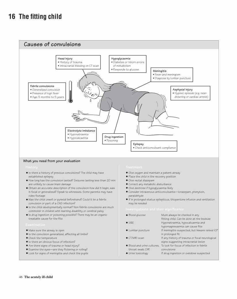
46 The acutely ill child
16 The fitting child
History Is there a history of previous convulsions? The child may have established epilepsy How long has the convulsion lasted? Seizures lasting less than 20 min are unlikely to cause brain damage Obtain an accurate description of the convulsion-how did it begin, was it focal or generalized? Speak to witnesses. Some parents may have video footage Was the child unwell or pyrexial beforehand? Could it be a febrile convulsion or part of a CNS infection? Is the child developmentally normal? Non-febrile convulsions are much commoner in children with learning disability or cerebral palsy Is drug ingestion or poisoning possible? There may be an organic treatable cause for the fits Examination Make sure the airway is open Is the convulsion generalized, affecting all limbs? Check the temperature Is there an obvious focus of infection? Are there signs of trauma or head injury? Examine the eyes—are they flickering or rolling? Look for signs of meningitis and check the pupils
Treatment
What you need from your evaluation
Give oxygen and maintain a patent airway Place the child in the recovery position Give rectal diazepam Correct any metabolic disturbance Give dextrose if hypoglycaemia likely Consider intravenous anticonvulsants—lorazepam, phenytoin, paraldehyde If in prolonged status epilepticus, thiopentone infusion and ventilation may be needed Investigations and their significance Blood glucose Must always be checked in any fitting child. Can be done at the bedside U&E Hyponatraemia, hypocalcaemia and hypomagnesaemia can cause fits Lumbar puncture If meningitis suspected, but beware raised ICP in prolonged fit CT/MRI scan If any history of trauma or focal neurological signs suggesting intracranial lesion Blood and urine cultures, To look for focus of infection in febrile throat swab, CXR convulsions Urine toxicology If drug ingestion or overdose suspected
Hypoglycaemia• Diabetes or inborn errors of metabolism• Responds to glucose Meningitis
• Fever and meningism• Diagnose by lumbar puncture
Head Injury• History of trauma• Intracranial bleeding on CT scan
Febrile convulsions• Generalized convulsion• Presence of high fever• Age: 5 months to 5 years
Asphyxial injury• Hypoxic episode (e.g. near- drowning or cardiac arrest)
Drug ingestion• Poisoning
Epilepsy• Check anticonvulsant compliance
Electrolyte imbalance• Hyponatraemia• Hypocalcaemia
Causes of convulsions

Generalized convulsionsThe term convulsion is synonymous with fit or seizure. Convulsions aredue to synchronous discharge of electrical activity from a number ofneurones, and this manifests itself as loss of consciousness and abnor-mal movements. In a generalized convulsion all four limbs and the faceare affected. Convulsions are common. They occur in 3–5% of children.They do not necessarily mean the child will go on to develop epilepsy,and many children only ever have one convulsion. However, 60% ofepilepsy develops in childhood. Children’s brains are particularly sus-ceptible to convulsions and the commonest trigger is the rise in tem-perature during a febrile illness. These are known as febrile convulsionsand are described below. Epilepsy is discussed in Chapter 60.
Febrile convulsionsThese usually occur between the ages of 5 months and 5 years in neuro-logically normal children and are triggered by fever, usually as part of aviral upper respiratory tract infection, although they may also be trig-gered by any febrile illness including acute otitis media, urinary tractinfection (UTI) or tonsillitis. The convulsion is usually short lived,lasting only a few minutes. Status epilepticus occurs in less than 1% offebrile convulsions. Febrile convulsions should be managed by identi-fying and treating the source of the fever, and cooling the child byundressing them and sponging their skin with tepid water. Antipyreticssuch as paracetamol or ibuprofen should be given. If the convulsionpersists for more than 10 min it should be terminated with rectaldiazepam. Investigations should be performed to exclude serious infec-tion, and if no obvious focus of infection is found a lumbar puncture isindicated to exclude meningitis.
Advice to parents is very important. About one-third of children willhave further febrile convulsions in the future. Parents must be taughthow to manage any future febrile illnesses and taught basic first aidmanagement of convulsions. The prognosis, which is good, should beexplained. Children with uncomplicated febrile convulsions are at nogreater risk of epilepsy, although if the convulsion lasts more than 20 min they may be at increased risk of temporal lobe epilepsy. Overallthe risk of epilepsy is 2–3%. Regular anticonvulsant medication is onlyvery rarely prescribed, if the child is having very frequent or prolongedfebrile convulsions.
Management of the fitting childMost parents who witness their child fitting imagine that the child isgoing to die and seek medical attention urgently. Children may still befitting when they present to the GP or to hospital. The most importantthing is to support the airway and turn the child into the recovery posi-tion, semi-prone with the knee flexed under the chest (see box oppo-
site). Objects should not be put into the mouth (except an artificialairway). If oxygen is available this should be given by facemask. If theconvulsion is ongoing, rectal diazepam should be given as an anti-convulsant to terminate the seizure. Occasionally some convulsionspersist; this is know as status epilepticus and is described below.
It is vital to check the blood glucose immediately in any fitting child,as hypoglycaemia is a common and rapidly treatable cause. Not all chil-dren with hypoglycaemic convulsions are diabetic; some may haveinborn errors of metabolism and may not tolerate even short periods ofstarvation. Once the convulsion has terminated the child may remaindrowsy or ‘postictal’ for some time. They should be observed carefullyand kept in the recovery position until they are able to maintain theirown airway. If it is the child’s first convulsion the parents will requiremuch reassurance and will need to be taught how to manage futureepisodes. Occasionally this will include prescribing rectal diazepam tobe administered at home.
Status epilepticusSeizures may be very prolonged and are an important cause of coma.Status epilepticus is defined as continuous seizure activity for morethan 30 min or a series of seizures without full recovery in between.Status may occur following febrile convulsions or more commonly inchildren with known epilepsy, or with other acute causes such as traumaor metabolic disturbance. The child’s airway should be maintained,oxygen given and blood glucose checked. If rectal diazepam has beengiven, IV lorazepam should also be given. Rectal paraldehyde or IVphenytoin may be tried. If all these have failed an anaesthetist should becalled and the child should undergo rapid sequence induction of anaes-thesia with thiopental. Any child with very prolonged seizures shouldbe monitored carefully on an intensive care unit, and urgent investiga-tions performed to identify the cause.
The fitting child 47
KEY POINTS• Febrile convulsions occur in 3% of children between age 5 months and 5years. The prognosis is usually excellent.• Children who are fitting must be placed in the recovery position and theirairway maintained.• Always check blood sugar as hypoglycaemia is a common and treatablecause.• Any convulsion lasting more than 10 min should be terminated with rectaldiazepam.• Status epilepticus is fitting for more than 30 min and requires urgent treatment.

48 The acutely ill child
17 The febrile child
Viral infections, e.g.• Non-specific viral infection• Upper respiratory tract infection (URTI)
Viral illness with rash, e.g.• Chicken pox• Measles• Rubella• Non-specific viral rash
Influenza• Fever, cough, headache, anorexia• Arthralgia
Pneumonia• Cough, tachypnoea, retractions• Signs of consolidation, crackles
Kawasaki's disease• Rash, conjunctivitis, lymphadenopathy, cracked lips, fever, skin peeling
Serious blood infection, e.g.• Meningococcal septicaemia• Streptococcal sepsis• Toxic shock syndrome• Malaria
Factitious• Taking temperature after hot drink• Deliberate manipulation of thermometer• Excessive crying or exertion• Overheating due to swaddling
Septic arthritis• Painful joint• Swelling and effusion
Tonsillitis• Commonly viral• Sore throat• Large tonsillar glands• Smelly breath
Otitis media• Tugging at ears, pain• Red tympanic membrane
Dehydration
Post immunizationPost surgery
Urinary tract infection(see Chapter 28)• Frequency, dysuria• Loin or suprapubic pain• Vomiting• Abnormal dipstick test and positive microscopy
History The parents have normally noticed the fever and may have checked the child's temperature Ask about the duration and pattern of the fever—does it occur at particular times of the day? Is there pain? Earache, difficulty swallowing, dysuria or frequency may point to the source Are there associated features such as malaise, anorexia, vomiting, coryza, cough or rash? Has there been contact with other children with infection such as meningitis or chicken pox? Has the child just been vaccinated? Is the child still drinking adequate amounts of fluid? What anti-pyretics and cooling measures have been tried?
Examination Check the temperature: oral, axilla or rectal Does the child look seriously ill? Is there a rash, tachypnoea, tachycardia or dehydration? Chest: are there signs of respiratory infection—tachypnoea, recession, crackles or grunting? Throat: feel for cervical lymphadenopathy and look at tonsils. Is there an exudate? Ears: are the tympanic membranes red or bulging? CNS: is the child orientated? Is there floppiness or signs of meningism? Urine: check the urine with dipstick or microscopy
Investigations and their significance Full blood count Leucocytosis with neutrophilia suggests bacterial infection Throat swab Streptococcus requires treatment with penicillin Blood culture If positive, suggests septicaemia. Treatment may have to commence before result known Lumbar To exclude meningitis and encephalitis. Should be performed in any seriously ill child when puncture no focus of infection can be found, especially in infants <1 year Urine analysis Pure growth of a single organism with leucocytosis confirms infection. Protein and red cells may be present. Dipsticks can be used to test for leucocytes, protein and nitrites Chest X-ray May reveal cause of fever in infants as chest signs may not always be apparent
What you need from your evaluation
Causes of fever

Management of fever in childrenTemperature can be measured rectally, orally, or in the axilla using athermometer. Thermal devices can also give an estimation of tempera-ture directly from the skin or from the ear canal. Fever is defined as anaxillary temperature above 37°C. The height of the fever doesn’t neces-sarily correlate with the severity of the illness and fever can commonlyoccur in children with minor illnesses. Fever is usually a response toinfection, and the child often appears flushed as blood vessels in theskin vasodilate in an attempt to lose heat. Fever is an unpleasantsymptom and should be treated. Some young children are at increasedrisk of febrile convulsions if their temperature rises very rapidly (see p. 47).
Fever can be treated by undressing the child and allowing them tolose heat through the skin. Sponging the skin with tepid water can alsobring down the temperature by evaporation. The mainstay of treatmentis antipyretics such as paracetamol (Calpol) or occasionally ibuprofen.Paracetamol can be used regularly in the correct dosage to keep thechild’s temperature down. Aspirin should not be used in children under12 years as it is associated with the development of severe liver failure(Reye’s syndrome).
Viral upper respiratory tract infections Upper respiratory tract infections (URTIs) are extremely common inchildren, occurring 6–8 times a year on average. They are especiallycommon when toddlers start at nursery or playgroup and when childrenstart school. At these times they are exposed to a large number of viralinfections to which they have no immunity. The child often has coryza(runny nose) or acute pharyngitis associated with fever. Young infantsmay have difficulty breathing and feeding because they are obligatenose breathers. The tympanic membranes are often inflamed. In acutepharyngitis the tonsillar fauces and palate are inflamed and cervicallymph nodes may be enlarged. Treatment is symptomatic, withantipyretics such as paracetamol. Saline drops may improve nasal con-gestion in infants. The infection usually lasts 3–4 days. Antibiotics arenot indicated!
TonsillitisTonsillitis is usually viral in origin. In older children the commonestbacterial organism is the group A beta-haemolytic streptococcus. Thechild may complain of a sore throat or dysphagia and they usually have a fever. There is often tender cervical lymphadenopathy whichmay cause neck stiffness. Associated adenitis in the mesenteric nodesmay cause abdominal pain. The tonsils will be enlarged and acutelyinflamed. They may have a white exudate in bacterial tonsillitis al-though this is not always a reliable sign. Exudates can also occur withinfectious mononucleosis (glandular fever) and with diphtheria (nowvery rare). The breath may smell offensive in bacterial tonsillitis. Acutetonsillitis should be distinguished from the hypertrophied but non-inflamed tonsils which are common in preschool children. Most chil-
dren do not require antibiotics and can be managed with saline garglesand paracetamol. If bacterial infection is suspected this should be con-firmed by a throat swab. Streptococcal tonsillitis should be treated withbenzyl penicillin for 10 days. Complications of tonsillitis are rare butinclude otitis media, peritonsillar abscess (quinsy) and poststreptococ-cal glomerulonephritis. Chronically enlarged tonsils can cause upperairway obstruction and obstructive sleep apnoea. This is an indicationfor tonsillectomy.
Acute otitis mediaThis is a very common disorder, especially in young children and canoccur in babies. The commonest causes are Streptococcus pneumoniae,Haemophilus influenzae and viruses. Otitis media is especially com-mon if there is Eustachian tube dysfunction, which can be associatedwith URTIs, obstruction from enlarged adenoids, cleft palate and Downsyndrome. Otitis media presents with fever, deafness and pain in the ear.The child may be irritable and may tug or pull at the affected ear, orinfection may be aymptomatic. Examination shows a red, inflamed andbulging tympanic membrane, with loss of the light reflex. Treatmentwith ampicillin shortens the duration of symptoms. Prognosis is gener-ally good even if the tympanic membrane has perforated.
Complications include secretory otitis media (glue ear), conductivedeafness and mastoiditis. In secretory otitis media, recurrent acuteinfections lead to a thick glue-like exudate building up in the middleear. On examination the tympanic membrane appears thickened andretracted with an absent light reflex. If there is significant hearing loss,ventilation tubes (grommets) may be inserted through the tympanicmembrane to allow the middle ear to drain. These are particularly indi-cated if there is language delay due to the conductive deafness associ-ated with glue ear.
Fever in newborn infantsFever in an infant less than 8 weeks old should always be taken seri-ously because this may represent late onset congenital infection. Signsof sepsis at this age can be quite non-specific, so a significant fevershould always prompt a careful examination and appropriate investiga-tions. If well, the child may just be observed, but if significantly ill willrequire a full infection screen including urine culture, chest X-ray andpossibly lumbar puncture.
The febrile child 49
KEY POINTS• Fever is a very common symptom in children, and can usually be managedby simple cooling measures and paracetamol.• Any ill child with a high fever should be examined carefully to excludeserious infections such as meningitis, UTI or pneumonia.• Any fever in a baby less than 8 weeks old should be taken seriously.• Otitis media and tonsillitis are common causes of fever in young children.• Most fevers are associated with non-specific viral infections or URTIs.

50 The acutely ill child
18 Acute diarrhoea and dehydration
What you need from your evaluation
Vomiting• Pyloric stenosis• Viral infections• Gastroenteritis
Excessive sweating• High fever• Hot climate• Cystic fibrosis
Inability to drink• Herpes stomatitis• Acute tonsillitis
Inadequate access to water
Fluid loss• Burns• Post surgery
Polyuria• Diabetes mellitus, especially diabetic ketoacidosis
Acute diarrhoea• Viral gastroenteritis• Bacterial gastroenteritis Shigella E. coli Salmonella Campylobacter• Antibiotic-induced• Food poisoning (toxins)• Any acute infection
Excessive fluid loss
Inadequate intake
History Has there been diarrhoea and/or vomiting? Is the vomiting projectile (pyloric stenosis)? How many loose stools have there been? Is the child passing less urine than normal? Ask when was the last wet nappy? How often and for how long has the child been vomiting? Does the child have cystic fibrosis or diabetes?
Investigations and their significance(Investigations are required only in moderate to severe diarrhoea or if the child is very ill) U&E For electrolyte imbalance and renal function Blood gas Metabolic acidosis or alkalosis Urinalysis For osmolality or specific gravity Blood sugar To exclude diabetic ketoacidodis Stool culture In gastroenteritis and food poisoning
Examination Weigh the child and compare with previous weight (if known) to assess dehydration In young infants feel for a pyloric mass during a test feed (pyloric stenosis) Assess the degree of dehydration (mild, moderate or severe) as follows: Mild Moderate Severe Mouth and lips Dry Dry Dry Urine output Normal Reduced None for 12 h Mental state Normal Lethargic Irritable or coma Pulse rate Normal Tachycardia Tachycardia Blood pressure Normal Normal Low Capillary refill time Normal Delayed Very delayed Fontanelle Normal Sunken Very sunken Skin and eye turgor Normal Reduced Very reduced Dehydration (%) <5 5–10 >10 (shock) Treatment Use oral rehydration therapy where possible Treat shock with boluses of IV fluids Rehydrate slowly to replace fluid loss over at least 24 h Correct any electrolyte imbalance
Causes of dehydration

DehydrationWater is the major component of the body, accounting for up to 80% ofan infant’s body weight. Loss of more than 5% of this water representssignificant dehydration. Fluid may be depleted in the intracellular orextracelluar compartments. If a significant amount of fluid is lostacutely from the intravascular part of the extracellular space, then shockmay ensue. Normal body fluid is a balance between intake (drinking)and output (urine output, stool volume, sweat and insensible losses suchas vapour in expiration). If intake does not keep up with losses, then thechild will become dehydrated. The commonest cause of dehydration inchildren is gastroenteritis due to diarrhoea and vomiting.
Acute diarrhoeaEpisodes of acute diarrhoea are not uncommon in children, and areusually due to infection, although not always GI infection. Sadly dehy-dration due to gastroenteritis is still a major cause of mortality in chil-dren in the developing world. Gastroenteritis is usually viral, androtavirus is the main agent causing winter epidemics. Diarrhoea follows1–2 days after low-grade fever, vomiting and anorexia. There may beacute abdominal pain and malaise. The diarrhoea resolves within aweek and the management is adequate rehydration (see below). Bacte-rial gastroenteritis has a similar presentation and the commonestpathogens are Escherichia coli, Shigella, Salmonella and Campylobac-ter. Meningism and febrile convulsions can occur with shigella, whilstbloody diarrhoea occurs in shigella and Campylobacter infection.Infection with the 0157 strain of E. coli can be followed by haemolyticuraemic syndrome—a life-threatening disease with haemolysis andacute renal failure. Antibiotics should not be prescribed for uncompli-cated gastroenteritis. If there is evidence of septicaemia the child shouldbe admitted for IV antibiotics.
Any form of infection or febrile illness can cause diarrhoea, espe-cially in infants. This includes viral URTIs, chest infections, otitismedia and UTI. Use of antibiotics may in itself cause diarrhoea due to adisturbance of the normal enteric flora. Recurrence of diarrhoea onrefeeding is most likely to be due to lactase deficiency and may requirea lactose free diet for a number of weeks.
Management of dehydrationTry to determine the cause of the diarrhoea and the degree of dehydra-tion. Ask about the duration of diarrhoea, whether there has been vom-iting and when the child last passed urine.
The degree of dehydration can be assessed by the pulse, blood pres-sure, mucous membranes, urine output, skin turgor and by feeling thefontanelle (see box opposite). You should be able to decide whether the child has mild (<5%), moderate (5–10%) or severe (>10%) dehy-dration.
In mild dehydration the only physical sign may be a dry mouth,whilst with severe dehydration the child may be semiconscious orshocked.
The child should be weighed, the difference between the weight atpresentation and a recent weight can be used to estimate the volume ofbody water that has been lost (1 kg approximates to 1 litre). If the childis significantly dehydrated blood should be taken for urea, electrolytesand bicarbonate.
Bicarbonate may be lost in diarrhoea leading to metabolic acidosis,or if there is persistent vomiting (e.g. pyloric stenosis) then loss of H+
ions may lead to metabolic alkalosis. Sodium may be low in hypona-traemic dehydration or high if more water than sodium has been lost(hypernatraemic dehydration) or if the child has been given over-con-centrated formula feeds or excessive salt. In hyponatraemic dehydra-tion (Na+ <130 mmol/l) the child is lethargic and the skin feels dry andinelastic. In hypernatraemic dehydration (Na+ >150 mmol/l) the child isvery thirsty and the skin may feel doughy.
Mild dehydration (<5%): may be treated at home using oral rehy-dration therapy, as long as the child is not vomiting excessively. Thechild should be encouraged to drink a rehydration solution which con-tains glucose and salt in the correct concentration to aid water absorp-tion and restore electrolyte balance. Breast-feeding may be continued,but if the infant is formula fed, milk can be reintroduced once the diar-rhoea has settled.
Moderate and severe dehydration: these children are usuallyadmitted to hospital and may require IV fluid therapy. If shock is presentthe circulation is restored by boluses of colloid. The volume of fluidnecessary to correct the deficit of water and to provide maintenancefluids and cover ongoing losses is then given over 24 h (see box below).The fluid used should be saline or dextrose saline. Too-rapid rehydra-tion can lead to dangerous fluid shifts and hyponatraemia. The elec-trolytes must be checked frequently and fluids adjusted to normalize thesodium and potassium concentrations. The urine output must be moni-tored and fluid balance calculated regularly.
Acute diarrhoea and dehydration 51
KEY POINTS• Gastroenteritis is the commonest cause of dehydration. In developing coun-tries it is a major cause of infant mortality.• It is important to accurately estimate the degree of dehydration by clinicalevaluation.• Wherever possible try to rehydrate the child with oral rehydration therapy.Breast feeds should be continued.• IV treatment of significant dehydration requires accurate calculation ofrequired fluid volumes and careful correction of electrolyte imbalance.
CALCULATING THE REPLACEMENT AND MAINTENANCE FLUID REQUIREMENTSAn infant weighing 7.5 kg is thought, on the basis of clinical examination, tobe 10% dehydrated.
Fluid deficit (ml) = weight ¥ percentage dehydration ¥ 10 = 750 ml
Maintenance fluids = 100 ml/kg/day for the first 10 kg of body weight= 100 ¥ 7.5 kg = 750 ml
This child therefore needs 750 + 750 = 1500 ml fluids over the first 24 h torehydrate and then maintain normal hydration.
Note: maintenance fluids covers essential urine output and insensiblelosses. If there are significant ongoing losses (e.g. diarrhoea) this volumemay need to be increased further. The best initial fluid is usually 0.45% salinewith 5% dextrose. The electrolyte content can be adjusted once serum elec-trolytes are known.

52 The acutely ill child
19 Vomiting
What you need from your evaluationHistory In infants it is important to differentiate posseting from serious vomiting. With significant vomiting the child will look ill and be failing to gain weight or may even be losing weight Take a thorough feeding history, as overfeeding is not uncommon in a thriving baby who seems hungry but vomits the excess milk after a feed Always ask about projectile vomiting (pyloric stenosis) and bile-stained vomiting. The latter suggests intestinal obstruction and must be investigated urgently The presence of diarrhoea suggests gastroenteritis Fever suggests infection, and it is important to look for infection outside the gastrointestinal system; UTI, otitis media and meningitis may all present with vomiting. Vomiting with infection tends not to be projectile Paroxysms of coughing followed by turning red or blue and vomiting suggests whooping cough Gastro-oesophageal reflux should be suspected in infants and children with disability such as Down syndrome or cerebral palsy
Investigations and their significanceInvestigations are required only in particular cases. Plasma urea and electrolytes To assess electrolyte imbalance in dehydration and in pyloric stenosis Plasma chloride, pH and bicarbonate To assess degree of metabolic alkalosis in pyloric stenosis pH monitoring and barium swallow May show significant gastro-oesophageal reflux Upper gastrointestinal contrast study Mandatory in bile-stained vomiting in newborn to exclude malrotation
Examination Check for dehydration, especially with gastroenteritis Feel for a palpable pyloric mass in any young infant Check for abdominal distension, which suggests intestinal obstruction Check for papilloedema and hyper- tension in cases of unexplained vomiting to exclude raised ICP as a cause Look for signs of meningitis
Overfeeding• Feeding >200ml/kg/day
Constipation
Whooping cough• Paroxysmal cough
Gastro-oesophageal reflux • Due to lax gastro-oesophageal sphincter: positional vomiting• May lead to oesophagitis or aspiration pneumonia• May cause apnoea and failure to thrive
Migraine• Characteristic headache
Raised intracranial pressure• Effortless vomiting• Usually neurological signs• Papilloedema
Systemic infection• Meningitis• UTI (pyelonephritis)
Gastroenteritis• Usually with diarrhoea• History of contact with infection• Check for dehydration• Usually self-limiting
Toxic ingestion or medications
Bulimia
Pregnancy
Small bowel obstruction(congenital atresia or malrotation)• Bile-stained vomiting• Presents soon after birth• May have abdominal distension
Pyloric stenosis• 4–6 weeks old• Projectile vomiting after feed• Hungry after vomiting• Constipated• Palpable pyloric mass
Newborn and infants Older children and adolescents
Causes of vomiting

Regurgitating a small amount of milk, called posseting, is normal inbabies. Vomiting refers to more complete emptying of the stomach.Vomiting is one of the commonest symptoms in childhood, and is oftendue to gastroenteritis. It may be associated with more serious infectionssuch as pyelonephritis, or may be the presenting symptom of life-threatening conditions such as meningitis or pyloric stenosis. In newborn infants bile-stained vomiting suggests a congenital obstruction to the GI system.
Gastro-oesophageal refluxGastro-oesophageal reflux (GOR) is a common symptom in babies andin children with cerebral palsy or Down syndrome. It is due to a weakgastro-oesophageal sphincter which allows stomach contents to refluxinto the oesophagus. It may present with trivial posseting or significantaspiration, oesophagitis or apnoea. Vomiting is worse after feeds and onlying down, and may cause failure to thrive. Abnormal posturing mayoccur with acid reflux. GOR is usually diagnosed on the basis of atypical history. Investigations should only be performed if the reflux issignificant. These include a barium swallow and monitoring theoesophageal pH for 24 h using a pH probe. The presence of acid in the oesophagus usually represents reflux of stomach acid and the percentage of time that this occurs can be calculated over 24 h.Oesophagoscopy is used to confirm oesophagitis. Simple reflux can bemanaged by thickening the feeds and nursing the infant in a moreupright position. In very severe reflux drugs that affect gastric emptyingand gut motiltiy can be used and a small number of children with recurrent aspiration require surgical fundoplication. Most gastro-oesophogeal reflux resolves over time as the infant is weaned onto amore solid diet.
Pyloric stenosisPyloric stenosis is caused by hypertrophy of the pylorus muscle. Itusually develops in the first 4–6 weeks of life and is said to be mostcommon in first-born male infants. The vomiting is characteristicallyprojectile, occurring immediately after a feed. The vomitus is not bile-stained and the infant is usually hungry. Examination may show weight
loss and dehydration and the infant may be irritable due to hunger.Careful palpation after a test feed may reveal a hard mobile mass in theepigastric area. If there is doubt ultrasound examination may show athickened and elongated pyloric muscle. Blood tests may show a lowserum chloride, potassium and sodium, and an alkalosis secondary toprotracted vomiting of stomach acid. The infant should be fully rehy-drated with careful correction of the electrolyte imbalance before definitive surgery is performed. Rehydration may take at least 24 h.Surgery involves splitting the pylorus muscle without cutting throughthe mucosa (Ramstedt’s pyloromyotomy). Oral feeds can usually becommenced soon after surgery.
Bowel obstructionBile-stained vomiting in the first days of life should always be investi-gated urgently. It may be due to congenital duodenal or ileal atresia orto a malrotation of the small bowel. Duodenal atresia is more commonin Down syndrome. Other causes of bowel obstruction include Hirshprung’s disease (colonic aganglionosis) and meconium ileus (incystic fibrosis). In older infants intussusception should be suspected(see p. 59). All infants with bile-stained vomiting should have a naso-gastric tube passed to aspirate the stomach and feeds should be stoppedpending investigation with an upper gastro-intestinal contrast study. Incongenital malrotation the small bowel is rotated on its mesentery and aDoppler ultrasound may show malalignment of the mesenteric vessels.Once the cause of the obstruction has been identified and the child hasbeen rehydrated, definitive surgery can take place.
Vomiting 53
KEY POINTS• Vomiting is often due to infection or gastroenteritis.• Pyloric stenosis presents at 4–6 weeks with projectile vomiting.• Gastro-oesophageal reflux is common and usually responds to simplythickening the feeds.• Bile-stained vomiting in an infant is a serious symptom which alwaysrequires investigation.

54 The acutely ill child
20 The chesty child
What you need from your evaluation
Pneumonia• Fever, cough• Respiratory distress• Chest or abdominal pain• Intercostal recession• Crackles and signs of consolidation
Viral-induced wheeze• Wheeze with URTI• Some progress to asthma• May respond to bronchodilators
Acute asthma• Known asthmatic• History of atopy• Wheeze• Cough(See Chapter 52 for details)
Cough without breathlessness• Gastro-oesophogeal reflux• Post-nasal drip• Tracheo-oesophageal fistula• Passive smoking• Cystic fibrosis
Bronchiolitis• Age: <2 years• Coryza• Respiratory distress• Difficulty feeding• Apnoea in young infants• Wheezing and crackles
Inhaled foreign body• Toddlers• History of choking• Unilateral wheeze• Sudden onset
Croup• Barking cough• Stridor
Heart failure• Left to right shunts, e.g. ASD, VSD
Tuberculosis• Contact with TB• Not immunized with BCG
Whooping cough (pertussis)• Paroxysmal cough, followed by vomiting, whoop or apnoea
History Are there features of infection such as pyrexia or poor appetite? Is there a history of previous episodic breathlessness suggesting recurrent asthma? Is the child atopic—asthma, hayfever, eczema? Is there a relevant family history? e.g. asthma, cystic fibrosis, TB Is there an underlying condition, such as congenital heart disease or prematurity, that increases the risk of severe bronchiolitis? Examination Are there signs of respiratory distress—grunting, nasal flaring, intercostal recession, tachypnoea? Are there any additional noises—wheeze, stridor, cough? Are there signs of consolidation—reduced air entry, crackles, bronchial breathing, dullness on percussion and reduced expansion? (NB: signs are often not focal in young children) Are there signs of a chronic respiratory condition, e.g. finger clubbing, chest deformity? Is there evidence of congenital heart disease? Is there cyanosis? Is the child pyrexial? Can the child talk in full sentences? Is the peak expiratory flow rate (PEFR) normal?
Investigations and their significance Chest X-ray Focal consolidation suggests bacterial infection; diffuse suggests viral or atypical pneumonia. Hyperinflation in asthma and bronchiolitis. May be patchy collapse in bronchiolitis Full blood count Neutrophilia in bacterial pneumonia Lymphocytosis in pertussis Sputum culture To isolate causative organisms. Acid- fast bacilli may be seen in TB Naso-pharyngeal Viral immunofluorescence for aspirate respiratory syncitial virus in bronchiolitis Per-nasal swab To isolate Bordetella pertussis Viral titres In atypical pneumonia, e.g. mycoplasma Blood cultures In suspected bacterial pneumonia may isolate Streptococcus pneumoniae or Staphylococcus aureus Mantoux test In suspected TB Bronchoscopy If inhaled foreign body likely, or for diagnostic bronchio-alveolar lavage
Causes of cough and breathlessness

Children very commonly present with respiratory symptoms which caninclude coryza, breathlessness, cough, wheeze or noisy breathing.Often these symptoms are associated with a viral URTI (see p. 49) orasthma (see Chapter 52). Common infectious causes of ‘chestiness’ arepneumonia, bronchiolitis and whooping cough. Some children willhave a persistent cough without any other chest signs, and some of thecauses are listed opposite.
PneumoniaPneumonia is the term used to describe an infection of the lower respi-ratory tract, which can be either bacterial or viral. Viral causes includerespiratory syncitial virus (see below), influenza virus, para-influenza,adenovirus and Coxsackie virus. Bacterial causes are Streptococcuspneumoniae, which often causes lobar pneumonia, Haemophilusinfluenzae, Mycoplasma pneumoniae and group B beta-haemolyticStreptococcus, which only occurs in the newborn. It is important to con-sider whether there are any predisposing factors such as a congenitalanomaly of the bronchi, inhaled foreign body, immunosuppression,recurrent aspiration (e.g. with a tracheo-oesophageal fistula) or cysticfibrosis.
Acute pneumonia usually presents with a short history of fever,cough and signs of respiratory distress, including tachypnoea and inter-costal recession. Grunting is common in infants. Signs include dullnessto percussion, bronchial breathing and crackles, reflecting the underly-ing consolidation. Clinical signs are often not reliable in infants and thediagnosis should always be confirmed by chest X-ray. This may show alobar pneumonia or a more widespread bronchopneumonia. With lobarpneumonia a repeat X-ray is usually performed 4–6 weeks later toconfirm that the infection has resolved. Blood and sputum cultures mayreveal the organism. Antibody titres or cold agglutinins may be usefulin diagnosing mycoplasma pneumonia, which often has a more insidi-ous onset and requires treatment with erythromycin. Penicillin is thefirst-line antibiotic for lobar pneumonia. Complications of pneumoniainclude pleural effusion, septicaemia, bronchiectasis, empyema(infected pleural effusion) or lung abscess (may follow staphylococcalpneumonia).
BronchiolitisBronchiolitis is an acute viral infection which causes respiratory dis-tress and wheezing in infants less than 18 months, due to obstruction ofthe small airways. It is usually caused by respiratory syncitial virus(RSV) and occurs in epidemics in the winter months. Adenovirus,influenza and para-influenza virus can also cause bronchiolitis. Coryzais followed by cough, respiratory distress and wheeze. Some infantswill have difficulty feeding or may have apnoea. Examination reveals
widespread wheeze and fine crackles and over-expansion of the chest.Chest X-ray will show hyperinflation and patchy collapse or consolida-tion. A nasopharyngeal aspirate (NPA) can be taken to look for RSV inthe respiratory secretions using immunofluoresence. Most childrenwith bronchiolitis do not require any specific treatment but indicationsfor admission to hospital include poor feeding, apnoea, increasing res-piratory distress or the need for oxygen. The illness usually lasts 7–10days and most recover fully although bronchiolitis may be associatedwith recurrent wheezing during infancy. A minority of children, partic-ularly those with chronic lung disease or an underlying congenital heartdefect will require mechanical ventilation on an intensive care unit, and bronchiolitis has a mortality of 1–2%. A monoclonal antibody(Palivizumab) against RSV can be given prophylactically to high-riskinfants throughout the winter months to provide passive immunityagainst infection.
Whooping coughThis is a form of pneumonia caused by Bordetella pertussis, whichtends to occur in young infants or in those who have not been fully vac-cinated. The same symptoms can be caused by parapertussis infection,which is not prevented by the pertussis vaccine. In older childrenwhooping cough presents with a coryzal illness followed by paradoxi-cal coughing spasms during expiration, followed by a sharp intake ofbreath—the whoop. They may turn red or blue in the face and may vomitdue to the coughing. In infants it can cause apnoea. Diagnosis is mainlyclinical, although a lymphocytosis (>20 ¥ 109/l) is usually present in theblood. The organism may be cultured from a per-nasal swab. Treatmentis supportive, although erythromycin can shorten the duration of theillness if it is given early during the coryzal phase.
The paroxysms of coughing can continue for several months after theinitial infection. The risks of hypoxic brain injury from acute whoopingcough far outweigh the risks of brain damage from the vaccine, and uni-versal vaccination is recommended at 2, 3 and 4 months of age.
The chesty child 55
KEY POINTS• The majority of children with ‘chestiness’ will have a self-limiting viral URTIand do not require antibiotics.• If a child has recurrent episodes of pneumonia, an underlying cause shouldbe sought and excluded.• Bronchiolitis is very common in winter and carries a significant mortality,especially amongst infants with chest or cardiac disease.• Whooping cough can usually be diagnosed by the characteristic paroxysmalcough and associated colour change.

56 The acutely ill child
21 Stridor
What you need from your evaluation
How long has the stridor been present? In a well baby stridor that comes and goes and has been present from birth is usually due to laryngomalacia(floppy larynx), which usually improves with time. Persistent fixed stridor may be due to a vascular ring or, more rarely, vocal cord palsy, or severe micrognathia (e.g. Pierre Robin sequence)Does the child look acutely ill? The commonest cause of stridor is croup—it is often worse at night and associated with a barking cough and preceding coryzal symptoms. Always consider epiglottitis, which presents more quickly in a very ill child who cannot swallow or speakIn any child with sudden onset of stridor, ask about choking as an inhaled foreign body must always be consideredIs there any history of allergy that would suggest anaphylaxis?
Assess the severity by the work of breathing, the presence of intercostal recession and the degree of oxygenation (by colour or by saturation monitoringif available)Unilateral wheeze or chest hyperexpansion suggests an inhaled foreign bodyAn urticarial rash and angioedema suggest anaphylaxisIf the child is sitting forwards, unable to swallow and is acutely unwell, consider epiglottitis-in this instance do not try to examine the throat until the airway has been secured. Call for senior anaesthetic help before examining the childIn chronic stridor assess the shape and size of the jaw. Listen for murmurs which may suggest congenital heart disease, where abnormal great vessels can compress the airways
Investigations will be determined by the likely diagnosis as follows:Foreign body Chest X-ray for unilateral hyperexpansion or radio-opaque objects
Rigid bronchoscopy to find and retrieve the objectCroup Usually none requiredEpiglottitis Do not perform investigations until airway secured!
Blood culture and FBCPersistent stridor Microlaryngoscopy (if infant not thriving or stridor very severe) to assess larynx and vocal cords
Barium swallow (may show indentation of vascular ring)
History
Examination
Investigations and their significance
Laryngomalacia• Present from birth• Worse on crying• Improves with age• Well, thriving child
Croup• Barking cough• Coryzal illness
Epiglottitis• Sudden onset• Septic• Drooling• Unable to speak• No Hib vaccination
Tonsillar abscess (quinsy)
Anaphylaxis
Subglottic stenosis• Previous intubation• Worse with URTI
Vascular ring• Congenital defect of great vessels• Worsens over time• Barium swallow may show indentation
Inhaled foreign body• Toddlers• Sudden onset• History of choking• Unilateral signs• Requires bronchoscopy
Chronic stridor Acute stridor

Stridor is an inspiratory noise usually caused by narrowing of theextrathoracic upper airway. It is a very common symptom in youngchildren and infants, but in a minority of cases can represent severe life-threatening disorders such as inhaled foreign body or epiglottitis. It maybe chronic, due to a congenital abnormality, or acute, usually due toinfection or obstruction.
Croup (acute laryngotracheobronchitis)This common condition affects children aged 6 months to 3 years and isdue to a parainfluenza infection of all the upper airways. It is commonerin winter and can be recurrent. Croup starts with coryzal symptoms,then proceeds to stridor, wheeze and a barking cough. Children mayhave a hoarse voice. It is usually self-limiting, lasting only a few days,but can occasionally be very severe requiring intubation and ventila-tion. Signs of severe croup include increased work of breathing,cyanosis and restlessness. Milder cases can be managed by observationand maintaining good hydration. Nebulized budesonide and oral dex-amethasone have been shown to reduce the severity of symptoms andthe need for hospital admission. Steam and humidity have not beenproven to be beneficial but may provide some symptomatic relief. Inthose not immunized against Haemophilus influenzae it is important toconsider epiglottitis as an alternative diagnosis. In toddlers always con-sider an inhaled foreign body.
Acute epiglottitisThis life-threatening infection is caused by Haemophilus influenzaeand is now rare thanks to immunization with Hib vaccine. It presents inolder children (2–4 years) with signs of sepsis, and an inability toswallow or talk. Children often lean forwards to maintain a patentairway and may drool saliva. If epiglottitis is suspected, examination ofthe throat is contraindicated as it may precipitate complete airwayobstruction. The child should be transferred immediately to an operat-ing theatre for intubation by an experienced anaesthetist. At laryn-goscopy a ‘cherry red’ swollen epiglottis confirms the diagnosis.Occasionally an emergency tracheostomy may be needed. Once theairway is protected, blood cultures can be taken and IV antibiotics(cefotaxime) given. Extubation is usually possible after 48 h. Once theairway has been protected the prognosis is excellent.
Distinguishing croup and epiglottitisIt can sometimes be difficult to distinguish between croup, epiglottitisand bacterial tracheitis (infection of the trachea). Epiglottitis usuallyaffects slightly older children than does croup, has a sudden onsetwithout a preceding coryza and children look acutely ‘septic’. They areusually unable to talk, with minimal cough, whereas in croup there is ahoarse voice and a barking cough.
LaryngomalciaLaryngomalacia (floppy larynx) usually presents with a variable stridorfrom soon after birth. It is loudest when the infant cries but disappearswhen they are settled. The stridor is caused by prolapse of the aryo-epiglottic folds which can obstruct the upper larynx at the glottis. Thiscondition usually resolves as the child grows older and a well, thrivingbaby with a characteristic mild stridor does not necessarily need furtherinvestigation.
Chronic or progressive stridorIf the infant has signs of respiratory distress or is failing to feed and gainweight adequately, or if the stridor is getting worse, then the childshould be investigated with a microlaryngo-bronchoscopy. Rarely thismay reveal a more serious cause such as vocal cord palsy, subglotticstenosis or tracheomalacia, which can be associated with a doubleaortic arch or other vascular ring compressing the trachea. Subglotticstenosis can be congenital but often is due to prolonged tracheal intuba-tion where the trachea has become scarred and narrowed. Papillomata(viral warts) can be acquired vertically from the mother’s birth canal—these may grow on the vocal cords and can cause a stridor which grad-ually progresses over a few weeks.
Stridor 57
KEY POINTS• Stridor suggests upper airway obstruction.• Always consider an inhaled foreign body.• Acute epiglottitis is a life-threatening infection.• Croup responds to corticosteroid therapy.

58 The acutely ill child
22 Acute abdominal pain
Causes of acute abdominal pain
History Pain in young children may present with intermittent unexplained screaming. Pallor and screaming are suggestive of intussusception. Older children may point to the site of pain. Pain migrating from the periumbilical area to the right iliac fossa suggests appendicitis. Sometimes children experience referred abdominal pain with lower lobe pneumonia Blood in the stool is a serious sign and may indicate intussusception, but also occurs in inflammatory bowel disease, Henoch-Schönlein purpura and some types of gastroenteritis It is important to ask about associated features such as vomiting, diarrhoea, recent viral infection, joint or urinary symptoms Loss of appetite (anorexia) is a particular feature of appendicitis
Investigations and their significance
Examination Examination should include an assessment of how ill the child looks, as well as assessing parameters such as pulse, capillary refill time and temperature The abdomen should be palpated very gently at first, while watching the child's face for signs of pain Signs of peritonism are a reluctance to move, rebound tenderness, guarding and rigidity In mesenteric adenitis there is often palpable lymphadenopathy elsewhere
Acute appendicitis• Anorexia• Pain centralÆright iliac fossa• Peritonism in right iliac fossa• Tachycardia
Mesenteric adenitis• Recent viral infection• No peritonism• Pain can mimic appendicitis
Renal calculi• Hydronephrosis
Gastroenteritis• Vomiting and diarrhoea
Intussusception• Intermittent screaming/colic• Shock/pallor• 'Redcurrent jelly' stool• Usually 3–24 months old
Henoch-Schönlein purpura• Purpuric rash on legs• Joint pain
Lower lobe pneumonia• Signs of pneumonia• Referred abdominal pain
Constipation• Hard or infrequent stools• Mass in left iliac fossa• Faecal loading on X-ray
Peptic ulcer• Pain at night• Relief with milk• Helicobacter pyloriUrinary tract infection
• Dysuria, frequency• Bedwetting• Back pain• Vomiting• Evidence of infection on urinalysis or microscopy
Diabetes• Diabetic ketoacidosis
Intestinal obstruction• Bile-stained vomiting• Abdominal distension
Inflammatory bowel disease• Blood/mucous in stools• Family history of diarrhoea• Weight loss and poor growth
What you need from your evaluation
Full blood count
Urine dipstix test
Urine microscopyand culture
Abdominal X-ray
Abdominalultrasound scan
Barium enema/air enema
CRP/ESR
Leucocytosis found in acute appendicitisand urinary tract infection
Nitrite test positive in urinary tract infectionHaematuria sometimes seen with HSP
Pyuria and presence of organisms indicate infection
Dilated bowel loops: intestinal obstructionAbnormal gas pattern: intussusceptionFaecal loading: constipation
To exclude renal tract abnormality and canbe very useful in diagnosis of intussusception
For diagnosis and treatment ofintussusception
May be elevated in infection and in inflammatory bowel disease

Abdominal pain is a very common symptom in childhood. Acute andchronic abdominal pain are discussed separately as their presentationsand causes are quite different. Chronic and recurrent abdominal pain isdiscussed in Chapter 26. The differential diagnosis of acute abdom-inal pain includes some important conditions which require surgicalintervention. Some of these can present in babies when often there is noclear history of an abdominal problem. These conditions should there-fore be considered in any seriously ill child when no other cause can befound. The common causes of acute abdominal pain are describedbelow.
Acute appendicitisThis is the commonest cause of an acute abdomen in childhood andoccurs in 3–4 per 1000 children. It can occur at any age but is morecommon beyond 5 years of age. There is no such condition as the‘grumbling appendix’—the child either has an acute appendicitis or not.Appendicitis is particularly difficult to diagnose in infants and veryyoung children. The presentation in older children is with pain in theumbilical area which moves over a few hours to the right iliac fossa.There is usually anorexia and a reluctance to move. There may be vom-iting and a low-grade fever. There may be a leucocytosis and the plasmaelectrolytes and urine should be checked. Abdominal X-ray is notusually helpful. Once the diagnosis is made the management is anappendicectomy. With skilled surgery the prognosis is excellent. If peri-tonitis has occurrred there may be severe illness and adhesions maycause later bowel obstruction.
IntussusceptionIntussusception is caused by the invagination of one part of the bowelinto another; often the terminal ileum into the caecum. It is commonestbetween the age of 3 months and 2 years. An enlarged Payer’s patch(part of the lymphatic system) may form the leading edge of the intus-susception and this often follows a viral URTI or gastroenteritis. Clas-sically the child presents with episodic screaming and pallor. Betweenepisodes the child may appear well. Passage of blood and mucous in the stool (so called ‘redcurrent jelly’ stool) occurs in 75%. A sausage-shaped mass may be palpable in the right side of the abdomen. Abdom-inal X-ray may show the rounded edge of the intussusception againstthe gas filled lumen of the distal bowel, with signs of proximal bowelobstruction. Ultrasound can confirm the presence of bowel withinbowel—the ‘doughnut sign’. The intussusception can often be reducedby an air or barium enema. If this fails or there is evidence of peritonitisthen a laparotomy is required for surgical reduction. Unfortunately children still die of intussusception because it can present very non-specifically and the diagnosis is not always considered. If intussuscep-tion recurrs the presence of an intestinal polyp should be suspected asthe a cause of repeated bowel invagination.
Mesenteric adenitisThis is caused by inflammation of intra-abdominal lymph nodes fol-lowing an upper or lower respiratory tract infection or gastroenteritis.
The inflamed, enlarged nodes cause acute pain which can mimicappendicitis. With mesenteric adenitis there is no peritonism or guard-ing and there may be evidence of infection in the throat or chest.
It is usually a diagnosis of exlusion. Treatment is with simple analge-sia and the prognosis is excellent.
Other causes of acute abdominal painSometimes acute abdominal pain may be the presenting feature ofpathology outside the abdomen. Diabetic ketoacidosis may causeabdominal pain and vomiting (see p. 114). Lower lobe pneumonia maygive a referred pain that is described as abdominal pain. InHenoch–Schönlein purpura (HSP) there may be acute abdominal painas part of a widespread vasculitis (see p. 54). These children are also atrisk of intussusception. Urinary tract infection, particularly ascendingpyelonephritis causes abdominal pain more than dysuria (see p. 69).
Acute abdominal pain 59
Intussusception
KEY POINTS• Intermittent screaming and pallor in an infant may be due to intussusception.• Appendicitis causes peritonism in the right iliac fossa and anorexia.• Mesenteric adenitis usually follows an URTI and is self-limiting.• Lower lobe pneumonia or diabetes can be causes of abdominal pain.• Urinary tract infection should always be excluded.

60 The acutely ill child
23 Accidents and burns
40%4% Trunk represents
20% back20% front
16%
4%
7%
7%
Every year 50 000 children attend Accident and Emergency departments with burns or scalds. Burns are the second commonest cause of accidental death in childhood after road traffic accidents, and account for about 90 deaths a year. Fatal burns are usually associated with house fires. Half are due to smoke inhalation and half to direct burns. Death from burns arises due to the massive fluid loss through the exposed tissues and due to infection. The severity of a burn is determined by the temperature and the duration of contact. Most skin burns are due to scalding with hot water or hot drinks
Accidents are the commonest cause of death in children after the first year of life and also cause significant morbidity. Each year about 700 children in England and Wales are killed and 10000 are disabled by accidents. Most accidents are not just chance events but are to some extent predictable, and therefore preventable. As most accidents occur in and around the home, one of the main accident-prevention strategies is parental education and improvingthe awareness of potential hazards. Some of the common causes of accidents and their prevention strategies are listed below.
Road traffic accidentsThis is the commonest cause of accidental death in childhood. The child is usually a pedestrian or cyclist. Road traffic accidents can be prevented by reducing the speed of traffic and by educating both drivers and children. • Use child car seats and seatbelts• Teach road safety to children from a young age• Traffic calming schemes around schools and playgrounds• Cycle helmets reduce the number of serious head injuries in cyclists• Enforce speed limits by use of speed cameras• Improve access to specialized trauma and neurosurgical centres
Drowning• Mostly occurs in fresh water (baths, swimming pools, rivers)• Outcome is better in very cold water due to protective effect of hypothermia• If the child is resuscitated from a near-drowning, the outcome is usually goodPrevention• Never leave children unattended in the bath• Swim only where a lifeguard is present• Fence off pools and ponds
Prevention• Caution in the kitchen• Reduce hot water temperature• Install smoke detectors• Avoid trailing flexes on kettles and irons• Use fire guards• Cover electrical sockets
Poisoning (See opposite)
Falls• Fit stair gates at home• Fit child-proof window locks• Soft surfaces in playgrounds
Choking• Keep small toys away from toddlers• No nuts for children under 5 years• Use pens with safe tops
ManagementRemove the heat source and any hot clothing immediately. Cool the skin under a cold tap and wrap the area in a clean sheet or cover with clean cling-film. If there has been smoke inhalation check for wheeze, cyanosis or respiratory distress. There may be soot in the nose and mouth. Check oxygen saturation and carboxy-haemoglobin level (in case of carbon monoxide poisoning). Give high flow oxygen and consider ventilation. The extent of the burns should be assessedand the percentage of the body surface area affecting full-thickness or partial thickness burns should be estimated (palms of hand = 1%). Burns affecting >10% are highly significant and intravenous fluid resuscitation will be required. The fluid management is complicated and depends on the percentage area affected. Give morphine to control pain. Full-thickness burns are less painful than partial ones. Most burns victims are now treated in specialized burns units. Skin grafting may be necessary and psychological support will be needed for the child and the family, especially if there is extensive scarring
Accidents
Burns

Poisoning 61
24 Poisoning
Parac
etamo
lTa
blets
WEEDKILLER
Parac
etamo
lTa
blets
Accidental ingestion in young children
Intentional overdose in older children and adolescents
Accidental poisoning is becoming less common as parents become more aware of the risks and drugs are sold in child-resistant containers. Accidental poisoning most commonly occurs in inquisitive toddlers, especially when they are staying in grandparents' homes where there are likely to be more medicines and household products may be stored less carefully
Common drugs ingested• Aspirin• Paracetamol• AntidepressantsCommon household agents• Disinfectants• Bleach• Weedkiller• Paraffin or white spirit• Dishwasher tablets
Agents used to overdose• Paracetamol• Aspirin• Alcohol• Drugs of abuse (e.g. opiates)• Sedatives and antidepressants
Risk factors for overdose• Children in care• Emotional upset• Child abuse or bullying• Psychiatric illness• Suicidal thoughts (usually rare)• Other self-harming behaviour
Paracetamol poisoning• Rarely severe enough to cause serious problems but liver failure can occur after ingestion of 20–30 tablets and is likely if >150mg/kg of paracetamol
has been ingested• Activated charcoal should be used where significant quantities have been ingested• Serum paracetamol levels should be measured 4 h after ingestion and the level plotted on a nomogram. If above the treatment level, an infusion of
N-acetyl cysteine (Parvolex) should be commenced and continued for at least 24 h. This reduces the risk of liver damage• The initial symptoms of nausea and vomiting usually settle within 24 h but hepatic necrosis can occur 3–4 days later with the onset of right upper
quadrant pain and later encephalopathy• In significant overdoses, serial measurements of liver enzymes and coagulation times should be made to monitor hepatic function. Serum urea and
electrolytes should be used to monitor renal function
Management• Discuss with nearest poisons unit• Where possible remove the poison. Gastric lavage may be necessary, particularly if ingestion was recent
(e.g. aspirin)• Activated charcoal can be given to absorb many drugs• Inducing vomiting with ipecacuanha syrup should only be used if the substance is not absorbed by charcoal
and ipecacuanha can be given within 1 h of ingestion of the poison. Avoid if the child is semi-conscious or if the substance is corrosive or likely to be aspirated (e.g. paraffin, petrol or turpentine)
• Give specific antidote if available (e.g. naloxone for opiates)• Supportive treatment for respiration. Monitor for cardiac arrhythmias and treat as necessary• Advice should be given to parents on safety within the home
Investigations• Blood and urine for toxicology if
the poison is not known• Paracetamol, alcohol, salicylate or
drug levels, as appropriate• Blood glucose, especially in alcohol
poisoning• Keep the product and packaging
for further analysis
Examination• What is the child's conscious level
and are the pupils reacting normally?• Check pulse and blood pressure and
monitor if arrhythmias are likely• Is there evidence in the mouth of
ingestion, e.g. ulcers, or clues from the clothing, such as burns or smell?
History and evaluation• Substance ingested• Time ingested• Calculate maximum quantity
that may have been ingested• Inspect the product container
Management• Evaluation, history and examination, as above• Removal of poison where possible or administration of
charcoal• Aspirin remains in the stomach for a considerable
time and gastric lavage should be considered• Treatment of the toxic effects of drug, as above• Admission for assessment by child psychiatrist in all
cases• Consider the possibility of serious risk factors, such
as abuse

62 Common symptoms
Toddler diarrhoeaToddlers often experience non-specific diarrhoea, probably due to arapid gastrocolic reflex. Features are drinking excessive fluids, particu-larly fruit juices and food particles in the stool. The diagnosis shouldonly be made if the child is thriving. Reassurance is all that is required.
25 Chronic diarrhoeaCauses of chronic or recurrent diarrhoea
History Bowel pattern. Get an idea of the volume, appearance and consistency of the stools. A diary is helpful in assessing the severity and pattern of symptoms. Is there blood or mucus? NB Odour and 'flushability' are usually not helpful Precipitating factors. Lactose intolerance is precipitated by acute diarrhoea. Are certain foods troublesome? Are others affected in the family or in childcare? Associated symptoms. Weight loss or abdominal pain are particularly significant Review of symptoms. Non-GI diseases often cause diarrhoea and a failure to thrive
Physical examination Growth. Obtain height, weight, head circumference and compare with earlier measurements. Chronic disease affects growth. Weight is useful as a baseline if symptoms persist Other features. Hydration, pallor, abdominal distension, tenderness and finger clubbing are particularly relevant General examination. Does the child look ill? Look for non-GI diseases that might cause diarrhoea Anorectal examination. Not routinely indicated
Toddler diarrhoea• Thriving toddler• Loose stools containing undigested food • May have a large fluid intake• Fast gut transit time
Non-specific diarrhoea• Loose watery stools• Thriving child, may follow on from acute gastroenteritis
NON-PATHOLOGICAL MALABSORPTION
Parasites: Giardia lamblia• Weight loss and abdominal pain• Watery stools• Common in nurseries
INFECTION
Overflow diarrhoea inconstipation• Soiling rather than diarrhoea• Constipated stool palpable abdominally or rectally
OTHER
Crohn's disease• Late childhood and adolescence• Weight loss and abdominal pain• Anorexia and fatigue• Exacerbations and remissions
Ulcerative colitis• Late childhood and adolescence• Bloody stools and abdominal pain• Exacerbations and remissions
Cow's milk protein intolerance (rare)• Occurs in babies• Watery stools, may be bloody• May have urticaria, stridor or bronchospasm, eczema
INFLAMMATION (rare)
Cystic fibrosis• Starts in infancy• Failure to thrive with chest infections• Fatty stools• Diagnosis by sweat test
Secondary lactose intolerance• Baby or toddler• Follows acute gastroenteritis• Watery stools with low pH and reducing substances
Coeliac disease• Failure to thrive with irritability• Muscle wasting, abdominial distension• Often presents after introduction of wheat into diet• Fatty stools• Diagnosis by jejunal biopsy
What you need from your evaluation
Investigations These are rarely necessary if a child is thriving and there are no accompanying symptoms or signs
Frequent stools are often normal in early childhood. Babies have one to seven loose stools per day, which become formed and adult-like in odour and colour after 12 months of age. If the child is thriving and there are no other symptoms or signs, investigations are rarely necessary. Pathological diarrhoeal illnessescan broadly be divided into malabsorption, inflammation and infections.
Lactose intoleranceLactose intolerance is common in babies and young children followinggastroenteritis. The superficial mucosal cells containing lactase arestripped off, causing high levels of lactose in the bowel, which prolongsthe diarrhoea. Congenital lactose intolerance is rare.

impairment. Immunosuppressant drugs also reduce the need forsteroids. Surgical resection may be indicated for localized disease.
Ulcerative colitisUlcerative colitis presents with diarrhoea containing blood and mucus.Pain, weight loss, arthritis and liver disturbance may also occur. Treat-ment is by corticosteroid enemas or suppositories. Sulfasalazine may begiven orally, and steroids, immunosuppressive therapy and even colec-tomy may be required in severe cases. Most cases starting in childhoodare severe in terms of activity and extent of involvement. There is a highrisk of colonic cancer developing later in life.
ParasitesGiardia lamblia not uncommonly causes outbreaks of diarrhoea indaycare nurseries. It may also be related to travel abroad. The child maybe asymptomatic or have diarrhoea, weight loss and abdominal pain.Diagnosis is made on microscopic examination of the stool. Three separate specimens are required as excretion of the cysts can be irregular. A blood count may show eosinophilia and the parasite canalso be detected in aspirates obtained at jejunal biopsy for coeliacdisease. Treatment is with metronidazole, and in an outbreak asymptomatic carriers should also be treated.
Cow’s milk protein intoleranceAllergy to cow’s milk protein is rare and often over-diagnosed. Thediarrhoea is often bloody, and urticaria, stridor and bronchospasm mayoccur. Very rarely it can be life-threatening. It is less common in babieswho have been breast-fed. The diagnosis is clinical, and symptomsshould subside within a week of withdrawing cow’s milk. The childshould be rechallenged after a period of time (in hospital if originalsymptoms were severe), and observed for a recurrence of symptoms.Treatment consists of substituting soy milk for cow’s milk. In mostcases the intolerance resolves in 1–2 years.
Overflow diarrhoea in constipationThe soiling that results from constipation is sometimes interpreted as diarrhoea. Treatment is directed towards resolving the constipation.
Chronic diarrhoea 63
Stool Occult blood Positive in cows milk intolerance, IBD Ova and parasites Parasitic infection (3 samples required) Reducing substances Present in sugar intolerance and low pH (usually lactose) Chymotrypsin Low in pancreatic insufficiency Microscopy for fat Globules seen in fat malabsorption globules (usually pancreatic insufficiency)
Other Urine culture Urinary tract infection Sweat test Cystic fibrosis Breath hydrogen test High H2 in sugar intolerance Jejunal biopsy Flattened villi in coeliac disease Barium meal and enema Characteristic signs in IBD Endoscopy Characteristic lesions in IBD
Blood Full blood count Anaemia indicates blood loss, malabsorption or poor diet Eosinophilia suggests parasites or atopy Plasma viscosity/ESR High in inflammatory bowel disease Coeliac antibodies A screening test for coeliac disease
Investigations and their significance
KEY POINTS• Check that the stool pattern is really abnormal for the age.• Identify any features suggestive of significant pathology, particularly weightloss or poor weight gain, and abdominal pain.• Investigations are only required if there are additional symptoms.
The diagnosis is suspected if gastroenteritis persists for several days,particularly if the temperature is not raised. A low pH (<6.0) and reduc-ing substances (lactose) are found in the stool. It is rarely necessary toperform lactose challenge or breath hydrogen tests. In bottle-fed babiesan empirical change of formula to soy milk (which contains non-lactosesugar) can be tried. The baby should revert to cow’s milk once symp-toms resolve. The breast-fed baby needs no change of milk.
Coeliac diseaseCoeliac disease results from a permanent inability to tolerate gluten, a substance found in wheat and rye. Most children present before theage of 2 years with failure to thrive, along with irritability, anorexia,vomiting and diarrhoea. Signs include abdominal distension, wastedbuttocks, irritability and pallor. The stools are pale and foul-smelling.There may also be mouth sores, a smooth tongue, excessive bruising,finger clubbing and peripheral oedema.
Investigations show iron deficiency anaemia and steatorrhoea withfat globules in the stool. Coeliac antibody levels can be used as a screen-ing test, but definitive diagnosis is made on finding subtotal villousatrophy on jejunal biopsy. The treatment is a gluten-free diet, eliminat-ing all wheat and rye products. An improvement in mood, resolution ofdiarrhoea and good growth occurs promptly. The diet is quite constrict-ing and must be continued indefinitely. The child is often rechallengedwith gluten after a period of 2 years (to allow for full villi regeneration)and the biopsy repeated before consigning the child to life-long restric-tion. Bowel lymphoma may develop as a long-term complication.
Cystic fibrosisInfants commonly present with diarrhoea and failure to thrive ratherthan respiratory symptoms. See also Chapter 54.
Inflammatory bowel diseaseInflammatory bowel disease is a cause of chronic diarrhoea in latechildhood and adolescence. Both Crohn’s disease and ulcerative colitisare characterized by unpredictable exacerbations and remissions. Theunderlying cause is unknown. The diagnosis is made by barium studiesand endoscopy.
Crohn’s diseaseThis presents with recurrent abdominal pain, anorexia, growth failure,fever, diarrhoea, oral and perianal ulcers and arthritis. Remission can beinduced by nutritional programmes based on elemental diets. Thisapproach is as effective as steroids and avoids the problem of growth

64 Common symptoms
26 Recurrent abdominal pain
Idiopathic recurrent abdominal pain
Hepatic• Hepatitis
Gastrointestinal• Irritable bowel syndrome• Oesophagitis• Peptic ulcer• Inflammatory bowel disease• Constipation• Malabsorption• Giardiasis
Gynaecological• Dysmenorrhoea• Pelvic inflammatory disease• Haematocolpos• Ovarian cyst
Pancreas• Pancreatitis
Other• Psychogenic• Abdominal migraine• Sickle cell disease
Urinary tract• Infection
History Obtain a good description of the pain. Where is it? (non-organic pain is classically periumbilical) Does it affect daily activities? Are there constitutional symptoms such as anorexia, weight loss or fever? Are there gastrointestinal, urinary or gynaecological symptoms? Are there emotional or family problems?
Physical examination Growth: weight loss or fall-off in growth indicates serious pathology General examination: look for pallor, jaundice and clubbing Abdominal examination: is there hepatomegaly, splenomegaly, enlarged kidneys or a distended bladder? Anorectal examination: not routine in children
Investigations and their significance
What you need from your evaluation
Full blood count Anaemia, eosinophilia, infection (leucocytosis)ESR or plasma viscosity Inflammatory bowel diseaseLiver function tests Liver dysfunction Urea and electrolytes Renal failure Amylase Pancreatitis Urinalysis and culture Urine infectionStool for ova and parasites GI parasites, e.g. giardiasis(3 samples)Occult blood GI blood loss, e.g. inflammatory bowel disease or peptic ulcer Abdominal and pelvic Urinary obstruction at all levels, organomegaly, ultrasound abscesses, pregnancy, ovarian cyst and torsionPlain abdominal X-ray Constipation, renal calculi if radiopaque, lead poisoning Barium swallow and follow- Oesophagitis and reflux, peptic ulcer, Crohn's through disease, congenital malformations of the gutBarium enema Ulcerative colitis Endoscopy Oesophagitis and reflux Peptic ulceration Colitis
Investigations are required only if your evaluation suggests an organic cause
Causes of recurrent abdominal pain

Ten to fifteen per cent of school-age children experience recurrentabdominal pain at some point. Only one in ten have an organic problem.A good clinical evaluation is essential as it is rare for organic problemsto present with abdominal pain alone, although inflammatory boweldisease, chronic urine infections and parasites may do so.
Idiopathic recurrent abdominal painThe majority of children presenting with recurrent abdominal pain haveno identifiable organic cause. In this circumstance the expression‘recurrent abdominal pain’ is often used as a diagnostic term in itselfimplying that the pain is functional rather than organic. The pain can bevery real and severe. The periodicity of the complaint and the interven-ing good health are characteristic. The children are often described asbeing sensitive, highly strung and high-achieving individuals, althoughthis is by no means always true. Management must be directed towardsreassurance, maximizing a normal lifestyle and minimizing schoolabsence. In the majority of children the pain resolves over time.
An approach to the child with non-organic recurrent pain(abdominal, headaches and ‘growing pains’):• Assure the parents and child that no major illness appears to bepresent.• In those children where neither an organic nor psychosomatic causeis found, it can be helpful to use terms such as tension headache, or growing pains, while qualifying this with an explanation that the aetiology is unknown.• Identify those symptoms and signs which the parents should watchfor and which would suggest the need for a re-evaluation.• Do not communicate to the parents that the child is malingering.• Liaise with the school to ensure consistent attendance.• Develop a system of return visits to monitor the symptom. Having the family keep a diary of pain episodes and related symptoms can behelpful.• During return visits allow time for both the child and parent touncover stresses and concerns.• Make every effort to normalize the life of the child, encouragingattendance at school and participation in regular activities.
Other causesPsychogenic abdominal pain In some children the abdominal pain is truly psychosomatic and relatedto stress at home or at school. Obviously these underlying causes mustbe addressed. In most cases simply indicating the link and explainingthat children tend to experience tummy-aches in a similar way to whichadults experience headache is enough to reassure the parents and child.An understanding attitude while maintaining that absence from schoolis unnecessary is a good approach.
Irritable bowel syndrome This term is sometimes used instead of ‘recurrent abdominal pain’, par-
ticularly if there are minor GI symptoms, and no psychological stressesidentified. It has been suggested that the discomfort results from a dys-function of the autonomic system of the gut. The bowel pattern may bedescribed as varying from pellets to unformed stool. Gas can also be afeature and many of these children give a history of colic as babies.Using the term irritable bowel syndrome often gives families the reassurance that a ‘diagnosis’ has been made. The symptoms usuallyresolve over time, but relapses are common
Peptic ulcer Peptic ulcer is now recognized as an important cause of childhoodabdominal pain. The features may be similar to adult ulcer symptoms—epigastric, relieved by food, and there may be a family history. If thediagnosis is suspected a trial of antacids may be used empirically, but ifsymptoms are persistent confirmation is required by barium studies orendoscopy. Treatment consists of H2-receptor antagonists and eradica-tion of Helicobacter pylori with antibiotics.
Parasitic infestations The commonest GI parasite in this country is Giardia lamblia. Inspec-tion of the stool (three separate samples are required) is merited in allchildren with recurrent abdominal pain. Threadworms do not causepain, nor are they detectable on examination of the stool.
ConstipationSee Chapter 27.
Inflammatory bowel disease See p. 63.
Urine infections See p. 69.
Sickle-cell disease Abdominal pain is a feature of sickle cell crisis. See p. 97.
Recurrent abdominal pain 65
KEY POINTSNonorganic pain characteristically is:• Periodic pain with intervening good health.• Often periumbilical.• May be related to school hours.Organic pain—consider this if there is:• Pain occurring at night.• Weight loss, reduced appetite, lack of energy, recurrent fever.• Organ-specific symptoms, e.g. change in bowel habit, polyuria, menstrualproblems, vomiting, occult or frank bleeding from any orifice.• Ill appearance, growth failure, swollen joints.

66 Common symptoms
27 Constipation
What you need from your evaluation
Fluid depletionCaused by fever or hot weatherMay require laxativesMay lead to chronic constipation
Acute causes Chronic causes
Bowel obstructionRare and due to congenital gut malformationsUsually presents as acute abdomen, but may present as constipationwith vomiting and abdominal pain
Functional constipationCommon, particularly in disabledchildrenOften stems from withholding from painful defaecationMay cause megacolonManagement involves laxatives, bowel training and dietOften recurs
Hirschsprung's diseaseOnset in newborn period or infancyFailure to thrive and abdominal distension are featuresDiagnosis is by rectal biopsy
Causes of constipation
HistoryHardness of the stool, painful defaecation, crampyabdominal pain and blood on the stool or toilet paper are symptoms of constipation. Infrequent but normal stools are not (although very long-standing constipation can be painless). History of an anal fissure is significant
Onset in infancy suggests Hirschsprung's disease— functional constipation has a later onset
Precipitating events include mismanagement of toilettraining, and fluid depletion caused by hot weather, a febrile illness or vomiting
Ask about diet as a basis for giving dietary advice on management of constipation
ExaminationGrowth Review the growth chart as Hirschsprung's disease is accompanied by failure to thrive
Abdominal examination Hard indentable faeces are often palpated in the left lower quadrant
Anorectal examination Rectal examination is not usuallyindicated, but will reveal hard stools. An anal fissure may be found on inspection of the anus
InvestigationsPlain abdominal X-ray is not usually required, but may show enormous quantities of faeces in the colon
Hirschsprung's disease is diagnosed by rectal biopsy, indicated if the history goes back to infancy and/or there is poorgrowth

In normal children the frequency of bowel movements ranges frommore than two per day to none for several days. Infrequent bowel move-ments are common in exclusively breast-fed babies.
Constipation is the passage of hard infrequent stools with painfuldefaecation. Asymptomatic infrequent bowel movements alone do notconstitute constipation.
Soiling refers to faecal staining of the underwear and results fromleakage of liquid stool around impacted faeces when a child is consti-pated. It can be mistaken for diarrhoea. The term is also sometimes usedwhen a child is delayed in gaining bowel control.
Encopresis is the voluntary passage of formed stool in inappropriateplaces (including underwear) by a child who is mature enough to becontinent. It is indicative of severe behavioural problems.
Functional constipationConstipation often stems from painful passage of a hard stool, causing
Constipation 67
KEY POINTS• Constipation is common and usually functional.• Constipation from infancy, in conjunction with failure to thrive suggestsHirschsprung’s disease.• Breast-fed babies often have infrequent stools, this is normal.
Management of constipation.
Stage 1: Evacuation of the bowelDiet: in simple cases diet alone is effective Laxatives: stool softeners (e.g. lactulose) and/or bowel
stimulants (e.g. senokot) may be needed. Increase the doseuntil the stools become liquid, then reduce
Enemas: rarely required Manual evacuation under general anaesthetic: occasionally
required in severe cases
Stage 2: MaintenanceStools should be kept soft by either diet or laxatives for
3–6 months Encourage daily bowel movements by sitting the
child on the toilet at a fixed time once or twice each day for5–10 min
Stage 3: VigilanceStart treatment at the first indication of recurrence of hard stools
High fibre foods Wholewheat bread and flourBranHigh-fibre breakfast cereals Fruit (particularly the peel)VegetablesBeansNuts
Stool softeners Fluids of any sortOrange juice, prune juiceFruit
Foods that can promote good bowel habits.
an anal fisssure. The child withholds further stools to avoid pain. Wateris reabsorbed from the colon making the stools harder and more painfulto pass. The cycle becomes self perpetuating and the rectum so stretchedthat colonic dilatation may occur (megacolon). Management is directedat evacuating the bowel, maintenance treatment and good diet. Consti-pation often recurs, but is controllable with active management.
Hirschprung’s diseaseHirschprung’s disease is caused by the absence of ganglion cells in thebowel wall nerve plexus. It usually presents in the newborn period withdelayed passage of meconium and abdominal distension, but maypresent later with constipation and failure to thrive. Diagnosis is madeby rectal biopsy when the abnormal nerve plexus is identified. Manage-ment is surgical with resection of the abnormal section of bowel.

68 Common symptoms
28 Urinary symptoms
Nocturnal enuresis refers to wetting whilst asleep. It usually occurs in normal children and is due to a delay in the development of the normal sphincter control mechanisms. Day and night wetting (diurnal enuresis) may be due to poor bladder sensation or bladder muscle instability. Secondary enuresis refersto wetting in a child who had previously been dry, and is often associated with psychological stress
Primary nocturnal enuresis• Common: 10% of 6-year-olds and 3% of 12-year-olds wet the bed once a week• Often familial. Twice as common in boys than girls Causes• Delayed maturation (often familial)• Emotional stress• Urinary tract infection• Polyuria due to diabetes or renal disease
Causes of secondary enuresis• Emotional upset• Urinary tract infection• Diabetes mellitus
Causes of diurnal enuresis• Urinary tract infection• Neurogenic bladder• Congenital abnormality (e.g. ectopic ureter)• Severe constipation• Psychogenic, due to stress• Sexual abuse• Physiological (urgency)
History Has the child ever been dry? If so, at what age? Was there a particular trigger that led to wetting again (e.g. birth of a sibling)? Is there a family history of primary nocturnal enuresis? Ask about siblings, parents and grandparents Is there anything to suggest stress as a cause? Is there any possibility of sexual abuse? Is there any dysuria, frequency or systemic upset to suggest a urinary tract infection? Has there been a sudden onset of polyuria, polydipsia, or weight loss to suggest diabetes mellitus or other renal disease? How have the parents dealt with the wetting? Have they punished or criticised the child for wetting in the past? What methods have they tried, e.g. fluid restriction, lifting onto the toilet at night, star charts? What is the pattern of the wetting—nocturnal only, day and night, with urgency or with dribbling incontinence? Are there any features in the history to suggest a neuropathic bladder?
Key points Enuresis is common—15% of 5-year-olds wet the bed There is rarely an organic cause The majority respond to behavioural management Psychological stress should be considered in secondary enuresis
Examination Is there any evidence of a neurological or congenital abnormality? Check leg reflexes and perineal sensation. Look for evidence of spina bifida occulta such as a lipoma or hairy patch over the sacral area Is the child constipated? Is there a palpable faecal mass? Is there evidence of renal disease? Check for hypertension
Management of primary nocturnal enuresis
Investigations and their significance
What you need from your evaluation
Intervention is not usually advised until the age of 7 years or more, when the child can take some responsibility and normal sphincter control has developed Behavioural management with star charts and rewards for dry nights is helpful An enuresis alarm, which wakes children when they start to urinate so that they learn to wake up and go to the toilet, is often the most effective treatment and works within a few weeks Avoid caffeinated drinks and fruit juice. Do not overly restrict fluid intake. Lifting is best avoided as it trains the child to void whilst half asleep Vasopressin (antidiuretic hormone) can be given by nasal spray or tablets at night. This reduces urine output and can be particularly useful for short periods such as going on camp or staying with friends. Oxybutynin reduces detrusor muscle instability in children with a small bladder capacity and urgency
Urine microscopy To exclude UTIand culture Urine dipstick To exclude glycosuriaRenal ultrasound If ectopic ureter stronglyand isotope scan, suspected. (This causes a constantIntravenous dribbling or incontinence as it urogram (IVU) connects to the vagina rather than the bladder)
Bedwetting and daytime wetting

Urinary symptoms 69
Vesicoureteric reflux• Retrograde flow of urine from the bladder up into the ureters, renal pelvis or pelvicalyceal system. Can cause hydronephrosis Poor hygiene
• Kept in wet nappies• Wiping 'back to front' in girls
Idiopathic• No cause is found for many UTIs
Constipation• Poor bladder emptying
Obstructed urinary system• Pelviureteric obstruction• Urinary stones• Posterior urethral valves (in boys with poor urinary stream)• Duplex kidney with obstructed pole• Horseshoe kidney (associated with Turner's syndrome)
History Ask about non-specific fever, irritability and vomiting, especially in infants Is there dysuria, frequency or bed-wetting? Are there signs of pyelonephritis such as loin pain, vomiting or systemic illness? Ask about constipation and assess fluid intake Examination UTI can present with prolonged jaundice, septic shock or failure to thrive in the neonatal period Is there any tenderness in the abdomen or over the kidneys? Check for palpable kidneys and bladder Always check blood pressure to exclude secondary renal impairment Examine the spine to exclude neuropathic bladder Examine the urine: leucocytes, protein and nitrites on dipstick testing suggest a UTI If possible, examine the urine under a microscope, looking for leucocytes, red blood cells and organisms. Abnormal crystals may suggest renal stone disease
Treatment Trimethoprim is the first-choice antibiotic. Nitrofurantoin, cephradine or amoxil may be effective. A 5-day treatment course is used. A prophylactic dose at night may be recommended for recurrent UTIs or in infants pending investigations If there are signs of systemic illness or of pyelonephritis, then intravenous antibiotics are indicated. Gentamicin is the first-line antibiotic in this case Analgesia may be required to relieve pain Treat any constipation and give advice on good hygiene and maintaining a high fluid intake. 'Double-voiding' of the bladder helps expel residual urine
Key points UTIs are common, especially in girls Fever may be the only symptom in infants Always confirm infection by culture Confirmed UTIs require investigation in all children In infancy check for obstruction and reflux
Investigations A pure culture of >105 colony-forming units with >50 white cells per high power view on microscopy confirms a UTI. Sterile pyuria (white cells without a growth) can occur in any febrile illness or in renal tuberculosis or inflammation. A mixed growth or growth without white cells suggests contamination Any organisms seen in a supra-pubic aspirate sample confirm infection Renal ultrasound to look for hydronephrosis, anatomical abnormalities and renal cortical damage. This should be performed on all children after their first confirmed UTI Abdominal X-ray if any suggestion of renal stones DMSA isotope scan is performed in children with recurrent UTIs to look for renal scarring DTPA or Mag3 isotope scan can be used to assess obstruction and is used in preference to intravenous urography In infants a micturating cystogram (MCUG) is also performed to exclude posterior urethral valves and to look for vesicoureteric reflux
What you need from your evaluation
Urinary tract infections (UTIs) are common: they occur in 3% of girls and 1% of boys and 90% are due to infection with Escherichia coli. It is important to makea definite diagnosis as a UTI may indicate a congenital renal anomaly or vesicoureteric reflux, which if left untreated may lead to renal failure
Urinary tract infection
Underlying causes of UTI

70 Common symptoms
Parac
etamo
lTa
blets
Glomerulonephritis• Preceding sore throat• Smoky urine• Red cell casts in urine• Malaise and oliguria may be present• Hypertension
Renal tumour• Abdominal mass• Abdominal pain
Urinary tract infection• Frequency and dysuria• Positive culture
Drugs• Aspirin, warfarin
Sickle cell diseaseExercise
Renal stone• Colicky pain
Renal trauma
Henoch–Schönlein purpura (See p. 84)
Causes of haematuriaNephrotic syndrome• Oedema (facial puffiness, limb and scrotal oedema)• Hypoalbuminaemia with ascites and pleural effusions• Hyperlipidaemia• Usually 'minimal change' glomerulonephritis• Commonly relapses
Urinary tract infection• With leucocytes and haematuria
Orthostatic proteinuria• After exercise or standing
Acute renal failure(haematuria and proteinuria)
Causes of proteinuria
Haematuria Make sure you are clear what is being described: is it frank blood, pink urine or a positive dipstick test.(Dipsticks are extremely sensitive to the presence of tiny quantities of blood) What colour is the urine? Brown suggests renal origin; fresh red blood or clots suggest bladder origin. Red urine can also be caused by eating beetroot or taking rifampicin Are there any other urinary symptoms? Frequency and dysuria suggest a UTI Is there severe pain? Renal colic or abdominal pain suggests a calculus (stone) or other obstruction Was there a precipitating factor? Enquire about trauma to the kidneys. Throat infections or skin infections may precede acute glomerulonephritis, or nephrotic syndrome and intense exercise may precipitate haematuria Is there a family history of renal disease or deafness? (Allport's syndrome causes deafness and nephritis, and is autosomal dominant)?
Nephrotic syndrome Has oedema around the eyes been noticed in the morning? Has there been any weight gain? What is the urine output? Is the child fluid restricted to a certain volume per day? Is this the first presentation or a relapse? If the latter, what has the child been treated with in the past?
Investigations and their significance Urinalysis and culture For presence of blood, protein, casts or white cells. Pyuria and bactiuria point to a UTI Full blood count For anaemia and to exclude HUS ASO titre/throat swab For evidence of streptococcal infection U&E To assess renal function Serum C3 complement level Will be low in some types of glomerulonephritis Serum albumin level Low in nephrotic syndrome Urinary protein/creatinine ratio High in nephrotic syndrome Triglycerides and cholesterol level High in nephrotic syndrome Renal ultrasound and AXR May show renal stones Renal biopsy If renal function impaired or if there is hypertension, proteinuria and haematuria
Examination Blood pressure measurement is mandatory. Hypertension suggests renal disease Palpate the abdomen for renal masses (tumour, polycystic kidneys or obstruction) and check for ascites Check for pitting oedema over the tibia and sacrum Examine for the presence of pleural effusions Measure weight and compare with previous values Look for any purpuric rash (Henoch–Schönlein purpura (p. 84) or haemolytic uraemic syndrome, HUS)
What you need from your evaluation
History
Haematuria and proteinuria

Acute glomerulonephritisAcute glomerulonephritis results from immune-mediated damage tothe glomerulus. The commonest type in childhood is due to immunecomplex formation following streptococcal infection. This is commonworldwide, although it is now relatively rare in the UK. The presentingcomplaint is usually haematuria, which is cola coloured and character-istically occurs 1–2 weeks after a throat or skin infection. The child mayhave malaise, loin pain and headache or be asymptomatic. There mayalso be mild peri-orbital oedema. Urinalysis shows gross haematuriawith granular and red cell casts and sometimes proteinuria. In most chil-dren there is mild oliguria (reduced urine output) but in a minority theremay be acute renal failure and hypertension. Useful investigationsinclude a throat swab and antistreptolysin O (ASO) titre to look for evi-dence of streptococcal infection and there may be a low C3 complementlevel (normal in nephrotic syndrome).
A 10-day course of penicillin is recommended to try to eradicate thestreptococcus, although there is no evidence that this alters the course of the disease. The management is similar to that of acute renal failurewith strict monitoring of fluid balance and renal function. Salt and fluidrestriction may be required and hypertension must be controlled. Veryrarely, acute glomerulonephritis leads to acute renal failure when renaldialysis (usually peritoneal dialysis) is required.
Other very rare causes of glomerulonephritis include IgA nephro-pathy, Alport syndrome (associated with deafness) and Goodpasturesyndrome (antiglomerular basement membrane disease, often withassociated haemoptysis).
Nephrotic syndromeNephrotic syndrome is characterized by proteinuria, low albumin,oedema and high triglycerides. There are a number of causes of neph-rotic syndrome, which is due to an increased capillary wall permeabil-ity in the glomerulus which allows protein to leak into the urine. Thecommonest cause by far (85%) is so called ‘minimal change’ glomeru-lonephritis, where the histological changes on renal biopsy are verymild. This type is most amenable to therapy. The presenting feature isoedema, which is usually most noticeable in the mornings around theeyelids and as pitting oedema on the legs. There may be history of arecent viral URTI.
With time, weight gain, ascites and pleural effusions develop secondary to the hypoalbuminaemia. Hypertension is rare, but theremay be anorexia, abdominal pain, diarrhoea and oliguria. There is anincreased risk of infection due to leakage of immunoglobulins, and anincreased risk of thrombosis. Urinalysis shows one, two or more‘plusses’ of protein and there is a low serum albumin, high triglycerideand cholesterol levels and normal C3 complement.
Treatment of minimal change nephrotic syndrome involves fluidrestriction, a low salt diet and corticosteroids (prednisolone). Pred-nisolone is continued until there is remission of the proteinuria, andthen continued at a low dose for 4–6 weeks. Parents should be warnedabout the immunosuppressive effects of steroids and should avoid livevaccines and chickenpox at this time. Relapses are common, occurringin up to 75% of those who initially respond to steroids. Those whobecome steroid resistant need a renal biopsy to confirm the pathologyand may need treatment with cyclophosphamide. The long-term prog-nosis is good although relapses may continue for up to 10 years. Othertypes of nephrotic syndrome (e.g. following HSP) carry a worse prog-nosis and may progress to chronic renal failure requiring dialysis andeventually transplantation.
Other renal conditionsAcute renal failureAcute renal failure is defined as a rapid onset of anuria or severe oliguria (<0.5 ml/kg/h). Causes can be divided into prerenal (i.e. poorperfusion), renal or postrenal (due to urinary obstruction). The com-monest prerenal cause is hypovolaemia due to gastroenteritis, sepsis or burns. In nephrotic syndrome there may be intravascular hypovolaemia despite extensive extravascular oedema. Intrinsic renalcauses include the following:• Acute tubular necrosis (often secondary to shock).• Haemolytic uraemic syndrome (HUS).• Vasculitis and glomerulonephritis.• Renal vein thrombosis.• Nephrotoxic drugs (e.g. gentamycin, vancomycin).
Prerenal failure can usually be managed with fluid replacement andinotropic support of the circulation.
Postrenal failure requires relief of the obstruction by catheterizationor nephrostomy. Established renal failure requires careful managementwith fluids restricted to those required to cover insensible losses. Nopotassium should be added and measures may be needed to controlhyperkalaemia. If conservative management is failing, there is severeelectrolyte imbalance, progressive acidosis or fluid overload, then renal dialysis is necessary. This can often be achieved using peritonealdialysis, which uses the peritoneum within the abdomen as a dialysismembrane.
Haemolytic uraemic syndromeHaemolytic uraemic syndrome is an important cause of renal failureassociated with thrombocytopenia, renal failure and haemolyticanaemia due to fragmentation of red blood cells. It often follows anepisode of bloody diarrhoea and is associated with a verotoxin produc-ing E. coli 0157:H7. The disease can also affect the brain causing anencephalopathy. The prognosis is good if intensive renal support is pro-vided early. Episodic HUS, not associated with diarrhoea, has a worseprognosis with a significant mortality.
Chronic renal failureChronic renal failure is unusual in childhood. The commonest cause is a structural renal abnormality such as ‘cystic-dysplastic’ kidneys orsevere obstructive nephropathy. Rarer causes include glomerulonephri-tis and renal disease as part of systemic disease.
Children with chronic renal failure are usually anaemic, lethargic andhave a poor appetite. There may be failure to thrive and renal osteodys-trophy due to abnormal vitamin D metabolism. Hypertension and pro-teinuria are important findings.
Management involves a high-calorie, low-protein diet that is low inphosphate. Vitamin D supplements are often required and anaemia maybe treated by recombinant erythropoietin injections. When the renaldisease becomes end stage, children will require dialysis to preventuraemia. Dialysis can either be haemodialysis or peritoneal dialysis,which can be administered at home. The best long-term treatment isrenal transplant from a cadaveric or living related donor.
Urinary symptoms 71
KEY POINTS• Haematuria should always be investigated once a UTI has been excluded.• Post-streptococcal glomerulonephritis and Henoch–Schönlein purpura arethe commonest causes of nephritis.• Nephrotic syndrome usually responds well to steroid treatment, butrelapses frequently occur.

72 Common symptoms
29 Headache
Tension headache• 'Band-like' pressure• Worse late in the day• Precipitated by stress
Raised intracranial pressure• Worse in mornings and with lying down• Vomiting• Papilloedema• Focal neurological signs• Hypertension and bradycardia if severe
Migraine• Sometimes unilateral• Episodic, but can be frequent• Nausea and vomiting• Family history
Sinusitis• Bony tenderness• Nasal congestion
Infection• Signs of meningitis• Intercurrent illness
Analgesic headache• With frequent use of non-steroidal
anti-inflammatory drugs (NSAIDs)• More common in teenagers
Eye strain
Dental cariesHypertension
HistoryIs there a family history of migraine? Migraine often tends to be familialObtain a good description of the headaches. Are they unilateral or bilateral? Tension headaches are described as a tight band around the head. Pain in the frontal bones may suggest sinusitis. Migraine is classically throbbingAre there associated symptoms? Ask about vomiting and blurred vision, which may be features of raised ICPA headache that is worse in the morning or when lying down suggests raised ICPVisual auras, such as halos or zigzag lines, are suggestive of migrainePhotophobia and neck stiffness in addition to headache would suggest meningitis, although they can occur in non-specific viral infectionsAsk about nasal congestion and pain in the teeth or ears as infection around the skull can present as headache
ExaminationRecord the blood pressureFeel the pulse: is there a relative bradycardia?Examine the fundi: look for signs of papilloedemaAre there any focal neurological signs?-cerebellar: nystagmus, ataxia, intention tremor-infratentorial: cranial nerve palsies-cerebral: focal seizures, spasticity-pituitary: endocrine dysfunction, visual field
defectsLook for evidence of dental caries, sinus tenderness, audible cranial bruits (suggests arteriovenous malformation)
Investigations and their significanceCT or MRI brain scan Indicated if signs of raised ICP or any focal neurological signs, or if headache is persisting and not responding to normal
analgesia. May show hydrocephalus or space-occupying lesion
What you need from your evaluation
Causes of headache

Headaches are usually benign and serious headaches due to raisedintracranial pressure can usually be differentiated on clinical grounds.If a headache is acute and severe, and the child is ill, then serious pathol-ogy such as intracranial infection, haemorrhage or tumour must be considered.
stricting band-like ache, which is usually worse towards the end of theday but does not interfere with sleep. The cause is often difficult to iden-tify, but a proportion of these children will be under some stress, eitherat home or school. Other family members may suffer similar headaches.Physical examination is normal. Management involves reassurance thatthere is no serious pathology, rest, sympathy and simple analgesia. Anyunderlying stress or anxiety in the child’s life should be addressed.School absence should be minimized, and the school may need to beinvolved in developing a management strategy for when the headachesoccur. Tension headaches usually become less frequent or resolve spon-taneously as the child gets older.
Raised intracranial pressureBrain tumours, subdural haematomas and abscesses are all rare causesof headache in children. Anxiety about brain tumours is commonamongst parents, though these rarely present with headache alone. If aheadache is particularly persistent then neuro-imaging may be requiredto put everyone’s mind at rest.
Headaches due to raised intracranial pressure are classically worseon lying down and worse in the mornings, and may wake the child fromsleep. There may be associated vomiting, often with surprisingly littlenausea. Raised intracranial pressure may also cause blurred vision, highblood pressure and focal nerve palsies (e.g. sudden onset of squint). Ifpapilloedema, hypertension, bradycardia or focal signs are present anurgent CT or MRI brain scan is indicated. The majority of brain tumoursare in the posterior fossa or brainstem, so the site of the pain is usuallynon-specific. They will often have cranial nerve palsies or cerebellarsigns. See also p. 118.
Other causes of headacheHeadaches are often a feature of minor non-specific viral infections.These should be treated with simple analgesia such as paracetamol.Dental caries, sinusitis and otitis media are all treatable local infectionsthat can cause headache. If headaches seem particularly related toschool it is worth checking the child’s visual acuity, and consideringwhether the headaches may be a manifestation of anxiety about school.
Headache 73
KEY POINTS• Tension headaches are like a constricting band.• Migraine often has visual symptoms, nausea and there may be a familyhistory.• Parents are often worried about brain tumours. Raised intracranial pres-sure, focal neurological signs or unusual features are indications for brainimaging.
Features of concern when evaluating headache.
Acute onset of severe painWorse on lying downAssociated vomitingDevelopmental regression or personality changeUnilateral painHypertensionPapilloedemaIncreasing head circumferenceFocal neurological signs
MigraineThis is a common condition in school-age children, and is thought to result from constriction followed by dilatation and pulsation ofintracranial arteries. Onset is usually in late childhood or early adoles-cence. Classically the attack starts with an aura such as zigzaggy vision,followed by a throbbing unilateral headache with nausea and vomiting.Sleep usually ends the attack. In younger children the headache may bebilateral with no preceding aura and no vomiting. Parents often describethe child going very pale. Migraines always cause some reduction in thechild’s ability to function normally during the attack. There is no diag-nostic test and physical examination is normal. The diagnosis is madeclinically on the basis of the following.• Episodic occurrence of headache (rarely every day, but can occurseveral times a week).• Aura (often visual), though aura is less common in childhood.• Nausea in 90% of cases, sometimes vomiting.• Throbbing headache, sometimes unilateral.• Positive family history, usually in the mother.• Impairment of normal function during an attack.
The first line treatment is rest and simple analgesia. Avoiding cheese,chocolate, nuts or caffeine may be helpful. Very frequent or severeattacks may warrant prophylaxis with beta-blockers or pizotifen.Migraine often persists into adulthood, but spontaneous remission doesoccur.
Tension headacheTension headaches are common in older school-age children. They aredue to contraction of neck or temporal muscles and are felt as a con-

74 Common symptoms
30 Fits, faints and funny turns
In infants and toddlers In school-age children
Obtain a description of the episode. Try to visualize the episode and 'replay' back to the witness. What was the child doing at the onset? Were there any precipitating factors? How long did it last? Was there loss or altered consciousness, involuntary movements, or a change in colour (pallor or cyanosis)? How did the child react to the event and was there a postictal phase? A developmental history is particularly important if infantile spasms or metabolic conditions are being considered Family history: is there anyone in the family with developmental problems, febrile seizures or a metabolic disorder?
Rarely helpful between episodes Undertake a careful cardiac and neurological examination Dysmorphic features, micro- or macrocephaly and hepatosplenomegaly suggest a metabolic disorder
The diagnosis is essentially clinical, but investigations must be considered if apnoea, epilepsy or a metabolic problem is suspected
EEG -Hypsarrhythmia seen in infantile spasms -3 per second spike and wave activity in absence seizures -Epileptiform activity may be seen in epilepsy (but may be present in normal children)
ECG If dysrhythmia is suspected as a cause of syncope or ALTE
Blood Hypoglycaemia, but unhelpful between chemistry episodes
pH monitoring Apnoea in infants may be due to GOR
What you need from your evaluation
Apnoea and acute life-threatening events (p. 42)• Found limp or twitching• Age: <6 months old• Usually no precipitating event, but consider reflux, sepsis, arrhythmia
Epilepsy (see Chapter 60)Simple absence• Fleeting vacant look• 3 per second spike and wave on EEG
Partial epilepsy• Twitching or jerking of face, arm or leg
Complex partial epilepsy• Altered or impaired consciousness with strange sensations or semi-purposeful movements such as chewing or sucking• May have postictal phase
Myoclonic epilepsy• Shock-like jerks causing sudden falls• Usually occurs in children with known neurological disability
Febrile convulsions (p. 47)• Age: 6 months to 5 years• Occurs on sudden rise of fever• Lasts a few minutes
Breath holding spells (cyanotic)• Older babies and toddlers• Always precipitated by crying from pain or anger• Stops breathing, becomes cyanotic and then limp• No postictal state
Reflex anoxic spells (pallid)• Precipitated by minor injury• Turns pale and collapses• Rapid recovery
Infantile spasms• A form of myoclonic epilepsy• Jacknife spasms occurring in clusters• Developmental regression
Syncope (vasovagal)• Precipitated by pain, emotion or prolonged standing• Blurred vision, light-headedness, sweating, nausea• Resolves on lying down
Hyperventilation• Precipitated by excitement• Excessive deep breathing, sometimes tetany• Resolves on breathing into a paper bag
Cardiac arrhythmias (rare)• Palpitations may occur
Hypoglycaemia and metabolic conditions• Rare
Investigations and their significanceHistory
Physical examination
Types of fits, faints and funny turns

Fits, faints and funny turns refer to episodes of transient altered con-sciousness, which usually present to the doctor after the event is overand may occur recurrently. Agood description of the event should allowthe different causes to be distinguished from each other, and it can behelpful to ask the family to video the episode. Most of the causes arebenign and resolve with age. However, some forms of epilepsy canpresent in this way and need to be considered in the differential diagno-sis. These include simple absence spells, complex partial epilepsy andmyoclonic epilepsy, which are covered in more detail on p. 124.
Breath-holding spellsBreath holding spells occur primarily in babies and toddlers.
Cyanotic spells are characteristically precipitated by crying frompain or temper. The child takes a deep breath, stops breathing, becomesdeeply cyanotic and the limbs extend. Transient loss of consciousnessmay occur and even convulsive jerks. The child then becomes limp,resumes breathing and after a few seconds is fully alert. The key to thediagnosis is the typical onset with crying and breathholding and theabsence of a postictal phase.
Pallid spells (reflex anoxic seizures) classically follow a bump on the head or other minor injury, which triggers vagal reflex overactivity,causing transient bradycardia and circulatory impairment. The childmay or may not cry, but then turns pale and collapses. There is transientapnoea and limpness followed by rapid recovery. The typical historyand absence of postictal drowsiness helps distinguish these spells fromepilepsy.
For both types of spell reassurance is all that is required, althoughparents may be quite terrified of recurrences. The attacks are alwaysbenign and disappear prior to school age
Syncope (fainting)Syncope occurs when there is hypotension and decreased cerebral per-
fusion. It occurs particularly in teenage girls reacting to painful or emo-tional stimuli, or prolonged standing. Blurring of vision, light headed-ness, sweating and nausea precede the loss of consciousness which israpidly regained on lying flat. It is rarely a symptom of cardiac arryth-mias or poor cardiac output in childhood. Evaluation includes a cardiacexamination, standing and lying blood pressure, and an electrocardio-gram (ECG) if there is doubt as to the cause of the faint.
HyperventilationExcitement in some children, particularly teenage girls, may precipitatehyperventilaton to the point of losing consciousness. The diagnosis isusually evident in that breathing is excessive and deep, and tetany mayalso occur. Rebreathing into a paper bag restores the child to normality.If episodes occur frequently, psychological therapy may be required.
Hypoglycaemia and other metabolic conditionsMetabolic disturbance, including hypoglycaemia, may cause loss ofconsciousness with seizures or a less dramatic alteration in conscious-ness. An underlying metabolic problem should be suspected in a child if there are features such as developmental delay, dysmorphism,hepatosplenomegaly, or micro- or macrocephaly. Hypoglycaemia maybe suspected if there is a temporal relationship of the episode to food.
Fits, faints and funny turns 75
KEY POINTS• Most fits, faints and funny turns are benign.• The history is of paramount importance as the episodes are rarely observedby the doctor.• The diagnosis is nearly always made on the basis of the history. Physicalexamination does not often contribute.• Only carry out investigations if merited by the nature of the episode.

76 Common symptoms
31 Leg pain and limp
Osteomyelitis Fever Swelling, erythema, tenderness, decreased movement of the limb High ESR, WCC Diagnosis by X-ray or bone scan
Neoplastic disease Benign or malignant Pain, tenderness and mass Destructive mass on X-ray Boring pain in leukaemia
Slipped capital femoral epiphysis Overweight teenage boys Gradual onset of pain in groin or knee Diagnosis by X-ray
Trauma
Legg-Perthes disease (avascular necrosisof the femoral head) Boys aged 4–10 years May follow transient synovitis Initially painless Pain and limp when fracture occurs Diagnosis by X-ray or bone scan
Growing pains Preschool children Pain often at night, but no limp Usually bilateral and located between joints Healthy child, no physical signs No interference with normal activities
Transient synovitis Benign and common in boys aged 2–8 years Sudden onset of limp No systemic symptoms Often preceded by URTI Normal investigations and X-ray
Septic arthritis (see also p. 77) Infant and toddler Looks septic Swollen hot joint (not obvious in the hip) Serious condition
Causes of leg pain and limp in childhood
History Organic pain tends to be persistent, occurring day and night, interrupts play as well as schooling, is often unilateral or located to a joint in particular
A limp or refusal to walk is significant
Weight loss, fever, night sweats, rash or diarrhoea point to organic causes
Physical examination Examine the child lying down and walking, and fully examine the leg and groin, not only the knee
The limb. Look for point tenderness, redness, swelling, muscle weakness or atrophy, and limitation of movement for each joint
General examination. Look for fever, rash, pallor, lymphadenopathy or organomegaly suggesting infectious or systemic causes
Investigations and their significance(Only if leg pain is thought to be organic)
Full blood count High white cell count in leukaemia, infections collagen vascular disease
Plasma viscosity High in infections, collagen vascular disease Inflammatory bowel disease, tumours
X-ray Bone tumours, infection, trauma, avascular necrosis, leukaemia, slipped capital femoral epihphysis
Bone scan Osteomyelitis, stress
Key points Organic and non-organic causes can be differentiated on clinical grounds. Leg pain alone is usually non-organic
Important features suggestive of organic disease are a child's refusal to walk, a limp, any physical signs, or constitutional symptoms
Pain in the hip is referred to the knee, so children presenting with knee pain require a full examination of the leg and groin
What you need from your evaluation

Swollen joints 77
32 Swollen joints
History Joint symptoms: pain in most conditons is exacerbated by activity, but stiffness alleviated by activity occurs in inflammatory arthropathies Are there systemic symptoms? Fever, anorexia, weight loss, rash, weakness and fatigue suggest systemic causes Past medical and family history: important information includes previous arthritis, inflammatory bowel disease, autoimmune conditions, blood dyscrasias and psoriasis Investigations and their significance Full blood count Signs of bacterial infection, anaemia in chronic disease, haemoglobinopathies, malignancy ESR and plasma viscosity Elevated in bacterial infection, collagen vascular disease and inflammatory bowel disease Blood culture Positive in septic arthritis ASO titre High in reactive arthritis or, very rarely, rheumatic fever Viral titres Viral arthritis Rheumatoid factor and Negative in most forms of juvenile chronic arthritis antinuclear antibodies X-ray of the joint Characteristics differ with underlying aetiology Joint aspiration Microscopy and culture to exclude or confirm septic arthritis
Physical examination Musculoskeletal system: examine all four limbs and the spine. Look for skin colour changes, heat, tenderness, range of motion and asymmetry Observe the child's gait General examination: look for anaemia, hepatosplenomegaly, cardiac murmurs and rash
Key points Causes other than traumatic are rare If the joint is acutely swollen, rule out septic arthritis as the cause Always enquire about systemic symptoms Clues to the underlying diagnosis are provided by the history and distribution of the joints involved
What you need from your evaluation
Septic arthritis• Hot, swollen, acutely tender joint• Fever may be present• High white cell count, CRP• X-ray shows widening of the joint space• Purulent joint fluid on aspiration
Henoch–Schönlein purpura• Painful joints +/- swelling• Purpuric rash on buttocks and thighs
Trauma• History of injury
Other causes• Viral infection• Leukaemia and other malignancies• Haemophilia• Sickle cell disease• Inflammatory bowel disease• Psoriasis
Juvenile chronic arthritis (see Chapter 55)• May be systemic, polyarticular or pauciarticular• Swelling due to oedema, effusion and synovial thickening• Child may be irritable with morning stiffness• Recurrent pattern• Anaemia and high CRP• Rheumatoid factor and ANA usually negative
Causes of joint swelling

78 Common symptoms
33 Swellings in the neck
Lymph glandsCervical adenitis• Tender swollen glands, usually along anterior cervical chain• Unilateral or bilateral• Acutely unwell• Fever, sore throat• High white cell count
Infectious mononucleosis• Fever, sore throat• Large purulent tonsils• Generalized lymphadenopathy and splenomegaly• Due to EB virus• Atypical lymphocytes on blood film
Lymphoma• Firm, non-tender nodes• Immobile or matted• Malaise, night sweats, persistent fever• Hepatosplenomegaly• Weight loss
Parotid glandsMumps• Swelling overlies the angle of the jaw• Ear displaced up and outward• Unilateral or bilateral• Fever and malaise• Pain on swallowing sweet or sour liquids
Thyroid glandThyroiditis• Anterior midline swelling• Smooth, diffusely enlarged, non-tender• Insidious onset• Signs of hypo-, hyper- or euthyroidism• Thyroid function tests usually abnormal with elevated thyroid antibodies
MastoidMastoiditis• Tender inflamed swelling behind ear• Ear pushed out• Complication of otitis media• Medical emergency
History Ask about malaise and sore throat What is the duration of the illness? In the case of thyroid swelling, ask about symptoms of hypothyroidism (tiredness, constipation, underachievement at school) or hyperthyroidism (hyperactivity, increased appetite, palpitations, heat intolerance)
Investigations and their significance
Physical examination Identify the site of the swelling: - Lymph nodes usually lie along the anterior cervical chain - Parotid glands overlie the angle of the jaw, with displacement of the ear up and out - The thyroid is midline anteriorly, and best palpated by standing behind the child - The mastoid is behind the ear and pushes the ear out Palpate the gland. Infected glands are mobile and tender. Malignant glands are fixed and matted Look for other sites of infection, e.g. tonsillitis, otitis media If the child is acutely unwell, look for signs of dehydration If cervical lymphadenopathy is present look for generalized lymphadenopathy and hepatosplenomegaly In the case of thyroid swelling, determine if the child is hypothyroid (poor growth, low pulse and BP, delayed tendon reflexes), hyperthyroid (tremor, sweating, fast pulse, high BP, eye signs) or euthyroid
What you need from your evaluation
Cervical lymph FBC High white cell count in bacterial infection; nodes atypical lymphocytes in infectious mononucleosis EB virus screen Positive in infectious mononucleosis Throat culture Group A haemolytic streptococcal infection needs antibiotics
Parotid glands Serum or urine Elevated in mumps, but not usually required amylase for diagnosis
Thyroid gland Thyroxine To assess if child is hypo-, hyper- or TSH euthyroid Thyroid antibodies Often positive in thyroiditis
Mastoid Tympanocentesis To identify responsible organism and drain process infection Key points Identify the gland involved If the process is thought to be infective, assess how sick the child is, and the state of hydration If cervical lymphadenopathy is identified, look for generalized lymphadenopathy and hepatosplenomegaly If a goitre is found, assess whether the child is hypo-, hyper- or euthyroid If mastoiditis is found, admit the child as an emergency
Causes of swellings in the neck

Swellings in the groin and scrotum 79
34 Swellings in the groin and scrotum
Inguinal hernia• Often increases in size when the child is crying• Swelling extends up into the groin, does not transilluminate• Testis is palpable, distinct from the swelling• Reduction of the swelling is diagnostic• No pain unless incarcerated• Requires surgery
Testicular torsion• Tender swollen scrotum• Intense pain• Occurs in pubertal boys• Requires urgent surgery
Impalpable testes
Retractile testis• Brought down by careful palpation• Examine the child with warm hands, and cross-legged or squatting
Undescended testis• May be unilateral or bilateral• More common in premature babies• May be associated with inguinal hernia• Surgery required by age 2 years to avoid infertility and malignancy
Hydrocele• Often present from birth and usually resolves by 12 months• No pain• Does not extend into the groin• Testis cannot be palpated through the fluid• Transilluminates
Inguinal lymphadenopathy• Firm nodules with clear borders• May be tender• Responsible infected lesion may be found on the leg
History Characteristics of the swelling: an incarcerated hernia and testicular torsion are both painful. Hernias usually cause intermittent swelling. Hydroceles are often present from birth
Physical examinationFor a swelling Observation: is the boy in pain? Does the swelling extend into the groin? Palpation: in an inguinal hernia the swelling extends right up into the groin, and the testis is palpable separate from the swelling. In a hydrocele the testis cannot be palpated through the fluid. Testicular torsion is acutely tender Reduction of the swelling by manipulation or spontaneously is diagnostic of a hernia Transillumination: when a torch is held to the scrotum a hydrocele transilluminates but a hernia does not General examination: if lymphadenopathy is suspected as a cause, look for an infected lesion on the leg, lymphadenopathy elsewhere, and check for hepatosplenomegaly
What you need from your evaluation
Swellings in the groin and scrotum and impalpable testes
Key points Incarcerated inguinal hernia and testicular torsion are emergencies Hydroceles are present from birth and usually resolve spontaneously Undescended testes must be referred by 2 years of age

80 Common symptoms
35 Pyrexia of unknown origin and serious recurrent infections
Factitious fever • No tachycardia or sweating
History Review symptoms related to all organ systems Ask about: -contact with infectious diseases -travel -exposure to animals
Physical examination(Repeat physical examinations may be required.) Check the temperature chart. Repetitive chills (rigors) and temperature spikes suggest septicaemia, abscess, pyelonephritis or endocarditis. There is no tachycardia or sweating in factitious fever Examine the mouth and sinuses. Oral candida may indicate immune deficiency. A red pharynx may suggest infectious mononucleosis. Tap the sinuses and teeth for tenderness, and transilluminate the sinuses Palpate muscles and bones. Point tenderness suggests osteomyelitis or neoplastic disease. Generalized muscle tenderness occurs in collagen vascular disease. Heart: a new murmur or changed murmur may suggest infective endocarditis
What you need from your evaluation
Urinary tract infection• Urinary symptoms often not evident in babies
Osteomyelitis• Painful immobile limb• Swelling and redness occur later
Pneumonia
Occult abscesses• e.g. dental
Infectious mononucleosis• Red pharynx• Lymphadenopathy
Infective endocarditis• Heart murmur• Fever, malaise and anorexia• Clubbing and splinter haemorrhages• Splenomegaly
Tuberculosis• Night sweats• Weight loss
Inflammatory bowel disease• Bowel symptoms may not be obvious
Collagen vascular disease• Remitting fever• Systemic juvenile chronic arthritis (Still's disease)
Hepatitis
Neoplastic disease
HIV infection
Full blood count High white cell count in bacterial infection. Very high in leukaemiaUrinalysis and culture Occult urinary tract infectionExamination of blood smear Parasitic infections, e.g. malariaESR or plasma viscosity High in bacterial infection Very high in collagen vascular disease, malignancyBlood cultures Bacterial infection(aerobic and anaerobic) Repeat samples needed to diagnose endocarditis, osteomyelitis and occult abscessesLiver function tests HepatitisTB skin test TBX-rays-chest, bones, Characteristic findings with bacterialsinuses, GI tract infection Bone marrow aspirate Leukaemia, metastatic neoplasms, rare infectionsSerological tests Infectious mononucleosis, other infections, rarely helpful in collagen vascular diseaseRadioactive scans Helpful for osteomyelitis, abdominal masses, tumours, abscessesEchocardiography Vegetations seen on heart valves in endocarditisUltrasonography Identification of intra-abdominal abscessesTotal body CT or MRI scanning Detection of neoplasms and abscesses
Investigations and their significance
Causes of pyrexia of unknown origin

Pyrexia of unknown originPyrexia of unknown origin (PUO) refers to prolonged fever, defined asmore than 1 week in young children and 2–3 weeks in adolescents. Inmost cases, the diagnosis becomes apparent or the fever resolves withina short period of time. The cause is usually an atypical presentation of a common illness such as urine infection or pneumonia, but more sig-nificant causes include endocarditis, collagen vascular diseases, malig-nancy and inflammatory bowel disease. At times no diagnosis is made,but the fever abates spontaneously.
The child should be hospitalized for careful observation. Anti-pyretics should not be given as they obscure the pattern of fever, andempirical trials of antibiotics should never be used. Blood cultures (at least three) should be obtained at fever peaks as the yield at that time is higher.
Infective endocarditisInfective endocarditis occurs as a complication of congenital heartdisease, such as ventriculoseptal defect, coarctation, patent ductus arte-riosus and aortic stenosis. The commonest causal organism is Strepto-coccus viridans which may be introduced during dental or othersurgery: because of this, prophylactic antibiotics are needed to coverany surgery in a child with congenital heart disease.
The child usually presents with fever, malaise and anorexia. Signsinclude clubbing, splinter haemorrhages in the nails and splenomegaly,and the pre-existing heart murmur may change in character. Micro-scopic haematuria may be found. The diagnosis is made on bloodculture which may need to be repeated on a number of occasions, andechocardiography shows vegetations on the heart valves. Intravenousantibiotics are required for a period of 6 weeks, and serum levels moni-tored to ensure adequate levels.
OsteomyelitisOsteomyelitis affects the metaphyses of long bones. The commonestorganisms are Staphylococcus pyogenes, Haemophilus influenzae andStreptococcus pyogenes. Although the child may present with PUO,more usually the infected limb is obviously painful and held immobile.Swelling and redness eventually appear, and the adjacent joint maycontain a sterile ‘sympathetic’ effusion. Repeated blood culture deter-mines the causative organism. X-rays are not helpful at presentation, as they take more than 10 days to show changes, but bone scans areuseful earlier. The child requires high-dose IV antibiotics for 6 weeksand if there is no immediate response surgical drainage is required.Inadequate treatment leads to bone necrosis, draining sinuses and limbdeformity.
Serious recurrent infectionMost children experience recurrent infections. These are commonlyrespiratory infections which peak when the child starts school ornursery, and despite parental concern they do not require diagnosticexploration. However, recurrent serious infections need to be thor-oughly evaluated for the underlying cause.
HIV infection and AIDSPaediatric acquired immunodeficiency syndrome (AIDS) is caused byHIV type 1. The two populations at risk are infants born to infectedmothers and adolescents who acquire infection sexually or by IV drugs.Young children usually present by the age of 3 years with features of immunodeficiency: failure to thrive, diarrhoea, candidiasis,hepatosplenomegaly, or severe bacterial infections such as pneumonia,septicaemia, persistent pulmonary infiltrates, Pneumocystis cariniipneumonia (PCP), TB and systemic candida.
Diagnosis is made by detection of HIV antibody. Interventionfocuses on the use of antiviral drugs, prophylactic antibiotics, viral vac-cines and, where necessary, immune serum globulin. The psychosocialand emotional needs of the family must also be addressed. The progno-sis is very variable, but in general the earlier and more severe the pre-sentation the worse the prognosis.
Without intervention, 20–30% of babies born to HIV positivemothers become HIV positive themselves, but maternal immunoglobu-lin G (IgG) may still be measurable up to 18 months in uninfectedinfants, and so can obscure the diagnosis. Both the administration ofzidovudine (azidothymidine, AZT) in pregnancy, and delivery by Caesarian section can reduce transmission to <3%, and the infantshould also receive zidovudine for some weeks. In Western countrieswhere the risk of gastroenteritis associated with bottle feeding is low,HIV positive mothers should not breast-feed, as the virus may be trans-mitted in breast milk.
Splenectomy and hyposplenismChildren who lack an effective spleen are at increased risk of sepsis.Hyposplenism may occur as a result of sickle-cell disease, splenectomyfor trauma and some metabolic and haematological conditions. The riskis especially high in children under 5 years old, and penicillin prophy-laxis is recommended.
Pyrexia of unknown origin and serious recurrent infections 81
KEY POINTS• A thorough history and repeat physical examinations are required.This may save the child from multiple, unpleasant investigations.• Hospitalization is needed to confirm and observe the pattern of the fever.• The characteristics of the fever may give a clue to diagnosis.• Samples for culture should be taken at the peak of the fever.

82 Common symptoms
36 Rashes—types of skin lesions
Papules• Solid palpable projections above skin surface• Example: insect bite
Purpura and petechiae• Purple lesions caused by small haemorrhages in the skin• Do not fade on pressure• Petechiae are tiny purpura• Examples: meningococcaemia, ITP, HSP, leukaemia
Vesicles• Raised fluid-filled lesions <0.5 cm in diameter• Bullae are large vesicles• Example: chickenpox
Wheals• Raised lesions with a flat top and pale centre• Example: urticaria
Desquamation• Loss of epidermal cells producing scaly eruption• Examples: post scarlet fever, Kawasaki's disease
Maculopapular• Mixture of macules and papules • Tend to be confluent• Examples: measles, drug rash
History Is the child ill or febrile? How long has the rash or skin lesion been present? Could it be an insect bite or allergic reaction? Is it a recurrent problem? Is it itchy? Has there been contact with anyone else with a rash?
Physical examinationDescribe the rash in the following terms: Raised or flat Crusty or scaly Colour Blanching on application of pressure Size of the lesions Distribution (discrete or generalized, or limited to certain sites on the body)
What you need from your evaluation
Classify the rash according to the following criteria: Acute onset Chronic rashes Discrete skin lesions Birth marks Nappy rashes Itchy conditions The following pages will then help you make the diagnosis.
Macules• Flat pink lesions• Examples: rubella, roseola, café au lait spot
See Plate 1

Rashes—acute rashes 83
37 Rashes—acute rashes
Rashes of acute onsetMost children presenting with acute onset of a rash and a temperaturehave a non-specific viral illness. These require only supportive treat-ment and specific diagnosis is not critical, other than when trying to rec-ognize an epidemic. Maculopapular rashes are often overdiagnosed asmeasles or rubella, and it is probably preferable to make the diagnosis
of viral exanthem if serological testing is not performed. In general, inany of the infectious diseases described below, children are infectiveduring the incubation period—before the rash emerges. If meningococ-caemia is suspected intramuscular penicillin must be given as rapiddeterioration can occur. The child with idiopathic thrombocytopaenicpurpura (ITP) must also be admitted.
History Is the child ill or febrile? Most exanthematous diseases are febrile. In measles and meningococcaemia the child is very ill Is the rash itchy? This is a feature of chicken pox and allergic rashes Are there associated symptoms? Bleeding occurs in ITP; arthritis and abdominal pain in HSP; and wheezing and stridor rarely with urticaria PMH. Exanthems are unlikely to recur (but diagnoses are often inaccurate). Ask about immunizations Has there been contact with anyone with an infectious disease?
Physical examination Describe the rash: Is it macular, papular, maculopapular, purpuric, petechial, vesicular or wheals? Does it blanch on pressure (purpura and petechiae do not)? Distribution: Measles and rubella spread down the body. HSP has a typical distribution Is there an enanthem? (rash on mucosal surface) Look for Koplick spots in measles and ulcers in chicken pox Carry out a full general examination. Fever and lymphadenopathy are common
Investigations
What you need from your evaluation
Generally these are notrequired unless rubella is suspected in a pregnant girl. Cultures are neededin meningococcaemia, and a platelet count in ITP
Rubella Scarlet fever Fifth disease
• Tiny pink macules on face and trunk, rapidly working downwards• Generally well, with or without fever • May have lymphadenopathy
• Fine punctate rash with sandpaper feel, blanches on pressure• Particularly in neck, axillae and groin• Fever, headache, sore throat• Red 'strawberry' appearance of tongue
Measles
• Rash on face and behind ears, spreading down the trunk• Fever, illness and irritability• Cough, coryza and conjunctivitis• Koplick spots in mouth during prodrome• Lymphadenopathy
• Mild illness with low-grade fever• Slapped cheek appearance• Lace-like rash on body• Lasts up to 6 weeks
Macular and maculopapular
See Plate 2

84 Common symptoms
Vesicular rashesChicken pox (varicella)Chicken pox is a common and highly contagious disease of childhoodwhich is usually mild, except in immunocompromised children. Afteran incubation period of 14–17 days the rash appears on the trunk andface. The spots occur in crops passing rapidly from macule to papule tovesicle to pustule and then crusting over. Itching is constant and annoy-ing. Vesicles in the mouth rapidly form shallow ulcers. The severity of the disease varies from a few lesions to many hundreds with severetoxicity. The commonest complication is secondary infection of thelesions, and scarring. More severe complications include encephalitiswhich produces cerebellar signs with ataxia, and thrombocytopeniawith haemorrhage. Varicella pneumonia is uncommon in children.
Itching can be reduced by cool baths, calamine lotion and antihista-mines. The child is contagious until all the lesions have crusted over. Ifan immunocompromised child is exposed, prophylaxis with zoster-immune globulin should be considered, and if chicken pox develops,urgent admission for IV acyclovir is indicated. A vaccine againstchicken pox may be introduced.
Macular and maculopapular rashesMeaslesMeasles is a miserable and very infectious viral illness, characterizedby its distinctive rash and the three ‘C’s’—cough, coryza and conjunc-tivitis. After an incubation period of 10–14 days there is a prodrome,when Koplick spots appear on the buccal mucosa of the cheeks, lookinglike grains of salt on a red background, and fever and upper respiratorysymptoms develop. The child is ill and irritable. The rash begins on thethird or fourth day on the face and behind the ears, and spreads down-wards to cover the whole body, beginning to fade after 3–4 days andbecomes blotchy. The child is contagious prior to the onset of the rashon the fifth day. Treatment of measles is supportive. Acute otitis mediaand bronchopneumonia are common complications and require antibi-otics. In developing countries there is a high morbidity and mortality,and diarrhoea is also common.
The serious complication of encephalitis occurs rarely, causingdrowsiness, vomiting, headache and convulsions. Subacute sclerosingencephalitis (SSPE) is a rare complication which occurs 4–10 yearsafter an attack and is characterized by slow progressive neurologicaldegeneration. High levels of measles antibody are found in the bloodand CSF, and the virus antigen has been demonstrated in brain tissue.Immunization with live attenuated vaccine is at age 12–18 months.
Rubella (german measles)Rubella is usually a mild illness and the rash may not even be noticed.After an incubation period of 14–21 days the rash appears as tiny pinkmacules on the face and trunk and works its way down the body. Thesuboccipital lymph nodes are enlarged and there may be generalizedlymphadenopathy. Thrombocytopenia, encephalitis and arthritis arerare complications. The rash is non-specific and diagnosis is often erro-neously and overconfidently made on clinical grounds.
The importance of rubella lies in the devastating effects it has if contracted during the first trimester of pregnancy. The fetus may die ordevelop congenital heart disease, mental retardation, deafness andcataracts. For this reason rubella immunization is given in early child-hood. If rubella is suspected in pregnancy, titres should be measuredimmediately and after 10 days to determine if recent infection hasoccurred.
Scarlet feverScarlet fever is the only common childhood maculopapular exanthemcaused by bacteria and so requiring antibiotic treatment. It is caused bya strain of Group A haemolytic streptococci.
After an incubation period of 2–4 days fever, headache and tonsil-litis appear. The rash develops within 12 h and spreads rapidly over thetrunk and neck, with increased density in the neck, axilla and groins. Ithas a fine punctate erythematous appearance, a ‘sandpapery’ feel andblanches on pressure. The face is spared, but the cheeks are flushed and there is usually perioral pallor. The tongue initially has a ‘whitestrawberry’ appearance, which desquamates leaving a sore ‘red strawberry’ appearance. The rash lasts about 6 days (less if treated) andis followed by peeling, which is useful in making a retrospective diagnosis.
Penicillin or erythromycin eradicates the organism and may preventother children from being infected. Sequelae such as rheumatic feverand acute glomerulonephritis are now rare in Western societies.
Chicken pox
• Mixture of papules, vesicles, pustules and crusts over trunk and face• Fever, itching, irritability• Ulcers in mouth• May look well or toxic
WhealsWheals are a feature of urticarial rashes, usually seen in acute allergicreactions.
Urticaria
• Well-circumscribed itchy wheals• Rarely accompanied by wheezing or anaphylactic shock

Rashes—acute rashes 85
Meningococcal septicaemiaMeningococcal septicaemia is a rapidly life-threatening condition.Within hours of onset of flu-like symptoms, the rash appears with mor-billiform, petechial or purpuric characteristics. If the septicaemia is ful-minant, the purpura rapidly progress with unrelenting shock and coma.As the consequences of delay are so serious, it must be suspected in anychild presenting with purpuria and fever, and intramuscular penicillingiven prior to transfer to hospital.
Henoch–Schönlein purpura (HSP, anaphylactoid purpura)This condition is a form of systemic vasculitis, presumed to be due to immune-complex mediated disease. The child presents with a purpuric rash in a typical distribution over the buttocks, thighs and legs. The lesions are purple, raised and a few millimetres in diameter.Arthritis or arthralgia and abdominal pain are commonly experiencedand occasionally melaena occurs. The diagnosis is usually made clini-cally with confirmation of a normal platelet count. Seventy per cent ofchildren develop haematuria and/or proteinuria, but the glomeru-lonephritis is usually asymptomatic and non-progressive. Treatment ofHSP is simply supportive. The rash resolves over a week or two,although microscopic haematuria can persist. Children with renal manifestations need periodic urinary examinations and blood pressuremeasurements to detect late development of hypertension and renalimpairment.
Idiopathic thrombocytopaenic purpura ITP presents with petechiae and superficial bruising, accompanied attimes by bleeding from the gums and nose. It often follows 1–2 weeksafter a viral infection. The onset is frequently acute, and the child
appears clinically well. The most serious complication is intracranialhaemorrhage, which occurs in less than 1% of cases.
Diagnosis is made on finding a platelet count reduced to <40 ¥ 109/land may be below 5 ¥ 109/l. The white cell count is normal and there isno anaemia unless significant blood loss has occurred. As the differen-tial diagnosis includes an aplastic or neoplastic process, bone marrowaspiration is indicated. A normal or increased number of megakary-ocytes is seen, reflecting the increased turnover resulting from theperipheral destruction of platelets.
In those who have only mild symptoms no treatment is necessary, butwhere there is a risk of severe bleeding a short course of steroids mayproduce a temporary rise in the platelet count. Platelet transfusion isneeded if life-threatening haemorrhage occurs. Intravenous gammaglobulin causes a sustained rise in the platelet count and may induceremission.
ITP has an excellent prognosis. Severe haemorrhage is usually confined to the initial phase of the disease. In a few children ITPbecomes chronic. Splenectomy and immunosuppressive therapy maybe required.
KEY POINTS• Decide if the rash is macular, papular, maculopapular, purpuric, petechial,vesicular or weals.• Determine if the child is febrile or ill.• If the rash is petechial or purpuric and the child is unwell treat with penicillinIM and admit.• Beware of making a specific diagnosis of measles or rubella clinically.Without serological confirmation ‘viral exanthem’ should be diagnosed.
Meningococcal septicaemia Henoch–Schönlein purpura (HSP) Idiopathic thrombocytopenic purpura (ITP)
• Petechial, purpuric or morbilliform rash• May progress rapidly to shock and coma
• Raised purple lesions, a few millimetres in diameter• Distributed over thighs, buttocks and legs• Abdominal pain +/- melaena• Arthralgia• Asymptomatic haematuria
• Petechial rash over body, with bruising• Bleeding from other sites, e.g. venepuncture, gums, nose• Platelet count < 40 x 109
Purpuric rashes
See Plate 2

86 Common symptoms
38 Rashes—chronic skin problems
Contact dermatitis• Erythema and weeping• Itching• Caused by irritants such as
saliva, detergents and synthetic shoes
• Looks like atopic dermatitis
Atopic dermatitis (eczema)• Erythema, weepiness and
crusting, leading to dry, thickened, scaling skin
• Intense itching• Often starts in infancy• Family history of atopy• Distribution varies with age
of the child
Psoriasis• Plaques of thick, silvery or
white scales with demarcated borders
• On scalp, knees, elbows and genitalia
• Pitting of the nail plate
Seborrhoeic dermatitis • Dry, scaly and erythematous • Cradle cap in infancy • Affects face, neck, axillae
and nappy area• May look like psoriasis
HistoryIs the rash itchy? This is characteristic of atopic and contact dermatitisAre there precipitating factors? Milk, wheat and eggs may precipitate or exacerbate atopic dermatitis. Enquire about irritants causing contact dermatitisIs there a family history? Children with eczema often have a family history of atopy. Psoriasis is often familial
Physical examinationCharacteristics of the rash. Fully undress the child to observe the distribution. Note the changing pattern of atopic dermatitis with ageOther features. Cradle cap and rash behind the ears suggests seborrhoeic dermatitis. Nail pitting and arthritis point towards psoriasis
What you need from your evaluation
Most chronic skin conditions in childhood are eczematous. Acuteeczema is characterized by erythema, weeping and microvesicle forma-tion within the epidermis. Chronic eczema is characterized by thick-ened, dry, scaly, coarse skin (lichenification). The commonest type ofeczema in children is atopic dermatitis, although contact dermatitis andseborrhoeic dermatitis are also relatively common. Topical corticos-teroids form an important part of the management of a variety ofchronic skin conditions. They must be used with care as long-term use,particularly of the fluorinated variety, leads to atrophy of the skin andincreased hair growth in some. Small amounts of cream applied fre-quently is more effective than large amounts infrequently. The morepotent topical steroids should not be applied to the face, and if appliedover the body using occlusive dressings, systemic absorption withadrenal suppression can occur.
Atopic dermatitisAtopic dermatitis is an inflammatory skin condition characterized byerythema, oedema, intense itching, exudation, crusting and scaling. It
often begins at 2–3 months, the onset coinciding with the introductionof cow’s milk, wheat and eggs into the diet. There is often a familyhistory of atopy, and allergic rhinitis and asthma often develop later.Genetically susceptible infants may be protected from developingeczema if they are exclusively breast-fed.
In infancy the lesions are erythematous, weepy patches on the cheekswhich subsequently extend to the rest of the face, neck, wrists, handsand extensor surfaces of the extremities. Pruritus is marked and scratch-ing leads to weeping, crusting, and commonly secondary infection.
By 3–5 years there is a tendency towards remission, although mild tomoderate dermatitis may persist in the popliteal and antecubital fossae,on the wrists, behind the ears and on the face and neck.
During school years recurrence tends to occur with antecubital andpopliteal involvement, extension to the neck, forehead, eyelids, wristsand dorsa of the hands and feet. The skin becomes dry and thickenedand the face can take on a whitish hue. Hyperpigmentation, scaling andlichenification become prominent.
The diagnosis is clinical. Total and allergen-specific serum
See Plate 3

immunoglobulin E (IgE) levels are often raised and eosinophilia maybe present. Although skin testing is frequently positive it is rarelyhelpful clinically.
Treatment is directed at trying to interrupt the itch–scratch–itchcycle. During an acute flare-up wet dressings are helpful, with topicalsteroids applied between dressing changes. Antihistamines can beuseful for their sedative and antipruritic effect. Topical or oral anti-biotics may be required for secondary infection.
After the acute phase, topical steroids are applied in the form ofcreams or ointments. The more potent steroid creams must be kept to aminimum and should not be applied to the face. Systemic corticos-teroids are only rarely used.
Lubricants are used after application of steroid creams and continuedon a prophylactic basis to keep the skin moist. Bath oils can be added tothe bath water, so that moisture is sealed into the well-hydrated skin.Dietary restriction is controversial and generally of limited value. Arbi-trary exclusion of a number of foods can lead to malnutrition.
The course of atopic dermatitis is fluctuating and fortunately resolvesentirely in some 50% of infants by the age of 2 years. A few continue tobe problematic beyond childhood. Reasonable control of this chroniccondition can usually be achieved in most children.
Contact dermatitisClinically, contact dermatitis may be indistinguishable from atopic der-matitis. It can be due to irritants or allergens and results from prolongedor repetitive contact with substances such as saliva, citrus juices, deter-gents, occlusive synthetic shoes, topical medication and jewellery. Ingeneral, contact dermatitis clears on removal of the irritant or allergenand temporary treatment with a topical corticosteroid preparation.
Seborrhoeic dermatitisSeborrhoeic dermatitis is a chronic inflammatory condition which iscommonest during infancy and adolescence. Cradle cap, a diffuse orfocal scaling and yellow crusting of the scalp is the commonest mani-festation. A dry scaly erythematous dermatitis may also involve theface, neck, axillae, nappy area and behind the ears. If the scaling isprominent it may look like psoriasis, and red scaly plaques may appear.Itching may or may not be present.
Scalp lesions are usually controlled with antiseborrhoeic shampoo.Inflamed lesions respond to topical corticosteroid therapy. Secondarybacterial infections and superimposed candidiasis are not uncommon.
PsoriasisPsoriasis is a common chronic skin disorder among adults, one-third ofwhom become affected during childhood. There is usually a familyhistory. The lesions consist of erythematous papules which coalesce toform plaques of thick silvery or white scales with sharply demarcatedborders. They tend to occur on the scalp, knees, elbows, umbilicus andgenitalia. Nail involvement, a valuable diagnostic sign, is characterizedby pitting of the nail plate. Guttate psoriasis is a variant where multiplesmall oval or round lesions appear over the body, often following arecent streptococcal infection.
Therapy is mainly palliative. The application of coal tar preparationsafter a bath is helpful. Salicylic acid ointment is useful in removingscale, but extensive application can result in salicylate poisoning par-ticularly in young children. Topical corticosteroids are effective butmust be used with caution. New treatments include UVB light and com-binations of steroids and Vitamin D.
Rashes—chronic skin problems 87

88 Common symptoms
39 Rashes—discrete skin lesions
Common birthmarks
Infectious lesions
Strawberry naevus• Bright red protuberant lesion• Usually enlarges for 1–2 years, then regresses• Resolves spontaneously without treatment
Pigmented naevus• Appears at age 2 years• If large, at risk of malignant change
Common warts• Roughened keratotic lesions with an irregular surface• Occur on hands, face, knees and elbows• Called verrucas if present on feet• Transferred by direct contact• Disappear spontaneously, but can be treated with salicylic acid or liquid nitrogen
Tinea corporis (ringworm)• Dry, scaly papule which spreads centrifugally with central clearing• Diagnosis confirmed microscopically by scrapings in a potassium hydroxide wet mount• Treat with topical antifungal agents for 2–4 weeks
Cold sore• Single or grouped vesicles or pustules sited periorally• Recurrent herpes simplex infection• Recur with colds and stress• May be treated with acyclovir
Impetigo• Sticky, heaped-up, honey-coloured crusts• Group A haemolytic streptococci or staphylococci• Contagious • Treat with antibiotics (erythromycin orally, or antibiotic cream if <5 lesions)
Molluscum contagiosum• Pearly dome-shaped papules with central umbilicus• Particularly on face, axillae, neck and thighs• Self-limited disease
Stork mark(naevus flammeus) • Occurs on eyelids, neck or forehead• Fades spontaneously
Portwine stain• Sharply circumscribed, pink to purple lesion• Consists of mature, dilated, dermal capillaries• May be a sign of Sturge–Weber syndrome with an underlying meningeal haemangioma, intracranial calcification and fits
Mongolian spots• Blue/grey lesions in the sacral area• Particularly common in black babies• Fade during the early years
See Plates 3 and 4

Rashes—nappy rashes and itchy lesions 89
40 Rashes—nappy rashes and itchy lesions
Candidal nappy rash• Bright red rash with clearly demarcated edge• Satellite lesions beyond border• Inguinal folds usually involved• May have oral thrush (white curds in mouth)• Treatment with nystatin cream, and orally if necessary
Ammoniacal dermatitis• Erythematous and/or papulovesicular lesions, fissures and erosions• Skin folds spared• Caused by irritation from excretions and chemicals• Secondary bacterial and candidal infection common, and limited use of hydrocortisone and nystatin cream. Treat by regular washing and changing, exposure to air, and use of protective creams
Head lice (Paediculosis capitis)• Itchy scalp• Nits (the eggs) are visible as white specks on hair shafts• Transmitted on clothing, combs or by direct contact• Treated by combing and use of anti-pediculosis shampoo
Scabies• Wheals, papules, vesicles with superimposed eczema• Intensely pruritic• Characteristic lesion is the mite burrow between the fingers• Head, neck, palms and soles are spared in children but not babies• Mites can be seen on scrapings• Treat all the household with scabicides and launder bedding
Threadworms• Anal (and also vulvar) itching• Diagnosis made by seeing tiny worms or identifying the eggs microscopically using sticky tape• Treated with mebendazole
Conditions covered elsewhere• Atopic dermatitis (p. 86)• Contact dermatitis (p. 87)• Urticaria (p. 84)• Chicken pox (p. 84)
Psoriatic nappy rash• Appearance similar to seborrhoeic dermatitis• Family history of psoriasis
Seborrhoeic nappy rash• Pink, greasy lesions with yellow scale• Often in skin folds• Cradle cap may be present• Treat with mild topical corticosteriods
Tongue dispenserwith tape wound round sticky-side-out
Press against glass slide. Eggs can be visualizedmicroscopicallyPress against anus on waking
Nappy rashes
Itchy lesions
See Plate 4

90 Problems presenting through child health surveillance
41 Short stature and poor growth
Steady growth below centiles Fall-off in growth across centiles
Constitutional (familial) short stature• Short parents• Normal history and examination• No delay in bone age
Chronic illness• Usually identifiable on history and physical
examination• Crohn's disease and chronic renal failure may
be occult• Some delay in bone age occurs
Acquired hypothyroidism• Clinical features of hypothyroidism• Goitre may be present• Low T4, high TSH and thyroid antibodies• Delayed bone age
Cushings' disease (rare)• Usually iatrogenic due to prescribed steroids• Cushingoid features• Delayed bone age
Growth hormone deficiency (rare)• Congenital or acquired• May occur with other hormone deficiencies• Delayed bone age
Maturational delay• Delayed onset of puberty• Family history of delay • Delayed bone age
Turner's syndrome• Features of Turner's syndrome
(not always present)• XO karyotype• No pubertal signs• No delay in bone age
IUGR• Low birth weight• The underlying reason for the IUGR
may be evident
Skeletal dysplasias (rare)• Body disproportion with shortened limbs• Achondroplasia is the commonest form
Psychosocial• Neglected appearance• Behavioural problems• Catch-up growth occurs when child is
removed from home
If a decrease in growth velocity has occurred, investigations are always required.Blood count and plasma viscosity Inflammatory bowel diseaseUrea and electrolytes Chronic renal failureCoeliac antibodies Screening test for coeliac diseaseThyroxine and TSH HypothyroidismKaryotype (in girls) Turner's syndromeGrowth hormone tests Hypopituitarism, growth hormone deficiencyX-ray of the wrist for bone age Delayed bone age suggests maturational
delay, hypothyroidism, GH deficiency or corticosteroid excess. A prediction of adult height can be made from it
Pattern of growth. Obtain previous growth measurements from the GP or child health service. A fall-off in growth suggests a medical condition requiring treatmentAnthropometric measures. Obtain accurate measures oflength (to 24 months of age) or height, and weight. Plot on a growth chartGeneral examination. Look for signs of hypothyroidism, body disproportion, stigmata of Turner's syndrome and dysmorphism. Each organ system should be examined for evidence of occult disease
Medical history and review of systems. Identify any chronic condition, such as asthma, arthritis or diabetes, which can affect growth. Ask about symptoms of raised intracranial pressure, malabsorption and hypothyroidism. Long-term steroid administration stunts growthFamily history. Compare the child's growth with parental heights. It normally lies on the centile between parents' height centiles. Late maternal menarche suggests familial maturational delayBirth history. A child born small for gestational age may have reduced growth potential. Enquire too about perinatal problemsPsychosocial history. Emotional neglect and abuse can stunt growth but also ascertain whether there are social or emotional difficulties resulting from short stature
What you need from your evaluation
Physical examination
History
Investigations and their significance

Short stature usually has a physiological basis and is due to a reducedgenetic potential or maturational delay (slow physical development).Fall-off in growth is much more concerning as it suggests an organicproblem. Short stature can cause social difficulties, particularly in ado-lescence for boys, and occasionally psychological counseling isrequired.
Constitutional or familial short statureShort parents tend to have short children. In this case the history andphysical examination are normal, and the bone age appropriate for age.Reassurance is often all that is required. The use of growth hormone inchildren with physiological short stature is controversial and probablyhas little effect on the child’s final adult height.
Maturational delayChildren with maturational delay are often called ‘late developers’ or‘late bloomers’. The child is short and reaches puberty late. The finalheight depends on the genetic constitution, and may be normal. There isoften a family history of delayed puberty and menarche, and the boneage is delayed. Most families simply require reassurance that finalheight will not be so affected. Sometimes, teenage boys find the socialpressures to be so great that it is helpful to trigger puberty early usinglow doses of testosterone, so causing an early growth spurt. This treat-ment does not have an effect on final height.
HypothyroidismHypothyroidism may result from Hashimoto’s autoimmune thyroiditiswhich is commoner in girls, or may be congenital. A lack of thyroidhormone has a profound effect on growth, and short stature is often thepresenting sign. Other features include a fall-off in school performance,constipation, dry skin and delayed puberty. Low thyroxine (T4) andhigh thyroid stimulating hormone (TSH) levels are found on investiga-tion with antithyroid antibodies if the cause is autoimmune. Treatmentconsists of lifelong replacement with thyroid hormone. Parents areoften alarmed when their placid, hypothyroid child is transformed intoa normal, active teenager. The prognosis is good.
Rarer hormonal problemsAlthough Cushing syndrome and disease are extremely rare in child-hood, growth suppression from exogenous steroids is not uncommon.In children requiring longterm high steroid therapy, the deleteriouseffects on growth can often be reduced by giving the steroids on alter-nate days.
Growth hormone deficiency is a rare cause of short stature. It mayoccur secondary to lesions of the pituitary such as tumours or cranialirradiation. It can be isolated, or accompanied by deficiency of otherpituitary hormones. The diagnosis is confirmed by growth hormonetesting. Brain imaging is needed to identify any underlying pathology.Treatment involves daily subcutaneous injections of synthetic growthhormone.
Chronic illnessAny chronic illness can lead to stunting of growth. However, chronicillnesses rarely present as short stature as the features of the illness areusually all-too evident. Chronic conditions that may present with poorgrowth, in advance of other clinical features, include inflammatorybowel disease, coeliac disease, and chronic renal failure.
Turner’s syndromeTurner’s syndrome or gonadal dysgenesis is an important cause of shortstature and delayed puberty in girls, caused by the absence of one Xchromosome, although mosaicism also occurs. The gonads are merelystreaks of fibrous tissue.
As neonates, Turner’s babies often have marked webbing of the neckand lymphoedematous hands and feet. In childhood, short stature ismarked and the classic features of webbing of the neck, shield shapedchest, wide-spaced nipples and a wide carrying angle may be evident.Some girls are only diagnosed in adolescence when puberty fails tooccur. Growth can be promoted by small doses of growth hormone andoestrogen in childhood. Puberty has to be initiated and maintained byoestrogen therapy. Despite treatment, in adult life women with Turner’ssyndrome are usually short. Recent advances in infertility treatmenthave resulted in a few women becoming pregnant through in vitrofertilization (IVF) with donated ova.
Short stature and poor growth 91
KEY POINTS• A good history and physical examination will identify most pathologicalcauses of short stature.• Focus on looking for evidence of intracranial pathology, hormone defi-ciency, chronic illness and GI symptoms.• The child’s height must be related to the parents’ heights.• Emotional and social consequences of the short stature should be identified.

92 Problems presenting through child health surveillance
42 Failure to thrive (weight faltering)
Environmental/psychosocial (non-organic)• Commonest cause of failure to thrive (FTT)• Weight is usually affected first, then height and head growth• Eating difficulties are common• Disturbed maternal–infant interaction may be present• Maternal depression/psychiatric disorder may be present• Neglect may be a factor
IUGR• Low birth weight• If birth length and head size were also small, catch-up is less likely• May have features of TORCH• May be due to a genetic syndrome
Cystic fibrosis (see Chapter 54)• Diarrhoea• Chest infections
Gastro-oesophageal reflux (see p. 53)• Pain from oesophagitis• Apnoea• Vomiting/possetting• Common in babies with neuro- developmental problems
Coeliac disease (see p. 63)• Weight falls off when gluten is introduced• Diarrhoea• Irritability
Chronic illness• Very rarely an occult cause• Features depend on the illness
Endocrine dysfunction• Developmental delay in hypothyroidism• Growth hormone deficiency very rare
Genetic syndromes• Low birth weight common• Dysmorphic features
Immunodeficiency (rare)• Recurrent infections• HIV, severe combined immuno- deficiency are causes
Height
Weight
'Fishing' for a diagnosis by carrying out multiple investigations is futile. Obtaining a blood count and ferritin level is useful as iron deficiency is common and affects development and appetite. Otherwise, investigations should be based on clinical findings
General observations. Does the child look neglected, ill or malnourished (thin, wasted buttocks, a protuberant abdomen and sparse hair)? How does the mother relate to the baby? Growth. Plot growth on a chart (remember to correct for prematurity!) Physical examination. Look for signs of chronic illness
Nutritional history. Take a dietary history (a food diary can be helpful). Ask about feeding difficulties: did they start at birth, weaning or as a toddler? Consider whether they are a result or cause of FTT Review of symptoms. A good history identifies most organic conditions. Look for diarrhoea, colic, vomiting, irritability, fatigue or chronic cough Developmental history. Are there neurodevelopmental problems? Has FTT affected the baby's developmental progress? Past medical history. Low birth weight and prenatal problems may jeopardize growth potential. Recurrent or chronic illness may affect growth Family history. Is there a family history of FTT or genetic problems? Are there psychosocial problems?
What you need from your evaluation
Examination
History
Investigations
Causes of failure to thrive (Note that babies may normally cross centiles in the first year)

The term ‘failure to thrive’ (FTT) implies not only growth failure, butalso failure of emotional and developmental progress, and usuallyrefers to babies or toddlers. The commonest causes of FTT are non-organic.
Concern about growth usually is raised when:• Weight is under the 2nd centile.• Height is below the 2nd centile.• Or, when height or weight cross down two centiles.
However, as normal babies commonly cross centiles in the first 2years, expertise is needed to differentiate this growth or weight falteringfrom failure to thrive.
It is very distressing when a young child fails to thrive and the evalu-ation needs to be carried out sensitively. The purpose of the evaluationis to differentiate the child with a problem from those showing normalgrowth faltering, and then to identify the contributing factors whetherorganic or non-organic (which may coexist). It is important that anormal, healthy but small baby is not wrongly labelled as having aproblem.
Investigations need to be requested judiciously.
FTT due to environmental or psychosocial causes (non-organic FTT)Psychosocial problems are the commonest cause of FTT. Problemsinclude difficulties in the home, limitations in the parents, disturbedattachment between mother and child, maternal depression or psychi-atric disorder and eating difficulties. Uncommonly, neglect is a factor.
Weight gain is usually first affected, followed in some by a fall inlength and head circumference. The child’s developmental progressmay also be delayed. Children with FTT range across a spectrum. Atone end there is the caring home where parents are anxious and con-cerned. The problem is often one of eating difficulties, where meals arevery stressful and parents do their utmost (often counterproductively)to persuade the child to eat. At the other end of the spectrum is theneglected child who shows physical signs of poor care and emotionalattachment. In this case the problem is often denied and compliancewith intervention poor.
Management must suit the underlying problem. The family healthvisitor should be involved for nutritional advice and help with eatingproblems. Occasionally it is helpful to admit the baby to hospital forobservation. Practical support can ease the stress, and nursery place-ment can be very helpful as well as helping to resolve eating difficulties.In those cases where neglect is the cause and the family are not
amenable to help, social services must be involved. A minority of children need to be removed from their homes.
MalabsorptionMalabsorption is an important cause of failure to thrive. Symptoms ofdiarrhoea and colic are usually present as diagnostic clues. The com-monest causes of malabsorption are coeliac disease and cystic fibrosis.In the former, the growth curve characteristically shows fall-off inweight coincident with the introduction of gluten to the diet.
Chronic illnessChildren and babies with any chronic illness not uncommonly growpoorly. They rarely present as a diagnostic dilemma as the manifesta-tions of the disease are usually evident. However, organic FTT may be compounded by psychosocial difficulties and these need to beaddressed. Very rarely, chronic disease can be occult and present asFTT.
Genetic causesSmall parents tend to have small children and the small healthy normalchild of short parents should not arouse concern. Usually in this casegrowth is steady along the lower centiles, but the large baby born to small parents may cross down centile lines before settling onto thedestined line.
Genetic syndromes are not uncommonly associated with shortstature. If dysmorphic features are present the diagnosis can be sus-pected. Intrauterine growth retardation results from adverse uterineconditions which may affect infant growth. When this occurs early ingestation, length and head circumference as well as weight may beaffected, and growth potential may be jeopardized. The cause of theintrauterine growth retardation should, where possible, be identified.
Failure to thrive (weight faltering) 93
KEY POINTS• Be sensitive. It can be very distressing if a baby has FTT.• Differentiate the baby who is failing to thrive from the normal baby who iscrossing centiles.• Identify any symptoms and signs suggestive of organic conditions.• Only perform laboratory investigations if there are clinical leads in thehistory and physical examination.• Identify psychosocial problems that are affecting the baby’s growth andprovide appropriate help and support.
Investigations and their significance
Full blood count, ferritin Iron deficiency is common in FTT and can cause anorexia Urea and electrolytes Unsuspected renal failure Stool for chymotrypsin Low chymotrypsin and the presence of fat and fat globules globules suggest malabsorption Coeliac antibodies, Coeliac disease and cystic fibrosis are the jejunal biopsy, sweat test most important causes of malabsorption
Thyroid hormone and TSH Congenital hypothyroidism causes poor growth and developmental delayKaryotype Chromosomal abnormalities are often associated with short stature and dysmorphismHospitalization Hospitalization can be a form of investigation. Observation of baby and mother over time can provide clues to the aetiology

94 Problems presenting through child health surveillance
43 Heart murmurs
Pulmonary flow murmur• Brief high-pitched murmur at second left intercostal space• Best heard with child lying down
Venous hum• Blowing continuous murmur in systole and diastole• Heard below the clavicles• Disappears on lying down
Aortic stenosis• Soft systolic ejection murmur at right upper sternal border• Radiates to neck and down left sternal border• Causes dizziness and loss of consciousness in older children
Ventricular septal defect• Harsh pansystolic murmur at lower left sternal border• Radiates all over chest• Signs of heart failure may be present
Atrial septal defect• Soft systolic murmur at second left intercostal space• Wide fixed splitting of the second sound• May first be detected at school entry
Coarctation of the aorta (see p. 111)• Systolic murmur on left side of chest• Heard in the back• Absent or delayed femoral pulses
Pulmonary stenosis• Short systolic ejection murmur in upper left chest• Conducted to back• Preceded by ejection click• Thrill in the pulmonary area
Systolic ejection murmur• Short systolic murmur at left sternal edge or apex• Musical sound• Changes with child's position• Intensified by fever, exercise and emotion
Innocent murmurs• Have no clinical significance• Are systolic and musical• Do not radiate• Vary with posture and position
Pathological murmurs• Are pansystolic or diastolic• Are harsh or long• May radiate and have a thrill• May involve cardiac symptoms
NB: Patent ductus arteriosus and tetralogy of Fallot are discussed in Chapter 51
These are required only if the murmur is thought to be pathological. Chest X-ray. Provides information about cardiac size and shape, and pulmonary vascularity ECG. Provides information about ventricular or atrial hypertrophy Echocardiography. Evaluates cardiac structure and performance, gradients across stenotic valves and the direction of flow across a shunt Cardiac catheterization. Rarely required for diagnosis
Murmur. The quality of the sound and the site where it is heard indicates if it is pathological. Listen for radiation over the precardium, back and neck, with the child both sitting and lying Signs of heart failure: Look for: failure to thrive and poor growth, tachycardia and tachypnoea, hepatomegaly and crepitations (peripheral oedema is rare in children) Pulse and blood pressure: Remember that femoral pulses are weak, delayed or absent in coarctation of the aorta. Blood pressure will be higher in the arms than the legs Cyanosis: is an unlikely finding in children presenting with a heart murmur
Fatigue is the most important symptom of cardiac failure. A baby in cardiac failure can take only small volumes of milk, becomes short of breath on sucking, and often perspires. The older child tires on walking and may become breathless too Take a family history. The risk of heart defects is higher in siblings of children with congenital heart disease
What you need from your evaluation
Physical examination
History
Investigations and their significance
Causes of heart murmers
Heart murmurs are very common in young children. Most are ‘func-tional’ or ‘innocent’ and are not associated with haemodynamic abnor-malities. It is important to learn to distinguish these from murmursassociated with cardiac disease. Good management includes the fol-
lowing: Discuss the benign nature of the murmur with the parents. It ishelpful to describe it as a simple ‘noise’ which itself does not indicate acardiac defect. No investigations are required and they need to be fullyreassured.

Defects causing a left-to-right shuntThese are the commonest defects. If large, a considerable volume isshunted, causing hypertrophy, dilatation and failure.
Ventricular septal defectThis is the commonest congenital heart lesion. If the defect is small thechild is asymptomatic but a large shunt causes breathlessness onfeeding and crying, FTT and recurrent chest infections. A harsh pansys-tolic murmur is heard at the lower left sternal border, and in largedefects the heart is enlarged, a thrill is present and the murmur radiatesover the whole chest. There may be signs of heart failure. Loudness ofthe murmur is not related to thesize of the shunt.
In large defects cardiomegalyand large pulmonary arteries areseen on X-ray and biventricularhypertrophy on ECG. Echocar-diography confirms the diagnosis.
Small defects usually closespontaneously. Large defects with cardiac failure are initiallymanaged medically, but surgicaltreatment may be required. Ifuncorrected, pulmonary hyperten-sion can result from the increasedblood flow, with reversal of theshunt and cyanosis. Antibioticprophylaxis is needed in any childwith a VSD.
Atrial septal defectAs the murmur is soft, it maynot be detected until the childstarts school. The systolicmurmur is heard in thesecond left interspace, and isdue to high flow across thenormal pulmonary valve(and not due to flow acrossthe defect). The second heartsound is widely split and‘fixed’ (does not vary withrespiration). The child mayexperience breathlessness,tiredness on exertion orrecurrent chest infections. If the defect is moderate or large, closure iscarried out at open heart surgery and has a good prognosis. If untreated,cardiac symptoms usually develop in the third decade of life or later.
Obstructive lesionsObstructive lesions can occur at the pulmonary and aortic valves andalong the aorta. The chamber of the heart proximal to the lesion hyper-trophies, and heart failure may develop.
Aortic stenosisAortic stenosis is usually identified at routine examination, but someolder children may experience faintness or dizziness on exertion. Thesystolic ejection murmur is heard at the right upper sternal border andradiates to the neck and down the left sternal border. It may be preceded
by an ejection click and theaortic second sound is softand delayed. The peripheralpulse is of small volume andblood pressure may be low.A thrill may be palpable atthe lower left sternal borderand over the carotid arteries.
Chest X-ray may show aprominent left ventricle andascending aorta. Left ven-tricular hypertrophy is foundon ECG. Echocardiogra-phy can evaluate the exactsite and severity of theobstruction.
Severe stenosis is relievedby balloon valvuloplasty—acatheter is passed from the femoral artery and a balloon inflated towiden the stenosis. If unsuccessful, open heart surgery is required. Re-operation is often required later. Children with aortic stenosis are at riskfor sudden death and so this is one defect in which strenuous activityshould be avoided. Infective endocarditis prophylaxis is required.
Pulmonary stenosisThe pulmonary valve is nar-rowed and the right ventriclehypertrophied. A short ejec-tion systolic murmur is heardover the upper left anteriorchest and is conducted to theback. It is usually precededby an ejection click. Withmild stenosis there are usu-ally no symptoms. In severestenosis a systolic thrill ispalpable in the pulmonaryarea. On chest X-ray dilata-tion of the pulmonary arteryis seen beyond the stenosis,and if severe an enlargedright atrium and ventricle. The extent of the stenosis can be demon-strated by echocardiography and cardiac catheterization. If severe,balloon valvuloplasty is performed. Surgery is generally successful.
Prophylaxis for infective endocarditisAny child with congenital heart disease is at risk for developing infec-tive endocarditis. The risk is reduced by surgical repair of the defect butpersists with valve replacements. Antibiotic prophylaxis is required fordental treatment, ear, nose and throat (ENT) or GI surgery. It is alsoimportant to ensure good dental hygiene and prompt treatment of skin sepsis.
Heart murmurs 95
Ventricular septal defectLarge flow causes cardiomegalyand prominent pulmonary arteries
Ventricularseptal defect
Atrial septal defectHigh flow through pulmonaryvalve causes a systolic murmur
Atrialseptaldefect
Aortic stenosisThe stenosis causes enlargementof the left ventricle and prominence of the ascending aorta
Narrowedaortic valve
Pulmonary stenosisRight venticle hypertrophiesto overcome the obstruction
Narrowedpulmonary valve
KEY POINTS• Learn to identify the innocent murmur.• Look for signs and symptoms of heart failure, including FTT.• With experience the pathological murmurs can be identified.• The child with congenital heart disease requires antibiotic prophylaxis.

96 Problems presenting through child health surveillance
44 Anaemia and pallor
Iron deficiency anaemia• Asymptomatic• Common in babies and toddlers• May be related to poor diet• Affects intellectual development• Low ferritin level
Leukaemia (see Chapter 56)• Insidious anorexia, irritability and lethargy• Pallor• Bleeding• Fever • Low Hb and platelets• High white cell count with blast cells
Sickle cell anaemia• Affects black individuals• Identified by Hb electropheresis
Other causes• Chronic infection• Chronic renal failure• Malignancies
Full blood count Severity and type of anaemia (microcytic, hypochromic, etc.) Presence of bizarre cells. Presence of blast cellsFerritin Low in iron deficiency Lead level High in lead toxicity Haemoglobin electrophoresis Abnormal in haemoglobinopathiesUrea and electrolytes Abnormal in renal failure Blood and urine culture Chronic infection Bone marrow aspiration Presence of leukaemic cells
Thalassaemia trait• Asymptomatic• Affects Asians and Mediterraneans• High HbA2 and HbF on electrophoresis
Lead poisoning• Irritability and anorexia• Colic• Pica• High lead level• Causes acute encephalopathy (now rare)
Hypochromic microcytic anaemias Other anaemias
As iron deficiency is so common it is usual to give a trial of iron before investigating hypochromic, microcytic iron deficiency.
Carry out a full physical examination Anaemia has to be significant to be clinically apparent. It is best seen in the conjunctivae and nail beds Look for evidence of hepatosplenomegaly. This suggests more severe causes of anaemia
What is the child's diet like? Ask about consumption of milk. Early introduction of 'doorstep milk' causes microscopic bleeding from the gut. Excessive milk intake (>1 pint/day) after 12 months of age can reduce solid, and therefore iron, intake. Is the diet varied? Many young children are faddy about eating iron-rich foods. Ask about pica, a symptom of lead toxicity Is there any history of bleeding? What is the child's ethnic origin and is there consanguinity? Relevant for haemoglobinopathies What are the home conditions like? Could there be exposure to fumes or old lead paint?
What you need from your evaluationPhysical examinationHistory
Investigations and their significance
Causes of anaemia and pallor

Anaemia is usually detected when a blood count is performed routinelyor on investigating another problem. It may also be suspected if a childlooks pale. Iron deficiency is very common and, in childhood, a trial ofiron treatment is usually given before investigating any further. Only if a child fails to respond to treatment are other causes of microcytichypochromic anaemia considered, namely thalassemia trait or lead poisoning. If a child is ill more serious causes of anaemia are likely.These include chronic infection and chronic renal failure which give a normochromic normocytic picture. The commonest malignancy is leukaemia which can usually be suspected on the peripheral blood count. The haemoglobinopathies have characteristic clinical features.
Iron deficiency anaemiaIn early childhood the combination of a high demand for iron to keep up with rapid growth and a poor intake of iron-rich foods makes irondeficiency very common. This can be exacerbated by chronic blood loss induced by early exposure to whole cow’s milk. Iron deficiencyanaemia can be as high as 50% in some populations, and in many coun-tries young children are screened routinely. Babies beyond 12 monthsshould be limited to one pint of milk daily to reduce blood loss andencourage the consumption of more iron-rich foods. Breast milk issomewhat protective as, although it has a relatively low iron content,the iron is absorbed more efficiently due to the iron binding protein,lactoferrin.
Iron deficiency anaemia is usually asymptomatic, but if the haemo-globin level falls significantly irritability and anorexia occur. Iron defi-ciency, even without anaemia, can affect attention span, alertness andlearning. The initial finding is a low ferritin level reflecting inadequateiron stores. As the deficiency progresses microcytosis, hypochromiaand poikilocytosis develop. The treatment is iron salts given orally for 2–3 months. The haemoglobin level starts to rise within 1 week oftreatment. Failure to do so suggests non-compliance or an incorrectdiagnosis.
ThalassaemiaThe thalassaemias are a group of heritable hypochromic anaemiasvarying in severity, caused by a defect in haemoglobin polypeptide synthesis. Beta thalassemia is the commonest, and affects Asian and Mediterranean individuals. Thalassaemia trait (the heterozygousform) causes a mild hypochromic, microcytic anaemia, which may beconfused with iron deficiency. Diagnosis is made by haemoglobin electrophoresis which demonstrates high levels of HbA2 and HbF. It requires no treatment.
Homozygous thalassemia results in a severe haemolytic anaemia,with compensatory bone marrow hyperplasia producing a characteris-tic overgrowth of the facial and skull bones. Major blood transfusionsare required on a regular basis to maintain haemoglobin levels. Haemo-siderosis is an inevitable consequence causing cardiomyopathy, dia-
betes and skin pigmentation, but can be minimized by continuous sub-cutaneous infusions of the chelating agent desferrioxamine.
Lead poisoningLead affects haem synthesis. The main source of lead is car exhaustfumes. Symptoms are usually non-specific consisting of irritability,anorexia and decreased playing. Colic and pica (eating non-nutrientsubstances such as earth) may occur, but acute encephalopathy withvomiting, ataxia and seizures is now rare. Chronic lead exposure has adetrimental effect on intellectual development. A hypochromic micro-cytic anaemia is found, and high lead levels confirm the diagnosis.Abdominal X-ray may demonstrate radiopaque flecks, and X-rays ofthe long bones may show leadlines (bands of increased density at thegrowing ends). Treatment consists of lead chelating agents whichincrease lead excretion. The source of lead must be identified andremoved.
Sickle-cell anaemiaSickle-cell anaemia is the commonest haemoglobinopathy, in whichone of the amino acid sequences in the globin chain is substituted,causing an unstable haemoglobin (HbS). When deoxygenated, thisforms highly structured polymers making brittle spiny red cells whichocclude blood vessels, causing ischaemia. In the homozygous condi-tion children experience recurrent acute painful crises which can beprecipitated by dehydration, hypoxia or acidosis. Painful swelling ofthe hands and feet is a common early presentation, and repeated splenicinfarctions eventually leave the child asplenic and susceptible toserious infections. Renal damage leads to a reduced ability to conc-entrate urine, making dehydration a severe problem. Its prevalence isconfined to black populations.
The peripheral blood smear typically shows target cells, poikilocytesand irreversibly sickled cells. Diagnosis is made by haemoglobin elec-trophoresis, which may also be used for screening in susceptible popu-lations. Treatment is largely symptomatic with analgesics, antibiotics,warmth and adequate fluids during crises. Immunization status must bemaintained and daily penicillin given in asplenic individuals. Sickle-cell trait is asymptomatic other than in conditions of low oxygen tensions such as occur at high altitude or under general anaesthesia.
Anaemia and pallor 97
KEY POINTS• Iron deficiency is common in the childhood years, as it is hard to sustainiron stores in the face of rapid growth and a toddler’s borderline intake of iron-rich foods.• It is usual to give a therapeutic trial of iron for anaemia first, and only toinvestigate if there is no response.• Principal causes of microcytic hypochromic anaemia include iron defi-ciency, lead poisoning and thalassemia trait.• If a child is ill consider the less common causes of anaemia.

98 Problems presenting through child health surveillance
45 Neglect and abuse
What you need from your evaluationHistory
Physical examination
Investigations and their significance How was the injury incurred? Characteristically, the explanation is unconvincing, does not match the injury and there is a delay on obtaining medical advice. It is particularly suspicious if young not-yet-mobile infants have been injured Past medical history. Ask about previous injuries Development and behaviour. Both are affected by neglect and abuse Social history and family history. Find out who is in the home and who cares for the child. Abuse is more likely where there are changes in partner. Other professionals (e.g. health visitors and nursery nurses) can often provide extra details
Non-accidental injury• Bruises of suspicious shape or site• Burns and scalds• Bites• Hidden head injuries• Suspicious fractures
Physical neglect• Unkempt dirty appearance• Sores• Uncared for nappy rash• Failure to thrive
Sexual abuse• Anogenital bruising and tears if acute• Pregnancy, sexually transmitted diseases• There may be no physical signs
Emotional abuse• 'Frozen watchful' appearance• Expressionless face, wary eyes• Abnormally affectionate to strangers
As the implications of non-accidental injury are so serious, rare medical causes of excessive bruising or fragile bones must be ruled out Photographs Useful for further consultation and evidence in court Full blood count, bleeding To rule out haematological causes of excessive bruising time, PT and PTT Skeletal survey (X-rays) Certain fractures (of ribs, spiral fractures and metaphyseal chips in the long bones) and fractures at various stages of healing are particularly suspicious Pregnancy test and The finding of sexually transmitted disease is strong cultures (in sexual abuse) corroborative evidence (and requires treatment)
General appearance. Are there signs of neglect? Is the child particularly wary or over-affectionate towards the examiner? Growth. Plot measurements and weight and compare with previous measurements. Abused and neglected children often fail to thrive Injuries. Many non-accidental injuries have a characteristic appearance. Multiple injuries are suspicious, particularly if sustained at different times. Bruises: Bruises, except on toddlers' legs, may be suspicious. The pattern may indicate how they were acquired. The age (identified from colour) may help in refuting an implausible explanation Burns and scalds: Inflicted scalds are classically symmetrical without splash marks. Inflicted cigarette burns cause deep circular ulcers Bites: The dental impression can be used forensically to identify the perpetrator Bony injuries: Clinical evidence of fractures may be found Neurological examination. Retinal haemorrhages are a clue to subdural haemorrhage, which can occur when a baby is shaken Signs of sexual abuse. If sexual abuse is suspected, the genitalia and anus must be examined by an experienced paediatrician. Signs may be overt, such as bruising and tears, or subtle. The absence of signs does not refute the diagnosis
Types of abuse and neglect
F 45

If there is any suspicion that a child is a victim of abuse or neglect, he orshe should be seen immediately by a paediatrician experienced in childprotection work, and the Social Services Department needs to beinformed. The evaluation should be conducted in privacy and thechild’s trust gained. Comprehensive clear notes must be made andwhere necessary photographs taken as they may be required for evidence. Helpful information can be obtained from other health professionals such as health visitors, nursery nurses, social workers, theGP and school, who may throw light on the child’s circumstances.
If abuse or neglect is confirmed, child protection procedures will beinitiated and may include admission to a place of safety, obtaining anemergency care order from court and a case conference. The outcomemay involve registration on the Child Protection Register (see p. 35),social services supervision and support at home, or foster care.
Physical abuse (non-accidental injury)In most cases the abuser is a related carer or male friend of the mother.Most have neither psychotic nor criminal personalities, but tend to beunhappy, lonely, angry adults under stress, who may have experiencedchild abuse themselves. Injuries may range in severity from minorbruises to fatal subdural haematomas. Abused children are commonlyfearful, aggressive and hyperactive, and many go on to become delin-quent, violent and the next generation of abusers. Children withrepeated injury to the CNS may develop brain damage with learningdisabilities or epilepsy. About 5% of abused children who are returnedto their parents without intervention are killed and 25% seriouslyinjured.
Subdural effusions and haematomasA subdural haematoma is a collection of bloody fluid under the dura. Itresults from rupture of the bridging veins that drain the cerebral cortex.Although any form of head trauma may produce subdural bleeding, theabused infant who is forcibly shaken is particularly susceptible to thisinjury. Subdural haematomas may be acute, or chronic, in which casethey may eventually be replaced by a subdural collection of fluid. Theycan lead to blockage of CSF flow and hydrocephalus. The infant usuallypresents with fits, irritability, lethargy, vomiting and FTT. Signs ofraised intracranial pressure and retinal haemorrhages are common. Thediagnosis is made by radiological imaging, and the treatment neurosur-
gical. The prognosis for recovery is variable and depends on the associ-ated cerebral insult.
Sexual abuseSexual abuse may take the form of inappropriate touching, forced expo-sure to sexual acts, vaginal, oral or rectal intercourse and sexual assault.Secrecy is often enforced by the offender who is usually male and afamily member or acquaintance of the family. It may come to light ifdisclosure is made, inappropriate sexual behaviour is exhibited or as aresult of trauma or genital infections. Signs of trauma may be found inthe mouth, anus or genitalia, but absence of signs is common and fewerthan half the victims have any substantiating physical evidence. Partic-ularly sensitive and skilled management is required and should only beundertaken by experienced professionals. All victims require psycho-logical support, and without intervention are likely to be seriously disturbed. They often grow up unable to form close relationships, and commonly enter abusive relationships later in life.
Non-organic FTT (see p. 92)A proportion of young children who fail to thrive do so as a result of neglect, the principal factor being inadequate nutrition. The motheris commonly deprived and unloved herself and often is clinicallydepressed. The child looks malnourished and uncared for, and immu-nizations are often not up to date. Delays in development are common,and signs of physical abuse may be seen. When admitted to hospitalthese babies often show rapid weight gain. With intervention, catch-upgrowth may occur, but brain growth may be jeopardized and emotionaland educational problems are common. Without detection and inter-vention a small proportion of these children die from starvation.
Neglect and abuse 99
KEY POINTSCharacteristics of non-accidental injury:• Injuries in very young children.• Explanations which do not match the appearance of the injury, and change.• Multiple types and age of injury.• Injuries which are ‘classic’ in site or character.• Delay in presentation.• Things the child may communicate during the evaluation.

100 Problems presenting through child health surveillance
46 The child with developmental delay
Developmental skills Assess each developmental area in turn: gross motor, fine motor/adaptive, language and social skills Attempt to evaluate vision and hearing Assess factors such as alertness, responsiveness, interest in surroundings, determination and concentration; these all have positive influences on a child's attainments General examination Dysmorphic signs suggest a genetic defect, chromosome anomaly or teratogenic effect Microcephaly at birth suggests fetal alcohol syndrome or intrauterine infections Poor growth is common, but may be due to hypothyroidism or non-organic failure to thrive (look for signs of neglect) Look for café-au-lait spots, depigmented patches and portwine stains which are indicative of neurocutaneous syndromes Hepatosplenomegaly suggests a metabolic disorder Neurological examination Look for abnormalities in tone, strength and coordination, deep tendon reflexes, clonus, cranial nerves and primitive reflexes, and ocular abnormalities Chromosome analysis, thyroid function tests and urine screen for metabolic defects are usually obtained in global developmental delay More sophisticated metabolic investigations and brain imaging may be indicated for some A hearing test is mandatory in language delay
Chromosomal abnormalities• Down syndrome• Fragile X
Central nervous system malformation• Neural tube defects• Hydrocephalus
Prenatal injury• Fetal alcohol syndrome• Intrauterine infections (TORCH)
Postnatal injury• Meningitis• Non-accidental injury• Neglect
Idiopathic• Autism• Various dysmorphic syndromes
Perinatal injury• Asphyxia• Birth trauma
Endocrine and metabolic defects• Congenital hypothyroidism• Phenylketonuria
Neurodegenerative disorders• Leucomalacia
Neurocutaneous syndromes• Sturge Weber• Neurofibromatosis• Tuberous sclerosis
Children are often uncooperative so parental report is particularly important.Developmental milestones Enquire systematically about milestones for the four developmental areas Ascertain the extent of delay and which areas are affected Remember to allow for prematurity during the first 2 years. Beyond that catch-up in development rarely occurs Loss in skills suggests a neurodegenerative condition Ask whether there are concerns about vision and hearingPast medical history Enquire into alcohol consumption, medical problems and medication during pregnancy Enquire about prematurity and neonatal complicationsFamily history Ask about learning difficulties and consanguinity
What you need from your evaluation
History Physical examination
Investigations
Causes of developmental delay

The term global developmental delay refers to delay in all milestones,but particularly language, fine motor and social skills and is particularlyworrying as it usually indicates significant learning disability (mentalretardation). Delay in a single area is much less concerning. Warningsigns indicative of significant developmental problems are described inthe Table on p. 27.
Repeat assessments may be needed to get an accurate view of achild’s difficulties, and referral to an appropriate therapist for furtherassessment and guidance may be required. When developmental diffi-culties are complex, the child should be referred to a Child Develop-ment Team (p. 120) for assessment and input. Parents concerns must beproperly addressed, as ongoing parental anxiety in itself can be damag-ing to the child.
Severe learning disabilities (mental retardation)The commonest causes of severe learning disability are Down syn-drome (p. 127), fragile X (p. 127) and cerebral palsy (see Chapter 59).With advances in the field of genetics more diagnoses are being made,particularly in children with congenital anomalies and dysmorphic fea-tures. This has been facilitated by the development of computerizeddatabases. As chromosomal anomalies are not uncommonly found inchildren with delay and dysmorphism, it is worth taking blood for akaryotype. However, in about one-third of children with global devel-opmental delay no specific cause is identified.
Intrauterine infectionsIf infection with organisms such as rubella, cytomegalovirus (CMV) ortoxoplasmosis occurs for the first time during pregnancy severe fetaldamage can result, leading to multiple handicaps and microcephaly.Visual and hearing deficits are common.
Fetal alcohol syndromeThe fetal alcohol syndrome is a common cause of learning disabilities,and is characterized by a characteristic facial appearance, cardiacdefects, poor growth and microcephaly, It is caused by a moderate to high intake of alcohol during pregnancy, with the severity of theproblems related to the quantity of alcohol consumed.
Congenital hypothyroidismLack of thyroid hormone in the first years of life has a devastating effecton both growth and development. However, since neonatal screeninghas been introduced, it is now a rare cause of developmental delay. Thedefect is due to abnormal development of the thyroid or inborn errors ofthyroxine metabolism.
Babies usually look normal at birth, but may have features of cre-tinism, including coarse facies, hypotonia, a large tongue, an umbilicalhernia, constipation, prolonged jaundice and a hoarse cry. In the olderbaby or child delayed development, lethargy and short stature arefound. Thyroid function tests reveal low T4 and high TSH levels.
Congenital hypothyroidism is one of the few treatable causes oflearning disabilities. Thyroid replacement is required lifelong and mustbe monitored carefully as the child grows. If therapy is started in thefirst few weeks of life and compliance is good, the prognosis for normalgrowth and development is excellent.
Inborn errors of metabolismThis group of disorders are caused by single gene mutations, inheritedin an autosomal recessive manner. Consanguinity is therefore common.They present in a variety of ways of which developmental delay is one,but may also include neonatal seizures, hypoglycaemia, vomiting and coma. Children sometimes have coarse features, microcephaly,FTT and hepatosplenomegaly. Inborn errors of metabolism are rare.Phenylketonuria is the commonest and is routinely screened for in allneonates.
Neurodegenerative disordersA neurodegenerative disease is characterized by progressive deteriora-tion of neurological function. The causes are heterogeneous and includebiochemical defects, chronic viral infections and toxic substances,although many remain of unknown aetiology. Children may havecoarse features, fits and intellectual deterioration, and microcephaly.The course for all of these conditions is one of relentless and inevitableneurological deterioration.
Neurocutaneous syndromesThe neurocutaneous syndromes are a heterogeneous group of disorderscharacterized by neurological dysfunction and skin lesions. In someindividuals there may be severe learning disabilities and in others intel-ligence is normal. Examples include Sturge–Weber syndrome, neurofi-bromatosis and tuberous sclerosis. The aetiology of these problems isnot known, but most are familial.
Abuse and neglectEmotional abuse and neglect can have serious consequences for achild’s developmental progress. The delay is often associated with FTT.On presentation the child may be apathetic, have evidence of physicalneglect such as dirty clothing, unkempt hair and nappy rash, and theremay be signs of non-accidental injury. If there is any suggestion ofregression of developmental skills, chronic subdural haematomas(which can occur as a result of shaking injuries) should be considered.
Intensive input and support is required. Day nursery placement canprovide good stimulation, nutrition and care. If the child continues to be at risk for on-going abuse or neglect the child must be removed fromthe home. The prognosis depends on the degree of the damage incurredand how early the intervention is provided. Children who requireremoval from the home often have irreversible learning and emotionaldifficulties.
The child with developmental delay 101
KEY POINTS• All developmental areas must be accurately assessed in turn.• Remember to correct for prematurity in the first 2 years, and carry out a fullphysical and neurological examination.• Repeat evaluations may be required over time.• Attempt to make a diagnosis or identify the aetiology for the difficulties.• Involve the Child Development Team if difficulties are complex.

102 The newborn infant
47 The newborn baby
The Apgar score
Gestation• Full-term: born between 37 and 42 weeks gestation• Preterm: born before 37 completed weeks gestation• Post-term: born after 42 completed weeks
Birthweight• Small for gestational age: birthweight <10th centile• Very low birthweight: <1500 g• Extremely low birthweight: <1000 g
The first breath• Clamping the umbilical cord and response to cold stimulates the baby to take its first breath within a minute of birth• Lung fluid is actively reabsorbed as well as being expelled during delivery• Surfactant in the alveoli helps them to expand and fill with air• Most babies can be dried and given straight to the mother. Occasionally, the onset of breathing may be delayed and the baby requires resuscitation
Mortality statistics• Stillbirth: a baby (>24 weeks gestation) who shows no signs of life after delivery (including no heart beat)• Perinatal mortality rate: Stillbirths and deaths within the first week of life per 1000 live-born and stillborn infants; approximately 8 per 1000 in the UK• Infant mortality rate: Number of deaths in the first year of life per 1000 live-born infants
Resuscitation • Usually, simple stimulation, drying and warming is sufficient but resusc- itation may be needed in some babies. Oxygen can be given by bag and mask and the baby placed under a warmer. Intubation may be needed, especially if the baby has aspirated meconium
Vernix• A white waxy substance that is often present, especially in preterm infants. Thought to provide insulation and lubrication
Breast feeding• A healthy baby can be put straight to the breast and will suckle soon after birth (see Chapter 7)
Meconium• Green–black stool passed by babies in the first days after birth• Fresh meconium-stained liquor can be a sign of fetal distress• The asphyxiated gasping baby can aspirate meconium into the lungs, causing respiratory distress
Vitamin K• Babies can sometimes be deficient in vitamin K, leading to severe bleeding disorders• Vitamin K is routinely offered to all babies, either orally or by intramuscular injection. Breast-fed babies require three oral doses as breast milk contains less Vitamin K than formula milks
Umbilical cord• Normally has two arteries and one vein• After clamping it dries up and usually separates from the baby within the first week. It can be a site of infection
Score 0 1 2
Heart rate Absent <100 >100 Respiration Absent Irregular Strong cry Tone Limp Some flexion Good flexion Reflex to suction None Grimace Cough or sneeze Colour White Blue periphery Pink
The Apgar score is often used to describe the infant'scondition at birth.The score (0–10) is recorded at 1 min and 5 min of life. A normal score at 1 min is 7–10. Babies with a score of 0–3 at 1 min require rapidresuscitation or they will die

The normal newbornThe vast majority of babies are born at term, in good condition and donot require any medical involvement. Nearly all babies in the UK areborn in hospital, where a paediatrician is usually available to attend‘high risk’ deliveries where it may be anticipated that some resuscita-tion will be required. A healthy newborn term infant will cry soon afterbirth, will be pink with good muscle tone, a normal heart rate andregular respiration. Once the cord has been clamped and cut, thesebabies can be dried and given straight to their mothers for skin to skincontact and to establish breast-feeding. Newborn babies, especially ifthey are premature, will be covered in a waxy material called vernix.Post-term infants may have very dry, cracked skin. Babies pass a green/black stool called meconium. This changes to a normalyellow/brown stool after a few days. It is recommended that infants aregiven Vitamin K at birth to prevent potentially catastrophic bleeding.Newborn infants are routinely examined within the first few days toexclude congenital abnormalities (see Chapter 48) and have bloodtaken from a heel-prick around day 6 to screen for hypothyroidism andmetabolic disorders (see p. 38).
Asphyxia and resuscitationThe perinatal mortality rate, which is currently 8 per 1000 has halved inthe UK over the last 20 years, largely due to improvements in obstetriccare. The reduction in neonatal mortality rate (now less than 5 per 1000live births) is due to improvements in the management of babies withcomplex congenital abnormalities and to improved care of preterminfants. Some babies do still require immediate resuscitation after birth,and personnel attending deliveries must be trained in effective and rapidresuscitation. The need for resuscitation can often be anticipated inadvance, and a paediatrician should be in attendance. Such situationsinclude:• Prematurity.• Fetal distress.• Thick meconium staining of the liquor.• Emergency caesarean section.• Instrumental delivery.• Known congenital abnormality multiple births.
The condition of the infant after birth is described by the Apgar score(see opposite). Each of five parameters is scored from 0–2. A totalApgar score of 7–10 at 1 min of age is normal. A score of 4–6 is a mod-erately ill baby and 0–3 represents a severely compromised infant whomay die without urgent resuscitation. Such babies will require intuba-tion and may require cardiac massage. In the most depressed babies IVdrugs such as epinephrine and bicarbonate may be necessary to re-establish cardiac output. The outcome for these infants may be poor.
Some infants with very low Apgar scores may have suffered ahypoxic or ischaemic insult during pregnancy or labour. A healthy fetuscan withstand physiological asphyxia for some time, but an alreadycompromised fetus may become exhausted and decompensate withbuild-up of lactic acid. These infants may develop irreversible organdamage, in particular to the brain. Evidence of severe asphyxia includes
a cord blood pH < 7.05, Apgar score of <5 at 10 min, a delay in sponta-neous respiration beyond 10 min and development of a characteristicencephalopathy with abnormal neurological signs including convul-sions. Death or severe handicap occurs in more than 25% of the mostseverely asphyxiated term infants.
Intrauterine growth retardationAbaby with a birthweight below the 10th centile is small for gestationalage (SGA). This may be normal or may be due to intrauterine growthretardation (IUGR). The pattern of growth retardation gives some indi-cation of the cause. An insult in early pregnancy, such as infection, willcause symmetrical growth retardation with short length and a smallhead. A later insult, usually placental insufficiency, will cause asym-metric growth retardation with relative sparing of the head (brain)growth due to selective shunting of blood to the developing brain.
Causes of IUGR include multiple pregnancy, placental insufficiency,maternal smoking, congenital infections (e.g. toxoplasmosis, rubella)and genetic syndromes (e.g. Down syndrome). Babies with severeIUGR should be screened for congenital infection (‘TORCH’(Toxoplasmosis, Other [syphilis], Rubella, Cytomegalovirus, Hepati-tis, HIV) screen).
In the first few days of life, babies with IUGR are at risk of hypogly-caemia and hypothermia due to low glycogen stores and lack of subcu-taneous fat. Symptomatic hypoglycaemia carries a poor prognosis fornormal intellectual development. If there has been poor head growthduring pregnancy intellect may be further impaired. Babies with IUGRusually show catch-up growth during infancy if they are givenincreased feeds. There is recent evidence that IUGR babies are atincreased risk of hypertension, ischaemic heart disease and diabetes inlater life.
Vitamin KVitamin K deficiency or persistent jaundice can lead to poor synthesisof vitamin K-dependent clotting factors and subsequent bleeding. Thebleeding may be minor bruising or significant intracranial haemor-rhage, which can cause disability or death. This is known as haemor-rhagic disease of the newborn (not to be confused with haemolyticdisease of the newborn, due to ABO incompatability). Breast milk isparticularly low in vitamin K, unlike formula milk which is supple-mented. For this reason vitamin K should be given routinely to allnewborn infants. It is either given intramuscularly or orally at birth, 1and 6 weeks. Babies with persistent jaundice should receive furtherdoses (see Chapter 50).
The newborn baby 103
KEY POINTS• Most babies are born healthy and do not require any resuscitation.• Vitamin K is recommended for all babies.• The Apgar score is used to describe the condition after birth.• Babies with severe IUGR are at increased risk of asphyxia, hypoglycaemiaand hypothermia, and may be at risk of intellectual impairment.

104 The newborn infant
48 Congenital abnormalities
The newborn examination
General observation• Weight, length and head circumference• Maturity• Muscle tone• Reflexes: Moro, grasp, suck and rooting• Is this a healthy baby who is feeding well?
Face—dysmorphic features?• Low set or simple ears• Inner epicanthic folds• Mongolian or anti-Mongolian slant of eyes• Symmetry of face and mouth• Accessory auricles and pre-auricular pits• Micrognathia (small chin)
Heart• Cyanosis• Heart failure (tachypnoea, hepatomegaly)• Heart murmur• Femoral pulses (coarctation)• Apex beat (dextrocardia)
Genitalia and anus• Hypospadias (urinary meatus on underside of penis)• Cryptorchidism (undescended testes)• Ambiguous genitalia: if both testes are impalpable, consider whether the baby could be a virilized female, due to congenital adrenal hypoplasia• Imperforate anus (may have fistula to bladder or vagina)
Back and spine• Spina bifida or posterior encephalocele• Midline naevus, lipoma or deep sacral pit can suggest an underlying spinal abnormality
Hips• Barlow and Ortolani tests for congenital dislocation of the hips (CDH)• Ask about risk factors (breech, family history of CDH)
Limbs• Talipes equinovarus (club foot)• Polydactyly (extra digits or toes) • Syndactyly (fused digits or toes)• Single palmar crease and 'sandal gap' between toes (Down syndrome)• Contractures (oligohydramnios or congenital muscular disorder)• Absent radii (VACTERL association)
Skin• Pallor• Jaundice• Cyanosis• Rashes (erythema toxicum is normal)• Birthmarks (see p. 88)
Eyes• Red reflex (to exclude cataract)• Jaundiced sclera• Colobomata (defect in the pupil)
Head• Anterior fontanelle • Cephalhaematoma (parietal swelling that does not cross suture lines)• Chignon from Ventouse suction cup
Mouth • Cleft lip/palate• Central cyanosis• Neonatal teeth
Chest• Respiratory rate• Respiratory distress• Symmetry of chest movement (pneumothorax, diaphragmatic hernia)
Abdomen• Abdominal distension or bile-stained vomiting suggest bowel obstruction (e.g. atresia)• Palpable kidneys (hydronephrosis)• Anterior abdominal wall defects (gastroschisis or exomphalos)• Three vessels in umbilical cord? (normal)
Common syndromes to be aware of:• Trisomy 21 (Down syndrome)• Trisomy 13 (Patau syndrome)• Trisomy 18 (Edward syndrome)• Turner syndrome (45 XO)• Noonan syndrome (lymphoedema)• VATER and VACTERL association• Pierre-Robin sequence
All newborn babies are carefully examined in the first 24 h of life to check that they are healthy and to detect congenital abnormalities, some of which may not be obvious to the parents. The baby should be fully undressed in a warm room and examined from head to toe. Ask the mother if she has any concerns and whether there is any family history of note, for example of deafness or congenital dislocation of the hips
Overall, the incidence of congenital abnormalities is 10–15 per 1000births. The commonest abnormality is a congenital heart defect, which occurs in 8 per 1000 births. Congenital heart disease is de-scribed in Chapter 51. Abnormalities may be minor such as a small naevus (birthmark) or very severe such as spina bifida. Some babies show characteristic patterns of abnormality which suggest a
‘syndrome’ diagnosis, such as Down syndrome (trisomy 21). Themajority of congenital abnormalities are genetically determined,though in many cases the exact defect has not been determined. Othersmay be due a combination of genetics and environment, such as spinabifida (see below) or due entirely to environment (e.g. fetal alcohol syndrome).

A syndrome is a consistent pattern of dysmorphic features seentogether, and suggests a genetic origin. A sequence is where one abnor-mality leads to another—for example the micrognathia in Pierre Robinsequence causes posterior displacement of the tongue, which preventsthe palate forming correctly, leading to cleft palate. An association is anon-random collection of abnormalities, as in the VACTERL associa-tion (see below).
Common syndromesDown syndrome (trisomy 21) occurs in 1 in 800 live births. There is anassociation with increased maternal age. Ninety-five per cent are due tonon-dysjunction during meiosis and 5% to an unbalanced translocation.Features include hypotonia, brachycephaly, mongolian slant to eyes, asingle palmar crease, wide-spaced first toe and congenital heart disease,especially atrioventricular septal defect (AVSD) and VSD. Theseinfants have mild to moderate learning difficulties, an increased risk ofhypothyroidism and leukaemia.
Patau syndrome (trisomy 13) occurs in 1 in 4000 to 1 in 10 000 livebirths. The main features are midline defects including cleft lip andpalate, areas of skin loss on the scalp, holoprosencephaly, severe mentalretardation, polydactyly, prominent heals and congenital heart defects.
Edward syndrome (trisomy 18) occurs in 1 in 8000 live births andusually leads to death in infancy. These infants have ‘rocker-bottom’feet, congenital heart defects, microcephaly with a prominent occiputand overlapping digits.
Turner syndrome (45XO) occurs in females, and leads to shortstature and ovarian failure (see p. 91).
Noonan syndrome is phenotypically similar to Turner’s but occursin males and females. There is short stature, neck webbing, lym-phoedema and congenital heart disease, especially pulmonary stenosis.
VACTERL association: Vertebral anomalies, Anal atresia, Cardiacanomalies, Tracheo-oEsophageal fistula, Renal anomalies, Limb de-fects. Infants usually have a tracheo-oesophageal fistula, often withoesophageal atresia and limb abnormalities, particularly small or absentradii leading to shortened, curved forearms.
Cleft lip and palateCleft lip is a distressing congenital abnormality because of the initialcosmetic appearance. It occurs in 1 in 1000 infants, and tends to recur infamilies although there is no autosomal inheritance. There is an associ-ated cleft palate in about 70% of cases. Increasingly cleft lip is diag-nosed by antenatal ultrasound scan, which allows the parents to becounselled and to prepare for the distressing appearance. After birth acleft palate is confirmed by feeling the defect in the palate with a cleanfinger inserted in the mouth. Some infants have a submucosal cleftwhich is palpable but not visible. These children are best managed by a multidisciplinary team including plastic surgeon, orthodontist andspeech therapist. Surgical repair of the lip is usually performed at 3months and of the palate at 9 months of age. The cosmetic appearancefollowing plastic surgery is usually excellent. Showing the parents‘before and after’photographs of other infants can help allay their anxi-eties. Expected difficulties include problems establishing milk feeds,aspiration of milk, speech problems, conductive hearing loss due toEustachian tube dysfunction and dental problems. Regular audiologicalassessments are essential.
Neural tube defects (spina bifida)Spina bifida results from the failure of the neural tube to close normallyin early pregnancy. It used to be a major cause of severe disability, but
the recommendation to take folic acid supplements before conceptionand in the first 3 months of pregnancy has reduced the incidence by75%. Routine antenatal ultrasound screening and selective terminationof pregnancy has made open spina bifida a rare condition. Neural tubedefects are always in the midline. The severity of the lesion depends onthe extent to which the neural tube has failed to develop:• Anencephaly. This is the most severe form where the cranial part ofthe neural tube does not exist and the brain does not develop.• Myelomeningocele. An open lesion where the spinal cord is coveredby a thin membrane of meninges. There is severe neurological weak-ness of the lower limbs with bladder and anal denervation and an asso-ciated hydrocephalus. Survivors have severe disability and are at risk ofrenal failure from recurrent urinary tract infections.• Meningocele. The spinal cord is intact and functionally normal, butthere is an exposed sac of meninges which can rupture, with the risk ofdeveloping meningitis and hydrocephalus. Surgical closure is requiredurgently to prevent infection.• Spina bifida occulta. This is a ‘hidden’ neural tube defect wherethere is failure of the vertebral bodies to fuse posteriorly. The only cluemay be a tuft of hair, naevus, lipoma or deep sacral pit in the midlineover the lower back. An ultrasound or MRI scan is indicated to excludetethering of the spinal cord which can cause neurological dysfunctionas the child grows.
Congenital dislocation of the hip Congenital dislocation of the hip (CDH) occurs in 2 per 1000 infants.Risk factors include breech delivery, a family history, female sex andneurological defects which impair lower limb movement, e.g. spinabifida. Examination includes observation of symmetrical skin creasesand leg length, the Ortolani test (a dislocated hip will not abduct fully)and the Barlow test (feeling for a clunk as a dislocatable hip slips out ofthe acetabulum). In some areas high-risk babies are routinely referredfor ultrasound examination of the hips in the neonatal period. Treatmentinvolves immobilizing the joint in abduction for several months.Surgery is rarely needed.
Congenital abnormalities 105
KEY POINTS• The overall incidence of congenital abnormality is approximately 2%.• The newborn examination is performed to detect significant abnormalitieswhich may not be obvious.• Congenital dislocation of the hips is commoner in breech babies.• Folic acid supplements have dramatically reduced the incidence of spinabifida.
Barlow test• Pushing backwards to try to
dislocate hip
Ortolani test• Abducting the hips to try to
relocate hip

106 The newborn infant
49 Prematurity
History Long-term complications
What you need from your evaluation
Respiratory• Respiratory distress syndrome (surfactant deficiency)• Apnoea and bradycardia• Pneumothorax• Chronic lung disease
Eyes• Retinopathy of prematurity due to abnormal vascularization of the developing retina• Requires laser treatment to prevent retinal detachment and blindness
Brain• Intraventricular haemorrhage• Post haemorrhagic hydrocephalus• Periventricular leucomalacia• Increased risk of cerebral palsy
Temperature control• Increased surface area to volume ratio leads to loss of heat• Immature skin cannot retain heat and fluid efficiently• Reduced subcutaneous fat reduces insulation
Cardiovascular (see Chapter 51)• Hypotension• Patent ductus arteriosus
Blood• Anaemia of prematurity• Neonatal jaundice (see Chapter 50)
Nutrition• May require parenteral nutrition• Nasogastric feeds until sucking reflex develops at 32–34 weeks• Difficult to achieve in-utero growth rates
Metabolic• Hypoglycaemia is common. Symptomatic hypoglycaemia must be treated promptly. Blood glucose should be maintained above 2.6 mmol/L to prevent neurological damage• Hypocalcaemia• Electrolyte imbalance• Osteopenia of prematurity (with risk of fractures)
Gastrointestinal• Necrotizing enterocolitis: a life-threatening inflammation of the bowel wall due to ischaemia and infection and which can lead to bowel perforation• Gastro-oesophageal reflux• Inguinal hernias (with high risk of strangulation)
Infection• Increased risk of sepsis, especially group B streptococcus and coliforms• Pneumonia is common• Infection is a common complication of central venous lines required for feeding
Mortality: only about 20% of infants born at 24 weeks gestation survive. By 27 weeks this rises to 70% and after 32 weeks the chances of survival are excellent Chronic lung disease: (bronchopulmonary dysplasia). This is a consequence of requiring ventilation and may require long-term oxygen treatment for months or sometimes years Neurological sequelae: there is a significant risk of hydrocephalus developing secondary to an intraventricular haemorrhage. A shunt may need to be inserted to relieve pressure. Hypotension may have been sustained, leading to peri-ventricular leucomalacia. This carries the risk of cerebral palsy, particularly of the diplegic type Blindness: as a consequence of severe retinopathy of prematurity (ROP). This is becoming less common with better prevention, detection and treatment of ROP Poor growth: especially if catch-up growth is not achieved
Risk factors for prematurity: young maternal age, multiple pregnancy, infection, maternal illness (e.g. pregnancy-induced hypertension), cervical incompetence, antepartum haemorrhage Full obstetric history Condition at birth: Apgar score, resuscitation required Birthweight: appropriate for gestational age? Gestation: must be known to give accurate prognosis. Calculate from menstrual period, by early dating ultrasound scan or by assessment of gestation after birth (Dubowitz score) Associated problems such as twin pregnancy (much higher risk of poor neurological outcome), congenital abnormalities or infection (chorioamnionitis may have been trigger for preterm labour) Antenatal steroids: if given, these reduce the incidence of respiratory distress syndrome and intraventricular haemorrhage
Complications of prematurity

A baby is premature if born before 37 weeks gestation: 7% of all babiesare premature and 1% are extremely premature with birthweight < 1500 g (very low birthweight). Premature babies are now viable from24 weeks gestation, although mortality is high (only 16% survival at 24weeks) and about 25% of those that survive will have severe disability.Beyond 32 weeks the prognosis is excellent. Premature infants are atrisk of a number of complications, many of which, such as hypother-mia, hypoglycaemia and difficulty feeding, are common to both prema-ture babies and babies with IUGR. Some preterm babies will also belight for dates due to IUGR.
Premature babies are cared for on a Special Care Baby Unit (SCBU)or Neonatal Intensive Care Unit (NICU), where they receive specialistmedical care. Incubators provide a warm humidified environment toprevent hypothermia and protect the infant’s skin, which is often thin,transparent and red, and does not provide an adequate barrier to heatand fluid loss. Feeding problems are common due to immaturity of theGI system. A strong suck reflex does not develop until 35 weeks gesta-tion, so premature babies often need feeding via a naso-gastric tube.Very sick premature babies, or those with concurrent growth retardationor asphyxia, may be at increased risk of necrotizing enterocolitis andare therefore fed intravenously using parenteral nutrition. Prematurebabies are at risk of infection, either acquired from the mother duringdelivery or from the hospital environment. Much of the disabilitycaused by prematurity is due to intracranial haemorrhage, and periven-tricular leucomalacia (PVL). Both may lead to the child developingcerebral palsy.
Respiratory distress syndromeRespiratory distress occurs very frequently in premature infants.Causes include pneumonia, pneumothorax, meconium aspiration,cardiac failure and diaphragmatic hernia. The commonest cause is res-piratory distress syndrome (RDS) due to surfactant deficiency
Signs of RDS include tachypnoea, intercostal and sternal recession,cyanosis and expiratory ‘grunting’. Diagnosis is confirmed by chest X-ray which shows a ‘ground glass’ appearance due to alveolar collapsewith the presence of an air-bronchogram (radiolucent air in thebronchial tree seen against the airless lung). RDS is caused by defi-ciency of surfactant, a phospholipid which reduces surface tension inthe alveoli. Surfactant is not normally produced until about 34–36weeks gestation, although the stress of birth usually stimulates produc-tion, and RDS is therefore usually self-limiting, lasting up to 7 days.Giving corticosteroids antenatally to mothers at risk of preterm deliverycan prevent RDS by stimulating surfactant production. IUGR babiesare physiologically ‘stressed’ and tend to get less RDS because ofendogenous corticosteroid release.
Management of RDS involves giving sufficient oxygen and support-ing respiration, either with continuous positive airway pressure (CPAP)via the nose, or by mechanical ventilation via an endotracheal tube.Exogenous surfactant can be administered via the endotracheal tube.This treatment has reduced the mortality of RDS by over 40%.
Some babies with RDS develop a complication known as chroniclung disease (broncho-pulmonary dysplasia). This may require long-term oxygen treatment at home.
Necrotizing enterocolitis (NEC)This is a rare complication due to impaired blood flow to the bowel.Mucosal ischaemia allows gut microorganisms to penetrate the bowelwall causing a severe haemorrhagic colitis. Babies with NEC presentwith acute collapse, abdominal distension, bile-stained vomiting and
Prematurity 107
KEY POINTS• 7% of infants are born before <37 weeks. 1% are extremely premature.• Complications are related to organ immaturity and include hypothermia,hypoglycaemia and RDS.• RDS is due to surfactant deficiency, and can be prevented by giving corti-costeroids antenatally.• Premature babies are at increased risk of cerebral palsy due to intracranialhaemorrhage and leucomalacia.
bloody diarrhoea. An X-raymay show gas in the bowel wallor portal tract. Managementinvolves stopping milk feeds,supporting the circulation andgiving antibiotics. Laparotomyis required if perforation oc-curs. Complications includeintestinal strictures and shortbowel syndrome.
Retinopathy ofprematurity (ROP)ROP is common in very prema-ture infants, occurring in up to50% of very low birthweight(<1500 g) babies. In the major-ity of cases it requires no treatment and resolves spontaneously. Inabout 1% of these babies it causes blindness. ROP is caused by prolif-eration of new blood vessels in an area of relative ischaemia in thedeveloping retina. Oxygen toxicity is one of the causes although thepathophysiology is not yet well understood. At-risk infants should bescreened for ROP. If detected, cryotherapy or laser treatment can beused to prevent the risk of retinal detachment and blindness.
Brain injuryPreterm infants are at particular risk of brain injury and this is the mostimportant factor in determining their long-term prognosis. Periventric-ular leucomalacia, large intraventricular haemorrhage and posthaemor-rhagic hydrocephalus are the most important in terms of causingdisability. Term infants are also at risk of developing hypoxic–ischaemic encephalopathy after an asphyxial insult (see p. 103).
Intraventricular haemorrhage (IVH) occurs in up to 40% of verylow birthweight infants. Haemorrhage develops in the floor of thelateral ventricle and ruptures into the ventricle. In less than 25% ofcases the haemorrhage extends into the white matter around the ventri-cle by a process of venous infarction. This carries a high risk of cerebralpalsy (see Chapter 59). IVH may be asymptomatic and is usually diag-nosed by cerebral ultrasound scan. The prognosis depends on the extentof IVH.
Post haemorrhagic hydrocephalus occurs in 15% and may requirethe insertion of a ventriculo-peritoneal shunt.
Periventricular leucomalacia is caused by ischaemic damage to theperiventricular white matter. It is less common than IVH, but is thecommonest cause of cerebral palsy in surviving infants. PVL is partic-ularly common where there has been perinatal infection, severehypotension or in monozygotic twins that share a placental circulation.The prognosis is worse if cystic change develops, with 80% developingcerebral palsy.
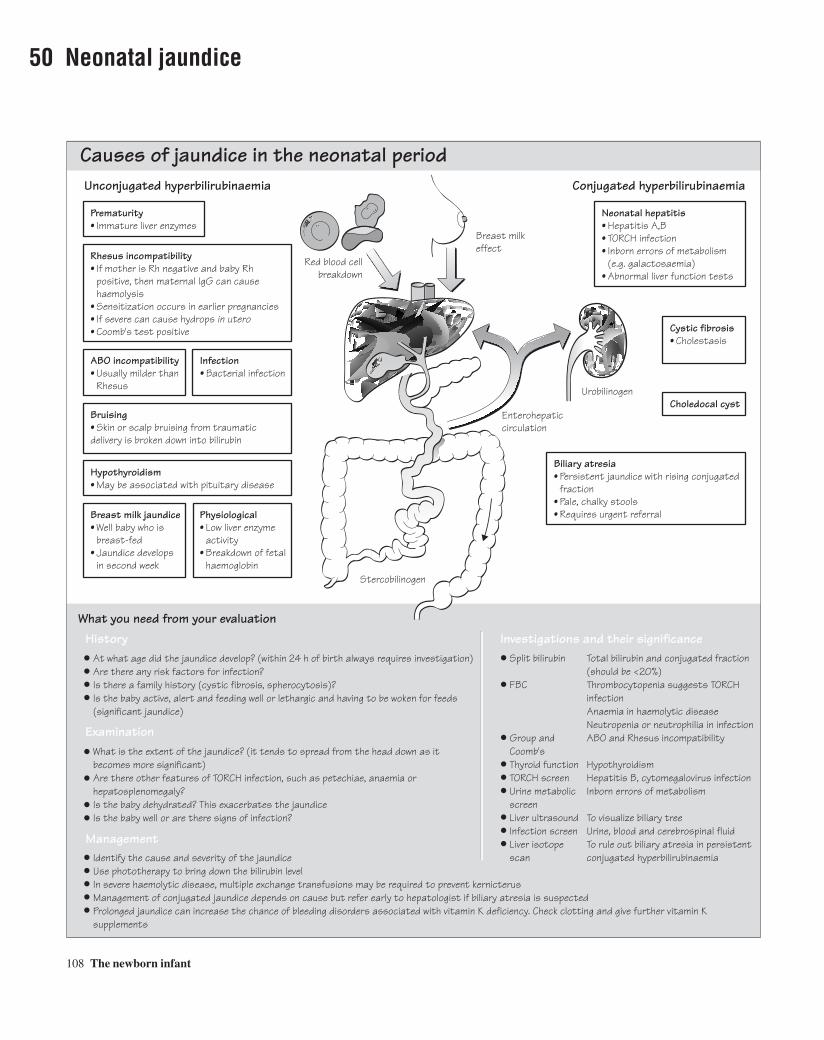
108 The newborn infant
50 Neonatal jaundice
ABO incompatibility• Usually milder than Rhesus
Cystic fibrosis• Cholestasis
Choledocal cyst
Red blood cellbreakdown
Breast milkeffect
Conjugation
Urobilinogen
Enterohepaticcirculation
Stercobilinogen
Biliary atresia• Persistent jaundice with rising conjugated fraction• Pale, chalky stools• Requires urgent referral
Prematurity• Immature liver enzymes
Unconjugated hyperbilirubinaemia
Neonatal hepatitis• Hepatitis A,B• TORCH infection• Inborn errors of metabolism (e.g. galactosaemia)• Abnormal liver function tests
Conjugated hyperbilirubinaemia
Infection• Bacterial infection
Rhesus incompatibility• If mother is Rh negative and baby Rh positive, then maternal IgG can cause haemolysis• Sensitization occurs in earlier pregnancies• If severe can cause hydrops in utero• Coomb's test positive
Bruising• Skin or scalp bruising from traumatic delivery is broken down into bilirubin
Hypothyroidism• May be associated with pituitary disease
Breast milk jaundice• Well baby who is breast-fed• Jaundice develops in second week
Physiological• Low liver enzyme activity• Breakdown of fetal haemoglobin
At what age did the jaundice develop? (within 24 h of birth always requires investigation)Are there any risk factors for infection?Is there a family history (cystic fibrosis, spherocytosis)?Is the baby active, alert and feeding well or lethargic and having to be woken for feeds (significant jaundice) What is the extent of the jaundice? (it tends to spread from the head down as itbecomes more significant)Are there other features of TORCH infection, such as petechiae, anaemia or hepatosplenomegaly?Is the baby dehydrated? This exacerbates the jaundiceIs the baby well or are there signs of infection?
Identify the cause and severity of the jaundiceUse phototherapy to bring down the bilirubin levelIn severe haemolytic disease, multiple exchange transfusions may be required to prevent kernicterusManagement of conjugated jaundice depends on cause but refer early to hepatologist if biliary atresia is suspectedProlonged jaundice can increase the chance of bleeding disorders associated with vitamin K deficiency. Check clotting and give further vitamin K supplements
Split bilirubin Total bilirubin and conjugated fraction (should be <20%) FBC Thrombocytopenia suggests TORCH infection Anaemia in haemolytic disease Neutropenia or neutrophilia in infection Group and ABO and Rhesus incompatibility Coomb's Thyroid function Hypothyroidism TORCH screen Hepatitis B, cytomegalovirus infection Urine metabolic Inborn errors of metabolism screen Liver ultrasound To visualize biliary tree Infection screen Urine, blood and cerebrospinal fluid Liver isotope To rule out biliary atresia in persistent scan conjugated hyperbilirubinaemia
What you need from your evaluation
Examination
Management
History Investigations and their significance
Causes of jaundice in the neonatal period

Jaundice is the yellow pigmentation of the skin that occurs whenplasma bilirubin levels are high. The bilirubin is formed from thebreakdown of haem in red blood cells and is transported to the liver asunconjugated bilirubin, bound to albumin. In order to be excreted itneeds to be made water soluble by conjugation in the liver. Conjugatedbilirubin is then excreted in the bile into the duodenum, where some isreabsorbed (the entero-hepatic circulation) and the remainder formsstercobilinogen, which gives the stools their yellow/brown pigment.Some of the reabsorbed conjugated bilirubin is excreted by the kidneysas urobilinogen.
Excessive haemolysis or impaired conjugation leads to a build-up of unconjugated bilirubin, and obstruction to drainage of bile leads toconjugated hyperbilirubinaemia. Unconjugated bilirubin can cross thebrain barrier. In high concentrations it is toxic to the basal ganglia andcauses ‘kernicterus’ which used to be a common cause of athetoid ce-rebral palsy. Kernicterus is now extremely rare due to better obstetricmanagement of Rhesus disease, careful monitoring of bilirubin levelsand early treatment with phototherapy.
TreatmentPhototherapy (blue light at 450 nm wavelength) helps convert unconju-gated bilirubin to an isomer which can be excreted by the kidneys. InRhesus or ABO incompatibility, if bilirubin levels rise significantly,despite phototherapy, then an exchange transfusion is required toremove the bilirubin and the maternal IgG antibodies from the circulation.
Physiological jaundiceJaundice in the neonatal period is very common and is usually due to aphysiological immaturity of the liver. It is self-limiting as the livermatures over the first week, and only occasionally needs phototherapytreatment. All premature infants become jaundiced in the first few daysof life, due to the immature hepatocytes not being able to adequatelyconjugate bilirubin. This never requires exchange transfusion but mayrequire phototherapy for a few days.
Haemolytic disease of the newbornHaemolysis occurs when maternal IgG antibody crosses the placentaand reacts with antigens on the fetal red blood cells. The commonestcauses of this are ABO or Rhesus incompatibility. In Rhesus disease thefetus is Rhesus positive and the mother Rhesus negative. The mother
will have been sensitized in a previous pregnancy by the passage of fetalred blood cells into her circulation, either at delivery or during a threat-ened miscarriage. Rhesus negative women are now routinely immu-nized with anti-D antibody, to ‘mop-up’fetal red blood cells before theystimulate maternal IgG production. Haemolysis in the fetus causesanaemia which can lead to hydrops (severe oedema). After birthaffected fetuses are anaemic and rapidly develop severe jaundice. Themanagement of Rhesus or severe ABO incompatibility is to deliver the baby before severe haemolysis has occurred and then to wash outthe maternal antibodies (and the bilirubin) by performing a series ofexchange transfusions, and also by the use of phototherapy.
Biliary atresiaBiliary atresia is a rare but important condition caused by the absence ofintra- or extra-hepatic bile ducts. An conjugated hyperbilirubinaemiadevelops over a period of weeks, and the stools become clay coloured.If undiagnosed the baby will develop liver failure and may die withouta transplant. If detected within the first 6 weeks then a hepatoporto-enterostomy (Kasai procedure) can achieve adequate biliary drainage.Because of this it is recommended that any baby still jaundiced after 2weeks has conjugated and unconjugated bilirubin levels checked.
Jaundice in older childrenJaundice is rare in older children. The commonest cause is hepatitis Ainfection. Other causes include chronic haemolysis due to hereditaryspherocytosis or glucose-6-phosphate dehydrogenase (G6PD) defi-ciency, or liver disease such as autoimmune chronic hepatitis. Reyesyndrome, an acute encephalopathy associated with fulminant liverfailure, can be induced by aspirin, and this is therefore contraindicatedin young children.
Neonatal jaundice 109
KEY POINTS• Mild jaundice is extremely common, especially in preterm infants.• Jaundice within the first 24 h or lasting beyond 2 weeks needs investigation.• Phototherapy and occasionally exchange transfusion are used to treat sig-nificant jaundice.• Biliary atresia causes an obstructive persistent jaundice with pale stools.Early treatment is essential.

110 The newborn infant
51 Congenital heart disease
Presentation of congenital heart defectsPresenting with heart failure Presenting with shock
Presenting with a murmur (see Chapter 43)
Presenting with cyanosis
Region of aortic narrowingVentricular
septal defect
Persistentductus
Patent ductusarteriosus
Narrowedpulmonary valve
Atrialseptaldefect
Narrowed pulmonary artery
Ventricularseptal defect
Pulmonary arteryfrom the left ventricle
Aorta from theright ventricle
Narrowedaortic valve
Ventricular septal defect (VSD)• 32% of congenital heart disease (CHD) (commonest)• Membranous or muscular• Can be asymptomatic• Harsh pansystolic murmur at lower left sternal edge• Parasternal thrill• Heart failure at 4–6 weeks
Pulmonary valve stenosis• 8% of CHD• If critical stenosis, may be cyanosed at birth and duct dependent• Ejection systolic murmur in pulmonary area radiating to back• Evidence of right ventricular hypertrophy on ECG
Atrial septal defect (ASD)• 6% of CHD• Ostium secundum (defect of foramen ovale and atrial septum)• Ostium primum (defect of atrioventricular septum)• Usually asymptomatic at birth• Fixed split second heart sound• Pulmonary flow murmur• Recurrent chest infections• Heart failure
Coarctation of aorta• 6% of CHD• If severe, presents as shock when duct closes in first few days of life• May present with heart failure and a murmur over the back• Causes hypertension in older children• Weak or absent femoral pulses• Associated with Turner's syndrome
Aortic valve stenosis• 5% of CHD• If severe, presents as shock or heart failure when duct closes• May present as asymptomatic murmur• Older children may have syncope or reduced exercise tolerance• Ejection systolic murmur radiating to neck
Tetralogy of Fallot• 6% of CHD• Ejection systolic murmur• Intermittent cyanosis from birth• Right ventricular hypertrophy on ECG• Boot-shaped heart on chest X-ray
Transposition of the great arteries• 5% of CHD• Separate pulmonary and systemic circulations, connected only by PDA (or sometimes VSD)• Severe cyanosis and acidosis at birth• Requires atrial septostomy urgently followed by definitive 'Switch' operation
Patent ductus arteriosus (PDA)• 12% of CHD• Continuous machinery murmur below left clavicle• Collapsing pulse• More common in premature infants

Congenital heart disease (CHD) is the most common malformation,with 7–8 per 1000 liveborn infants having a significant cardiac abnor-mality. About 8% are associated with chromosomal abnormality (e.g.AVSD in Down syndrome) or more subtle genetic abnormalities, suchas a deletion at chromosome 22q11 which is associated with aortic archdefects and hypocalcaemia (diGeorge syndrome). The risk of CHD ishigher if there is a family history. Teratogens may cause CHD (e.g. VSDand tetralogy of Fallot in fetal alcohol syndrome).
It is increasingly common for some heart defects to present antena-tally, with abnormalities detected on ultrasound. Others present at birth, with cyanosis (e.g. transposition of the great arteries) or shock(hypoplastic left heart syndrome). Some duct-dependent lesions willpresent when the arterial duct starts to close within the first few days oflife (e.g. coarctation of the aorta, critical pulmonary stenosis). Defectswith a left to right shunt such as a VSD often present with heart failureand difficulty feeding some weeks after birth. Finally, some lesions maybe asymptomatic and are first detected as a heart murmur (e.g. atrialseptal defect, aortic stenosis) (see Chapter 43).
Medical management involves the use of diuretics and other drugs tocontrol heart failure, pending definitive surgical repair. Many heartdefects can just be monitored for years, and may not need surgical cor-rection. It is important to prevent of bacterial endocarditis.
Coarctation of the aortaThis is a narrowing of the aorta. Severe coarctation will present in thefirst few days of life when the arterial duct closes and insufficient bloodis able to reach the lower limbs and perfuse vital organs such as thekidneys, causing circulatory collapse and acidosis. The key feature isweak or impalpable femoral pulses. The blood pressure may be higherin the arms than the legs and oxygen saturation will be lower in the legs. Milder forms of coarctation will present with heart failure and amurmur or with hypertension in a young adult. The management is IVprostaglandin E2 to keep the duct patent. Once the diagnosis is con-firmed, by echocardiography, the narrowed segment is repaired surgi-cally or dilated using a balloon.
Transposition of the great arteries (TGA)In TGA, the aorta and main pulmonary artery are transposed, so that theaorta comes off the right ventricle and the pulmonary artery off the leftventricle. It always presents soon after birth with profound cyanosis andacidosis. The only way oxygenated blood from the lungs can reach thesystemic circulation is across the arterial duct, or a VSD, if present. The
emergency management of TGA is to commence a prostaglandin infu-sion, provide ventilatory and circulatory support and perform an atrialseptostomy which allows mixing of oxygenated and deoxygenatedblood. The definitive treatment is surgical correction—the Switch op-eration, where the two great vessels are switched over and the coronaryarteries connected to the new aorta.
Tetralogy of FallotThis is the commonest cyanotic CHD. The ‘tetralogy’ refers to a largeVSD, an aorta that sits over the ventricular septum, pulmonaryinfundibular stenosis and right ventricular hypertrophy. Some willpresent with cyanosis at birth but others may be diagnosed by an ejec-tion systolic murmur. Classically these children develop hypercyanotic‘spells’which are relieved by squatting down (to reverse the right to leftshunt by increasing left ventricular pressure). Chest X-ray may show a‘boot-shaped’ heart. Surgical correction is usually performed at 2–3months of age.
Patent ductus arteriosus (PDA)During fetal life the ductus arteriosus (arterial duct) shunts blood fromthe pulmonary artery to the aorta, bypassing the unexpanded lungs. Innormal babies the duct closes within a few days of birth, first by con-striction and then by fibrosis. In sick preterm babies, or those exposedto hypoxia, the duct remains open. As the right-sided pressures are nowless than the aortic pressure, blood shunts from left to right and cardiacfailure and pulmonary oedema may develop. The clinical signs of aPDA are a continuous ‘machinery’ murmur below the left clavicle andcollapsing pulses. A patent arterial duct is particularly common in premature infants receiving mechanical ventilation. Management in-volves diuretics and fluid restriction, and sometimes administration ofprostaglandin synthetase inhibitors. Rarely the duct needs to be closedsurgically by ligation or insertion of an occlusion device.
Congenital heart disease 111
Atrioventricular septal defect (AVSD): especially common in Down syndrome (40%)Interrupted aortic archHypoplastic left heart syndromePeripheral pulmonary stenosis: commoner in Noonan syndromeSupravalvular aortic stenosis: with Williams syndromeTricuspid atresiaEbstein's anomaly: in infants exposed to lithiumTotal anomalous pulmonary venous drainage (TAPVD): the only lesion to present with cyanosis and heart failure
Pulse oximetry To determine degree of cyanosisChest X-ray Cardiomegaly in heart failure
Boot-shaped heart (Fallot's tetralogy)Increased vascular markings with left to right shunts (VSD, ASD, PDA)
ECG Right ventricular hypertrophySuperior QRS axis (AVSD, primum ASD)
Echo Ultrasound examination of the heart, usually performed by a paediatric cardiologist, can diagnose the vast majority of congenital heart defects
Fetal Echo Many defects can be detected antenatallyCardiac catheter To define very complex anatomy
Rarer forms of congenital heart disease Investigations and their significance
KEY POINTS• CHD is the commonest congenital abnormality.• The key to diagnosis is understanding the different modes of presentation.• Duct-dependent defects will present in the first days of life and need urgenttreatment with prostaglandin to keep the duct open.• With good management and corrective surgery the prognosis for most CHDis excellent.

112 Chronic illness in childhood
52 Asthma
What you need from your evaluation
Ask about the cough and wheeze. What triggers it and at what time of day does it occur? How many acute exacerbations have there been? How severe was the worst attack? How does the asthma affect the child's life? Does it limit activities; have they missed any school? How often has the child had to use reliever treatment? How effective was it? Are there other atopic symptoms such as hay fever (allergic rhinitis) or eczema or a family history of atopy?
In well-controlled asthmatics there may be no physical signs between acute exacerbations Listen for wheeze. Beware the 'silent chest' of severe asthma when there is almost no air moving Look for chronic chest deformity: barrel chest and Harrison's sulcus in severe uncontrolled asthma Measure PEFR using handheld peak flow meter Check height and weight and plot on centile chart. Poorly controlled asthma will stunt growth, as will overuse of oral corticosteroids Check inhaler technique periodically
Chronic asthma
Cough• Recurrent dry cough• Worse at night• Worse with exercise
Acute asthma
Acute asthma attack• Acutely short of breath• Cough and wheeze• Work of breathing increased• Child often frightened• May be triggered by viral illness, exposure to allergens, exercise or cold air
Assessing severity
Mild• Breathless but not distressed• Peak expiratory flow rate (PEFR) reduced but still >50% of normalSevere• Too breathless to talk or feed• Respiratory rate >50 breaths/min, pulse >140 beats/min• PEFR <50% of expectedLife-threatening• PEFR <33% of expected• 'Silent chest' or cyanosis• Fatigue, drowsiness
Wheeze• Expiratory noise due to airway narrowing• Often triggered by viral infections• Responds to bronchodilators
Shortness of breath• Exercise limitation• Triggers can be exercise, cold, allergens, smoke
Uncontrolled asthma• Poor growth• Chronic chest deformity• Time off school• Frequent acute exacerbations
Pathology• Environmental triggers cause bronchoconstriction, mucosal oedema and excess mucous production in a genetically predisposed child• Airway narrowing causes wheeze and shortness of breath
Examination
PEFR Peak expiratory flow rate is best recorded in a peak flow diary to monitor change over time Chest X-ray To exclude pneumothorax in severe asthma. Avoid excessive X-rays Allergy tests Skin prick or serum radio-allergosorbent tests (RAST) if history suggests a specific allergic trigger (e.g. cat dander)
Medication: 'preventers' (inhaled steroids or sodium chromoglycate) and 'relievers' (bronchodilators) Environmental control: avoid passive smoking and reduce house dust mite exposure if possible Self-monitoring of disease severity: PEFR and symptom diary, management plan for each child Education: of the child, the family and the school on good control of asthma, inhaler technique and emergency treatment of an acute exacerbation
Management
History
Investigations and their significance

Asthma is the commonest chronic illness of childhood, occurring in upto 15% of all children at some point. The incidence has risen signifi-cantly in recent years, partly due to better recognition but possibly alsodue to environmental pollutants. The symptoms of cough, wheeze anddyspnoea are due to narrowing of the bronchi and bronchioles by bron-choconstriction, mucosal inflammation and thick mucous obstructingthe lumen. In a susceptible individual this process is initiated by envi-ronmental factors such as dust mite allergens, air pollution, cigarettesmoke, cold air, viral infections, stress and exercise.
PresentationChildren with asthma usually present in infancy or early childhood.This diagnosis is clinical, based on recurrent cough or wheeze thatresponds to bronchodilator treatment. Ahistory of atopy (eczema or hayfever) or a family history of asthma supports the diagnosis. In infancy itis often unclear whether recurrent wheeze is the first manifestation ofasthma or merely airway obstruction associated with viral respiratorytract infections such as RSV or adenovirus. As the airways are narrow,mucous and mucosal oedema contribute more to obstruction than bron-choconstriction, and there may be poor response to bronchodilators. Ifan infant is atopic, or there is a strong family history then it is morelikely that these wheezy episodes are an early presentation of asthma.
In older children recurrent episodes of wheeze and cough, especiallyif triggered by exercise, viral infections or allergens, suggest a di-agnosis of asthma. A good response to bronchodilator therapy, either in symptom reduction or improvement in peak expiratory flow rate(PEFR) confirms the diagnosis. A CXR should always be obtained atfirst presentation to exclude other pathology such as inhaled foreignbody. In asthma the CXR may show hyperinflation (due to air-trapping)and areas of collapse (due to mucous plugging). RAST may be helpfulin some cases.
Management of chronic asthmaThe goal of good asthma management is to relieve the symptoms andallow normal activity, school attendance and growth. A stepwiseapproach is used—increasing the amount of treatment until control isobtained, then stepping back to the minimum treatment required tomaintain good symptom control.Step 1—Inhaled beta2-agonists (e.g. salbutamol), as required up to threetimes a week. This is known as ‘reliever’ medicine. Check the deliverydevice is appropriate (see below) and the technique is correct.Step 2—Add in a regular ‘preventer’ medicine—either sodium chro-moglicate or low-dose inhaled corticosteroids. Continue the beta2-agonists as required.Step 3—Inhaled beta2-agonists as required plus higher dose regularinhaled corticosteroid. Consider a long-acting beta2-agonist (e.g. sal-meterol) or modified release theophylline.Step 4—Inhaled beta2-agonists as required plus regular high-doseinhaled steroid and consider a short course of oral steroids.Step 5 (only in older children)—Inhaled beta2-agonists as requiredplus regular high-dose inhaled steroid and regular oral steroids (butthere is a serious risk of affecting growth with systemic steroids).
Inhaler devicesTreatment will only be effective if the drug is delivered in sufficientquantity to the small airways of the lungs. This is best achieved using anaerosolized drug delivery system, via a metered dose inhaler (MDI).One ‘puff’of the MDI delivers a known dose of the drug. A high degreeof coordination is required to activate the MDI during inhalation, and this method is really only suitable for teenagers. Some MDIs arebreath-activated.
For children under 8 years, the MDI can be used in conjunction witha spacer device (e.g. aerochamber, volumatic), so that the child breathesin and out of a chamber containing the aerosolized drug. In infants theseshould be fitted with a mask to place over the child’s mouth and nose.
In children over 8 years, a dry powder device (e.g. Turbohaler,Accuhaler) is used—the child sucks in a fine powder, containing thedrug, during inspiration. These devices deliver the required dose morereliably and are easier to use in this age group than MDIs. Nebulizerscan be used in infants and for emergency treatment of acute exacer-bations although there is evidence that MDIs via a spacer are moreeffective.
Treatment of severe exacerbationAcute exacerbations should be treated promptly at home by using moreof the reliever medication. If the symptoms continue or worsen thenaggressive treatment with high flow oxygen, regular beta2-agonists oripratroprium bromide via a nebuliser or spacer device (e.g. 15 puffs)and systemic corticosteroids is indicated. In life-threatening asthma(see opposite) an infusion of salbutamol or aminophylline is used. Allchildren should have oxygen saturations measured and be admitted forclose observation.
Asthma 113
Metered doseinhaler
Spacer device Dry powderdevice
KEY POINTS• Asthma is the commonest chronic childhood illness, occurring in 10–15%of all children.• Bronchoconstriction, viscid mucous and mucosal oedema causes airwaynarrowing with wheeze, cough and dyspnoea.• Treatment is increased and decreased step-by-step to gain symptom controland maintain a normal lifestyle.• It is crucial to use an inhaler device suitable for the child’s age.

114 Chronic illness in childhood
53 Diabetes
What you need from your evaluation
Ask about polyuria, polydipsia, lethargy and weight loss Ask about bedwetting (secondary eneuresis) Review the diabetic diary and ask about hypoglycaemic and hyperglycaemic episodes —what triggered them and were they managed appropriately? How is the child coping at home and at school? Also ask about siblings Is the child managing to eat a healthy diet and modify the diet to certain situations (e.g. snacks before heavy exercise) Is insulin being administered correctly with rotation of injection sites?
Monitor height and weight as poor growth reflects poor control Check for signs of lipodystrophy or lipoatrophy at injection sites Check blood pressure annually and fundi in older children (>14 years) Check for signs of coexistent coeliac disease or hypothyroidism
Blood glucose Monitor regularly at home using finger-prick samples and handheld glucometerHbA1c Reflects control over (% of glycosylated last 2–3 monthshaemoglobin) Urinalysis For glycosuria, ketones, proteinuriaBlood gases, U&E Need to be monitored carefully during acute diabetic ketoacidosisAnti-endomysial Screen for coeliac diseaseantibody Thyroid function Screen for hypothyroidismtests
History
Examination
Investigations and their significance
Initial presentation• Polyuria, polydipsia and weight loss over a few weeks• Polyuria may cause enuresis• Diagnosis made by finding high blood sugar and glycosuria (glucose tolerance test rarely needed)
Ongoing management• Requires multidisciplinary approach• Initial correction of metabolic state and education of family• Treatment with insulin and specific dietary advice• Monitoring blood sugar levels at home and HbA1c regularly• Education about diabetic control, injection technique, diet. • Dealing with emergencies and liaison with school
Aetiology• 1 in 500 children• Destruction of beta islet cells in pancreas leads to insulin deficiency• Genetic predisposition plus infection
Prognosis• No cure. Insulin controls the metabolic disturbance but good control is mandatory to prevent long-term complications in adult life• Retinopathy, neuropathy, renal impairment and atherosclerosis are the long-term effects of poor control of blood glucose levels
Diabetic ketoacidosis (DKA)• May be triggered by infection or poor compliance• Thirst and polyuria• Vomiting• Abdominal pain• Acetone smell on the breath • Very high blood sugar level, ketonuria• Metabolic acidosis on blood gas• Urea raised, electrolytes disturbed• Signs of dehydration• Kussmaul acidotic breathing• Hypovolaemic shock, drowsiness and coma if not treated urgently• Requires urgent treatment with fluids and insulin
Hypoglycaemia• The result of excess insulin or inadequate carbohydrate intake• Feel hungry and shaky• Pale, sweating, tremors• Tachycardic• Drowsy or irritable• Convulsions or coma• Hypoglycaemia on testing• May get rebound hyperglycamia afterwards• Requires urgent treatment
Poor diabetic control• Polydipsia, polyuria• Hypoglycaemic episodes• Poor growth• Hyperglycaemia, high HbA1c• Lipodystrophy if inadequate rotation of injection sites

Diabetes mellitusDiabetes affects more than 1 in 500 children. The diagnosis has a majorimpact on the child and the family in terms of their daily life: the pos-sibility of emergencies such as hypoglycaemia and diabetic ketoacido-sis (DKA), and the fear of long-term complications such as retinopathy,renal failure, cardiovascular disease and neuropathy. Diabetes in chil-dren is usually insulin-dependent diabetes mellitus (IDDM) due todestruction of the beta cells in the islets of Langerhans in the pancreas,resulting in a lack of insulin. The islet cell destruction is genetically pre-disposed but probably triggered by a viral illness. The lack of insulinmeans that glucose cannot be utilized, resulting in hyperglycaemia. Thehigh glucose concentration in the blood spills over into the urine,causing an osmotic diuresis with polyuria and dehydration. This leadsto excessive thirst, and weight loss. Because the cells cannot utilizeglucose they switch to metabolizing fats, leading to the production ofketones and acidosis. Maintenance treatment of diabetes thereforerequires regular administration of insulin to allow normal glucosemetabolism.
Initial presentation of diabetesChildren usually present with a short (2–3 week) history of lethargy,weight loss, polyuria and thirst. The polyuria may cause a recurrence ofbedwetting. If the symptoms are not recognized the child may developsigns of diabetic ketoacidosis with abdominal pain, vomiting and even-tually coma. Newly diagnosed children are usually admitted to hospitalto start their insulin treatment and to allow intensive education of thechild and family, although more and more this is undertaken by a dia-betic nurse specialist who visits the family at home. The family needs tobe taught how to inject the insulin, how to monitor blood glucose andtest urine for ketones, and how to recognize the signs of hypoglycaemia.They also need advice from a dietician, although the diet should not be over-restrictive, but a healthy balanced diet high in fibre. Childrenare encouraged to wear a medic-alert bracelet, giving details of theircondition.
Growing up with diabetesThe education given to families at the time of diagnosis is crucial tothem developing the right approach to their child’s diabetes. They needto be careful without being over-restrictive. As the child gets older theycan gradually take on more of the responsibility themselves, includinginjecting insulin and monitoring blood glucose levels. Food intakeshould not be restricted so that normal growth can occur. Snacks needsto be given as slowly absorbed foods, such as cereal bars or biscuits,rather than chocolate and sweets. Many children go through a ‘honey-moon’period soon after diagnosis where they need very little insulin, asthey still have some endogenous insulin. More insulin is often requiredas they go through the pubertal growth spurt.
Insulin therapyInsulin is usually delivered by an injection pen which injects a prede-termined dose of insulin with each activation. The insulin is given as a
mixture of short-acting (peak at 2–4 h) and intermediate-acting insulin(peak at 4–12 h). For example, Mixtard-30 or Humulin M3 contain 30%short-acting and 70% long-acting insulin. This is administered subcuta-neously in the arms, thighs, buttocks or abdomen, before breakfast andbefore the evening meal. If injection sites are not rotated regularly thenlipodsytrophy and lipoatrophy can occur, leading to erratic absorptionof insulin. About two-thirds of the insulin is given in the morning andone-third in the evening, to cover the increased carbohydrate intakeduring the day and prevent nocturnal hypoglycaemia. Occasionallyinsulin is given four times a day to achieve tighter control.
MonitoringControl is assessed by keeping a blood sugar diary and measuringHbA1c levels, which reflect the degree of hyperglycaemia in the pre-ceding few months. The family need to be warned of the symptoms of hypoglycaemia (see opposite) and have a supply of sugary drinks,glucose gel or glucagon injections available at all times. As childrenbecome adolescents they should be gradually integrated into the adultdiabetic service. At this stage, if not already, they will begin screeningfor retinopathy and renal disease.
Diabetic ketoacidosisDKA occurs when there is inadequate insulin to meet the metabolicdemands. This is due to either poor compliance with insulin treatment,a common problem in adolescence, or to an intercurrent illness whichincreases insulin demands. A common mistake is to give less insulinbecause a child is ill, not eating, and vomiting. In fact in this situationthey will often need more insulin than usual.
The lack of insulin leads to hyperglycaemia and dehydration due toan osmotic diuresis. A switch to fat metabolism leads to ketone produc-tion, resulting in acidosis, vomiting and abdominal pain and gastricstasis. If not treated the child will become comatose. Treatmentinvolves treatment of shock, then gradual rehydration over 24 h. Aninsulin infusion is used to gradually bring down the blood glucose; atthis point some dextrose should be given to allow the cells to switchback to metabolizing glucose. Serum electrolytes and fluid balancemust be checked regularly. There is a danger of hypokalaemia as insulinpushes potassium into the intracellular compartment. A naso-gastrictube should be passed to empty the stomach, as there is often gastricparesis. Infection should be sought and treated. Once the child is eatingand drinking they can be switched back to their normal insulin.
Diabetes 115
KEY POINTS• Diabetes is very common: it occurs in 1 in 500 children.• Presentation is with a short history of weight loss, polyuria and polydipsia.• DKA can be life-threatening and needs careful treatment with fluids andinsulin.• Insulin is given subcutaneously as a mixture of short- and long-actinginsulin.• Patients must be able to recognize and treat hypoglycaemia.

116 Chronic illness in childhood
54 Cystic fibrosis
Recurrent chest infections• Cough• Purulent sputum• Pneumonia• Chronic pseudomonas infection• Bronchiectasis• Chest deformity• Eventual respiratory failure
Finger clubbing• Seen with chronic lung infection
Male infertility• Congenital absence of the vas deferens
Ear, nose and throat• Nasal polyps
Liver disease• Obstructive jaundice in neonatal
period (rare)• Biliary stasis may require treatment
with ursodeoxycholic acid• Eventually liver cirrhosis may develop
High salt losses in sweat• Salty taste to skin• Risk of salt-losing crisis during very hot weather
Poor growth• Require 40% extra energy intake compared with
normal child• Poor weight gain• Short stature• Normal growth is achievable with pancreatic
replacement, physiotherapy and aggressive treatment of chest infections
Pancreatic insufficiency• Poor fat absorption• Steatorrhoea (fatty stools)• Distended abdomen• Rectal prolapse• Meconium ileus at birth (15%)• Meconium ileus equivalent—can mimic acute
appendicitis• Need to take pancreatic enzymes with meals• May develop diabetes
There is currently no cure for cystic fibrosis. Gene therapy trials are ongoing but results have been disappointingManagement should involve a specialist multidisciplinary team of respiratory paediatrician, physiotherapist, dietician, specialist nurses and psycho-social support. Treatment should be delivered at home whenever possibleParents are taught chest physiotherapy, which must be performed regularly to clear secretionsAntibiotics are often given prophylactically to prevent chest infections. When infections occur they are treated aggressively, and once colonized with Pseudomonas aeruginosa the child may require regular courses of intravenous and nebulized antibiotics. These are often given at home via an indwelling central venous catheter (e.g. portacath)Pancreatic enzyme capsules containing lipases, amylases and proteases are taken with meals to aid absorptionDietary supplements may be needed to provide sufficient calorie intake. Energy requirements can be 140% of normal due to recurrent infection, coughing and malabsorption. Supplements of fat-soluble vitamins are requiredHeart–lung transplant is offered when the disease has progressed to the end stage. The heart from the cystic fibrosis patient can be used in anotherrecipient (the domino transplant)Life expectancy has improved considerably but the median survival is still only 32 years
Genetics and aetiology
Management
Investigations and their significance
Commonest serious autosomal recessive condition in Caucasian populationAffects 1 in 2500 children; 1 in 25 are carriersCFTR (cystic fibrosis transmembrane regulator) protein is coded for on chromosome 7. Commonest CFTR mutation (78%) is DF508Abnormal CFTR protein leads to poor function of epithelial membrane chloride channel with viscid secretions that obstruct pancreatic exocrine ducts and bronchiolesAbnormal sweat gland function leads to high concentrations of sodium and chloride in the sweat
DNA: Analysis of the CFTR gene may reveal a common mutation (screens for only 30 of 800 mutations)Immunoreactive trypsin (IRT): Level is high in neonates with cystic fibrosis and is used for newborn screening of 'Guthrie' cardsSweat test: High concentrations of sodium (>60 mmol/l) and chloride can be measured. This is the basis of the diagnostic test for cystic fibrosis. A minimum of 100 mg sweat is neededFaecal chymotrypsin and faecal elastase levels are low in cystic fibrosis. Faecalfat concentrations can be used to monitor pancreatic enzyme supplementationLung function tests: Will show a restrictive and obstructive pattern. CXR: May show signs of consolidation. With chronic pseudomonas infection cystic changes become evident

Juvenile chronic arthritis 117
55 Juvenile chronic arthritis
Juvenile chronic arthritis is a group of conditions that present in childhood with joint swelling lasting more than 6 months. The classification has recently been revised (see box below) but is based on the way the condition initially presents. Treatment is aimed at reducing symptoms of pain and preserving joint function and mobility. In the majority of cases the symptoms resolve but in about 25% of those with the systemic form, the disease continues into adulthood and can be disabling. A multidisciplinary approach with psychological support is necessary for these children.
The aims of management are to preserve joint function, to minimize complications, including complications of the treatment, and to aid the psychological adjustment to what can be a chronic disabling condition in some.The aim is to reduce joint inflammation using non-steroidal anti-inflammatory drugs (NSAIDs). Steroids may be injected into affected joints, but cannotbe used too frequently. Systemic steroids may be required for severe systemic disease. Other steroid-sparing therapies include hydroxychloroquine, penicillamine, gold injections, methotrexate and immune regulator drugs, which can be used in severe disease. Physiotherapy, hydrotherapy and wearing splints can help maintain joint function and mobility. Occupational therapy can help with aids to improve function. The family needs psychological support. Children with residual handicap may require help in planning a suitable career
ComplicationsFlexion contractures of the joint may develop without regular therapy and splinting. Joint destruction may require eventual joint replacement (e.g. knees, hips) in some children. Growth failure can occur due to the chronic illness, anorexia and the growth suppression effect of corticosteroid therapy. Patients may develop Cushingoid features. Chronic anterior uveitis (iridocyclitis) is asymptomatic but if missed can lead to visual impairment. Whilst the prognosis differs between subgroups, overall the prognosis is good and most children recover completely
Classification• Systemic (Still's disease): 9%• Polyarticular: 19%• Oligoarticular (≤4 joints): 49%• Enthesitis associated (HLA B27): 7%• Juvenile psoriatic arthritis: 7%• Other
Presentation• Usually affects large joints (knees, ankles, elbows)• Commonly affects girls under the age of 4 years• Minimal systemic symptomsFeatures and prognosis• Rheumatoid factor negative, antinuclear antibody
(ANA) may be positive• High risk of chronic iridocyclitis (inflammation of
the anterior structures of the eye). Regular examination with slit lamp is required to screenfor this
• Arthritis usually resolves completely
Pauciarticular (up to four joints)
Presentation• Symmetrical involvement of large and small joints• There may be poor weight gain and mild anaemia• Morning stiffness• Irritability in young childrenFeatures and prognosis• Rheumatoid factor negative in 97%, ANA may be
positive• No eye involvement• 12% develop severe arthritis but prognosis is
generally good
Polyarticular (five or more joints)
Presentation• Spiking fever, severe malaise• Salmon-pink rash• Anaemia, weight loss• Hepatosplenomegaly• Arthralgia and myalgia but may have minimal
joint symptoms• May resemble malignancy
Features• Both large and small joints affected• 25% have severe arthritis• Rheumatoid factor negative• Tempero-mandibular joint may be involved,
causing micrognathia
Prognosis• In 25%, arthritis persists into adulthood with
disability• May require joint replacements
Systemic onset (Still's disease)
Management

118 Chronic illness in childhood
56 Childhood cancer
Leukaemia• Commonest childhood malignancy (35%)• 80% acute lymphoblastic leukaemia• Presents with -malaise -anaemia -bruising -bone pain -lymphadenopathy• Chemotherapy used to induce remission and prevent relapse• Overall prognosis good (up to 80% survival)
Retinoblastoma• Rare but important cause of blindness• Presents within the early years• White pupillary reflex or squint
Brain tumours• Second commonest presentation of childhood cancer (25%)• Usually primary brain tumour• Present with raised intracranial pressure or neurological signs: -headache -nausea and vomiting -blurred vision -squint (Vl nerve palsy) -ataxia, clumsiness -nystagmus -endocrine dysfunction• Most tumours occur in the brain stem or cerebellum• Treatment involves neurosurgical resection and radiotherapy• Long-term sequelae include endocrine and growth problems
Neuroblastoma• Usually in children under 5 years• Arises in neural crest tissue (adrenal medulla and sympathetic nervous system)• Presents with abdominal mass. Peri-orbital bruising or skin nodules can occur• Increase in urinary catecholamines
Bone tumours• Usually occur in older children• Present with bone pain or mass, usually in the long bones• Ewing's sarcoma• Osteosarcoma• Treatment involves chemotherapy and surgery with bone prostheses
Lymphoma• Hodgkin's disease and non-Hodgkin's lymphoma (NHL)• Usually present with lymphadenopathy• Mediastinal lymph node involvement is common• Diagnostic excision biopsy followed by chemotherapy
Wilm's tumour (nephroblastoma)• Presents with large renal mass• Occasionally causes haematuria or hypertension• Metastasize via IVC to the lungs• Associated with aniridia (absent iris) coded for on chromosome 11
Rhabdomyosarcoma• Arises in mesenchymal tissue• Head and neck and genitourinary tract are the commonest sites• 15% present with metastases
Germ cell tumours• Sacrococcygeal tumour• Gonadal tumours
DiagnosisThe diagnosis of cancer in childhood is devastating. Children should be referred to specialized paediatric oncology centres for optimum management, although shared care with the local hospital is often possible laterTreatmentThe aim of treatment is eradication of the cancer, whilst minimizing damage to the normal tissues. Cancer therapy is toxic and the child requiresintensive support treatment including prophylactic antibiotics and good nutritional support– Surgery is often required for diagnostic biopsy and excision of solid tumours, and for inserting indwelling central venous catheters necessary for chemotherapy– Radiotherapy is used to treat local disease and for total body irradiation in conjunction with bone marrow transplantation. Adjacent tissues are often damaged and there may be long-term effects on growth if the spine or pituitary gland is irradiated– Chemotherapy acts by killing cells during cell division. The aim is to kill the rapidly dividing malignant cells without killing normal cells. The drugs are usually given in combination at regular intervals. Side effects include hair loss, nausea, immunosuppression and bone marrow suppression. There is a particular risk of sepsis if the child becomes neutropenic, and any febrile episodes while the child is neutropenic should be treated aggressively with broad-spectrum intravenous antibiotics pending the results of blood and other cultures– Bone marrow transplantation involves either harvesting bone marrow or using compatible donated bone marrow to replace the patient's suppressed marrow; this allows more intensive chemotherapy to be used. This technique is being used more commonly and earlier in the disease. Side effects include severe immunosuppression and graft-versus-host disease
Management of childhood cancer
Types of childhood cancer

The incidence of malignant disease in childhood is about 1 per 10 000children per year and causes 14% of all childhood deaths. The com-monest malignancies are acute leukaemia, brain tumours and lym-phoma. Overall, there has been a significant improvement in prognosisover recent years due to the use of well-researched and standardizedchemotherapy regimes delivered in specialized paediatric oncologycentres. The prognosis still depends largely on the particular type of malignancy and on the progression of the disease at the time of diagnosis.
Acute leukaemiaLeukaemia is due to the malignant proliferation of white cell precursorswithin the bone marrow. These ‘blast’ cells escape into the circulationand may be deposited in lymphoid tissue. The commonest type ofleukaemia in childhood is acute lymphoblastic leukaemia (ALL),where the blast cells are precursors of lymphocytes. Acute myeloidleukaemia (AML) is commoner in Down syndrome. Chronicleukaemias are very rare in childhood.
ALL can occur at any age, but the peak is around 5 years. The prog-nosis is worse for those presenting under the age of 2 or over 10 yearsold. The onset is insidious with malaise, anorexia and then pallor, bruis-ing or bleeding. Lymphadenopathy and splenomegaly may be present,and bone pain may occur. Peripheral blood usually shows anaemia,thrombocytopenia and a raised white cell count. Those with anextremely high white count (>50 ¥ 109/L) carry a worse prognosis.Blast cells may be seen on the peripheral blood film. The diagnosis isconfirmed by a bone marrow aspirate, which shows the marrow infil-trated with blast cells. Cells are examined for immunological pheno-typing and for cytogenetic analysis as these give important prognosticinformation. ALL can be subdivided into common (75%), T-cell (15%),null (10%) and B-cell (1%).
Treatment of ALL involves combination chemotherapy to induceremission (i.e. remove all blast cells from the circulation and restorationof normal marrow function). Intensification chemotherapy maintainsremission, and methotrexate or cranial irradiation protects the CNSfrom involvement. Monthly cycles of maintenance chemotherapy arethen given for up to 2 years. Prophylactic antibiotics (cotrimoxazole)are given to prevent opportunistic infections such as Pneumocystiscarinii. Care must be taken to avoid live vaccines and contact withinfections such as chicken pox. Children who relapse are often offeredhigh-dose chemotherapy and bone marrow transplantation. The overallprognosis for acute leukaemia is good with up to 65% cured and 5-yearsurvival approaching 80%.
Short-term side-effects of treatmentTumour lysis syndrome. The breakdown of large numbers of malig-nant cells either before or during treatment can lead to very high serum
urate, phosphate and potassium levels and urate crystals can precipitatein the kidneys causing renal failure. Tumour lysis syndrome can be pre-vented by good hydration and the use of allopurinol (a xanthine oxidaseinhibitor).
Bone marrow suppression and febrile neutropenia. Bone marrow suppression may be due to invasion by tumour cells or the effect ofchemotherapy. Anaemia and thrombocytopenia can be treated withinfusions of red cells and platelets. Neutropenia (neutrophil count <1.0¥ 109) is difficult to treat and means the patient is at risk of serious infec-tion. Consequently any significant fever (>38ºC) while neutropenicshould be investigated and treated aggressively with broad-spectrumIV antibiotics until culture results are known.
Immunosuppression. Severe immunosuppression may result fromtreatment. This leaves the child at risk from normally trivial infections.Patients should not be given live vaccines and if exposed to varicella(chickenpox) should be given specific immunoglobulin. If the patientgoes on to develop chicken pox they should be treated with acyclovirand immunoglobulin.
Inflammation. Inflammation of gut mucosa and mouth ulcers as wellas anorexia can lead to poor calorie intake. Nutritional support withfood supplements may be necessary.
Late sequelae of treatmentShort stature or asymmetrical growth may be caused by radiotherapy tothe spine or pituitary fossa. The latter may also cause delayed pubertyand other endocrine dysfunction including growth hormone deficiency,hypothyroidism and gonadal failure. Cranial irradiation, especially in very young children, can lead to neuro-cognitive effects such asmemory loss and poor attention. Chemotherapy can lead to male sterility, nephrotoxicity, deafness, pulmonary fibrosis and cardiomy-opathy. There is a significant risk (about 12%) of second cancers due tothe carcinogenic effect of chemo- and radiotherapy and an increasedgenetic tendency. Chronic ill health and poor school attendance mayhave long-term effects on educational achievement, although this maybe minimized by good liaison with school and specialist staff.
Childhood cancer 119
KEY POINTS• Acute lymhoblastic leukaemia is the commonest childhood malignancy, butwith effective treatment the 5-year survival is above 75%.• Immunosuppression and neutropenia increase the risk of infection. Suspected infection must be treated aggressively.• Survivors of childhood cancer may suffer long-term effects including poorgrowth and endocrine dysfunction.

120 The child with a disability
57 The child with a disability
Giving the diagnosis: this must be carried out in a skilled way by a senior professional Medical management: therapeutic input should be provided initially at home or the child development centre, and then in nursery and school Genetic counselling: required by many families even if no obvious genetic cause is identified Education (see p. 36): A Statement of Special Educational Needs describes the educational provision that must be made for a child with disabilities. Where possible the child should be integrated into mainstream school Provision of services: Social Services are responsible for preschool childcare, respite care, home help, advice about benefits and assessment for services on leaving school. Voluntary agencies may provide support and information
Prevalence of disability10-20 per 1000 children have a significant disability • Physical and multiple disabilities Cerebral palsy 2.5 per 1000 Spina bifida 0.3 per 1000 Muscular dystrophy 0.2 per 1000 • Severe learning difficulties Chromosomal abnormalities 4.0 per 1000 CNS abnormality 0.2 per 1000 Idiopathic 0.5 per 1000 • Special senses Severe visual handicap 1 per 2500 Severe hearing loss 1 per 1000
How disability presents• Antenatally or at birth if anomalies are present• In the first year for motor handicaps and severe learning disabilities • In the second or third year for moderate learning disabilities, language disorder and autism• After cranial insults
Assessment of disability This involves:• A detailed assessment of the child's abilities • Recognition of any underlying medical problem • An assessment of the likely long-term difficulties
A Child Development Team is involved for complex difficultiesProfessional
Developmental paediatrician Physiotherapist
Occupational therapist
Speech and language therapist Psychologist Special needs teacher Social worker
Liaison health visitor
Role
Diagnosis of medical problems Advice on medical issues Assessment and management of gross motor difficulties, abnormal tone and prevention of deformities in cerebral palsyProvision of special equipment Assessment and management of fine motor difficulties Advice on toys, play and appliances to aiddaily living Advice on feeding Assessment and management of speech, language and all aspects of communication Support and counselling of family and team Advice on special educational needs Support for the familyAdvice on social service benefits, respite care, etc. Support for the familyLiaison with local health visitor
Management of disability
Living with a disabilityParents' initial reaction to news of their child's disability is similar to bereavement; they may feel shock, fear and loss, anger and guilt. Each stage of childhood then requires further adaptation, and independenceis an ongoing issue. Schools need to be prepared for any anticipated difficulties and to accommodate physical disabilities. Staff must work with therapists to implement their recommendations. Young adult disability teams can advise about options beyond high school
Key points The child requires a detailed assessment of their difficulties and abilities Parents need an explanation of the nature and causes of the child's disability A coordinated programme must be developed to cover the child's and family's needs Support is important to help the family cope practically and emotionally Educational needs and schooling must be met

The child with visual and hearing impairment 121
58 The child with visual and hearing impairment
Visual impairmentAchild is defined as blind if education can only be provided by methodssuch as Braille that do not involve sight. A child is partially sighted ifeducational methods such as large print books can be used.
Prevalence1 in 2500 children are registered blind or partially sighted and 50% haveadditional handicaps.
AetiologyThe commonest causes are optic atrophy, congenital cataracts andchoroidoretinal degeneration.
Clinical featuresThe eyes may look abnormal or have unusual movements. If the deficitis congenital, psychomotor development is altered. Early smiling isinconsistent and there is no turning towards sound. Reaching for objectsand the pincer grip is delayed. Early language may be normal, butcomplex language may be delayed. ‘Blindisms’ (eye poking, eyerubbing and rocking) may occur. Hearing deficit or severe learning difficulties are commonly associated.
How visual impairment presentsIn neonates the diagnosis is suspected if cataracts, nystagmus or pur-poseless eye movements are present. Otherwise it may be identified byparents or child health surveillance. Ophthalmological examination(and often visual evoked response (VER) testing is required.
ManagementEarly intervention focuses on developmental progress, reducingblindisms and increasing parental confidence. A peripatetic teacherfrom the Royal National Institute for the Blind (RNIB) is provided forpreschool children, and advises on school, mobility training and sup-portive services.
Growing up with visual impairmentParents need advice on caring for their child, non-visual stimulation andadaptations for the home. Mainstream preschool is usually appropriatewith support, but beyond this, placement at mainstream school, a par-tially sighted unit or school for the blind in part depends on learningabilities. Mobility training is important.
Hearing impairmentPrevalenceFour per cent of children have hearing deficits. Most are mild but 2 per1000 need a hearing aid and 1 per 1000 needs special education.
AetiologyMost mild to moderate hearing loss is conductive, resulting from sec-ondary otis media. Sensorineural deafness may be genetic, result frompre- or perinatal problems or follow a cerebral insult.
Factors for risk of deafness• History of meningitis.• Children with cleft palate.• History of recurrent otitis media.• Children with cerebral palsy.• Family history of deafness.
How hearing impairment presentsUniversal neonatal screening with otoautistic emissions (OAE) is nowbeing introduced to identify congenital sensorineural loss. Audiologicaltesting is required for any child with significantly delayed or unclearspeech, children at risk (see above) or where there is parental suspicionof deafness. Testing includes brainstem evoked responses (BSER) if thechild cannot cooperate.
Clinical featuresLack of response to sound.Delayed speech.Behavioural problems.Associated problems: learning disabilities, neurological disorders,visual deficits.
ManagementGrommets are inserted in children with conductive hearing loss.Hearing aids are fitted for sensorineural deafness, and early speechtherapy is needed to develop communication. Genetic counselling maybe needed.
Growing up with hearing impairmentParents need to learn to communicate with the child, which may includesign language. Moderately deaf children can attend a normal school.The severely deaf child requires specialist education at a school for thedeaf or a hearing unit attached to a normal school.

122 The child with a disability
59 The child with cerebral palsy
Cerebral palsy is a disorder of movement caused by a permanent, non-progressive lesionin the developing brain. Spastic cerebral palsy is the commonest form
Hemiplegia • One side of the body• Arm often more involved than the leg • Delayed walking • Tiptoe gait, with arm in a dystonic posture when running
Athetoid cerebral palsy • Due to basal ganglia damage• Walking movements• Intelligence often normal
Diplegia • Both legs involved with arms less affected or unaffected • Excessive hip adduction (hard to put on a nappy)• Scissoring of legs • Characteristic gait: feet in equinovarus and walking on tiptoe
Quadriplegia • Most severe form • All extremities involved• High association with severe learning disabilities and fits • Swallowing difficulties and gastro-oesophageal reflux common • Flexion contractures of the knees and elbows often present by late childhood
Children with cerebral palsy may commonly have additional problems (especially if they are quadriplegic or severe hemiplegic): Learning difficulties Epilepsy Visual impairment Squint Hearing loss Speech disorders Behaviour disorders Undernutrition and poor growth Respiratory problems
Associated problems2–3 per 1000 children
Prevalence
Prenatal Cerebral malformations Congenital infection Metabolic defects Perinatal Complications of prematurity Intrapartum trauma Hypoxic–ischaemic insult Postnatal Non-accidental injury (incurred before Head trauma 2 yrs of age) Meningitis/encephalitis Cardiopulmonary arrest
Aetiology
Diagnosis is clinical, based on the findings of abnormalities of tone, delays in motor development, abnormal movement patterns and persistent primitive reflexes. Diagnosis may be suspected in neonates but can only be made months later
Diagnosis
PrognosisDepends on the degree and type of cerebral palsy, level of learningdiasability and presence of other associated problems. The degree of independent living achieved relates to: Type and extent of cerebral palsy Degree of learning disability Presence of associated problems, e.g. visual impairment, epilepsy
Hemiplegia Diplegia
Quadriplegia

Cerebral palsy is a disorder of movement and posture caused by a per-manent and non-progressive cerebral lesion acquired early in braindevelopment. It is often complicated by other neurological and learningdifficulties. Although the brain lesion itself in cerebral palsy is non-progressive, the clinical picture changes as the child grows and devel-ops. The underlying brain lesion may result from different insultsoccurring at various times in the developing brain.
In the neonatal period the diagnosis may be suspected if a baby hasdifficulty sucking, irritability, convulsions, or an abnormal neurologicalexamination. The diagnosis is usually made later in the first year whenthe following features emerge.• Abnormalities of tone. Initially the tone may be reduced, but even-tually spasticity develops.• Delays in motor development: e.g. marked head lag, delays insitting and rolling over.• Abnormal patterns of development. Movements are not onlydelayed, but also abnormal in quality.• Persistence of primitive reflexes: such as the Moro, grasp and asym-metric tonic neck reflex.
The diagnosis is made on clinical grounds, with repeated examina-tions often required to establish the diagnosis. Once made, a multidisci-plinary assessment is needed to define the extent of the difficulties. CTor MRI scan may be useful in demonstrating cerebral malformations,delineating their extent and ruling out very rare progressive or treatablecauses such as tumours.
Management of cerebral palsyMost children with cerebral palsy have multiple difficulties and requirea multidisciplinary input. This is best provided by a Child DevelopmentTeam, who should structure a coordinated programme of treatment to meet all the child’s needs, and ensure good liaison between profes-sionals and parents.
TherapyPhysiotherapyPhysiotherapists advise on handling and mobilization, and their role iscrucial. The family must be taught how to handle the child in dailyactivities such as feeding, carrying, dressing and bathing in ways thatlimit the effects of abnormal muscle tone. They are also taught a seriesof exercises to prevent the development of deforming contractures. Thephysiotherapist may also provide a variety of aids, such as firm boots,lightweight splints and walking frames for the child who is beginning towalk.
Occupational therapyThe role of the occupational therapist overlaps with that of the physio-therapist. The occupational therapist is trained to advise on equipmentsuch as wheelchairs and seating, and on play materials and activitiesthat best encourage the child’s hand function.
Speech therapyThe speech and language therapist is involved on two accounts—feeding and language. In the early months advice may be required for
feeding and swallowing difficulties. Later, a thorough assessment of thechild’s developing speech and language is required and help given onall aspects of communication, including non-verbal systems when necessary.
Paediatric managementThe paediatrician’s key role is supportive, along with liaison with otherprofessionals, including at school. In the long term the child needs to bemonitored for developmental progress, medical problems, develop-ment of contractures or dislocation, behavioural difficulties and nutri-tional status. Drugs, other than anticonvulsants for epilepsy, have alimited role in cerebral palsy. If spasticity is severe and causing pain,medication to reduce muscle spasm is sometimes prescribed.
Orthopaedic surgeryEven with adequate physiotherapy, orthopaedic deformities maydevelop as a result of long-standing muscle weakness or spasticity. Dislocation of the hips may occur as a result of spasticity in the thighadductors and fixed equinus deformity of the ankle as a result of calfmuscle spasticity. These may require orthopaedic surgery.
NutritionUndernutrition commonly occurs in children with cerebral palsy, andcan reduce the child’s chances of achieving his or her physical and intel-lectual potential. Food must be given in a form appropriate to the child’sability to chew and swallow. Energy-rich supplements and medicaltreatment for reflux may be required. If the child is unable to eat ade-quate amounts, a gastrostomy may need to be placed.
Growing up with cerebral palsyThe family has to cope with all the difficulties facing any family with adisabled child. However, cerebral palsy, if severe, places particularlyheavy demands in terms of time and input. Everyday tasks such asdressing and bathing take time, and feeding, in particular, may takehours each day. The child also needs regular physiotherapy at home,and needs to attend appointments, both for medical follow-up andtherapy. In view of this the family needs support, often beyond whatfamily and friends can supply. Voluntary and social service agenciescan provide babysitting, respite care and benefits.
Children with milder forms of cerebral palsy can cope at mainstreamschool, provided minor learning difficulties and physical access areaddressed. Children with more severe cerebral palsy need specialschooling in a school for the physically or severely learning disabled,depending on the degree of their difficulties.
The child with cerebral palsy 123
KEY POINTS• Physiotherapy is needed to minimize the effects of spasticity and preventcontractures.• Associated problems must be identified and managed.• Any special educational needs must be met.• The family needs adequate financial, practical and emotional support.• The child’s integration into society should be maximized.

124 The child with a disability
60 Epilepsy
Approximately 8 per 1000 schoolchildren Learning difficulties are a common association
Generalized tonic–clonic seizures (grand mal) • Tonic phase: sudden loss of consciousness limbs extend, back arches teeth clench, breathing stops tongue may be bitten• Clonic phase: intermittent jerking movements irregular breathing may urinate and salivate• Postictal phase: child sleepy and disorientated
Simple absence seizures (petit mal) • Fleeting (5–20 seconds) impairment of consciousness (daydreaming) • No falling or involuntary movements• EEG: characteristic bursts of 3/second spike and wave activity
Infantile spasms• Onset usually at 3–8 months of age• Flexion spasms ('jacknife' or 'salaam') • Last a few seconds, in clusters lasting up to half an hour• Regression of developmental skills • May have a history of perinatal asphyxia or meningitis • EEG—characteristic hypsarrhythmic pattern
Monitor: frequency of fits side effects of drugs psychosocial and educational problems anticonvulsant levels if uncontrolled
Simple partial seizures• Twitching or jerking of face, arm or leg• Consciousness usually retained• May start focally and spread (Jacksonian pattern) • Involved part of the body may be temporarily weak after attack • May progress to full-blown tonic–clonic attacks
Complex partial seizures (temporal lobe seizures) • Altered or impaired consciousness associated with strange sensations, hallucinations or semi-purposeful movements • May show chewing, sucking or swallowing movements• Postictal phase with amnesia• EEG may show discharges arising from the temporal lobe
Myoclonic seizures • Shock-like jerks, often causing sudden falls• Usually occur in children with a structural neurological/cerebral degenerative condition
Paroxysmal involuntary disturbances of brain function result in recurrent fits
The diagnosis is largely clinical, based on the description of the attacks. EEG has a limited value in the diagnostic process
Generalized seizures
Partial seizures
Generally good, with resolution of fits in 60% of children with idiopathic epilepsy. Poor prognosis for those with infantile spasms Paediatric follow-up
PrognosisPrevalence
Pathophysiology
How the diagnosis is made
Seizures, convulsions or fits are non-specific terms describing animpairment of consciousness, abnormal motor activity, sensory distur-bance or autonomic dysfunction. Epilepsy is defined as a condition ofrecurrent fits resulting from paroxysmal involuntary disturbances ofbrain function and unrelated to fever or acute cerebral insult. Seizuresmay be generalized from the onset, or partial, beginning in a localizedor focal area of the brain. Partial seizures are defined as complex if thereis an associated impairment of consciousness. Epilepsy is usually idio-pathic, but may result from severe cerebral insult. In some children, par-
ticularly those with neurological signs or learning difficulties, there isan underlying anatomical lesion.
The diagnosis of epilepsy is clinical, the key being a good detailedhistory. Physical examination is usually normal, but the finding of neu-rological signs suggests possible underlying pathology. Investigationsare not usually helpful as 50% of children with epilepsy have normalEEGs on first testing, and 3% of normal children have abnormal EEGs.EEG is useful in diagnosing simple absences and infantile spasms asthese have characteristic findings. Twenty-four hour and video EEG

recordings are sometimes helpful. CT or MRI is indicated in childrenwith focal neurological deficits, focal EEG changes, increased intracra-nial pressure, progressive CNS disease or intractable seizures as there isa high chance of a brain lesion or pathology.
Medical management of epilepsyThe goal is to achieve the greatest control of fits while producing theleast degree of side-effects. This is best achieved through a mono-therapy approach.• Treatment is started with the most effective drug for the type of fit.• The dose is gradually increased to maximum recommended levels.• A second drug is added if the first is ineffective, and the doseincreased.• The first drug, where possible, is gradually discontinued.• Drugs should be given at intervals no longer than one half-life. Drugswith sedative effects should be given at bedtime and if there is a pattern,the peak level should be timed to coincide with the seizures.
If medical treatment fails, surgery may rarely be tried in childrenwith intractable fits and clinical and electrographic evidence of a dis-crete epileptic focus. For most children with epilepsy restriction ofphysical activity is unnecessary, other than attendance by a responsibleadult while bathing and swimming. Avoiding cycling in traffic andclimbing high gymnastic equipment is prudent.
Management of a tonic–clonic seizure (see p. 46)In a tonic–clonic seizure, the child should be placed in the recoveryposition. Parents should be instructed to end the fit by giving diazepamrectally if it lasts more than 10 min. Intravenous drugs should only begiven in hospital where facilities are available in the event of respiratoryarrest. Children do not need to be hospitalized each time a fit occurs.Emergency treatment is not required for other forms of epileptic fits.
Monitoring a child with epilepsyThe family should be encouraged to keep a diary recording any fitsalong with medications received, side-effects and behavioural changes.
This allows accurate review of the child’s condition and the effect ofdrugs. Physical examination is only required if there is a deteriorationin control. Monitoring of anticonvulsant blood levels is only required iffits remain uncontrolled or drug toxicity is suspected. Levels below thetherapeutic range can result from inadequate dosage, poor absorption,rapid drug metabolism, drug interactions and deliberate or accidentalnon-compliance.
Living with epilepsyEpilepsy is a difficult condition for children to live with as it periodi-cally and unpredictably places them in embarrassing situations. Theymay suffer from stigmatization and social difficulties, and their integra-tion into school may become affected. Too often physical activities arelimited for fear that a fit will place them in danger.
Most parents are initially frightened by the diagnosis of epilepsy andrequire support and accurate information about the condition. Theyneed to know how to safely manage an acute fit including using rectaldiazepam, about side-effects of drugs, the dangers of sudden with-drawal of medication, and social and academic repercussions. There areoften concerns about genetic implications, and the teratogenic effects ofanticonvulsants must be discussed with the teenage girl.
The family should be encouraged to treat the child as normally aspossible and not to thwart the child’s independence. This becomes aparticular issue in adolescence when compliance too can be a problem.Career guidance is important as some occupations are closed to indi-viduals with epilepsy. Application for a driving licence can only bemade after 3 fit-free years whether the person is on or off medication.
Staff at school must be taught the correct management oftonic–clonic fits, although most will not take the responsibility foradministering rectal diazepam. Teachers need to be aware of other typesof fit such as absence spells, as well as side-effects of drugs, and reportthese to the parents or school nurse. When epilepsy is associated withlearning difficulties, appropriate help needs to be provided.
Epilepsy 125
KEY POINTS• Ensure the diagnosis is correct.• Only treat if fits are recurrent.• Use monotherapy when possible.• Check plasma levels if control is inadequate and, if low, consider non-compliance.• For tonic–clonic epilepsy, rectal diazepam should be prescribed for homeuse.• Ensure any learning difficulties are addressed.• Help the child live a normal life with full participation at school and home.
Seizure type and drug therapy.
Type of epilepsy Drug of first choice Other drugs
GeneralizedTonic–clonic (grand mal) Valproate CarbamazepineSimple absences (petit mal) Valproate EthosuxamideMyoclonic Clonazepam ValproateInfantile spasms Vigabatrin ACTH
Partial Carbamazepine Phenytoin

126 The child with a disability
61 Learning disability
Diagnosis of underlying cause Early intervention and educational programmes to stimulate cognitive, language and motor development School: statementing is required with placement in mainstream or special school Behavioural difficulties must be addressed Family support and benefits should be provided General paediatric care must not be neglected
Developmental progress and physical growth require review Screening for specific associated problems in some conditions Behaviour is often an issue Liaison with other professionals is important, particularly regarding education The family needs support
Depends on the underlying cause Degree of independent living achieved relates to the level of learning disability and the underlying aetiology
How learning disability presents• Dysmorphic features may be evident at birth• SLD presents as developmental delay before 12 months• MLD presents with delayed language in toddlers• The diagnostic process is discussed in Chapter 46 (global developmental delay)Aetiology/pathophysiology
• Chromosome disorders 30%• Identifiable disorders or syndromes 20%• Associated with cerebral palsy, microcephaly, 20% infantile spasms, postnatal cerebral insults • Metabolic or degenerative disease <1%• Idiopathic 25%
Clinical features• Reduced intellectual functioning • Delay in reaching developmental milestones, particularly language and social skills, in early childhood• Often associated with: Epilepsy Vision and hearing deficits Communication problems Attention deficit/hyperactivity Feeding problems and failure to thrive
Prevalence• 4 per 1000 children
Management (needs to be multidisciplinary)
Paediatric follow-up Prognosis
Learning disability (or difficulty) has replaced the terms ‘mental retar-dation’and ‘mental handicap’. The disability is classified as mild, mod-erate or severe according to the intellectual limitation and degree ofindependence anticipated or achieved. Individuals with severe learningdifficulties can learn minimal self-care and simple conversation skills,and need much supervision throughout their lives. Those with profoundlearning disability require total supervision, few become toilet-trainedand language development is generally minimal.
Children with severe learning disabilities are spread throughout thesocial classes, and usually have an organic basis for their problem. Thiscontrasts with children with milder learning disabilities, where mostlyno organic cause is found and there is a predominance of children fromlower socioeconomic classes.
Paediatric management of children with learning disabilityThe role of the paediatrician is to support and help the family in comingto terms with their child’s limitations, to attempt to diagnose the under-lying cause, and to manage medical problems. A diagnosis is often notpossible, but is of great importance to the family and allows for moreaccurate genetic counselling. Other aspects of management involveadvising on appropriate educational and therapeutic input. Liaison withother professionals is an important aspect of the work.
Growing up with learning disabilityThe diagnosis of severe learning disability is devastating and familiesrequire particularly sensitive support at diagnosis and beyond. Eachstage of childhood brings its own issues. Adolescence is usually a par-

ticularly difficult time when issues related to sexuality, vocational train-ing and community living must be addressed.
It is important to begin input early to stimulate cognitive, languageand motor development. Therapists from the Child Development Teamshould provide advice on play activities and suitable toys, give guid-ance in the development of skills such as feeding, washing and dress-ing, and instruct parents on the principles of language development,introducing alternative communication systems where appropriate.Attendance at special nurseries, such as Mencap, can be stimulating forthe child while providing contact with other families.
Many children with learning disabilities can cope with and benefitfrom mainstream nursery and primary school, with appropriate helpprovided. Others, particularly if they have additional disabilities, maybe better placed in a special school. A statement of special educationalneeds (see p. 36) is required. Education must be realistic, and shouldinclude teaching skills such as personal care, hygiene and safety, devel-opment of acceptable social behaviour, and maximizing independence.On leaving school, facilities should be available for the young adult,including an adult training centre, special hostels, and vocational train-ing schemes.
Behaviour problems occur with greater frequency in children withdevelopmental disabilities. This may include attention difficulties orhyperactivity (see p. 37) or stereotypic or self-injurious behaviour. Psychological help is then needed, and occasionally medication too.Genetic counselling is important, whether there is a clearly inheriteddisorder or not, as the family will want to know the chances of havinganother affected child. In children with no identified cause, the risk ofanother sibling being affected is about 1 in 25. However, if multiplecongenital anomalies are also present the risk falls to 1 in 40.
Down syndrome (trisomy 21)Down syndrome is the commonest congenital anomaly causing learn-ing disability. The extra chromosome is usually maternal, and the incidence of Down syndrome increases with maternal age (2% at age 40 years).
Features include upward sloping palpable fissures, epicanthal folds,Brushfield spots (speckled iris), a protruding tongue, flat occiput, singlepalmar creases, and mild to moderate developmental delay with socialskills often exceeding other milestones. One-third are born with GIproblems, most commonly duodenal atresia, and one-third have cardiacanomalies (most commonly atrioventricular canal defects). Secretoryotitis media, strabismus, hypothyroidism, atlanto-axial instability andleukaemia are common problems. Children with Down syndrome canusually be integrated into mainstream primary school with additionalsupport.
A cardiac evaluation should be performed at birth, routine audio-logical and thyroid tests are needed throughout childhood and ophthal-mological assessment if there is any evidence of a squint. The child’sgrowth needs to be followed on special Down growth charts. Thefamily requires genetic counselling.
Fragile XFragile X is an important genetic cause of learning disabilities amongstboys. The chromosomal anomaly consists of a ‘fragile site’at the end ofone of the long arms of the X chromosome. The diagnosis should besought in any boy who has unexplained moderate or severe learning disability. Some girls carrying the chromosome have mild learning disabilities.
AutismAutism is a condition where there is an inability to relate to others, andlanguage development is very delayed. It often occurs in conjunctionwith learning disabilities. Characteristically the autistic child fails todevelop social relationships, has little non-verbal communication andoften has ritualistic behaviour patterns. Special education is oftenrequired and the family need good support as it is extremely stressful tohave an autistic child in the family.
Learning disability 127
KEY POINTS• Where possible the underlying condition should be diagnosed.• The child’s developmental progress should be monitored.• Appropriate input should be provided in the preschool years and appropri-ate school placement made.• The child and parents need a supportive framework.• Good general paediatric care must be provided.
Generalhypotonia
Space between1st and 2nd toes
Trisomy 21
5th fingerclinodactyly
Brachydactyly(short fingers)
Most common features:• Flat facial profile• Flat occiput• Epicanthal fold
Variable features:• Congenital heart defect (especially AVSD)• Increased risk of leukaemia• Duodenal atresia• Hirschprung disease• Hypothyroidism
Simiancrease
Down syndrome


abdominal examination 14newborn infant 104
abdominal migraine 64abdominal pain 14
acute 58–9recurrent 64–5
abdominal wall defects 104ABO incompatibility 108, 109abortion 33abscess 80
lung 55peritonsillar (quinsy) 49, 56
absence seizures 74, 75, 124abused child 10, 98–9
child protection procedures 99failure to thrive 98intentional overdose 61see also non-accidental injury (physical
abuse)accidents
adolescents 32, 33causes/prevention 60health education 35see also poisoning
acidosis 19acne 32activated charcoal 61acutely ill child 40–3
assessment checklist 43adenovirus infection 55adolescents 32–3
developmental tasks 32diet 29intentional overdose 61learning disability 126–7oral contraception 10psychological changes 32psychological disorders 32sexual health 33vomiting 52vulnerable groups 32
adrenal insufficiency 45aggressive behaviour 30, 31, 37air-trapping 12, 21airway opening manoeuvres 43
basic life support 42convulsions 46, 47unconscious child 45
alcohol 32, 33intentional overdose 61
alkalosis 19allergy 11
rash 83ambiguous genitalia 104amblyopia 16amenorrhoea 33aminophylline 113ammoniacal dermatitis 89anaemia 14, 96–7
chronic illness 18, 96, 97chronic renal failure 71, 96, 97investigations 18premature infant 106
anal fissure 14, 66, 67anal trauma 14, 98, 99analgesic headache 72anaphylactoid purpura see Henoch–Schönlein
purpura
anaphylaxis 40acute stridor 56
anencephaly 105anorexia 58, 59anorexia nervosa 32, 33antacids 65antenatal steroids 106anterior fontanelle 104anterior uveitis (iridocyclitis) 117anti-D antibody 109antibiotic prophylaxis 13antibiotic-induced gastroenteritis 50, 51anticonvulsants 125antidepressants poisoning 61antihistamines 87anus, newborn examination 104aortic arch abnormalities 111aortic stenosis 13, 95, 110
heart murmur 94, 95supraventricular 111
apex beat 13newborn examination 104
Apgar score 102, 103apnoea 74appendicitis, acute 58, 59appetite 14arteriovenous malformation 72artificial ventilation 40, 41, 42, 43
respiratory distress syndrome 107ascites 14, 70, 71asphyxia-related convulsions 46asphyxial event, acute 44aspirin 49, 70, 109
poisoning 61asthma 12, 32, 40, 41, 54, 55, 112–13
management 112, 113severe exacerbation/acute attack 112, 113severity assessment 112
asymmetric tonic neck reflex 15ataxia 15, 72atopic asthma 113atopic dermatitis (atopic eczema) 86–7atrial septal defect 13, 54, 95, 110
heart murmur 94, 95atrioventricular septal defect 111attention deficit disorder 37, 127auroscope examination 12auscultation
abdominal system 14cardiovascular system 13chest 12
autism 100, 127autoimmune chronic hepatitis 109avascular necrosis of femoral head (Legg-
Perthes disease) 76AVPU scale 44
baby care advice 35back to sleep campaign 43bacterial meningitis 45bacterial pneumonia 55Barlow test 39, 104, 105barrel chest 12basic life support 42bedwetting (nocturnal enuresis) 30, 68behaviour problems 30–1
adolescents 32, 33health education 35
129
learning disabled children 127at school 37
beta2-agonists 113biliary atresia 108, 109bilirubin metabolism 109birth asphyxia 19, 44, 100, 103birth injury 100birthmarks 88, 104birthweight 102bites, non-accidental injury 98bleeding time 18blood film 18blood gases 19blood glucose monitoring 115blood loss
anaemia 18shock (circulatory failure) 40
blood pressure measurement 13blood in stool 58
‘redcurrant jelly’ 59bone marrow suppression 119bone marrow transplantation 118, 119
ethical issues 10bone tumours 118
leg pain/limp 76Bordetella pertussis 55
see also pertussis (whooping cough)bowel gaseous distension 14bowel movements 62, 67bowel obstruction 14, 52, 53, 58
constipation 66newborn examination 104
brain abscess 73brain tumour 15, 73, 118, 119breast development 24, 32breast-feeding 28, 29, 31, 35, 51, 97, 102, 103
advantages 28HIV transmission 81problems 28
breast milk jaundice 108breath, first in newborn 102breath sounds 12breath-holding spells 74, 75breathlessness 13, 54
asthma 112, 113bronchiectasis 55, 116bronchiolitis 12, 41, 54, 55bronchodilators 112bronchopulmonary dysplasia 106, 107bruising
neonatal jaundice 108non-accidental injury 98
Brushfield spots 127bulimia 32, 33, 52bullae 82bullying 37burns 50, 60
extent assessment 60management 60non-accidental injury 98prevention 60shock (circulatory failure) 40
café-au-lait spots 82, 100Campylobacter 50, 51cancer 118–19
pyrexia of unknown origin 80, 81treatment sequelae, short-/long-term 119
Index

130 Index
candidiasis 87HIV infection/AIDS 81nappy rash 89
capillary refill time 13, 43carbohydrate intake in milk 29carbon monoxide poisoning 60cardiac arrhythmias 74, 75
poisoning 61cardiac massage 42, 43cardiorespiratory arrest 40, 42, 44
airway management 42causes 42convulsions 46
cardiothoracic ratio 20cardiovascular examination 13
newborn infant 104cataract 16
congenital 16, 120screening 39
central nervous system malformation 100cephalhaematoma 104cephradine 69cerebral palsy 15, 17, 101, 106, 107, 120,
122–3, 126aetiology 122associated problems 122athetoid 122convulsions 46diagnostic features 123gastro-oesophageal reflux 53kernicterus 109management 123nutrition 123
cerebrospinal fluid (CSF) analysis 21cerebrovascular accident 44cervical lymphadenopathy 12, 78chaperone 11chemotherapy 118, 119chest auscultation 12chest deformity 12, 54, 116chest radiograph 20–1chest wall palpation 12chicken pox (varicella) 48, 83
clinical features 84complications 84rash 82
child care 36Child Development Team 101, 120, 127child health service 34–5child protection
child health services 34, 35procedures 35, 99
Child Protection Register 34, 35, 99childminders 36choking 41
prevention 60choledochal cyst 108choreoathetoid movements 15choroidoretinal degeneration 120chromosomal abnormalities 100, 101, 111, 120,
126chronic illness 32, 112–19
failure to thrive 92, 93growth impairment 90, 91
circulation 13cleft lip/palate 49, 104, 105, 120clinical chemistry 19clotting investigations 18club foot (talipes equinovarus) 104clumsiness (dyspraxia) 37coarctation of aorta 13, 110, 111
clinical features 111heart murmur 94
coeliac disease 14, 29, 62, 63failure to thrive 92, 93
growth chart 23cold sore 88colic 30, 31collagen vascular disease 80, 81collapse 13, 15collapsing pulse 13colobomata 104colonic agangliosis see Hirschsprung’s diseasecolostrum 29colour vision 39coma 44–5
investigations 44metabolic causes 45unexplained 45
communication skills 10, 11community paediatrician 34competence 10complex partial seizures 74, 75, 124conductive hearing loss 49, 120confidentiality
adolescents 32ethical issues 10
congenital abnormalities 103newborn examination 104–5
conjugated hyperbilirubinaemia 108consciousness level 11
assessment 44poisoning 61transient alteration (fits/faints) 74, 75
consent 10, 11, 32constipation 14, 52, 58, 64, 66–7
definition 67functional 66, 67management 67overflow diarrhoea 62, 63urinary symptoms 68urinary tract infection 69
constitutional (familial) short stature 90, 91consultation 10–11
adolescents 32contact dermatitis 86, 87contraception 32, 33, 35contractures 15
cerebral palsy 123juvenile chronic arthritis 117newborn examination 104
convulsions 15, 40, 44, 124causes 46febrile 46, 47, 49, 74generalized 47investigations 46treatment 46, 47
coordination 15corneal light reflex 16corrosives ingestion 61corticosteroid therapy
antenatal 106, 107asthma 112, 113juvenile chronic arthritis 117
coryza 48, 49, 55cough 12, 54
asthma 112, 113cover test 16cow’s milk protein intolerance 62, 63Coxsackie virus pneumonia 55crackles 12, 41, 54, 55cradle cap 86, 87cranial irradiation 119cranial nerve examination 15cranial nerve palsy 72Crohn’s disease 32, 62, 63croup (acute laryngotracheobronchitis) 40, 41,
54, 56, 57epiglottits differentiation 57
crying baby 30, 31, 35, 58
cryptorchidism 14, 79, 104screening 39
Cushing’s disease 35growth impairment 90, 91
cyanosis 11, 12, 13, 40, 41, 54, 104breath-holding spells 74, 75congenital heart disease 94, 110, 111
cystic fibrosis 12, 19, 32, 41, 50, 54, 55, 62, 63,92, 108, 116
failure to thrive 93genetic aspects 116management 116meconium ileus 53
cystic-dysplastic kidneys 71cytomegalovirus 101
decerebrate posture 44decorticate posture 44defibrillation 42dehydration 11, 19, 48, 50, 51
assessment 50, 51over-rapid correction 45shock (circulatory failure) 40, 51treatment 50, 51
delayed menarche 91delayed puberty 24, 91
cancer therapy sequelae 119dental caries 29, 72, 73dental health promotion 35depression 32dermatitis
ammoniacal 89atopic 86–7contact 86, 87
desquamation 82development 27
assessment 27, 34fine motor 25, 27gross motor 25health education/promotion 35history-taking 11social 26speech/language 26, 27surveillance 39warning signs 27
developmental delay 100–1, 126global 101
developmental milestones 15, 27, 100diabetes mellitus 16, 21, 32, 114–15
complications 114hyperglycaemia 114, 115hypoglycaemia 45, 46, 47, 114ketoacidosis 19, 21, 44, 45, 50, 58, 59, 114, 115management 114, 115urinary symptoms 50, 68
diaphragmatic hernia 104, 107diarrhoea 14, 19, 58
acute 50, 51chronic/recurrent 62–3dehydration 50, 51HIV infection/AIDS 81
diet see nutritiondieting behaviour 32, 33diffuse axonal injury 44diGeorge syndrome 111diphtheria 38, 49
immunization 38diplegia 122dipstick tests 21, 69disabled children 120
assessment 120management 120see also learning disability
disseminated intravascular coagulation 18diurnal enuresis 68

doctor–patient relationship 10–11adolescents 32
double aortic arch 57Down syndrome (trisomy 21) 100, 101, 103,
104associated disorders 49, 53, 111, 119, 127clinical features 105, 127newborn examination 104
drowning 46, 60prevention 60
drug abuse 32, 33intentional overdose 61
drug history 11drug overdose 32, 44
intentional 61drug-induced haematuria 70drug-induced rash 82Dubowitz score 106Duchenne muscular dystrophy 15duodenal atresia, congenital 53duplex kidney 69dyslexia 37dysmenorrhoea 33, 64dysmorphic features 104, 126dysmorphic syndromes 93, 100, 101, 105dyspraxia (clumsiness) 37dysuria 14
ear, nose and throat examination 12eating behavioural problems 30, 31, 35
failure to thrive 92, 93eating disorders 32, 33Ebstein’s anomaly 111ectopic ureter 68eczema 86education 36
cerebral palsy patient 123disabled child 120epilepsy patient 125hearing impaired child 120learning disabled child 127problems at school 37visually impaired child 120
Edward syndrome (trisomy 18) 104, 105electrolyte balance 19
premature infant 106emergency caesarean section 103emergency care order 35, 99emotional abuse 35, 98
developmental delay 101psychosocial growth impairment 90
empyema 55encephalitis 44, 45encephalopathy 45encopresis 30, 67energy requirements 29entero-hepatic circulation 109enuresis 14
diabetes mellitus presentation 114, 115diurnal 68nocturnal (bedwetting) 30, 68secondary 68
enuresis alarm 68epicanthic folds 16epiglottitis 12, 41, 56, 57
croup differentiation 57epilepsy 44, 46, 47, 74, 75, 124–5
definition 124drug therapy 124, 125management 125monitoring 125seizure types 124, 125
erythema toxicum 104erythropoietin 71Escherichia coli 50, 51, 69
Escherichia coli O157 51, 71ethical issues 10Ewing’s sarcoma 118examination procedure 11exercise 35exercise-related asthma 113exercise-related haematuria/proteinuria 70exomphalos 104external cardiac massage 42extradural haematoma 44extremely low birthweight 102eye strain 72
facial hair 32factitious fever 48, 80failure to thrive 13, 92–3
HIV infection/AIDS 81neglect 99, 101non-organic (psychosocial) 92, 93, 99
fainting (syncope) 13, 74, 75falls, prevention 60familial (constitutional) short stature 90, 91family history 11fat intake in milk 29febrile convulsions 46, 47, 49, 74febrile neutropenia 119femoral pulses 13fetal alcohol syndrome 100, 101, 104, 111fetal distress 103
meconium-stained liquor 102fever 48–9, 54
causes 48factitious 48, 80management 49newborn infant 49pyrexia of unknown origin 80–1
fifth disease 83finger clubbing 12, 13, 54, 81, 116finger–nose test 15fits 74–5, 124floppy baby 15fluid depletion
constipation 66shock (circulatory failure) 40
focal neurological signs 72, 73focal seizures 72fontanelle palpation 15
dehydration assessment 51newborn examination 104
food poisoning 50foreign travel 11, 12formula milk feeds 28, 29fractures, non-accidental injury 98fragile X syndrome 100, 101
clinical features 127frog’s leg position 15full-term infant 102fundoscopy 16funny turns 74–5
gait 15, 17galactosaemia 108gallop rhythm 13gastro-oesophageal reflux 14, 52, 53, 54, 92,
106gastroenteritis 19, 50, 58
dehydration 40, 50, 51vomiting 52, 53
gastroschisis 104general practitioners 34generalized tonic-clonic (grand mal) seizures
124, 125genetic counselling 127genetic syndromes, failure to thrive 92, 93genital development 24
Index 131
genitalia examination 14newborn infant 104
german measles see rubellagestation 102Giardia lamblia 62, 63, 64, 65glandular fever see infectious mononucleosisGlasgow coma scale 44global developmental delay 101glomerulonephritis
acute 70, 71minimal change 70, 71renal failure 71
glucose-6-phosphate dehydrogenase deficiency109
glue ear (secretory otitis media) 49goitre 78Gower sign 15grand mal (generalized tonic-clonic) seizures
124, 125grasp reflex 104grasping 25groin swellings 79grommets 49, 120growing pains 65, 76growth
assessment 11, 13, 22–3, 34nutritional requirements 29
growth chart 22, 23growth hormone deficiency 23, 90, 91, 92growth impairment 90–1
cancer therapy sequelae 119cystic fibrosis 116juvenile chronic arthritis 117
growth spurt 24, 32grunting respiration 12Guthrie test 39gynaecological disorders 64gynaecomastia 32
H2-receptor antagonists 65haematocolpos 64haematocrit 18haematological investigations 18haematuria 21, 70–1haemodialysis 71haemoglobin 18haemolysis 18haemolytic disease of newborn 109haemolytic uraemic syndrome 51, 71haemophilia 18, 77Haemophilus influenzae 38, 45, 49, 55, 57, 81
immunization 38, 57haemorrhagic disease of newborn 29, 103hair pulling 30, 31Harrison’s sulcus 12Hashimoto’s autoimmune thyroiditis 91head banging 30head circumference
measurement 22newborn examination 104
head injurycoma 44convulsions 46respiratory failure 41
head lice (Pediculosis capitis) 89headache 15, 16, 72–3
features of concern 73non-organic recurrent pain 65
health education/promotion 34, 35health visitors 34hearing impairment 101, 120, 121
screening/testing 15, 39, 120heart disease, congenital 12, 13, 54, 104,
110–11heart murmurs 94–5

132 Index
heart disease, congenital (cont’d)infective endocarditis 81, 95left-to-right shunts 95newborn examination 104obstructive lesions 95screening 39
heart failure 13breathlessness 54congenital heart disease 94, 110premature infant 107respiratory failure 41shock (circulatory failure) 40
heart murmurs 13, 39, 94–5congenital heart disease 110innocent 94newborn examination 104
heart sounds 13heel–shin test 15height 11
measurement 22surveillance 39
Heimlich manoeuvre 41Helicobacter pylori 58, 65hemiplegia 122hemivertebrae 20Henderson–Hasselbach equation 19Henoch–Schönlein purpura 17, 58, 59, 70
clinical features 85joint swelling 77skin lesions 82, 83, 85
hepatitis 64, 80neonatal jaundice 108
hepatitis A 108, 109hepatitis B 108hepatomegaly 13hepatosplenomegaly 81hereditary spherocytosis 109hernia 14herpes simplex
cold sores 88meningo-encephalitis 21, 45stomatitis 50
high fibre foods 67hip dislocation, congenital 15, 17, 105
newborn examination 104screening 39
Hirschsprung’s disease 53, 66, 67history-taking 10, 11Hodgkin’s disease 118horseshoe kidney 69human immunodeficiency virus (HIV) 33, 80,
81human papilloma virus (HPV) 33hydrocele 14, 79hydrocephalus 100
growth chart 23post-haemorrhagic 106, 107
hydronephrosis 58, 69, 104hydroxychloroquine 117hyperactivity 37, 127hyperbilirubinaemia, conjugated/unconjugated
in newborn 108, 109hyperglycaemia 45, 114, 115hypernatraemia see sodium balance disordershypertension 44
headache 72intracranial pressure elevation 73renal disease 70, 71
hyperventilation 19, 74, 75hypoalbuminaemia 70, 71hypocalcaemia
convulsions 46premature infant 106
hypoglycaemia 44, 45, 74, 75convulsions 46, 47
diabetes mellitus 114low birthweight newborn 103premature infant 106, 107
hyponatraemia 19convulsions 46see also sodium balance disorders
hypoplastic left heart syndrome 111hypospadias 104hyposplenism 81hypothalamic syndromes 35hypothermia
low birthweight newborn 103premature infant 106, 107
hypothyroidism 35congenital 100, 101failure to thrive 92growth impairment 90, 91neonatal jaundice 108neonatal screening 39, 103
hypotonia 15
idiopathic recurrent abdominal pain 64, 65idiopathic thrombocytopenic purpura (ITP) 18
clinical features 85management 83skin lesions 82, 83, 85
idiopathic urinary tract infection 69ileal atresia, congenital 53immune thrombocytopenia 18immunization 11, 34, 38, 48
precautions 38schedule 38
immunodeficiencycancer therapy sequelae 119failure to thrive 92
impalpable testes 79imperforate anus 104impetigo 88inborn errors of metabolism 19, 21, 44, 45, 47
convulsions 46developmental delay 100, 101neonatal jaundice 108
infant mortality rate 102infant nutrition 28–9
premature infants 107infantile spasms 15, 74, 124, 126infants, neurological assessment 15infection
chronic/recurrent diarrhoea 62haematological investigations 18intrauterine 100, 101, 103neonatal jaundice 108premature infant 106, 107skin lesions 88
infectious mononucleosis (glandular fever) 49,78, 80
infective endocarditis 80, 81prophylaxis 95, 111
inflammatory bowel disease 14, 58, 62, 63, 64,77, 80, 81
influenza 48pneumonia/bronchiolitis 55
inguinal hernia 79, 106inguinal lymphadenopathy 79inhaled foreign body 12, 40, 41, 54, 55, 56, 57inhaled steroids 112, 113inhaler devices 113insect bite lesions 82instrumental delivery 103insulin therapy 115intensive care, ethical issues 10intention tremor 72intestinal perforation 20intracranial haemorrhage 21, 106, 107intracranial pressure elevation 15, 40, 44, 52
brain tumours 118headache 72, 73respiratory failure 41subdural haematoma 99
intrauterine growth retardation 90, 92, 93, 103growth chart 23premature infants 107symmetrical/asymmetrical 103
intrauterine infection 100, 101, 103intravenous fluids
burns 60dehydration 51excess 19
intraventricular haemorrhage 106, 107intussusception 30, 31, 53, 58, 59investigations 18–21iridocyclitis (anterior uveitis) 117iron deficiency 18iron deficiency anaemia 29, 96, 97
coeliac disease 63irritable bowel syndrome 64, 65itchy skin lesions 89
jaundice 14, 69neonatal period 104, 108–9older children 109premature infant 106, 108, 109
jaw thrust 42joint disorders 17
swelling 77juvenile chronic arthritis 77, 117
Kawasaki’s disease 18, 48, 82kernicterus 109Kernig’s sign 45knee effusion 17Koplick spots 84Kussmaul respiration 44
lactase deficiency 51lactation 28, 29lactose intolerance, secondary 62–3language development 26, 27laryngomalacia 56, 57laryngotracheobronchitis see crouplaxatives 66, 67lead poisoning 18, 96, 97learning disability 120, 126–7
convulsions 46management approaches 126–7mild 126paediatrician’s role 126profound 126severe 101, 126
left shift 18left ventricular function 13left ventricular outflow tract obstruction 13leg pain 76Legg-Perthes disease (avascular necrosis of
femoral head) 76length measurement 22, 39
newborn examination 104let-down reflex 28leucocytosis 18leucomalacia 100leukaemia 18, 77, 82, 96, 118, 119life-threatening events, acute (ALTEs) 43, 74limbs, newborn examination 104limp 76liver disease 14, 18
cystic fibrosis 116jaundice 109
liver palpation 14lower motor neurone lesions 15lumbar puncture 21
meningitis 45lung abscess 55lung collapse 21lung consolidation 12, 20, 21, 54
pneumonia 55lymphoblastic leukaemia, acute (ALL) 119lymphocytosis 18lymphoma 118, 119
cervical lymph node involvement 78
macules 82maculopapular rash 82, 83, 84malabsorption 64
chronic diarrhoea 62cystic fibrosis 116failure to thrive 93
malaria 48malrotation of small bowel 53mastoiditis 49, 78masturbation 30, 31maturational delay 90, 91mean cell haemoglobin (MCH) 18mean cell volume (MCV) 18measles 38, 48, 83, 84
clinical features 84complications 84immunization 38, 84rash 82, 83, 84
meconium 102, 103aspiration 102, 107ileus 53, 116ileus equivalent 116
meconium-stained liquor 102, 103megacolon 66, 67menarche 24, 32meningitis 30, 38, 41, 44, 45, 52, 53, 72, 120
causes 45complications 46, 100
meningo-encephalitis 45meningocele 105meningococcaemia (meningococcal
septicaemia) 38, 40, 45, 48, 83clinical features 85management 83skin lesions 82, 85
meningococcal meningitis 21, 45meningococcus C 38menorrhagia 33menstrual disorders 33mesenteric adenitis 49, 58, 59metabolic acidosis 19, 51metabolic alkalosis 19, 51metabolic disorders
coma 44premature infant 106transient altered consciousness (fits/faints)
74, 75see also inborn errors of metabolism
microcephaly 100, 101, 126micrognathia 56, 104, 105mid upper arm circumference 14migraine 52, 72, 73
abdominal 64milk composition 29minerals 29molluscum contagiosum 88mongolian spots 88morning-after pill 33Moro reflex 15, 104morphine 60movement abnormalities 15multiple births 103mumps 38, 78
immunization 38muscle power 15
muscle tone, newborn examination 104muscle wasting 15muscle weakness, respiratory failure 41muscular dystrophy 15, 120musculoskeletal system 17Mycoplasma pneumoniae 45, 55myeloid leukaemia, acute (AML) 119myelomeningocele 105myoclonic seizures 15, 74, 75, 124
naevusflammeous (stork mark) 88midline, newborn examination 104pigmented 88strawberry 88
nail biting 30, 31nannies 36nappy rash 89nasal obstruction 12nasal polyp 12naso-gastric tube feeding 106, 107near-drowning 46near-miss cot death 44neck swelling 78necrotizing enterocolitis 106, 107neglect 35, 98–9
developmental delay 100, 101failure to thrive 92, 93, 98, 99
Neonatal Intensive Care Unit (NICU) 107nephroblastoma (Wilm’s tumour) 118nephrotic syndrome 21, 70, 71nephrotoxic drugs 71neural tube defects 100, 105neuroblastoma 118neurocutaneous syndromes 100, 101neurodegenerative disorders 101neurofibromatosis 100, 101neurogenic bladder 68, 69neurological assessment 15neutropenia 18neutrophilia 18newborn infant 102–5
examination 104–5fever 49first breath 102immediate post-natal care 103jaundice 104, 108–9lung disease 41, 104, 106, 107mortality statisitcs 102resuscitation 102, 103screening tests 39, 103teeth 104vomiting 52
night terrors 30, 31nightmares 30, 31nocturnal enuresis (bedwetting) 30, 68non-accidental injury (physical abuse) 35, 45, 98, 99
coma 44developmental delay 100, 101see also abused child
non-organic (psychosocial) failure to thrive 92,93, 99
non-organic recurrent pain 65non-specific diarrhoea 62non-steroidal anti-inflammatory drugs (NSAIDs)
117Noonan syndrome 104, 105, 111nurseries 36nutrition 14, 29
cancer patients 119cerebral palsy management 123constipation management 66, 67cystic fibrosis 116diabetes mellitus 115health education/promotion 35
Index 133
infant 28–9non-organic (psychosocial) failure to thrive
92, 93, 99premature infant 106preschool years 29problem eating habits 30, 31, 35
toddlers 30school-age child 29, 35
nystagmus 16, 72
obesity 32health education 35
observations 10, 11abdominal system 14cardiovascular system 13musculoskeletal system 17neurological assessment 15respiratory system 12visual system 16
obstructive nephropathy 71obstructive sleep apnoea 49ocular movements 16oedema 14oesophagitis 64oligohydramnios 104opiates overdose 61optic atrophy 120oral contraceptive pill 10, 33oral rehydration therapy 51orthostatic proteinuria 70Ortolani test 39, 104, 105osteomyelitis 76, 80, 81osteopenia of prematurity 106osteosarcoma 118otitis media 12, 30, 48, 49, 51, 73, 120
acute 49secretory (glue ear) 49
ovarian cyst 64overflow diarrhoea 62, 63oxygen therapy 40, 41, 42, 43
asthma, severe exacerbation 113burns 60convulsions 46, 47respiratory distress syndrome 107resuscitation of newborn baby 102
pallor 11, 12, 13, 96–7, 104reflex anoxic spells 74, 75
palmar grasp reflex 15palpation
abdominal system 14cardiovascular system 13musculoskeletal system 17
palpitations 13pancreatic enzyme replacement therapy 116pancreatic insufficiency 116pancreatitis 64papilloedema 44, 52, 72, 73papules 82paracetamol 47, 49, 73
poisoning 61parainfluenza virus 55, 57paraldehyde 46parapertussis 55parent-held record (red book) 14, 34parents
child health surveillance 34doctor–patient relationship 10
parotid gland swelling 78partial seizures 74
complex (temporal lobe) 124simple 124
passive smoking 35, 54past medical history 11Patau syndrome (trisomy 13) 104, 105

134 Index
patent ductus arteriosus 13, 110clinical features 111premature infant 106
peak expiratory flow rate (PEFR) 54, 112pectus excavatum 12Pediculosis capitis (head louse) 89pelvic inflammatory disease 64pelviureteric obstruction 69pencil skills 25peptic ulcer 58, 64, 65percussion
abdominal system 14respiratory system 12
perinatal injury 100perinatal mortality 102, 103
premature infants 106periorbital oedema 14peripheral pulmonary stenosis 111peritoneal dialysis 71peritonsillar abscess (quinsy) 49, 56periventricular leucomalacia 106, 107pertussis (whooping cough) 18, 38, 52, 54, 55
immunization 38, 55petechiae 82pharyngitis 49phenylketonuria 100, 101
neonatal screening 39, 101phototherapy 109physical abuse see non-accidental injuryphysiological jaundice in newborn 108, 109Pierre Robin sequence 56, 104, 105pigeon chest 12pigmented naevus 88placental insufficiency 103plantar reflex 15platelet count 18pleural effusions 12, 21, 55, 70, 71Pneumocystis carinii 81, 119pneumonia 20, 41, 48, 54, 55, 116
complications 55HIV infection/AIDS 81lower lobe 58, 59premature infant 106, 107pyrexia of unknown origin 80, 81
pneumothorax 12, 21, 104, 107poisoning 40, 52, 61
convulsions 46drug overdose 32, 44
intentional 61management 61respiratory failure 41unexplained coma 45
polio 38immunization 38
polydactyly 104polydipsia 114polyuria 50, 68, 114, 115portwine stain 88posseting 14, 53posterior encephalocele 104posterior urethral valves 69post-haemorrhagic hydrocephalus 106, 107post-nasal drip 54poststreptococcal glomerulonephritis 49, 71,
84post-term infant 102posture 15potassium balance abnormalities 19precocious puberty 24pregnancy 32, 33, 52premature infant 16, 103, 106–7
brain injury 106, 107complications 106definition 102, 107developmental assessment 27
ethical issues 10growth chart 23jaundice 106, 108, 109temperature control 106
prenatal injury 100preschool education 36primitive reflexes 15prolactin 28protein intake in milk 29proteinuria 70–1
chronic renal failure 71pseudosquint 16psoriasis 86, 87
joint swelling 77nappy rash 89
psychogenic diurnal enuresis 68psychogenic recurrent abdominal pain 64, 65psychological disorders
adolescents 32intentional overdose 61
psychosocial factorsfailure to thrive 92, 93, 99growth impairment 90
puberty 24boys 24girls 24growth spurt 24, 32
pubic hair 24, 32pulmonary flow murmur 94pulmonary hypertension 13pulmonary infiltrates 81pulmonary stenosis 110, 111
heart murmur 94, 95pulse 13, 43
rate 13pupillary responses 16, 44purpuric rash 82, 85pyloric stenosis 19, 50, 51, 52, 53pyrexia of unknown origin 80–1
quadriplegia 122questioning 11quinsy (peritonsillar abscess) 49, 56
radiotherapy 118, 119range of movements 17rash 82–9, 104
acute onset 83–4chronic disorders 86–7classification 82discrete skin lesions 88itchy lesions 89nappy 89types of skin lesions 82viral infection 48
records 34recovery position 46, 47rectal examination 14recurrent serious infections 81red reflex 16, 104‘redcurrant jelly’ stool 59reflex anoxic spells 74, 75reflexes 15
newborn examination 104primitive 15
reflux oesophagitis 30refusal of treatment 10registers 34regurgitation 14renal anomaly, congenital 69renal calculi 58, 70renal enlargement 14renal failure 19
acute 19, 70, 71chronic 71, 96
renal osteodystrophy 71renal transplantation 71renal tubular acidosis 19renal tumour 70renal vein thrombosis 71respiratory acidosis 19respiratory alkalosis 19respiratory distress 11, 12, 54
pneumonia 55respiratory distress syndrome 104, 106, 107respiratory examination 12
newborn 104respiratory failure 19, 41
acute illness 40clinical features 41investigations 41management 41symptoms/signs of underlying disease 41
respiratory rate 12, 104respiratory syncitial virus 55resuscitation, newborn infant 102, 103reticulocytes 18retinal haemorrhages 98, 99retinitis pigmentosa 16retinoblastoma 118retinopathy of prematurity 106, 107retractile testes 14, 79Reye’s syndrome 49, 109rhabdomyosarcoma 118rhesus incompatibility 108, 109rheumatic fever 84rheumatoid disease 17rib fracture 20right ventricular heave 13ringworm (tinea corporis) 88risk-taking behaviour 32road traffic accidents 60rooting reflex 104roseola 82rotavirus gastroenteritis 51rubella 38, 48
clinical features 84complications 84immunization 38, 84intrauterine infection 84, 101, 103rash 82, 83, 84
safe sex 32, 33, 35salicylate poisoning 19, 61Salmonella 50, 51scabies 89scarlet fever 84
rash 82, 83, 84school failure 37school health education/promotion 35school non-attendance 37school nurses 34school problems 37school refusal 37school stress
recurrent abdominal pain 65tension headache 73
scissoring of lower limbs 15scoliosis 17screening tests 39
newborn infant 103scrotal swellings 79seborrhoeic dermatitis 86, 87seborrhoeic nappy rash 89sedatives poisoning 61seizures 124
absence 74, 75, 124complex partial 74, 75, 124generalized tonic-clonic (grand mal) 124, 125myoclonic 15, 74, 75, 124

simple partial 74, 124types 124, 125
self-harm 32intentional overdose 61
sensorineural deafness 120septic arthritis 48, 76, 77septic shock 40, 69septicaemia 55
HIV infection/AIDS 81premature infant 106
sequence 105sexual abuse 35, 68, 98, 99sexual health 33sexually transmitted disease 32, 33, 35shaking injury 45, 98, 99Shigella 50, 51shock 11, 13, 19, 40
acute illness 40acute renal failure 71clinical features 40congenital heart disease 110, 111dehydration 51management 40signs of underlying cause 40
short stature 90–1sickle-cell disease 70, 77, 96
clinical features 97hyposplenism 81recurrent abdominal pain 64
sinusitis 72, 73sitting 25skeletal dysplasia 90skin lesions
newborn examination 104see also rash
sleeping problems 30, 31, 35slipped capital femoral epiphysis 76small bowel obstruction 52, 53small for gestational age 102, 103smoke inhalation 60smoking 32, 33, 35
passive 35, 54in prepgnancy 103
Snellen chart 39social development 26social history 11social services
child protection procedures 99disability services 120special needs services 36
sodium balance abnormalities 19, 45dehydration 51
soiling 67soy milk formula 63spastic diplegia 15spasticity 15, 72Special Care Baby Unit (SCUB) 107special educational needs 36
learning disabled child 127mainstream versus special schools 36statement of needs 36
speech development 26spider naevi 14spina bifida 104, 105, 120
newborn examination 104spina bifida occulta 68, 105spleen palpation 14splenectomy 81splenomegaly 81splinter haemorrhages 81squint 16standing 25Staphylococcus 55, 88Staphylococcus pyogenes 81star charts 30, 37, 68
statement of special educational needs 36, 126,127
status epilepticus 44, 46, 47steatorrhoea 62, 63, 116stercobilinogen 109stillbirth 102Still’s disease 117stool softeners 67stork mark (naevus flammeous) 88strangulated inguinal hernia 30strawberry naevus 88Streptococcus 48, 49
group A 84, 88group B 55, 106
Streptococcus pneumoniae 49, 55Streptococcus pyogenes 81Streptococcus viridans 81stridor 12, 41, 54, 56–7
acute 56chronic/progressive 56, 57
Sturge–Weber syndrome 88, 100, 101subacute sclerosing encephalitis 84subdural haematoma 44, 73
non-accidental injury 98, 99subglottic stenosis 56, 57substance abuse 32, 33suck reflex 104sudden infant death syndrome (SIDS) 43suicide 32sulfasalazine 63supraventricular tachycardia 42surfactant deficiency 107surgery
fever following 48fluid loss following 50
surveillance tests 39sweat test 116syncope (fainting) 13, 74, 75
vasovagal 74syndactyly 104syndrome of inappropriate antidiuretic hormone
secretion (SIADH) 19systems examination 12–17systolic ejection murmur 94
talipes equinovarus (club foot) 104teasing at school 37teething 30temper tantrums 30, 31, 35temporal lobe seizures 124tension headache 72, 73testicular enlargement 24, 32testicular torsion 79tetanus 38
immunization 38tetralogy of Fallot 110, 111thalassaemia trait 18, 96, 97thalassaemias 97theophylline 113thiopentone 46, 47threadworms 89thrills (palpable murmurs) 13thrombocytopenia 18thrombotic disorder 44thumb sucking 30, 31thymus 20thyroid gland swelling 78thyroiditis 78time-out 30, 31, 37tinea corporis (ringworm) 88toddler diarrhoea 62tone 15tonsilitis 12tonsillar abscess (quinsy) 49, 56tonsillitis 48, 49
Index 135
acute 50topical corticosteroids 86, 87TORCH infection 100, 103
neonatal jaundice 108total anomalous pulmonary venous drainage 111toxic shock syndrome 40, 48toxoplasmosis 101, 103tracheo-oesophageal fistula 54, 55tracheomalacia 57tracheostomy 41, 57transient synovitis 76transposition of great arteries 110, 111tricuspid atresia 111trimethoprim 69trisomy 13 (Patau syndrome) 104, 105trisomy 18 (Edward syndrome) 104, 105trisomy 21 see Down syndrometruancy 37tuberculosis 12, 38, 54
HIV infection/AIDS 81immunization 38pyrexia of unknown origin 80
tuberous sclerosis 100, 101tubular necrosis, acute 71tumour lysis syndrome 119Turner syndrome (45 XO) 69, 104, 105
clinical features 91, 105growth chart 23management 91short stature 90, 91
ulcerative colitis 62, 63umbilical cord 102, 104unconjugated hyperbilirubinaemia 108undescended testes see cryptorchidismupper airway obstruction 41upper motor neurone lesions 15upper respiratory tract infection 49, 51, 55uraemia 45urea cycle disorders 45urgency 68urinalysis 21, 69urinary frequency 14urinary stones 69urinary symptoms 68–71urinary tract infection 21, 30, 48, 51, 52, 64, 68,
69acute abdominal pain 58, 59causes 69haematuria/proteinuria 70management 69pyrexia of unknown origin 80, 81
urinary tract obstruction 71urine samples 21urobilinogen 109urticaria 82, 83, 84
VACTERL association 104, 105varicella see chicken poxvascular ring 56, 57vasculitic disorder 44
acute renal failure 71vasopressin (antidiuretic hormone) therapy 68vasovagal syncope 74VATER association 104venous hum 94Ventouse suction cup 104ventricular ectopic beats 13ventricular fibrillation 42ventricular septal defect 54, 95, 110
heart murmur 94, 95ventricular tachycardia 42vernix 102, 103very low birthweight 102
premature infants 107

136 Index
vesicoureteric reflux 69vesicular rash 82, 84viral infection
exanthema 83fever 48gastroenteritis 50, 51meningitis 45pneumonia 55rash 48upper respiratory tract 49, 55wheeze 54
vision assessment 16, 39newborn 104
visual acuity 16visual field defects 72visual fields 16visual impairment 101, 120, 121
vitamin C 29vitamin K 18, 29, 102, 103vitamins in milk 29vocal cord palsy 56, 57vocal cord papilloma 57vomiting 14, 15, 19, 50, 52–3, 58, 59
dehydration 50, 51intracranial pressure elevation 72, 73newborn infants 52older children/adolescents 52
von-Willebrand’s disease 18
walking 25warfarin 70warts 88weakness on standing 15weaning 28, 29, 35
weight 11, 14measurement 22
dehydration assessment 50, 51newborn examination 104surveillance 39
weight faltering see failure to thrivewheals 82, 84wheeze 12, 41, 54, 55
asthma 112, 113white cell count 18whooping cough see pertussisWilliams syndrome 111Wilm’s tumour (nephroblastoma) 118
45 XO see Turner syndrome
young adult disability team 120

Plate 1: Rashes—types of skin lesions
Desquamation
Maculopapular
Vesicles
Wheals
Papules
Purpura and petechiae
Macules

Plate 2: Rashes—acute rashes
Scarlet fever
RubellaFifth disease
Measles
Henoch–Schönlein purpura
Idiopathic thrombocytopenic purpura
Meningococcal septicaemia
Urticaria
Chicken pox

Common birthmarks
Pigmented naevus Strawberry naevus Stork mark Portwine stain
Mongolian spots
Plate 3: Rashes—chronic skin problems
Atopic dermatitisContact dermatitis
Seborrhoeic dermatitis Psoriasis

Plate 4: Infectious lesions
Tinea corporis Common warts
Impetigo
Cold sore Molluscum contagiosum
seborrhoeic nappy rash
Candidal nappy rash
Ammoniacal dermatitis
Psoriatic nappy rash
Scabies Head lice
Nappy rashes
Itchy lesions