pain management techniques and practice: new approaches
TRANSCRIPT

Pain Management Techniques and Practice: New Approaches, Modifications of Techniques, and Future DirectionsGuest Editors: Andrea Trescot, Hans Hansen, Standiford Helm, Giustino Varrassi, and Magdi Iskander
Anesthesiology Research and Practice

Pain Management Techniques and Practice:New Approaches, Modifications of Techniques,and Future Directions
Anesthesiology Research and Practice
Pain Management Techniques and Practice:New Approaches, Modifications of Techniques,and Future Directions
Guest Editors: Andrea Trescot, Hans Hansen, Standiford Helm,Giustino Varrassi, and Magdi Iskander

Copyright © 2012 Hindawi Publishing Corporation. All rights reserved.
This is a special issue published in “Anesthesiology Research and Practice.” All articles are open access articles distributed under theCreative Commons Attribution License, which permits unrestricted use, distribution, and reproduction in any medium, provided theoriginal work is properly cited.

Editorial Board
Peter Andrews, UKNeal H. Badner, CanadaEnrico M. Camporesi, USAJacques E. Chelly, USAHans De Boer, The NetherlandsD. John Doyle, USAJames B. Eisenkraft, USAMichael R. Frass, AustriaYoshitaka Fujii, JapanYukio Hayashi, JapanSteven K. Howard, USAGirish P. Joshi, USAMasahiko Kawaguchi, Japan
S. Kozek-Langenecker, AustriaPeter Kranke, GermanyArthur M. Lam, USAJean Jacques Lehot, FranceAlex Macario, USAColin McCartney, CanadaFrancis McGowan, USAOlivier Mimoz, FranceKouichiro Minami, JapanMohamed Naguib, USAS. Neustein, USATakashi Nishino, JapanKeiichi Omote, Japan
Nicholas A. Pace, UKRonald G. Pearl, USAFerenc Petak, HungaryUwe Rudolph, USAGerhard Schneider, GermanyGeorge Silvay, USAAudun Stubhaug, NorwayBenoit Vallet, FranceRuth E. Wachtel, USAChih Shung Wong, TaiwanMichael W. Zenz, GermanyHaibo Zhang, Canada

Contents
Pain Management Techniques and Practice: New Approaches, Modifications of Techniques, and FutureDirections, Andrea Trescot, Hans Hansen, Standiford Helm, Giustino Varrassi, and Magdi IskanderVolume 2012, Article ID 239636, 1 page
Recent Advances in Epidural Analgesia, Maria Bauer, John E. George III, John Seif, and Ehab FaragVolume 2012, Article ID 309219, 14 pages
Lasting Developmental Effects of Neonatal Fentanyl Exposure in Preweanling Rats, Dora Catre,Maria Francelina Lopes, and Antonio Silverio CabritaVolume 2012, Article ID 180124, 10 pages
Spinal Cord Stimulation: The Clinical Application of New Technology, Dominic HegartyVolume 2012, Article ID 375691, 5 pages
A New Look at Trigger Point Injections, Clara S. M. Wong and Steven H. S. WongVolume 2012, Article ID 492452, 5 pages
Transforaminal Blood Patch for the Treatment of Chronic Headache from Intracranial Hypotension: ACase Report and Review, Kirk Bowden, Adam Wuollet, Amol Patwardhan, Theodore J. Price, John Lawall,Jeffery Annabi, Steven Barker, and Emil AnnabiVolume 2012, Article ID 923904, 4 pages
Hindawi Publishing CorporationAnesthesiology Research and PracticeVolume 2012, Article ID 239636, 1 pagedoi:10.1155/2012/239636
Editorial
Pain Management Techniques and Practice: New Approaches,Modifications of Techniques, and Future Directions
Andrea Trescot,1 Hans Hansen,2 Standiford Helm,3 Giustino Varrassi,4 and Magdi Iskander5
1 Trescot Pain Fellowship, Wasilla, AK, USA2 Pain Relief Centers, Conover, NC, USA3 Pacific Coast Pain Management Center, Laguna Hills, CA, USA4 Department of Anesthesiology, University of L’Aquila, L’Aquila, Italy5 Anaesthesia and Pain Relief, National Cancer Institute, Cairo University, Cairo, Egypt
Correspondence should be addressed to Andrea Trescot, [email protected]
Received 29 July 2012; Accepted 29 July 2012
Copyright © 2012 Andrea Trescot et al. This is an open access article distributed under the Creative Commons Attribution License,which permits unrestricted use, distribution, and reproduction in any medium, provided the original work is properly cited.
The practice of pain medicine has radically changed overthe last twenty years, morphing from an almost exclusivelyanesthesia-based, recovery room, and procedure-orientedpart-time practice into a multidisciplinary, multimodality,multispecialty field. These changes have been the conse-quence and the stimuli for the expansion of new medicationsand techniques, which have improved the diagnosis andtreatment of painful conditions. This issue attempts tohighlight some of the advances in anesthesiology and pain,including epidural analgesia, spinal cord stimulation, andtrigger point diagnosis and treatment. There is also acase report of a technique utilizing transforaminal bloodpatches to treat intracranial hypotension, analogous topostdural puncture headaches. Particularly intriguing, giventhe current controversy regarding the role of opioids in themanagement of chronic pain, is the report of the lastingdevelopmental delays seen in infant rats exposed to fentanyl.This observation could have a significant impact on thedecision to initiate opioids in human infants and childrenand adds data to the current dilemma.
Pain medicine is a rapidly growing field, and theinnovations described in this issue move the field further intothe future. Hopefully, the reader will be encouraged to utilizeand expand on these topics in their own practice.
Andrea TrescotHans Hansen
Standiford HelmGiustino VarrasMagdi Iskander

Hindawi Publishing CorporationAnesthesiology Research and PracticeVolume 2012, Article ID 309219, 14 pagesdoi:10.1155/2012/309219
Review Article
Recent Advances in Epidural Analgesia
Maria Bauer,1 John E. George III,2 John Seif,2 and Ehab Farag1, 2
1 Department of Outcomes Research, Cleveland Clinic, Cleveland, OH 44195, USA2 Anesthesiology Institute, Cleveland Clinic, Cleveland, OH 44195, USA
Correspondence should be addressed to Ehab Farag, [email protected]
Received 24 May 2011; Accepted 13 August 2011
Academic Editor: Andrea Trescot
Copyright © 2012 Maria Bauer et al. This is an open access article distributed under the Creative Commons Attribution License,which permits unrestricted use, distribution, and reproduction in any medium, provided the original work is properly cited.
Neuraxial anesthesia is a term that denotes all forms of central blocks, involving the spinal, epidural, and caudal spaces. Epiduralanesthesia is a versatile technique widely used in anesthetic practice. Its potential to decrease postoperative morbidity and mortalityhas been demonstrated by numerous studies. To maximize its perioperative benefits while minimizing potential adverse outcomes,the knowledge of factors affecting successful block placement is essential. This paper will provide an overview of the pertinentanatomical, pharmacological, immunological, and technical aspects of epidural anesthesia in both adult and pediatric populationsand will discuss the recent advances, the related rare but potentially devastating complications, and the current recommendationsfor the use of anticoagulants in the setting of neuraxial block placement.
1. Introduction
Neuraxial anesthesia is the term for central blocks involvingthe spinal, epidural, and caudal spaces. While it is nowan invaluable adjunct and even occasionally an alternativeto general anesthesia, its use is not a new phenomenon.Physicians such as Corning published studies documentingsuccess with neuraxial blocks as early as 1885 [1]. Even moreambitious physician-scientists such as Bier became knowl-edgeable about spinal anesthesia, in particular, through self-investigation [2]. It unfortunately was also through this typeof dedication that he became all too familiar with postduralpuncture headaches. Despite its early use, though, muchof the gains we have with neuraxial blocks did not occuruntil the early 1900’s. Limitations in this particular area ofanesthesia were limited to lack of drug diversity and a lack ofadequate equipment. Prior to 1904, the only drug availablefor neuraxial use was cocaine, and development of epiduraltechnology was still a ways off. With a larger drug base andequipment advancements came an expansion of the role ofneuraxial anesthesia in anesthesia practice.
Excluding the obvious fact that surgical conditionsprimarily dictate the type of anesthesia performed, mostoperations below the neck can be performed under neuraxialanesthesia. Various studies have shown a decrease in post-operative morbidity and even mortality when used either
with general anesthesia or alone. Neuraxial blocks have evenbeen shown to reduce the incidence of venous thrombosisand pulmonary embolism while also minimizing transfu-sion requirements and respiratory compromise followingthoracic and upper abdominal surgery. A decreased stressresponse has also been noted which may have positive cardiacbenefits such as reduced perioperative and postoperativeischemia. Despite these proposed advantages of neuraxialblocks, adverse reactions and complications can occur. Thesecan range from self-limited back soreness to permanentneurologic deficits and even death. Because an expansivereview of neuraxial blocks is beyond the scope of this review,we have chosen to focus our discussion to epidural andcaudal anesthesia. In doing so, we will review pertinentepidural knowledge, and present cutting edge advancesspecific to epidural and caudal anesthesia.
2. Anatomy for Epidural Placement
The anatomy for the placement of an epidural goes beyondthe epidural space itself. It is for this reason that this sectionwill not only cover anatomy of this space, but also importantsurrounding anatomy.
The epidural space extends from the base of the skullto the sacral hiatus. Its lateral boundaries are the vertebralpedicles, while the anterior and posterior boundaries are

2 Anesthesiology Research and Practice
the dura mater and ligamentum flavum, respectively. Thecontents of the space include fat, lymphatics, and veins withnerve roots that cross it. Determinants of epidural fat includeage and body habitus with obese patients having the greatestamount of epidural fat [2]. The amount of epidural fat withinthe space is just one of the factors that determine volumenecessary for adequate anesthesia or analgesia.
Veins within the epidural space form a plexus calledBatson’s venous plexus. These veins connect with the iliacand azygos veins and are significant because of a lack of valvescommonly found in veins. It is the lack of these valves inconjunction with a compressed inferior vena cava from agravid uterus, which results in the venous engorgement ofepidural veins found in parturients.
Traditional thought on epidural anatomy was that it isone continuous space. A more recent thought is the conceptof it being a potential space with septations or crevicesformed by layering of epidural contents (fat). The anatomiclayering and texture of epidural contents create inconsistentpaths that ultimately make flow through it less uniform [3].The idea of these septations or crevices forming variablepaths for the flow of a solution is the rationale given forunilateral or partial epidural blockade [4].
Vertebral spinous processes help define the midline. Inthe cervical and lumbar areas they are horizontal, whilein the thoracic vertebrae (specifically T4 through T9) theyare caudally angulated. The space between these caudallyangulated spinous processes are often difficult to accessleading some to favor a paramedian approach to thoracicepidural placement as opposed to the traditional midlineapproach. While the surgical site dictates the level of theepidural placement, the safest location is one wherebyinadvertent spinal cord damage can be avoided. In adults,the spinal cord typically ends at the lower border of the L1vertebra while in children it is at the level of the lower borderof L3. By the age of 8 years, one can safely target the samelumbar levels for safe epidural placement as in the adult,while under the age of 7 years, a caudal approach to theepidural space is safest. One generally accepted landmark forassessing lumbar level for epidural placement is the superioraspect of the iliac crest. A horizontal line drawn between thesuperior borders of either iliac crest corresponds to the L4vertebral body or the L4-5 interspace. For thoracic epiduralplacement, the inferior border of the scapula is the usual siteof the T7 vertebral body/spinous process, and is typicallyused to approximate thoracic level of epidural placementfor thoracic or intraabdominal surgical procedures. Theapproximate distance from the skin to the epidural space in80% of individuals is 4–6 cm with the caveat that thin andobese patients may vary outside of this range [5].
3. Choices for Epidural Infusions
Local anesthetics are the mainstay of therapy for obtaininganalgesia or anesthesia with an epidural. Understanding thepharmacology of local anesthetics is therefore paramount.Specifically, factors such as surgical location and duration,desire to have a sensory and/or motor block, or the expectedpotency and duration of a specific local anesthetic agent
should be considered prior to placing an epidural block.The choice of which local anesthetic agent to use can becategorized based on desired length of action. Regardlessof the class of local anesthetic, these drugs can be dividedinto ones that are short, intermediate, or long acting. Theshortest-acting local anesthetic agent is chloroprocaine. Itsshort length provides ample anesthesia for short surgicalprocedures, and its quick elimination obviates the need forprolonged recovery room discharges.
Lidocaine has traditionally been the agent of choicefor slightly longer surgical procedures that require anintermediate-acting local anesthetic. In place of lidocaine,some centers have also adopted the use of mepivacaine for itslonger length of action with a similar onset profile. The inter-mediate length of action of either agent can be prolonged bythe addition of epinephrine. Of note is the potential for anincreased incidence of hypotension due to venous poolingfrom the beta effects of epinephrine containing solutions.This phenomenon seems to be especially true of patientsreceiving lumbar epidural analgesia.
Longer-acting local anesthetics used for epidural block-ade typically consist of either bupivacaine or ropivacaine invarying concentrations. Greater concentrations of either willproduce a greater motor block in addition to the sensoryblock that is typically desired. Ropivacaine, an analog ofmepivacaine, has a lesser intense and shorter duration ofmotor block in addition to a lower toxicity profile thanan equipotent dose of bupivacaine [6]. The cardiac toxicityprofile of bupivacaine is the highest among all the choices oflocal anesthetics. It is due to a high degree of protein bindingand a greater blocking effect on cardiac sodium channels.
Multiple attempts have been made to find various addi-tives to improve the onset and duration of an epidural block.Alkalinization with sodium bicarbonate has proven effectivein a dose of 1 mEq/10 mL local anesthetic for chloroprocaine,lidocaine, or mepivacaine. A lower concentration of sodiumbicarbonate (0.1 mEq/10 mL of local anesthetic) is necessaryfor bupivacaine and ropivacaine due to the potential ofprecipitation with higher concentrations. The addition ofepinephrine to a local anesthetic increases the duration ofaction by decreasing the vascular absorption. While thisphenomenon has been shown to be true with the short-and intermediate-acting local anesthetic agents, it appearsto be less effective with longer-acting agents. With thelow doses typically used in the epidural space, the overallcardiovascular response seems to be vasodilation (causing adecrease in mean arterial pressure), in addition to an increasein heart rate and contractility. These effects ultimately resultin maintenance of cardiac output. Phenylephrine has alsobeen used to prolong the effects of neuraxial local anesthetics.In contrast to the use of epinephrine in the epiduralspace, it causes an increase in peripheral vascular resistancewithout the added benefits of an increase in chronotropyor contractility. The resulting drop in cardiac output is thereason most anesthesiologists avoid phenylephrine in theepidural space.
Opioids remain the analgesic adjuvant of choice for aug-menting the effects of local anesthetics in the epidural space.Epidural administration of fentanyl intraoperatively has been
Anesthesiology Research and Practice 3
shown to significantly reduce volatile agent requirements bymore than twofold in some instances [7]. Despite the benefitsof neuraxial opioids, side effects do occur. Some of the morecommon side effects are pruritus (specifically in the mid-facial area), nausea, and urinary retention. Hypotension canalso occur which is attributed to the reduction of sympatheticoutflow via opioid receptors in the sympathetic ganglia.
Another class of analgesic adjuvants includes alpha-adrenergic agonists. Clonidine is the main drug used inthis class due to its production as a preservative-free prepa-ration. The effects of epidurally administered clonidineare seen as early as 20 minutes after injection, with peakeffects occurring in 1 hour. The analgesic potency has beendescribed as being comparable to epidurally administeredmorphine [8]. Adding clonidine to opioids in the epiduralspace has an additive effect, which results in a lower doseof narcotic necessary for optimal pain control. This as aconsequence diminishes the incidence of respiratory depres-sion that potentially occurs with neuraxial opioids. Cloni-dine is lipophilic, and as a result is quickly redistributedsystemically despite neuraxial injection. It therefore has bothcentral and peripheral effects. At lower doses, the centraleffects cause sympatholysis leading to hypotension, whilethe peripheral effects at higher doses cause vasoconstriction.Clonidine administered in the low thoracic or lumbar regiontypically produces blood pressure effects similar to thatseen with intravenous administration [9]. When given inthe mid or upper thoracic regions, epidurally administeredclonidine causes an even greater decrease in blood pressure[10]. This more substantial drop in blood pressure isattributed to blocking thoracic dermatomes that contributeto sympathetic fibers innervating the heart. In additionto the hypotensive potential of clonidine, bradycardia, andnausea with or without vomiting are also potential sideeffects. The cause of bradycardia is twofold. Clonidine hasvagomimetic effects in addition to inhibiting norepinephrinerelease. Additional side effects such as sedation and drymouth are possible, but seem to be dose related. Evenmore esoteric compounds such as neostigmine, ketamine,ketorolac, midazolam, and dexamethasone are being studiedwith hopes to develop additional tools to supplement oreven replace the neuraxial analgesia and anesthesia of localanesthetics. While this discussion focuses on epidural useof these agents, their clinical use may have far greaterapplication. Current studies are not only investigating theseagents in the acute pain setting, but are also for use in variouschronic pain disorders.
4. The Effect of Anesthetic Technique onImmune Function
Surgery is associated with a wide range of metabolic, end-ocrine, hematological, and inflammatory/immunologicalresponses, known collectively as surgical stress response.Surgical stress response has been identified as a majorfactor accounting for perioperative immune suppression[11]. The extent to which this adaptive response can bemodified appears to be dependent on the anesthetic andanalgesic technique used, and with regards to postoperative
outcomes, has been extensively studied [12, 13]. Thereis evidence that regional anesthesia, particularly epiduralblockade, attenuates or inhibits surgical stress by blockingafferent neural stimuli from reaching the central nervoussystem, as well as by blocking the efferent activation of thesympathetic nervous system [14, 15]. The nervous systemaccounts for the main common pathway mediating thesurgical stress response [16]. Immune response is subjectto neuroendocrine regulation and elicits neuroendocrinechanges [17], augmenting or blunting the neuroendocrineresponse. It therefore affects postoperative immune function,and ultimately long-term outcomes [18, 19].
5. Perioperative Immunosuppression andthe Impact of Anesthetic Technique onPostoperative Outcomes
Impaired perioperative immunity is related to the neuroen-docrine stress response. Evidence suggests that the factorsthat are associated with immunosuppression during surgeryare surgical stress response, general anesthesia, and opioidanalgesia.
Surgical trauma in itself induces the release of cat-echolamines, adrenocorticotropic hormone, and cortisol,depresses cell-mediated immune responses including naturalkiller cell and cytotoxic T-cell function [13, 19–21], andpromotes tumor vascularization [22, 23]. Additionally, riskfactors, such as pain [24], blood transfusion [25], hypother-mia [26], and hyperglycemia [27], further impair immunity.Pain activates the HPA axis, and may induce acceleratedlymphocyte apoptosis [28]. Hypothermia impairs neutrophiloxidative killing by causing thermoregulatory vasoconstric-tion and thus decreasing oxygen supply [29]. Perioperativehyperglycemia impairs phagocytic activity and oxidativeburst, as there is less NADPH available due to the activationof the NADPH consuming polyol pathway [30–32]. Earlierstudies suggested that cell-mediated immune function [33,34] is reduced by allogenic blood transfusion. Transfusionhas more recently been suggested to facilitate host Th2 cellsto produce immunosuppressive IL-4 and IL-10; however, theexact mechanism of causality is yet unclear [25].
General anesthesia is also considered to be immunosup-pressive, either by directly affecting immune mechanisms,or by activating the hypothalamic-pituitary-adrenal (HPA)axis and the sympathetic nervous system [12, 29]. Volatileanesthetics, by mechanisms that are only partially elucidated,impair NK cell, T cell, dendritic cell, neutrophil, andmacrophage functions. Furthermore, opioid analgesics werefound to inhibit both cellular and humoral immune functionin humans [35, 36]. Melamed and colleagues showed thatketamine, thiopental, and halothane, but not propofol, hadinhibitory effects on NK cell activity and increased metastaticburden in rats [37].
Opioids suppress the innate and adaptive immuneresponses [38, 39]. While neural and neuroendocrineresponses are also involved [40], the presence of opioid-related receptors on the surface of immune cells increasesthe likelihood of a direct immune-modulating effect [41]. De
4 Anesthesiology Research and Practice
Waal and colleagues found different opioids to have differingimmunosuppressive effects [42]. Synthetic opioids, however,do not appear to attenuate immune response [43, 44].
These immunosuppressive factors occur simultaneouslyduring surgery and in the immediate postoperative period.The perioperative period is therefore a decisive periodduring which interventions that promote host defense mayespecially benefit the patient [11]. This may be of particularinterest in patients undergoing tumor resection. Whilesurgery is essential to reduce tumor burden, and amongvarious treatment options, it is considered to be the mosteffective treatment for solid tumors; a rapid spread andgrowth of malignant tissue is often observed after tumorresection [45]. Cancer surgery, even with the best technique,is usually associated with dissemination of malignant cellsthrough the lymphatics and the systemic circulation, and, atthe time of surgery, many patients have already establishedmicrometastases [46]. The clinical manifestation of thisminimal residual disease is a function of both the hostimmune competence (particularly NK cell function) and thetumor’s proliferative and angiogenic abilities [22, 23, 45, 47].Regional anesthesia reduces the amount of intraoperativegeneral anesthetics required, has opioid sparing effects, andmarkedly attenuates the neuroendocrine stress response tosurgery as well as preserving NK cell function and Th1 cellactivity better than general anesthesia [48]. It is hypothesizedthat regional anesthesia and analgesia help preserve controlof tumor progression. Modification to anesthetic manage-ment might thus reduce the risk of recurrence [18].
6. Imaging Techniques duringEpidural Catheterization
Identifying the epidural space and correct needle positioningis often challenging for the novice anesthesiologist. Epiduralcatheter placement is thought to be among the most difficulttechniques to acquire [49], with a success rate of as low as60% at the first attempt [50], and an overall success rateof nearly 90% [51]. Factors contributing to the success orfailure of catheter placement can be surgery related, as thetype of surgery determines the specific region of the vertebralcolumn for block placement [52]; patient dependent, such asbody habitus, presence or absence of identifiable anatomicallandmarks, or spinal anatomy; or operator dependent, suchas the degree of personal experience, patient positioning,needle size, or the use of conventional “blind” versusimaging-guided techniques [53]. Previous reports suggestthat the conventional “loss of resistance” technique used inthe thoracic and lumbar region may have a false-positivesuccess rate of as high as 30%, and, although generallyconsidered reliable for epidural anesthesia, when used asa sole tool, this clinical sign may not offer the samepotential to accurately identify the epidural space, as whencomplemented with an imaging tool [54, 55]. Visualizationof the interlaminar space, accurate estimation of the depth tothe epidural space, and optimal needle insertion angle areknown to facilitate epidural block placement [50, 56, 57].With the rapidly evolving imaging technology, there has
been an increasing interest in the use of various imagingtools, to improve success rates of neuraxial blocks. Severalstudies have shown the usefulness of both ultrasound guidedand fluoroscopically guided catheter insertion techniques[49, 58, 59].
6.1. Ultrasound Guided Epidural Catheter Placement. Ultra-sound is a radiation-free imaging tool that is now widelyused in clinical practice. The first successful sonographicmeasurement of the epidural space dates back to the1980s, when Cork and colleagues [60], and Currie [61]were able to localize and estimate the distance from theskin to the epidural space. More recently, Bonazzi and deGracia identified the ligamentum flavum in the lumbarvertebral region [62]. Technical improvement in sonographicvisualization, such as the ability to digitally depict anatomicalstructures at high resolution, has much increased the clinicalfeasibility of ultrasound in epidural catheter insertion andvisualization [57, 58]. The increasing popularity of thistechnique over the past three decades has been attributedto a more accurate estimation of epidural space depth, amore optimal determination of the needle insertion point,and insertion angle particularly in cases of difficult anatomy(such as obesity, especially during obstetric anesthesia, orscoliosis), or the presence of implanted hardware [63], andreduced failure rate [56]. While the use of ultrasound offersa greater likelihood of successful catheter placement in theobese patient, morbid obesity may pose technical difficultiesto the visualization of the vertebral anatomy and the epiduralspace.
Besides the obvious benefits of this radiation-free tech-nique compared to the conventional “blind” method, thereare disadvantages of ultrasound use in the setting ofepidural block placement. Technically, it can be difficult tosimultaneously stabilize and advance the Tuohy needle, andmaintain the acoustic window, holding the ultrasound probein the optimal position. Also, it can be difficult to maintaincontinuous visualization of the Tuohy needle tip duringadvancement. The use of ultrasound in adults is helpfulfor anatomical identification, but there is limited publishedevidence available for the same degree of usefulness of realtime needle insertion, compared to the pediatric population.A recent study by Belavy and colleagues, evaluating thefeasibility of real-time 4D ultrasound for epidural catheterplacement in cadavers, found that 4D ultrasound potentiallyimproves operator orientation of the vertebral column atthe cost of needle visibility and resolution [64–67]. Slightdiscrepancy between the sonographically and clinically mea-sured epidural space depth should be anticipated, likely dueto factors such as tissue deformation during needle passage,deviation from the midline, and deviation from the 90 degreeinsertion angle that has been found to most precisely corre-late with the sonographically measured skin-epidural spacedistance. When compared to the fluoroscopic visualization,ultrasound guidance does not offer the advantage of placingthe epidural catheter exactly at the desired vertebral level;also, the depth of the inserted needle may not always beadequately assessed.
Anesthesiology Research and Practice 5
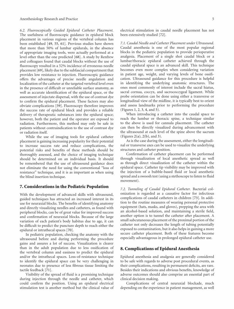
6.2. Fluoroscopically Guided Epidural Catheter Placement.The usefulness of fluoroscopic guidance in epidural blockplacement in various regions of the vertebral column hasbeen established [49, 55, 61]. Previous studies have shownthat more than 50% of lumbar epidurals, in the absenceof appropriate imaging tools, were actually performed at alevel other than the one predicted [68]. A study by Renfrewand colleagues found that caudal blocks without the use offluoroscopy resulted in a 52% incidence of erroneous needleplacement [69], likely due to the subfascial compartment thatprovides low resistance to injection. Fluoroscopic guidanceoffers the advantages of precise needle angulation andlocalization of the catheter at the targeted vertebral level evenin the presence of difficult or unreliable surface anatomy, aswell as accurate identification of the epidural space, or theassessment of injectate dispersal, with the use of contrast dyeto confirm the epidural placement. These factors may alsoobviate complications [59]. Fluoroscopy therefore improvesthe success rate of epidural block and provides a reliabledelivery of therapeutic substances into the epidural space;however, both the patient and the operator are exposed toradiation. Furthermore, this method may only be safe inpatients without contraindication to the use of contrast dyeor radiation itself.
While the use of imaging tools for epidural catheterplacement is gaining increasing popularity for their potentialto increase success rate and reduce complications, thepotential risks and benefits of these methods should bethoroughly assessed, and the choice of imaging techniqueshould be determined on an individual basis. It shouldbe remembered that the use of ultrasound guidance doesnot eliminate the need for using the conventional “loss ofresistance” technique, and it is as important as when usingthe blind insertion technique.
7. Considerations in the Pediatric Population
With the development of advanced skills with ultrasound,guided techniques has attracted an increased interest in itsuse for neuraxial blocks. The benefits of identifying anatomyand directly visualizing needles and catheters, as found withperipheral blocks, can be of great value for improved successand confirmation of neuraxial blocks. Because of the largevariation of each patient’s body habitus due to age, it canbe difficult to predict the puncture depth to reach either theepidural or intrathecal spaces [70].
In pediatric population, checking the anatomy with theultrasound before and during performing the proceduregains and assures a lot of success. Visualization is clearerthan in the adult population due to less ossification ofthe vertebral column and easiness to predict the epiduraland/or the intrathecal spaces. Loss-of-resistance techniqueto identify the epidural space can be very challenging inneonates due to presence of less fibrous tissue limiting thetactile feedback [71].
Visibility of the spread of fluid is a promising techniqueduring injection through the needle and catheter, whichcould confirm the position. Using an epidural electricalstimulation test is another method but the clinical value of
electrical stimulation in caudal needle placement has notbeen extensively studied [72].
7.1. Caudal Needle and Catheter Placement under Ultrasound.Caudal anesthesia is one of the most popular regionalblocks in the pediatric population to provide perioperativeanalgesia. Placement of a single shot caudal block or alumbar/thoracic epidural catheter achieved through thecaudal epidural space is an advanced skill. This techniquebecomes even more complex when considering variationin patient age, weight, and varying levels of bone ossifi-cation. Ultrasound guidance for this procedure is helpfulin identifying the underlying anatomic structures. Theones most commonly of interest include the sacral hiatus,sacral cornua, coccyx, and sacrococcygeal ligament. Whileprobe orientation can be done using either a transverse orlongitudinal view of the midline, it is typically best to orientand assess landmarks prior to performing the procedure(Figures 1, 2, 3, 4, and 5).
When introducing a catheter into the caudal space toreach the lumbar or thoracic spine, a technique similarto the above is used for cannula placement. The cathetercan then be directly visualized during advancement withthe ultrasound at each level of the spine above the sacrum(Figures 2(a), 2(b), and 3).
As is the case during the assessment, either the longitudi-nal or transverse axes can be used to visualize the underlyingstructures and catheter position.
Confirmation of catheter placement can be performedthrough visualization of local anesthetic spread as wellas through direct visualization of the catheter within theepidural space. Catheter tip visibility may be improved withthe injection of a bubble-based fluid or local anestheticspread and a swoosh test (using a stethoscope to listen to fluidmovement).
7.2. Tunneling of Caudal Epidural Catheter. Bacterial col-onization is regarded as a causative factor for infectiouscomplications of caudal catheters in children [73]. In addi-tion to the routine measures of wearing personal protectiveequipment (hats, masks, and gloves), prepping the area withan alcohol-based solution, and maintaining a sterile field,another option is to tunnel the catheter after placement. Asmall subcutaneous placement of the proximal portion of thecatheter not only decreases the length of tubing potentiallyexposed to contamination, but it also helps in gaining a moresecure catheter placement. Both of these features becomeespecially advantageous in prolonged epidural catheter use.
8. Complications of Epidural Anesthesia
Epidural anesthesia and analgesia are generally consideredto be safe with regards to adverse post procedural events, astheir complications, resulting in permanent deficits, are rare.Besides their indications and obvious benefits, knowledge ofadverse outcomes should also comprise an essential part ofclinical decision making.
Complications of central neuraxial blockade, muchdepending on the experience in patient management, as well

6 Anesthesiology Research and Practice
Sacrococcygeal ligament
Sacral comua
Figure 1: Placing the probe transverse plane at the coccyx, thesacral cornua (represented in white arrows heading down) areviewed laterally as humps. Sacral hiatus is located between an upperhyperechoic line, representing the sacrococcygeal membrane orligament and an inferior hyperechoic line representing the dorsumof the pelvic surface of the sacrum (bidirectional sided arrow).
Dorsal surface of thesacrum
Pelvic surface
of sacrum
Caudalcatheter
(a)
Caudalcatheter
Dorsal surface of thesacrum
Local anestheticspread
Pelvic
surfa
ceof sacru
m
(b)
Figure 2: (a) Placing the probe longitudinally between the sacralcornua will capture the dorsal surface of the sacrum, the dorsalaspect of the pelvic surface of the sacrum, and the sacrococcygealligament. Angiocatheter penetrated the sacrococcygeal ligamentand lies in the epidural space. (b) Local Anesthetic spread throughCaudal Angiocatheter in caudal epidural space.
as materials, equipment, and the presence of risk factors,have been reported to occur at various frequencies [74, 75].An epidemiologic study conducted in Sweden over a periodof 10 years revealed an increasing trend (1 in 10,000 neuraxialanesthetics) of severe complications after central neuraxialblockade [74]. Relatively recent literature suggests that most
Dorsal surface of sacrum
Pelvic surface of sacrum
Catheter
Figure 3: Caudal epidural catheter passing through Angiocatheterin the epidural space.
Figure 4: Longitudinal view at the thoracic spine level, viewingthe advancement of the caudal epidural catheter. Curved linesshow spinous processes. Arrows show epidural catheter in betweenspinous processes.
of these occur with the perioperative use of epidural block[74, 76]. The incidence of major complications (permanentharm including death) of epidural and combined spinal-epidural anesthesia were at least twice as high as those ofspinal and caudal blocks, as reported by Cook and colleagues.This study also found that the incidence of epidural catheter-related serious morbidity and mortality was higher whenblocks were placed in the perioperative setting, as opposedto catheter placement in obstetric and pediatric populations,when inserted for chronic pain management, or when placedby non-anesthetists [77]. While prognosis is infrequentlyreported, retrospective reviews report full recovery in 61–75% of patients, epidural hematoma accounting for two-thirds of residual neurological deficits [77, 78]. Seriouscomplications, if not recognized and treated at an earlystage, may thus result in permanent loss of function [74,79]. With regards to the timing of catheter placement,there is still substantial controversy: while many anesthesiaproviders believe that epidural catheters should be placedin awake or mildly sedated patients capable of providingfeedback [80], Horlocker’s retrospective review found noevidence of an increased risk for neural injury in anes-thetized patients receiving epidural anesthetic [81]. Thoracicepidural placement, however, should never be attemptedon an anesthetized patient. Having increasingly become

Anesthesiology Research and Practice 7
Figure 5: Longitudinal view at the lumbar spine. Visualization ofthe local anesthetic spread confirmed the position.
the focus of attention, and as a result of both meticulousadherence to sterile, atraumatic catheter insertion techniqueand management, as well as careful risk-benefit assessment,major complications of epidural anesthesia are now rare,particularly those not involving infection or bleeding, andmany resolving within 6 months [74]. The estimation ofthe incidence of all adverse outcomes, however, is ofteninaccurate.
Complications may occur early if related to traumaticcatheter insertion, or later in the operative-postoperativecourse if caused by catheter-related spinal space-occupyinglesions such as epidural hematoma or abscess formation,and are infrequent among the general population. Althoughits incidence is lower than when associated with spinalanesthesia [80], transient neurological injury has been foundto account for the majority of short-term epidural catheterrelated complications (1 in 6,700) in a meta-analysis byRuppen and colleagues, followed by deep epidural infections(1 in 145,000), epidural hematoma (1 in 150,000–168,000),and persistent neurological injury (1 in 257,000) in womenreceiving epidural catheter for childbirth [82, 83]. Spinalepidural hematoma, however, has been recently suggestedto occur in a rate as high as 1 in 3,600 in female patientsundergoing knee arthroplasty [74, 84, 85]. These findingswere consistent with those previously reported in the ASAClosed Claims Project database analysis by Lee et al; however,limitations of that study design and database do not allowrisk quantification specific to regional anesthetic techniquesor populations [86].
Adverse events may result from direct mechanical injuryor adverse physiological responses. Neurological complica-tions resulting from accidental penetration of the dura aresimilar to those that occur with spinal anesthesia. Inad-vertent dural puncture and postdural puncture headache,direct neural injury, total spinal anesthesia, and subduralblock have been commonly reported. The incidence ofinadvertent dural puncture ranges between 0.19–0.5% ofepidural catheter placements. Postdural puncture headache(PDPH), described as a positional, bilateral frontal-occipital,nonthrobbing pain, may develop in as much as 75% ofpatients [87–89]. PDPH is thought to develop as a resultof persistent transdural leakage of cerebrospinal fluid (CSF)at a rate that is faster than that of CSF production. Thesubsequently decreasing CSF volume and pressure causes
traction on the meninges and intracranial vessels, whichrefer pain to the frontal-occipital region, often extending tothe neck and shoulders, more pronounced in the uprightposition. Available measures of prevention besides conserva-tive measures are immediate intrathecal catheter placement,prophylactic epidural blood patch, epidural or intrathecaladministration of saline, and epidural administration ofmorphine [90]. Direct neural injury has a reported incidenceof 0.006% [82], and has been associated with paresthesiasduring needle placement and pain on injection [80]. Totalspinal anesthesia may occur if the solution used for epiduralanesthesia is inadvertently administered into the intrathecalspace in large volumes. Symptoms are of a rapidly arisingsubarachnoid block, potentially resulting in cardiovascularcollapse and apnea requiring prompt resuscitation. Providedthat immediate, skilled resuscitative efforts are made, com-plete recovery should be expected [91]. While clinically notalways distinguishable from epidural blocks, the incidenceof clinically recognized subdural block was found to be0.024% in a prospective study [92]. A subdural block maypresent as high sensory block, often with sparing of motorand sympathetic fibers, is slow in onset, and the blockadeis disproportionately extensive for the volume of anestheticinjected. Clinical signs and symptoms may be mistakenfor accidental intrathecal injection, migration of epiduralcatheter, or an asymmetrical, patchy or inadequate epiduralblock. Subdural placement is thought to occur independentlyof the operator’s expertise. Although there are no establishedrisk factors, recent lumbar puncture and rotation of theneedle may predispose to subdural injection [93].
Hemorrhagic complications are serious adverse out-comes that may arise from neuraxial anesthesia. Epiduralhematoma is a rare, but potentially devastating, complicationthat requires emergency decompression in case of clinicaldeterioration. It is rarely attributed to an arterial source,and can develop spontaneously [94, 95]. While paralysis mayoccur even after hematoma evacuation, it is still not preciselyunderstood why several of the spinal epidural hematomasassociated with concurrent anticoagulant use involving lessblood than the volume injected when performing a therapeu-tic blood patch [85]. Clinically significant bleeding is morelikely with congenital or acquired coagulation abnormalities,thrombocytopenia, vascular anomalies or anatomical abnor-malities, advanced age and female gender, repetitive attemptsat catheter insertion, and traumatic block placement [74, 96–98]. The risk is reported to increase 15-fold when thereis a concomitant use of anticoagulants, and appropriateprecautions are not taken [85]. Appropriate timing ofanticoagulant administration is important in decreasing therisk of bleeding [99]. The commonest presenting symptomsof spinal epidural hematoma are new back pain, radicularpain, and progressive lower extremity weakness. Symptomsrarely present immediately after surgery, but may developwhile the catheter is still in place. These symptoms canoccur 15 hours to 3 days after catheter insertion [78, 98].The diagnostic investigation of choice is MRI. A delay indiagnostic imaging may lead to devastating outcomes, andis a common error, as manifesting neurological symptomsand back pain may be attributed to the use of epidural

8 Anesthesiology Research and Practice
infusion and a prolonged effect of local anesthetic, and tomusculoskeletal origin [78, 100]. Cauda equina syndromedue to hematoma formation, a rare complication with areported incidence of 2.7/100,000 epidural blocks, was foundto result in permanent deficit in more than two-third of thecases [74]. Classic manifestation is low back pain, alteredproprioception and decreased sensation to pinprick andtemperature in the lumbar and sacral nerve distribution,voiding and defecation disturbances, and progressive loss ofmuscle strength. Outcomes are primarily function of intervalto hematoma evacuation and the severity of the neurologicaldeficit, and are favorable if decompression is performedwithin 8 hours of the development of symptoms [98].
Epidural catheter related infections are rare compli-cations both in adult and in pediatric patients. A ret-rospective database analysis by Sethna et al. found anexpected incidence ranging between 3–13/10,000 cathetersin children [101]. Epidural abscess and meningitis hasbeen reported to occur in 1 : 1000 and 1 : 50,000 catheterplacements, respectively [74]. Although epidural cathetersare placed under aseptic conditions, needle or cathetercontamination does occur even during aseptic puncture andsterile handling of devices [102]. Of patient risk factors, skincolonization at the puncture site and bacterial migrationalong the catheter is proposed to be the most likely route ofinfection; however, immunosuppression [74, 103], diabetesmellitus [104], chronic renal failure, steroid administration,cancer, herpes zoster, rheumatoid arthritis [105], systemicor local sepsis, and prolonged infusion duration are alsoidentifiable risk factors. The rate of skin colonization atpuncture sites is reported to be higher in children thanin adults, with an overall incidence as high as 35% [101].The incidence of infection increases after three days [106].The classic presentation signs and symptoms are severemidline back pain, fever, and leukocytosis, with or withoutneurological symptoms (worsening lower limb weakness andparaplegia, incontinence, irradiating pain, nuchal rigidity,and headache). Symptoms commonly appear after removalof the epidural catheter [78]. Neurological deficits havebeen found to be persistent in more than 50% of patientsdeveloping epidural abscess [105]. Barrier precautions, skindisinfection [107], as well as the use of closed epiduralsystem, and patient-controlled epidural analgesia [101] havebeen suggested as ways to decrease the incidence of epiduralcatheter-associated infections. Frequent syringe changes, onthe other hand, may be associated with a higher rate ofepidural infections [108]. Frequently implicated infectingorganisms are Methicillin-resistant Staphylococcus aureus(MRSA), Staphylococcus aureus, and Coagulase-negativeStaphylococcus [101, 109]. Outcomes are favorable whendiagnosed and treated promptly. Adhesive arachnoiditis, pre-senting in various forms, is a sterile inflammatory responseto accidental subarachnoid injection of local anesthetics,preservatives, detergents, or antiseptics [110–112], and hasalso resulted from traumatic puncture or epidural abscess.Medical literature suggests an extremely low incidence [113,114].
Complications of epidural anesthesia are rare eventsthat may result in detrimental sequelae. Strict adherence
to prophylactic measures and treatment without delay isessential to further lower the incidence of adverse outcomes.
9. Epidural Anesthesia andThromboprophylaxis
Some controversy exists with regards to reduced coagulationand neuraxial anesthesia and challenges are emerging asnew agents are introduced into clinical practice. Spinalepidural hematoma, although still considered to be a rarecomplication occurring at a previously reported rate of lessthan 1 in 150,000 epidural and less than 1 in 220,000 spinalanesthetics in patients with normal coagulation status, isnow suggested to occur in a rate as high as 1 in 3,000in some patient populations [84, 85]. Patients receivingantithrombotic or antiplatelet therapies are more at risk forthis potentially dramatic adverse event, in particular afterinvasive procedures [98]. In the United States, the estimatedincidence of spinal epidural hematoma with concurrentadministration of antithrombotic drugs (low molecularweight heparins) is 1 : 40,800 for spinal anesthesia, 1 : 6,600for single-shot epidural anesthesia, and 1 : 3,100 for continu-ous epidural anesthesia [85]. Risk factors for epidural bleed-ing were established as coagulation disorders, antithromboticor fibrinolytic therapy, or the use of any agents interferingwith coagulation, female gender, age, difficult vertebral orspinal cord anatomy, difficult or traumatic catheter insertion,and lack of guidelines [96, 98, 115, 116]. Catheter removalcarries nearly the same risk as insertion [98]. Appropriatetime intervals between the administration of anticoagulants,neuraxial block placement, and catheter removal are crucialin the prevention of hematoma formation [117, 118].
The American Society of Regional Anesthesia and PainMedicine (ASRA), and more recently, the European Societyof Anaesthesiology (ESA) published their consensus state-ments on neuraxial anesthesia and the use of antithromboticand thrombolytic agents [99, 119]. While providing guide-lines in clinical decision making, and having the aim of min-imizing hemorrhagic complications, these recommendationsdo not guarantee a specific outcome, and allow of variationsbased on the judgment of the anesthesiologist. The guidelinesof the American Society of Regional Anesthesia and PainMedicine and the European Society of Anaesthesiology arebased on previously published national recommendations,hematology, pharmacology, and risk factors for surgicalbleeding, and incorporate updated information since thetime of their publication.
With regards to epidural catheter placement, the ASRArecommends that patients receiving thrombolytic therapybe queried and their medical records reviewed for a recenthistory of lumbar puncture or neuraxial analgesia. Neuraxialanesthesia should be avoided, or, if received concurrentlywith the fibrinolytic/thrombolytic therapy, close neuro-logical monitoring should be continued along with theadministration of neuraxial solutions that minimize sensoryand motor block. There is no definitive recommendation forepidural catheter removal in patients receiving fibrinolyticand thrombolytic therapy. Thrombolytics, if scheduled,

Anesthesiology Research and Practice 9
should be avoided for 10 days after puncture of noncom-pressible vessels [99].
Patients receiving unfractionated heparin (UFH) thrice aday, if recommended by recent thromboprophylaxis guide-lines, may be at an increased risk of surgical-related bleed-ing. The ASRA recommends that the patient’s—potentiallysimultaneous—anticoagulant and antiplatelet medication bedaily reviewed. There is no contraindication to epiduralblockade in patients receiving subcutaneous UFH prophy-laxis at daily doses of 2 × 5000 U. The risk of bleedingmay be increased in debilitated patients receiving prolongedtherapy, and may be decreased by delaying the heparininjection until after neuraxial block placement. The safety ofcentral neural block in patients receiving subcutaneous UFHin a dosing regimen of more than 10,000 U daily has notbeen established, and an increased risk of a spinal epiduralhematoma has also not been elucidated. Patients receivingheparin for greater than 4 days should be assessed forheparin-induced thrombocytopenia (HIT). In patients withknown coagulopathies, combining neuraxial techniques withintraoperative heparinization should be avoided however,this technique is acceptable in patients with no othercoagulation disorders, if
(1) heparin administration is delayed for 1 hour afterpuncture,
(2) epidural catheters are removed 2 to 4 hours after thelast heparin dose and the patient’s coagulation statusis assessed. the next heparin dose may be adminis-tered 1 hour after catheter removal,
(3) patient is closely monitored for early signs of neuro-logic dysfunction while receiving neuraxial solutionsthat minimize sensory and motor block postopera-tively.
Per the ASRA guidelines, in contrast with the ESArecommendations that suggest considering postponement ofthe procedure, difficult or traumatic block placement shouldnot necessarily prompt postponing surgery; however, thepotential benefits should be carefully weighed against allpotentially detrimental outcomes in each individual. Withregards to the full anticoagulation of patients undergoingcardiac surgery, the ASRA finds insufficient evidence avail-able to determine an increased risk of neuraxial hematoma.Close postoperative monitoring of neurologic function, aswell as administration of neuraxial solutions that minimizesensory and motor block to facilitate detection of signsand symptoms of cord compression, is however suggested[99, 119].
Patients on low molecular weight heparin (LMWH)anticoagulation have not been found to be at an increasedrisk of bleeding in high-risk groups, contrasting withpatients receiving UFH-thromboprophylaxis. Also, com-pared to UFH, LMWH-therapy has been associated witha significant decrease in the risk of HIT, as demonstratedby Warkentin and colleagues [120]; nonetheless, LMWHsare contraindicated in such condition due to the high levelof cross-reactivity. To avoid an elevated risk of bleedingcomplications, an interval of 10 to 12 hours between
preoperative LMWH administration at prophylactic dosesand needle placement or catheter removal is recommended.Administration of LMWH the night before surgery doesnot thus interfere with epidural block placement on theday of surgery. In patients on therapeutic doses of LMWH,catheter placement should be delayed for a minimum of24 hours after the last dose. Patients undergoing generalsurgery and receiving LMWH 2 hours prior to surgery arenot ideal candidates for a neuraxial blockade, and are thusrecommended against neuraxial techniques. Patients receiv-ing postoperative LMWH thromboprophylaxis may safelybe administered both single-dose and continuous cathetertechniques. With regards to management, timing of the firstpostoperative dose, dosing schedule, and total daily doseare authoritative. Concerning the management of patientsreceiving LMWH, the ASRA recommends against the routinemonitoring of anti-Xa level and concurrent administrationof medication affecting hemostasis, regardless of LMWHdosing regimen [99, 119].
The management of patients receiving perioperativeoral anticoagulants is still controversial. In the UnitedStates, much like in Europe, therapeutic oral anticoagulationis considered as a contraindication to central neuraxialblockade. As opposed to Europe, however, perioperativethromboprophylaxis is still possible in the United States.According to the recommendation of the American Societyof Regional Anesthesia and Pain Medicine, warfarin therapymust be stopped ideally 4-5 days before the scheduledprocedure, and the INR checked before neuraxial blockplacement. In patients receiving an initial preoperativedose of warfarin, INR should be measured before needlepuncture if the administration of the first dose exceeded 24hours, or if a second dose of such anticoagulant has beenadministered. In patients at risk for an enhanced responseto oral anticoagulants, a reduced dose of drug should beadministered. In patients receiving low-dose warfarin duringepidural analgesia, INR should be monitored daily. Epiduralcatheters should be removed when the INR is less than 1.5.If the INR is greater than 1.5 but less than 3, indwellingepidural catheters should be done with caution. The ASRArecommends against concurrent use of agents, such as UFH,LMWH, or platelet aggregation inhibitors, that influenceother components of the clotting system, as these, withoutaffecting the INR, may increase the risk of bleeding. Medicalrecords should be reviewed for such agents. Neurologictesting of sensory and motor function should be performedroutinely during epidural analgesia for patients on oralanticoagulants, and should be continued for at least 24hours after catheter removal, until the INR returns to thedesired prophylactic range. In patients with INR greater than3, the American Society of Regional Anesthesia and PainMedicine recommends that the warfarin be held or reduced,without making a definitive recommendation regarding themanagement to facilitate catheter removal in these patients[99, 119].
Platelet aggregation inhibitors, such as acetylsalicylicacid, thienopyridines (clopidogrel, ticlopidine, and prasug-rel), glycoprotein (GP) IIb/IIIa antagonists (eptifibatide,tirofiban, and abciximab), the novel ADP P2Y12 receptor
10 Anesthesiology Research and Practice
antagonist ticagrelor, and the selective phosphodiesteraseIIIA inhibitor cilostazol, have diverse effects on plateletfunction. No wholly accepted test exists to guide antiplatelettherapy. It is therefore critical to perform a careful pre-operative risk assessment to identify factors that mightpotentially contribute to bleeding. Although administrationof nonsteroidal anti-inflammatory drugs (including aspirin)does not appear to significantly increase the incidence ofhematoma formation, concurrent administration of LMWH,UFH, or oral anticoagulants resulted in a higher rate ofcomplications in both surgical and medical patients, theiruse along with NSAIDs, including aspirin, is therefore notrecommended. Cyclooxygenase-2 inhibitors have minimalinhibitory effect on platelet aggregation, and should beconsidered in patients requiring anti-inflammatory therapyin the presence of anticoagulation. The actual incidence ofspinal epidural hematoma related to thienopyridines andGP IIb/IIIa inhibitors is not known. Management shouldbe based on labeling precautions and the experience ofprofessionals involved in the clinical care of the patient.However, as it has been suggested by recent guidelines,ticlopidine and clopidogrel therapy should be discontinued14 and 7days prior to neuraxial block, respectively. Ifneedle puncture is indicated between 5 and 7 days ofdiscontinuation of clopidogrel, normalization of plateletfunction should be documented. GP IIb/IIIa antagonistsexert a dose-dependent effect on platelet aggregation. Afterthe last administered dose, the time to normal aggregationis 4 to 8 hours for eptifibatide and tirofiban, and 24 to 48hours for abciximab. Neuraxial blockade should be avoideduntil normal platelet function is achieved. Should a patient,despite the contraindication, be administered GP IIb/IIIainhibitors within 4 weeks of surgery, careful neurologicalmonitoring should be performed [99, 119].
Both the ASRA and the ESA guidelines recommendagainst the mandatory discontinuation of herbal agents(most commonly: garlic, Echinacea, Gingko biloba, ginseng,aloe vera, and ephedra of dwarf palm), neither shouldneuraxial techniques be avoided, as there is insufficientevidence that these, by themselves, significantly increase therisk for spinal hematoma formation. There is insufficientevidence to conclude that thrombin inhibitors, such aslepirudin, desirudin, bivalirudin, or argatroban, are saferto use in patients receiving spinal or epidural anesthesia;performance of these techniques in the presence of theseagents is thus not recommended. Until sufficient evidenceis available, neuraxial techniques in patients receiving fon-daparinux should only be performed if single needle pass,atraumatic block placement, and avoidance of indwellingcatheters are feasible, or a different method of prophylaxisshould be considered [99, 119].
10. Summary
Epidural and caudal anesthesia is a versatile neuraxialanesthetic technique with an expanding area of indication. Itcan be used in the perioperative setting as the sole anesthetic,or in combination with general or spinal anesthesia. Itspotential to decrease postoperative complication rate by its
beneficial physiological effects has been clearly demonstratedin several studies. The absolute contraindications to its usehave traditionally been well defined. Despite its rare, butpotentially devastating complications, neuraxial anesthesiais considered to be safe. Performing such procedures in thepresence of anticoagulants is however controversial. Withpatients presenting with medical conditions that predisposeto clinically significant bleeding and an increased numberof patients taking various anticoagulants, there is greaterconcern for an increased incidence of epidural hematomas.The key to maximizing the advantages while minimizingthe disadvantages of epidural and caudal anesthesia is tobecome familiar with the anatomical, physiological, phar-macological, and technical aspects of block placement. Thereview and advances discussed here allow both adult andpediatric populations a form of care that is often consideredindispensable.
Conflict of Interests
The authors declare no conflict of interest.
References
[1] J. Corning, “Spinal anesthesia and local medications of thecord,” New York Journal of Medicine, vol. 42, pp. 483–485,1885.
[2] D. Brown, “Spinal, epidural, and caudal anesthesia,” inMiller’s Anesthesia, R. D. Miller, Ed., pp. 1653–1683, Elsevier,Philadelphia, Pa, USA, 6th edition, 2005.
[3] Q. H. Hogan, “Lumbar epidural anatomy. A new look bycryomicrotome section,” Anesthesiology, vol. 75, no. 5, pp.767–775, 1991.
[4] Q. Hogan, “Distribution of solution in the epidural space:examination by cryomicrotome section,” Regional Anesthesiaand Pain Medicine, vol. 27, no. 2, pp. 150–156, 2002.
[5] B Deschner, M. Allen, and O. de Leon, “Epidural blockade,”in Textbook of Regional Anesthesia and Acute Pain Manage-ment, A. Hadzic, Ed., pp. 237–269, McGraw–Hill, New York,NY, USA, 1st edition, 2006.
[6] J. H. McClure, “Ropivacaine,” British Journal of Anaesthesia,vol. 76, no. 2, pp. 300–307, 1996.
[7] I. Harukuni, H. Yamaguchi, S. Sato, and H. Naito, “Thecomparison of epidural fentanyl, epidural lidocaine, andintravenous fentanyl in patients undergoing gastrectomy,”Anesthesia and Analgesia, vol. 81, no. 6, pp. 1169–1174, 1995.
[8] A. Tamsen and T. Gordh, “Epidural clonidine producesanalgesia,” The Lancet, vol. 2, no. 8396, pp. 231–232, 1984.
[9] M. De Kock, B. Crochet, C. Morimont, and J. L. Scholtes,“Intravenous or epidural clonidine for intra- and postoper-ative analgesia,” Anesthesiology, vol. 79, no. 3, pp. 525–531,1993.
[10] M. De Kock, “Site of hemodynamic effects of alpha sub 2-adrenergic agonists,” Anesthesiology, vol. 75, pp. 715–716,1991.
[11] B. Biki, E. Mascha, D. C. Moriarty, J. M. Fitzpatrick, D. I.Sessler, and D. J. Buggy, “Anesthetic technique for radicalprostatectomy surgery affects cancer recurrence: a retrospec-tive analysis,” Anesthesiology, vol. 109, no. 2, pp. 180–187,2008.

Anesthesiology Research and Practice 11
[12] J. P. Desborough, “The stress response to trauma and sur-gery,” British Journal of Anaesthesia, vol. 85, no. 1, pp. 109–117, 2000.
[13] S. Ben-Eliyahu, G. G. Page, R. Yirmiya, and G. Shakhar,“Evidence that stress and surgical interventions promotetumor development by suppressing natural killer cell activ-ity,” International Journal of Cancer, vol. 80, no. 6, pp. 880–888, 1999.
[14] S. C. O’Riain, D. J. Buggy, M. J. Kerin, R. W. G. Watson, andD. C. Moriarty, “Inhibition of the stress response to breastcancer surgery by regional anesthesia and analgesia does notaffect vascular endothelial growth factor and prostaglandinE2,” Anesthesia and Analgesia, vol. 100, no. 1, pp. 244–249,2005.
[15] H. Kehlet, “Modification of responses to surgery by neu-ral blockade: clinical implications,” in Neural Blockade inClinical Anesthesia and Management of Pain, M. Cousinsand P. Bridenbaugh, Eds., pp. 129–178, J. B. Lippincott,Philadelphia, Pa, USA, 1998.
[16] H. Kehlet, “Surgical stress: the role of pain and analgesia,”British Journal of Anaesthesia, vol. 63, no. 2, pp. 189–195,1989.
[17] H. O. Besedovsky, A. E. Del Rey, and E. Sorkin, “Immune-neuroendocrine interactions,” Journal of Immunology, vol.135, no. 2, pp. 750–754, 1985.
[18] D. I. Sessler, “Long-term consequences of anesthetic manage-ment,” Anesthesiology, vol. 111, no. 1, pp. 1–4, 2009.
[19] S. Ben-Eliyahu, G. Shakhar, G. G. Page, V. Stefanski, and K.Shakhar, “Suppression of NK cell activity and of resistance tometastasis by stress: a role for adrenal catecholamines and β-adrenoceptors,” NeuroImmunoModulation, vol. 8, no. 3, pp.154–164, 2000.
[20] K. Buttenschoen, K. Fathimani, and D. C. Buttenschoen,“Effect of major abdominal surgery on the host immuneresponse to infection,” Current Opinion in Infectious Diseases,vol. 23, no. 3, pp. 259–267, 2010.
[21] D. J. Buggy and G. Smith, “Epidural anaesthesia and anal-gesia: better outcome after major surgery?” British MedicalJournal, vol. 319, no. 7209, pp. 530–531, 1999.
[22] M. S. O’Reilly, T. Boehm, Y. Shing et al., “Endostatin: anendogenous inhibitor of angiogenesis and tumor growth,”Cell, vol. 88, no. 2, pp. 277–285, 1997.
[23] M. S. O’Reilly, L. Holmgren, Y. Shing et al., “Angiostatin: anovel angiogenesis inhibitor that mediates the suppression ofmetastases by a Lewis lung carcinoma,” Cell, vol. 79, no. 2,pp. 315–328, 1994.
[24] M. Yokoyama, Y. Itano, S. Mizobuchi et al., “The effects ofepidural block on the distribution of lymphocyte subsets andnatural-killer cell activity in patients with and without pain,”Anesthesia and Analgesia, vol. 92, no. 2, pp. 463–469, 2001.
[25] S. A. Kirkley, “Proposed mechanisms of transfusion-inducedimmunomodulation,” Clinical and Diagnostic LaboratoryImmunology, vol. 6, no. 5, pp. 652–657, 1999.
[26] L. Reynolds, J. Beckmann, and A. Kurz, “Perioperativecomplications of hypothermia,” Best Practice and Research,vol. 22, no. 4, pp. 645–657, 2008.
[27] M. Turina, D. E. Fry, and H. C. Polk, “Acute hyperglycemiaand the innate immune system: clinical, cellular, and molec-ular aspects,” Critical Care Medicine, vol. 33, no. 7, pp. 1624–1633, 2005.
[28] G. Delogu, S. Moretti, G. Famularo et al., “Mitochon-drial perturbations and oxidant stress in lymphocytes frompatients undergoing surgery and general anesthesia,” Archivesof Surgery, vol. 136, no. 10, pp. 1190–1196, 2001.
[29] G. P. Chrousos, F. Epstein, J. Flier, S. Reichlin, and S. Pavlou,“The hypothalamic-pituitary-adrenal axis and immune-mediated inflammation,” New England Journal of Medicine,vol. 332, no. 20, pp. 1351–1362, 1995.
[30] A. J. Rassias, A. L. Givan, C. A. S. Marrin, K. Whalen, J.Pahl, and M. P. Yeager, “Insulin increases neutrophil countand phagocytic capacity after cardiac surgery,” Anesthesia andAnalgesia, vol. 94, no. 5, pp. 1113–1119, 2002.
[31] C. P. Nielson and D. A. Hindson, “Inhibition of polymor-phonuclear leukocyte respiratory burst by elevated glucoseconcentrations in vitro,” Diabetes, vol. 38, no. 8, pp. 1031–1035, 1989.
[32] A. J. Rassias, C. A. S. Marrin, J. Arruda, P. K. Whalen,M. Beach, and M. P. Yeager, “Insulin infusion improvesneutrophil function in diabetic cardiac surgery patients,”Anesthesia and Analgesia, vol. 88, no. 5, pp. 1011–1016, 1999.
[33] I. Beck, J. S. Scott, M. Pepper, and E. H. Speck, “Theeffect of neonatal exchange and later blood transfusionon lymphocyte cultures,” American Journal of ReproductiveImmunology, vol. 1, no. 5, pp. 224–225, 1981.
[34] P. I. Tartter, B. Steinberg, D. M. Barron, and G. Martinelli,“Transfusion history, T cell subsets and natural killer cyto-toxicity in patients with colorectal cancer,” Vox Sanguinis, vol.56, no. 2, pp. 80–84, 1989.
[35] K. Yuki, N. S. Astrof, C. Bracken, G. S. Sulpicio, and M. Shi-maoka, “Sevoflurane binds and allosterically blocks integrinlymphocyte function-associated antigen-1,” Anesthesiology,vol. 113, no. 3, pp. 600–609, 2010.
[36] P. Sacerdote, M. Bianchi, L. Gaspani et al., “The effects oftramadol and morphine on immune responses and pain aftersurgery in cancer patients,” Anesthesia and Analgesia, vol. 90,no. 6, pp. 1411–1414, 2000.
[37] R. Melamed, S. Bar-Yosef, G. Shakhar, K. Shakhar, and S.Ben-Eliyahu, “Suppression of natural killer cell activity andpromotion of tumor metastasis by ketamine, thiopental, andhalothane, but not by propofol: mediating mechanisms andprophylactic measures,” Anesthesia and Analgesia, vol. 97, no.5, pp. 1331–1339, 2003.
[38] J. M. Risdahl, K. V. Khanna, P. K. Peterson, and T. W. Molitor,“Opiates and infection,” Journal of Neuroimmunology, vol. 83,no. 1-2, pp. 4–18, 1998.
[39] S. Roy and H. H. Loh, “Effects of opioids on the immunesystem,” Neurochemical Research, vol. 21, no. 11, pp. 1375–1386, 1996.
[40] T. Hori, T. Katafuchi, S. Take, Y. Kaizuka, T. Ichijo, andN. Shimizu, “The hypothalamo-sympathetic nervous systemmodulates peripheral cellular immunity,” Neurobiology, vol.3, no. 3-4, pp. 309–317, 1995.
[41] M. H. Makman, “Morphine receptors in immunocytes andneurons,” Advances in Neuroimmunology, vol. 4, no. 2, pp.69–82, 1994.
[42] E. J. De Waal, J. W. Van Der Laan, and H. Van Loveren,“Effects of prolonged exposure to morphine and methadoneon in vivo parameters of immune function in rats,” Toxicol-ogy, vol. 129, no. 2-3, pp. 201–210, 1998.
[43] K. Jaeger, D. Scheinichen, J. Heine et al., “Remifentanil,fentanyl, and alfentanil have no influence on the respiratoryburst of human neutrophils in vitro,” Acta AnaesthesiologicaScandinavica, vol. 42, no. 9, pp. 1110–1113, 1998.
[44] B. Larsen, G. Hoff, W. Wilhelm, H. Buchinger, G. A.Wanner, and M. Bauer, “Effect of intravenous anesthetics onspontaneous and endotoxin- stimulated cytokine response incultured human whole blood,” Anesthesiology, vol. 89, no. 5,pp. 1218–1227, 1998.

12 Anesthesiology Research and Practice
[45] P. Buinauskas, G. McDonald, and W. Cole, “Role of operativestress on the resistance of the experimental animal toinoculated cancer cells,” Annals of Surgery, vol. 148, pp. 642–648, 1958.
[46] M. G. Denis, C. Lipart, J. Leborgne et al., “Detection ofdisseminated tumor cells in peripheral blood of colorectalcancer patients,” International Journal of Cancer, vol. 74, no.5, pp. 540–544, 1997.
[47] G. Shakhar and S. Ben-Eliyahu, “Potential prophylacticmeasures against postoperative immunosuppression: couldthey reduce recurrence rates in oncological patients?” Annalsof Surgical Oncology, vol. 10, no. 8, pp. 972–992, 2003.
[48] W. A. Koltun, M. M. Bloomer, A. F. Tilberg et al., “Awakeepidural anesthesia is associated with improved natural killercell cytotoxicity and a reduced stress response,” AmericanJournal of Surgery, vol. 171, no. 1, pp. 68–72, 1996.
[49] T. Nagaro, T. Yorozuya, M. Kamei, N. Kii, T. Arai, and S. Abe,“Fluoroscopically guided epidural block in the thoracic andlumbar regions,” Regional Anesthesia and Pain Medicine, vol.31, no. 5, pp. 409–416, 2006.
[50] P. Marhofer, M. Greher, and S. Kapral, “Ultrasound guidancein regional anaesthesia,” British Journal of Anaesthesia, vol.94, no. 1, pp. 7–17, 2005.
[51] P. H. Pan, T. D. Bogard, and M. D. Owen, “Incidence andcharacteristics of failures in obstetric neuraxial analgesiaand anesthesia: a retrospective analysis of 19,259 deliveries,”International Journal of Obstetric Anesthesia, vol. 13, no. 4, pp.227–233, 2004.
[52] P. Lirk, H. Messner, M. Deibl et al., “Accuracy in estimatingthe correct intervertebral space level during lumbar, thoracicand cervical epidural anaesthesia,” Acta AnaesthesiologicaScandinavica, vol. 48, no. 3, pp. 347–349, 2004.
[53] H. Willschke, P. Marhofer, A. Bosenberg et al., “Epiduralcatheter placement in children: comparing a novel approachusing ultrasound guidance and a standard loss-of-resistancetechnique,” British Journal of Anaesthesia, vol. 97, no. 2, pp.200–207, 2006.
[54] A. H. White, R. Derby, and G. Wynne, “Epidural injectionsfor the diagnosis and treatment of low-back pain,” Spine, vol.5, no. 1, pp. 78–86, 1980.
[55] A. H. White, “Injection techniques for the diagnosis andtreatment of low back pain,” Orthopedic Clinics of NorthAmerica, vol. 14, no. 3, pp. 553–567, 1983.
[56] C. P. C. Chen, S. F. T. Tang, T. C. Hsu et al., “Ultrasoundguidance in caudal epidural needle placement,” Anesthesiol-ogy, vol. 101, no. 1, pp. 181–184, 2004.
[57] T. Grau, R. W. Leipold, R. Conradi, E. Martin, and J. Motsch,“Ultrasound imaging facilitates localization of the epiduralspace during combined spinal and epidural anesthesia,”Regional Anesthesia and Pain Medicine, vol. 26, no. 1, pp. 64–67, 2001.
[58] T. Grau, R. W. Leipold, R. Conradi, E. Martin, and J.Motsch, “Efficacy of ultrasound imaging in obstetric epiduralanesthesia,” Journal of Clinical Anesthesia, vol. 14, no. 3, pp.169–175, 2002.
[59] B. A. Johnson, K. P. Schellhas, and S. R. Pollei, “Epidurogra-phy and therapeutic epidural injections: technical consider-ations and experience with 5334 cases,” American Journal ofNeuroradiology, vol. 20, no. 4, pp. 697–705, 1999.
[60] R. C. Cork, J. J. Kryc, and R. W. Vaughan, “Ultrasoniclocalization of the lumbar epidural space,” Anesthesiology,vol. 52, no. 6, pp. 513–516, 1980.
[61] J. M. Currie, “Measurement of the depth to the extraduralspace using ultrasound,” British Journal of Anaesthesia, vol.56, no. 4, pp. 345–347, 1984.
[62] M. Bonazzi and L. B. de Gracia, “Individuazione ecoguidatadello spazio epidurale lombare,” Minerva Anesthesiol, vol. 61,pp. 201–205, 1995.
[63] D. H. Wallace, J. M. Currie, L. C. Gilstrap, and R. Santos,“Indirect sonographic guidance for epidural anesthesia inobese pregnant patients,” Regional Anesthesia, vol. 17, no. 4,pp. 233–236, 1992.
[64] H.-J. Rapp, A. Folger, and T. Grau, “Ultrasound-guidedepidural catheter insertion in children,” Anesthesia andAnalgesia, vol. 101, no. 2, pp. 333–339, 2005.
[65] D. Belavy, M. J. Ruitenberg, and R. B. Brijball, “Feasibilitystudy of real-time three-/four-dimensional ultrasound forepidural catheter insertion,” British Journal of Anaesthesia,vol. 107, no. 3, pp. 438–445, 2011.
[66] M. K. Karmakar, X. Li, A. M.-H. Ho, W. H. Kwok, and P.T. Chui, “Real-time ultrasound-guided paramedian epiduralaccess: evaluation of a novel in-plane technique,” BritishJournal of Anaesthesia, vol. 102, no. 6, pp. 845–854, 2009.
[67] H. Yamagami, Y. Yuda, M. Shiotani, K. Ooseto, Y. Naganuma,and H. Karasawa, “The administration of continuous epidu-ral block under proneposition with fluoroscopic guidance,”Japanese Journal of Anesthesiology, vol. 38, no. 2, pp. 229–235,1989.
[68] B. Fredman, M. B. Nun, E. Zohar et al., “Epidural steroids fortreating “failed back surgery syndrome”: is fluoroscopy reallynecessary?” Anesthesia and Analgesia, vol. 88, no. 2, pp. 367–372, 1999.
[69] D. L. Renfrew, T. E. Moore, M. H. Kathol, G. Y. El-Khoury, J.H. Lemke, and C. W. Walker, “Correct placement of epidu-ral steroid injections: fluoroscopic guidance and contrastadministration,” American Journal of Neuroradiology, vol. 12,no. 5, pp. 1003–1007, 1991.
[70] O. J. Arthurs, M. Murray, M. Zubier, J. Tooley, and W. Kelsall,“Ultrasonographic determination of neonatal spinal canaldepth,” Archives of Disease in Childhood, vol. 93, no. 6, pp.f451–f454, 2008.
[71] J. G. McCormack and S. Malherbe, “Applications of ultra-sound in paediatric anaesthesia,” Current Anaesthesia andCritical Care, vol. 19, no. 5-6, pp. 302–308, 2008.
[72] B. C. H. Tsui, P. Tarkkila, S. Gupta, and R. Kearney, “Confir-mation of caudal needle placement using nerve stimulation,”Anesthesiology, vol. 91, no. 2, pp. 374–378, 1999.
[73] W. Fujinaka, N. Hinomoto, S. Saeki, A. Yoshida, and S.Uemura, “Decreased risk of catheter infection in infantsand children using subcutaneous tunneling for continuouscaudal anesthesia,” Acta Medica Okayama, vol. 55, no. 5, pp.283–287, 2001.
[74] V. Moen, N. Dahlgren, and L. Irestedt, “Severe neurologicalcomplications after central neuraxial blockades in Sweden1990–1999,” Anesthesiology, vol. 101, no. 4, pp. 950–959,2004.
[75] N. Dahlgren and K. Tornebrandt, “Neurological complica-tions after anaesthesia. A follow-up of 18,000 spinal andepidural anaesthetics performed over three years,” ActaAnaesthesiologica Scandinavica, vol. 39, no. 7, pp. 872–880,1995.
[76] U. Aromaa, M. Lahdensuu, and D. A. Cozanitis, “Severe com-plications associated with epidural and spinal anaesthesiasin Finland 1987–1993. A study based on patient insurance

Anesthesiology Research and Practice 13
claims,” Acta Anaesthesiologica Scandinavica, vol. 41, no. 4,pp. 445–452, 1997.
[77] T. M. Cook, D. Counsell, and J. A. W. Wildsmith, “Majorcomplications of central neuraxial block: report on the ThirdNational Audit Project of the Royal College of Anaesthetists,”British Journal of Anaesthesia, vol. 102, no. 2, pp. 179–190,2009.
[78] I. W. Christie and S. McCabe, “Major complications ofepidural analgesia after surgery: results of a six-year survey,”Anaesthesia, vol. 62, no. 4, pp. 335–341, 2007.
[79] A. A. N. M. Royakkers, H. Willigers, A. J. Van der Ven, J.Wilmink, M. Durieux, and M. Van Kleef, “Catheter-relatedepidural abscesses—Don’t wait for neurological deficits,”Acta Anaesthesiologica Scandinavica, vol. 46, no. 5, pp. 611–615, 2002.
[80] Y. Auroy, P. Narchi, A. Messiah, L. Litt, B. Rouvier, and K.Samii, “Serious complications related to regional anesthesia:results of a prospective survey in France,” Anesthesiology, vol.87, no. 3, pp. 479–486, 1997.
[81] T. T. Horlocker, M. D. Abel, J. M. Messick, and D. R.Schroeder, “Small risk of serious neurologic complicationsrelated to lumbar epidural catheter placement in anesthetizedpatients,” Anesthesia and Analgesia, vol. 96, no. 6, pp. 1547–1552, 2003.
[82] W. Ruppen, S. Derry, H. McQuay, and R. A. Moore,“Incidence of epidural hematoma, infection, and neuro-logic injury in obstetric patients with epidural analge-sia/anesthesia,” Anesthesiology, vol. 105, no. 2, pp. 394–399,2006.
[83] C. L. Wu, R. W. Hurley, G. F. Anderson, R. Herbert, A.J. Rowlingson, and L. A. Fleisher, “Effect of postoperativeepidural analgesia on morbidity and mortality followingsurgery in medicare patients,” Regional Anesthesia and PainMedicine, vol. 29, no. 6, pp. 525–533, 2004.
[84] A. Tyagi and A. Bhattacharya, “Central neuraxial blocksand anticoagulation: a review of current trends,” EuropeanJournal of Anaesthesiology, vol. 19, no. 5, pp. 317–329, 2002.
[85] D. R. Schroeder, “Statistics: detecting a rare adverse drugreaction using spontaneous reports,” Regional Anesthesia andPain Medicine, vol. 23, no. 6, pp. 183–189, 1998.
[86] L. A. Lee, K. L. Posner, K. B. Domino, R. A. Caplan, and F.W. Cheney, “Injuries associated with regional anesthesia inthe 1980s and 1990s: a closed claims analysis,” Anesthesiology,vol. 101, no. 1, pp. 143–152, 2004.
[87] C. M. Gleeson and F. Reynolds, “Accidental dural puncturerates in UK obstetric practice,” International Journal ofObstetric Anesthesia, vol. 7, no. 4, pp. 242–246, 1998.
[88] B. Darvish, A. Gupta, S. Alahuhta et al., “Management ofaccidental dural puncture and post-dural puncture headacheafter labour: a Nordic survey,” Acta Anaesthesiologica Scandi-navica, vol. 55, no. 1, pp. 46–53, 2011.
[89] M. Van de Velde, R. Schepers, N. Berends, E. Vandermeersch,and F. De Buck, “Ten years of experience with accidentaldural puncture and post-dural puncture headache in a ter-tiary obstetric anaesthesia department,” International Journalof Obstetric Anesthesia, vol. 17, no. 4, pp. 329–335, 2008.
[90] C. C. Apfel, A. Saxena, O. S. Cakmakkaya, R. Gaiser, E.George, and O. Radke, “Prevention of postdural punctureheadache after accidental dural puncture: a quantitativesystematic review,” British Journal of Anaesthesia, vol. 105, no.3, pp. 255–263, 2010.
[91] K. Hara and T. Sata, “Unintentional total spinal anesthesiaduring cervical epidural block with ropivacaine,” JapaneseJournal of Anesthesiology, vol. 55, no. 9, pp. 1168–1169, 2006.
[92] J. G. Jenkins, “Some immediate serious complications ofobstetric epidural analgesia and anaesthesia: a prospectivestudy of 145 550 epidurals,” International Journal of ObstetricAnesthesia, vol. 14, no. 1, pp. 37–42, 2005.
[93] D. Agarwal, M. Mohta, A. Tyagi, and A. K. Sethi, “Subduralblock and the anaesthetist,” Anaesthesia and Intensive Care,vol. 38, no. 1, pp. 20–25, 2010.
[94] S. M. Hussenbocus, M. J. Wilby, C. Cain, and D. Hall,“Spontaneous spinal epidural hematoma: a case report andliterature review,” Journal of Emergency Medicine. In press.
[95] T. T. Horlocker, “What’s a nice patient like you doing with acomplication like this? Diagnosis, prognosis and preventionof spinal hematoma,” Canadian Journal of Anesthesia, vol. 51,no. 6, pp. 527–534, 2004.
[96] H. Wulf, “Epidural anaesthesia and spinal haematoma,”Canadian Journal of Anaesthesia, vol. 43, no. 12, pp. 1260–1271, 1996.
[97] H. Renck, “Neurological complications of central nerveblocks,” Acta Anaesthesiologica Scandinavica, vol. 39, no. 7,pp. 859–868, 1995.
[98] E. Vandermeulen, H. van Aken, and J. Vermylen, “Anti-coagulants and spinal-epidural anaesthesia,” Anesthesia &Analgesia, vol. 79, pp. 1165–1177, 1994.
[99] T. T. Horlocker, D. J. Wedel, J. C. Rowlingson et al.,“Regional Anesthesia in the patient receiving antithromboticor thrombolytic therapy; American Society of RegionalAnesthesia and Pain Medicine evidence-based guidelines(Third Edition),” Regional Anesthesia and Pain Medicine, vol.35, no. 1, pp. 64–101, 2010.
[100] F. W. Cheney, K. B. Domino, R. A. Caplan, and K. L. Posner,“Nerve injury associated with anesthesia: a closed claimsanalysis,” Anesthesiology, vol. 90, no. 4, pp. 1062–1069, 1999.
[101] N. F. Sethna, D. Clendenin, U. Athiraman, J. Solodiuk, D.P. Rodriguez, and D. Zurakowski, “Incidence of epiduralcatheter-associated infections after continuous epidural anal-gesia in children,” Anesthesiology, vol. 113, no. 1, pp. 224–232,2010.
[102] C. Raedler, C. Lass-Florl, F. Puhringer, C. Kolbitsch, W.Lingnau, and A. Benzer, “Bacterial contamination of needlesused for spinal and epidural anaesthesia,” British Journal ofAnaesthesia, vol. 83, no. 4, pp. 657–658, 1999.
[103] L. P. Wang, J. Hauerberg, and J. F. Schmidt, “Incidence ofspinal epidural abscess after epidural analgesia: a national 1-year survey,” Anesthesiology, vol. 91, no. 6, pp. 1928–1936,1999.
[104] A. S. Baker, R. G. Ojemann, M. N. Swartz, and E. P.Richardson, “Spinal epidural abscess,” New England Journalof Medicine, vol. 293, no. 10, pp. 463–468, 1975.
[105] W. D. Ngan Kee, M. R. Jones, P. Thomas, and R. J. Worth,“Extradural abscess complicating extradural anaesthesia forCaesarean section,” British Journal of Anaesthesia, vol. 69, no.6, pp. 647–652, 1992.
[106] S. Grewal, G. Hocking, and J. A. W. Wildsmith, “Epiduralabscesses,” British Journal of Anaesthesia, vol. 96, no. 3, pp.292–302, 2006.
[107] I. Haraga, S. Shono, S. Abe, and K. Higa, “Aseptic precautionsin epidural catheterization for surgery,” Japanese Journal ofAnesthesiology, vol. 59, no. 5, pp. 585–588, 2010.
[108] D. B. Scott and B. M. Hibbard, “Serious non-fatal complica-tions associated with extradural block in obstetric practice,”British Journal of Anaesthesia, vol. 64, no. 5, pp. 537–541,1990.
[109] H. B. Yuan, Z. Zuo, K. W. Yu, W. M. Lin, H. C. Lee, andK. H. Chan, “Bacterial colonization of epidural catheters

14 Anesthesiology Research and Practice
used for short-term postoperative analgesia: microbiologicalexamination and risk factor analysis,” Anesthesiology, vol.108, no. 1, pp. 130–137, 2008.
[110] K. Drasner, M. L. Rigler, D. I. Sessler, and M. L. Stoller,“Cauda equina syndrome following intended epidural anes-thesia,” Anesthesiology, vol. 77, no. 3, pp. 582–585, 1992.
[111] A. Sghirlanzoni, R. Marazzi, D. Pareyson, A. Olivieri, andM. Bracchi, “Epidural anaesthesia and spinal arachnoiditis,”Anaesthesia, vol. 44, no. 4, pp. 317–321, 1989.
[112] J. A. Aldrete, “Neurologic deficits and arachnoiditis followingneuroaxial anesthesia,” Acta Anaesthesiologica Scandinavica,vol. 47, no. 1, pp. 3–12, 2003.
[113] I. Rice, M. Y. K. Wee, and K. Thomson, “Obstetric epiduralsand chronic adhesive arachnoiditis,” British Journal of Anaes-thesia, vol. 92, no. 1, pp. 109–120, 2004.
[114] D. M. Long, “Chronic adhesive spinal arachnoiditis: patho-genesis, prognosis, and treatment,” Neurosurgery Quarterly,vol. 2, no. 4, pp. 296–319, 1992.
[115] D. K. Wysowski, L. Talarico, J. Bacsanyi, P. Botstein, P.Chaikin, and J. Lim, “Spinal and epidural hematoma andlow-molecular-weight heparin,” New England Journal ofMedicine, vol. 338, no. 24, pp. 1774–1775, 1998.
[116] W. H. Geerts, D. Bergqvist, G. F. Pineo et al., “Preventionof venous thromboembolism: American College of ChestPhysicians evidence-based clinical practice guidelines (8thedition),” Chest, vol. 133, no. 6, pp. 381S–453S, 2008.
[117] M. Stafford-Smith, “Impaired haemostasis and regionalanaesthesia,” Canadian Journal of Anaesthesia, vol. 43, no. 5,pp. R129–R141, 1996.
[118] E. Vandermeulen, F. Singelyn, M. Vercauteren, J. F. Brichant,B. E. Ickx, and P. Gautier, “Belgian guidelines concern-ing central neural blockade in patients with drug-inducedalteration of coagulation: an update,” Acta AnaesthesiologicaBelgica, vol. 56, no. 2, pp. 139–146, 2005.
[119] W. Gogarten, E. Vandermeulen, H. Van Aken, S. Kozek,J. V. Llau, and C. M. Samama, “Regional anaesthesia andantithrombotic agents: Recommendations of the EuropeanSociety of Anaesthesiology,” European Journal of Anaesthesi-ology, vol. 27, no. 12, pp. 999–1015, 2010.
[120] T. E. Warkentin, M. N. Levine, J. Hirsh et al., “Heparin-induced thrombocytopenia in patients treated with low-molecular- weight heparin or unfractionated heparin,” NewEngland Journal of Medicine, vol. 332, no. 20, pp. 1330–1335,1995.

Hindawi Publishing CorporationAnesthesiology Research and PracticeVolume 2012, Article ID 180124, 10 pagesdoi:10.1155/2012/180124
Research Article
Lasting Developmental Effects of Neonatal Fentanyl Exposure inPreweanling Rats
Dora Catre,1, 2 Maria Francelina Lopes,1, 3 and Antonio Silverio Cabrita1
1 Faculty of Medicine, University of Coimbra, 3004-504 Coimbra, Portugal2 Department of Anesthesiology, Sao Teotonio Hospital, EPE, 3504-509 Viseu, Portugal3 Pediatric Surgery Department, Pediatric Hospital, Centro Hospitalar e Universitario de Coimbra, EPE, 3000-602 Coimbra, Portugal
Correspondence should be addressed to Maria Francelina Lopes, [email protected]
Received 31 July 2011; Accepted 13 August 2011
Academic Editor: Andrea Trescot
Copyright © 2012 Dora Catre et al. This is an open access article distributed under the Creative Commons Attribution License,which permits unrestricted use, distribution, and reproduction in any medium, provided the original work is properly cited.
The present study aimed to determine whether neonatal treatment with fentanyl has lasting effects on stressed developing brain.Six-day-old rats were assigned to one of three groups (10 males/group): (1) fentanyl (incision+fentanyl), (2) saline (incision+0.9%saline), and (3) unoperated (unoperated sham). Pups with a plantar paw incision received repetitive subcutaneous injectionsof fentanyl or vehicle through postnatal days (PNDs) 6 to 8. A nonoperated sham group served as nonstressed control. Studiesincluded assessment of development from PND 6 to PND 21 (growth indices and behavioral testing). Fentanyl administered twicedaily for three days after surgical incision had no impact on early growth and development, as measured on PND 9, but showed alasting impact on later growth, enhanced behavioral development, and lower anxiety, as measured through PNDs 10–21. While thisdoes not completely support a benefit from such treatment, our findings may contribute to support the neonatal use of fentanyl,when indicated, even in premature newborns.
1. Introduction
Fentanyl, a potent μ-opioid agonist, is a synthetic drug thathas been widely used for pain management [1, 2] and as ageneral anesthetic for surgical procedures in pediatrics [3–5], namely in surgical neonatal intensive care units. In aprevious study, fentanyl was identified as being within the5 medications with the highest exposure rates in a pediatricintensive care unit [2].
Fentanyl is an appropriate medication that has rarelybeen linked to significant adverse effects on the central ner-vous system (CNS) or other systems, with proper monitoring[6]. However, concerning fentanyl use in pediatric criticalcare population, this is one of the many medications whichare not properly tested for pediatric use [2].
It is well documented in clinics [7] and in experimentalwork [8, 9] that gradual increases of standard doses of fen-tanyl [7], illicit fentanyl abuse [10], drug interactions [11], orindividual susceptibility may lead to severe neurotoxicity anddeath [10]. Animal studies have also reported adverse effects.Kofke et al. [8] evaluated the neuropathological effects offentanyl in the brain and showed that it produces limbic
system brain damage in rats and that the damage occursover a broad range of doses. In another study, regardingfentanyl effects in rat brain ischemia, Kofke et al. [9] showedthat fentanyl, in both high and low doses, can exacerbateincomplete forebrain ischemia in rats. Additionally, it iswell known from the literature that large-dose opioids inrats produce hippocampal hypermetabolism, epileptiformactivity, and neuropathologic lesions [12]. These doses inrats are comparable in potency to a large-dose regimen thatmight be used in humans [12].
The neonatal period is a time of rapid growth anddevelopment of the brain, and perturbations to the normalseries of developmental events during this time can lead toadverse functional consequences that manifest later in life.Lack of data on the impact of fentanyl’s repeated use duringthis vulnerable period of brain development raises specialconcern, as is well known that the developing CNS of theneonate is recognized as very sensitive to most anesthetics,in animal research studies.
Our study’s primary goal was to further investigatethe possibility that repeated administration of this opiate,

2 Anesthesiology Research and Practice
in a window of developmental susceptibility, could havelasting impact on neurodevelopment. It was hypothesizedthat neonate rat pups, exposed to both postoperative andrepetitive parenteral fentanyl, would show growth restric-tion and abnormal neurobehavioral functions. As postnatalgrowth and development are sensitive measures of centralneurotoxicity and brain maturation, we assessed growth anddevelopment in the infant male rat after exposing a neonaterat model of postoperative pain to repeated administrationof subcutaneous fentanyl during early postnatal life.
2. Materials and Methods
2.1. Ethics Statement. Animal protocols for this study werewritten in strict accordance with the recommendations in theEuropean legislation and meet the standards of the NationalInstitutes of Health, as set forth in the Guide for the Careand Use of Laboratory Animals [13]. These protocols wereapproved in advance by the Ethics Committee of the Facultyof Medicine of the University of Coimbra.
2.2. Subjects. For this experiment, the progeny (∼12 perlitter) of 9 multiparous Wistar rats (Charles River Labora-tories, L’Arbresle Cedex, France) was used. Litters (3 pergroup) were assigned to the following groups: (1) fentanyl(incision + fentanyl, n= 10 males), (2) saline (incision +0.9% saline control, n= 10 males), and (3) unoperated(unoperated sham control, n= 10 males). Lactating damswere maintained with their litters for 21 days, housed inpolypropylene cages in a temperature-controlled environ-ment (20–22◦C) with a 12-hour light dark cycle and adlibitum access to water and pelleted rat chow.
2.3. Procedures. Figure 1 summarizes study tests and proce-dures timeframe.
Each litter was transferred together with the dam to aclean cage with fresh bedding on postnatal day 6 (PND6). After baseline testing and weighing, rat pups receivedsubcutaneously in the neck either the first dose of fentanyl(B|Braun Medical Lda. Barcarena, Portugal), 25 μg per kgbody weight (0.1 mL per g weight of solution of fentanyldiluted to 0.25 μg/mL in 0.9% saline solution), or 0.9% salinesolution, 0.1 mL per g body weight; this was immediatelyfollowed by creation of a deep plantar paw incision, aspreviously described [14]. Briefly, the plantar aspect of theright hindpaw was prepared in a sterile manner with a 10%povidone-iodine solution. Using a no. 11 surgical blade weperformed a midline incision from the heel to the baseof the toes under local anesthetic (ethyl chloride spray).The underlying flexor muscle was elevated and incisedlongitudinally. The skin incision was closed with nylonsutures (6/0). Equivalent subcutaneous doses of fentanyl, or0.9% saline, were repeatedly given 8–12 hours apart from thefirst dose for three consecutive days, with a total of 6 doses(average total dose per animal: 135 μg/kg weight).
Any incidence of adverse effects (namely, respiratorydepression) was recorded. Control sham animals underwentlocal anesthesia with skin preparation, but no incision.
On PND 9, at least eight hours after the last injection,assessment of growth and development was performed.Growth and physical development, such as teething, numberof eyes open, was monitored daily through PND 6–21. Thenumber of eyes open was scored; the observations for eachitem were coded as 0 (both eyes closed), 1 (1 eye open), and2 (both eyes open).
Rats were assessed for motor and cognitive performancebetween postnatal days 18 and 21 through behavioral studiesin the open-field, elevated plus maze, wire hanging maneu-ver, novel object recognition test of short-term memoryand accelerating rotarod. Between test phases and animals,apparatus, and objects were thoroughly cleansed with 70%ethanol. Behavior experiments were recorded using a cameramounted above the testing apparatus and data were reviewedwithout knowledge of each rats’ group.
Although every litter contained pups of both sexes,growth and behavioral results reported here include datacollected only from males (10 per group).
Anesthetized rats were killed on PND 21.
2.4. Early Outcome Assessment (Acute Effects of
Fentanyl Exposure)
2.4.1. Righting Reflex. This test took place on PND 6(baseline information) and PND 9. It consists in the time inseconds required for a pup placed on its back to right itself(all 4 paws flat on the surface). The amount of time requiredfor the pup to right itself on all 4 limbs was measuredusing a stopwatch, with a maximum of 30 seconds. Thistest, performed as previously described, assesses subcorticalmaturation [15].
2.4.2. Negative Geotaxis. This test took place on PND 6(baseline information) and PND 9. It consists on the time inseconds required for a pup placed head down on a 25◦ inclineto turn 180◦ and begin crawling up the slope. The cutoff timewas 60 sec. The time spent for a turn of 180◦ upward wasrecorded using a stopwatch. If unsuccessful, each pup wasgiven up to 3 trials. Each failed trial was recorded as value61. Negative geotaxis, performed as previously described,is believed to test reflex development, motor skills andvestibular labyrinth, and cerebellar integration [15].
2.4.3. Locomotor Activity. This test took place on PND 6(baseline information) and on PND 9, at least eight hoursafter the last fentanyl injection. Rat pups were individuallyplaced in a circular hole (6 cm diameter/2 cm height), andlocomotor activity was scored during 3 min as follows: 1:immobility and head down; 2: raises head up; 3: forepawsover borders; 4: climbs the borders.
2.5. Later Outcomes Assessment
2.5.1. Behavior in the Open-Field Arena. This test took placeon PND 18 and was used with the aim of assessing locomotoractivity. The test was performed as previously described[16], with slight modifications. Briefly, rats were placed

Anesthesiology Research and Practice 3
PND 6 7 8 9 [10–16] 17 18 19 20 21
Ad1, Ad2, Ad3, Ad4, Ad5, Ad6
Incis
PhDev
OpenFMazeWire
WirePrRotaTr Wire
RecogRotaTr
RotaSac
EOutBase
EOut
Figure 1: Study tests and procedures timeframe. PhDev: growth and physical development assessment (PND 6–21); EoutBase: baselinetesting (righting reflex, negative geotaxis, and locomotor activity) (PND 6); EOut: early outcome assessment (PND 9); Adm: administrationprocedure, according to group (administration of fentanyl, administration of 0.9% saline solution, or manipulation) (PND 6–8); Incis:incision or manipulation, according to group (PND 6); WirePr: wire hanging maneuver pretest (PND 17); Wire: wire hanging maneuver test(PND 18 + 20); RotaTr: accelerating rotarod training (pretest) (PND 17 + 19); Rota: accelerating rotarod test (PND 21); OpenF: behavior inopen-field arena (PND 18); Maze: elevated plus maze test (PND 18); Recog: novel object recognition test of short-term memory (PND 19);Sac: sacrifice (PND 21).
Table 1: Early outcomes.
MeasuresGroups
PFentanyl Saline Unoperated
Weight, mean (±SD), g:
PND 6 12 (±1) 11 (±2) 11 (±2) >.05
PND 9 18 (±2) 16 (±3) 16 (±2) >.05
Righting latency, median (IQR), sec:
PND 6 2 (1-2) 2 (2-2) 2 (2-2) >.05
PND 9 1 (1–1.3) 1 (1-2) 1 (1–1.3) >.05
Geotactic latency, median (IQR), sec:
PND 6 183 (152–183) 29 (20–145) 34 (12–48) <.05∗
PND 9 19 (13–27) 23 (11–44) 22 (12–48) >.05
Locomotor activity score, median (IQR):
PND 6 1 (1-2) 2 (1–2.3) 1 (1-2) >.05
PND 9 2 (1.8–3) 3 (2-3) 3 (2.8–3) >.05
PND: postnatal day; IQR: interquartile range; P: significance for independent samples.Locomotor activity scores: 1: immobility and head down; 2: raises head up; 3: forepaws over borders; 4: climbs the borders.Fentanyl versus saline (P = 0.005) and fentanil versus unoperated (P = 0.007), both ∗P < .05.
Table 2: Later Outcomes.
MeasuresGroups
PFentanyl Saline Unoperated
Weight PND 21, mean (±SD), g 43 (±3)∗§ 36 (±6)∗ 36 (±4)§∗F versus S =.001;§F versus Un <.001;
Eye opening score, PND 14, median (IQR) 2 (1-2) 1 (0–2) 0 (0–2) NS
Open field, line crossing, mean (±SD) 68 (±23) 62 (±24) 48 (±27) NS
Latency time wire, median (IQR), sec:
PND 18 127 (20–183)∗ 183 (183-183)∗ 183 (162–183) ∗F versus S =0.035
PND 20 25 (15–98) 75 (12–183) 76 (9–183) NS
Recognition index, mean (±SD) 0.8 (±0.3)∗ 0.6 ± (0.3)∗§ 0.8 (±0.2)§∗F versus S =0.045;§Un versus S =0.025
Time (%) spent open arms, median (IQR) 18 (17–32)∗§ 7 (3–17)∗ 10 (7–16)§∗F versus S =0.034;§F versus Un =0.045
Best latency time fall rod, mean (±SD), sec 173 (±24)∗§ 123 (±73)∗ 128 (±24)§∗F versus S =0.022;§F versus Un =0.04
PND: postnatal day; IQR: interquartile range; P: significance for independent samples: NS: nonsignificant; F: Fentanyl; S: Saline; Un: Unoperated.

4 Anesthesiology Research and Practice
0
5
10
15
20
25
30
35
40
45
50P
ND
6
PN
D7
PN
D8
PN
D9
PN
D10
PN
D11
PN
D12
PN
D13
PN
D14
PN
D15
PN
D16
PN
D17
PN
D18
PN
D19
PN
D20
PN
D21
Bod
yw
eigh
t(g
)
FentanylSalineUnoperated
Figure 2: Growth curves representing mean body weight (g)through postnatal days (PNDs) 6–21. Fentanyl group showedenhanced weight gain compared to controls, after PND 12.
individually in a transparent box (60 × 40 × 25 cm) with thefloor divided into twelve identical areas in a dim room. Linecrossings (with all four paws placed into an adjacent area)were recorded in a 5 min period.
In addition, the presence/absence of exploratory behav-iors such as rearing (standing on hind legs), grooming (usingpaws or tongue to clean/scratch body), and corner-facing(standing or sitting with the face directed toward the cornerof the box) was recorded.
2.5.2. Anxiety-Like Behavior. This test was performed onPND 18 to assess anxiety-like behavior as previouslydescribed [17], with slight modifications. Rats were placedin an elevated plus maze which consisted of a cross-shapedplatform (height: 49.5 cm) with four arms (width: 10 cm;length: 110.5 cm), two of which were enclosed by walls30.5 cm high. Each rat was placed into the central areafacing an open arm and allowed to explore for 5 min. Thepercentage of time spent on the open arms and number ofentries into the open arms were used as measures of anxiety-like behavior.
2.5.3. Wire Hanging Maneuver. Wire hanging maneuverassesses neuromuscular and locomotor development [16].This test was conducted over 3 days (maneuvers performedon PNDs 18 and 20). Rats were allowed 1 pretest on PND 17.Normal pups suspended by the forelimbs from a horizontalwire supported between two platforms (15 cm above thetable top) tend to support themselves with their hind limbs,
2.5
2
1.5
1
0.5
0
−0.5
Fentanyl Saline Unoperated
No.
ofey
esop
enon
PN
D14
Group
Figure 3: Number of eyes open on postnatal day (PND) 14. Thenumber of eyes open was higher in the fentanyl group than in boththe saline and unoperated groups (P > 0.05). Values represent meanand 95% confidence interval for mean.
preventing falling and aiding in progression along the wire toreach the platform [16]. A sponge at the base of the apparatusserved as protection for the falling rats. Latency to reach oneof the platforms from the wire was measured and recorded inseconds, with a cutoff time of 60 sec. Each unsuccessful trialwas recorded as value 61, with a maximum of 3 trials allowedeach day. Therefore, latency values may vary from 1 to 183per test.
2.5.4. Novel Object Recognition Test of Short-Term Memory.This test, based on the natural tendency of rodents toinvestigate a novel object instead of a familiar one, wascarried out as previously described [18]. On PND 19 eachrat was allowed to move freely in an open-field box for 3min, as habituation, followed by an exposition trial in whichthe rat was placed in the center of the box containing twoidentical objects (transparent white blocks) located in twoadjacent corners. The cumulative time spent exploring eachobject was recorded during a 5 min period. Exploration wasdefined as actively touching or directly facing the object.One hour later the rats were tested for memory using thesame procedure, except that one of the familiar objects wasreplaced with a novel different looking object. The timeof exploration of each object (tn and t f for novel andfamiliar objects, resp.) was recorded for determination of therecognition index (RI): RI = tn/(tn + t f ).
2.5.5. Rotarod. This test was performed as previously de-scribed [15], with slight modifications, to examine potentialeffects of fentanyl exposure on motor balance and coordina-tion, using the accelerating rotarod (Rota Rod LI 8200; Letica

Anesthesiology Research and Practice 5
0
50
100
150
200
250
Group
Fentanyl Saline Unoperated
Late
ncy
tim
eP
ND
18(s
) ∗
∗
(a)
0
50
100
150
200
250
Fentanyl
Group
Saline Unoperated
Late
ncy
tim
eP
ND
20(s
)(b)
Figure 4: Latency times to complete the task of reaching the platform in the wire hanging maneuver on (a) postnatal day 18 and (b)postnatal day 20. Both on PND 18 and 20, rats in the fentanyl group were faster than controls, with statistical difference to the saline group(∗P = 0.035) on PND 18. Values represent mean and 95% confidence interval for mean.
SA Scientific Instruments). The rotarod test was conductedover 3 days and consisted of 2 pretests which took place onPND 17 and 19 and a test performed on PND 21. In the2 pretests, the rats underwent habituation and training, byplacement on the still rod to acclimate, followed by trainingon the moving rotarod, beginning at a constant speed of 5–10–20 revolutions per minute (rpm) on a schedule of three5-minute trials. This training was repeated under the sameprotocol on PND 19.
On PND 21 the rats were tested using the acceleratingrotarod: the apparatus was set to accelerate linearly from 4to 40 rpm over 300 seconds. The sessions consisted of three5-minute trials. The latency to fall from the rotarod during a300 sec trial was recorded. Each animal was given 3 trials, andthe best latency of three trials was calculated for each animal.
2.6. Statistics. Statistical analysis was performed usingSPSS statistical software package (version 17, SPSS, Inc.,Chicago, Il). Normality of distribution was determined usingKolmogorov-Smirnov and Shapiro-Wilk tests. Comparisonswere made using multigroup, one-way ANOVA to test forthe significance of changes among the different groups,followed by the Least Significant Difference test to comparedifferences between groups. If the data were not normallydistributed, the Kruskal-Wallis test (nonparametric ANOVA)was used, and, where differences were identified, pairwisecomparisons were performed using Mann-Whitney U testwith appropriate correction by Holm-Bonferroni method.All differences were considered significant at P < 0.05.Values are expressed as means ± standard deviation (±SD)or as median and interquartile range (IQR): 25th and 75th
percentiles, mean and 95% confidence interval for the mean(95% Cl), or number (percentage), as appropriate.
3. Results
3.1. Early Outcomes. There were no deaths. All animalsreached PND 9. Table 1 summarizes the most relevantoutcomes.
Baseline characteristics of the groups were equivalent.No significant (P > 0.05) intergroup differences were foundin the baseline (PND 6) body weight, locomotor activityscore, and righting reflex latency. However, rats in thefentanyl group showed significantly longer baseline geotacticresponses than saline (P = 0.005) and unoperated rats (P =0.007).
During the early period of the experiment, from PND 6to PND 9, all rats increased body weight in a steady manner,showing no delay in physical development.
When compared to baseline results, outcomes of thefentanyl, saline and unoperated groups, recorded on PND 9,showed an improvement in all parameters, such as weightgain, enhancement of locomotor activity score, and reducedpostural reflex latencies.
Comparison of the PND 9 results between fentanyl groupand the controls did not show significant differences (P >0.05) on righting reflex latency, negative geotactic latency, orlocomotor activity score (Table 1).
3.2. Later Outcomes. Table 2 summarizes the most relevantoutcomes.
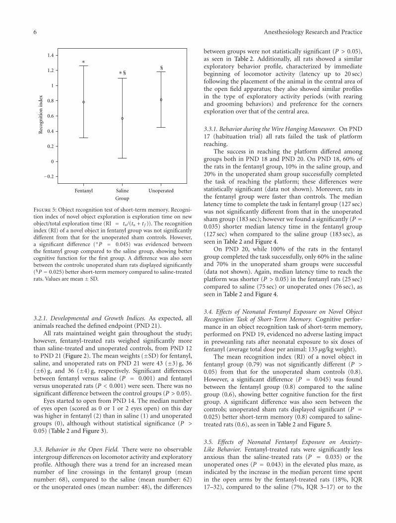
6 Anesthesiology Research and Practice
1.4
1.2
1
0.8
0.6
0.4
0.2
0
−0.2
∗§
Group
Fentanyl Saline Unoperated
§∗
Rec
ogn
itio
nin
dex
Figure 5: Object recognition test of short-term memory. Recogni-tion index of novel object exploration is exploration time on newobject/total exploration time (RI = tn/(tn + t f )). The recognitionindex (RI) of a novel object in fentanyl group was not significantlydifferent from that for the unoperated sham controls. However,a significant difference (∗P = 0.045) was evidenced betweenthe fentanyl group compared to the saline group, showing bettercognitive function for the first group. A difference was also seenbetween the controls: unoperated sham rats displayed significantly(§P = 0.025) better short-term memory compared to saline-treatedrats. Values are mean ± SD.
3.2.1. Developmental and Growth Indices. As expected, allanimals reached the defined endpoint (PND 21).
All rats maintained weight gain throughout the study;however, fentanyl-treated rats weighed significantly morethan saline-treated and unoperated controls, from PND 12to PND 21 (Figure 2). The mean weights (±SD) for fentanyl,saline, and unoperated rats on PND 21 were 43 (±3) g, 36(±6) g, and 36 (±4) g, respectively. Significant differencesbetween fentanyl versus saline (P = 0.001) and fentanylversus unoperated rats (P < 0.001) were seen. There was nosignificant difference between the control groups (P > 0.05).
Eyes started to open from PND 14. The median numberof eyes open (scored as 0 or 1 or 2 eyes open) on this daywas higher in fentanyl (2) than in saline (1) and unoperatedgroups (0), although without statistical significance (P >0.05) (Table 2 and Figure 3).
3.3. Behavior in the Open Field. There were no observableintergroup differences on locomotor activity and exploratoryprofile. Although there was a trend for an increased meannumber of line crossings in the fentanyl group (meannumber: 68), compared to the saline (mean number: 62)or the unoperated ones (mean number: 48), the differences
between groups were not statistically significant (P > 0.05),as seen in Table 2. Additionally, all rats showed a similarexploratory behavior profile, characterized by immediatebeginning of locomotor activity (latency up to 20 sec)following the placement of the animal in the central area ofthe open field apparatus; they also showed similar profilesin the type of exploratory activity periods (with rearingand grooming behaviors) and preference for the cornersexploration over that of the central area.
3.3.1. Behavior during the Wire Hanging Maneuver. On PND17 (habituation trial) all rats failed the task of platformreaching.
The success in reaching the platform differed amonggroups both in PND 18 and PND 20. On PND 18, 60% ofthe rats in the fentanyl group, 10% in the saline group, and20% in the unoperated sham group successfully completedthe task of reaching the platform; these differences werestatistically significant (data not shown). Moreover, rats inthe fentanyl group were faster than controls. The medianlatency time to complete the task in fentanyl group (127 sec)was not significantly different from that in the unoperatedsham group (183 sec); however we found a significantly (P =0.035) shorter median latency time in the fentanyl group(127 sec) when compared to the saline group (183 sec), asseen in Table 2 and Figure 4.
On PND 20, while 100% of the rats in the fentanylgroup completed the task successfully, only 60% in the salineand 70% in the unoperated sham groups were successful(data not shown). Again, median latency time to reach theplatform was shorter (P > 0.05) in the fentanyl rats (25 sec)compared to saline (75 sec) or unoperated ones (76 sec), asseen in Table 2 and Figure 4.
3.4. Effects of Neonatal Fentanyl Exposure on Novel ObjectRecognition Task of Short-Term Memory. Cognitive perfor-mance in an object recognition task of short-term memory,performed on PND 19, evidenced no adverse lasting impactin preweanling rats after neonatal exposure to six doses offentanyl (average total dose per animal: 135 μg/kg weight).
The mean recognition index (RI) of a novel object infentanyl group (0.79) was not significantly different (P >0.05) from that for the unoperated sham controls (0.8).However, a significant difference (P = 0.045) was foundbetween the fentanyl group (0.8) compared to the salinegroup (0.6), showing better cognitive function for the firstgroup. A significant difference was also seen between thecontrols; unoperated sham rats displayed significant (P =0.025) better short-term memory (0.8) compared to saline-treated rats (0.6), as seen in Table 2 and Figure 5.
3.5. Effects of Neonatal Fentanyl Exposure on Anxiety-Like Behavior. Fentanyl-treated rats were significantly lessanxious than the saline-treated rats (P = 0.035) or theunoperated ones (P = 0.043) in the elevated plus maze, asindicated by the increase in the median percent time spentin the open arms by the fentanyl-treated rats (18%, IQR17–32), compared to the saline (7%, IQR 3–17) or to the
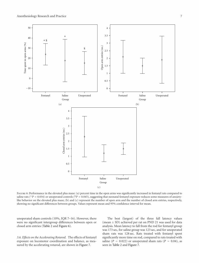
Anesthesiology Research and Practice 7
−10
0
10
20
30
40
50
∗§
Fentanyl Saline Unoperated
Tim
esp
ent
inop
enar
ms
(%)
Group
∗
§
(a)
Fentanyl Saline Unoperated
GroupO
pen
arm
entr
ies
(no.
)
0
0.5
1
1.5
2
2.5
3
3.5
4
(b)
Fentanyl Saline Unoperated
Clo
sed
arm
entr
ies
(no.
)
Group
0
0.5
1
1.5
2
2.5
3
3.5
4
(c)
Figure 6: Performance in the elevated plus maze: (a) percent time in the open arms was significantly increased in fentanyl rats compared tosaline rats (∗P = 0.034) or unoperated controls (§P = 0.045), suggesting that neonatal fentanyl exposure reduces some measures of anxiety-like behavior on the elevated plus maze; (b) and (c) represent the number of open arm and the number of closed arm entries, respectively,showing no significant differences between groups. Values represent mean and 95% confidence interval for mean.
unoperated sham controls (10%, IQR 7–16). However, therewere no significant intergroup differences between open orclosed arm entries (Table 2 and Figure 6).
3.6. Effects on the Accelerating Rotarod. The effects of fentanylexposure on locomotor coordination and balance, as mea-sured by the accelerating rotarod, are shown in Figure 7.
The best (largest) of the three fall latency values(mean ± SD) achieved per rat on PND 21 was used for dataanalysis. Mean latency to fall from the rod for fentanyl groupwas 173 sec, for saline group was 123 sec, and for unoperatedsham rats was 128 sec. Rats treated with fentanyl spentsignificantly more time on rod, compared to rats treated withsaline (P = 0.022) or unoperated sham rats (P = 0.04), asseen in Table 2 and Figure 7.

8 Anesthesiology Research and Practice
∗ §
Fentanyl Saline Unoperated
−100
0
100
200
300
Bes
tti
me
tofa
ll(s
)
∗
§
Group
Figure 7: Best latency time to fall from the accelerating rotarod(speed 5–40 rpm). Rats administered fentanyl spent significantlymore time on rod, compared to rats administered saline (∗P =0.022) or unoperated sham rats (§P = 0.040). Values are mean ±SD.
4. Discussion
The present study was designed to assess whether exposureto repetitive injections of fentanyl during brain developmentinfluences later physical and neurological outcomes. Using aneonatal postoperative pain model, this study demonstrates,for the first time, lasting effects on growth and behaviorof rat pups that underwent repeated fentanyl exposureduring early postnatal life, when tested as later pre-weanlingrats. The results showed that repeated fentanyl exposure ofan immature stressed animal significantly interferes withgrowth, cognitive function, behavioral reactivity to stress,neuromuscular and locomotor development, and balanceand coordination. All these outcomes (Table 2) suggesta neurological impact with possible consequences, eitherpositive or negative, later in life.
To examine the role of fentanyl administration in thedevelopment of behaviors that occur following repeatedexposure to this medication, both in immature CNS andpain settings, we combined two strategies: the model forthe study of neonatal neurodevelopment (6-day-old rat pup)was combined with the postincisional pain of Brennan pawincision [14], as a model for neonatal neurodevelopmentaland postoperative pain. Translation of developmental agesfrom rodents to humans continues to be debated. A reviewpaper by Vidair [19], which discusses the adequacy of thepostnatal rat to serve as a model for neurodevelopment in thepostnatal human, concludes that the rat in the third postnatalweek is the neurodevelopmental equivalent of the newbornhuman and that the two species share numerous pathways
of postnatal neurodevelopment. Therefore, our neonatalrat model roughly corresponds to a human premature.Brennan’s model of incisional pain [14] was chosen sinceit simulates the usual clinical setting involving critically illprematures in neonatal intensive care units.
Premature newborns typically present a broad range ofcomorbidities which make them a complex group to study,given the many variables, painful/stressful procedures, andpharmacologic exposures involved. Therefore, experimentalstudies using animals allow us to exclude potential con-founding variables. In our study we used a model withoutcomorbidities in a postoperative pain and stress setting.Such a preclinical model, which leads to pain-related eventsthat mirror the symptoms observed in patients undergoingsurgery [20], gives us the opportunity to explore whetherrepetitive fentanyl exposure, early in neonates subject topainful stimuli, leads to later neurodevelopmental anomalies.The postoperative pain model we used was previouslydescribed by Brennan and coworkers [14]. This rat modelconsists of an incision of the plantar paw skin, with damageof the underlying muscle, which results in localized mechan-ical hypersensitivity that lasts 3–5 days. Further research byBrennan’s group showed release of excitatory amino acids,such as glutamate and aspartate, activation of dorsal horncells, and central sensitization [21].
Concerning protocol design, the dose of fentanyl usedin this study, although at first sight much higher than theneonatal human recommended dose, was chosen accordingto the species known metabolism to relate to that typicallyencountered in clinical settings reflecting antinociceptiveED50 values for PND6 rats [22]. We assessed behavioralproblems in our neonatal stressed model using a validatedset of tests usually chosen for drug toxicity screening.
Among major findings in the present study, we highlightthe significant enhancement of weight gain in fentanyl groupcompared to controls, as summarized in Table 2. Neitherfentanyl nor control conditions had significant effects onnormal early pup weight gain. In contrast, there weresignificant group differences in rat weights on PND 21.Rats in the fentanyl group weighed more than those inthe saline and unoperated sham groups, with the differencebecoming significant around PND 12 and expanding asthe pups aged until weaning. These outcomes suggest thatthe effects of the early postnatal exposure were subtlebut, nonetheless, predisposed the pups to abnormal weightgain. Many hypotheses are possible to explain this find-ing, namely, metabolic derangements, behavior anomaliesrelated to eating disorders, or decreased physical activity.An important issue that can be raised is whether theweight change is transitory or if it can continue intoadulthood.
Other major findings in the present study were behav-ioral changes induced by administration of fentanyl in ourmodel. Somewhat surprisingly, the results point towards anoverall apparently “positive” effect on neurodevelopment,instead of the expected negative one. This “positive” impactwas evidenced by an apparent lack of significant acute toxiceffects on early development. Moreover, later, in infant ratswho were treated with fentanyl, we found enhancement
Anesthesiology Research and Practice 9
of the recognition index of a novel object, lesser anxiety-like behavior, and better performances on the wire hangingmaneuver and on the accelerating rotarod. Furthermore,there was a trend for sooner eye opening in this group,suggesting that the eye command center of CNS of rats inthe fentanyl group ages earlier.
Interestingly, aversive stressful procedures performed inthe current study, which should be associated with increasedanxiety, seemed blunted by fentanyl treatment. In fact,fentanyl-treated rats were significantly less anxious than thesaline and the unoperated rats in the elevated plus maze. Thisoutcome is not clearly explained, but calmer subjects canprobably better explain other outcomes found in this study,such as enhanced cognitive function, motor, and balance andcoordination. It is possible that all these results are at leastpartially explained by a fentanyl impact on the developmentof central neuronal circuits, given the great plasticity of theCNS characteristic of the immature mammalian brain [23].
The effects of the impact of fentanyl on SNC areprobably complex and multivariate with different possiblemechanisms found in the literature, both potentially pro-tective or detrimental, such as faster CNS myelination andenhanced neurogenesis by NeuroD activity level increase(a transcription factor essential for the development of theCNS) [24] eventually translating into enhanced performanceor, on the other hand, cytotoxic lesion/blockade of theventral hippocampus by N-methyl-D-aspartate (NMDA)receptor interference, manifesting as reduced anxiety [25].It is well known from the literature that fentanyl modulatesimportant cellular and molecular neuronal mechanisms,interfering not only in anatomically distributed neuralnetwork involved in generating states of anesthesia but also inmechanisms involved in hippocampus neurogenesis. In thissetting, fentanyl may regulate the functions of the developinghippocampus, a region highly related to learning, memory,stress responses, and emotionality [25].
There is a growing body of evidence showing that drugsinterfering in the SNC functions may cause pharmacologicneuroprotection or, on the opposite, detrimental effects,depending on the pathological conditions [19, 26–28]. Neg-ative impact alerts are particularly alarming in the contextof very ill preterm infants who usually present a multitudeof physiological derangements and pathological pain con-ditions coupled with a very immature brain, therefore it isimportant to define safe indications and doses for the use ofthese drugs, such as fentanyl, in this stage.
In conclusion, the current study is the first to demon-strate that rat pups exposed to parenteral fentanyl in a painfulcontext have lasting growth and behavioral changes. Thestudy highlights behavioral changes that could potentiallyaffect brain function either in a positive or negative manner.These results should serve as a basis for further researchand should lead investigators to focus on specific pathwaysrelevant to the changes in behavior we have shown. Ourfindings may contribute to support the neonatal use offentanyl, when indicated, namely in postsurgical settings,even in premature newborns. However, extrapolating ourdata to a clinical setting must be done with caution, as withevery animal study.
Authors’ Contribution
Dora Catre is responsible for design, intellectual and sci-entific content, and writing of the paper. Maria FrancelinaLopes supervised all phases of the experimental study andwriting of the paper. Antonio Silverio Cabrita is responsiblefor collecting and processing of study information.
References
[1] T. Lasky, F. R. Ernst, J. Greenspan, S. Wang, and L. Gonzalez,“Estimating pediatric inpatient medication use in the UnitedStates,” Pharmacoepidemiology and Drug Safety, vol. 20, no. 1,pp. 76–82, 2011.
[2] B. Hsu and T. Brazelton, “Off-label medication use in anacademic hospital pediatric critical care unit,” WisconsinMedical Journal, vol. 108, no. 7, pp. 343–348, 2009.
[3] A. N. Naguib, P. Winch, L. Schwartz et al., “Anestheticmanagement of the hybrid stage 1 procedure for hypoplasticleft heart syndrome (HLHS),” Paediatric Anaesthesia, vol. 20,no. 1, pp. 38–46, 2010.
[4] J. D. Tobias, “Sedation and analgesia in the pediatric intensivecare unit,” Pediatric Annals, vol. 34, no. 8, pp. 636–645, 2005.
[5] J. G. Klamt, W. V. A. de Vicente, L. V. Garcia, and C.A. Ferreira, “Effects of dexmedetomidine-fentanyl infusionon blood pressure and heart rate during cardiac surgeryin children,” Anesthesiology Research and Practice, vol. 2010,Article ID 869049, 7 pages, 2010.
[6] M. Palot, H. Visseaux, and C. Botmans, “Conduction anesthe-sia and the newborn infant,” Cahiers d’Anesthesiologie, vol. 43,no. 6, pp. 547–553, 1995.
[7] T. R. Okon and M. L. George, “Fentanyl-induced neurotoxicityand paradoxic pain,” Journal of Pain and Symptom Manage-ment, vol. 35, no. 3, pp. 327–333, 2008.
[8] W. A. Kofke, R. H. Garman, R. L. Stiller, M. E. Rose, andR. Garman, “Opioid neurotoxicity: fentanyl dose-responseeffects in rats,” Anesthesia and Analgesia, vol. 83, no. 6, pp.1298–1306, 1996.
[9] W. A. Kofke, R. H. Garman, R. Garman, and M. E. Rose, “Opi-oid neurotoxicity: fentanyl-induced exacerbation of cerebralischemia in rats,” Brain Research, vol. 818, no. 2, pp. 326–334,1999.
[10] J. E. Bailey, E. Campagna, and R. C. Dart, “RADARS SystemPoison Center Investigators. The underrecognized toll ofprescription opioid abuse on young children,” Annals ofEmergency Medicine, vol. 53, no. 4, pp. 419–424, 2009.
[11] T. T. Levin, M. H. Bakr, and T. Nikolova, “Case report: delir-ium due to a diltiazem-fentanyl CYP3A4 drug interaction,”General Hospital Psychiatry, vol. 32, no. 6, pp. 648.e9–648.e10,2010.
[12] E. H. Sinz, W. A. Kofke, and R. H. Garman, “Phenytoin,midazolam, and naloxone protect against fentanyl-inducedbrain damage in rats,” Anesthesia and Analgesia, vol. 91, no.6, pp. 1443–1449, 2000.
[13] Institute of Laboratory Animal Research, Commission on LifeSciences, and National Research Council, Guide for the Careand Use of Laboratory Animals, The National Academies Press,Washington, DC, USA, 1996.
[14] T. J. Brennan, E. P. Vandermeulen, and G. F. Gebhart,“Characterization of a rat model of incisional pain,” Pain, vol.64, no. 3, pp. 493–501, 1996.
[15] K. Wallace, S. Veerisetty, I. Paul, W. May, J. J. Miguel-Hidalgo,and W. Bennett, “Prenatal infection decreases calbindin,

10 Anesthesiology Research and Practice
decreases purkinje cell volume and density and produces long-term motor deficits in Sprague-Dawley rats,” DevelopmentalNeuroscience, vol. 32, no. 4, pp. 302–312, 2010.
[16] L. W. Fan, R. F. Chen, H. J. Mitchell et al., “α-Phenyl-n-tert-butyl-nitrone attenuates lipopolysaccharide-induced braininjury and improves neurological reflexes and early senso-rimotor behavioral performance in juvenile rats,” Journal ofNeuroscience Research, vol. 86, no. 16, pp. 3536–3547, 2008.
[17] Y. Silberman, O. J. Ariwodola, A. M. Chappell, J. T. Yorgason,and J. L. Weiner, “Lateral paracapsular GABAergic synapses inthe basolateral amygdala contribute to the anxiolytic effects ofβ3 adrenoceptor activation,” Neuropsychopharmacology, vol.35, no. 9, pp. 1886–1896, 2010.
[18] J. A. Able, G. A. Gudelsky, C. V. Vorhees, and M. T.Williams, “3,4-Methylenedioxymethamphetamine in adultrats produces deficits in path integration and spatial referencememory,” Biological Psychiatry, vol. 59, no. 12, pp. 1219–1226,2006.
[19] C. A. Vidair, “Age dependence of organophosphate andcarbamate neurotoxicity in the postnatal rat: extrapolation tothe human,” Toxicology and Applied Pharmacology, vol. 196,no. 2, pp. 287–302, 2004.
[20] T. J. Martin, N. L. Buechler, W. Kahn, J. C. Crews, and J. C.Eisenach, “Effects of laparotomy on spontaneous exploratoryactivity and conditioned operant responding in the rat: amodel for postoperative pain,” Anesthesiology, vol. 101, no. 1,pp. 191–203, 2004.
[21] P. K. Zahn, K. A. Sluka, and T. J. Brennan, “Excitatory aminoacid release in the spinal cord caused by plantar incision in therat,” Pain, vol. 100, no. 1-2, pp. 65–76, 2002.
[22] S. R. Thornton and F. L. Smith, “Characterization of neonatalrat fentanyl tolerance and dependence,” Journal of Pharmacol-ogy and Experimental Therapeutics, vol. 281, no. 1, pp. 514–521, 1997.
[23] S. Trojan, M. Langmeier, D. Maresova, J. Mourek, and J.Pokorny, “Plasticity of the brain in neuroontogenesis,” PragueMedical Report, vol. 105, no. 2, pp. 97–110, 2004.
[24] H. Zheng, Y. Zeng, J. Chu, A. Y. Kam, H. H. Loh, and P. Y. Law,“Modulations of NeuroD activity contribute to the differentialeffects of morphine and fentanyl on dendritic spine stability,”Journal of Neuroscience, vol. 30, no. 24, pp. 8102–8110, 2010.
[25] C. Barkus, S. B. McHugh, R. Sprengel, P. H. Seeburg, J.N. Rawlins, and D. M. Bannerman, “Hippocampal NMDAreceptors and anxiety: at the interface between cognition andemotion,” European Journal of Pharmacology, vol. 626, no. 1,pp. 49–56, 2010.
[26] V. Laudenbach, G. Calo, R. Guerrini et al.,“Nociceptin/orphanin FQ exacerbates excitotoxic whitematterlesions in the murine neonatal brain,” Journal of ClinicalInvestigation, vol. 107, no. 4, pp. 457–466, 2001.
[27] K. J. Anand, S. Garg, C. R. Rovnaghi, U. Narsinghani, A.T. Bhutta, and R. W. Hall, “Ketamine reduces the cell deathfollowing inflammatory pain in newborn rat brain,” PediatricResearch, vol. 62, no. 3, pp. 283–290, 2007.
[28] S. R. Hays and J. K. Deshpande, “Newly postulated neurode-velopmental risks of pediatric anesthesia,” Current Neurologyand Neuroscience Reports, vol. 11, no. 2, pp. 205–210, 2011.

Hindawi Publishing CorporationAnesthesiology Research and PracticeVolume 2012, Article ID 375691, 5 pagesdoi:10.1155/2012/375691
Review Article
Spinal Cord Stimulation: The Clinical Application ofNew Technology
Dominic Hegarty1, 2
1 Department of Anaesthesia and Intensive Care, Cork University Hospital, Cork, Ireland2 Department of Pharmacology and Therapeutics, University College Cork, Clinical Sciences Building, Cork, Ireland
Correspondence should be addressed to Dominic Hegarty, [email protected]
Received 28 May 2011; Revised 11 August 2011; Accepted 13 August 2011
Academic Editor: Andrea Trescot
Copyright © 2012 Dominic Hegarty. This is an open access article distributed under the Creative Commons Attribution License,which permits unrestricted use, distribution, and reproduction in any medium, provided the original work is properly cited.
The use of neuromodulation for pain relief is among the fastest-growing areas of medicine, involving many diverse specialtiesand impacting on hundreds of thousands of patients with numerous disorders worldwide. As the evidence of efficacy improves,the interest in spinal cord stimulation (SCS) will increase because it is minimally invasive, safe, and a reversible treatmentmodality with limited side effect profile. While the mechanism of action evades complete understanding, the technologicalimprovements have been considerable and current neuromodulation developments have been coupled with the rapid growthof the neuromodulation device industry resulting in the development of the next-generation neuromodulation systems. Thedevelopment, the newest technicaliti and the future for the clinical application of spinal cord stimulation (SCS) are reviewedhere.
1. Introduction
Neuromodulation is among the fastest-growing areas ofmedicine, involving many diverse specialties and impactingon hundreds of thousands of patients with numerous dis-orders worldwide [1]. Historically, electricity, either in theform of the torpedo fish or man-made electrotherapy, hasbeen used to try and cure various ailments [2]. For ex-ample, in the middle of the 18th century “electroanalgesia”became advocated for the treatment of angina pectoris, gout,headaches, pleuritic pain, and sciatica. However, by the 20thcentury the enthusiasm for the medical use of electricitybecame associated with “quackery” [3] and was bannedfrom clinical practice. In 1965 Melzack and Wall presentedthe “Gate Theory” [4], which postulated that stimulationof nonpainful stimuli can inhibit painful afference, therebyoffering the opportunity to align basic research with theclinical application of electricity which has resulted in thedevelopment of neuromodulation techniques as we knowthem today [5–7]. While the mechanism of action evadescomplete understanding, the technological improvementshave been considerable and current neuromodulation devel-opments have been coupled with the rapid growth of the
neuromodulation device industry resulting in the develop-ment of the next-generation neuromodulation systems. Thedevelopment, the newest technicalities, and the future forthe clinical application of spinal cord stimulation (SCS) arereviewed here.
2. Principles of Neuromodulation
Essentially there are two components of a fully implantedSCS system: the electrodes (or lead) and an implantablepulse generator (IPG). In SCS the placement of epiduralelectrodes is generally targeted at the dorsal column of thespinal cord; however, in patients with segmental pain (singledermatome), stimulation is focused at the correspondingdorsal root. This is where the ascending tracts pass withoutdecussation to the gracile and cuneate nuclei of the medullaoblongata. These tracts are composed of a wide range offiber diameters which are the central processes of the primaryafferent neurons located in the spinal ganglia. As the tractsascend, they receive accession from the dorsal roots, resultingin a somatotopic organization [6]. The recruitment of fibersis correlated directly with the diameter of the fiber andinversely with the distance between the electrode contacts
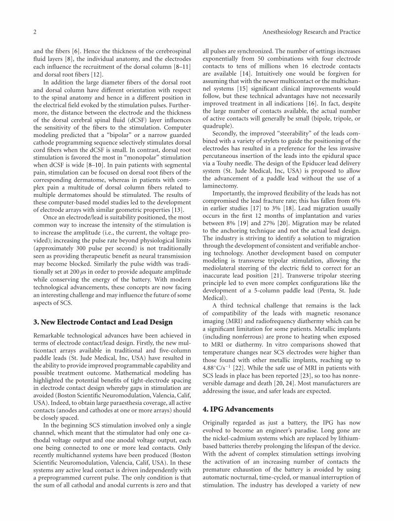
2 Anesthesiology Research and Practice
and the fibers [6]. Hence the thickness of the cerebrospinalfluid layers [8], the individual anatomy, and the electrodeseach influence the recruitment of the dorsal column [8–11]and dorsal root fibers [12].
In addition the large diameter fibers of the dorsal rootand dorsal column have different orientation with respectto the spinal anatomy and hence in a different position inthe electrical field evoked by the stimulation pulses. Further-more, the distance between the electrode and the thicknessof the dorsal cerebral spinal fluid (dCSF) layer influencesthe sensitivity of the fibers to the stimulation. Computermodeling predicted that a “bipolar” or a narrow guardedcathode programming sequence selectively stimulates dorsalcord fibers when the dCSF is small. In contrast, dorsal rootstimulation is favored the most in “monopolar” stimulationwhen dCSF is wide [8–10]. In pain patients with segmentalpain, stimulation can be focused on dorsal root fibers of thecorresponding dermatome, whereas in patients with com-plex pain a multitude of dorsal column fibers related tomultiple dermatomes should be stimulated. The results ofthese computer-based model studies led to the developmentof electrode arrays with similar geometric properties [13].
Once an electrode/lead is suitability positioned, the mostcommon way to increase the intensity of the stimulation isto increase the amplitude (i.e., the current, the voltage pro-vided); increasing the pulse rate beyond physiological limits(approximately 300 pulse per second) is not traditionallyseen as providing therapeutic benefit as neural transmissionmay become blocked. Similarly the pulse width was tradi-tionally set at 200 µs in order to provide adequate amplitudewhile conserving the energy of the battery. With moderntechnological advancements, these concepts are now facingan interesting challenge and may influence the future of someaspects of SCS.
3. New Electrode Contact and Lead Design
Remarkable technological advances have been achieved interms of electrode contact/lead design. Firstly, the new mul-ticontact arrays available in traditional and five-columnpaddle leads (St. Jude Medical, Inc, USA) have resulted inthe ability to provide improved programmable capability andpossible treatment outcome. Mathematical modeling hashighlighted the potential benefits of tight-electrode spacingin electrode contact design whereby gaps in stimulation areavoided (Boston Scientific Neuromodulation, Valencia, Calif,USA). Indeed, to obtain large paraesthesia coverage, all activecontacts (anodes and cathodes at one or more arrays) shouldbe closely spaced.
In the beginning SCS stimulation involved only a singlechannel, which meant that the stimulator had only one ca-thodal voltage output and one anodal voltage output, eachone being connected to one or more lead contacts. Onlyrecently multichannel systems have been produced (BostonScientific Neuromodulation, Valencia, Calif, USA). In thesesystems any active lead contact is driven independently witha preprogrammed current pulse. The only condition is thatthe sum of all cathodal and anodal currents is zero and that
all pulses are synchronized. The number of settings increasesexponentially from 50 combinations with four electrodecontacts to tens of millions when 16 electrode contactsare available [14]. Intuitively one would be forgiven forassuming that with the newer multicontact or the multichan-nel systems [15] significant clinical improvements wouldfollow, but these technical advantages have not necessarilyimproved treatment in all indications [16]. In fact, despitethe large number of contacts available, the actual numberof active contacts will generally be small (bipole, tripole, orquadruple).
Secondly, the improved “steerability” of the leads com-bined with a variety of stylets to guide the positioning of theelectrodes has resulted in a preference for the less invasivepercutaneous insertion of the leads into the epidural spacevia a Touhy needle. The design of the Epiducer lead deliverysystem (St. Jude Medical, Inc, USA) is proposed to allowthe advancement of a paddle lead without the use of alaminectomy.
Importantly, the improved flexibility of the leads has notcompromised the lead fracture rate; this has fallen from 6%in earlier studies [17] to 3% [18]. Lead migration usuallyoccurs in the first 12 months of implantation and variesbetween 8% [19] and 27% [20]. Migration may be relatedto the anchoring technique and not the actual lead design.The industry is striving to identify a solution to migrationthrough the development of consistent and verifiable anchor-ing technology. Another development based on computermodeling is transverse tripolar stimulation, allowing themediolateral steering of the electric field to correct for aninaccurate lead position [21]. Transverse tripolar steeringprinciple led to even more complex configurations like thedevelopment of a 5-column paddle lead (Penta, St. JudeMedical).
A third technical challenge that remains is the lackof compatibility of the leads with magnetic resonanceimaging (MRI) and radiofrequency diathermy which can bea significant limitation for some patients. Metallic implants(including nonferrous) are prone to heating when exposedto MRI or diathermy. In vitro comparisons showed thattemperature changes near SCS electrodes were higher thanthose found with other metallic implants, reaching up to4.88◦C/s−1 [22]. While the safe use of MRI in patients withSCS leads in place has been reported [23], so too has nonre-versible damage and death [20, 24]. Most manufacturers areaddressing the issue, and safer leads are expected.
4. IPG Advancements
Originally regarded as just a battery, the IPG has nowevolved to become an engineer’s paradise. Long gone arethe nickel-cadmium systems which are replaced by lithium-based batteries thereby prolonging the lifespan of the device.With the advent of complex stimulation settings involvingthe activation of an increasing number of contacts thepremature exhaustion of the battery is avoided by usingautomatic nocturnal, time-cycled, or manual interruption ofstimulation. The industry has developed a variety of new

Anesthesiology Research and Practice 3
generation of compact rechargeable IPGs to meet the newrequirements of SCS; thereby, energy consumption becomesless of a problem.
As previously mentioned the recruitment of dorsal hornfibers is correlated inversely with the distance between theelectrode contact and the fiber [6]. Hence the thickness of thecerebrospinal fluid layers [8] the recruitment of the dorsalcolumn [8–11] and dorsal root fibers [12]. Therefore aselectrode/leads placement varies and stimulation intensitycan vary depending on the position of the patient (e.g.,supine or standing) to such an extent that patients cannot usethe IPG without manually changing the program in order toavoid painful overstimulation.
The RestoreSensor (Medtronic Inc, USA) is the firstimplantable neurostimulator for spinal cord stimulation(SCS) that automatically adapts stimulation settings in re-sponse to position changes and provides objective patientactivity data. The adaptive SCS is based on accelerationsensor that enables chronic motion sensing in battery-powered applications. It is robust to shock and representsthe first practical, packaged, three-axis accelerometer suitablefor chronic physiological measurements. The sensor’s perfor-mance is also desirable for more general micropower appli-cations like package tracking, vibration, and tilt detection.
Initial results from the Testing RestoreSensor Usabilityand Satisfaction (TRUST) survey [25] in 30 patients, mainlysuffering from predominant leg pain due to failed backsurgery syndrome or complex regional pain syndrome typeI, followed up over a 10-week period are very promising.80% of patients reported more effective pain relief. Useof the patient programmer became less difficult, and lessnecessary, with adaptive stimulation. Adaptive stimulationhad a positive effect on sleep quality in all patients, whichmay have led to a perception of greater sleep quantity. Intotal, 58% of patients reported the ability to perform moreactivities with the therapy (e.g., standing, walking, sleeping,and staying in a particular position longer). Overall patientsatisfaction was 97% at the end of the 10-week follow-upperiod [25].
5. Modification of the Pulse Width
In SCS, the pulse amplitude is usually the focus of stim-ulation control as it is intuitively understood by clinicianand patient alike [26–29]. With advances in SCS technology,particularly rechargeable IPG implantable devices, pulsewidth (PW) programming ranges of now match that ofolder radiofrequency systems (with programmability up to1000 µs). Traditionally PW was only changed when otherparameter adjustments fail to achieve therapeutic goals. Inneurostimulation the pulse amplitude and width relate di-rectly to the depolarization of the cell membrane and aretherefore critical parameters for determining the locus ofexcited tissue [30]. The value of PW programming wasinvestigated in 19 subjects who had a fully implanted SCSin place for over 3 months to treat chronic intractable lowback and/or leg pain. It was shown that the baseline medianPW parameter was 295 µs (range 242–326 µs) with a median
amplitude of 2.5 mA (1.3–3.3 mA). Following independentmodification of the PW, the median PW of all patients’ pro-grams increased to 400 µs, approximately 48% higher (P =0.01) and showed a significant increase in the paraesthesia-pain overlap (56%, P = 0.04). It was estimated that 10/19patients appeared to have greater paraesthesia coverage, 7/19patients selected the new PW programs, and 8/19 patientsappeared to display a “caudal shift” of paraesthesia coveragewith increased PW [31].
Mathematical modelling suggests that the mechanismbehind such paraesthesia steering is due to the differentselectivity of PW for larger and smaller fibers. The modelconsidered incorporated realistic fiber size, density, anddistributions in the dorsal columns, based upon humananatomic data. With a greater relative density of smallerfibers located more medial in the dorsal columns, an increasein PW will recruit smaller fibers more readily and thus resultin greater midline axon recruitment. Clinically, this appearedto manifest as a caudal shift in paraesthesia. In summaryvariable PW programming in SCS appears to have clinicalvalue, demonstrated by some patients improving theirparaesthesia-pain overlap, as well as the ability to increaseand even “steer” paraesthesia coverage [31].
6. High-Frequency Stimulation
Although SCS is a recommended treatment for patients withfailed back surgery syndrome (FBSS) [32], if paraesthesiaover the lumbar dermatomes cannot be obtained, then axiallow back pain is very difficult to treat and clinical resultsare poor [33]. Ongoing multicentred European prospectivetrials [34] using dual octapolar, percutaneous leads placedsequentially near anatomic midline and connected to a re-chargeable IPG capable of delivering waveforms with fre-quencies up to 10 kHz. (Nevro, Menlo Park, Calif) haveshown that of 34 cases with full implantation the averageback pain VAS decreased by 77% (8.9 cm baseline to 2.0 cmat 6 months, P < .001) and leg pain VAS decreased by 82% at6-month follow-up. (5.5 cm baseline to 0.7 cm at 6 months,P < .001). In addition the average Oswestry Disability Indexscore decreased by 36% (from 58 to 37, P < .001). Thisapproach is novel for several reasons: (a) the use of high-frequency stimulation provides sustained analgesia in apreviously difficult patient cohort without paraesthesia—thus adequate axial low back pain relief is achieved withoutthe overwhelming leg sensation one would have expectedby increasing the frequency using a traditional IPG; (b)anatomical placement of the leads is possible and intraop-erative paraesthesia mapping is avoided; (c) it has decreasedprogramming requirements; (d) continued use of the systemindependent of position including night-time use is possible.To date, no adverse effect of such high-frequency stimulationhas been reported however, the clinical outcome in thelonger term is awaited. Pre-clinical studies in goats whoreceived 10 days of continuous stimulation at amplitudesup to the sensory/motor threshold showed no differencein the behaviour or spinal cord neural histology betweenthe therapy and control groups. Why such stimulation has

4 Anesthesiology Research and Practice
this remarkable effect still remains to be understood andmay influence our approach to this co-cohort heretofore un-satisfactorily managed with conventional SCS technology.
7. New Clinical Applications
There are several established indications for SCS such asneuropathic back and leg pain, complex regional painsyndrome, spinal cord injury, and ischemic pain (vascularand angina pectoris). While it is beyond the remit of thispaper to discuss each clinical indication, there is a growingdatabase of clinical-based evidence to support the use of SCS.The economic evaluation in these areas is limited but theinitial costs of SCS is generally both more effective and lesscostly then conventional management over a period of 3–5 years [33]. Unfortunately SCS is regarded as a last-resortoption by many healthcare providers, and the real economicbenefits may lie in the earlier introduction of the technique.
The recognition of new treatment modalities and thenew application of SCS techniques will redefine our under-standing of the pathophysiological concepts involved indifferent medical conditions. For example, painful bladdersyndrome/interstitial cystitis and diffuse chronic abdom-inal/pelvic pain may be considered as neuropathic painthereby offering the potential for exciting development. Theevidence that cervical SCS increases cerebral blood flow(CBF) may lead to a role in cerebral ischemia. The modifi-cation of the autonomic system, particularly its sympatheticcomponent by SCS, suggests that body functions undersignificant autonomic control could be subjected to mod-ulation. It is suggested that in the future bronchospasm,gastrointestinal motility, and possibily metabolic disorderscould become the focus of neuromodulation [35].
8. Conclusion
Modern medicine requires that any treatment modality isbased on rational knowledge and well-documented theo-ries; however, some conditions, particularly those involvingchronic pain, often remain imprecise. SCS may be oneof the few examples of a treatment that has significantlycontributed to a change in attitudes and providing satis-factory relief to patients who in the past would have beenleft untreated.
Spinal cord stimulation has significant implications forthe healthcare system offering a safe reversible treatmentmodality with a limited side effect profile. To ensure thedeliverance of a high-standard quality of care spinal cordstimulation should be provided in small well-resourced cen-tres able to address the aftercare needs of this patient cohort.Cost-effectiveness and efficacy are fundamental if SCS is to beaccepted as the therapy of choice by the public, physicians,and the healthcare decision makers. Earlier introduction ofthe technique may prove to be critical. Research into themechanism of pain, the diseases, and the action of SCSrequires randomized control trials (RCTs). The inability (a)to blind patients (owning to the paraesthesia), (b) to selecta comparative therapy (medical, surgical, rehabilitation),
and/or (c) to address the ethical implications to participatein a trial are specific issues in the design of such a RCT.
As the evidence of efficacy improves and the number ofindications increases, the interest in the neuromodulationand SCS will undoubtedly increase. SCS is minimally inva-sive, safe, and reversible treatment modality with limited sideeffect profile. It is only through the combined efforts of thebiomedical industry, basic science researchers, and frontlinehealthcare providers will the technological advancementsalready made in this area continue to make significant clinicalimpact on the patients of tomorrow.
References
[1] E. S. Krames, P. H. Peckham, A. R. Rezai, and F. Aboelsaad,“What is neuromodulation?” in Neuromodulation, Krames etal., Ed., pp. 3–8, Elsevier, 2009.
[2] D. Kellaway, “The william osler medical essay; the part playedby electric fish in the early history of bioelectricity and elec-trotherapy,” Bulletin of the History of Medicine, vol. 20, pp.112–137, 1946.
[3] R. M. Macklis, “Magnetic healing, quackery, and the debateabout the health effects of electromagnetic fields,” Annals ofInternal Medicine, vol. 118, no. 5, pp. 376–383, 1993.
[4] R. Melzack and P. D. Wall, “Pain mechanisms: a new theory,”Science, vol. 150, no. 3699, pp. 971–979, 1965.
[5] U. Rossi, “The history of electrical stimulation of the nervoussystem for the control of pain,” in Electric Stimulation and theRelief of Pain, B. A. Simpson, Ed., pp. 5–16, Elsevier, London,UK, 2003.
[6] A. Foletti, A. Durrer, and E. Buchser, “Neurostimulationtechnology for the treatment of chronic pain: a focus on spinalcord stimulation,” Expert Review of Medical Devices, vol. 4, no.2, pp. 201–214, 2007.
[7] C. N. Shealy, J. T. Mortimer, and J. B. Reswick, “Electricalinhibition of pain by stimulation of the dorsal columns: pre-liminary clinical report,” Anesthesia and Analgesia, vol. 46, no.4, pp. 489–491, 1967.
[8] J. Holsheimer and J. J. Struijk, “How do geometric factorsinfluence epidural spinal cord stimulation? A quantitativeanalysis by computer modeling,” Stereotactic and FunctionalNeurosurgery, vol. 56, no. 4, pp. 234–249, 1991.
[9] J. Holsheimer and W. A. Wesselink, “Optimum electrodegeometry for spinal cord stimulation: the narrow bipole andtripole,” Medical and Biological Engineering and Computing,vol. 35, no. 5, pp. 493–497, 1997.
[10] J. Holsheimer, J. J. Struijk, and N. R. Tas, “Effects of electrodegeometry and combination on nerve fibre selectivity in spinalcord stimulation,” Medical and Biological Engineering andComputing, vol. 33, no. 5, pp. 676–682, 1995.
[11] N. J. M. Rijkhoff, J. Holsheimer, F. M. J. Debruyne, and H.Wijkstra, “Modelling selective activation of small myelinatednerve fibres using a monopolar point electrode,” Medical andBiological Engineering and Computing, vol. 33, no. 6, pp. 762–768, 1995.
[12] J. J. Struijk, J. Holsheimer, and H. B.K. Boom, “Excitationof dorsal root fibers in spinal cord stimulation: a theoreticalstudy,” IEEE Transactions on Biomedical Engineering, vol. 40,no. 7, pp. 632–639, 1993.
[13] L. Manola, J. Holsheimer, P. H. Veltink, K. Bradley, and D.Peterson, “Theoretical investigation into longitudinal cathodal

Anesthesiology Research and Practice 5
field steering in spinal cord stimulation,” Neuromodulation,vol. 10, no. 2, pp. 120–132, 2007.
[14] B. A. Simpson, “Spinal cord stimulation,” Pain Reviews, vol. 1,pp. 199–230, 1994.
[15] R. B. North, M. G. Ewend, M. T. Lawton, and S. Piantadosi,“Spinal cord stimulation of chronic, intractable pain: superi-ority of ’multi-channel’ devices,” Pain, vol. 44, no. 2, pp. 119–130, 1991.
[16] R. B. North, D. H. Kidd, J. Olin, J. N. Sieracki, and L. Petrucci,“Spinal cord stimulation for axial low back pain: a prospectivecontrolled trial comparing 16-contact insulated electrodeswith 4-contact percutaneous electrodes,” Neuromodulation,vol. 9, no. 1, pp. 56–67, 2006.
[17] R. Davis and E. Gray, “Technical factors important to dorsalcolumn stimulation,” Applied Neurophysiology, vol. 44, no. 1-3, pp. 160–170, 1981.
[18] G. H. Spincemaille, H. M. Klomp, E. W. Steyerberg, H. VanUrk, and J. D.F. Habbema, “Technical data and complicationsof spinal cord stimulation: data from a randomized trialon critical limb ischemia,” Stereotactic and Functional Neu-rosurgery, vol. 74, no. 2, pp. 63–72, 2000.
[19] M. S. May, C. Banks, and S. J. Thomson, “A retrospective, long-term, third-party follow-up of patients considered for spinalcord stimulation,” Neuromodulation, vol. 5, no. 3, pp. 137–144,2002.
[20] M. J. L. De Jongste, D. Nagelkerke, C. M. Hooyschuur etal., “Stimulation characteristics, complications, and efficacyof spinal cord stimulation systems in patients with refractoryangina: a prospective feasibility study,” Pacing and ClinicalElectrophysiology, vol. 17, no. 11 I, pp. 1751–1760, 1994.
[21] J. C. Oakley, F. Espinosa, H. Bothe et al., “Transverse tripolarspinal cord stimulation: results of an international multicenterstudy,” Neuromodulation, vol. 9, no. 3, pp. 192–203, 2006.
[22] P. S. Ruggera, D. M. Witters, G. von Maltzahn, and H. I. Bas-sen, “In vitro assessment of tissue heating near metallic med-ical implants by exposure to pulsed radio frequency diather-my,” Physics in Medicine and Biology, vol. 48, no. 17, pp. 2919–2928, 2003.
[23] R. V. Sha, H. K. Smith, J. Chung, A. Hegazi, and G. B. Racz,“Cervical spinal cord neoplasm in a patient with an implantedcervical spinal cord stimulator: the controversial role of mag-netic resonance imaging,” Pain Physician, vol. 7, no. 2, pp.273–278, 2004.
[24] J. M. Henderson, J. Thach, M. Phillips, K. Baker, F. G. Shellock,and A. R. Rezai, “Permanent neurological deficit related tomagnetic resonance imaging in a patient with implanteddeep brain stimulation electrodes for Parkinson’s disease: casereport,” Neurosurgery, vol. 57, no. 5, p. E1063, 2005.
[25] J. W. Kallewaard, J. Koy, P. Rigoard, D. Abejon, and K. Gat-zinksy, “Adaptive spinal cord stimulation: results from the test-ing restore sensor usability and satisfaction (TRUST) survey,”INS London, 2011.
[26] M. Tulgar, G. Barolat, and B. Ketcik, “Analysis of parametersfor epidural spinal cord stimulation,” Stereotactic and Func-tional Neurosurgery, vol. 61, no. 3, pp. 129–139, 1993.
[27] J. D. Law and L. V. Miller, “Importance and documentationof an epidural stimulating position,” Applied Neurophysiology,vol. 45, no. 4-5, pp. 461–464, 1982.
[28] R. Segal, B. R. Stacey, T. E. Rudy, S. Baser, and J. Markham,“Spinal cord stimulation revisited,” Neurological Research, vol.20, no. 5, pp. 391–396, 1998.
[29] J. P. van Buyten, “The performance and safety of animplantable spinal cord stimulation system in patients with
chronic pain: a 5-year study,” Neuromodulation, vol. 6, no. 2,pp. 79–87, 2003.
[30] J. T. Mortimer, “Motor prostheses,” in Handbook of Phys-iology—The Nervous System III, V. Brooks, Ed., pp. 155–187,American Physiological Society, Bethesda, Md, USA, 1981.
[31] T. L. Yearwood, B. Hershey, K. Bradley, and D. Lee, “Pulsewidth programming in spinal cord stimulation: a clinicalstudy,” Pain Physician, vol. 13, no. 4, pp. 321–335, 2010.
[32] K. Kumar, R. S. Taylor, L. Jacques et al., “Spinal cord stim-ulation versus conventional medical management for neu-ropathic pain: a multicentre randomised controlled trial inpatients with failed back surgery syndrome,” Pain, vol. 132, no.1-2, pp. 179–188, 2007.
[33] J. C. Oakley, “Spinal cord stimulation in axial low back pain:solving the dilemma,” Pain Medicine, vol. 7, no. 1, pp. S58–S63,2006.
[34] I. Smet, J. P. van Buyten, and A. Al-Kaisy, “Successful treat-ment of low back pain with a novel neuromodulation device,”in Proceedings of the 14th North American NeuromodulationSociety Annual Meeting, Las Vegas, Nev, USA, 2010.
[35] E. Buchser and S. Thompson, “The future of spinal cordstimulation and related “neuroaugmentative” procedures,” inElectrical Stimulation and the Relief of Pain, B. A. Simpson, Ed.,pp. 251–267, Elsevier, London, UK, 2003.

Hindawi Publishing CorporationAnesthesiology Research and PracticeVolume 2012, Article ID 492452, 5 pagesdoi:10.1155/2012/492452
Review Article
A New Look at Trigger Point Injections
Clara S. M. Wong and Steven H. S. Wong
Department of Anaesthesiology, Queen Elizabeth Hospital, 30 Gascoigne Road, Kowloon, Hong Kong
Correspondence should be addressed to Steven H. S. Wong, [email protected]
Received 31 May 2011; Revised 28 July 2011; Accepted 30 July 2011
Academic Editor: Andrea Trescot
Copyright © 2012 C. S. M. Wong and S. H. S. Wong. This is an open access article distributed under the Creative CommonsAttribution License, which permits unrestricted use, distribution, and reproduction in any medium, provided the original work isproperly cited.
Trigger point injections are commonly practised pain interventional techniques. However, there is still lack of objective diagnosticcriteria for trigger points. The mechanisms of action of trigger point injection remain obscure and its efficacy remainsheterogeneous. The advent of ultrasound technology in the noninvasive real-time imaging of soft tissues sheds new light onvisualization of trigger points, explaining the effect of trigger point injection by blockade of peripheral nerves, and minimizing thecomplications of blind injection.
1. Introduction
Myofascial pain syndrome is a common, painful musculo-skeletal disorder characterized by the presence of triggerpoints. They have been implicated in patients with headache,neck pain, low back pain, and various other musculoskeletaland systemic disorders [1–4]. The prevalence of myofascialtrigger points among patients complaining of pain anywherein the body ranged from 30% to 93% [5]. Although the mostimportant strategy in treatment of myofascial pain syndromeis to identify the etiological lesion that causes the activationof trigger points and to treat the underlying pathology [6],trigger point injections are still commonly practised paininterventional technique for symptomatic relief.
Despite the popularity of trigger point injections, the pa-thophysiology of myofascial trigger points remains unclear.Localization of a trigger point is often based on the physi-cian’s examination. However, such physical examination isoften unreliable. Lack of objective clinical measurements hasalso been a barrier for critically evaluating the efficacy of thetherapeutic methods.
Ultrasound is used extensively for noninvasive real-timeimaging of soft tissues including muscle, nerve, tendon,fascia, and blood vessels. With the advent of portable ultra-sound technology, ultrasound is now commonly employedin the field of regional analgesia. In this paper, we will lookat the potential application of ultrasound in trigger point in-jections.
2. Diagnosis of Trigger Points
Physician’s sense of feel and patient expressions of pain uponpalpation are the most commonly used method to localizea trigger point. The most common physical finding is pal-pation of a hypersensitive bundle or nodule of muscle fibreof harder than normal consistency. The palpation will elicitpain over the palpated muscle and/or cause radiation of paintowards the zone of reference in addition to a twitch response[7].
In myofascial pain syndrome, trigger points have beenclassified into active or latent. In an active trigger point, thereis an area of tenderness at rest or on palpation, a taut band ofmuscle, a local twitch response, and referred pain elicited byfirm compression similar to the patient’s complaint. Latenttrigger points are more commonly seen. They may displayhypersensitivity and exhibit all the characteristics of an activetrigger point except that it is not associated with spontaneouspain [7].
Trigger points have also been further classified into key orsatellite. An active key trigger point in one muscle can inducean active satellite trigger point in another muscle. Inactiva-tion of the key trigger point often also inactivates its satellitetrigger point without treatment of the satellite trigger pointitself [7].
The diagnosis of trigger points depends very much on thesubjective experience of the physician. Pressure algometryhas been used to quantify the sensitivity of trigger points.

2 Anesthesiology Research and Practice
A hand-held pressure meter with a 1 cm2 rubber disc at-tached to a force gauge calibrated up to 10 kg is applied overa trigger point to measure its pain threshold [8]. However,this method is not commonly employed clinically, and therehave not been any imaging criteria for the diagnosis of triggerpoints.
3. Pathophysiology of Trigger Points
Trigger points are defined as palpable, tense bands of skeletalmuscle fibres. They can produce both local and referred painwhen compressed.
The local pain could be explained by the tissue ischemiaresulting from prolonged muscle contraction with accumu-lation of acids and chemicals such as serotonin, histamine,kinins, and prostaglandins [9]. These changes are fed intoa cycle of increasing motor or sympathetic activity and canlead to increased pain. A painful event can sustain itself oncea cycle is established even after the initial stimulus has beenremoved [10].
The pathogenesis of trigger points is probably relatedto sensitized sensory nerve fibres (nociceptors) associatedwith dysfunctional endplates [11]. In fact, endplate noise wasfound to be significantly more prevalent in myofascial triggerpoints than in sites that were outside of a trigger point butstill within the endplate zone [12].
Studies have found that development of trigger points isdependent on an integrative mechanism in the spinal cord.When the input from nociceptors in an original receptivefield persists (pain from an active trigger point), central sen-sitization in the spinal cord may develop, and the receptivefield corresponding to the original dorsal horn neuron maybe expanded (referred pain). Through this mechanism, new“satellite trigger points” may develop in the referred zone ofthe original trigger point [11].
4. Mechanisms of Action of TriggerPoint Injections
Noninvasive measures for treatment of trigger points in-clude spray and stretch, transcutaneous electrical stimula-tion, physical therapy, and massage. Invasive treatments in-clude injections with local anaesthetics, corticosteroids, orbotulinum toxin, or dry needling [13–18].
Hong reported that with either lidocaine injection or dryneedling of trigger points, the patients experienced almostcomplete relief of pain immediately after injection if localtwitch responses were elicited. On the other hand, they ex-perienced only minimal relief if no such response occurredduring injection. Hong has suggested that nociceptors (freenerve endings) are encountered and blocked during triggerpoint injection if local twitch response can be elicited [19].
The mechanism of action of trigger point injections isthought to be disruption of the trigger points by the me-chanical effect of the needle or the chemical effect of theagents injected, resulting in relaxation and lengthening ofthe muscle fibre. The effect of the injectate may includelocal vasodilation, dilution, and removal of the accumulated
nociceptive substrates. Botulinum toxin A has been used toblock acetylcholine release from the motor nerve ending andsubsequently relieve the taut band [6].
While the relief of local pain could easily be explained bythe relaxation of the muscle fibre, the relief of referred paincould not be explained without attributing it to a peripheralnerve blockade. However, little has been said in the literatureregarding the mechanism of trigger point injection in thisrespect.
5. Could the Application of UltrasoundSolve the Mystery of Trigger Points?
5.1. Direct Visualization of Trigger Points. As mentionedabove, the most common physical finding of a trigger pointis palpation of a hypersensitive bundle or nodule of musclefibre of harder than normal consistency. Attempts to confirmthe presence of myofascial trigger points using imaging havebeen demonstrated by magnetic resonance elastography[20]. For ultrasound, earlier studies have failed to find anycorrelation between physical findings and diagnostic ultra-sound [21]. This may be attributed to poorer quality of ul-trasound imaging in earlier dates.
Recently, Sikdar et al. have tried to use ultrasound tovisualize and characterize trigger points. They found thattrigger points appeared as focal, hypoechoic regions of el-liptical shape, with a size of 0.16 cm [22]. This is promisingas ultrasound can provide a more objective diagnosis oftrigger point. Even if visualization of individual trigger pointis difficult due to the small size, some advocate the use ofultrasound to guide proper needle placement in muscletissue and to avoid adipose or nonmusculature structuresduring trigger point injections [23].
5.2. Injection of Peripheral Nerves. Trigger point injectionshave been implicated in patients with headache, low backpain, and various other musculoskeletal and systemic disor-ders. Some of these injections may involve injectate depo-sition directly to the nerves supplying the region. Indeed,entrapment, compression, or irritation of the sensory nervesof local regions has been implicated in various conditions.
5.2.1. Greater Occipital Nerve. Entrapment of the greater oc-cipital nerve is often implicated as the cause of cervicogenicheadache, and the characteristic occipital headache can bereproduced by finger pressure over the corresponding occip-ital nerve over the occipital ridge [3, 24–26]. This referralpattern of pain coincides with that of the properties of atrigger point, and it could explain the mechanism of referredpain for trigger points.
Simons has considered that the effect of greater occipitalnerve injection is due to the release of the entrapment byrelaxation of semispinalis muscle [7]. However, injection oflocal anaesthetics with or without steroid over the occipitalnerve has been found to result in alleviation of occipitalheadache [27]. In migraine headaches, local injection oflocal anaesthetics or botulinum toxin type A to the greater
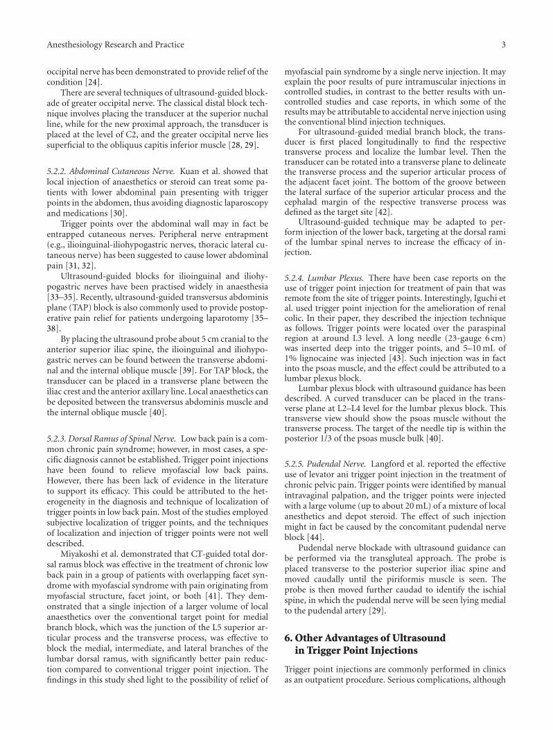
Anesthesiology Research and Practice 3
occipital nerve has been demonstrated to provide relief of thecondition [24].
There are several techniques of ultrasound-guided block-ade of greater occipital nerve. The classical distal block tech-nique involves placing the transducer at the superior nuchalline, while for the new proximal approach, the transducer isplaced at the level of C2, and the greater occipital nerve liessuperficial to the obliquus capitis inferior muscle [28, 29].
5.2.2. Abdominal Cutaneous Nerve. Kuan et al. showed thatlocal injection of anaesthetics or steroid can treat some pa-tients with lower abdominal pain presenting with triggerpoints in the abdomen, thus avoiding diagnostic laparoscopyand medications [30].
Trigger points over the abdominal wall may in fact beentrapped cutaneous nerves. Peripheral nerve entrapment(e.g., ilioinguinal-iliohypogastric nerves, thoracic lateral cu-taneous nerve) has been suggested to cause lower abdominalpain [31, 32].
Ultrasound-guided blocks for ilioinguinal and iliohy-pogastric nerves have been practised widely in anaesthesia[33–35]. Recently, ultrasound-guided transversus abdominisplane (TAP) block is also commonly used to provide postop-erative pain relief for patients undergoing laparotomy [35–38].
By placing the ultrasound probe about 5 cm cranial to theanterior superior iliac spine, the ilioinguinal and iliohypo-gastric nerves can be found between the transverse abdomi-nal and the internal oblique muscle [39]. For TAP block, thetransducer can be placed in a transverse plane between theiliac crest and the anterior axillary line. Local anaesthetics canbe deposited between the transversus abdominis muscle andthe internal oblique muscle [40].
5.2.3. Dorsal Ramus of Spinal Nerve. Low back pain is a com-mon chronic pain syndrome; however, in most cases, a spe-cific diagnosis cannot be established. Trigger point injectionshave been found to relieve myofascial low back pains.However, there has been lack of evidence in the literatureto support its efficacy. This could be attributed to the het-erogeneity in the diagnosis and technique of localization oftrigger points in low back pain. Most of the studies employedsubjective localization of trigger points, and the techniquesof localization and injection of trigger points were not welldescribed.
Miyakoshi et al. demonstrated that CT-guided total dor-sal ramus block was effective in the treatment of chronic lowback pain in a group of patients with overlapping facet syn-drome with myofascial syndrome with pain originating frommyofascial structure, facet joint, or both [41]. They dem-onstrated that a single injection of a larger volume of localanaesthetics over the conventional target point for medialbranch block, which was the junction of the L5 superior ar-ticular process and the transverse process, was effective toblock the medial, intermediate, and lateral branches of thelumbar dorsal ramus, with significantly better pain reduc-tion compared to conventional trigger point injection. Thefindings in this study shed light to the possibility of relief of
myofascial pain syndrome by a single nerve injection. It mayexplain the poor results of pure intramuscular injections incontrolled studies, in contrast to the better results with un-controlled studies and case reports, in which some of theresults may be attributable to accidental nerve injection usingthe conventional blind injection techniques.
For ultrasound-guided medial branch block, the trans-ducer is first placed longitudinally to find the respectivetransverse process and localize the lumbar level. Then thetransducer can be rotated into a transverse plane to delineatethe transverse process and the superior articular process ofthe adjacent facet joint. The bottom of the groove betweenthe lateral surface of the superior articular process and thecephalad margin of the respective transverse process wasdefined as the target site [42].
Ultrasound-guided technique may be adapted to per-form injection of the lower back, targeting at the dorsal ramiof the lumbar spinal nerves to increase the efficacy of in-jection.
5.2.4. Lumbar Plexus. There have been case reports on theuse of trigger point injection for treatment of pain that wasremote from the site of trigger points. Interestingly, Iguchi etal. used trigger point injection for the amelioration of renalcolic. In their paper, they described the injection techniqueas follows. Trigger points were located over the paraspinalregion at around L3 level. A long needle (23-gauge 6 cm)was inserted deep into the trigger points, and 5–10 mL of1% lignocaine was injected [43]. Such injection was in factinto the psoas muscle, and the effect could be attributed to alumbar plexus block.
Lumbar plexus block with ultrasound guidance has beendescribed. A curved transducer can be placed in the trans-verse plane at L2–L4 level for the lumbar plexus block. Thistransverse view should show the psoas muscle without thetransverse process. The target of the needle tip is within theposterior 1/3 of the psoas muscle bulk [40].
5.2.5. Pudendal Nerve. Langford et al. reported the effectiveuse of levator ani trigger point injection in the treatment ofchronic pelvic pain. Trigger points were identified by manualintravaginal palpation, and the trigger points were injectedwith a large volume (up to about 20 mL) of a mixture of localanesthetics and depot steroid. The effect of such injectionmight in fact be caused by the concomitant pudendal nerveblock [44].
Pudendal nerve blockade with ultrasound guidance canbe performed via the transgluteal approach. The probe isplaced transverse to the posterior superior iliac spine andmoved caudally until the piriformis muscle is seen. Theprobe is then moved further caudad to identify the ischialspine, in which the pudendal nerve will be seen lying medialto the pudendal artery [29].
6. Other Advantages of Ultrasoundin Trigger Point Injections
Trigger point injections are commonly performed in clinicsas an outpatient procedure. Serious complications, although

4 Anesthesiology Research and Practice
of rare occurrence, have been reported (e.g., pneumothorax,haematoma, intravascular injection of local anaesthetics, andintrathecal injections) [45]. Direct visualization of surround-ing soft tissues and important structures can reduce the riskof such complications. Moreover, ultrasound allows real-time imaging of the spread of the injectate around the rele-vant structures and increases the success rate of injection.
7. Future Directions
The nonspecific diagnosis and lack of objective clinical meas-urements for trigger points mean that the evidence for the ef-fectiveness of trigger point injection remains heterogenous.There is so far no strong evidence for the effectiveness oftrigger point injections, and many physicians consider triggerpoint injections a little more than, if not equivalent to, pla-cebo effects.
With the advancement of ultrasound technology, thequality of scans for soft tissues and musculature has im-proved dramatically. Future studies may focus on more ob-jective diagnostic criteria of trigger points using ultrasoundimaging. For the technique of trigger point injections, real-time visualization of trigger points, relaxation of locally con-tracting muscles, and visualization of surrounding tissues orimportant structures may improve the outcome and mini-mize complications of such treatments.
Moreover, efficacy of some of the trigger point injectionstraditionally performed may be related to some kind of pe-ripheral nerve blocks, the implication which is yet to be ex-plored.
References
[1] S. C. Han and P. Harrison, “Myofascial pain syndrome andtrigger-point management,” Regional Anesthesia, vol. 22, no.1, pp. 89–101, 1997.
[2] T. A. Garvey, M. R. Marks, and S. W. Wiesel, “A prospective,randomized, double-blind evaluation of trigger-point injec-tion therapy for low-back pain,” Spine, vol. 14, no. 9, pp. 962–964, 1989.
[3] A. Ashkenazi, A. Blumenfeld, U. Napchan et al., “Peripheralnerve blocks and trigger point injections in headache man-agement—a systematic review and suggestions for futureresearch,” Headache, vol. 50, no. 6, pp. 943–952, 2010.
[4] C. Bron, A. de Gast, J. Dommerholt, B. Stegenga, M. Wensing,and R. A.B. Oostendorp, “Treatment of myofascial triggerpoints in patients with chronic shoulder pain: a randomized,controlled trial,” BMC Medicine, vol. 9, 2011.
[5] D. G. Simons, “Clinical and etiological update of myofascialpain from trigger points,” Journal of Musculoskeletal Pain, vol.4, no. 1-2, pp. 93–121, 1996.
[6] C. Hong, “Myofascial Pain Therapy,” Regional MusculoskeletalPain, vol. 12, no. 3, pp. 37–43, 2004.
[7] D. Simons, J. Travell, and L. Simons, Travell & Simons’Myofascial Pain and Dysfunction: The Trigger Point Manual,Williams & Wilkins, Baltimore, Md, USA, 2nd edition, 1999.
[8] J. L. Reeves, B. Jaeger, and S. B. Graff-Radford, “Reliability ofthe pressure algometer as a measure of myofascial trigger pointsensitivity,” Pain, vol. 24, no. 3, pp. 313–321, 1986.
[9] J. Travel and S. H. Rinzler, “The myofascial genesis of pain,”Postgraduate Medicine, vol. 11, no. 5, pp. 425–434, 1952.
[10] A. Sola and J. Bonica, “Myofascial pain syndromes,” in Bonica’sManagement of Pain, J. Loeser et al., Ed., Lippincott Williams& Wilkins, Philadelphia, Pa, USA, 3rd edition, 2001.
[11] C. Z. Hong and D. G. Simons, “Pathophysiologic and electro-physiologic mechanisms of myofascial trigger points,” Archivesof Physical Medicine and Rehabilitation, vol. 79, no. 7, pp. 863–872, 1998.
[12] D. G. Simons, C. Z. Hong, and L. S. Simons, “Endplate po-tentials are common to midfiber myofacial trigger points,”American Journal of Physical Medicine and Rehabilitation, vol.81, no. 3, pp. 212–222, 2002.
[13] H. Iwama and Y. Akama, “The superiority of water-diluted0.25% to neat 1% lidocaine for trigger-point injections in my-ofascial pain syndrome: a prospective randomized, double-blinded trial,” Anesthesia and Analgesia, vol. 91, no. 2, pp. 408–409, 2000.
[14] H. Iwama, S. Ohmori, T. Kaneko, and K. Watanabe, “Water-diluted local anesthetic for trigger-point injection in chronicmyofascial pain syndrome: evaluation of types of local anes-thetic and concentrations in water,” Regional Anesthesia andPain Medicine, vol. 26, no. 4, pp. 333–336, 2001.
[15] J. Borg-Stein and D. Simons, “Focused review: myofascialpain,” Archives of Physical Medicine and Rehabilitation, vol. 83,no. 3, supplement 1, pp. S40–S49, 2002.
[16] C. L. Graboski, D. Shaun Gray, and R. S. Burnham, “Botuli-num toxin A versus bupivacaine trigger point injections for thetreatment of myofascial pain syndrome: a randomised doubleblind crossover study,” Pain, vol. 118, no. 1-2, pp. 170–175,2005.
[17] K. Y. Ho and K. H. Tan, “Botulinum toxin A for myofascialtrigger point injection: a qualitative systematic review,” Euro-pean Journal of Pain, vol. 11, no. 5, pp. 519–527, 2007.
[18] C. T. Tsai, L. F. Hsieh, T. S. Kuan, M. J. Kao, L. W. Chou, andC. Z. Hong, “Remote effects of dry needling on the irritabilityof the myofascial trigger point in the upper trapezius muscle,”American Journal of Physical Medicine and Rehabilitation, vol.89, no. 2, pp. 133–140, 2010.
[19] C. Z. Hong, “Lidocaine injection versus dry needling to my-ofascial trigger point: the importance of the local twitch re-sponse,” American Journal of Physical Medicine and Rehabilita-tion, vol. 73, no. 4, pp. 256–263, 1994.
[20] D. G. Simons, “New views of myofascial trigger points: etiolo-gy and diagnosis,” Archives of Physical Medicine and Rehabili-tation, vol. 89, no. 1, pp. 157–159, 2008.
[21] J. Lewis and P. Tehan, “A blinded pilot study investigating theuse of diagnostic ultrasound for detecting active myofascialtrigger points,” Pain, vol. 79, no. 1, pp. 39–44, 1999.
[22] S. Sikdar, J. P. Shah, T. Gebreab et al., “Novel applications ofultrasound technology to visualize and characterize myofascialtrigger points and surrounding soft tissue,” Archives of PhysicalMedicine and Rehabilitation, vol. 90, no. 11, pp. 1829–1838,2009.
[23] K. P. Botwin, K. Sharma, R. Saliba, and B. C. Patel, “Ultra-sound-guided trigger point injections in the cervicothoracicmusculature: a new and unreported technique,” Pain Physi-cian, vol. 11, no. 6, pp. 885–889, 2008.
[24] J. E. Janis, D. A. Hatef, E. M. Reece, P. D. McCluskey, T. A.Schaub, and B. Guyuron, “Neurovascular compression of thegreater occipital nerve: implications for migraine headaches,”Plastic and Reconstructive Surgery, vol. 126, no. 6, pp. 1996–2001, 2010.
[25] A. Blumenfeld and A. Ashkenazi, “Nerve blocks, trigger pointinjections and headache,” Headache, vol. 50, no. 6, pp. 953–954, 2010.

Anesthesiology Research and Practice 5
[26] A. Blumenfeld, A. Ashkenazi, B. Grosberg et al., “Patterns ofuse of peripheral nerve blocks and trigger point injectionsamong headache practitioners in the USA: results of the Amer-ican headache society interventional procedure survey (AHS-IPS),” Headache, vol. 50, no. 6, pp. 937–942, 2010.
[27] A. Ashkenazi and W. B. Young, “The effects of greater occipitalnerve block and trigger point injection on brush allodynia andpain in migraine,” Headache, vol. 45, no. 4, pp. 350–354, 2005.
[28] M. Greher, B. Moriggl, M. Curatolo, L. Kirchmair, and U.Eichenberger, “Sonographic visualization and ultrasound-guided blockade of the greater occipital nerve: a comparison oftwo selective techniques confirmed by anatomical dissection,”British Journal of Anaesthesia, vol. 104, no. 5, pp. 637–642,2010.
[29] S. Narouze, Ed., Atlas of Ultrasound-Guided Procedures in In-terventional Pain Management, Springer, New York, NY, USA,2011.
[30] L. C. Kuan, Y. T. Li, F. M. Chen, C. J. Tseng, S. F. Wu, andT. C. Kuo, “Efficacy of treating abdominal wall pain by localinjection,” Taiwanese Journal of Obstetrics and Gynecology, vol.45, no. 3, pp. 239–243, 2006.
[31] J. L. Whiteside, M. D. Barber, M. D. Walters, T. Falcone, andA. Morse, “Anatomy of ilioinguinal and iliohypogastric nervesin relation to trocar placement and low transverse incisions,”American Journal of Obstetrics and Gynecology, vol. 189, no. 6,pp. 1574–1578, 2003.
[32] R. Peleg, J. Gohar, M. Koretz, and A. Peleg, “Abdominal wallpain in pregnant women caused by thoracic lateral cutaneousnerve entrapment,” European Journal of Obstetrics Gynecology,vol. 74, no. 2, pp. 169–171, 1997.
[33] S. L. Lim, S. Ng, and G. M. Tan, “Ilioinguinal and iliohypogas-tric nerve block revisited: single shot versus double shot tech-nique for hernia repair in children,” Paediatric Anaesthesia, vol.12, no. 3, pp. 255–260, 2002.
[34] F. Oriola, Y. Toque, A. Mary, O. Gagneur, S. Beloucif,and H. Dupont, “Bilateral ilioinguinal nerve block decreasesmorphine consumption in female patients undergoing nonla-paroscopic gynecologic surgery,” Anesthesia and Analgesia, vol.104, no. 3, pp. 731–734, 2007.
[35] C. Aveline, H. Le Hetet, A. Le Roux et al., “Comparisonbetween ultrasound-guided transversus abdominis plane andconventional ilioinguinal/iliohypogastric nerve blocks for day-case open inguinal hernia repair,” British Journal of Anaesthe-sia, vol. 106, no. 3, pp. 380–386, 2011.
[36] J. Chiono, N. Bernard, S. Bringuier et al., “The ultrasound-guided transversus abdominis plane block for anterior iliaccrest bone graft postoperative pain relief: a prospective de-scriptive study,” Regional Anesthesia and Pain Medicine, vol. 35,no. 6, pp. 520–524, 2010.
[37] J. M. Baaj, R. A. Alsatli, H. A. Majaj, Z. A. Babay, and A.K. Thallaj, “Efficacy of ultrasound-guided transversus abdo-minis plane (TAP) block for post-cesarean section deliveryanalgesia—a double-blind, placebo-controlled, randomizedstudy,” Middle East Journal of Anesthesiology, vol. 20, no. 6, pp.821–826, 2010.
[38] Y. S. Ra, C. H. Kim, G. Y. Lee, and J. I. Han, “The analgesiceffect of the ultrasound-guided transverse abdominis planeblock after laparoscopic cholecystectomy,” Korean Journal ofAnesthesiology, vol. 58, no. 4, pp. 362–368, 2010.
[39] U. Eichenberger, M. Greher, L. Kirchmair, M. Curatolo, andB. Moriggl, “Ultrasound-guided blocks of the ilioinguinal andiliohypogastric nerve: accuracy of a selective new techniqueconfirmed by anatomical dissection,” British Journal of Anaes-thesia, vol. 97, no. 2, pp. 238–243, 2006.
[40] V. Chan, S. Abbas, R. Brull, B. Moriggl, and A. Perlas, Ultra-sound Imaging for Regional Anesthesia—A Practical Guide,Vincent Chan, 3rd edition, 2010.
[41] N. Miyakoshi, Y. Shimada, Y. Kasukawa, H. Saito, H. Kodama,and E. Itoi, “Total dorsal ramus block for the treatment ofchronic low back pain: a preliminary study,” Joint Bone Spine,vol. 74, no. 3, pp. 270–274, 2007.
[42] M. Greher, L. Kirchmair, B. Enna et al., “Ultrasound-guidedlumbar facet nerve block: accuracy of a new technique con-firmed by computed tomography,” Anesthesiology, vol. 101, no.5, pp. 1195–1200, 2004.
[43] M. Iguchi, Y. Katoh, H. Koike, T. Hayashi, and M. Nakamura,“Randomized trial of trigger point injection for renal colic,”International Journal of Urology, vol. 9, no. 9, pp. 475–479,2002.
[44] C. F. Langford, S. U. Nagy, and G. M. Ghoniem, “Levator anitrigger point injections: an underutilized treatment for chron-ic pelvic pain,” Neurourology and Urodynamics, vol. 26, no. 1,pp. 59–62, 2007.
[45] L. S. Nelson and R. S. Hoffman, “Intrathecal injection: unusualcomplication of trigger-point injection therapy,” Annals ofEmergency Medicine, vol. 32, no. 4, pp. 506–508, 1998.

Hindawi Publishing CorporationAnesthesiology Research and PracticeVolume 2012, Article ID 923904, 4 pagesdoi:10.1155/2012/923904
Review Article
Transforaminal Blood Patch for the Treatment ofChronic Headache from Intracranial Hypotension:A Case Report and Review
Kirk Bowden,1 Adam Wuollet,1 Amol Patwardhan,1 Theodore J. Price,2 John Lawall,3
Jeffery Annabi,4 Steven Barker,1 and Emil Annabi1
1 Department of Anesthesiology and Pain Management, University of Arizona, 1501 N. Campbell Avenue, Room 5301,P.O. Box 245114, Tucson, AZ 85724, USA
2 Department of Pharmacology, University of Arizona, Tucson, AZ 85724, USA3 Department of Neurology, University of Arizona, Tucson, AZ 85724, USA4 El Paso Orthopaedic Surgery Group, El Paso, TX 79930, USA
Correspondence should be addressed to Emil Annabi, [email protected]
Received 8 April 2011; Accepted 16 June 2011
Academic Editor: Andrea Trescot
Copyright © 2012 Kirk Bowden et al. This is an open access article distributed under the Creative Commons Attribution License,which permits unrestricted use, distribution, and reproduction in any medium, provided the original work is properly cited.
This case report describes the successful treatment of chronic headache from intracranial hypotension with bilateral transforaminal(TF) lumbar epidural blood patches (EBPs). The patient is a 65-year-old male with chronic postural headaches. He had not had aheadache-free day in more than 13 years. Conservative treatment and several interlaminar epidural blood patches were previouslyunsuccessful. A transforaminal EBP was performed under fluoroscopic guidance. Resolution of the headache occurred within5 minutes of the procedure. After three months without a headache the patient had a return of the postural headache. A secondtransforaminal EBP was performed again with almost immediate resolution. The patient remains headache-free almost six monthsfrom the time of first TF blood patch. This is the first published report of the use of transforaminal epidural blood patches for thesuccessful treatment of a headache lasting longer than 3 months.
1. Introduction
Headaches secondary to intracranial hypotension or cere-brospinal fluid hypovolemia have been well documentedfor over 100 years. Dr. Bier experienced such a headachefirst hand in 1898 which lead to the first report of whatis now known as postdural puncture headache (PDPH)[1, 2]. Forty years later Dr. Schaltenbrand described spon-taneous intracranial hypotension (SIH) [3] which hasrecently become a more recognized cause of severe persistentheadache. PDPH and SIH are very similar in mechanism,symptomatology as well as treatment. A relative decrease inintracranial pressure is thought to cause irritation of painsensitive structures such as the meninges and bridging veins.Patients typically present with a postural occipital-frontalheadache that resolves in the supine position and is greatlyexacerbated by sitting or standing. The headaches can be
associated with neck pain, nausea, vomiting photophobia,and cranial nerve palsies [4–6]. In severe cases, SIH has beenassociated with dementia, encephalopathy, paralysis, coma,and even death [7–9]. In 2004 the International Classifica-tion of Headache Disorders, 2nd edition provided specificdiagnostic criteria for SIH [10]. These criteria are shownin Table 1. Conservative therapy including bed rest, oralhydration, increased salt intake along with intravenous fluid,caffeine, and the use of an abdominal binder have all beenrecommended [4, 6]. Refractory cases of both PDPH andSIH typically resolve with the use of an epidural blood patch(EBP). Dr. Gormley described this technique in 1960 andit remains the treatment of choice when conservative man-agement has been ineffective [4, 6, 11]. Traditionally, EBP isperformed by placing a needle in the epidural space throughan interlaminar approach and injecting 10–30 mL of sterileautologous blood. At times the traditional interlaminar

2 Anesthesiology Research and Practice
Table 1: Diagnostic criteria for headache due to spontaneousspinal CSF leak and intracranial hypotension according to theInternational Classification of Headache Disorders, 2nd edition,2004 [10].
(A) Diffuse and/or dull headache that worsens within 15 minafter sitting or standing, with at least one of the following andfulfilling criterion D:
(1) Neckstiffness
(2) Tinnitus
(3) Hypacusia
(4) Photophobia
(5) Nausea
(B) At least one of the following:
(1) Evidence of low CSF pressure on MRI (e.g., pachymeningealenhancement)
(2) Evidence of CSF leakage on conventional myelography, CTmyelography or cisternography
(3) CSF opening pressure <60 mm H2O in sitting position
(C) No history of dural puncture or other cause of CSF fistula
(D) Headache resolves within 72 h after epidural blood patching
approach is either impractical due to surgical scar or localinfection. We present a case of successful treatment ofchronic headache secondary to SIH using a transforaminalepidural blood patch (Figures 1 and 2). Using a transforam-inal approach allowed for placement of blood directly at thepresumed site of CSF leak when an interlaminar approachwas not practical because of a previous laminectomy.
2. Case Report
This patient is a 65-year-old male with a history of chronicpostural headache for 13 years. The headaches started aftersustaining a ground level fall in 1997 shortly after havinga L4-L5 laminectomy in 1997 for spinal stenosis. He waseventually seen by a specialist in low pressure headaches andwas subsequently diagnosed with spontaneous intracranialhypotension. Computed tomographic melography (CTM)demonstrated a likely CSF leak at L4-L5. The headacheswere initially managed conservatively with bed rest, caffeine,increase oral intake, intravenous fluid, and an abdominalbinder. These measures provided only minimal temporaryrelief. Multiple interlaminar epidural blood patches wereperformed but none of them were effective. The patientalso underwent C6–C8 rhizotomy as well as multiple C2-C3 epidural steroid injections. Discouraged and not wantingto consider surgical intervention the patient decided tosimply try and cope with the pain. He continued to useacetaminophen, ibuprofen, and oxycodone 40 mg q12 hrsbut continued to have daily headaches. Unable to tolerate theheadaches any longer the patient once again sought medicalintervention in 2010. At that time he was referred to thecurrent authors for evaluation and potential nonsurgicalintervention.
At the time of consultation the patient complained ofdaily dull, achy frontal headache with some radiation to the
Figure 1: Fluoroscopic image of epidural contrast injected throughright L4-L5 foramen.
Figure 2: Fluoroscopic image of epidural contrast injected throughleft L5-S1 foramen.
neck that was significant worse when sitting or standingand resolved when lying supine. His pain was reportedto be 9/10 with verbal numeric rating scale (VNRS). Theheadaches are frequently associated with recent nausea,vomiting, and photophobia. On physical exam he was foundto be afebrile, normotensive, and with no gross neurologicaldeficits. Heavily T2-weighted magnetic resonance myelogra-phy (MRM) was performed which showed a CSF collectionin the posterior epidural space at the level of L5 presumablyrepresenting the site of CSF leak. MRM was chosen to helplocated the exact site of CSF leak because addition lumbarpuncture for intrathecal contrast for a CTM could exacerbatethe patient’s symptoms [12]. The case was discussed with thepatient’s neurosurgeon and the decision was made to attemptan EBP by entering the bilateral intervertebral foramen. Thepotential risks and benefits were explained to the patient ingreat detail.
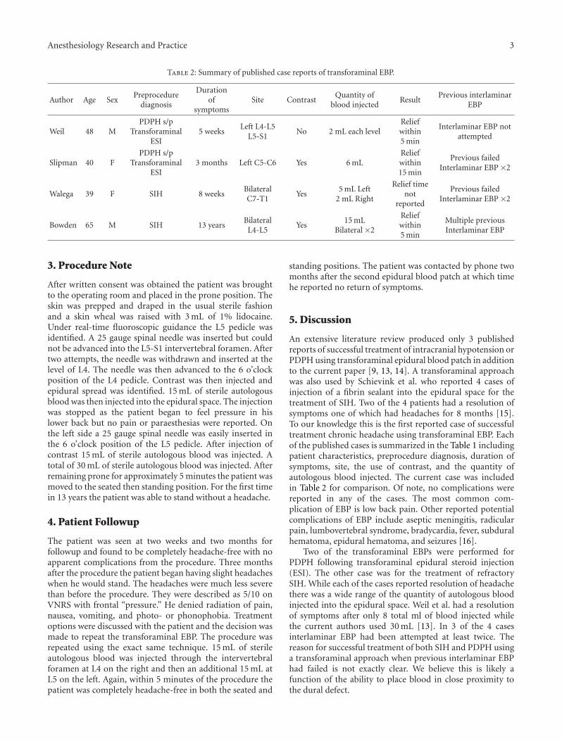
Anesthesiology Research and Practice 3
Table 2: Summary of published case reports of transforaminal EBP.
Author Age SexPreprocedure
diagnosis
Durationof
symptomsSite Contrast
Quantity ofblood injected
ResultPrevious interlaminar
EBP
Weil 48 MPDPH s/p
TransforaminalESI
5 weeksLeft L4-L5
L5-S1No 2 mL each level
Reliefwithin5 min
Interlaminar EBP notattempted
Slipman 40 FPDPH s/p
TransforaminalESI
3 months Left C5-C6 Yes 6 mLReliefwithin15 min
Previous failedInterlaminar EBP ×2
Walega 39 F SIH 8 weeksBilateralC7-T1
Yes5 mL Left
2 mL Right
Relief timenot
reported
Previous failedInterlaminar EBP ×2
Bowden 65 M SIH 13 yearsBilateral
L4-L5Yes
15 mLBilateral ×2
Reliefwithin5 min
Multiple previousInterlaminar EBP
3. Procedure Note
After written consent was obtained the patient was broughtto the operating room and placed in the prone position. Theskin was prepped and draped in the usual sterile fashionand a skin wheal was raised with 3 mL of 1% lidocaine.Under real-time fluoroscopic guidance the L5 pedicle wasidentified. A 25 gauge spinal needle was inserted but couldnot be advanced into the L5-S1 intervertebral foramen. Aftertwo attempts, the needle was withdrawn and inserted at thelevel of L4. The needle was then advanced to the 6 o’clockposition of the L4 pedicle. Contrast was then injected andepidural spread was identified. 15 mL of sterile autologousblood was then injected into the epidural space. The injectionwas stopped as the patient began to feel pressure in hislower back but no pain or paraesthesias were reported. Onthe left side a 25 gauge spinal needle was easily inserted inthe 6 o’clock position of the L5 pedicle. After injection ofcontrast 15 mL of sterile autologous blood was injected. Atotal of 30 mL of sterile autologous blood was injected. Afterremaining prone for approximately 5 minutes the patient wasmoved to the seated then standing position. For the first timein 13 years the patient was able to stand without a headache.
4. Patient Followup
The patient was seen at two weeks and two months forfollowup and found to be completely headache-free with noapparent complications from the procedure. Three monthsafter the procedure the patient began having slight headacheswhen he would stand. The headaches were much less severethan before the procedure. They were described as 5/10 onVNRS with frontal “pressure.” He denied radiation of pain,nausea, vomiting, and photo- or phonophobia. Treatmentoptions were discussed with the patient and the decision wasmade to repeat the transforaminal EBP. The procedure wasrepeated using the exact same technique. 15 mL of sterileautologous blood was injected through the intervertebralforamen at L4 on the right and then an additional 15 mL atL5 on the left. Again, within 5 minutes of the procedure thepatient was completely headache-free in both the seated and
standing positions. The patient was contacted by phone twomonths after the second epidural blood patch at which timehe reported no return of symptoms.
5. Discussion
An extensive literature review produced only 3 publishedreports of successful treatment of intracranial hypotension orPDPH using transforaminal epidural blood patch in additionto the current paper [9, 13, 14]. A transforaminal approachwas also used by Schievink et al. who reported 4 cases ofinjection of a fibrin sealant into the epidural space for thetreatment of SIH. Two of the 4 patients had a resolution ofsymptoms one of which had headaches for 8 months [15].To our knowledge this is the first reported case of successfultreatment chronic headache using transforaminal EBP. Eachof the published cases is summarized in the Table 1 includingpatient characteristics, preprocedure diagnosis, duration ofsymptoms, site, the use of contrast, and the quantity ofautologous blood injected. The current case was includedin Table 2 for comparison. Of note, no complications werereported in any of the cases. The most common com-plication of EBP is low back pain. Other reported potentialcomplications of EBP include aseptic meningitis, radicularpain, lumbovertebral syndrome, bradycardia, fever, subduralhematoma, epidural hematoma, and seizures [16].
Two of the transforaminal EBPs were performed forPDPH following transforaminal epidural steroid injection(ESI). The other case was for the treatment of refractorySIH. While each of the cases reported resolution of headachethere was a wide range of the quantity of autologous bloodinjected into the epidural space. Weil et al. had a resolutionof symptoms after only 8 total ml of blood injected whilethe current authors used 30 mL [13]. In 3 of the 4 casesinterlaminar EBP had been attempted at least twice. Thereason for successful treatment of both SIH and PDPH usinga transforaminal approach when previous interlaminar EBPhad failed is not exactly clear. We believe this is likely afunction of the ability to place blood in close proximity tothe dural defect.

4 Anesthesiology Research and Practice
A transforaminal approach for the EBP was chosen forthe current case to obtain a more direct approach to thedural leak. We felt a direct interlaminar approach at L4-L5 would be unsafe as the integrity of the ligamentumflavum was likely compromised during the laminectomy.The lack of an intact ligamentum flavum would increasethe possibility of inadvertent dural puncture and potentialworsening of symptoms. An interlaminar approach at a levelabove or below the defect would likely be ineffective as thishad previously been attempted. The two prior EBPs at L2-L3 and through a caudal approach, respectively, were likelyineffective because they failed to reach the site of CSF leak.The spread of epidural blood was likely limited because ofpostsurgical adhesions. Entering the intervertebral foramenallowed us to avoid possible adhesions and place blooddirectly at the site of the CSF leak.
Headaches related to intracranial hypotension eitherfrom dural puncture or SIH can be severe and very difficultto treat. EBP appears to be the treatment of choice whenconservative measures has failed. When EBP does notprovide relief patient may benefit from surgical interventionif the site of the CSF leak has been identified [17]. Inthe case presented the patient suffered from a chronicpostural headache for more than 13 years despite medicalmanagement and repeated interlaminar EBP. He was referredto clinic as he did not want to consider surgery. The use of arelatively novel approach to a treatment that has been usedfor 50 years eliminated the patient’s headache and restoredhis quality of life.
6. Conclusion
This case demonstrates that transforaminal epidural bloodpatch can be an effective in the treatment of chronicheadache secondary to intracranial hypotension when tra-ditional interlaminar technique is either impractical or hasbeen previously ineffective.
References
[1] A. Bier, “Experiments on the cocainization of the spinal cord,”Deutsche Zeitschrift fur Chirurgie, vol. 51, pp. 361–369, 1899.
[2] H. F. Wulf, “The centennial of spinal anesthesia,” Anesthesiol-ogy, vol. 89, no. 2, pp. 500–506, 1998.
[3] V. G. Schaltenbrand, “Neuere anschauungen zur pathophysi-ologie der liquorzirkulation,” Zentralblatt fur Neurochirurgie,vol. 3, pp. 290–299, 1938.
[4] D. K. Turnbull and D. B. Shepherd, “Post-dural punctureheadache: pathogenesis, prevention and treatment,” TheBritish Journal of Anaesthesia, vol. 91, no. 5, pp. 718–729, 2003.
[5] B. Mokri and J. B. Posner, “Spontaneous intracranial hypoten-sion: the broadening clinical and imaging spectrum of CSFleaks,” Neurology, vol. 55, no. 12, pp. 1771–1772, 2000.
[6] M. Paldino, A. Y. Mogilner, and M. S. Tenner, “Intracranialhypotension syndrome: a comprehensive review,” Neurosurgi-cal Focus, vol. 15, no. 6, pp. 1–8, 2003.
[7] A. Francia, P. Parisi, and A. M. Vitale, “Life-threateningintracranial hypotension after diagnostic lumbar puncture,”Neurological Sciences, vol. 22, pp. 385–389, 2001.
[8] F. T. Sayer, M. Bodelsson, and E. M. Larsson, “Spontaneousintracranial hypotension resulting in coma: case report,”Neurosurgery, vol. 59, no. 1, p. E204, 2006.
[9] D. Walega, E. McComb, and J. Rosenow, “Bilateral cervicotho-racic transforaminal blood patches for persistent headachefrom spontaneous intracranial hypotension: a case report andreview,” The Clinical Journal of Pain, vol. 27, no. 4, pp. 357–364, 2011.
[10] Headache Classification Subcommittee of the InternationalHeadache Society, “The International Classification ofHeadache Disorders: 2nd edition,” Cephalalgia, vol. 24, pp.79–80, 2004.
[11] J. B. Gormley, “Treatment of post-spinal headache,” Anesthesi-ology, vol. 21, pp. 565–566, 1960.
[12] Y. F. Wang, J. F. Lirng, J. L. Fuh, S. S. Hseu, and S. J. Wang,“Heavily T2-weighted MR myelography vs CT myelographyin spontaneous intracranial hypotension,” Neurology, vol. 73,no. 22, pp. 1892–1898, 2009.
[13] L. Weil, R. I. Gracer, and N. Frauwirth, “Transforaminalepidural blood patch,” Pain Physician, vol. 10, no. 4, pp. 579–582, 2007.
[14] C. W. Slipman, O. H. El Abd, A. Bhargava, M. J. DePalma,and K. R. Chin, “Transforaminal cervical blood patch for thetreatment of post-dural puncture headache,” The AmericanJournal of Physical Medicine and Rehabilitation, vol. 84, no. 1,pp. 76–80, 2005.
[15] W. I. Schievink, M. M. Maya, and F. M. Moser, “Treatmentof spontaneous intracranial hypotension with percutaneousplacement of a fibrin sealant: report of four cases,” Journal ofNeurosurgery, vol. 100, no. 6, pp. 1098–1100, 2004.
[16] J. H. Diaz, “Permanent paraparesis and cauda equina syn-drome after epidural blood patch for postdural punctureheadache,” Anesthesiology, vol. 96, no. 6, pp. 1515–1517, 2002.
[17] B. Mokri, “Expert commentary: role of surgery for themanagement of CSF leaks,” Cephalalgia, vol. 28, no. 12, pp.1357–1360, 2008.