pakistan journal of pharmacy - university of the...
TRANSCRIPT
1
ISSN: 1019-956XCoden: Pak. J. Pharm. 16-19 (1&2) 2003-2006
(Printed in 2010)
Pakistan Journal of Pharmacy
University College of PharmacyUniversity of the Punjab,
Allama Iqbal Campus, LAHORE-54000Tel: + 92 42 99211616
Email: [email protected]
PAKISTAN JOURNAL OF PHARMACY
CHIEF EDITOR:Dean, University College of Pharmacy, University of the Punjab, Lahore
EXECUTIVE EDITORS:Prof. Dr. Bashir Ahmad
Prof. Dr. Syed Saeed ul HassanProf. Dr. Mobasher Ahmed Butt
SENIOR EDITOR:Dr. Nadeem Irfan Bukhari
EDITORS:Dr. Tahir Javaid Khan, Dr. Khalid Hussain, Dr. Tariq Saeed
EDITORIAL BOARD
Dr. Ignacio Segarra TausDepartment ofPharmaceutics, San JorgeUniversity, Autovia A-23Zaragoza-Huesca Km 510, 50830Villanueva de Gallego (Zaragoza), Spain.
Prof. Dr. Karamt A Javed,RIFA International University, Islamabad, Pakistan
Prof. Dr. M. Ashraf,University of Veterinary and Animal Sciences, Lahore,Pakistan
Dr. Khalid Ahmad Sheikh,School of Pharmacy, University of London, London,The United Kingdom
Prof. Dr. Tayyab Ansari,Faculty of Pharmacy, B.Z University, Multan, Pakistan
Dr. Zaheer ud Din Babar,School of Pharmacy, University of Auckland,New Zealand
Prof. Dr. Nisar ur RehmanDepartment of Pharmacy, COMSET Institute,Abbott Abad
Dr. Nisar Hussain ShahFaculty of Pharmacy, B. Z. University, Multan, Pakistan
Dr. Rosa Pereira,Department of Health Sciences, Iniversidade Lusofona,Lisbon and CQUMED’UP, Porto, Portugal
INFORMATION FOR SUBSCRIPTIONPakistan Journal of Pharmacy is published twice a year, first issue in June and the second in December.Subscription rate in Pakistan: Rs 500 per issue (Annual Rs 1000), Foreign: $50 per issue (Annual $ 100)
(Printed in 2010)
II
CONTENTS
LIST OF ARTICLES PAGE
Pakistan Journal of Pharmacy (Author Guidelines) 1
Extraction and Characterization of Polar Terpenic Constituents from Artemisia maritimaIrshad Khokhar, Shahzad Mehmood Siddiqui, Syed Saeed ul Hassan and Tayyab Ansari
3
Antibacterial Activity of Stachys palustrisMuhammad Tahir Javed Khan, Alexander I. Gray, John Michael Midgley and Michael D. Cole
7
Effect of Essential Oils on the Percutaneous Absorption of Diclofenac DiethylamineSyed Nisar Hussain Shah, Muhammad Salaman, Mashhood Ahmad, Yasser Shahzad,Sajid Asghar and Asma Safdar
13
Study of Non-Compliance and Its Reasons in Outdoor Patients With Mental Illness of aPublic HospitalAmjad Hussain, Khalid Hussain, Nadeem Irfan Bukhari, Furqan Khurshid Hashmi,Asima Asgher, Tayyaba Rasool, Madeeha Shakeel, Sameera Anjum, Saira Rehman andMuhammad Tayyab
21
Studies of Phenylbutazone Toxicity in Avian SpeciesAsif Farooq Awan, Muhammad Ashraf, Taha Nazir, Owais Umer and Habib ur Rehman
25
Phytochemical and Pharmacological Studies of Euphorbia pilulifera L.Syed Saeed-ul-Hassan, Muhammad Khalil ur Rehman, Tayyab Ansari andMuhammad Usman Bhatti
29
The Pharmacy Act XI of 1967: Quackery and Irrational Use of DrugsKhalid Hussain, Bashir Ahmad, Furqan K. Hashmi, Amjad Hussain and Abida Latif
35
1
PAKISTAN JOURNAL OF PHARMACYAUTHOR GUIDELINES
SCOPE
Pakistan Journal of Pharmacy is an official biannualpeer-reviewed publication of the University College ofPharmacy, University of the Punjab, Lahore (Pakistan).Full length original articles (not exceeding 10 printedpages) or short papers (not exceeding 4 printed pages)and letter to Editor are considered for publication in theJournal. The Journal accepts manuscript in any of thefields of Pharmacy or experimental medicine. TheJournal also publishes reviews, usually by invitation andis available for publication of pharmaceuticalsymposium/conference proceedings. The Journal will notresponsible for the opinions of the authors expressed inand the authenticity and correctness of the publishedarticles.
INSTRUCTION FOR AUTHORS
Preparation of manuscripts
Manuscripts should be prepared using 12-point byemploying Microsoft Word. The manuscript should beorganized as follows:
Title page
Title is the first page of the manuscript, it should provide(a) title of the manuscript, (b) full name of all authors, (c)full addresses of the departments and institutions wherethe work was performed, (d) a running head with 70characters inclusive of spaces, and (e) name, address,telephone and fax numbers, and e-mail address of thecorresponding author.
Abstract
On the second page, repeat names of the authors and themanuscript’s title at the top of the page followed by theAbstract. The abstract should be structured, withoutheadings into “Introduction”, “Objective,” “Methods,”“Results” and “Conclusion.” Keywords not exceeding 5should be indicated.
Main body text
The main body of the manuscript should consist of (a)“Introduction”, (b) “Materials and Methods”, “Subjects,Materials and Methods” or “Patients, Materials andMethods”, whichever is applicable, (c) “Results”“Discussion”, (d) “Acknowledgments”, if applicable and(e) “References”.
Introduction: This section should contain the studyobjectives, rationale, problem statement and previousworks of other researchers.
Materials and Methods: This section should includedescription of the methods used in the study. If the studyuses the previously reported method, a brief descriptionwith reference is enough. The study involvingexperimental animals and human subjects/patients shouldconforms with the “Guide for the Care and Use ofLaboratory Animals” and “Declaration of Helsinki (NIHPublication No. 85-23)” Cardiovascular Research 1997;35: 2-4), respectfully.
Results: The data are presented in Results section. Useeither Tables or Figures for presentation of data. Thedata given in Tables or Figures should not be repeated in

2
the main body of the text. The Tables, labelled as Romannumbers and Figures, labelled as Arabic numbers shouldbear self-explanatory legends/captions and should beembedded nearest where they are referred in the text. SIunits should be used. The Figures should be of resolutionof 300 dpi or higher. The size of the text, letter or markin the Figures should be appropriate to withstandreduction.
Discussion: This section can be combined with Resultssection. It should contain explanation or the reasoning ofthe findings of the research as compared to the previousfindings, if any.
Acknowledgments: The help of the individuals otherthan the authors should be included in this Section. Anyresearch support from any source must be acknowledgedin the Acknowledgments section.
References: The reference should be verifiable andcorrect citation is the responsibility of the submittingauthor. Now onward, the references should be cited inthe text by last name for single author, two names fortwo authors with ‘and’ in between and name of authorwith et al for more than two authors followed by the yearin ascending chronological order. Bibliography style isas follows:
Papers
Amidon, G.L., Lennernas, H., Shah, V.P. and Crison, J.R. (1995). A theoretical basis for the biopharmaceuticdrug classification. The correlation of in vitro drugproduct dissolution and in vivo bioavailability.Pharmaceutical Research. 12(3), 413-420
Books
Aulton, M. E (2002). Pharmaceutics: The Science ofDsage Form Design (2nd ed.), Churchill Livingstone,Edinburgh. pp. 211-274.
Ashford, M. (2002). Introduction to biopharmaceutics.In: Aulton, M. E. (Ed.), Pharmaceutics: The Science ofDosage Form Design (2nd ed.), Churchill LivingstoneEdinburgh. pp. 211-274.
Thesis
Pereira, R. E. V (2008). pH sensitive nanoparticles ascarriers for oral delivery of a model peptidomimeticdrug. PhD thesis submitted, University TechnologyMARA, ShahAlam, Malaysia.
Webpage/Data bases
The information should be retrievable through thecitation. The author name, if any is to be stated followedby the web address and the retrieval date.
Manuscript submission
Manuscripts should be submitted electronically by thecorresponding author at [email protected] or onan appropriate storage media to the Office of PakistanJournal of Pharmacy (Address is given below). Acovering letter should accompany the manuscript.
Cover letter
The covering letter should indicate the category of themanuscript as original article, short communication orreview article. It must also include declaration that themanuscript is original, has not published or under reviewprocess elsewhere, that the co-author(s) has read themanuscript and approved its submission to the Journaland that, in case of publication, the authors are agree totransfer its copyright to the Pakistan Journal ofPharmacy. The Journal also requires disclosing of anyconflict, financial arrangements with commercialcompanies. The cover letter should be signed by allauthors and mailed or faxed to the Office of the PakistanJournal of Pharmacy. Attachment of the signed coverletter as scanned document with the submission is alsoacceptable.
For any question, the office of the Journal may becontacted at the following address.
Dr. Nadeem Irfan BukhariOffice of the Pakistan Journal of PharmacyUniversity College of Pharmacy,University of the Punjab,Shahrah-e-Quaid-e-Azam,Lahore 54000Pakistan
Tel: 042-99211616 (0092-42-99211616 from outsidePakistan)
Fax: 042-99211624 (0092-42-99211624 from outsidePakistan)
Email: [email protected]

Pak. J. Pharm.16-19 (1 & 2) 2003-2006 ISSN: 1019-956X
3
EXTRACTION AND CHARACTERIZATION OF POLAR TERPENIC CONSTITUENTSFROM Artemisia maritima
Irshad Khokhar1*, Shahzad Mehmood Siddiqui1, Syed Saeed ul Hassan2, Hashim Naveed2 and Tayyab Ansari3
1Institute of Chemistry, University of the Punjab, Lahore, Pakistan2University College of pharmacy, University of the Punjab, Lahore, Pakistan
3Faculty of Pharmacy, Bahauddin Zakariya University Multan, Pakistan
ABSTRACT
The chemica1 investigation based on finding the polar terpenic constituents in the Artemesia maritima has been carriedout. Plant material was extracted with n-hexane and ethyl acetate. The ethyl acetate extract showed presence of polarterpenes, which were isolated by column chromatography. Further isolation and purification was carried out by thinlayer chromatography and their tentative structures were explained on the basis of their physical properties.
Keywords: Terpenes, Artemisia maritima
INTRODUCTION
Terpenes are one of the most important classes of naturalproducts and widely distributed in nature. Most ofnaturally occurring terpenes are either hydrocarbonshaving formula (C5H8)n or their oxygenated derivatives.Isoprene (2-methyl-l, 3-butadiene) is the basic unit of allterpenoids. They are the important constituent ofmedicinally important essential oils.1
Artemisia maritima is a perenial, woody, branched, hairyherb. It is found in most parts of Khyber Pakhtoonkhwa,a province of Pakistan. Artemisia species are widely usedas medicinal plants in folk medicine, and have beenincorporated into the pharmacopoeias of severalEuropean and Asian countries. Ethnobotanical studies ofplant have also been carried out by various workers.2-7
Santonin is an important constituent of Artemisia. It ischiefly reported in Artemisia maritima while otherspecies show almost negligible content of it. It is ananthelmintic agent widely used previously.
A study on the extraction of santonin has been carriedout by S. M. Khafagy et a,l8 It also contains manybioactive sesquiterpenes and flavanoids. Extraction andanalysis of sesquiterpenes and flavanoids from the planthas been described by Bilia et al.9 Terpenoidal structuredetermination and analysis has been explained by variousworkers and biotransformation of terpenes has also beenstudied by Carla et al.10-13
Many pharmacologic activities have been reported forArtimisia plant. It is found to be antimicrobial,anticoccidial, antiplasmodial and anthelmintic.14-16
Its use in urinary tract infections and infections due toTrichomonas vaginalis has been studied. Fumiganttoxicity of its essential oil against common pests instored products has been studied by Negahban et al. Ithas also been used as a Forage Plant in northern areas ofPakistan.17-20
*Corresponding author’s address: Irshad Khokhar, Institute of Chemistry, University of the Punjab, Quaid-e-Azam Campus, Lahore, Pakistan
Khokhar et al.
4
MATERIALS AND METHOD
Artemisia maritima was collected from Mansehra,Hazara Division, Khyber Pakhtoonkhwa (Pakistan).
Melting points were recorded in glass capillary tubeon Gallenkamp.
The ultraviolet (UV) spectra were recorded inmethanol on U-2000 Spectrophotometer.
The infrared (IR) spectra were scanned on 270-30Hitachi Infrared spectrophotometer.
Thin layer chromatography was performed on glassplates (5 x 20cm) coated with slurry of silica gel G60 (0.25 mm thick); the plates were activated inoven at 150°C for one hour. While the columnchromatography was performed with silica gel 60(70-230 mesh, E. Merk). The solvents used aregiven in Table I.
Table I: The solvents and their brands
Solvent BrandMethanol E. Merk (DAB)Chloroform E. Merk (MW11938)n-hexane LAB-SCANEthyl acetate BDH Chemicals (Analar)
Spraying reagent used was ceric sulphate (0.1 g)suspended in 4 ml of water. One gram of trichloroacetic
acid was added, the solution was boiled and sulphuricarid (conc.) was added drop wise until the turbiditydisappeared.
Extraction and identification of terpenoids
The powdered plant (175g) was extracted with 700ml ofn-hexane and filtered after three days of maceration. The3.410 g residue obtained after filtration was extractedwith 700 ml of ethyl acetate. The solvent was evaporatedunder reduced pressure. Removal of the solvent fromdried filtrate gave 1500 mg of dark brown residue.Terpenoids were identified by thin layer chromatography(TLC) using methanol-chloroform (99:1) as mobilephase. The chromatographic plates were developed withceric sulphate-sulphuric acid reagent which gave positivetest for terpenoids.
Isolation of terpenoids
A column of kiesel Gel G type 60 was taken. Hundredgram kiesel Gel G 60 was prepared using n-hexane ethylacetate (99:1) and dark brown residue extracted from theplant was chromatographed on this column usingfollowing ratios of eluting solvent as shown in Table II.
As a result of this separation twelve 12 fraction wereobtained (Table II). Out of these 12 fractions, ninefractions (2, 3, 4, 5, 6, 7, 8, 9, 10) gave positive test forterpenoids while remaining gave no spots with cericsulphate–sulsulphuric acid reagent, So furtherinvestigation was done on these fractions.
Table II: Isolation of terpenoids fraction using different concentrations of elution solvents by column chromatography
Fraction n-hexane ethylacetate ratio Yield (mg) Consistency of color Result after spray with
ceric sulphate reagent
1 99:1 61 White No spot2 95:5 212 Dark Greenish brown Two spots3 90:10 107 Light Greenish brown One spot4 80:20 76 Dirty yellow One spot5 70:30 118 Greenish yellow One spot6 60:40 78 Greenish yellow One spot7 50:50 240 Faint green One spot8 40:60 63 Faint green One spot9 30:70 77 Greenish yellow One spot
10 20:80 81 Bright yellowish green One spot
11 10:90 68 Bright yellow No spot12 0:100 54 Colorless No spot

Pak. J. Pharm.16-19 (1 & 2) 2003-2006 ISSN: 1019-956X
5
Purification of terpenoids
Terpenoids from each of the fraction 5, 6, 7, 8, 9 and 10were purified by preparative TLC plates using thesolvent system chloroform - methanol (Table III).
Table III: Fractions obtained by increasingconcentrations of ethyl acetate
Fraction Yield (mg) Pure component (mg)
5 118 93
6 78 61
7 240 213
8 63 50
9 77 63
10 81 69
RESULTS AND DISCUSSION
The present study was undertaken to investigate the polarconstituents from Artemisia maritima. The fractionsobtained from column chromatography of the extractobtained from plant showed presence of terpenoids onlyin those fractions with increasing ethyl acetateconcentration, when analyzed by TLC using cericsulphate-sulphuric acid reagent. Other fractions were nonterpenoid.
The physical constants like melting point, lambda maxand infrared (IR) spectra of terpenoids were determinedfor each fraction. The findings of the above are given inTable-IV.
The UV results indicated that the faction 7 showed themaximum absorbance at 441.5 nm whereas Fraction 9showed minimum absorbance at 295.5 nm. The IR dataof fractions 5 to 10 also indicated that the presentcomponents were aromatic in nature along with thepresent of C-O, C-H, C=O, C=C bonds.
Table IV: Analysis of pooled fractions by UV/VS spectroscopy and IR spectroscopy
IR (cm-1)Fraction M.P (C°)
UV max (nm)
(Absorbance) C-O C-H C=O C=C Aromatic
5 169 426.5 1050 2900 1640 1640 1460
6 172 417.5 1048 2900 1820 1620 1460
7 167 441.5 1048 2900 1800 1620 1460
8 174 298 1048 2900 1720 1660 1440
9 176 295.5 1045 2925 1740 1640 1430
10 164 421 1050 2940 1640 1640 1480
REFERENCES1. Eberhard Breitmaier. (2006). Terpenes: flavors,
fragrances, pharmaca, pheromones Wiley-VCHVerlag GmbH. pp. 1-9
2. Ashraf, M., Hayat, M.Q., Jabeen, S., Shaheen, N.,Khan, M.A. and Yasmin, G. (2010). Artemisia L.species recognized by the local community ofnorthern areas of Pakistan as folk therapeutic plants.Journal of Medicinal Plants Research. 4(2), 112-119.
3. Kaul, M. K. and Bakshi S.K. (1984). Studies ongenus Artemisia L. in North-West Hamalaya withparticular reference to Kashmir. Folia Geobotanica.19 (3), 299-316.
4. Shinwari, M.I. and Khan, M.A.(2000). Folk use ofmedicinal herbs of Margalla Hills National Park,Islamabad. Journal of Ethnophar-macology. 69, 45-56.
Khokhar et al.
6
5. Ibrar, M., Hussain, F. and Sultan, A. (2007).Ethnobotanical studies of plant resources of RanyalHills, District Shangla, Pakistan. Pakistan Journal ofBotany. 39(2), 329-337.
6. Ashraf, M., Hayat, M.A., Jabeen, S., Shaheen, N.,Khan, M.A. and Yasmin, G. (2008).Ethnotaxonomical approach in the identification ofuseful medicinal flora of Tehsil Pindigheb (DistrictAttock) Pakistan. Ethnobot. Research Applications.6, 35-62.
7. Afshan, N.S., Khalid, N. and Javed, H. (2008).Further additions to the rust flora of Pakistan.Pakistan Journal of Botany. 40(3), 1285-1289.
8. Khafagy, S.M., Gharbo, S.A. and Sarg, T.M. (1971).Phytochemical investigation of Artemisia herba-alba. Planta Medica. 20(3), 90-96.
9. Bilia, A.R., Lazari, D., Messori, L. and Taglioli, V.(2006). Simultaneous analysis of artemisinin andflavonoids of several extracts of Artemisia annua L.obtained from a commercial sample and a selectedcultivation. Phytomedicine. 13, 487.
10. James, R., Hanson (2002). The development ofstrategies for terpenoid structure determination.ChemInform. 33(13).
11. Quan-Xiang Wu, Yan-Ping Shi, Zhong-Jian Jia(2006) Eudesmane sesquiterpenoids from theAsteraceae family. Natural Product Report 23(5),699-734
12. Avula, B., Wang, Y-H., Smillie, T.J., Mabusela, M.,Vincent, L., Weitz, F. and Khan, I.A. (2009).Comparison of LC–UV, LC–ELSD and LC–MSmethods for the determination of Sesquiterpenoidsin various species of Artemisia. Chromatographia.70(5-6).
13. Carla C.C.R., DeCarvalho and Manuela, M. andDaFonseca, R. (2005). Biotransformation ofterpenes Biotechnology Advances. 24(2), 134-142.
14. Arab, H.A., Mardjanmehr, S.H., Shahbazfar, A.,Rassouli, A., Abdollahi, M. and Nekouie, O. (2006).Determination of artemisinin in Artemisia sieberiand anticoccidial effects of the plant extract inbroiler chickens. Tropical Animal Health andProduction. 38, 497-503.
15. Ene, A.C., Atawodi, S.E., Ameh, D.A., Kwanashie,H. O. and Agomo, P.U. (2009). In vivoantiplasmodial effect of chloroform extracts ofArtemisia maciverae Linn and Artemisia maritimaLinn. African Journal of Biotechnology. 8(23),6612-6616.
16. Jabbara, A., Zaman, M.A., Iqbala, Z., Yaseen, M.,and Shamim, A. (2004). Anthelmintic activity ofArtemisia brevifolia in sheep. Journal ofEthnopharmacology. 930(2-3), 265-268.
17. Pais, P., Khurana, R. and George, J. (2002). Urinarytract infections: a retrospective survey of causativeorganisms and antibiotics prescribed in a tertiarycare setting. Indian Journal of Pharmacology. 34(4),278-280.
18. Bakht, M.A., Ziaei, H., Abdollah, F., khani, S.B.(2003). Effect of essential oils of Artemisia, Zatariaand Myrtus on Trichomonas vaginalis. Journal ofMedicinal Plants. 8, 35-40.
19. Negahbana, M., Moharramipoura, S. and Sefidkonb,F., (2007). Fumigant toxicity of essential oil formArtemisia sieberi Besser against three stored productinsects. Journal of Stored Product Research. 43,123-128.
20. Hussain, F. and Durrani, M.J. (2009). NutritionalEvaluation of some forage plants from HarboiRangeland, Kalat, Pakistan. Pakistan Journal ofBotany. (3), 1137-1154.

Pak. J. Pharm.16-19 (1 & 2) 2003-2006 ISSN: 1019-956X
7
ANTIBACTERIAL ACTIVITY OF Stachys palustris
Muhammad Tahir Javed Khan1, Alexander I. Gray1, John Michael Midgley 1 and Michael D. Cole2
1Department of Pharmaceutical Sciences, University of Strathclyde, Glasgow, GI 1XW, Scotland, UK2Forensic Science, Department of Applied Chemistry, University of Strathclyde, Glasgow, GI 1XW, Scotland, UK
ABSTRACT
In vitro antibacterial studies were carried out with the hexane, chloroform and ethanol extracts of leaves, stems and rootsof Stachys palustris. All of them, a part from leaves, stems, roots hexane extracts and ethanol extract of leaves, exhibitedantibacterial activity against both gram positive and gram negative bacteria. The chloroform extracts were more potentthan the ethanolic extract.
KEY WORDS: Stachys palustris, Antibacterial activity
INTRODUCTION
Various herbs of the genus of the Stachys(Labiatae/Lamiaceae) are used in the traditional medicineas astringent, tonic, stomachache, vulnerary antiseptic,sedative, antidiarrhoeal, purgative, emetic, cystitis, andalso used in asthma, neuralgia, swelling and infectedwounds.l Many other species of the genus Stachys arealso used as a general remedy for the treatment of spasm,cardiac debility (tachycardia), amenorrhoea andneurousis.2Uterotionic, antitumor, cytotoxic and antiviral activities3-6
have been reported in plant species of the genus Stachysacteside. Isolated compounds from Stachys sieboldiiexhibited antinephritic and antianoxia (Hyaluronidaseactivity) actions.7-8 Stachyrene obtained from Stachysrecta showed antiinflammatory, antitoxic andhypoazothemic responses.9 Xanthine oxidase andantioxidative effects have been reported by theisoacteside and tubloside isolated from the stems ofStachys deserticola.10 Previous chemical investigation ofStachys species proved presence of alkaloids,11 flavo-onoids,12-15 iridoids tannins, diterpenoids, triter-penoids,16-18 and other constituent i.e. citric acid, malicacid, oleic acid, bitter principles are also present alongwith the carbohydrates,19-21 from different parts. Although
the medicinal importance of Stachys palustris L.
has not been claimed by any worker. Therefore, thisstudy was carried out on extracts of the different parts ofStachys palustris for their antibacterial activity.
MATERIALS AND METHODS
Plant material
Stachys palustris L. (Labiatae /Lamiaceae) was collected byGray and MeGill in September, 1997 from the Ground ofRosspriory, University of Strathclyde, Glasgow. Scotland,U.K, and identified by Alan. The voucher specimen (Col5321) was deposited in the herbarium of Royal BotanicalGarden, Kew, Richmond, Surry, TW93AB, U.K.
Preparation of plant extracts
Leaves, stems and roots were separated, dried under shadeand successively extracted in soxhlet apparatus with hexane,chloroform and ethanol. Evaporation of solvents in vacuo(ROTAVAPOR-R BUCK SWITZERLAND) providedLeaves: hexane (yield 2.79%), chloroform (C, 2.79%) andethanol extract (E, 6.99%). Stems: hexane (0.51%),chloroform (C, 0.49%) and ethanol extract (E, 7.69%) roots:hexane (0.68%), chloroform (C, 0.79%) and ethanol (E,19.40%).
*Correspondence author’s address; Present address: Dr. Muhammad Tahir Javed Khan, Associate Professor,University College of Pharmacy, University of the Punjab, Lahore-54000, Pakistan
Khan et al.
8
Microorganisms
The above mentioned extracts were tested againsts apanel of bacteria including the Gram-positive bacteriaBacillus pumilus NCTC 10327, Bacillus subtilis NCTC10073, Staphylococcus aureus NCTC 10788, Straptococcusfaecalis NCTC 775, and Gram-negative bacteria, Serratiamarcescens NCTC 1377, Proteus vulgaris NCTC 4175,Escherichia coli NCTC 9001 and Pseudomonas aerugrinosaNCTC 6750. The organisms were obtained from themicrobiology lab., Department, of Pharmaceutical Sciences,Strathclyde University, Glasgow, Scotland, U.K., andmaintained as a slant on nutrient agar in McCartney bottlesand stored at 4C .A loopful of the bacteria from the stockculture was aseptically transferred to sterile nutrient brothmedium. The organisms were allowed to grow on rotaryshaker for 18h harvested and used for the experiments.
Bioassay
The antibacterial study was carried out according to thereported procedure22, by incorporating into molten
nutrient agar plates inocula containing 105 to 106 bacteriaper ml approximately, the plates were allowed to solidifyand 6 wells of 8mm diameter were made in each plate. 5.50and 100 mg/ml of S. palustris extracts, and standardantibacterial agents (ampicillin and streptomycin 1 mg/ml)were introduced into the wells. Gum acacia (4.5%) was thevehicle that served as a control. The plates were incubated at37C for 24h. The zones of inhibition were determined asmeans of six replicates.
RESULTS
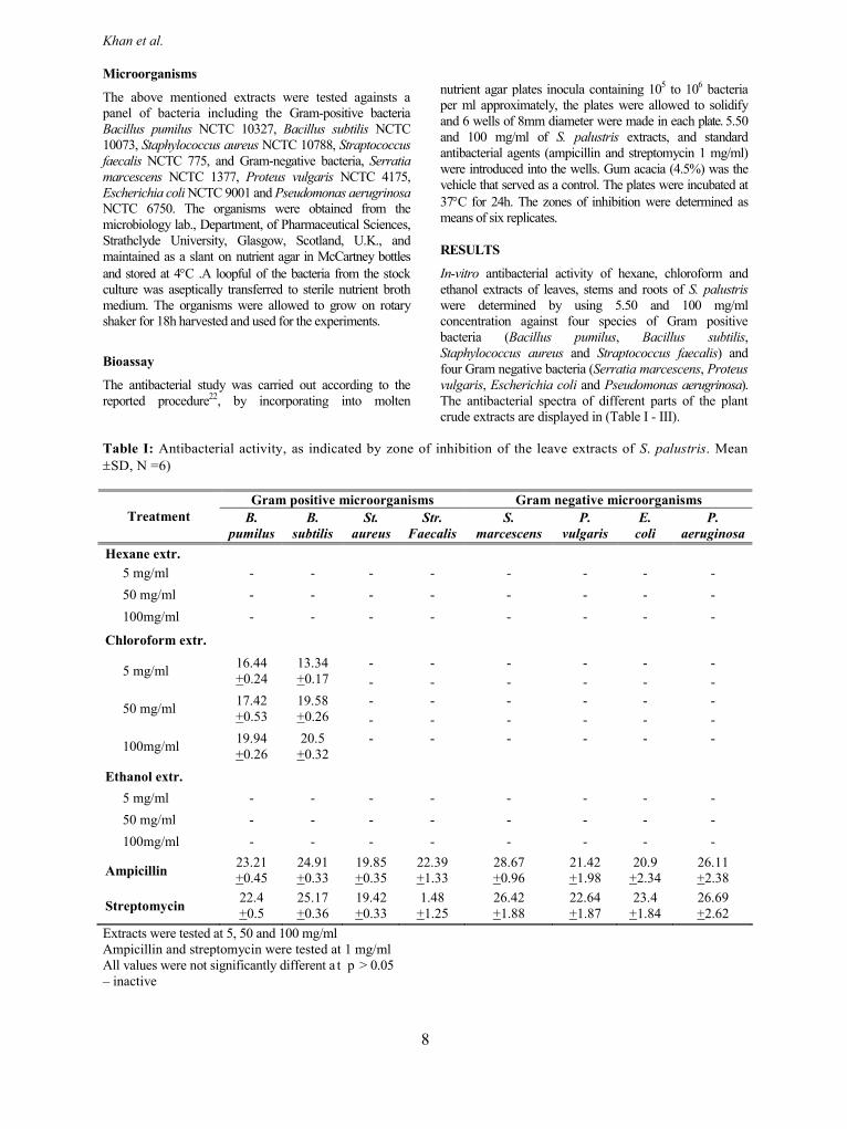
In-vitro antibacterial activity of hexane, chloroform andethanol extracts of leaves, stems and roots of S. palustriswere determined by using 5.50 and 100 mg/mlconcentration against four species of Gram positivebacteria (Bacillus pumilus, Bacillus subtilis,Staphylococcus aureus and Straptococcus faecalis) andfour Gram negative bacteria (Serratia marcescens, Proteusvulgaris, Escherichia coli and Pseudomonas aerugrinosa).The antibacterial spectra of different parts of the plantcrude extracts are displayed in (Table I - III).
Table I: Antibacterial activity, as indicated by zone of inhibition of the leave extracts of S. palustris. MeanSD, N =6)
Gram positive microorganisms Gram negative microorganismsTreatment B.
pumilusB.
subtilisSt.
aureusStr.
FaecalisS.
marcescensP.
vulgarisE.
coliP.
aeruginosaHexane extr.
5 mg/ml - - - - - - - -50 mg/ml - - - - - - - -100mg/ml - - - - - - - -
Chloroform extr.- - - - - -5 mg/ml 16.44
+0.2413.34+0.17 - - - - - -
- - - - - -50 mg/ml 17.42+0.53
19.58+0.26 - - - - - -
- - - - - -100mg/ml 19.94+0.26
20.5+0.32
Ethanol extr.5 mg/ml - - - - - - - -50 mg/ml - - - - - - - -100mg/ml - - - - - - - -
Ampicillin 23.21+0.45
24.91+0.33
19.85+0.35
22.39+1.33
28.67+0.96
21.42+1.98
20.9+2.34
26.11+2.38
Streptomycin 22.4+0.5
25.17+0.36
19.42+0.33
1.48+1.25
26.42+1.88
22.64+1.87
23.4+1.84
26.69+2.62
Extracts were tested at 5, 50 and 100 mg/mlAmpicillin and streptomycin were tested at 1 mg/mlAll values were not significantly different a t p > 0.05– inactive

Pak. J. Pharm.16-19 (1 & 2) 2003-2006 ISSN: 1019-956X
9
Table II: Antibacterial activity, as indicated by zone of inhibition of the stem extracts of S. palustris. (Mean SD, N =6)
Gram positive microorganisms Gram negative microorganisms
Treatment B.pumilus
B.subtilis
St.aureus
Str.Faecalis
S.marcescens
P.vulgaris
E.coli
P.aeruginosa
*Hexane extr.5 mg/ml - - - - - - - -50 mg/ml - - - - - - - -100mg/ml - - - - - - - -Chloroformextr.5 mg/ml - - - -22.13
+0.8820.44+0.83
11.04+0.31
10.75+0.13 - - - -
50 mg/ml - - - -23.18+1.09
22.55+0.71
10.50+0.23
11.03+0.05 - - - -
100mg/ml - - - -23.76+0.65
23.80+0.80
11.64+0.19
11.18+0.17
Ethanol extr.5 mg/ml - - - - - -10.75
+0.2611.56+0.17
50 mg/ml - - - - - - -9.58+0.13
100mg/ml - - - - - - - -Ampicillin 24.40
+0.5825.40+0.43
21.39+0.48
22.75+0.20
27.46+0.27
27.23+1.29
25.96+0.66
29.98+0.82
Streptomycin 25.79+0.80
24.33+0.20
19.68+0.31
19.18+0.62
26.57+0.40
25.46+0.68
25.04+0.44
28.20+0.43
Extracts were tested at 5, 50 and 100 mg/mlAmpicillin and streptomycin were tested at 1 mg/mlAll values were not significantly different a t p > 0.05- inactive
All the hexane extracts as well as ethanol extract ofleaves did not display any antibacterial activity againstany of the tested bacteria in this study. All chloroformextracts exhibited the most prominent antibacterial effectagainst two gram-positive microorganism (B. pumilusand B. subtilis) when compared with the referenceantibiotics such as ampicillin and streptomycin.
DISCUSSION
The ethanol extract of stems and roots displayed
antibacterial activity which may be due to polar componentslike alkaloids, glycosides, saponins, polyols, resins andamino acids. The antibacterial activity exhibited by thechloroform extract of leaves, stems and roots may be dueto slightly polar components which are present in theseparts of the plant. Slightly polar components seem to bemore potent in their antibacterial action than the polarcomponents.

Khan et al.
10
Table III: Antibacterial activity, as indicated by zone of inhibition of the root extracts of S. palustris. (Mean SD,N =6)
Gram positive microorganisms Gram negative microorganismsTreatment B.
pumilusB.
subtilisSt.
aureusStr.
FaecalisS.
marcescensP.
vulgaris E. coli P.aeruginosa
Hexane extr.5 mg/ml - - - - - - - -50 mg/ml - - - - - - - -100mg/ml - - - - - - - -Chloroformextr.5 mg/ml - - - - - - - -
- - - - -50 mg/ml - - - - -22.23
+0.8323.46+0.77 - -
11.23+0.21 - - -
100mg/ml - - - - -24.30+0.97
24.13+0.66 - -
13.10+0.05 - - -
Ethanol extr.5 mg/ml - - - - - - - -
- - - - - -50 mg/ml - - - - - -
- -10.50+0.14
11.23+0.26 - - - -
100mg/ml - - - - - -- -
11.53+0.17
10.95+0.04 - - - -
Ampicillin 24.38+0.37
25.15+0.30
20.91+0.31
22.95+0.17
27.48+0.35
25.02+0.99
23.26+0.67
27.85+0.60
Streptomycin 24.77+0.83
24.04+0.28
19.14+0.23
20.45+0.36
27.04+0.55
23.86+0.47
23.68+0.31
26.50+0.38
Extracts were tested at 5, 50 and 100 mg/mlAmpicillin and streptomycin were tested at 1 mg/mlAll values were not significantly different a t P > 0.05– inactive
REFERENCES
1. Stodolu, J., Volak, J. and Severa, F. (1986). In: Theillustrated book of herbs their Medicinal and culinaryuses. Buiiney, S. (Ed). Octopus Books, Ltd, London, p275.
2. Newall, C.A., Andirson, L.A. and PhiJIipson, J.D.(1996). Herbal Medicines A guide for Health-CareProfessionals. The Pharmaceutical Press, London. p197.
3. Yeung, W., Kong, Y.C., Lay, W.P. and Cheng, K.F.(1977). Cervical cancer and motherwort. PlantaMedica. 31, 51.
4. Kong, Y.C., Yeung, W., Cheung, Y.M., Hwang, J.C.,Chan, W.Y, Law, Y.P., Ng, K.H. and Yeung, C.H.(1976). Cytotoxicity of Chinese motherwort aqueousehtnaol extracts in non-apoptotic and estrogen receptordependent on human breast cancer cells. Journal ofChinese Medicine. 4, 373.
5. Vanxing, X. (1983). The constituents of the essential oilfrom Lavendulla stoechas growing with in Greece.Journal of Traditional Chinese Medicine. 3, 185.
Phytochemical and Pharmacological Studies of Euphorbia Pilulifera L
11
6. Lee, K., Lin, Y.M., Wu, S.T., Zhang, C.D, Yamagishi,T., Hayashi, T., Hail, H.T., Chaug. J.J. and Yang, H.T.(1988). The constituents of the essential oil fromLavendulla stoechas growing with in Greece. PlantaMedica. 54, 308.
7. Iuiyaslf, K., Nagamatsu, T., Ito. M., Agita, H. andSuzuki, Y. (1996). Release of preprotachykinin-AmRNA from rabbit iris upon c-fiber stimulation.Japanese Journal of Pharmacology. 70, 157.
8. Hayashi, K., Nagamatsu, T., Ito, M., Hattorl, T.and Suzuki. Y. (1994). Potent anti-inflammatoryactivities of hydroalcoholic extract from aerial partof Stacys inflata on rats. Japanese Journal ofPharmacology. 65, 143.
9. Zinchenko, T.V., Voitenko, G.N. and Lipkan,G.N. (1981). Anti-inflammatory, antitoxic, andhypoazotemic effect of a Stachys rectapreparation,stachyrene. Farmakol Toksikol. 44,191-194
10. Xioig, G.B., Kadota, S., Tani, T. and Namba, T.,(1996). Serotonin 5-HT2A receptors function as acontributory factor to both neuropsychiatric andcardiovascular diseases. Biological andPharmaceutical Bulleti. 19, 1580.
11. Gidubov, A. Z. and Biol, M.F. (1970). Change in ATPpool of paroited and submaxillary glands of rats afterstimulation with isoproterenol. 8, 129-131.
12. El-Ansari, M.A., Nawwar, M.A. and Saleh, N.A.M.,(1995). Stachysetin, a diapiggenin-7-glucoside-р, p׳ –dihydroxy-truxinate from Stacys algyptica.Phytochemistry. 40, 1543.-1548
13. Cl-Ansari, M.A., Barren, D., AbdalIa, M.F., Saleh,N.A.M. and Le Quere, J.L. (1991). Culturedhepatocytes bind and internalize bovine serumamineoxidase- gold complexes. Phytochemistry. 30,1169-1174.
14. Sclyultz, O.E. and AJhyane, M., (1973). In vitro aldosereductase inhibitory activity of substitutions N-benzenesulfonylglycin derivatives. ScinticiaPharmaceutica. 41, 149-152.
15. Kailnig T., Gruber, A., Menziiiger, S. and Prod, J.N.(1985). Flavanoid-o-glycosides from the herbs ofLeonurus cardiaca. 48, 494.
16. Calls, B.A.A., Saracogtu, I. and Stidier, O.(1992). 4-oxo-β-ionol and linolol glycosides fromraspberry fruit. Phytochemistry. 31, 4187-1490.
17. Buzogany, K. and Cucu, V., (1983) Myelofibrosis of thefacial bone Cpu journal of Medicine. 56, 385. 32-38
18. Yauiamoto, R., Miyase, T. and Ueno, (1994).Annonaceaeous acetogenins from the seeds ofannonasquamosa. adjacent bis tetra hydrofuranicacetogenins. A Chemical & Pharmaceutical Bulletin,42, 1163-1174.
19. Takeda, Y., Zhang, H.J., Masuda. T., Honda, G.,Otsuka, H., Sezik. E., Yesilada, E. and Sun (1997).Megastigmane glucosides from Stacy byzantina.H.D.Phytochemistry. 44, 1335-1337.
20. Biieskorn, C.H., Hofmann R. and Lett, T. (1979).Original reactions of α,α-dithio arylalkanes withbutyllithiums 27, 2509-2512.
21. Ikieskorn, C.R. and Broschek, W., (1972). Analysis ofbitter elements and furanoid derivatives fromLeonurus cardiaca L. Pharmaceutica ActaHelvetiae. 47, 123.
22. Flaavik, H.I., Johanssen, S., (1973).Transformation reveals a chromosomal locus ofthe gene(s) for methicillin resistance inStaphylococcus aureus. Journal of GeneralMicrobiology. 76, 45.

Pak. J. Pharm.16-19 (1 & 2) 2003-2006 ISSN: 1019-956X
13
EFFECT OF ESSENTIAL OILS ON THE PERCUTANEOUS ABSORPTION OFDICLOFENAC DIETHYLAMINE
Syed Nisar Hussain Shah1*, Muhammad Salaman1, Mashhood Ahmad1, Yasser Shahzad, Sajid Asgharand Asma Safdar1
1Faculty of Pharmacy, Bahauddin Zakariya University, Multan, Pakistan1*SNS Pharmaceutical Research Laboratory, Multan, Pakistan
ABSTRACT
The aim of present study was to evaluate the effect of various essential oils (eucalyptus, peppermint, turpentine and cod-liver oil) as enhancers on transdermal absorption of diclofenac diethylamine across full thickness, hairless rabbit skinusing modified Franz diffusion cell. The receptor compartment was constantly stirred normal saline solution at 37°C. Atset intervals up to 24hr, 5ml samples were removed from the receptor compartment and the amount of diclofenacdiethylamine permeated through the skin were calculated by the UV absorbance at 276 nm. In the interpretation ofresults the lag time played an important role. Peppermint oil showed the smallest lag time indicating its rapid enhancingeffect. The permeability coefficient calculated for diclofenac under the influence of enhancers showed that peppermintoil was a better enhancer as compared to others at the concentration under study. The enhancing effects were ranked as:peppermint oil > cod liver oil > turpentine oil > eucalyptus oil in this study. The ‘Benchmark’, flux rate of diclofenacunder the influence of enhancer showed that almost all the enhancers increased the penetration of DDA through hairlessrabbit skin. The rate constant showed fluctuations at various time intervals. With all enhancers decreased partitioncoefficients were observed but the diffusion coefficient values obtained were comparatively higher. The mode of actionof these accelerants may be described by combined process of partition and diffusion, the diffusion process beingdominant.
Keywords: Enhancers, Transdermal absorption, Franz diffusion cell, Partition coefficients
INTRODUCTION
During the past few years, skin has been shown to be asuitable delivery route for drugs formulated intransdermal therapeutic system.1 Transdermal drugdelivery involved the continuous administration oftherapeutic molecules through the skin. It has theadvantage of maintaining constant drug plasma levelsand improving patient compliance.2 The amount of drugbioavailable for targeting the sites of action is lower thanvia the oral route, but the absorbed dose appears to theadequate for therapeutic use, particularly because of theabsence of side effects.3
The skin surface consisted of a highly shiny lipid film ofvarious depths, depending on the location on the body.The stratum corneum, however, is first barrier and muchinterest has been shown in the percutaneous absorptionof chemicals and drugs that elicit a therapeutictoxicological response following skin contact.
Volatile or essential oils are volatile in steam and differentirely into both chemical and physical properties fromfixed oils. With the exception of oils such as oil of bitteralmonds, which are produced by hydrolysis of glycol-
*Corresponding author’s address: Syed Nisar Hussain Shah Faculty of Pharmacy, Bahauddin Zakariya University,Multan, Pakistan. Tel: +92619239322, Email: [email protected]

Shah et al.
14
sides, these oils are obtained largely as such from theplant. Volatile oils are used for their therapeutic action,for flavouring (e.g. oil of lemon), in perfumery (e.g. oilof rose) or as starting material for the synthesis of othercompounds (e.g. oil of turpentine). For therapeuticpurposes they are administered as inhalation (e.g.eucalyptus oil), orally (e.g. peppermint oil), as garglesand mouthwashes (e.g. thymol) and transdermally (manyessential oils including those of levander, rosemary andbergamot are employed in practice of aromatherapy.4
Diclofenac {(2-[2,6-dichlorophenyl) armino] phenyl-acetate} is a phenyl acetic acid non steroidal anti-inflammatory drug (NSAID) and is a potent inhibitor ofprostaglandin synthesis. For the treatment of rheumatoidarthritis, osteoarthritis, ankylosing spondylitis and acutegouty arthritis therapeutic doses of diclofenac have beenproven to be equi-efficacious when compared with othercommonly used NSAIDs.5-6 Diclofenac exhibited potentanalgesic effects and is used clinically for the short termalleviation of post-operative pain, dysmenorrhoea and invarious ocular conditions.7 The aim of present study wasto evaluate the effect of various essential oils(eucalyptus, peppermint, turpentine and cod-liver oil) asenhancers on transdermal absorption of diclofenacdiethylamine.
MATERIALS AND METHODS
Materials and apparatus
Diclofenac diethylamine was supplied by Novartis(China origin), turpentine oil, eucalyptus oil, cod-liveroil and peppermint oil were purchased from MD Traders.Double distilled water, pH 6.8 was used throughout theexperiment. Ethanol and sodium chloride were obtainedfrom Merck.
Software-assisted U. V. Spectrophotometer (Agilent2005) was used for determination of drug in sample(Agilent, Germany). Franz diffusion cell, fabricated byHEJ glass apparatus repairing workshop, Karachi(Pakistan) was used for the permeation experiments.
Animal Skin
In-vitro technique that was used to study transdermalabsorption involves the use of animal excised skin; inmany cases full thickness was used.8 Dorsal fullthickness skin of male rabbit (white, n=5, weighing 1-2kg) was used as a permeation membrane. The fat wasremoved with the aid of scissor.
Control Solution
Ten milligram of diclofenac diethylamine was dissolvedin 5 ml methanol in 100 ml volumetric flask and thevolume was made up to the mark with normal saline.
This was used as reference control solution without anyenhancers.
Test Solution
Test solutions were prepared by dissolving 10 mg ofdiclofenac diethylamine in 5 ml methanol in 100 mlvolumetric flasks and the solutions were made up to themark with previously constituted solutions of enhancer(5% v/v) in normal saline.
Diffusion Cell
Diffusion cell9 was fabricated locally after somemodifications. The cell was in the form of twocylindrical glass half cells termed as upper half cell(donor compartment) and the lower half cell (receptorcompartment) and inside diameter was 2 cm. The volumecapacity of the donor and receptor compartments was40ml and 35ml, respectively. The membrane wasmounted in between the two half cells and the exposedpenetration area was approximately 3.14cm2. From thelower half of the receptor compartment at a distance ofabout 3.8 cm a side arm 4 cm in length is used for takingthe sample and correcting the volume of receptorcompartment with the help of normal saline solution byexposing the epidermal side toward the donor half cell.The two half cells, after clamping were mounted on amagnetic stirrer and small magnetic fleas were placed inthe receptor compartment, and the receptor solution wasstirred at 60 rpm.
Membrane Preparation
The membrane, full thickness skin was taken from theabdominal surface of the hairless rabbit. The skin at thelower abdomen was marked and hairs were cut and thenrabbit skin was sacrificed and whole skin was removedand a rectangular section marked was excised from theanimal with surgical scissors. Since the skin was notfirmly attached to the viscera it was lifted easily from theanimal after the incision was made. Prior to the skinremoval, a uniform circle was made on the abdomen,marking the precise skin section to be positionedbetween the two half cells after the excised skin wastrimmed into an oversized rough circle it was mountedbetween the half cells with the marked section centered.The skin was placed in a normal saline solution beforemounting on to the diffusion cell.1, 10
Charging the cell
The receptor cell filled with normal saline was stirred bymagnetic stirrer at 60 rpm for 30 minutes, at which timethe compartments were evacuated with a syringe andrefilled with fresh normal saline. Then the compartmentswere evacuated a second time, refilled, evacuated a thirdtime and finally refilled with normal saline. The donorcompartment of the cell was charged with a test solution.

Pak. J. Pharm.16-19 (1 & 2) 2003-2006 ISSN: 1019-956X
15
Permeation
The donor compartment of the cell was charged with atest solution containing 1% of diclofenac diethylamineplus 5% v/v of each enhancer dissolved in 100ml ofnormal saline. The receptor cell contents were stirred andat predetermined time intervals, samples were taken andtransferred to the small bottles having stoppers, using 10ml syringe the time of charging the donor compartmentwas noted at the beginning of the diffusion runs and thereceptor samples were reference to this time.
Sampling
From the side arm of the receptor compartment, 5 ml ofthe sample was drawn at each time interval with the helpof 5 ml syringe and correcting the receptor half cellvolume with pre-thermostated normal saline. The sampletaken from the receptor cell, a portion of 3 ml was takenand was run on U.V. Spectrophotometer (Agilent2005;software version 2005) at λ 276 nm.
RESULTS AND DISCUSSIONThe permeation profile of the receptor phaseconcentrations in microgram per 100ml is summarized inTables I-II.
The lag time of the plots was calculated graphically byextrapolation from the pseudo steady state region of thegraph of the total amount penetrated versus time to theX-axis.
The Flux (J) of a drug is directly proportional to itsthermodynamic activity of the drug (Equation 1).
X
c
dd
DJ (g.cm–2.h–1) Equation 1
Where D is the diffusion coefficient and is a function ofthe size, shape and flexibility of the diffusing moleculeas well as the membrane resistance; C is theconcentration of the diffusing species; X is the spatialcoordinate.
Although the solution for J with various boundaryconditions and membrane heterogeneities can be verycomplex, the basic concepts regarding flux enhancementcan be found in Equation (5). The concentration gradientis thermodynamic in origin, and the diffusion coefficientis related to the size and shape of permeant and theenergy required to make a hole for diffusion.
Thus enhancement of flux across membranes reduces toconsideration of:
Thermodynamics (lattice energies and distributioncoefficients).
Molecular size and shape.
Reducing the energy required to make a molecularhole in the membrane
The agents to alter the barrier energy to form hole is asomewhat empirical process but the task is beingapproached from a basic study of kinetics of expandingthe structure of proteins and lipids.11. For example, theeffects of agents on the compressibility of monolayerfilm are studied through Langmuir troughs. Variousspectroscopic techniques are employed to investigatedetailed molecular interactions.
Flux which measures the mass of material transportedthrough the skin is more relevant parameter,therapeutically, than the permeability coefficient.12
The diffusion coefficients (D) of different concentrationsof enhancers are calculated by dividing the square of thethickness of the rabbit incised skin by 6 lag time(Equation 2).13
L6hD
2 (cm2.h–1) Equation 2
Where h is the thickness of the rabbit skin and L is thelag time.
Permeability coefficient (P) was calculated by dividingthe diffusion coefficient by square of the effectiveabsorption area of the skin in contact.14
2ADP (cm.h–1) Equation 3
A is the effective absorption area of the skin in contact.
As a measure of the penetration enhancing activity ofenhancers, the enhancement ratio (ER) was calculated asunder.15
b
a
PP
ER Equation 4
Where Pa is P after application of penetration enhancerand Pb is P before application of penetration enhancer.
The values indicate that the penetration may bedependent on the lipoidal solubility of the drug moiety.However, the permeation may be complicated by chargeeffect and also may depend on the skin partitioncoefficient of the drug between the aqueous phase andlipid phase of the barrier.13
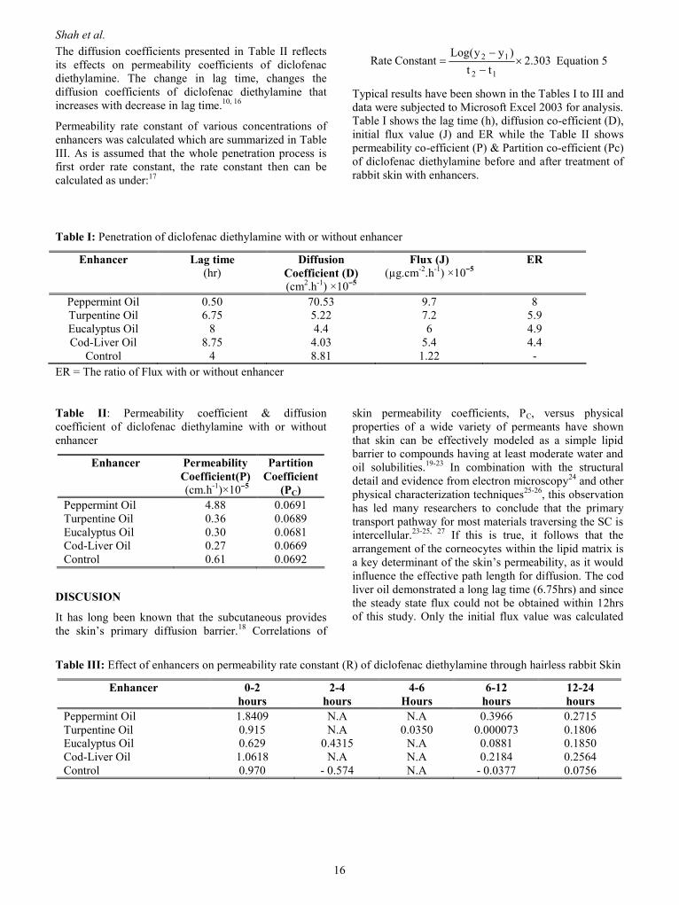
DPPC Equation 5
Shah et al.
16
The diffusion coefficients presented in Table II reflectsits effects on permeability coefficients of diclofenacdiethylamine. The change in lag time, changes thediffusion coefficients of diclofenac diethylamine thatincreases with decrease in lag time.10, 16
Permeability rate constant of various concentrations ofenhancers was calculated which are summarized in TableIII. As is assumed that the whole penetration process isfirst order rate constant, the rate constant then can becalculated as under:17
303.2tt
)yy(LogttanConsRate
12
12
Equation 5
Typical results have been shown in the Tables I to III anddata were subjected to Microsoft Excel 2003 for analysis.Table I shows the lag time (h), diffusion co-efficient (D),initial flux value (J) and ER while the Table II showspermeability co-efficient (P) & Partition co-efficient (Pc)of diclofenac diethylamine before and after treatment ofrabbit skin with enhancers.
Table I: Penetration of diclofenac diethylamine with or without enhancer
Enhancer Lag time(hr)
DiffusionCoefficient (D)(cm2.h-1) ×10ˉ5
Flux (J)(µg.cm-2.h-1) ×10ˉ5
ER
Peppermint Oil 0.50 70.53 9.7 8Turpentine Oil 6.75 5.22 7.2 5.9Eucalyptus Oil 8 4.4 6 4.9Cod-Liver Oil 8.75 4.03 5.4 4.4
Control 4 8.81 1.22 -ER = The ratio of Flux with or without enhancer
Table II: Permeability coefficient & diffusioncoefficient of diclofenac diethylamine with or withoutenhancer
Enhancer PermeabilityCoefficient(P) (cm.h-1)×10ˉ5
PartitionCoefficient
(PC)Peppermint Oil 4.88 0.0691Turpentine Oil 0.36 0.0689Eucalyptus Oil 0.30 0.0681Cod-Liver Oil 0.27 0.0669Control 0.61 0.0692
DISCUSION
It has long been known that the subcutaneous providesthe skin’s primary diffusion barrier.18 Correlations of
skin permeability coefficients, PC, versus physicalproperties of a wide variety of permeants have shownthat skin can be effectively modeled as a simple lipidbarrier to compounds having at least moderate water andoil solubilities.19-23 In combination with the structuraldetail and evidence from electron microscopy24 and otherphysical characterization techniques25-26, this observationhas led many researchers to conclude that the primarytransport pathway for most materials traversing the SC isintercellular.23-25, 27 If this is true, it follows that thearrangement of the corneocytes within the lipid matrix isa key determinant of the skin’s permeability, as it wouldinfluence the effective path length for diffusion. The codliver oil demonstrated a long lag time (6.75hrs) and sincethe steady state flux could not be obtained within 12hrsof this study. Only the initial flux value was calculated
Table III: Effect of enhancers on permeability rate constant (R) of diclofenac diethylamine through hairless rabbit Skin
Enhancer 0-2hours
2-4hours
4-6Hours
6-12hours
12-24hours
Peppermint Oil 1.8409 N.A N.A 0.3966 0.2715Turpentine Oil 0.915 N.A 0.0350 0.000073 0.1806Eucalyptus Oil 0.629 0.4315 N.A 0.0881 0.1850Cod-Liver Oil 1.0618 N.A N.A 0.2184 0.2564Control 0.970 - 0.574 N.A - 0.0377 0.0756

Pak. J. Pharm.16-19 (1 & 2) 2003-2006 ISSN: 1019-956X
17
The control value for "P" of diclofenac diethylamine inthe untreated skin at 37 ± 0.5°C was 0.61×10ˉ5 cm.hˉ¹with a lag time of 4 hour.
The oils very significantly increased drug permeationacross the skin. The most effective oil as enhancer for thedrug permeation across the skin was peppermint oil(ER=8 ) (P<0.01) followed by turpentine oil (ER =5.9),eucalyptus oil (ER =4.9) and cod-liver oil (ER =4.4)showed less activity as enhancers than both the aboveoils. The steady-state permeation was observed for only½ -1 hours increase of peppermint oil, turpentine oil andeucalyptus oil, while cod-liver oil showed negativesteady-state permeation. As the diclofenac diethylamineconcentration in the donor compartment of the cell wassignificantly higher than the receptor compartment;therefore, the decrease of permeability after the steadystate may contribute to the wash up effect of enhancer inthe diffusion cell.28
The treatment with cod-liver oil did not improve thepartitioning whereas peppermint oil, turpentine oil andeucalyptus oil enhanced the partitioning. From thediffusion co-efficient values, it can be seen (Table II)that use of oils as enhancer have decreased the resistanceto diffusion of drug.29, 34
Current drug permeation enhancers (oils) enhanced drugdelivery through biological membranes (such as skin ormucosa) by causing some physicochemical changes within the lipophilic membrane barrier and has been observedthat the aqueous exterior of membranes could be just aseffective barrier as the membrane itself.30
Cod liver oil is rich in unsaturated fatty acids and hasbeen reported that the extracted fatty acids from the cod-liver oil showed significant enhancing effect and thefatty acid profile of the extract was almost identical tothat of the oil . So it has been observed that penetrationenhancing effect of cod-liver oil may be associated withunsaturated fatty acid portion of the extract, interestingly,cod-liver oil itself did not enhance transdermal drugdelivery.30
Some essential oils and their terpenes constituents havebeen investigated as potential enhancers. Eucalyptus oilincreased the total flux of diclofenac diethylamine (6.0×10ˉ5 (µg.cm-2.h-1) permeating excised hairless rabbitskin. A series of terpenes and some essential oils havebeen investigated from their penetration enhancing effecttoward drugs. In the present study, eucalyptus oil wasfound to be active as compared to other oils. So oils canbe used as effective enhancers because their safety iswell documented.31-32 From the enhancing effect of thegraded fractions of eucalyptus oil it can be observed thatthe fractions with higher boiling points exert more
significant effect than the original oil. Eucalyptus oil ismainly composed of 1, 8-cineol, which has a boilingpoint higher than the constituents present. Therefore,fraction of eucalyptus oil obtained at 140°C undervacuum has caused about 83 fold increase in the drugflux, and this is in agreement with William et al. 2003,33
who found that 1, 8-cineol caused a 95 fold increase ofdrug permeability.
CONCLUSION
The partitioning ratio suggested that the enhancersincreased the partition of DDA into the skin. As the drugis less soluble in oil than water, the increased partition ofdrug into the skin can be contributed to the structuremodification of the stratum corneum lipid bilayer.Therefore, it could be concluded that the enhancedpermeation of drug may not be only increasing thepartition of drug into the stratum corneum, but also bymodifying intercellular lipids, disrupting their highlyordered structure and thus increasing the permeation ofDDA through the membrane. Furthermore, the latter wasmore important than the former in permeation becausethe increased amounts of drug in the skin may also be theretention of the drug by the skin.
This study also verifies the idea that essential oils mayoffer a large and useful selection of relatively safepenetration enhancers to aid topical drug delivery.
ACKNOWLEDGMENT
Authors are thankful to Research Committee forfinancial support and the Institutional Animal EthicsCommittee of Bahauddin Zakariya University forapproving all in vitro experimental procedures.
REFERENCES1. Cordero, J.A., Alarcon, L., Escribano, E., Obach, R.,
Domenech, J. (1997). A comparative study of thetransdermal penetration of a series of non-steroidalanti-inflammatory drugs. Journal of PharmaceuticalSciences. 86 (4), 503–508.
2. Brown, L. and Langer, R. (1988).Transdermaldelivery of drugs. Annual Review of Medicine.39,221-229.
3. Devi, K. and Paranjothy, K.L. (1999).Pharmacokinetic profile of a new diethyl ammoniumpatch. Drug Development and IndustrialPharmacy.25, 695–700.
4. Evans, W.C. (2002). Trease and EvansPharmacognosy (16th ed.), pp.253–288.
Shah et al.
18
5. Brogden, R.N., Heel, R.C., Pakes, G.E., Speight,T.M. and Avery, G.S. (1980). Diclofenac sodium: Areview of its pharmacological properties andtherapeutic use in rheumatic disease and pain ofvarying origin. Drugs. 20, 24-48.
6. Todd, P.A. and Sorkin, E.M. (1988). Diclofenacsodium: A reappraisal of its pharmacodynamic andpharmacokinetic properties and therapeutic efficacy.Drugs. 35, 244-285.
7. Goa, K.L. and Chrisp, P. (1992). Ocular diclofenac:A review of its pharmacology and clinical use incataract surgery, and potential in other inflammatoryocular conditions. Drugs and Aging. 2, 473–486.
8. Hadgraft, J., Whitefield, M. and Rosher, P.H.(2003). Skin penetration of topical formulations ofIbuprofen 5%: an in-vitro comparative study. SkinPharmacology and Applied Skin Physiology. 16,137-142.
9. Franz, T.J. (1975). Percutaneous absorption: On therelevance of in vitro data. Journal of InvestigativeDermatology. 64, 190-195.
10. Durrheim, H., Flynn, G.L., Higuchi, W.I. and Behl,C.R. (1980). Permeation of hairless mouse skin; I.Experimental methods and comparison with humanepidermal permeation by alkanols. Journal ofPharmaceutical Sciences. 69(7), 781-786.
11. Bitterly, F.R. (1965). The influence of detergentsand surfactants on epidermal permeability. BritishJournal of Dermatology. 77(12), 98-100.
12. Rautio, J., Nevalainen, T., Taipale, H., Vepsäläinen,J. and Gynther, J. (2000). Synthesis and in vitroevaluation of novel morpholinyl- andmethylpiperazinylacyloxyalkyl prodrugs of 2-(6-methoxy-2-naphthyl) propionic acid (Naproxen) fortopical drug delivery. Journal of MedicinalChemistry. 43, 1489−1494.
13. Shah, S.N.H., Rabbani, M. and Amir, M.F. (2006).In-vitro study of percutaneous absorption ofDiclofenac in the Cetrimide through hairless rabbitskin. Journal of Research Science B. Z. Univ.17(1), 45-50.
14. Tsai, J.C., Chaung, S.A., Hsu, M.Y. and Sheu, H.M.(1999). Distribution of salicylic acid in humanstratum corneum following topical application invivo: a comparison of six different formulations.International Journal of Pharmaceutics. 188, 145-153.
15. Abdullah, D., Ping, Q.N. and Liu, G.J. (1996).Enhancing effect of essential oils on thepenetration of 5-Florouracil through rat skin. ActaPharmaceutica Sinica. 31(3), 214-221.
16. Aguiar, A.J. and Weiner, M.A. (1969). Percutaneousabsorption of chloramphenicol solutions. Journal ofPharmaceutical Sciences. 58,: 210-215.
17. Badar, R. (1992). Effect of various enhancers ontransdermal absorption of triamcinolone acetonidethrough hairless mouse skin. M. Phil. Thesissubmitted, Gomal Univeristy, D.I. Khan, Pakistan.
18. Scheuplein, R.J. and Blank, I.H. (1971).Permeability of the skin. Physiological Review. 51,702-747.
19. Michaels AS, Chandrasekaran SK, Shaw JE.(1975). Drug permeation through human skin:theory and in vitro experimental measurement.American Institute of Chemical Engineers Journal.21, 985-996.
20. Ackermann, C., Flynn, G.L. and Smith, W.M.(1987). Ether-water partitioning and permeabilitythrough nude mouse skin in vitro. II. Hydrocortisone21-n-alkyl esters, alkanols and hydrophiliccompounds. International Journal of Pharmacology.36, 67-71.
21. Potts, R.O. and Guy, R.H. (1992). Predicting skinpermeability. Pharmaceutical Research. 9, 663-669.
22. Kasting, G.B., Smith, R.L. and Anderson, B.D.(1992). Prodrugs for dermal delivery: solubility,molecular size, and functional group effects. In:Sloan, K.B. (Ed) Prodrugs: Topical and OcularDrug Delivery. NY Marcel Dekker, New York.117-161.
23. Johnson, M.E., Blankschtein, D. and Langer, R.(1997). Evaluation of solute permeation through thestratum corneum: lateral bilayer diffusion as theprimary transport mechanism. Journal ofPharmaceutical Sciences. 86, 1162-1172.
24. Bodde, H.E., van den Brink, I., Koerten, H.K., andde Haan, F.H.N. (1991). Visualization of in vitropercutaneous penetration of mercuric chloridetransport through intercellular space versus cellularuptake through desmosomes. Journal of ControlledRelease. 15, 227-236.
25. Flynn, G.L (1985). Mechanism of percutaneousabsorption from physicochemical evidence. In:Bronaugh, R.L. and Maibach, H.I. (Eds),Percutaneous Absorption. NY Marcel Dekker, NewYork. 27-51.
26. Turner, N.G. and Nonato, L.B. (1997). Visualizationof stratum corneum and transdermal permeationpathways. In: Potts, R.O. and Guy, R.H. (Ed).Mechanisms of Transdermal Drug Delivery. NYMarcel Dekker, New York. 1-40.
Pak. J. Pharm.16-19 (1 & 2) 2003-2006 ISSN: 1019-956X
19
27. Potts, R.O. and Francoeur, M. (1991). The influenceof stratum corneum morphology on waterpermeability. Journal of Investigative Dermatology.96, 495- 499.
28. Fukushima, K., Ise, A., Morita, H., Hasegawa, R.,Ito, Y., Sugioka, N. and Takada, K. (2011). Two-Layered Dissolving Microneedles for PercutaneousDelivery of Peptide/Protein Drugs in Rats.Pharmaceutical Research. 28, 7–21.
29. Saify, Z.S., Ahsan, O. and Dayo, A. (2000). Cineoleas skin penetration enhancer. Pakistan Journal ofPharmaceutical Sciences. 13(1), 29-32.
30. Loftsson, T., Guomundsdottir, T.K., Frioriksdottir,H., Siguroardottir, A.M., Thorkelsson, J.,Guomundsson, G. and Hjaltason B. (1995). Fattyacids from cod-liver oil as skin penetrationenhancers. Pharmazie, 50, 188-190.
31. Charles, S.A. and Bozena, B.M. (2000).Percutaneous penetration enhancers: local versustransdermal activity. Pharmaceutical Science andTechnology Today. 3 (1), 36-41.
32. Edris, A.E. (2007). Pharmaceutical and TherapeuticPotentials of Essential Oils and Their IndividualVolatile Constituents: A review. PhytotherapyResearch (in press) Published online in WileyInterScience (www.interscience.wiley.com) DOI:10.1002/ptr.2072.
33. Williams, A.C. (2003). Transdermal and TopicalDrug Delivery. Pharmaceutical Press, London.
34. Barakat, N.S. (2010). Optimization of physicalcharacterization, skin permeation of naproxen fromglycofurol-based topical gel. Asian JournalPharmacy. 4, 154-62.

Pak. J. Pharm.16-19 (1 & 2) 2003-2006 ISSN: 1019-956X
21
STUDY OF NON-COMPLIANCE AND ITS REASONS IN OUTDOOR PATIENTS WITHMENTAL ILLNESS OF A PUBLIC HOSPITAL
Amjad Hussain1, Khalid Hussain1, Nadeem Irfan Bukhari1 , Furqan Khurshid Hashmi1, Asima Asgher1, Tayyaba Rasool1, Madeeha Shakeel1, Sameera Anjum1, Saira Rehman1, Muhammad Tayyab2.
1University College of Pharmacy, University of the Punjab, Lahore, Pakistan2Department of Pharmacy, the Mental Hospital and Punjab Institute of Mental Health, Lahore, Pakistan
ABSTRACT
Compliance with prescribed therapeutic regimen of outdoor patients was studied by interrogating patients aboutplanning they make to take the medicine. A questionnaire was prepared to evaluate patient’s understanding, behaviorand motivation to take medication in accordance with prescribed regimen. The results showed that about 13% ofpatients did not come to refill the prescription, because of distant healthcare facility. Non-compliance was found to be20% due to patient’s own choice, 26% due to dissatisfaction on the treatment, 24% because patient could not understandthe instructions of prescription and 17% because of multiple attendants, disturbance of working routine and culturalbeliefs.
Keywords: Non-compliance, Prescription, Patient understanding
INTRODUCTION
Compliance to medication regimens has been monitoredsince the time of Hippocrates.1 In the recent years, it hasbecome a focus of increasing concern in the treatment ofpsychiatric disorders. Non-compliance is a common,prevalent and important issue in the treatment ofpsychiatric illnesses.2 Compliance to treatment is thedegree to which a patient carries out the clinicalrecommendations of the physician or pharmacist or inother words non-compliance is the failure of the patientto follow the prescribed treatment regimen. Non-compliance is a significant problem in all patientpopulation ranging from pediatrics to the elderlypatients.3, 4 It applies nearly to all chronic disease statesand settings. Nowadays, non-compliance is considered tobe the major problem in the health services of bothdeveloped and developing countries.5
Non-compliance to drug therapy is very common inPakistan. The reasons for non-compliance reported werethe lack of awareness about the benefits of treatment,non availability of healthcare services, non affordabilityof medicine, side effects and unfriendly attitude ofphysicians.6. Poor infrastructure of society, lack ofproper knowledge of mental illness to patients andmultiple caregivers could also be considered as some ofbasic reasons for non-compliance.7 The other reportedreasons of non-compliance were the beliefs of patientsthat the medications were not working and the medicineshave physical side effects.8
MATERIALS AND METHODS
Outdoor patients of Punjab Institute of Mental HealthLahore (PIMH) were selected as subjects for the study.The patients were included in the study on the basis of
*Corresponding author’s address: Amjad Hussain, University College of Pharmacy, University of the Punjab,Lahore, Pakistan

Hussain et al.
22
their willingness to discuss their drug related problemswith pharmacist openly. This is a cross sectional study inwhich the behavior and motivation of patients towardsmedication therapy was assessed by interviewing thepatients. Trained pharmacist with validated questionnaireinterviewed the patients to note the compliant or non-compliant behavior of the patients. A total of 150patients with their prescription were selected randomlyover a period of fifteen days. According to thisquestionnaire, patient’s personal information includingname, age, weight, gender, marital status, education,attendant’s relation, address and socio-culturalbackground was recorded. Then patients were askedabout their compliance with medication according toformat of the prepared questionnaire.
Most of patients (about 64%) were willing to giveinformation. A total of 150 patients were asked about thecompliance with prescription medicines. Some of them(36%) did not respond properly and were excluded fromthe study, while others were happy to receive extraattention from healthcare professionals. Most of thepatients were illiterate (92%) and belonged to the ruralareas (67%) and not have good understanding about theirprescriptions.
A large number of patients were not complying with theprescribed medication therapy. The major reasons fornon compliance observed in patients are summarized inFigure 1.
Intentionalnoncompliance
20%
Poor understanding ofprescription23%
Cultural believes2%
Multiple attendants10%
Dissatisfaction ontreatment26%
Side effects5%
Lack of prescription refill8%
Non availability forinjection6%
Figure 1: Several reasons of non-compliance along with their respective percentages
The result showed that the commonest reason of noncompliance in psychiatric illness could be thehopelessness of patients that might lead to dissatisfactionon treatment (26%) and intentional non-compliance(20%). The reasons of hopelessness might be long termtherapy and the supernatural believes. Patients did notwant to continue the therapy for long time and wereeager to get positive results of therapy as early aspossible. As the psychiatric medications showed theirresults after a long time, hence, the patients become
unsatisfied. The lack of prescription refill could be due tosocio-cultural background and distant residences ofpatients from healthcare services (Average 120km) in8% of patients. The other reasons might include patient’sown choice to follow the schedule of doses because ofdisturbance of their routine work as the side effects ofmedication appeared (5%), multiple care givers (10%),fear of treatment (6%) and patient’s inability tounderstand the prescription (23%) as shown in Figure 1.Many of these reasons of non-compliance were not
Pak. J. Pharm.16-19 (1 & 2) 2003-2006 ISSN: 1019-956X
23
reported in any study previously. Our results conform thefindings of Mahmood et al.12
The social and cultural stigma related to psychiatricillnesses and their treatment and doctor- patientrelationship also play a significant role in patientcompliance. The patients do not follow the medicationregimen as per advice either due to aggressive behavioror forgetfulness to take medicine. Patients stopmedication without consulting the doctor when they findthemselves stable or skip the medicines due to sleep.Some patients also decrease the dose by their ownperception as they feel side effects with prescribed doses.The illiteracy of patients was another reason of noncompliance due to which they were unaware of theirdisease and mechanism of cure and time required fortreatment with such medication.
Multiple care givers including mother, father, sister,brother or some times other relatives involved in the careof psychiatric patients might pose another problem incompliance. It is better to have only one care giver. Incase of multiple care givers, patient and caregiversdepend on each other and dose may be skipped.Medications used to treat mental illnesses are known tohave an array of potentially unpleasant side effectsranging from restlessness and pacing to excessivesedation, tremor, dry mouth, constipation, impotence,weight gain, missed menstrual cycles and many others.This study showed that side effects of psychotropic drugswere also a reason for non-compliance.
In majority of the cases only attendants come to hospitalto get medicine for their psychiatric patients. This isbecause many people were coming from distant areasand traveling with such patients in public transport wasdifficult and also there was chance that patient might fledaway or lost. In this situation if the prescription had oneor two injectable (s) non-compliance results. Many ofsuch patients did not purchase injectables to beadministered at home.
CONCLUSION
Non-compliance is common, prevalent and importantissue in the treatment of psychiatric illnesses. Healthcareprofessionals especially pharmacists should take leadingrole to educate the patients and their caregivers about thecourse of disease and importance of complying withmedication therapy along with others measures in thetreatment of psychiatric illness.
ACKNOWLEDGMENT
The author is grateful to Miss Shazia Iqbal (Pharmacist),Dr. Tariq Aziz, Dr Tahir Pervaiz, Dr. Khalid Mughal andDr Abdul Jaleel (Physicians) of Punjab Institute ofMental Health Lahore for their help during datacollection.
REFERENCES1. Osterberg, L. and Blaschke, T. (2005). Adherence to
medication. New England Journal of Medicine. 353:487-497.
2. Cramer, J.A. and Rosenheck, R. (1998). Compliancewith medication regimens for mental and physicaldisorders. Psychiatric Services. 49: 196-201.
3. Matsui, D.M. (1997). Drug compliance in pediatrics.Clinical and research issues. Pediatric Clinics ofNorth America. 44: 1-14.
4. Spagnoli, A., Ostino, G., Borga, A.D., D'Ambrosio,R., Maggiorotti, P., Todisco, E. et al. (1989). Drugcompliance and unreported drugs in the elderly.Journal of America Geriatric Society. 37: 619-624.
5. Taj, F., Tanwir, M., Aly, Z., Khowajah, A.A., Tariq.A., Syed, F.K., Waqar, F. and Shahzada, K. (2008).Factors associated with Non-adherence amongPsychiatric patients at a Tertiary Care Hospital,Karachi, Pakistan: a questionnaire based cross-sectional study. Journal of Pakistan MedicalAssociation. 58(8), 432-436.
6. Taj, R. and Khan, S. A. (2005). Study of reasons ofnon-compliance to psychiatric treatment. Journal ofAyub Medical College Abbott Abad. 17(2), 26-28.
7. Roy, R., Jahan, M., Kumari, S. and Chakraborty,K.P. (2005). Reasons for drug non-compliance ofpsychiatric patients: A centre based study. Journalof the Indian Academy of Applied Psychology. 31(1-2), 24-28
8. Ruscher, S.M., de-Wit, R. and Mazmanian, D.(1997). Psychiatric patients’ attitudes aboutmedication and factors affecting non-compliance.Psychiatric Services. 48: 82-85.
9. Francis, S.A. (2001). People with mental healthproblems. In: Pharmacy Practice. Kelvin, M.G.,Taylor. Geoffery (Eds.) T.J. International Ltd.Padstow, Cornwall. pp 330-344.
10. Horne, R. (2001). Compliance, Adherence andconcordance In: Pharmacy Practice. Kelvin, M.G.,Taylor. Geoffery (Eds.) T.J. International Ltd.Padstow, Cornwall. pp 165-186
Hussain et al.
24
11. Bryson, S.M. and Lawson, D.H. (1982). Non-compliance In: Clinical Pharmacy and HospitalDrug Management Lawson, D.H. and Michael, R.,Richards, E. (Eds) London Chapman and Hall Ltd.pp. 133-155.
12. Mahmood. K.T. Khalid, N. and Makhdum, Z.(2010). Adherence to drug therapy in psychiatricpatients. Journal of Pharmaceutical Science andResearch. 2 (11), 700-703.

Pak. J. Pharm.16-19 (1 & 2) 2003-2006 ISSN: 1019-956X
25
STUDY OF PHENYLBUTAZONE TOXICITY IN AVIAN SPECIES
Asif Farooq Awan1,2, Taha Nazir2*Muhammad Ashraf3, Owais Umer3 and Habib ur Rehman3
1Services Hospital, Department of Health, Government of the Punjab, Jail Road, Lahore, Pakistan2 Department of Pharmacy, University of Sargodha, Sargodha, Pakistan;
3 Faculty of Biosciences, University of Veterinary and Animal Sciences, Lahore, Pakistan
ABSTRACT
A vulture crisis is an important environmental problem, which happened because of utilization of unsafe anti-inflammatory and analgesic drugs. The clinical profile of phenylbutazone is not very much different from otherNSAID’s. Despite of sharing pharmacological usefulness, phenylbutazone also shares unwanted effects which mayeventually lead to the serious therapeutic complications and ecological imbalance. Therefore; we have aimed this studyto evaluate the effects of toxic doses of phenylbutazone in broiler chickens. Two hundred and twenty five (225) healthybroiler chickens were reared up to 28 days and were divided into 5 groups each comprising 25 birds. On day 29 fourgroups were dosed 50mg/kg body weight twice a day intra-muscularly for 4 days. Food and water were provided adlibitum. A physical examination, toxicity and mortality rate were recorded daily. Blood samples were drawn todetermine the serum values of aspartame transaminase (AST), alanine transaminase (ALT), uric Acid, alkalinephosphatase (ALP), and creatinine. Postmortem was performed on day 41. In second experiment other 100 birds weredivided into 5 groups, each comprising 20 birds. One of the groups was injected I/M phenylbuazone 100 mg/kg twice aday. Postmortem was performed after medication on day 5. Based on the necropsy findings and biochemical analysis,phenylbuazone was not found to be safe in the avian species. Thus, it is suggested that the veterinary use ofphenylbuazone should be avoided.
Keywords: Phenylbutazone toxicity, Broiler birds, LFTs
INTRODUCTION
Phenylbutazone; (3, 5-Pyrazolidinedione, 4-butyl-1, 2-diphenyl-Butazolidin, C19H20N2O2) is a white to off-white, odorless, crystalline powder. Soluble in alcohol,water, acetone and ether. It has similar anti-inflammatoryeffects and different toxicity profile as compared to theother salicylates.1 Like aminopyrine, phenylbutazone cancause retention of sodium and chloride ion, edema,nausea, vomiting epigastric discomfort.2,skin rashes,peptic ulcer hemorrhage3 perforation, hypersensitivtyreaction, serum sickness, ulcerative stomatitis, hepatitis,nephritis, aplastic anemia, leukopenia, agranulocytosisand thrombocytopenia4 A number of deaths have also
been reported, especially from aplastic anemia andagranulocytosis. Keeping the above in view, we aimedthis study to investigate the toxicity and evaluate thesafety of phenylbutazone in avian species to avoidhazards in wild life.
MATERIALS AND METHODS
The experiment was conducted at experimental sheds ofthe Department of Pharmacology and Toxicology,University of Veterinary and Animal Sciences, Lahore.One hundred and fifty (150) day old broiler chickscollected from the “Pakistan Hatchery, Lahore” were
*Corresponding author’s current address: Dr. Taha Nazir, Associate Dean, School of Pharmacy, The University ofLahore – 24 Jinnah Avenue, Blue Area, Islamabad, Pakistan. Fax: +92 51 282 9238, Email:. [email protected]

Awan et al.
26
vaccinated according to the vaccination schedule givenin Table I. The phenylbutazone (Orient Labs. Pvt.Limited) was injected twice a day.
Table I: Vaccination schedule
Age Vaccine Route
6th day Newcastle disease Eye15th day Gumboro (I.B.D) Oral*21st day Newcastle disease Oral*25th day Gumboro (I.B.D) Oral *
*With drinking water
MATERIALS AND METHODS
Experimental Design
On day 28, 75 birds were randomly divded into 2 groupscomprising; group A with 50 and group B with 25 birds.On day 29, phenylbutazone I/M 50mg/kg body weightwas injected twice a day up to four days indivdually toeach bird of group A, for four consecutive days. Group Bwas kept as control. The remaining 75 birds were dividedinto two groups C with 50 and D with 25 birds.
Each bird of group C was injected phenylbuazone I/M100 mg/kg body weight twice a day for four days andgroup D was kept as controlled without medication. Foodand water was provided ad libitum. A daily basis recordof physical examination, sign and symptoms and toxicitywas maintained regularly.
Sample Schedule and Parameters Determined
The sampling schedule and different parameters wereinvestigated by Asif et al5, 11. Three ml blood samplefrom birds of each group (A, B, C and D) was collected
before the start of medication on day 29. Then bloodsample from the same birds were drawn from wing vein(vena cutanea ulnaris) on days 33, 37 and 41 aftermedication for determination of serum values offollowing parameters; aspartate transaminase, alaninetransaminase,6 uric acid, alkaline phosphatase (ALP)7
concentration of creatinine in serum8.
Clinical Findings and Statistical Analysis
The clinical findings mortality and postmortem wererecorded during the study. The collected data wereanalyzed statistically with one way analysis of variance.9
On days 41 and 47 the postmortem were done. Threeparameters; postmortem, liver, kidney biopsies andstaining of specimens were examined.
RESULTS
The biochemical parameters including uric acid,creatinine, alanine transaminase, aspartate transaminaseand alkaline phosphatase were noted for test andcontrolled birds before and after phenylbutazone dose.The necropsy findings of experimental chicks were alsorecorded, as given below.
Biochemical Parameters of Phenylbutazone
Phenylbutazone I/M 50mg/kg and 100 mg/kg bodyweight were injected to each bird of group A and Brespectively; twice a day for four days and followingparameters were measured.
Uric Acid: As shown in Table II, the mean values of uricacid of phenylbutazone were 5.960040 mg/dl, 5.130200mg/dl, 5.532480 mg/dl and 5.234160 mg/dl beforemedication, 1st, 5th and 9th days after medication,respectively. There was no significant difference in themean values of uric acid of phenylbutazone Table III.
Table II: Biochemical parameters of phenylbutazone group. Mean±SEM, N=5
Time ofsample
collection
Uric Acidmg/dl
Creatininemg/dl
ALTµg/L
ASTµg/L
ALPµg/L
Beforemedication
5.960040 ±.331098
1.334880±0.1094 10.979760 ±0.4342
184.13982 ±6.05195
29.252640±2.70735
1st day aftermedication
5.130200 ±.218903
1.283580 ±0.1301 15.639820 ±1.3141
299.63830 ±4.95355
59.784000±2.07406
5th day aftermedication
5.532480 ±.292441
1.269640 ±0.1134 15.633520 ±1.9852
242.31240 ±7.90280
65.208000±2.05354
9th day aftermedication
5.234160 ±.363122
1.151620±0.1050 11.045560 ±0.5830
187.74400±14.48778
59.564000±4.11303

Pak. J. Pharm.16-19 (1 & 2) 2003-2006 ISSN: 1019-956X
27
Table III. Statistical analysis of phenylbutazone data
Parameters Sum of Squares Df Mean Square F Sig.Uric acid Between groups
Within groupsTotal
2.0757.4989.573
31619
0.6920.469
1.476 0.259
Creatinine Between groupsWithin groups
Total
9.001E-021.0561.146
31619
3.000E-026.599E-02
.455 0.718
ALT Between groupsWithin groups
Total
106.918123.928230.846
31619
35.6397.745
4.601 0.017
AST Between groupsWithin groups
Total
44401.5526670.280
51071.832
31619
14800.517416.892
35.502 0.000
ALP Between groupsWithin groups
Total
4006.317655.312
4661.629
31619
1335.43940.957
32.606 0.000
Biochemical Parameters in Control Group
No medication was given to group B and D.
Uric Acid: As shown in Table II, the mean values of uricacid of normal or control bird were 5.031800 mg/dl,4.776720 mg/dl, 5.479140 mg/dl and 4.874880 mg/dl at1st, 5th and 9th day, respectively. There was no significantdifference in the mean values of uric acid of controlbirds.
Creatinine: The mean values of creatinine in controlbirds were 1.052080 mg/dl, 0.972660 mg/dl, 1.134440mg/dl and 1.066040 mg/dl at 1st, 5th and 9th day,respectively. There was no significant difference in themean values of creatinine in control birds.
Alanine Transaminase (ALT): The mean values ofalanine transaminase of control birds were found to be10.149740 µ/L, 10.205000 µ/L, 10.269660 µ/L and10.351780 µ/L at 1st, 5th and 9th day, respectively. Therewas no significant difference in the mean values of ALTin controlled birds.
Aspartate Transaminase (AST): As shown in table IV,the mean values of aspartate transaminase of control
birds were found to be 193.555 µ/L, 199.435 µ/L,158.98660 µ/L and 166.81000 µ/L. There was nosignificant difference in the mean values of aspartatetransaminase of control birds.
Alkaline Phosphatase (ALP): The mean values ofalkaline phosphatase of piroxicam were 27.252000 µ/L,34.470000 µ/L, 33.548000 µ/L and 27.824400 µ/L.There was no significant difference in the mean values ofalkaline phosphatase of control birds.
Necropsy of Experimental Chicks in DifferentGroups
The birds were slaughtered at the end of experiment anddifferent lesions in kidney, liver and muscles wererecorded. Each bird of group C was injected with I/Mphenylbutazone 100 mg/kg body weight, twice a day forfour days. Each bird of group C was injected with I/Mphenylbutazone 100 mg/kg body weight, twice a day forfour days. Findings are given in Table IV.
Control Group: Group B and D acted as control(without medication).
Table IV: Necropsy of various drugs in broilers chicks, N=15Postmortem lesions
Drug Dose Site of injection Liver Kidney50 mg/kg 10/15 7/15 0/15Phenylbutazone
100 mg/kg 15/15 12/15 0/15No medication 0/15 0/15 0/15
Control No medication 0/15 0/15 0/15
Awan et al.
28
DISCUSSION
Phenylbutazone is a widely used non-steroidal anti-inflammatory drug with little anti-pyretic and analgesiceffects. The adverse effects associated withPhenylbutazone include; peptic ulcer hypersensitivityreaction ulcerative stomatitis, hepatitis nephritis aplasticanemia and agranulocytosis. The current study showedthat there was no toxic effect on kidneys in broiler chicksas indicated by the necropsy finding and biochemicalanalysis of serum uric acid and creatinine. GIT toxicity,edema of small intestine, erosions, and ulcers of largecolon and development of renal crest necrosis wereobserved.4 The results of the present study differed fromthe above observations which might be due to differentspecies used for experiment. The findings of presentstudy are partially in agreement with the observations ofKari and co-workers,3 who investigated the toxic effectsof phenylbutazone on the kidneys and liver in mice andreported different lesions as hemorrhage, centrilobularcytomegaly, fatty metamorphosis, cellular degeneration,coagulative necrosis and clear cell foci in liver. Theyalso observed lesions in kidney which were not found inthe present study which might be due to different animalspecies used for experiments.
Hepatotoxicity was observed in phenyl butazone treatedgroup, while there no nephrotoxicity was found. Musclenecrosis was observed in almost all of the birds at the siteof injection. There was no significant difference in serumuric acid and creatinine levels which indicated thatphenylbutazone had no toxic effects on kidneys, butthere was significant difference in the values of serumALT, AST and ALP in phenylbutazone treated group asstated by Embert, 198610
The levels of ALT, AST and ALP might be elevated dueto cellular degeneration or destruction in liver musclesand acute hepato cellular necrosis or bilary obstruction.Similar observations were recorded in the present study.It could be concluded that phenyl butazone is hepatotoxic in avan species even at the dosage rate of 50 mg/kgbody weight.
The postmortem of the control group revealed noabnormalities particularly in liver, kidneys and muscles.Similarly there was no significant difference in the serumvalues of uric acid and creatinine ALT, AST and ALP inthe samples collected at the different times duringexperiments in the group.
CONCLUSION
The phenylbutazone was studied for its toxicity in broilerchicks. No mortality was recorded in all groups. Basedon the necropsy and biochemical studies, phenylbutazone
was not found to be safe in avian species. In context withvulture’s crises, phenylbutazone should be avoided inveterinary practice.
REFERENCES
1. Chatterjee, S., Das, S. N. and Agrawala, S. K.(1996). Antiarthritic effect of ART-400 in rats.Indian Journal of Indigenous Medicine. 18(1), 83-85
2. Valk, N., Doherty T. J., Blackford, J. T., Abraha T.W. and Frazier D.L. (1998). Phenylbutazoneprevents the endotoxin-induced delay in gastricemptying in horses. Canadian Journal of VeterinaryResearch. 62(4), 320.
3. Kari, F., Bucher, J., Haseman, J., Eustis S. and Huff,J. (1995). Long-term exposure to the anti-inflammatory agent phenylbutazone induces kidneytumors in rats and liver tumors in mice. JapaneseJournal of Cancer Research. 86(3), 252-263.
4. MacAllister, C. G., Morgan, S. J., Borne A. T. andPollet R. A. (1993). Comparison of adverse effectsof phenylbutazone, flunixin meglumine, andketoprofen in horses. Journal of the AmericanVeterinary Medical Association 202(1), 71-77.
5. Awan A.F., Nazir, T., Ashraf, M, Umer, Owais andRehman, H.R. (2011). Studies of ketoprofentoxicity in avian species. Journal of Basic andApplied Sciences 7(2), 1-6.
6. Thomas, L. (1998). Alanine aminotransferase(ALT), Aspartate aminotransferase (AST) In:Thomas L., Editor. Clinical LaboratoriesDiagnostics 1st edn. Frankfurt: TH-BooksVerlagsgesellschaft, 55-65 and 208-214.
7. Bessey, O. A., Lowry, O. H. and Brock, M. J.(1946). Method for the determination of alkalinephosphatase. Journal of Biological Chemistry. 164-321.
8. Tietz, N. W. (1986). Kinetic method ofdeproteinization Text book of Clinical Chemistry,W. B. Saunders: 1271.
9. Steel, R.G.D. and Torries, J.H. (1982). Principal andProcedures of Statistics (2nd ed.). McGraw HillInternational Book Company, Tokyo, Japan.
10. Embert, H.C. (1986). Veterinary Clinical Pathology.4th edn, Nueva Editorial Interamericana, C-6450Mexico.
11. Awan, A.F., Ashraf, M., Umer, O., Nazir, T.,Akhtar, M.S., Habib-ur-Rehman. (2009). Studies ofdipyrone (metamizole sodium) toxicity in avianspecies. Journal of Applied Pharmacy. 1(1), 1-6.

Pak. J. Pharm. 16-19 (1 & 2) 2003-2006 ISSN: 1019-956X
29
A REVIEW OF Euphorbia pilulifera L.
Syed Saeed-ul-Hassan1*, Muhammad Khalil ur Rehman1, Tayyab Ansari2 and Muhammad Usman Bhatti1
1University College of Pharmacy, University of the Punjab, Lahore, Pakistan2Faculty of Pharmacy, B. Z. University, Multan, Pakistan
ABSTRACT
Plant extracts, that have common utilization in the traditional medicine as curative agents, are being used in modernmedicine. Euphorbiaceae is a big plant family and contain many plants that are medicinal very important and havecommon utilization for the treatment of different disease conditions in the conventional medicine systems. Euphorbiapilulifera L is one of the plants of this family and have constituents that are valuable curative agents. In addition, thelatex from its leaf and other parts of the plant is irritant to skin. It is used in many female disorders; it has property toincrease the milk flow in woman and is also used in respiratory problems. In subcontinent it is used to treat worms inchildren. It is employed in a variety of treatments because of its different constituents. A review of this plant is beingpresented in this article.
Keywords: Euphorbia, Asthma, Irritancy
INTRODUCTION
Role of plants in the treatment of disease is exemplifiedby their employment in all the major systems ofmedicine irrespective of the underlying philosophicalpremise. For example, we have western medicine withorigins in Mesopotamia and Egypt, the unani (Islamic)and Ayurvedic (Hindu) systems centered in western Asiaand Indian subcontinent and those of the orient (China,Japan, Tibet, etc.)1. How and when such medicinal plantswere first used is, in many cases, lost in pre-history. Theplant kingdom thus presents an enormous reservoir ofpharmacologically valuable materials still to bediscovered2. Nearly 100,000 secondary plant productshave so far been isolated and characterized.3
Medicinal plants used in the traditional medicines shouldtherefore be screened for their safety and efficacy, in thelight of modern scientific investigations.4-7 In recentyears, there has been growing trends to evaluate the
chemical constituents of the medicinal plants, used in thetraditional medicine on different biological andpharmacological parameters, which lead to a systematictherapeutic utilization.
During a general screening of medicinal plants fromlocal flora, it was observed that most of the medicinalplants belong to famous family Euphorbiaceae. Severalplants of this family are of high economic value.8-9 Mostof the species are poisonous, causing sicknesses or deathif ingested. Dermatitis was also caused by many species,if juice of the plant contacts the skin. Even the rain waterdripping from certain plants is enough to causedermatitis.8-9 Euphorbia pilulifera L. is used in differentfemale disorders and also in the treatment of respiratorydiseases like asthma and bronchitis. The chloroformextract of Euphorbia pilulifera L. is irritant to skin.10
*Corresponding author’s address: Syed Saeed-ul-Hassan, University College of Pharmacy, University of the Punjab,Lahore, Pakistan
Review article
Hussan et al.
30
DESCRIPTION
Euphorbia pilulifera L. or Euphorbia hirta L.
Family: Euphorbiaceae
Euphorbia pilulifera
Vernacular Names and classification:
Urdu DudhiUnani Dhudi Kalan, sheer jiyahSanskrit RaktavinduchadaEnglish Australian asthma weed, snake, weed,
cat’s hair, milk weedBengal Barakeru, BrokerueeHind DudhiMah Nayeti, Dudhali, GoverdhanTelugu Bidarie, NanbalaTami Amumpatchaiyariss
Classification of Euphorbia pilulifera
Kingdom Plantae – PlantsSubkingdom Tracheobionta– Vascular plants
Superdivision Spermatophyta – Seed plantsDivision Magnoliophyta–Flowering plantsClass Magnoliopsida – Dicotyledons
Subclass RosidaeOrder EuphorbialesFamily Euphorbiaceae – Spurge familyGenus EuphorbiaSpecies Euphorbia pilulifera L
PLANT FAMILY EUPHORBIACEAEcs
Euphorbiaceae is a large family of angiosperms. Itincludes 300 genera and around 7500 species.11 Most ofthe members of this family are herbs, sometime shrubsand trees. Some species of genera Euphorbia arexerophytic.12 Family Euphorbiaceae is widelydistributed throughout both hemispheres and ranges inmorphological form, from largest desert succulent herbsto trees.13
GENERAL BOTANICAL DESCRIPTION
Euphorbia is one of the most diverse genera in the plantkingdom. Plants are often annual or perennial herbs,woody shrubs or trees with a caustic, poisonous milkysap (latex). The roots are fine or thick and fleshy ortuberous. Many species are more or less succulent,thorny or unarmed. The main stem and side branches ofthe succulent species are thick and fleshy, 15-91 cm (6-
36 inches) tall. The deciduous leaves are opposite,alternate or in whorls. In succulent species the leaves aremostly small and short-lived. The stipules are mostlysmall, partly transformed into spines or glands. Like allmembers of family Euphorbiaceae, it has unisexualflowers.14
Distribution
Euphorbia pilulifera L. is common weed foundthroughout India on the waste grounds and in loamysoils.15-16 It occur mostly in summer and rainy seasonsbut found occasionally in winter season along road sideand waste grounds. It is also found in Pakistan and manytropical countries but it is exported mainly from India.15-
18
Morphology
The plant is an annual, erect or ascending, 14-50cm high,covered with long yellowish hairs; stem usually terete,branched often four angled; leaves opposite, 2-3.5 X 1-1.5 cm. obliquely oblong or lanceolate or obovate-lanceolate, acute or sub-acute, serrate, dark green above,pale beneath. Base of the leaves usually unequal, acute orrounded, main veins 3-4, distinct; petioles short about 2-3mm. long; stipules pectinate, caduceus; flowers minute,numerous in globular, axillary, short peduncled, clusters;capsules hairy; seeds ovaoid; trigonous, light reddishbrown.15-16, 19-20 Capsules 1/20 inches across hairy; coccicompressed, keeled; seeds pale brown, ovoid, acutelyangled, faintly transversely wrinkled. Flowers and fruitformed through out the year.15 The flowers are veryminute and crowded in dense axillary or terminal cymes;about 1cm in diameter. The fruit is minute, yellow, threecelled capsule of about 1mm, each of the three carpelsbeing distinctly keeled; containing a single seed.17 Thewhole plant produces milky latex which is often irritatingto the mucous membranes and skin.21
ETHNOPHARMACOLOGY
According to the doctrine of signatures, Euphorbiapilulifera L., has reputation for increasing milk flow inwomen, because of its milky latex and is used for otherfemale complaints as well as in diseases of respiratorytract.22 The plant has been used for female disorders butis now more important in treating respiratory ailments,especially cough, coryza, bronchitis and asthma. In Indiait is used to treat worm infections in children and fordysentery, gonorrhoea, jaundice, pimples, digestiveproblems and tumors. The fresh milky latex is applied towounds and warts. Roots of the plant are used in sprainsand inflammation, miscarriage, epilepsy, maggots inwounds and irregular growth of teeth.23
Pak. J. Pharm. 16-19 (1 & 2) 2003-2006 ISSN: 1019-956X
31
Chemical constituents
Euphorbia pilulifera L. contained 0.4 % of a lycosidalsubstance, tannin, fatty acids, phorbic acid, sterols,eciphosterol, Jambulol melissic acid and sugars, 0.1 %alkaloids.24 Important constituents of the aerial partswere terpenoids, including triterpenes: α-amyrin, β-amyrin, friedlin, taxaxerol and esters of it: taraxerone,11α-oxidotaraxerol, 12α-oxidotaraxerol, cycloartenol,24-methylene-cycloartenol and euphorbolhexacosoate.16,25 The aerial parts and roots also containditerpenoid esters of the phorbol type and ingenol type,including 12-deoxyphorbol-13-dodecanoate-20-acetate,12-deoxyphorbol-13-phenylacetate-20-acetate-ingenoltriacetate, as well as highly toxic tinyatoxin, aresiniferonol derivative.25 Other terpenoids isolated aresterols including β-sistosterol, campesterol, cholesteroland stigmasterol.19, 25
Tannins isolated include the dimeric hydrolysabledehydroellagitannins, euphorbin A, B, C, E andterchebin, the monomeric hydrolyable tannins geraniin,2, 4, 6 tri-O-galloyl-β-D-glucose abd 1, 2, 3, 4, 6 penta-O-galloyl-β-D-glucose and the ester 5-O-caffeoylquinicacid (neochlorogenic acid) and 3,4-di-O-galloylquinicacid, and benzyl gallate.26-27 Acids isolated includeellagic acid, gallic acid, tannic acid, maleic acid andtartaric acid.26-27
Flavonoids isolated include quercetin, quercitrin,quercitrol and derivatives containing rhamnose,quercetin rhamoside, a chlorophenolic acid, rutin,leucocyanidin, leucocyanidol, myricitrin, cyaniding 3, 5-diglucoside, pelargonium 3, 5-diglucoside and camphol.The flavonol glycoside xanthorhmnin was alsoisolated.19, 28-29
The latex contained inositol, taraxerol, friedelin, β-sitosterol, ellagic acid, kaempferol, quercitol andquercitrin.19, 28-29 The mineral contents of dried leavessample were: Ca 1.1 % and P 0.3%, Fe 0.03%, Mg 0.5%,Mn 0.01%, Zn 0.01% and Cu 0.002%. Fresh leaves fromEuphorbia pilulifera of Nigerian origin were found tocontain high levels of Mn (189ppm), Cu (30.5 ppm), Zn(152ppm) and NO3 (4600ppm). Varying proportions ofFe, Mg, K, Ca, and Na were found.30
Pharmacological actions and medicinal uses
Polyphenolic extract of the whole plant inhibited thegrowth of Entamoeba histolitica with a minimal activeconcentration of less than 10µg /ml31 and with 80µg /mlexhibited more than 70% inhibition of acetylcholine and/ or KCl solution induced contractions on isolated guineapig ileum.32 It exhibited anti diarrheal activity against
castor oil and prostaglandin E2 induced diarrhea inmice.33 The solvent extract of Euphorbia piluliferashowed selective cytotoxicity against several cancer celllines. The plant was useful in effective treatment ofcancer, particularly malignant melanomas and squamouscell carcinomas.34
Methanolic extract of the plant is non-cytotoxic and haveantibacterial properties. The plant also hadimmunomodulatory activity. It affects lectin-inducedlymphoblast transformation in vitro.35
Ethanolic extract of the plant exhibited antifungalactivity when tested against the plant fungal pathogenssuch as Colletotrichum capsici, Fusarium palcidoroseum,Botryodiplodia theobromae, Alternaria alternate,penicillium citrinum; Phomopsis caricae-ppauae andAspergillus niger.36 An aqueous extract of Euphorbiapilulifera L. significantly inhibits aflotoxin production onrice, wheat, maize and ground nut.37 E. pilulifera L. at adose of 50mg/kg body weight reduced the sperm motilityand density of cauda epididymal and testis spermsuspension significantly, leading eventually to 100%infertility.38 This drug is also reported to have arelaxation effect on the bronchial tubes and a depressantaction on respiration. It was shown that plant given tofemale guinea pigs before puberty, increased thedevelopment of mammary glands and induced secretion.Drug processed galactogenic activity.39 Euphorbiaextract has also been found to have depressant action onthe cardiovascular system in general; musculature ofheart is slightly depressed, a sedative effect on themucous membrane of the respiratory tract and genitor-crinary tract; and produces a relaxation of thebronchioles by central action. The liquid extract ofEuphorbia pilulifera L. was irritant to the mucousmembrane of the stomach. In animals Euphorbia extractsproduced broncho-dilatation.40 Different fractionsisolated through column chromatography, from thechloroform extract of the Euphorbia pilulifera L. wereirritant to rabbit’s skin.11
CONCLUSION
Euphorbia pilulifera Linne is a common herb. It growsamong the grasses and in moist soil. It is very usefultherapeutically. Valuable constituents of this herb can bescreened and investigated for their potential aspharmacological candidates. Such herbs can be a sourceof revenue generation only once their chemical,structural and pharmacological studies have beenestablished.
Hussan et al.
32
REFERENCES1. Evans, W.C. (2002). Trease and Evan’s
Pharmacognosy, (15th Edn.) W. B. Suanders,Edinburgh, London, p. 3
2. Hostettmann, K., Potterat, O. and Wolfender, J.L.(1998). The potential of higher plants as a source ofnew drugs. Chimia. 52: 10-17.
3. Buckingham, J. (1994). Dictionary of NaturalProducts. Vol. I-II Chapman and Hall, London.
4. Rehman, S., Hasnat, A., Hasan, C.M., Rashid, M.Aand Ilias, M. (2001). Pharmacological evaluation ofBangladeshi medicinal [lants - A Review.Pharmacology and Biology. 37: 202-207.
5. Adebanjo, A.O., Adewumi, C.O. and Essein, E.E.(1983). International Symposium on MedicinalPlants. University of Ife, Nigeria, pp.152-158.
6. Farnsworth, N.R. (1994). Ethnopharmacology andDrug Development In Ethnobotany and the Searchfor New Drugs, Prince, G.T. (Ed.). Ciba FoundationSymposium 185, pp.42-59.
7. Farnsworth, N.O. (1984). The role of medicinalplants in drug development. In: Natural Products andDrug Development, Krogsgaac Larsen, P.,Christensen, S.B. and Kofod, H. (Eds.). Balliere,Tinda and Cox, London., pp. 8-98.
8. Michall, Hickey, M. and Clieve, K. (1997).Common Families of Flowering plants. CambrigeUniversity Press, p. 97.
9. Sharma, O. P. (1993). Plant Taxonomy (17th Edn.)Tata McGraw Hill publishing company limited,New Dehli pp.377-383
10. Muhammad Khalil ur Rehman. (2009).Dermatological studies of Euphorbia pilulifera L.M. Phil thesis, submitted to College of Pharmacy,University of the Punjab, Lahore (Pakistan)
11. Charles, C.D., Maribeth, L., Daniel, L.N., Kenneth,J.W. and David, A.B. (2007). Floral gigantism inRafflesiaceae Science Express Publishers.c.f.http://www.wikipedia.org.
12. Evans W.C., (1996). Trease and EvansPharmacognosy. (14th Edn.) Harcourt Brace abdcompany Asia PTE Ltd. p. 41.
13. Cateni, F., Falsone, G. and Zilic, J. (2003)Terpenoids and Glycolipids from Euphorbiaceae.Mini. Review. Med. Chem. 3(5), 425 – 437. c.f.Chem. Abst. 139, 288913 j, 2003.
14. Kashyap, S.R. and Joshi, A.C. (1936). Compositae.In: Lahore District Flora, The University of thePunjab, Lahore. pp. 139-140,149.
15. NIIR Board of consultants and engineers (2005).Hand book on Unani Medicines with formulae,processing and analysis. Asia Pacific Bussinesspress Inc. p71.
16. Sivarajan, V.V., Bachanardran, I. (2002). AyurvedicDrugs and their plant sources. Oxford & IBHpublishing co. pvt. Ltd. Kolkata, New Delhi. Pp.141-143
17. Henry, G., Greenish, F.I.C, F.L.S. (1999). MateriaMediica (3rd Edn.) Scientific Publishers (India),p210.
18. BHMA. (1983). British Herbal Pharmacopoeia,BHMA, Bournemouth. c.f. www.wikepedia.org.
19. Hickey, M. and King. C. (1997). Common Familiesof Flowering Plants. Cambridge University Press.p.97.
20. Behl, P.N., Srivastava, G. (2002). Herbs useful indermatological theray. (2nd Edn.) CBS Publishers &Distribution, New Delhi, pp. 73-74.
21. Williamson, E.M. and Curchill Livingstone (2003).Major Herbs of Ayurveda. Daber Researchfoundation and Daer Ayurvet Limited. pp 141-144.
22. Jha, M.K. (1992). The folk veterinary system ofBihar. A research survey. NDDB, Anand, Gujrat c.f.Williamson, E.M., Curchill Livingstone (2003).Major Herbs of Ayurveda. Daber Researchfoundation and Daer Ayurvet Limited. pp 141-144
23. Tona L, Kambu K, Ngimbi, N. (2000). Antiamoebicand spasmolytic activities of extracts from someantidiarrhoel traditional preparations used inKinshasa, Congo. Phytomedicine. 7(1), 31.
24. Kirtikar, K.R. and Basu, B.D. (1999). IndianMedicinal Plants. (2nd Edn.) Vol. 1, Internationalbook distributors India, p. 838.
25. Yoshida, T., Namba, O., Yokoyama, K., Okuda, T.and Chen, L. (1989). Hydrolyzable tannin oligomersfrom Euphorbiaceae. Tennen Yuki KagobutsuToronkai Koen Yoshishu, pp31-601.
26. Yoshida T, Chen L, ShinguT and Okuda T (1988).Tannins and related polyphenols of Euphorbiaceaeplants. IV. Euphorbins A and B, novel dimericdehydroellagitannins from Euphorbia hirta L.Chemical and Pharmaceutical Bulletin. 36(8), 2940.
27. Blanc, P. and DeSaqui-Sannes, G. (1972).Flavonoids of Euphorbia hirta (Euphorbiaceae).Plantes Medicinales Phytotherapie. 6(2), 106.
28. Khan, M.A. (1999). Euphorbianin, a new flavonolglycoside from Euphorbia hirta Linn. GlobalJournal of pure and Applied Science. 5(3) p38.
29. Nguyen, N.T. and Sosef, M.S.M. (1999). EuphorbiaL. In: de Padua,L.S., Bunyapraphastsara, N. andLemmens, R.H.M.J. ( Eds.) Plant Resources ofSouth East Asia No. 12(1). Medicinal and poisonousplants 1. Backhuys Publishers, Leiden, Netherlands.pp. 263 – 272.
Pak. J. Pharm. 16-19 (1 & 2) 2003-2006 ISSN: 1019-956X
33
30. Chopra, N., Chopra. I.C. (1958). Chopra’sIndigenous Drugs of India. (2nd Ed.) U.N. Dhur &sons private Ltd. p 507
31. Gnecco, S., Perez, C., Bittner, M., Becerra, J., andSilva Y.M. (1996). Distribution pattern of n-alkanesin Chilean species from the Euphorbiaceae family.Bolletino Sociedad Chilena de Quimica. 41(3), 29
32. Galvez, J., Zarzuelo, A., Crespo, M.E., Lorente MD,Ocete MA. and Jimenez J. (1993). Antidiarrhealactivity of Euphorbia hirta extract and isolation of anactive flavonoid constituent. Planta Medica. 59(4),333
33. Aylward, JH, (1999). Peplin pvt. Ltd. Patent. Appl.Wo 9908994 c.f. Williamson, E.M., CurchillLivingstone (2003). Major Herbs of Ayurveda. DaberResearch foundation and Daer Ayurvet Limited. pp141-144
34. Vijaya K, Ananthan S. and Nalini R. (1995).Antibacterial effects of theaflavin, polyphenon 60(camellia sinensis) and Ephorbia hirta on shiegellaspp. – a cell culture study. Journal ofEthnopharmacology 49 (2) p115
35. Szenasi T.E., (1992). Euphorbia hirta extracts asimmunostimulant. German patent DE 4102054 c.f.
Williamson, E.M., CurchillLiving stone (2003).Major Herbs of Ayurveda. Daber Researchfoundation and Daer Ayurvet Limited. pp 141-144
36. Singh, P., and Sinha K.K., (1986). Inhibition ofaflotoxin production on some agriculturalcommodities through aqueous plant extracts.Journal of Indian botanical society. 65(1), 30
37. Mathur A. Dixit V.P. and Dobal MP, (1995). Antifertility plant product: Euphorbia hirta in males.Proceedings of the International symposium on malecontraception: Present and Future, New Dehli.
38. Chopra R.N., Chopra I.C., Handa, KL and KapurL.D. (1994). Indigenous drugs of India, Academicpublishers, Calcutta c.f. Williamson, E.M.,Curchillliving stone (2003). Major Herbs ofAyurveda. Daber Research foundation and DaerAyurvet Limited. pp 141-144
39. Nadkarni K.M. (2002). The Materia Medica PopularPrakashan Pvt. Ltd. pp. 526-527
40. Brain, K.R. and Turner, T.D. (1975). The PracticalEvaluation of Phytopharmaceuticals. Wright-Scientehnica, Bristal, U.K.
Pak. J. Pharm.16-19 (1 & 2) 2003-2006 ISSN: 1019-956X
35
THE PHARMACY ACT XI OF 1967: QUACKERY AND IRRATIONAL USE OF DRUGS
Khalid Hussain*, Bashir Ahmad, Furqan K. Hashmi, Amjad Hussain and Abida Latif1University College of Pharmacy, University of the Punjab, Lahore, Pakistan
Dear Editor!
The Pharmacy Act XI of 1967 is an Act to establishPharmacy Councils to regulate the practices ofpharmacy. And according to this act “Pharmacist” meansa person who is registered under section 24 in Register Aor Register B or Register C.1 Among these threecategories, the person registered in Register A is the onlya pharmacy degree holding person, whereas, thoseregistered in Register B and Register C are not havingany formal or informal pharmacy education or evenscience education. Since the enforcement of this Act andPunjab Drug Rules 1988, the three types of“Pharmacists” were eligible to get license- 4 types- topractice pharmacy and provide pharmaceutical care tocommunity.2 Being competitive, the hiring of personsregistered in Registers B and C is preferred by medicalstore/ retail pharmacy owners, which resulted inirrational dispensing of drugs and quackery. An effortwas made in Punjab Drug Rules 2007 to provide betterpharmaceutical care by drug sales license to only thoseregistered in Register A and Register B.3 In these rules,the role of person registered in Register B was restrictedby prohibiting the selling drugs enlisted in Schedule G. 3
But the presence of provisions regarding the registrationof pharmacists in the Pharmacy Act 1967 has encouragedinfluential people to cash the opportunity to make moneyby facilitating the medical store owners to getregistration as a pharmacist in Register B and C, and atpresent Pharmacy Council of Pakistan has accreditedfour institutes to facilitate persons to be registered inRegister B. Another astonishing practice is beingexercised by Punjab Pharmacy Council for registration
in Register B viz each pharmacist working in publicsector in various capacities can grant certificate ofeligibility to five persons to take examination of theCouncil, which has not only opened another door ofcorruption but quackery also. Nowadays, many quacksare in search of getting registration as a pharmacist inRegister B to give cover to their illegal activity.
At present many public and private sector institutes areoffering 5 years Pharm. D programme and adding morethan 3000 pharmacists each year in a pool ofunemployed/underpaid pharmacists. Additionally, anumber of pharmacy institutes are in queue foraccreditation of Pharmacy Council of Pakistan and feware in a process of development. From the prevailingscenario we can expect a tsunami of pharmacists in soonin Pakistan. Right now there is unemployment ofpharmacists and those getting employment are receivingvery low remuneration (Rs 5000.00 to 7000.00 permonth) and situation will certainly be worsted in nearfuture, if attention is not paid. Hence, it is a high time forgovernment authorities, academia and practicingpharmacists to take inflexible notice to regulate/restrictpharmacy education and opening job opportunities forfuture pharmacists.
Keeping in view the above stated facts the suggestionsmade are given as follows:
1. The Federal Government of Pakistan need to amendthe Pharmacy Act 1967 regarding the registration ofPharmacists. Only a person having pharmacy degreeshould be eligible for registration as a pharmacist.
*Corresponding author’s address: Dr. Khalid Hussain, University College of Pharmacy, University of the Punjab,Allama Iqbal Campus, Lahore-54000, Pakistan. Fax: + 92-42-99211624 E-mail: [email protected],[email protected]

Hussain et al.
36
2. The Health Department, Government of Pakistanshould acknowledge the role of pharmacist as amember of healthcare team for betterpharmacotherapy outcome.
3. As highlighted by Hussain (2009) there is a direneed of a qualifying examination to get registrationwith the provincial pharmacy councils to enhancethe standard of pharmacy education and pharmacyservices, and to standardize the pharmacyexamination system that is being observed in manypharmacy institutes.4
4. Mushroom growth of pharmacy institutes must berestricted and at the same time Pharmacy Council ofPakistan need to limit the number of admissions inits accredited institutes.
5. The Pharmacy Council of Pakistan and HigherEducation Commission of Pakistan are required toput sincere efforts to bring curriculum of Pharm. Dat par with the international standards because thepresent curriculum has many insufficiencies.5
REFERENCES1. Khan, S.A. and Asad, M.U. (2010). The Pharmacy
Act XI of 1967. In: Manual of Drug Laws. ManssorBook House, Lahore, Pakistan. pp. 334-348.
2. Khan, S.A. and Asad, M.U. (2010). The PunjabDrug Rules, 1988. In: Manual of Drug Laws.Manssor Book House, Lahore, Pakistan. pp. 334-348.
3. Khan, S.A. and Asad, M.U. (2010). The PunjabDrug Rules, 2007. In: Manual of Drug Laws.Manssor Book House, Lahore, Pakistan. pp. 673-709.
4. Hussain, K. (2010). Un-standardized and defectiveevaluation practices in the examination system inpharmacy institutes of Pakistan. American Journalof Pharmaceutical Education. 74(1): 2-3.
5. Zaheer-ud-din Babar. (2011). Pharmacy Educationand Practice in Pakistan. American Journal of Phar-maceutical Education.(http://www.ajpe.org/view.asp?art=aj6905105_2).