pal nagy dmd department of periodontology -...
TRANSCRIPT

2017.12.06.
1
Local plaque retentive factorsPal Nagy DMD
Department of Periodontology

2017.12.06.
2
Risk factors:
• Genetics
•Behavioural
•Systemic
conditions
•Local factors
Etiology
Risk factors of periodontitis
Local
Anatomical
Tooth position and diseases
Iatrogenic !!!
Local
Anatomical
Tooth position and diseases
Iatrogenic !!!
Systemic
Modifiable:
- Oral Hygiene
- Specific plaque microbiota
- Smoking
- Stress
- Nutrition
- Syst. diseases: diabetes,osteoporosis
Non-modifiable (Determinants):
- Genetics
- Age
- Hormonal influences

2017.12.06.
3
THE PRIMARY FACTOR IN THE ETIOLOGY OF
PERIODONTAL DISEASES IS THE ACCUMULATION AND
MATURATION OF A BACTERIAL PLAQUE ON THE TEETH
NEAR THE GINGIVAL MARGIN OR/AND IN THE SULCUS
OR POCKET
HOWEVER, PLAQUE ACCUMULATION IS INFLUENCED BY
NUMEROUS LOCAL ANATOMICAL AND IATROGENIC
FACTORS
Etiological factors which modifies plaque
accumulation
Anatomical factors
Tooth position and it’s
anomalies
Iatrogenic factors

2017.12.06.
4
ETIOLOGIC FACTORS FOR THE DEVELOPMENT OF
DENTAL PLAQUE
I. ANATOMICAL FACTORS
PALATAL SULCUS OF UPPER INCISORS
FURCATION AREAS
BUCCAL AND LINGUAL FRENULUMS
ENAMEL PROJECTIONS AND PEARLS
GINGIVAL RECESSION
ETIOLOGIC FACTORS FOR THE
DEVELOPMENT OF DENTAL PLAQUE
I. ANATOMICAL FACTORS
STARTS FROM THE PALATAL
TUBERCLE, BECOMES A FOCUS
FOR PLAQUE ACCUMULATION AND
ENHANCES POCKET FORMATION
PALATAL SULCUS OF
UPPER INCISORS
FURCATION AREAS
ENAMEL PROJECTIONS
AND PEARLS
BUCCAL AND LINGUAL
FRENULUMS
GINGIVAL RECESSION
Lee KW, Lee EC, Poon KY. Palato-gingival grooves in maxillary
incisors. A possible predisposing factor to localised periodontal
disease. Br Dent J. 1968 Jan 2;124(1):14-8.

2017.12.06.
5
Destruction caused by palatal sulcus
ETIOLOGIC FACTORS FOR THE
DEVELOPMENT OF DENTAL PLAQUE
I. ANATOMICAL FACTORS
PALATAL SULCUS OF
UPPER INCISORS
FURCATION AREAS
ENAMEL PROJECTIONS
AND PEARLS
BUCCAL AND LINGUAL
FRENULUMS
GINGIVAL RECESSION

2017.12.06.
6
Furcation region
Hardly reach for subgingival scaling, root
planing and plaque control

2017.12.06.
7
CLASS III. FURCATION
LAESION, TUNNEL
BETWEEN THE MESIAL
AND DISTAL ROOTS
LOWER MOLAR TOOTH
AFTER THE DISSECTION
AND RESECTION OF THE
MESIAL ROOT

2017.12.06.
8
PSEUDOFURCATION
ON THE ROOTS OF
CENTRAL INCISIORS

2017.12.06.
9
ETIOLOGIC FACTORS FOR THE
DEVELOPMENT OF DENTAL PLAQUE
I. ANATOMICAL FACTORS
PALATAL SULCUS OF
UPPER INCISORS
FURCATION AREAS
ENAMEL PROJECTIONS
AND PEARLS
BUCCAL AND LINGUAL
FRENULUMS
GINGIVAL RECESSION

2017.12.06.
10
Cervical enamel projections
No connective tissue attachement
Furcation laesion can evolve
Cervical enamel projections
The frequency of furcation laesion was up
to 82.5% next to enamel projections
Furcation laesion on control tooth is only
17.5%
Hou G-L, Tsai C-C. Relationship between periodontal furcation involvement and
molar cervical enamel projections. J Periodontol 1987: 58: 715–721

2017.12.06.
11
Enamel pearl
1.1–9.7% prevalence
almost 70% at upper wisdomteeth.
Moskow BS, Canut PM. Studies on root enamel. (2) Enamel pearls. A review of their morphology,
localization, nomenclature, occurrence, classification, histogenesis and incidence. J Clin
Periodontol 1990: 17:
Enamel pearl and
attachement loss on first
molar tooth
No enamel pearl and
attachement loss on
contralateral tooth

2017.12.06.
12
ETIOLOGIC FACTORS FOR THE
DEVELOPMENT OF DENTAL PLAQUE
I. ANATOMICAL FACTORS
FRENUM PULL CAUSES
INTERDENTAL PAPILLARY
INFLAMMATION AND DESTRUCTION,
ALONG WITH GINGIVAL RECESSION
THEY PREVENT SUFFICIENT
TOOTHBRUSHING
PALATAL SULCUS OF
UPPER INCISORS
FURCATION AREAS
ENAMEL PROJECTIONS
AND PEARLS
BUCCAL AND LINGUAL
FRENULUMS
GINGIVAL RECESSION

2017.12.06.
13
1. Preoperational
3. Postoperational
2. Frenulectomy
4. Years after surgery
ETIOLOGIC FACTORS FOR THE DEVELOPMENT OF
DENTAL PLAQUE
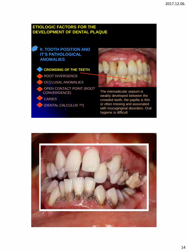
II. TOOTH POSITION AND IT’S PATHOLOGICAL
ANOMALIES
CROWDING OF THE TEETH
ROOT DIVERGENCE
OCCLUSAL ANOMALIES
OPEN CONTACT POINT
(ROOT CONVERGENCE)
CARIES
(DENTAL CALCULUS ??)
2017.12.06.
14
ETIOLOGIC FACTORS FOR THE
DEVELOPMENT OF DENTAL PLAQUE
II. TOOTH POSITION AND
IT’S PATHOLOGICAL
ANOMALIES
The interradicular septum is
weakly developed between the
crowded teeth, the papilla is thin
or often missing and associated
with mucogingival disorders. Oral
hygiene is difficult
CROWDING OF THE TEETH
ROOT DIVERGENCE
OCCLUSAL ANOMALIES
OPEN CONTACT POINT (ROOT
CONVERGENCE)
CARIES
(DENTAL CALCULUS ??)

2017.12.06.
15
ETIOLOGIC FACTORS FOR THE
DEVELOPMENT OF DENTAL PLAQUE
II. TOOTH POSITION AND IT’S
PATHOLOGICAL ANOMALIES
CROWDING OF THE TEETH
ROOT DIVERGENCE
OCCLUSAL ANOMALIES
OPEN CONTACT POINT (ROOT
CONVERGENCE)
CARIES
(DENTAL CALCULUS ??)
The divergence of the roots is also
accompanied by small interdental
space, and it makes plaque removal
more difficult
Partially erupted, impacted wisdom
teeth

2017.12.06.
16
ETIOLOGIC FACTORS FOR THE
DEVELOPMENT OF DENTAL PLAQUE
TRAUMATIC OCCLUSION CAN
NOT BE REGARDED AS A
DIRECT ETIOLOGIC FACTOR
FOR PERIODONTITIS
II. TOOTH POSITION AND IT’S
PATHOLOGICAL ANOMALIES
CROWDING OF THE TEETH
ROOT DIVERGENCE
OCCLUSAL ANOMALIES
OPEN CONTACT POINT (ROOT
CONVERGENCE)
CARIES
(DENTAL CALCULUS ??)
ETIOLOGIC FACTORS FOR THE
DEVELOPMENT OF DENTAL PLAQUE
CAUSING DEGENERATIVE CHANGES
IN THE DEEP PERIODONTAL
STRUCTURES. THE INFLAMMATORY
PROCESS IS ALLOWED TO SPREAD
APICALLY MORE RAPIDLY, RESULT IN
MORE SEVERE PERIODONTAL
DESTRUCTION
II. TOOTH POSITION AND IT’S
PATHOLOGICAL ANOMALIES
CROWDING OF THE TEETH
ROOT DIVERGENCE
OCCLUSAL ANOMALIES
OPEN CONTACT POINT (ROOT
CONVERGENCE)
CARIES
(DENTAL CALCULUS ??)

2017.12.06.
17
ETIOLOGIC FACTORS FOR THE
DEVELOPMENT OF DENTAL PLAQUE
II. TOOTH POSITION AND IT’S
PATHOLOGICAL ANOMALIESMISSING TEETH CAN LEAD TO
MESIAL DRIFTING, TILTING AND
EXTRUSION OF TEETH. RESULT:
OCCLUSAL DISHARMONIES,
INCREASE PLAQUE FORMATION,
FOOD IMPACTIONCROWDING OF THE TEETH
ROOT DIVERGENCE
OCCLUSAL ANOMALIES
OPEN CONTACT POINT (ROOT
CONVERGENCE)
CARIES
(DENTAL CALCULUS ??)
ETIOLOGIC FACTORS FOR THE
DEVELOPMENT OF DENTAL PLAQUE
II. TOOTH POSITION AND IT’S
PATHOLOGICAL ANOMALIES
LEADS TO FOOD IMPACTION
CROWDING OF THE TEETH
ROOT DIVERGENCE
OCCLUSAL ANOMALIES
OPEN CONTACTPOINT (ROOT
CONVERGENCE)
CARIES
(DENTAL CALCULUS ??)

2017.12.06.
18
ETIOLOGIC FACTORS FOR THE
DEVELOPMENT OF DENTAL PLAQUE
Ainamo (1970) drawed attention first to the strong
relation between the GI values and caries
II. TOOTH POSITION AND IT’S
PATHOLOGICAL ANOMALIES
Ainamo J.: Concominant periodontal disease and dental caries in young adult
males. Suomen Hammaslaakariseuran Toimituksa 66:303, 1970.
CROWDING OF THE TEETH
ROOT DIVERGENCE
OCCLUSAL ANOMALIES
OPEN CONTACTPOINT (ROOT
CONVERGENCE)
CARIES
(DENTAL CALCULUS ??)
Dental caries
Dental caries enhace plaque retention promoting periodontal disease.
Ainamo J.: Concominant periodontal disease and dental caries in young
adult males. Suomen Hammaslaakariseuran Toimituksa 66:303, 1970

2017.12.06.
19
ETIOLOGIC FACTORS FOR THE
DEVELOPMENT OF DENTAL
PLAQUE
CARIES
Significantly more secondary caries around subgingivally
placed margins than around supragingival margins.
secondary caries after 5 years15.4% of the supragingivally located amalgam restoration
30.4% of the subgingivally located amalgam restoration
Hammer B, Hotz P. [Inspection of 1 to 5-year-old amalgam, composite, and cast gold fillings].
SSO Schweiz Monatsschr Zahnheilkd. 1979 Apr;89(4):301-14. German.
Dental calculus ?
Sterile calculus of its own would not cause inflammation!
The rough surface of calculus is always covered by fresh, vital biofilm
and bacterial aggregation. There is strong correlation between the
amount of calculus and the severity and incidence of gingival
inflammation.
2017.12.06.
20
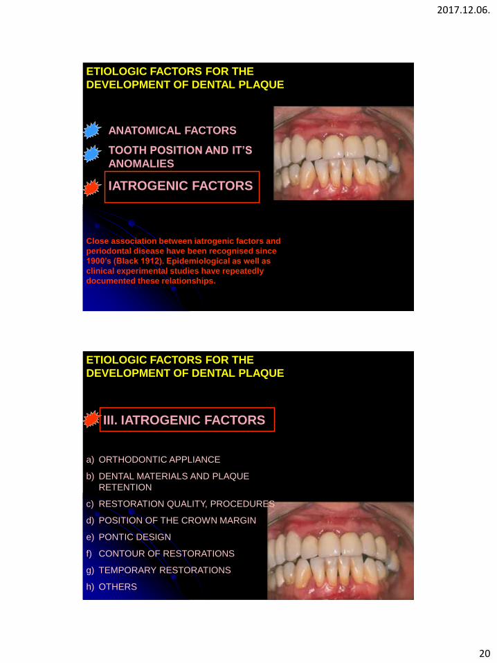
ETIOLOGIC FACTORS FOR THE
DEVELOPMENT OF DENTAL PLAQUE
ANATOMICAL FACTORS
TOOTH POSITION AND IT’S
ANOMALIES
IATROGENIC FACTORS
Close association between iatrogenic factors and
periodontal disease have been recognised since
1900’s (Black 1912). Epidemiological as well as
clinical experimental studies have repeatedly
documented these relationships.
ETIOLOGIC FACTORS FOR THE
DEVELOPMENT OF DENTAL PLAQUE
III. IATROGENIC FACTORS
a) ORTHODONTIC APPLIANCE
b) DENTAL MATERIALS AND PLAQUE
RETENTION
c) RESTORATION QUALITY, PROCEDURES
d) POSITION OF THE CROWN MARGIN
e) PONTIC DESIGN
f) CONTOUR OF RESTORATIONS
g) TEMPORARY RESTORATIONS
h) OTHERS
2017.12.06.
21
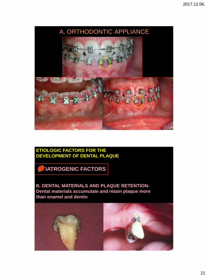
A. ORTHODONTIC APPLIANCE
ETIOLOGIC FACTORS FOR THE
DEVELOPMENT OF DENTAL PLAQUE
IATROGENIC FACTORS
B. DENTAL MATERIALS AND PLAQUE RETENTION-
Dental materials accumulate and retain plaque more
than enamel and dentin

2017.12.06.
22
THE PLAQUE RETENTIVE PROPERTY OF A DENTAL
MATERIAL DEPENDS ON SURFACE POROSITY

2017.12.06.
23
SEM images
Smoothness
Glassionomer Composite Porcelain Metal alloy
Glassionomer
Composite
Porcelain
Metal alloy
CROWNS MADE WITH METAL- OR ZIRCONIUM
MARGIN ARE THE LESS PLAQUE RETENTIVE

2017.12.06.
24
Dental gold foil and
porcelain irritate tissues
hardly if at all.
Porosity of all these
materials and especially of
acrylics, contribute to the
plaque retentive potentials.
The degree of porosity is
directly dependent upon
the way the materials were
handled and finished.
Case of Windisch Péter DMD: frameworks and implant abutments made of precious metal

2017.12.06.
25
Polymethyl-methacrylate
accumulates plaque faster
than gold and porcelain,
because the absorption of
fluids may increase its
tendency for plaque
adhesion. (+ bigger
porosity)
ETIOLOGIC FACTORS FOR THE
DEVELOPMENT OF DENTAL PLAQUE
IATROGENIC FACTORS
B. DENTAL MATERIALS
AND PLAQUE RETENTION
NO COMMERCIALLY AVAILABLE LUTING CEMENT PROVIDES A
PERFECT SEAL, THE SURFACE OF THE CEMENT IS ALWAYS
ROUGH AND POROUS.
Waerhaug’s histological investigations have shown that subgingival
cement roughness enhances plaque accumulation.
TRANSITION ZONE=
PREDILECTION SITE

2017.12.06.
26
ETIOLOGIC FACTORS FOR THE
DEVELOPMENT OF DENTAL PLAQUE
IATROGENIC FACTORS
d) C. RESTORATION QUALITY
Black stated as far back as in 1912,
that the inadequate marginal
crown-fit is responsible for the
presence of gingivitis.
He found in patients, from 20 to 35
years old, that from 1820 inflamed
areas, 663 had inadequate margins
and 421 had inadequate contact to
the adjecent teeth.
ETIOLOGIC FACTORS FOR THE
DEVELOPMENT OF DENTAL PLAQUE
IATROGENIC FACTORS
d) C. RESTORATION QUALITY
The World Workshop in
Periodontics (1966) reported
that the overhanging at the
margins of a restoration are
local factors promoting
periodontitis.

2017.12.06.
27
ETIOLOGIC FACTORS FOR THE
DEVELOPMENT OF DENTAL PLAQUE
IATROGENIC FACTORS
d) C. RESTORATION QUALITY
Bjorn et al. reported a generally
poor marginal fit of the examined
crowns.
80% of the radiographically studied
reconstructions exhibited marginal
defects on the proximal surfaces.
Margins were either overhangs or
open.
MARGIN WITH OPEN EDGE
2017.12.06.
28
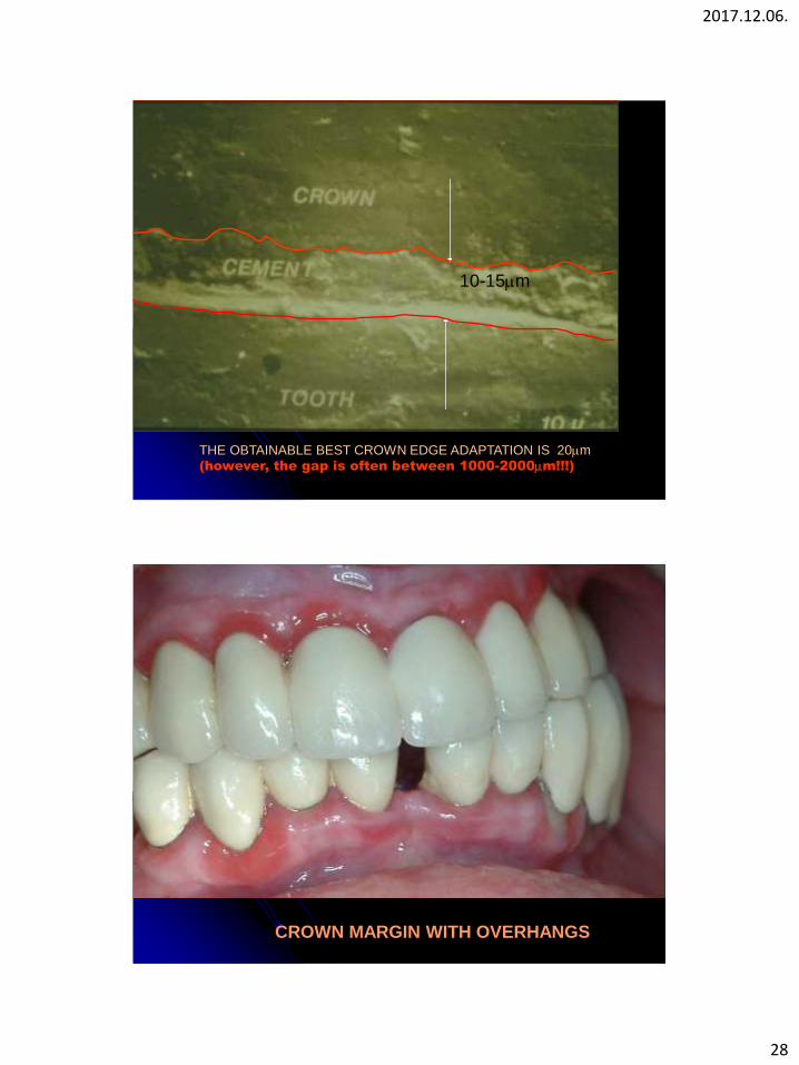
10-15mm
THE OBTAINABLE BEST CROWN EDGE ADAPTATION IS 20mm
(however, the gap is often between 1000-2000mm!!!)
CROWN MARGIN WITH OVERHANGS
2017.12.06.
29
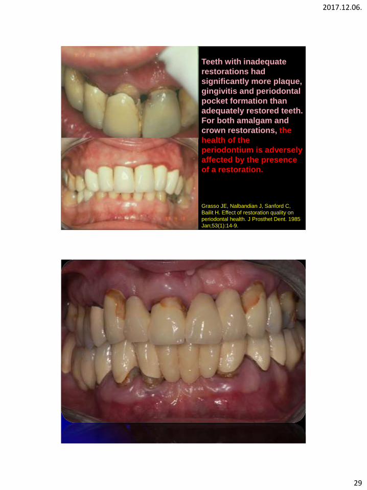
Teeth with inadequate
restorations had
significantly more plaque,
gingivitis and periodontal
pocket formation than
adequately restored teeth.
For both amalgam and
crown restorations, the
health of the
periodontium is adversely
affected by the presence
of a restoration.
Grasso JE, Nalbandian J, Sanford C,
Bailit H. Effect of restoration quality on
periodontal health. J Prosthet Dent. 1985
Jan;53(1):14-9.

2017.12.06.
30
The frequency of bad restorations
Reference Diagnostic method for detection % restored surfaces with
overhangs
(n = number of subjects)
Gilmore & Sheiham, 1971 Bitewing radiographs 25% (n = 1976)
Burch et al., 1976 Bitewing radiographs 30% (n = 825)
Hakkrainen & Ainamo,
1980
Orthopantograms 50% (n = 85)
Than et al., 1982 Calculus probe 60% (n = 240)
Lervik & Riordan, 1984 Bitewing radiographs,
microscope
25% (n = 175)
Keszthelyi & Szabo, 1984 Bitewing radiographs,
microscope
86% (n = 176)
Coxhead, 1985 Bitewing radiographs,
mirror, probe
76% (n = 50)
Claman et al., 1986 Bitewing radiographs 27% (n = 826)
Jansson et al., 1994 Bitewing radiographs 18 % (n = 162)
BACTERIAL SAMPLES GATHERED UNDER OVERHANGING
MARGINS SHOWED HIGH CORREALTION WITH
PERIODONTOPATHOGENIC ORGANISMS,
GRAM-NEGATIVE ANAEROBIC BACTERIAS (Porphyromonas,
Prevotella, Fusobacterium )
THE OVERHANGING RESTORATIONS DISTURB THE
ECOLOGIVAL BALANCE IN THE PERIODONTAL POCKET AND
ALLOW A GROUP OF DISEASE ASSOCIATED ORGANISMS.
SAMPLES COMING FROM THE CLINICALLY PERFECT MARGINS
WERE CHARACTERISTIC OF GINGIVAL HEALTH.
Lang P. N., Kiel A. R. , Anderhalden: Clinical and
microbiological effects of subgingival restorations with
overhangings or clinically perfect margins. J. Clini
Periodontol 1983; 10: 563-578

2017.12.06.
31
The subgingivally
located overhanging
crown- and filling
margins result
periodontal
attachement loss in
patients with
susceptibility.

2017.12.06.
32
Correctional possibilities I.
Correction of overhanging crown margins
Superficial, local or block anesthesia
Removal of porcelain edge with fissure or
torpedo shape diamond burs (turbine),
position crown margin supragingivally
Metal edge contouring with carbide crown-
bill (accelerator)
Smooth surface formation with carbide
and arkansas stone finishing- and
polishing burs (contra angel hand piece)
Correction of overhangs

2017.12.06.
33
1997 10.27.
2000. 07.02.
1997 07.02.
Correctional possibilities II.
Correction of open crown margins
2012 03.20.

2017.12.06.
34
Correctional possibilities III.
Change the crown
ETIOLOGIC FACTORS FOR THE
DEVELOPMENT OF DENTAL PLAQUE
IATROGENIC FACTORS
C. QUALITY OF RESTORATIONS
– EVERY PROCEDURE STEPS:
•PREPARATION
•IMPRESSION
•CEMENTATION

2017.12.06.
35
PREPARATION:
Shoulder
Supra- or paragingival
contourpreparation
IMPRESSION:
Correct sulcus retraction
CEMENTATION:
Total removal of luting material

2017.12.06.
36
ACCEPTABLE QUALITY
Conclusion: better restoration will help, but improving restorative quality alone
is unlikely to have major effects on the health of the periodontium without
effective plaque control.
Grasso JE, Nalbandian J, Sanford C, Bailit H. Effect of restoration quality on periodontal health. J
Prosthet Dent. 1985 Jan;53(1):14-9.
ETIOLOGIC FACTORS FOR THE
DEVELOPMENT OF DENTAL PLAQUE
IATROGENIC FACTORS
D. THE POSITION OF THE
CROWN MARGIN: SUPRA-
OR SUBGINGIVAL???
BLACK’S THEORY (1908): „EXTENSION
FOR PREVENTION” = SUBGINGIVALY
PLACED MARGINS

2017.12.06.
37
ETIOLOGIC FACTORS FOR THE
DEVELOPMENT OF DENTAL PLAQUE
IATROGENIC FACTORS
D. THE POSITION OF THE CROWN
MARGIN: SUPRA- OR
SUBGINGIVAL
1. Bodecker and Applebaum (1934) were the first to question black’s theory.
2. Waerhaug (1967, 1968) gave scientific proof that subgingival crown margins
create periodontal destruction due to plaque retention.
3. Loe(1968), Zander and Kennedy (1970) supported the position of the crown
margins above the free gingiva.
Subgingivally localised crown margins,
made with not shoulder preparation
2017.12.06.
38
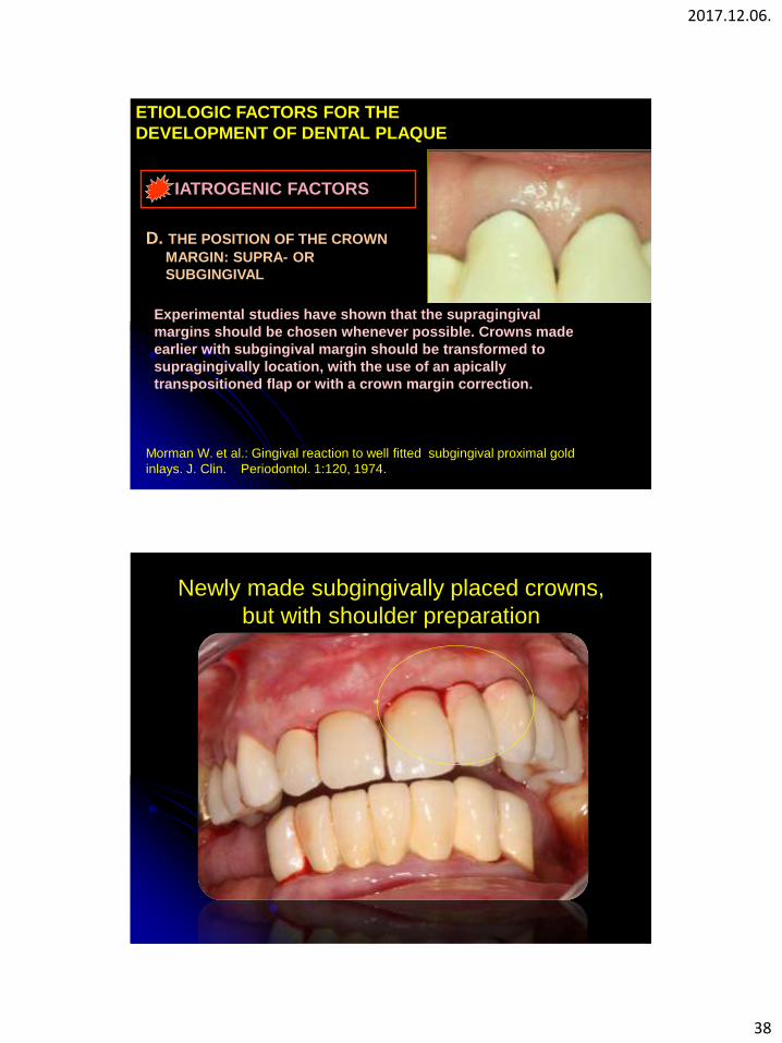
ETIOLOGIC FACTORS FOR THE
DEVELOPMENT OF DENTAL PLAQUE
IATROGENIC FACTORS
D. THE POSITION OF THE CROWN
MARGIN: SUPRA- OR
SUBGINGIVAL
Experimental studies have shown that the supragingival
margins should be chosen whenever possible. Crowns made
earlier with subgingival margin should be transformed to
supragingivally location, with the use of an apically
transpositioned flap or with a crown margin correction.
Morman W. et al.: Gingival reaction to well fitted subgingival proximal gold
inlays. J. Clin. Periodontol. 1:120, 1974.
Newly made subgingivally placed crowns,
but with shoulder preparation

2017.12.06.
39
Apically transpositioned flap

2017.12.06.
40
ETIOLOGIC FACTORS FOR THE
DEVELOPMENT OF DENTAL PLAQUE
IATROGENIC FACTORS
D. THE POSITION OF THE CROWN
MARGIN: SUPRA- OR SUBGINGIVAL
• There is no significant difference in the incidence of secunder
caries comparing the supra- and subgingivally positioned crown
margins.
•From a secunder caries preventive point of view, the location of
crown margins does not seem to be of great importance, if the
patient maintains a satisfactory oral hygiene.
Valderhaug J., H.Loe .: Oral hygiene in a group of supervised patients with fixed
prosthesis. J. Periodontol. 1977; 48:221- 224
Secunder caries ????
Biological width
2017.12.06.
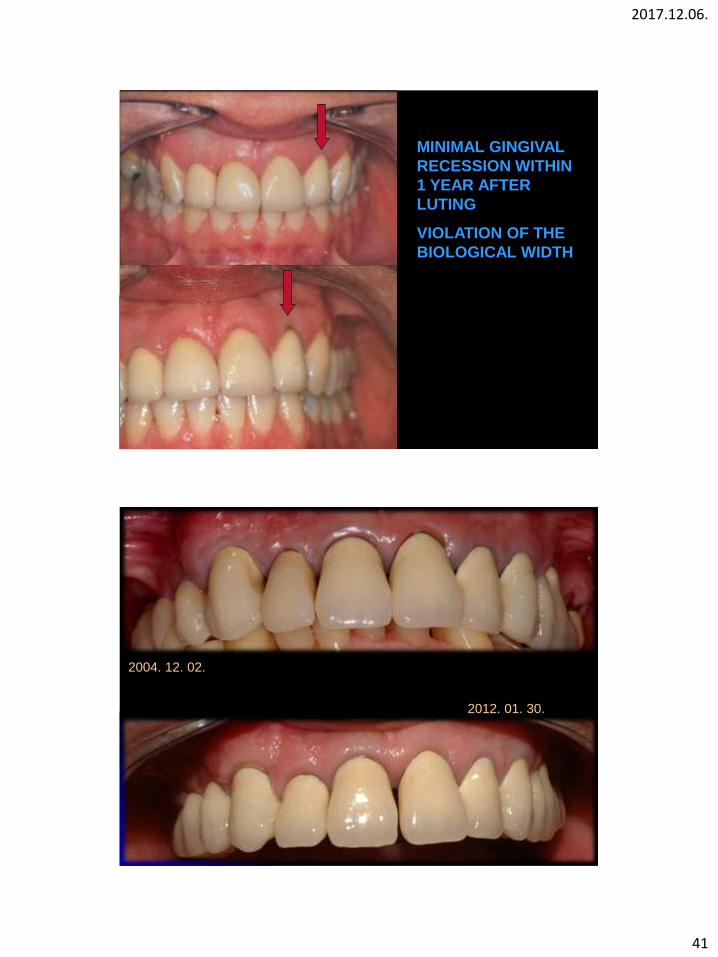
41
MINIMAL GINGIVAL
RECESSION WITHIN
1 YEAR AFTER
LUTING
VIOLATION OF THE
BIOLOGICAL WIDTH
2004. 12. 02.
2012. 01. 30.

2017.12.06.
42
ALTHOUGH ESTHETICALLY PLEASING, SUBGINGIVAL CROWN MARGINS ARE
CONSIDERED BIOLOGICAL UNDESIRABLE, BUT CAN BE DONE IF THE QUALITY IS
PERFECT!!
Today supragingivalmargins can provide excellent aesthetic results!

2017.12.06.
43
ETIOLOGIC FACTORS FOR THE
DEVELOPMENT OF DENTAL PLAQUE
IATROGENIC FACTORS
E. PONTIC DESIGN AND IT’S
CORRELATION TO THE EDENTOLOUS
MUCOSAL AREA
ETIOLOGIC FACTORS FOR THE
DEVELOPMENT OF DENTAL PLAQUE
IATROGENIC FACTORS
E. PONTIC DESIGN AND IT’S
CORRELATION TO THE EDENTOLOUS
MUCOSAL AREA
Badly designed pontics are
very frequently the cause
of tissue damage, gingival
inflammation, hyperplasia
of the underlying mucosa
and bone resorption.

2017.12.06.
44
A PLAKK AKKUMULÁCIÓT ELŐSEGÍTŐ
ETIOLÓGIAI TÉNYEZŐK
IATROGENIC FACTORS
E. PONTIC DESIGN AND IT’S
CORRELATION TO THE EDENTOLOUS
MUCOSAL AREA
It is forbidden to put the
pontic intermediary into
the fresh extraction socket.

2017.12.06.
45
ETIOLOGIC FACTORS FOR THE
DEVELOPMENT OF DENTAL PLAQUE
IATROGENIC FACTORS
E. PONTIC DESIGN AND IT’S
CORRELATION TO THE EDENTOLOUS
MUCOSAL AREA
The most ideal is the total
convex, egg shape
gingival surface, which is
not in touch with the
mucosa of the gingiva, or
just slightly touches it

2017.12.06.
46
ETIOLOGIC FACTORS FOR THE
DEVELOPMENT OF DENTAL PLAQUE
IATROGENIC FACTORS
F. Contour of restorations
GINGIVAL PROTECTION
THEORY: OUT OF DATE,
DOES NOT PROTECT
SULCUS FROM FOOD
IMPACTION!!
SCHLUGER: „THE SO
CALLED PROTECTIVE
CERVICAL CONVEXITY
PROTECTS NOT THE
GINGIVA, RATHER THE
DENTAL PLAQUE BEDDING
THERE ARE NO SELF-
CLEANSING MECHANISMS
AROUND THE SULCUS
ORAL HYGIENE
PRACTICES MAY BE
SEVERLY JEOPARDIZED BY
OVERCONTOURED
RESTORATIONS

2017.12.06.
47
THE INTERDENTAL AREAS OF
CROWNS AND BRIDGES
SHOULD BE ACCESSIBLE
WITH INTERDENTAL
BRUSHES OR WITH
SUPERFLOSS
TO ENSURE THIS WE HAVE
TO CREATE ADEQUTE
INTERDENTAL SPACES
CORRECT CONTACTPOINTS!
(EVEN THE QUITE HUGE
INTERDENTAL AREAS WILL
NOT LEAD TO FOOD
IMPACTION)
THE CORRECT CONTOUR OF THE CROWNS DEPEND NOT ONLY ON DENTAL
TECHNICIANS, DENTISTS HAVE TO DO ABUTMENT PREPARATION
ADEQUATELY IN ORDER TO MAKE A GOOD QUALITY CROWN WITH CORRECT
CONTOUR AND PROPER MARGINAL ADAPTATION IN THE LABORATORY.
CONTACTPOINTS ARE IN THE CORONAL THIRD OF THE CROWN.

2017.12.06.
48
CLASS II. FURCATION
LAESION TOTALLY
COVERED BY
OVERHANGING CROWN
MARGIN
OVERCONTOURED
CROWN MARGIN
WITH SEVERE
OVERHANG
COVERING THE
GINGIVAL
RECESSION WITH
OVERCONTOURED
CROWN MARGIN
WITH SEVERE
OVERHANG
THE WHOLE DENTAL PROBE
CAN BE PUT UNDER THE
CROWN MARGINE!!!!!!
2017.12.06.
49
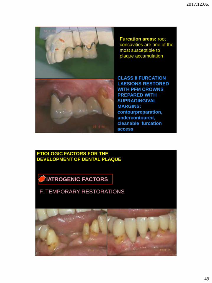
CLASS II FURCATION
LAESIONS RESTORED
WITH PFM CROWNS
PREPARED WITH
SUPRAGINGIVAL
MARGINS:
contourpreparation,
undercontoured,
cleanable furcation
access
Furcation areas: root
concavities are one of the
most susceptible to
plaque accumulation
ETIOLOGIC FACTORS FOR THE
DEVELOPMENT OF DENTAL PLAQUE
IATROGENIC FACTORS
F. TEMPORARY RESTORATIONS

2017.12.06.
50
The quality of the temporary restoration can not
be either bad, it’s margin and adaptation can
neither enhance plaque accumulation.
IATROGENIC FACTORS
E. OTHERS
Radiofrequency
used for root canal
desinfection may
result in recession
of the gingival
margin and loss of
attachmnet, if the
needle goes trough
the apex.

2017.12.06.
51
IATROGENIC FACTORS
E. OTHERS- reconstruction procedures
During cavity and
tooth preparation
the rotating
instruments used
beneath the
gingival margins
traumatized the
gingiva and even
the attachement.
IATROGENIC FACTORS
E. OTHERS- Sulcus retraction
The retraction cord and the
astringent solution put in the
gingival sulcus cause damage
to the periodontal tissue

2017.12.06.
52
Further
aviodable
and preferable
EXAMPLES
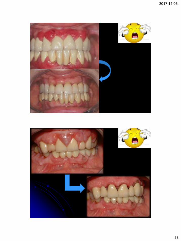
2017.12.06.
53

2017.12.06.
54

2017.12.06.
55
PERIIMPLANTITIS

2017.12.06.
56
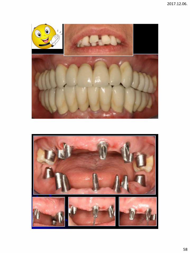
ACCEPTABLE
MARGINAL FIT
WITH CORRECT
MARGINAL
ADAPTATION

2017.12.06.
57
2017.12.06.
58

2017.12.06.
59
BASELINE AND FINAL

2017.12.06.
60
MAINTENANCE THERAPY

2017.12.06.
61
THANK YOU FOR YOUR KIND
ATTENTION!!