pamidronate resistance and associated low ras levels in breast
TRANSCRIPT

263
Address correspondence to Robert E. Brown, M.D., Depart-ment of Laboratory Medicine, Geisinger Medical Center, 100North Academy Ave., Danville, PA 17822, USA; tel 570 2716333; fax 570 271 6105; e-mail: [email protected].
Pamidronate Resistance and Associated Low Ras Levels inBreast Cancer Cells: A Role for Combinatorial Therapy
Ping L. Zhang,1,3 Mingyue Lun,3 Nava Siegelmann-Danieli,2 Thomas M. Blasick,3 and Robert E. Brown1
1Department of Laboratory Medicine, 2Department of Adult Hematology and Oncology, and 3Weis Centerfor Research, Geisinger Clinic, Danville, Pennsylvania
Abstract. To identify markers sensitive to inhibitors of the farnesylation pathway, we used 3 breast cancercell lines (SKBR-3, MDA-175, and MDA-231) to evaluate the in vitro effects of pamidronate, an inhibitorof farnesyl diphosphate synthase. In response to pamidronate, there was significant inhibition of cell prolif-eration in MDA-231 and SKBR-3 cells, compared to MDA-175 cells. This correlated with their respectivebasal levels of N-ras and H-ras. N-ras and H-ras protein levels were both reduced in MDA-231 cells, andto lesser extent in SKBR-3 cells, following exposure to pamidronate, whereas these markers were not alteredin MDA-175 cells. Combinatorial therapy with pamidronate and Gleevec, an inhibitor of several tyrosinekinases; Velcade, a proteasome inhibitor; or rapamycin, an inhibitor of the mammalian target of rapamycin(m-TOR) all showed additive effects in causing proliferative inhibition in MDA-175 cells. In summary,resistance to pamidronate may result from low levels of GTPase-activating proteins, such as N-ras and H-ras, in tumor cells. Combinatorial therapies directed against other signaling pathways, not dependent uponras, may be required to overcome such resistance. (received 6 May 2004; accepted 24 May 2004)
Keywords: pamidronate, H-ras, N-ras, apoptosis, breast cancer, Gleevec, Velcade
Introduction
Ras is an important signal transducing protein forgrowth factor activated pathways. Approximately20-30% of human tumors contain mutated versionsof ras proteins. Because ras is mutated so often inhuman cancers, much effort has been devoted todevising means to control the activity of renegaderas. Normal ras binds GTP and in the GTP-boundstate interacts with numerous effectors including theRaf proto-oncogene kinase and phosphatidyl-inositol-3-kinase (PI3K). Its intrinsic GTPaseactivity terminates the signal. Ras function requireslipophilic anchorage to the cell membrane by lipidprenylation and this prenylation reaction is mediatedby the enzyme farnesyl protein transferase (FT).Preventing ras protein association with the plasma
membrane is a strategy to impair ras transformation.Therefore drugs that target FT and interfere withactivated ras formation are good candidates for anti-tumor agents [1].
Farnesyl pyrophosphate synthase in the chole-sterol synthesis pathway is the specific moleculartarget of bisphosphonates, analogues of farnesylpyrophosphate [2]. Inhibition of farnesyl pyro-phosphate synthase by bisphosphonates leads todecreased production of isoprenoid intermediateslike farnesyl needed for the prenylation of ras viaFT [1]. Among 3 forms of ras proteins (H-ras, N-ras, K-ras), K-ras has been found to be resistant toFT inhibitors [3]. The basis for the resistance maybe related to high affinity of K-ras to FT and itscapacity to be prenylated by the related enzymegeranylgeranyl transferase (GGT), in the presenceof FT. Several bisphosphonates have been approvedby the Food and Drug Administration (FDA) totreat malignant lytic lesions (multiple myeloma or
0091-7370/04/0300-0263 $2.00. © 2004 by the Association of Clinical Scientists, Inc.
Annals of Clinical & Laboratory Science, vol. 34, no. 3, 2004

264
metastatic cancer) in bone [4,5]. But a cellularmarker in malignant tumors to predict the effectsof bisphosphonates on tumor cells per se has notbeen defined.
We selected 3 breast cancer cell lines that arenegative for hormonal receptors in order to studythe cell-growth inhibitory effects of pamidronate andto determine whether the inhibitory effects werecorrelated with proteins levels of several makers suchas N-ras, H-ras, or FTα/GGTα. We found thatcancer cells with less expression of N-ras and H-rashad high resistance to pamidronate treatment. Thisresistance can be overcome by the additive effects ofcombinatorial drugs, directed at different cellularpathways.
Materials and Methods
Materials. Pamidronate (BC Cancer Agency,Vancouver, BC, Canada) and Velcade (MilleniumPharmaceuticals, Inc, Cambrige, MA) were obtainedfrom the pharmacy of Geisinger Medical Center.Gleevec was a gift from Novartis Pharma AG (Basel,Switzerland). Ramamycin was purchased fromCalbiochem (San Diego, CA). N-ras, FTα/GGTα,and actin antibodies were purchased from SantaCruz Technology, Inc. (Santa Cruz, CA). H-ras(p21ras) was purchased from DAKO Cytomation(Carpinteria, CA). p-ERK1/2 and cleaved caspase-3 antibodies were purchased from Cell SignalingTechnology, Inc. (Beverly, MA).
Cells and cell culture. SKBR-3, MDA-175, andMDA-231 breast cancer cell lines (American TypeCulture Collection, Manassas, VA) were incubatedin Dulbecco’s Modified Eagle’s Medium (DMEM,Mediatech, Herndon, VA) supplemented with 10%fetal bovine serum (Gibco/Invitrogen, Grand Island,NY) at 37°C in a humidified atmosphere of 5% CO2in air.
Cell proliferation assay. The 3 lines of breast cancercells were grown in 96-well plates, and treated withpamidronate at specified concentrations for 4 days.Viable cells in each respective well were determinedcolorimetrically (CellTiter 96 one solution prolif-eration assay, Promega, Madison, WI). The cells
were washed once with Hank’s Balanced SaltSolution (HBSS, Gibco/Invitrogen), and then with100 µl of HBSS. One Step Assay Solution (20 µl,Promega) containing a tetrazolum compound wasadded to each well. The tetrazolium compound wasbioreduced by viable cells to a colored formazanproduct. After a 30-min incubation, absorbancemeasurements at 490 nm were made using a platereader. The proliferation rates of the control cellsand inhibitor-treated cells were compared.
Separation of particulate and soluble proteins. Todetermine if pamidronate diminished the ras activityby dislocating ras from cell membranes into thecytoplasmic compartment, cells with or withoutpamidronate treatments were processed to separateparticulate (membrane) and soluble (cytosolic)proteins, as described previously [3]. In brief, cellswere suspended in ice-cold hypotonic buffer andsonicated for 5 sec. The cell debris was pelleted at1500 x g for 4 min at 4°C and discarded. Thesupernatant was transferred to Beckman polyallomertubes and spun at 46,000 rpm (100,000 x g) for 45min. The pellet was washed once with ice-coldhypotonic buffer with protease inhibitors, re-pelletedat 46,000 rpm for 15 min, and resuspended in 200µl of ice-cold hypotonic buffer with proteaseinhibitors to prepare the particulate sample. Using10K NMWL concentrators (Millipore, Bedford,MA), the supernatant was concentrated to preparethe soluble sample. The resulting particulate andsoluble samples were analyzed by Western blotting.
Western blots. Control and pamidronate-treatedbreast cancer cells were harvested and sonicated. Cellhomogenates (30 µg total protein per lane) wereelectrophoresed on 6-12% SDS PAGE. Fractionatedproteins were transferred onto PVDF membranes.For immunostaining of N-ras, monoclonal mouseanti-N-ras antibody (1:500 dilution) was used. Thesecond antibody was horseradish peroxidase-linkedanti-mouse whole antibody (from donkey; 1:1,000dilution). Immunoreactive proteins were visualizedby an enhanced chemiluminescence-Westernblotting system (Amersham Biotech, Piscataway,NJ). Western blots for other antibodies wereperformed as described above, using primary and
Annals of Clinical & Laboratory Science

265
secondary antibodies as recommended by therespective producers.
Statistics In vitro inhibitory rates were expressed asmean ± SE. One-way ANOVA was used to comparethe inhibitory rates among the 3 types of breastcancer cells; p <0.05 was considered statisticallysignificant.
Results
Ras protein levels are correlated with the inhibitoryeffects of pamidronate. In Fig. 1, N-ras in theparticular part (membranous protein) of MDA-231and SKBR-3 cells showed a reduction in proteinexpression after pamidronate treatment. In responseto pamidronate, dislocation from the membranousto the cytoplasmic compartment was observed onlyin N-ras of MDA-231 cells, since soluble proteinshowed a mild increase in N-ras expression post-treatment in MDA-231. Based on densitometricanalysis of the corresponding band, the ratio ofparticulate N-ras to soluble N-ras was reduced from8.5 to 1.7 in MDA-231 cells after pamidronate
treatment. SKBR-3 and MDA-175 cells revealedminimal ratio changes of N-ras before and after the48-hr pamidronate challenge. In contrast, H-rasremained largely in the particulate component inall 3 types of cells. Membranous FTα/GGTα(particulate component) was slightly reduced afterpamidronate treatment in the MDA-231 cells.Otherwise, soluble FTα/GGTα remained stableafter pamidronate treatment in all 3 types of cells.
Whole cell lysates showed basal protein expres-sions of N-ras and H-ras to be high in MDA-231cells, moderate in SKBR-3 cells, and low in MDA-175 cells (Fig. 2). Subjected to pamidronatetreatment, the whole N-ras protein levels were notaltered in any of the 3 cell lines. The whole H-raslevel was prominently reduced in the MDA-231 cellsafter pamidronate treatment, but was essentiallyunchanged in the SKBK-3 and MDA-175 cells.Basal levels of FTα/GGTα were slightly higher inSKBK-3 cells, when compared to either MDA-175cells or MDA-231 cells. No alterations in the wholeFTα/GGTα expression was seen in any of 3 celltypes following pamidronate treatment.
Fig. 1. Particulate (P) and soluble (S) protein expression of N-ras, H-ras, and FTα/GGTα in SKBR-3, MDA-175, and MDA-231 cells before and after 24 or 48 hr incubation with 90 µM pamidronate.
Pamidronate resistance in breast cancer cell lines

266
The proliferation assay showed markedlyenhanced inhibitory rates in the pamidronate-treatedMDA-231 cells in a dose-dependent manner (Table1). This dose-dependent pattern was also seen inpamidronate-treated SKBR-3 cells. However, theinhibitory rates in the pamidronate-treated MDA-175 cells were significantly lower than in thecorresponding MDA-231 or SKBR-3 cells. Asshown in Figs. 1 and 2 and Table 1, there was generalcorrelation between the basal ras expression and thediminution of inhibitory rate following pamidronatetreatment.
Pamidronate inhibits cell proliferation by blockingthe Erk pathway and promoting apoptosis. Toillustrate the alteration of downstream signals alongthe ras pathway responsible for the inhibitory effectsof pamidronate, Western blotting was performedusing a series of antibodies in the 3 types of cellsincubated with pamidronate for 1 and 2 days. Thep-Erk expression was markedly reduced at 48 hr inboth SKBR-3 and MDA-231 cells, but not inMDA-175 cells (Fig. 2). The expression of p-JNKwas also reduced after 24 hr incubation withpamidronate in MDA-231 and SKBR-3 cells, but
Fig. 2. Protein expression of N-ras, H-ras, FTα/GGTα, p-Erk, p-JNK, cleaved (C) caspase-3, and actin in SKBR-3, MDA-175,and MDA-231 cells before and after 24 or 48 hr incubation with 90 µM pamidronate.
Annals of Clinical & Laboratory Science

267
not in MDA-175 cells. This reduced expression ofp-JNK was seen following 48 hr of incubation withpamidronate in all 3 cell lines. To examine the effectsof pamidronate on the apoptotic pathway, cleavedcaspase-3 antibody was used to detect this proteinexpression. Subjected to pamidronate treatment,there was a sequential increase in cleaved caspase-3
from the MDA-175 cells to the SKBR-3 cells, andfinally to the MDA-231 cells, indicating a graduatedenhancement in apoptotic activity in the 3 cell lines.Actin protein was expressed equally in each cell linebefore and after pamidronate treatment for either24 or 48 hr.
Additive effects of drugs at varying cellular pathwaysovercame drug resistance to pamidronate. To demon-strate the “additive” effects of pamidronate withGleevec, an inhibitor of farnesylation-independentplatelet-derived growth factor receptor and c-kitsignaling, pamidronate at 30 µM and Gleevec at 20µM were both added to MDA-175 cells to result in75.2 ± 1.9% inhibition; pamidronate alone caused39.5 ± 2.2% inhibition, whereas Gleevec alonecaused 50.2 ± 3.7% inhibition in MDA-175 cells(p <0.05 vs combinatorial effects) (Fig. 3).
To test whether the Akt pathway (associatedwith the mammalian target of rapamycin [mTOR]and nuclear factor-kappaB [NF-κB]) may be analternative farnesylation-independent growthpathway associated with cell proliferation in MDA-175 cells, we combined rapamycin at 1.0 µM (aninhibitor of mTOR) and Velcade at 30 nM (aninhibitor of proteasome and in turn NF-κB). Theseadditive effects resulted in 76.9 ± 0.9 % inhibition,which was significantly more than the solo effect ofrapamycin (28.1 ± 4.3% inhibition) or Velcade (64.3± 1.3% inhibition) (Fig. 3).
Table 1. Inhibitory effects of pamidronate in 3 cell lines
Pamidronate Inhibitory rates in 3 cell lines (%)exposure SKBR-3 MDA-175 MDA-231
10 µM, 4 days 7.0±1.8 5.0±2.5 4.2±1.8
30 µM, 4 days 47.4±1.1 15.0±1.9a 53.9±1.0a,b
90 µM, 4 days 74.1±5.0 23.6±2.7a 84.7±4.0a,b
a p <0.05 vs SKBR-3 cells; b p <0.05 vs MDA-175 cells.Inhibitory rates = (controls-treated)/controls.
Fig. 3. Upper panel: inhibitory effects of pamidronate at 30µM, Gleevec at 20 µM, or both in MDA-175 cells. Lowerpanel: inhibitory effects of rapamycin at 1 µM, Velcade at 30nM, or both in MDA-175 cells. *p <0.05 vs drug 1; #p<0.05vs drug 2.
Pamidronate resistance in breast cancer cell lines
268
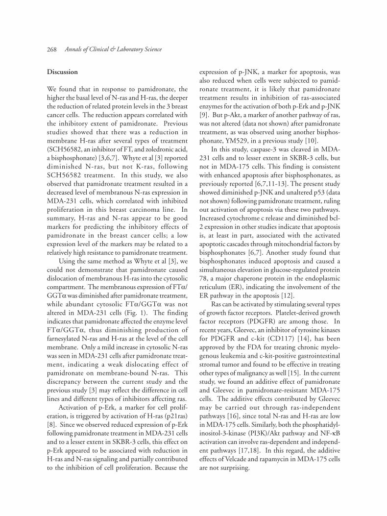
Discussion
We found that in response to pamidronate, thehigher the basal level of N-ras and H-ras, the deeperthe reduction of related protein levels in the 3 breastcancer cells. The reduction appears correlated withthe inhibitory extent of pamidronate. Previousstudies showed that there was a reduction inmembrane H-ras after several types of treatment(SCH56582, an inhibitor of FT, and zoledronic acid,a bisphosphonate) [3,6,7]. Whyte et al [3] reporteddiminished N-ras, but not K-ras, followingSCH56582 treatment. In this study, we alsoobserved that pamidronate treatment resulted in adecreased level of membranous N-ras expression inMDA-231 cells, which correlated with inhibitedproliferation in this breast carcinoma line. Insummary, H-ras and N-ras appear to be goodmarkers for predicting the inhibitory effects ofpamidronate in the breast cancer cells; a lowexpression level of the markers may be related to arelatively high resistance to pamidronate treatment.
Using the same method as Whyte et al [3], wecould not demonstrate that pamidronate causeddislocation of membranous H-ras into the cytosoliccompartment. The membranous expression of FTα/GGTα was diminished after pamidronate treatment,while abundant cytosolic FTα/GGTα was notaltered in MDA-231 cells (Fig. 1). The findingindicates that pamidronate affected the enzyme levelFTα/GGTα, thus diminishing production offarnesylated N-ras and H-ras at the level of the cellmembrane. Only a mild increase in cytosolic N-raswas seen in MDA-231 cells after pamidronate treat-ment, indicating a weak dislocating effect ofpamidronate on membrane-bound N-ras. Thisdiscrepancy between the current study and theprevious study [3] may reflect the difference in celllines and different types of inhibitors affecting ras.
Activation of p-Erk, a marker for cell prolif-eration, is triggered by activation of H-ras (p21ras)[8]. Since we observed reduced expression of p-Erkfollowing pamidronate treatment in MDA-231 cellsand to a lesser extent in SKBR-3 cells, this effect onp-Erk appeared to be associated with reduction inH-ras and N-ras signaling and partially contributedto the inhibition of cell proliferation. Because the
expression of p-JNK, a marker for apoptosis, wasalso reduced when cells were subjected to pamid-ronate treatment, it is likely that pamidronatetreatment results in inhibition of ras-associatedenzymes for the activation of both p-Erk and p-JNK[9]. But p-Akt, a marker of another pathway of ras,was not altered (data not shown) after pamidronatetreatment, as was observed using another bisphos-phonate, YM529, in a previous study [10].
In this study, caspase-3 was cleaved in MDA-231 cells and to lesser extent in SKBR-3 cells, butnot in MDA-175 cells. This finding is consistentwith enhanced apoptosis after bisphosphonates, aspreviously reported [6,7,11-13]. The present studyshowed diminished p-JNK and unaltered p53 (datanot shown) following pamidronate treatment, rulingout activation of apoptosis via these two pathways.Increased cytochrome c release and diminished bcl-2 expression in other studies indicate that apoptosisis, at least in part, associated with the activatedapoptotic cascades through mitochondrial factors bybisphosphonates [6,7]. Another study found thatbisphosphonates induced apoptosis and caused asimultaneous elevation in glucose-regulated protein78, a major chaperone protein in the endoplasmicreticulum (ER), indicating the involvement of theER pathway in the apoptosis [12].
Ras can be activated by stimulating several typesof growth factor receptors. Platelet-derived growthfactor receptors (PDGFR) are among those. Inrecent years, Gleevec, an inhibitor of tyrosine kinasesfor PDGFR and c-kit (CD117) [14], has beenapproved by the FDA for treating chronic myelo-genous leukemia and c-kit-positive gastrointestinalstromal tumor and found to be effective in treatingother types of malignancy as well [15]. In the currentstudy, we found an additive effect of pamidronateand Gleevec in pamidronate-resistant MDA-175cells. The additive effects contributed by Gleevecmay be carried out through ras-independentpathways [16], since total N-ras and H-ras are lowin MDA-175 cells. Similarly, both the phosphatidyl-inositol-3-kinase (PI3K)/Akt pathway and NF-κBactivation can involve ras-dependent and independ-ent pathways [17,18]. In this regard, the additiveeffects of Velcade and rapamycin in MDA-175 cellsare not surprising.
Annals of Clinical & Laboratory Science
269
In recent years, the proteasome inhibitor,Velcade, has been found to have potent inhibitoryeffects on tumor growth, at least partially resultingfrom its inhibitory effects on NF-κB binding activityto DNA for proliferation [19,20]. Rapamycin is amacrolide fungicide that binds intracellularly to theimmunophilins FKBP12, and the resultant complexinhibits the activity of a 290-kDa kinase known asmammalian target of rapamycin (mTOR), adownstream signal of Akt. Velcade has beenapproved by the FDA to treat malignant myelomaand CCI-779, an ester of rapamycin, has been testedin clinical trials for human cancer. Combination ofthe 2 drugs showed more profound inhibition oncell growth than either drug alone. The additiveinhibition most likely represents simultaneous effectson ras-dependent and ras-independent pathways inpamidronate-resistant MDA-175 cells.
In summary, at both 30 and 90 µM of pamid-ronate, MDA-175 cells showed higher resistancethan SKBR-3 and MDA-231 cells, which may berelated to the higher levels of N-ras and H-ras inthe latter two cell lines compared to MDA-175 cells.N-ras and H-ras may be useful protein markers topredict effects of pamidronate in breast cancer cells.Inhibitory effects of pamidronate on breast cancercells were, at least partially, mediated by a p-Erkmediated pathway and by promoting apoptosis. InMDA-175 cells, combinatorial use of pamidronateand Gleevec resulted in an additive effect onproliferative inhibition. To test the Akt pathway asan alternative farnesylation-independent growthpathway associated with cell proliferation in MDA-175 cells, combinatorial therapy with rapamycin (aninhibitor of m-TOR) and Velcade (an inhibitor ofproteasome and NF-κB) also led to additive inhib-ition of proliferation in MDA-175 cells. Resistanceto pamidronate in breast cancer cells may not besimply determined by the level of FT. Combinatorialtherapy with inhibition of other signal pathways,which can be ras-independent, may provide additiveeffects and overcome the drug resistance.
Acknowledgments
The authors thank Xin S. Xin and Ann O. Karosasfor assistance and Novartis for the gift of Gleevec.
References
1. Cox AD. Farnesyltransferase inhibitors–potential role inthe treatment of cancer. Drugs 2001;61:723-732.
2. Bergstrom JD, Bostedor RG, Masarachia PJ, Rezka AA,Rodan G. Alendronate is a specific, nanomolar inhibitorof farnesyl disphosphonate synthase. Arch BiochemBiophys 2000;373:231-241.
3. Whyte DB, Kirschmeier P, Hockenberry TH, Numez-Oliva I, James L, Catino JJ, Bishop WR, Pai J. K- and N-ras are geranylgeranylated in cells treated with farnesylprotein transferase inhibitors. J Biol Chem 1997;272:14459-14464.
4. Sausville EA, Elsayed Y, Monga M, Kim G. Signaltransduction-directed cancer treatment. Ann RevPharmacol Toxicol 2003;43:199-231.
5. Berenson J, Hillner BE, Kyle RA, Anderson K, Lipton A,Yee GC, Biemann JS. American Society of ClinicalOncology clinical practice guidelines: the role ofbisphosphonates in multiple myeloma. J Clin Oncol2002;17:3719-3736.
6. Senaratne SG, Mansi JL, Colston KW. The bisphos-phonate zoledronic acid impairs membrane localisationand induces cytochrome c release in breast cancer cells.Br J Cancer 2002;86:1479-1486.
7. Oades GM, Senaratne SG, Clarke IA, Kirby RS, ColstonKW. Nitrogen containing bisphosphonates induce apop-tosis and inhibit the mevalonate pathway, impairing rasmembrane localization in prostate cancer cells. J Urol2003;170:246-252.
8. Ming XF, Burgering BM, Wennstrom S, Claesson-WelshL, Heldin CH, Bos JL, Kozma SC, Thomas G. Activationof p70/p85 S6 kinase by a pathway independent of p21ras.Nature 1994;371:426-429.
9. Raman M, Cobb MH. MAP kinase modules: many roadshome. Cur Biol 2003;13:R886-R888.
10. Nishida S, Fujii Y, Yoshioka S, Kikuichi S, Tsubaki M,Irimajiri K. A new biosphosphonate, YM529, inducesapoptosis in HL60 cells by decreasing phosphorylationof single survival signal ERK. Life Sci 2003;73:2655-2664.
11. Salomo M, Jurlander J, Bo Nielsen LB, Gimsing P. Howmyeloma cells escape bisphosphonate-mediated killing:development of specific resistance with preserved sensit-ivity to conventional chemotherapeutics. Br J Haematol2003;122:202-210.
12. Beaupre DM, Cepero E, Obeng EA, Bosie LH, Lichten-held MG. R115777 induces ras-independent apoptosisof myeloma cells via multiple intrinsic pathways. MolCancer Therap 2004;3:179-186.
13. Riebeling C, Forsea AM, Raisova M, Orfanos CE, GeilenCC. The bisphosphonate pamidronate induces apoptosisin human melanoma cells in vitro. Br J Cancer 2002;87:366-371.
14. Heinrich MC, Griffith DJ, Druker BJ, Wait Cl, Ott KA,Zigler AJ. Inhibition of c-kit receptor tyrosine kinaseactivity by STI571, a selective tyrosine kinase inhibitor.
Pamidronate resistance in breast cancer cell lines

270
Blood 2000;96:925-932.15. Sausville EA, Elsayed Y, Monga M, Kim G. Signal
transduction-directed cancer treatment. Ann RevPharmacol Toxicol 2003;43:199-231.
16. Besset V, Scott RP, Ibanez CF. Signaling complexes andprotein-protein interactions involved in the activation ofthe Ras and phosphatidylinositol 3-kinase pathways bythe c-Ret receptor tyrosine kinase. J Biol Chem 2000;275:39159-39166.
17. Cox AD, Der CJ. The dark side of Ras: regulation ofapoptosis. Oncogene 2003;22:8999-9006.
18. Wu K, Wang C, D’Amico M, Lee RJ, Albanese C, PestellRG, Mani S. Flavopiridol and trastuzumab synergisticallyinhibit proliferation of breast cancer cells: association withselective cooperative inhibition of cyclin D1-dependentkinase and Akt signaling pathways. Mol Cancer Thera2002;1:695-706.
19. Adams J. Potential for proteasome inhibition in thetreatment of cancer. Drug Discov Today 2003;8:307-315.
20. Lenz HJ. Clinical update: proteasome inhibitors in solidtumors. Cancer Treat Rev 2003;29(suppl 1):41-48.
Annals of Clinical & Laboratory Science