pan london haemato oncology clinical guidelines
TRANSCRIPT
Pan-London Haemato-Oncology Clinical Guidelines Acute Leukaemias and Myeloid Neoplasms
Part 5: Myelodysplastic Syndromes
January 2020
CONTENTS
1
Contents
1 Introduction ........................................................................................................................ 3
2 Referral Pathways .............................................................................................................. 4
3 Investigation and Diagnosis ............................................................................................... 5
3.1 Pathology ............................................................................................................... 12
4 Risk Stratification ............................................................................................................. 13
5 Patient Information/Support .............................................................................................. 16
6 Treatment ........................................................................................................................ 17
6.1 Low or intermediate-risk MDS ................................................................................ 17
6.2 High risk MDS ........................................................................................................ 18
6.3 Chronic myelomonocytic leukaemia (CMML) .......................................................... 20
6.4 Fertility ................................................................................................................... 21
7 Management of Disease and Treatment-related Complications ....................................... 22
7.1 Anaemia ................................................................................................................. 22
7.2 Severe neutropenia ................................................................................................ 22
7.3 Neutropenic sepsis ................................................................................................. 23
7.4 Severe thrombocytopenia ....................................................................................... 23
7.5 Haemostasis and thrombosis ................................................................................. 23
7.6 Transfusional iron overload .................................................................................... 24
8 Supportive Care ............................................................................................................... 25
8.1 Anaemia ................................................................................................................. 25
8.2 Transfusions ........................................................................................................... 25
8.3 Haemostasis and thrombosis ................................................................................. 25
8.4 Infection prophylaxis ............................................................................................... 25
9 Treatment Summary and Care Plan ................................................................................. 26
9.1 Treatment summary and care plan ......................................................................... 26
10 Follow-up Arrangements ................................................................................................ 27
11 End-of-life Care .............................................................................................................. 27
12 Data Requirements ........................................................................................................ 27
References ............................................................................................................................ 28
Annex 1: Data Requirements ................................................................................................. 29
CONTENTS
2
Lead authors 2020:
Dr Shreyans Gandhi, King’s College Hospital NHS Foundation Trust
Dr Agapi Parcaridou, London North West University Healthcare NHS Trust
Dr Beth Payne, University College London Hospitals NHS Foundation Trust
2018 Edition;
Dr Beth Payne, University College London Hospitals NHS Foundation Trust
Dr Kavita Raj, Guys and St Thomas' NHS Foundation Trust
Disclaimer
These guidelines should be read in conjunction with the latest NICE guidance, and all applicable
national/international guidance. The prescribing information in these guidelines is for health professionals
only. It is not intended to replace consultation with the Haematology Consultant at the patient’s specialist
centre. For information on cautions, contra-indications and side effects, refer to the up-to-date prescribing
information. While great care has been taken to see that the information in these guidelines is accurate,
the user is advised to check the doses and regimens carefully and if there is any uncertainty about the
guidance provided, you should discuss your queries with a Haematology Consultant or Senior Pharmacist.
No set of guidelines can cover all variations required for specific patient circumstances. It is the
responsibility of the healthcare practitioners using them to adapt them for safe use within their institutions
and for the individual needs of patients.
Contact us
The writing cycle for the guidelines will be from May-July each year. If you wish to be part of the writing
group, please contact us through the following link: Pan London Blood Cancer (or via
If you wish to report errors or omissions that require urgent attention please contact us via the same email
addresses.
© RM Partners, South East London Cancer Alliance, North Central and East London Cancer Alliance
2020
INTRODUCTION
3
1 Introduction
This guidance should be read in conjunction with the British Committee for Standards
in Haematology (BCSH) myelodysplastic syndrome (MDS) guideline1 and the
European Leukaemia Net (ELN) guidelines on myelodysplastic syndromes.2
The myelodysplastic syndromes (MDS) are a group of clonal stem cell disorders characterised
by qualitative and quantitative defects in haemopoiesis that predispose individuals to anaemia,
life-threatening bleeds and infection concomitant with a risk of transforming to acute myeloid
leukaemia (AML).
The incidence of MDS is 4–5 per 100,000, but it increases with age such that the incidence is
30 per 100,000 in those aged over 70; and 40 per 100,000 in those aged over 80. Some 10% of
MDS are secondary, most often due to radiotherapy or chemotherapy for cancer; with increasing
numbers of patients surviving chemotherapy, the incidence of therapy-related MDS may also be
set to increase.
Cytogenetic abnormalities are present in 40–50% of patients and are of value both in confirming
the diagnosis and indicating the risk of disease progression. More recently, molecular
abnormalities that commonly occur have been identified and their prognostic value is being
clarified. Thus, several prognostic indices incorporating these features have been developed in
order to guide optimal management.
REFERRAL PATHWAYS
4
2 Referral Pathways
Patients with suspected MDS in primary care should be referred to a haematologist for
assessment. It may be appropriate for patients with severe neutropenia, thrombocytopenia or
blasts in peripheral blood to be referred via the 2 week wait pathway (often picked up on a routine
blood test via the laboratory).
All new patients should be referred to the MDT for confirmation of diagnosis, prognosis and
management plan taking into account their performance status, needs and co-morbidities. A joint
approach with elderly care physicians and palliative care teams may be appropriate as per local
guidelines.
The following patients should be brought to the MDT:
all new patients with MDS in order to confirm the diagnosis and treatment plan
all patients where a new line of therapy needs to be considered
all patients with a restaging assessment of response to treatment (e.g. hypomethylating
agents or immunosuppression)
all patients in whom an allogeneic stem cell transplant is a consideration.
The MDT outcome should be documented and communicated to a primary care and secondary
care referring centre (where relevant).
Patients with MDS IPSS-low and IPSS INT1 and INT2 may be managed at facilities with at least
British Committee for Standards in Haematology (BCSH) Level 1 designation. MDS IPSS-high or
complex patients may be referred to centres with at least BCSH Level 2 designation and with
specific expertise, or which have available trials. Candidates for transplantation should be referred
to a JACIE-accredited centre. For complex MDS cases, a centre with a specific interest and
expertise in MDS may be asked to review the case with the requesting site.
INVESTIGATION AND DIAGNOSIS
5
3 Investigation and Diagnosis
Investigations are aimed at excluding secondary causes of dysplasia or cytopenias, and tests to
confirm the diagnosis of MDS and exclude other clonal stem cell disorders. Investigation of MDS is
usually initiated with the findings of:
a macrocytic anaemia (or persistent macrocytosis)
unexplained neutropenia with a blood film that suggests dysplastic features
(pseudo Pelger-Huët abnormality)
unexplained thrombocytopenia (especially when not responsive to immunosuppressive
therapy/ITP treatment)
blasts in the peripheral blood
a persistent unexplained monocytosis >1x10⁹/L.
Appropriate investigations should exclude the following alternative causes:
haematinic deficiency (vitamin B12, folate, selenium in appropriate patients)
liver dysfunction
thyroid dysfunction
haemolysis
autoimmune disorders
viral infections such as HIV, HBV and HCV
other primary cancers
systemic inflammatory response syndrome (SIRS)/cytokine storm
other causes of inflammation (e.g. concurrent infection).
A pertinent history should be taken including:
smoking and alcohol intake history
family history of thrombocytopenia, breast and other cancers, lymphoedema, pulmonary
fibrosis or MDS/AML
in younger patients, a family history of constitutional bone marrow failure (such as Fanconi’s
anaemia, Schwachman-Diamond syndrome and dyskeratosis congenita should be sought) or
even abnormal familial traits (eg. premature greying of hair, hearing loss)
prior exposure to chemotherapy particularly alkylating agents, topoisomerase inhibitors and
radiotherapy (especially to the pelvis)
occupational exposure to chemicals (e.g. benzenes)
current medications (such as methotrexate, azathioprine, quinine)
bleeding and infection history.
INVESTIGATION AND DIAGNOSIS
6
Physical examination of the patient should include the assessment of:
abnormal skin, hair and nail changes/lesions (vasculitis, Sweet’s syndrome, E.
nodosum/pyoderma gangrenosum lesions, café au lait spots, premature grey, etc.)
arthritis
lymphoedema (Emberger syndrome)
splenomegaly and lymphadenopathy.
Initial investigations that can be requested/performed are:
FBC and blood film for morphologic assessment
reticulocyte count
DAT
haematinics: vitamin B12 (if available, methylmalonic acid), red cell folate, ferritin
haemoglobin electrophoresis
thyroid function tests
LDH and uric acid
U&Es
LFTs
CRP and ESR
serum protein electrophoresis (SPEP) with immunoglobulins (a paraprotein may occur with
MDS) and (T-cell subsets including LGL, if available)
viral screen: HIV, hepatitis B and hepatitis C
autoimmune screen
beta 2 microglobulin
serum erythropoietin levels
parvovirus if appropriate
haptoglobins.
Investigations after referral to haematology:
bone marrow aspirate and trephine (BMAT)
Conventional karyotyping (if not enough dividing metaphases seen, flow-FISH for
chromosome 5 and 7)
PNH screen
specific genetic tests where there is a suspicion of an inherited or acquired bone marrow
failure syndrome (e.g. telomere lengths, chromosome fragility, genetic testing).
INVESTIGATION AND DIAGNOSIS
7
The diagnosis of MDS is made based on the current World Health Organization (WHO) 2008
classification and morphologic assessment:
To enable better evaluation of blasts, a haemogram of >500 cells that include >100
non-erythroid cells (where erythroid cells >50% of the count) is necessary for both
peripheral blood films and aspirates. In performing a haemogram, due consideration
must be given in identifying blasts, promyelocytes, monoblasts and promonocytes
and examining >100 erythroblasts and 30 megakaryocytes. In cases where the
diagnosis is difficult, i.e. normal / non-informative cytogenetics, no excess
myeloblasts or ring sideroblasts, it may be appropriate to repeat the marrow (weeks
to months apart) prior to confirming a diagnosis. An observation interval of 6 months
is recommended in those with unilineage dysplasia, no increase in blasts (peripheral
blood or bone marrow) and where ring sideroblasts <15%.
The WHO classification requires the assessment of dysplasia in the following samples:
Peripheral blood film
It is recommended that at least 200 cells are examined. Features of dysplasia include:
red cell anisocytosis, poikilocytosis, basophilic stippling
myeloid nuclear hypolobation, pseudo Pelger-Huët anomaly, hypo- or degranulation
the presence of myeloblasts
platelet anisocytosis or giant platelets.
Bone marrow aspirate
Dysplastic features should be present in ≥10% of the cells of the lineage in consideration to give a
definition of MDS. The WHO classification stratifies patients based on the presence of dysplasia
>10% of cells in any lineage. Multilineage dysplasia is defined as the presence of 10% dysplastic
cells in at least two cell lineages and confers a poorer prognosis.
Features of dysplasia that are diagnostic of MDS are the presence of an acquired Pelger-Huët
abnormality in the peripheral blood and presence of micromegakaryocytes in the bone marrow.
The presence of circulating blasts of <1%, 1%, 2–4% or 5% alters the WHO classification, as does
the presence of 5–10% and 11–20% blasts.
Iron stain with Prussian blue must be performed in order to identify the presence of significant
number of ring sideroblasts (≥5 siderotic granules covering at least a third of the nuclear
circumference in ≥15% of erythroid cells).
Flow cytometry
This is not currently used in standard practice and is not an essential test in MDS. In this context, it
may be helpful in identifying dysplasia where no clear cytogenetic or clonal marker is present, in
distinguishing refractory anaemia from refractory anaemia with multilineage dysplasia (scatter
properties), and in enumerating myeloblasts, although all of the above should primarily be a
morphologic diagnosis. If undertaken, this test is best performed at a centre with experience with
the ELNET recommendations for MDS/AML.
INVESTIGATION AND DIAGNOSIS
8
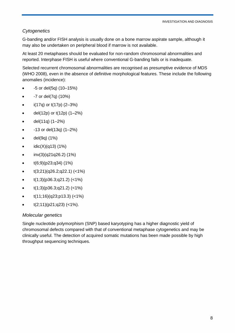
Cytogenetics
G-banding and/or FISH analysis is usually done on a bone marrow aspirate sample, although it
may also be undertaken on peripheral blood if marrow is not available.
At least 20 metaphases should be evaluated for non-random chromosomal abnormalities and
reported. Interphase FISH is useful where conventional G-banding fails or is inadequate.
Selected recurrent chromosomal abnormalities are recognised as presumptive evidence of MDS
(WHO 2008), even in the absence of definitive morphological features. These include the following
anomalies (incidence):
-5 or del(5q) (10–15%)
-7 or del(7q) (10%)
i(17q) or t(17p) (2–3%)
del(12p) or t(12p) (1–2%)
del(11q) (1–2%)
-13 or del(13q) (1–2%)
del(9q) (1%)
idic(X)(q13) (1%)
inv(3)(q21q26.2) (1%)
t(6;9)(p23;q34) (1%)
t(3;21)(q26.2;q22.1) (<1%)
t(1;3)(p36.3;q21.2) (<1%)
t(1;3)(p36.3;q21.2) (<1%)
t(11;16)(q23;p13.3) (<1%)
t(2;11)(p21;q23) (<1%).
Molecular genetics
Single nucleotide polymorphism (SNP) based karyotyping has a higher diagnostic yield of
chromosomal defects compared with that of conventional metaphase cytogenetics and may be
clinically useful. The detection of acquired somatic mutations has been made possible by high
throughput sequencing techniques.
INVESTIGATION AND DIAGNOSIS
9
Commonly mutated genes in MDS (but not exclusive to MDS) include those of the spliceosome
component:
Gene mutation Incidence
SF3B1 25–30%
TET2 20–25%
RUNX1 10–20%
ASXL1 10–15%
SRSF2 10–15%
TP53 5–10%
U2AF1 5–10%
NRAS/KRAS 5–10%
DNMT3A 5%
ZRSR2 5%
EZH2 5%
IDH1&2 2–3%
ETV6 2%
CBL 1–2%
NPM1 1–2%
JAK2 1–2%
SETBP1 1–2%
ZRSF1 1–2%
U2AF65 1–2%
PRPF40B 1–2%
At least 52% of patients with a normal karyotype harbour at least one mutation and 74% have at
least a copy number variation or molecular mutation, thus these tests can help confirm the
diagnosis.
While these tests are not essential for the diagnosis of MDS, and do not currently form part of the
classification or risk stratification in standard practice, prognostic information from such tests may
assist treatment decisions in some cases. Therefore, where appropriate such tests should be used
in conjunction with standard prognostic scoring systems (IPSS, IPSS-R). Incorporation of these
prognostic values are likely to become part of the next revision of the IPSS.
Bone marrow trephine
This test will assess marrow cellularity, topography, presence of reticulin fibrosis and blasts, and
exclude other metastatic disease or infections. The trephine biopsy should be stained with
haematoxylin and eosin (H&E) or equivalent, Giemsa, myeloperoxidase, glycophorin A and C or
equivalent, CD34, CD117, CD61 or CD42b for megakaryocytes, CD68 or CD68R for monocytes,
CD20, CD3 and Gomori silver stain for reticulin.
Bone marrow cellularity in MDS is usually hyper- or normo-cellular but is hypocellular in 10% of
patients (hypocellular MDS) and needs to be differentiated from aplastic anaemia (AA). The
presence of dysplasia, reticulin fibrosis, ring sideroblasts, CD34+ cells and micro-megakaryocytes
favours a diagnosis of MDS. A scoring system based on cyto-histological features (h-score) when
INVESTIGATION AND DIAGNOSIS
10
integrated with chromosomal make-up and genetic analysis in a hypocellular bone marrow, can
differentiate with high specificity for hypoplastic MDS, and furthermore also help predict risk of
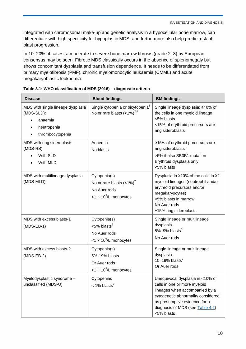
blast progression.
In 10–20% of cases, a moderate to severe bone marrow fibrosis (grade 2–3) by European
consensus may be seen. Fibrotic MDS classically occurs in the absence of splenomegaly but
shows concomitant dysplasia and transfusion dependence. It needs to be differentiated from
primary myelofibrosis (PMF), chronic myelomonocytic leukaemia (CMML) and acute
megakaryoblastic leukaemia.
Table 3.1: WHO classification of MDS (2016) – diagnostic criteria
Disease Blood findings BM findings
MDS with single lineage dysplasia
(MDS-SLD):
anaemia
neutropenia
thrombocytopenia
Single cytopenia or bicytopenia1
No or rare blasts (<1%)2,4
Single lineage dysplasia: ≥10% of
the cells in one myeloid lineage
<5% blasts
<15% of erythroid precursors are
ring sideroblasts
MDS with ring sideroblasts
(MDS-RS)
With SLD
With MLD
Anaemia
No blasts
≥15% of erythroid precursors are
ring sideroblasts
>5% if also SB3B1 mutation
Erythroid dysplasia only
<5% blasts
MDS with multilineage dysplasia
(MDS-MLD)
Cytopenia(s)
No or rare blasts (<1%)2
No Auer rods
<1 × 10⁹/L monocytes
Dysplasia in ≥10% of the cells in ≥2
myeloid lineages (neutrophil and/or
erythroid precursors and/or
megakaryocytes)
<5% blasts in marrow
No Auer rods
±15% ring sideroblasts
MDS with excess blasts-1
(MDS-EB-1)
Cytopenia(s)
<5% blasts2
No Auer rods
<1 × 10⁹/L monocytes
Single lineage or multilineage
dysplasia
5%–9% blasts3
No Auer rods
MDS with excess blasts-2
(MDS-EB-2)
Cytopenia(s)
5%-19% blasts
Or Auer rods
<1 × 10⁹/L monocytes
Single lineage or multilineage
dysplasia
10–19% blasts3
Or Auer rods
Myelodysplastic syndrome –
unclassified (MDS-U)
Cytopenias
< 1% blasts2
Unequivocal dysplasia in <10% of
cells in one or more myeloid
lineages when accompanied by a
cytogenetic abnormality considered
as presumptive evidence for a
diagnosis of MDS (see Table 4.2)
<5% blasts
INVESTIGATION AND DIAGNOSIS
11
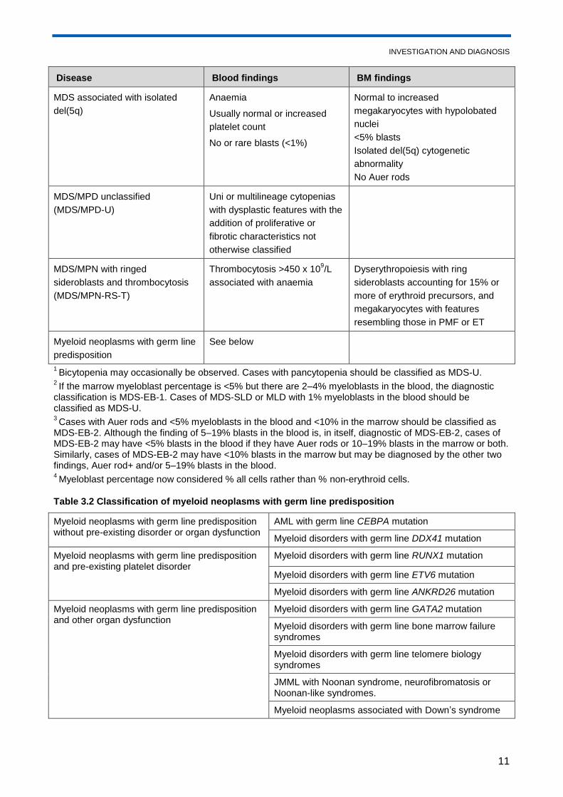
Disease Blood findings BM findings
MDS associated with isolated
del(5q)
Anaemia
Usually normal or increased
platelet count
No or rare blasts (<1%)
Normal to increased
megakaryocytes with hypolobated
nuclei
<5% blasts
Isolated del(5q) cytogenetic
abnormality
No Auer rods
MDS/MPD unclassified
(MDS/MPD-U)
Uni or multilineage cytopenias
with dysplastic features with the
addition of proliferative or
fibrotic characteristics not
otherwise classified
MDS/MPN with ringed
sideroblasts and thrombocytosis
(MDS/MPN-RS-T)
Thrombocytosis >450 x 109/L
associated with anaemia
Dyserythropoiesis with ring
sideroblasts accounting for 15% or
more of erythroid precursors, and
megakaryocytes with features
resembling those in PMF or ET
Myeloid neoplasms with germ line
predisposition
See below
1 Bicytopenia may occasionally be observed. Cases with pancytopenia should be classified as MDS-U.
2 If the marrow myeloblast percentage is <5% but there are 2–4% myeloblasts in the blood, the diagnostic
classification is MDS-EB-1. Cases of MDS-SLD or MLD with 1% myeloblasts in the blood should be classified as MDS-U. 3 Cases with Auer rods and <5% myeloblasts in the blood and <10% in the marrow should be classified as
MDS-EB-2. Although the finding of 5–19% blasts in the blood is, in itself, diagnostic of MDS-EB-2, cases of MDS-EB-2 may have <5% blasts in the blood if they have Auer rods or 10–19% blasts in the marrow or both. Similarly, cases of MDS-EB-2 may have <10% blasts in the marrow but may be diagnosed by the other two findings, Auer rod+ and/or 5–19% blasts in the blood. 4 Myeloblast percentage now considered % all cells rather than % non-erythroid cells.
Table 3.2 Classification of myeloid neoplasms with germ line predisposition
Myeloid neoplasms with germ line predisposition without pre-existing disorder or organ dysfunction
AML with germ line CEBPA mutation
Myeloid disorders with germ line DDX41 mutation
Myeloid neoplasms with germ line predisposition and pre-existing platelet disorder
Myeloid disorders with germ line RUNX1 mutation
Myeloid disorders with germ line ETV6 mutation
Myeloid disorders with germ line ANKRD26 mutation
Myeloid neoplasms with germ line predisposition and other organ dysfunction
Myeloid disorders with germ line GATA2 mutation
Myeloid disorders with germ line bone marrow failure syndromes
Myeloid disorders with germ line telomere biology syndromes
JMML with Noonan syndrome, neurofibromatosis or Noonan-like syndromes.
Myeloid neoplasms associated with Down’s syndrome
INVESTIGATION AND DIAGNOSIS
12
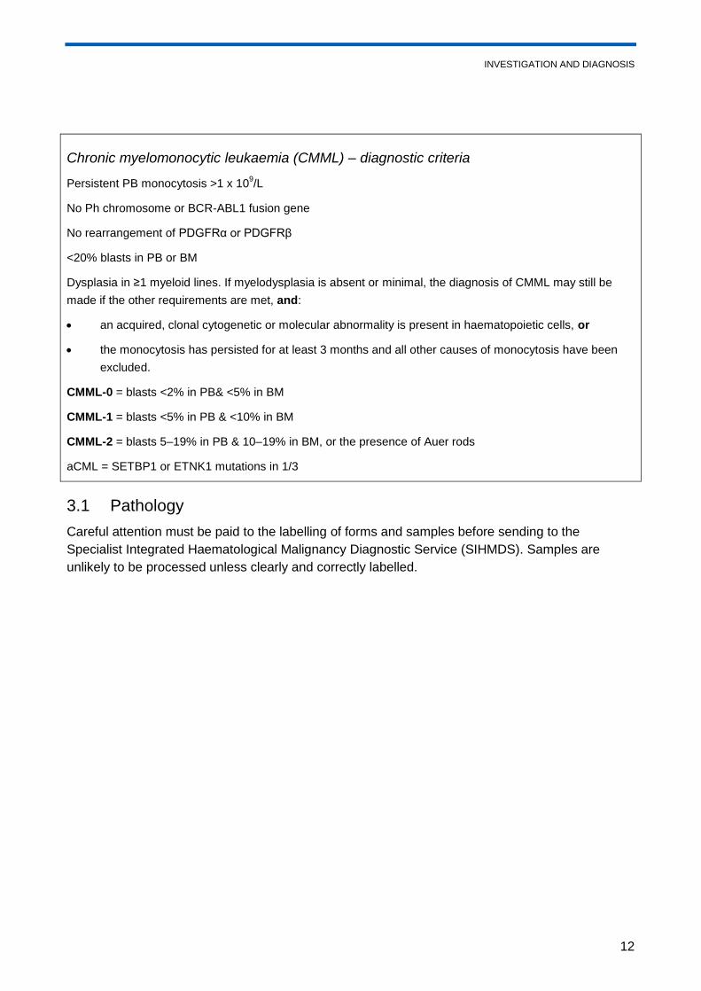
Chronic myelomonocytic leukaemia (CMML) – diagnostic criteria
Persistent PB monocytosis >1 x 10⁹/L
No Ph chromosome or BCR-ABL1 fusion gene
No rearrangement of PDGFRα or PDGFRβ
<20% blasts in PB or BM
Dysplasia in ≥1 myeloid lines. If myelodysplasia is absent or minimal, the diagnosis of CMML may still be
made if the other requirements are met, and:
an acquired, clonal cytogenetic or molecular abnormality is present in haematopoietic cells, or
the monocytosis has persisted for at least 3 months and all other causes of monocytosis have been
excluded.
CMML-0 = blasts <2% in PB& <5% in BM
CMML-1 = blasts <5% in PB & <10% in BM
CMML-2 = blasts 5–19% in PB & 10–19% in BM, or the presence of Auer rods
aCML = SETBP1 or ETNK1 mutations in 1/3
3.1 Pathology
Careful attention must be paid to the labelling of forms and samples before sending to the
Specialist Integrated Haematological Malignancy Diagnostic Service (SIHMDS). Samples are
unlikely to be processed unless clearly and correctly labelled.
RISK STRATIFICATION
13
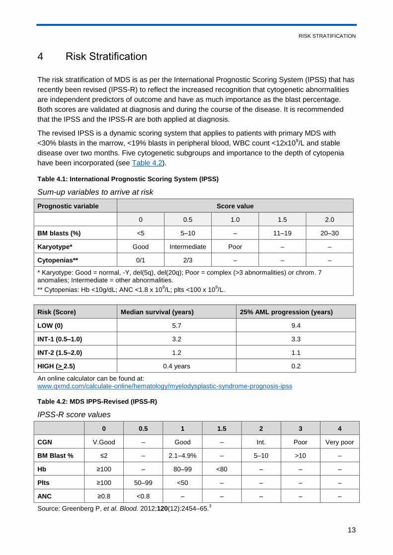
4 Risk Stratification
The risk stratification of MDS is as per the International Prognostic Scoring System (IPSS) that has
recently been revised (IPSS-R) to reflect the increased recognition that cytogenetic abnormalities
are independent predictors of outcome and have as much importance as the blast percentage.
Both scores are validated at diagnosis and during the course of the disease. It is recommended
that the IPSS and the IPSS-R are both applied at diagnosis.
The revised IPSS is a dynamic scoring system that applies to patients with primary MDS with
<30% blasts in the marrow, <19% blasts in peripheral blood, WBC count <12x109/L and stable
disease over two months. Five cytogenetic subgroups and importance to the depth of cytopenia
have been incorporated (see Table 4.2).
Table 4.1: International Prognostic Scoring System (IPSS)
Sum-up variables to arrive at risk
Prognostic variable Score value
0 0.5 1.0 1.5 2.0
BM blasts (%) <5 5–10 – 11–19 20–30
Karyotype* Good Intermediate Poor – –
Cytopenias** 0/1 2/3 – – –
* Karyotype: Good = normal, -Y, del(5q), del(20q); Poor = complex (>3 abnormalities) or chrom. 7 anomalies; Intermediate = other abnormalities.
** Cytopenias: Hb <10g/dL; ANC <1.8 x 10⁹/L; plts <100 x 10
9/L.
Risk (Score) Median survival (years) 25% AML progression (years)
LOW (0) 5.7 9.4
INT-1 (0.5–1.0) 3.2 3.3
INT-2 (1.5–2.0) 1.2 1.1
HIGH (> 2.5) 0.4 years 0.2
An online calculator can be found at: www.qxmd.com/calculate-online/hematology/myelodysplastic-syndrome-prognosis-ipss
Table 4.2: MDS IPPS-Revised (IPSS-R)
IPSS-R score values
0 0.5 1 1.5 2 3 4
CGN V.Good – Good – Int. Poor Very poor
BM Blast % ≤2 – 2.1–4.9% – 5–10 >10 –
Hb ≥100 – 80–99 <80 – – –
Plts ≥100 50–99 <50 – – – –
ANC ≥0.8 <0.8 – – – – –
Source: Greenberg P, et al. Blood. 2012;120(12):2454–65.3
RISK STRATIFICATION
14
IPSS-R cytogenetic prognostic subgroups
Very good Good Intermediate Poor Very poor
Single
-Y
Del(11q)
Normal
Single
Del(5q)
Del(12p)
Del(20q)
Double
Incl. del(5q)
Single
Del(7q)
+8
I(17q)
+19
Any other
independent clone
Double
Any other
Single
der(3q)
-7
Double
Incl. -7/7q-
Complex
3 abnormalities
Complex
>3 abnormalities
IPSS-R prognostic risk categories/scores
Risk category Risk score Survival
(median years)
AML 25% evolution
Very low ≤1.5 8.8 –
Low >1.5–3 5.3 10.8
Intermediate >3–4.5 3.0 3.2
High >4.5–6 1.6 1.4
Very high >6 0.8 0.73
Table 4.3: CMML-Specific Prognostic Scoring System (CPSS)
Variable 0 1 2
WHO subtype CMML-1 CMML-2 –
FAB subtype CMML-MD (WBC <13) CMML-MP (WBC >13) –
CGN* Low Intermediate High
RBC dependent No Yes –
*Low = normal, -Y; Intermediate = other abnormalities; High = +8, complex (≥3 anomalies), chrom. 7 anomalies.
Source: Such E, et al. Blood. 2013;121:3005–15.4
Risk group Overall score
Low 0
Intermediate-1 1
Intermediate-2 2–3
High 4–5
RISK STRATIFICATION
15
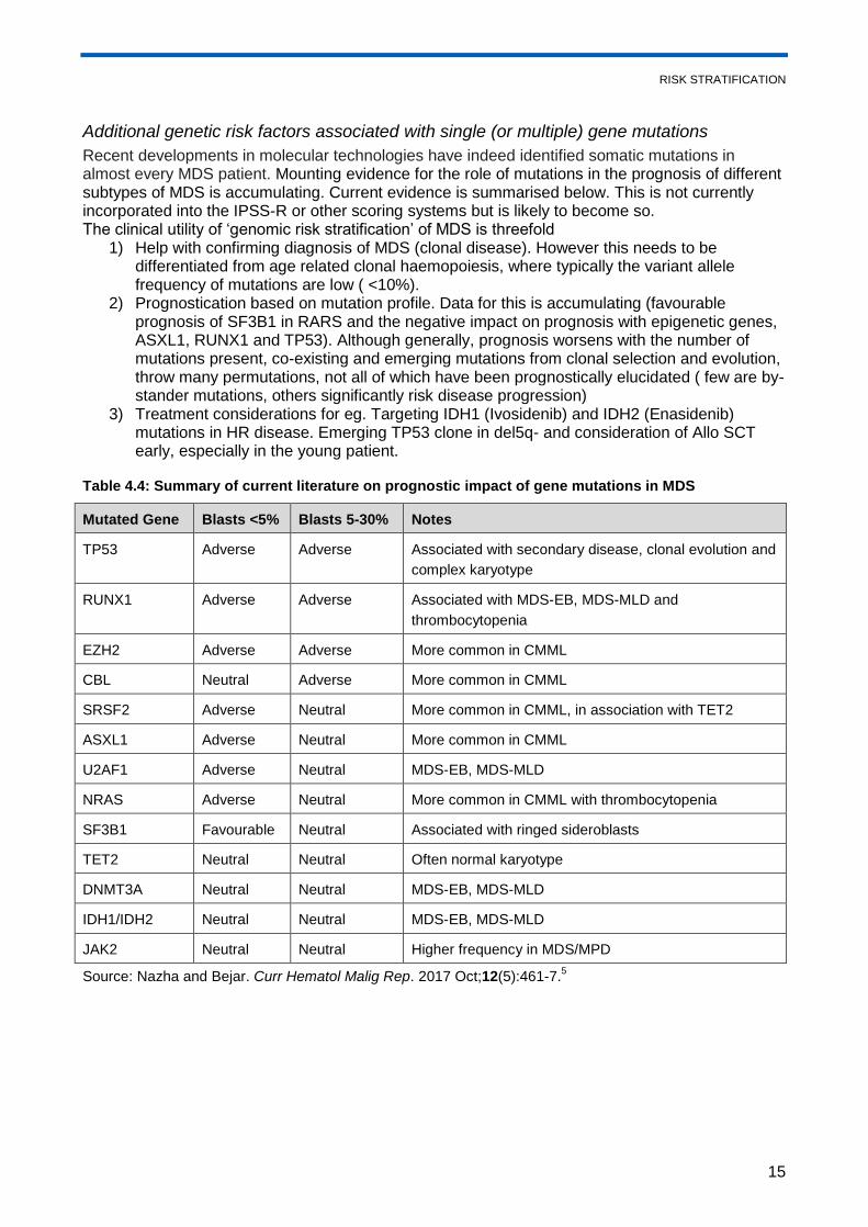
Additional genetic risk factors associated with single (or multiple) gene mutations
Recent developments in molecular technologies have indeed identified somatic mutations in almost every MDS patient. Mounting evidence for the role of mutations in the prognosis of different subtypes of MDS is accumulating. Current evidence is summarised below. This is not currently incorporated into the IPSS-R or other scoring systems but is likely to become so. The clinical utility of ‘genomic risk stratification’ of MDS is threefold
1) Help with confirming diagnosis of MDS (clonal disease). However this needs to be differentiated from age related clonal haemopoiesis, where typically the variant allele frequency of mutations are low ( <10%).
2) Prognostication based on mutation profile. Data for this is accumulating (favourable prognosis of SF3B1 in RARS and the negative impact on prognosis with epigenetic genes, ASXL1, RUNX1 and TP53). Although generally, prognosis worsens with the number of mutations present, co-existing and emerging mutations from clonal selection and evolution, throw many permutations, not all of which have been prognostically elucidated ( few are by-stander mutations, others significantly risk disease progression)
3) Treatment considerations for eg. Targeting IDH1 (Ivosidenib) and IDH2 (Enasidenib) mutations in HR disease. Emerging TP53 clone in del5q- and consideration of Allo SCT early, especially in the young patient.
Table 4.4: Summary of current literature on prognostic impact of gene mutations in MDS
Mutated Gene Blasts <5% Blasts 5-30% Notes
TP53 Adverse Adverse Associated with secondary disease, clonal evolution and
complex karyotype
RUNX1 Adverse Adverse Associated with MDS-EB, MDS-MLD and
thrombocytopenia
EZH2 Adverse Adverse More common in CMML
CBL Neutral Adverse More common in CMML
SRSF2 Adverse Neutral More common in CMML, in association with TET2
ASXL1 Adverse Neutral More common in CMML
U2AF1 Adverse Neutral MDS-EB, MDS-MLD
NRAS Adverse Neutral More common in CMML with thrombocytopenia
SF3B1 Favourable Neutral Associated with ringed sideroblasts
TET2 Neutral Neutral Often normal karyotype
DNMT3A Neutral Neutral MDS-EB, MDS-MLD
IDH1/IDH2 Neutral Neutral MDS-EB, MDS-MLD
JAK2 Neutral Neutral Higher frequency in MDS/MPD
Source: Nazha and Bejar. Curr Hematol Malig Rep. 2017 Oct;12(5):461-7.5
PATIENT INFORMATION/SUPPORT
16
5 Patient Information/Support
If the diagnosis of MDS is certain, patients should be informed that MDS is a clonal disorder and
that it is considered malignant/neoplastic. Their prognosis based on the IPSS/ IPSS-R should be
discussed, along with possible treatment options.
All patients must have access to a key worker. This is usually (but not always) the clinical nurse
specialist.
The clinical nurse specialist/key worker should be present at diagnosis and at any significant
discussion where treatment changes and outcomes are discussed. Where it is not possible for the
clinical nurse specialist or a deputy to be present, patients should be given the clinical nurse
specialist’s contact numbers. The clinician leading the consultation should advise the clinical nurse
specialist who should then arrange to make contact with the patient.
The clinical nurse specialist should ensure that all patients are offered a Holistic Needs
Assessment (HNA) at key pathway points, including: within 31 days of diagnosis; at the end of
each treatment regimen; and whenever a person requests one. Following each HNA, every patient
should be offered a written care plan. This plan should be developed with the patient and
communicated to all appropriate healthcare and allied healthcare professionals.
Written and verbal information are essential and the key worker/clinical nurse specialist plays a key
role in ensuring that patients have access to appropriate and relevant written information about
their condition.
The Macmillan Cancer Support, MDS Foundation and MDS UK Patient Support Group websites
and information booklets are good sources of patient information at diagnosis and are available for
download on the following websites:
www.macmillan.org.uk/Cancerinformation/Cancerinformation.aspx
https://www.mds-foundation.org/patient-caregiver-resources/#Programs
http://mdspatientsupport.org.uk/what-is-mds/information-material
TREATMENT
17
6 Treatment
The management of MDS may vary from monitoring blood counts for evidence of disease
progression in early MDS and supportive care, to intensive chemotherapy followed by stem cell
transplantation in those with advanced disease. Patients with ICUS should be followed up in the
same way as for low risk MDS until the diagnosis is clear. Responses to treatment should be
recorded using the Cheson 2006 criteria.
A focus on overall response rate (ORR) defined by improvement in blood counts or reduction in the
proportion of bone marrow blasts is, by its self, not enough to validate a new drug‘s usefulness in
MDS unless it correlates with improvement in quality of life, a meaningful reduction in transfusion
frequency, or longer survival.
The goals of treating MDS are to prolong survival, improve quality of life and improve the blood
counts. In order to achieve these goals, treatment options vary from best supportive care,
replacement therapy, low intensity therapy and high intensity therapy. In early MDS, the
predominant goal is haematological improvement with best supportive care. The imminent threat to
life in high risk MDS makes disease-modification the primary goal.
As patients with MDS are usually older and likely to have co-morbidities, the use of the Cumulative
Illness Rating Scale (CIRS) to assess the impact that co-morbidities may have on treatment is
recommended when planning treatment for MDS. Combined assessments with Geriatricians,
Haematologists and other specialty inputs can help, not only in planning treatments, mitigating
side-effects of treatments that could affect organ function, but also set realistic targets and
milestones that define holistic care of the patient.
Clear discussions with patients (and their families), at the point of diagnosis, detailing their
baseline disease characteristics (including the genetic profile of the disease, although it is not
currently incorporated into the disease staging algorithm), expected risk of progression and likely
survival outcomes, and most importantly, patient’s wishes, expectations and treatment preferences
should be clearly documented. Time points for review and re-discussion of treatment goals should
also be documented (for eg. Awaiting genetic/molecular test results, or defined number of cycles of
HMA).
Formal written consent should be obtained for all patients before commencing any
cytoreductive or epigenetic therapy including HU.
6.1 Low or intermediate-risk MDS
Also see section 7: Management of Disease and Treatment-related Complications.
Patients with IPSS-low or IPSS-INT1 may be eligible for a clinical trial.
6.1.1 Growth factor support (EPO +/- G-CSF)
For those patients with low risk disease and primarily anaemia and an EPO predictive score that is
low (serum erythropoietin <500IU and less than 2U blood transfusion/monthly), treatment with
recombinant human erythropoietin (EPO) 30,000 to 60,000 IU SC weekly for at least 8 weeks,
followed by a higher dose for 8 weeks, is recommended. The addition of G-CSF 300µg once a
TREATMENT
18
week (to maintain neutrophils between 5–10 x 109/L) should be considered in all patients with
refractory anaemia with ring sideroblasts (RARS) and other patients where the response to EPO
alone is suboptimal. The target Hb is 10-12g/dl but dose adjustments need to be made prior to this
to prevent overshooting. The ferritin should be maintained >100µg/ml for those on erythropoietin
replacement with IV iron infusions. Regular blood pressure monitoring and appropriate therapy
should accompany EPO treatment.
6.1.2 Anti-thymocyte globulin (ATG)/ciclosporin
Hypocellular/normocellular patients who have a normal karyotype or trisomy 8 may respond to
immunosuppressive therapy with ATG/ciclosporin. The HLA-DR15 haplotype is a good predictor of
response to immunosuppressive therapy, especially ATG. Patients not suitable for ATG may
receive Campath-1H currently available under compassionate program. Single agent ciclosporin is
a suitable alternative for elderly patients not deemed fit for more intensive immunosuppressive
therapy.
6.1.3 Lenalidomide
Patients with transfusion dependent anaemia and del(5q) with low risk or Int-1 (IPSS) who are
unresponsive to or unsuitable for ESAs are eligible for treatment with lenalidomide. Where blast
percentage is >5%, response rates may be lower and advice from a haematologist with sub-
specialist expertise is advisable. Responses occur rapidly at a median of 4 weeks from starting
therapy, and therapy should be continued until loss of response or disease progression. Starting
dose for patients with creatinine clearance >=60ml/min is 10mg daily for 21 out of 28 days. Dose
modifications as follows for renal impairment are shown below:
CrCl 30 to 60 mL/min 5 mg once daily
CrCl <30 mL/min (not requiring dialysis) 2.5 mg once daily
CrCl <30 mL/min (requiring dialysis) 2.5 mg once daily. On dialysis days,
administer the dose following dialysis
Cytopenias are the most common toxicity with lenalidomide. Recommendations for dose
reductions for cytopenias can be found here:
https://www.revlimid.com/mds-hcp/dosing/how-to-dose-modify/
Patients with del 5q MDS, developing cytopenias after a period of treatment with lenalidomide,
should also be screened for clonal evolution, to look for blast progression or emergence of a TP53
clone.
6.2 High risk MDS
Patients should be assessed for eligibility to undergo an allogeneic stem cell transplant: assess
fitness and co-morbidities using the HCT-CI, and carry out tissue typing for potential donors,
including siblings where familial MDS is not suspected.
6.2.1 Allogeneic stem cell transplant
If patients are transplant-eligible, a donor should be identified at the earliest possible opportunity.
Patients may be treated either directly with a myeloablative transplant (if fit, young and blasts
TREATMENT
19
<10%) or following induction chemotherapy with daunorubicin/cytarabine (DA 3+10) or a similar
regimen to remission (blasts <5% and no MRD by normal karyotype). For older patients, or for
those with co-morbidities, a RIC transplant is preferred, although the risk of relapse is higher.
Results from an allele-matched 10/10 VUD donor approximate those of a matched sibling
transplant and is a viable option.
For those who have tolerated chemotherapy and regenerated within 4–5 weeks, one cycle of
treatment to consolidate the remission is preferably to be administered prior to an allogeneic stem
cell transplant. It is recognised that a proportion of patients with MDS may develop chemotherapy-
induced aplasia and experience a prolonged time to recover counts, in which case a rescue
allograft may be necessary.
For patients with a complex karyotype or monosomal karyotype, there is some evidence to suggest
that treatment with hypomethylating agents such as 5’-azacitidine may be a good option.
In cases that are refractory to chemotherapy, the use of sequential chemotherapy and
transplantation is experimental; if it is being considered, it should be undertaken early in the course
of treatment.
6.2.2 Hypomethylating agents: 5’-azacitidine (5’-aza)
Where a patient declines, or is not a suitable candidate for, allogeneic stem cell transplantation,
the standard of care is treatment with 5’-aza based on a Phase III open label randomised
controlled trial that demonstrated disease-modifying activity in IPSS-INT2 high risk patients,
non-proliferative CMML and in AML with less than 30% blasts. The licensed regimen consists of
5’-aza 75mg/m2 SC daily for 7 days every 28 days, for at least 4–6 cycles to assess response,
and continued until loss of response or disease progression. The trial demonstrated improved
overall survival at 2 years of approximately 50%, compared with 24% for low dose cytarabine,
but there was no difference compared with chemotherapy. Treatment with 5’-aza may result in a
complete remission in 16% of cases, but is more likely to show responses with haematological
improvement. Given that the median time of response to 5’-aza is 18–24 months, in suitable cases
an allogeneic stem cell transplant in CR may be considered.
6.2.3 Hypomethylating agents: decitabine
Decitabine 20mg/m2 IV for five days every 28 days and, more recently, an extended 10-day
schedule may be useful in high risk MDS as an alternative to 5’-aza. However, the drug is not
licensed for MDS in Europe, and so IFR funding or treatment on a clinical trial should be sought.
6.2.4 Emerging and combination treatment options
Azacytidine remains the only licensed disease modifying drug in MDS with excess of blasts, in
Europe. In HR MDS, Azacytidine has been studied with various combinations of histone
deacetylase inhibitors (Valproic acid, Vorinostat and Entinostat), immunomodulating agents
(Lenalidomide and Thalidomide) and Gemtuzumab. However, none have shown any response or
survival advantage of those combinations over azacitidine alone. Similarly, check-point inhibitors
when combined with HMA, have failed to show any superiorty to HMA alone therapy.
BCL-2 inhibition with Venetoclax when combined with Decitabine, has shown promise in
relapse/refractory setting of AML with overall RR of 60%. However, there is no published data of
this combination therapy in HR MDS.
TREATMENT
20
IDH1 and 2 inhibitors have shown promise in the R/R – AML patients with ORR of ~40% and
median survival of 8.8 months. They remain potential therapeutic targets, and hence patients
should be screened for these mutations at disease progression even when these mutations were
absent at presentation. IDH1/2 inhibition with HMA is currently being studied in clinical trials, or
access to these drugs is currently limited to compassionate access programmes.
Drugs of promise in HR MDS (Guadecitabine, APR-246, High dose Decitabine and oral
azacytidine) are currently being studied. If Allo SCT cannot be considered, supportive care and
access through means of clinical trials or compassionate grounds remain promising options.
Caution needs to be exercised to drugs being added to HMA’s, in view of the added toxicity of
myelosuppression, especially for the elderly patient being treated in the out-patient setting.
6.2.5 Patients refractory to induction chemotherapy or hypomethylating agents
Best supportive care
Treatment on a clinical trial
Referral to palliative care teams may be considered.
6.3 Chronic myelomonocytic leukaemia (CMML)
CMML was part of the original FAB classification. However, in WHO (2008) it has been included in
the MDS/MPN overlap category. Patients with CMML may have varying prognosis and the
Dusseldorf scoring system or the CMML-specific prognostic score is recommended in order to
determine prognosis.
6.3.1 Active monitoring
For some patients (CMML-1 and some stable CMML-2 patients), active monitoring may be
sufficient.
6.3.2 Supportive care
Treatment with supportive care and hydroxycarbamide to control counts is recommended in the
absence of excess blasts.
6.3.3 5’-azacitidine
The National Institute for Health and Care Excellence (NICE) has approved the use of 5’-aza for
patients requiring treatment for CMML-2 only. For proliferative (WBC <13,00) CMML, 5’-aza at
conventional dosing may be used, but a funding application may be required.
6.3.4 Intensive chemotherapy
For patients with disease progression to CMML-2, AML induction chemotherapy followed by an
allogeneic stem cell transplant may be considered, based on patient characteristics and donor
availability.
TREATMENT
21
6.4 Fertility
For young patients with MDS due to undergo AML induction-type chemotherapy and/or a stem cell
transplant, the options for fertility preservation should be discussed and the patient referred to an
onco-fertility specialist for preservation of sperm, ovarian tissue or fertilised embryos.
Consideration of fertility preservation should be made for those of reproductive age (men below the
age of 55 and women below the age of 40).
MANAGEMENT OF DISEASE AND TREATMENT-RELATED COMPLICATIONS
22
7 Management of Disease and Treatment-related Complications
7.1 Anaemia
The onset of symptomatic anaemia is an independent prognostic factor in MDS. It is important to
record the number of transfusions a patient has had, with transfusion-dependence defined as the
need for >2 units per month for 4 months.
Assessment of anaemia should include haematinics, screening for haemolysis, blood loss and
infection. Identification of 5q- syndrome, a PNH clone or hypocellularity may alter therapies. Serum
erythropoietin levels may be low in elderly patients and should be measured in all patients with MDS.
For those with an EPO predictive score that is low (serum erythropoietin <500IU and less than 2U
blood transfusion), treatment with recombinant human erythropoietin (EPO) 30,000 to 60,000IU SC
weekly for at least 8 weeks, followed by a higher dose for 8 weeks, is recommended. The addition
of G-CSF 300µg once a week (to maintain neutrophils between 5–10 x 109/L) should be
considered in all patients with RARS) and in other patients where the response to EPO alone is
suboptimal. The target Hb is 10–12g/dl, but dose adjustments need to be made prior to this to
prevent overshooting.
The ferritin should be maintained at >100µg/ml for those on erythropoietin replacement with IV iron
infusions.
Blood transfusions may be the mainstay for those predicted to have a low response to EPO (serum
erythropoietin >500IU and >2U blood transfused). Patients facing primary or secondary ESA
failure, can be considered for clinical trials, with Lenalidomide or hypomethylating agents. In
patients with RS, refractory to ESA and low transfusion requirements, Luspatercept achieves
transfusion independence in 38% patients at a median of 30 weeks of treatment duration. In LR
MDS, erythroid response and haematological improvement is seen in 63%. Luspatercept is under
consideration with EMA, to get license for these indications, and access may have to be on
compassionate grounds. Clinical trials with other agents in LR MDS for improvement in anaemic
indices include Imtelstat, a telomerase inhibitor and Roxadustat, a hypoxia inducible factor inhibitor
and referral to centres where patients can be enrolled into clinical trials should be encouraged.
Patients with ongoing red cell transfusion dependency, especially young patients, should be
identified early on for consideration of allogeneic stem cell transplantation.
It is important to identify patients who may need iron chelation.
7.2 Severe neutropenia
There is no evidence to support routine use of G-CSF in neutropenic patients.
There is also no evidence to support routine prophylaxis with antimicrobials or antifungal drugs.
Most patients are unlikely to get serious infections till Neut > 0.5 x 10⁹/L. However, serious
infections can be seen, even in the absence of significant neutropenia, if neutrophil dysfunction is
present.
Door to needle time (DTN) to administration of broad spectrum antibiotics, especially with gram
negative coverage, is an independent variable to OS in patients with neutropenic sepsis.
MANAGEMENT OF DISEASE AND TREATMENT-RELATED COMPLICATIONS
23
Every haematology unit should have a neutropenic sepsis protocol in place, including in accident
and emergency wards, in close consultation with the Microbiology team.
In patients with hypoplastic MDS, the use of immunosuppression with anti-thymocyte globulin
(horse ATG preferable) and ciclosporin (CYA) or single agent ciclosporin may be helpful
(responses more likely to be seen with - absence of ring sideroblasts, a hypoplastic bone marrow,
HLA‐DR15, younger age (<60 years), female gender, normal karyotype or trisomy 8, presence of a
paroxysmal nocturnal haemoglobinuria clone, and short duration of transfusion dependence.
It is important to examine a peripheral film to exclude T-LGL, as treatment with ciclosporin and
methotrexate may be beneficial.
7.3 Neutropenic sepsis
Patients with neutropenic pyrexia or sepsis should be treated according to local protocols for neutropenic sepsis (and following National Institute for Health and Care Excellence/NICE guidance).
7.4 Severe thrombocytopenia
Platelet anisocytosis/clumping may give artefactually low platelet counts. Other causes of
thrombocytopenia also need to be considered, in particular immune thrombocytopenia in LR MDS.
Often the thrombocytopenia is out of proportion to the other cytopenias associated with low risk
disease.
Platelet transfusion may be used in MDS if there is bruising or bleeding. Steroids, IV
immunoglobulin in doses used to treat ITP may be tried if an immune component is suspected. For
non-bleeding patients and those not at high risk of spontaneous bleeding (i.e. not hypertensive),
transfuse platelets only when clinically indicated. Consider tranexamic acid in order to maintain
haemostasis. When a platelet transfusion programme is initiated, use single-donor apheresis
platelet products preferably, in order to avoid platelet refractoriness, unless in an emergency.
In refractory cases, patients should be assessed for the presence of HLA antibodies and
splenomegaly. Eltrombopag and Romiplostim can be useful, particularly if there’s an element of
immune thrombytopenia in MDS (unlicensed and unfunded indication). Trilineage responses to
these drugs have been reported, but the concerns with blast progression and clonal evolution have
not been entirely excluded with these agents, and hence are best considered as options within a
clinical trial.
7.5 Haemostasis and thrombosis
Although counts may be adequate, platelets (and neutrophils) may be dysfunctional in MDS. Such
patients may need platelet transfusions regardless of count for surgical procedures and/or
tranexamic acid in order to maintain haemostasis.
Platelet transfusion may be used in MDS if there is severe bruising or bleeding. For non-bleeding
patients and those not at high risk of spontaneous bleeding (i.e. not hypertensive), transfuse
platelets only when clinically indicated. Consider tranexamic acid in order to maintain haemostasis
when platelets <20 x 109/L or in the bleeding/high risk patient.
Ensure that patients have good control of blood pressure (if they are known to be hypertensive)
and do not suffer from constipation – if not appropriately managed, both conditions can increase
the risk of severe life-threatening haemorrhage.
MANAGEMENT OF DISEASE AND TREATMENT-RELATED COMPLICATIONS
24
7.6 Transfusional iron overload
Blood transfusions contribute to iron overload and transfusion in excess of 100 units may result in
evidence of end-organ damage (abnormal liver function, glucose intolerance or reduced left
ventricular ejection fraction). Iron chelation therapy is recommended for patients with a serum
ferritin >1000ng/ml or who have received in excess of 20 blood transfusions or evidence of
transfusional iron overload by ferriscan or cardiac MRI, and are expected to have a life expectancy
in excess of 3 years. If a patient has a life expectancy of <3 years when the transfusion regimen
commences, they are unlikely to become symptomatically iron-overloaded and chelation therapy
should not normally be started. An exception to this may be patients with underlying cardiac
problems (e.g. AF or CHD) who may also be more susceptible to the effects of iron overload and
patients in whom a transplant may be considered since iron overload is a negative prognostic
factor in transplant outcomes. It is recommended that all patients receiving chelation have a
baseline surrogate measure of labile plasma iron using ferriscan along with baseline audiology.
The serum ferritin is the most convenient way to monitor iron accumulation. However, it is an acute
phase reactant and may be elevated in liver disease as well. It is not clear in MDS at what levels of
ferritin end-organ iron-overloading occurs. However, iron-overload may contribute to
dyserythropoiesis. It is also an independent predictor of poor outcomes following stem cell
transplantation.
It is recommended that a cumulative record of number of units transfused be kept in the notes and
serum ferritin be checked after 20 units of blood have been transfused. Thereafter, ferritin levels
should be measured after every further 10 units transfused until a decision is made to chelate.
Consider a clinical trial for this patient group, if available.
The choice of iron chelator in the UK lies between Desferrioxamine and Desferasirox. Currently,
funding for Deferasirox is recommended for inherited transfusion dependent anaemias by NHS
England. However, cost analysis by NHS England for this purpose indicated similar costs for
Desferrioxamine when infusers and needles were incorporated compared to Deferasirox.
Furthermore, compliance is improved using oral therapy. Patients being initiated on iron chelation,
should have baseline audiometry, ophthalmic and renal function evaluations. Clinical judgement
relating to suitability, patient compliance and co-morbidities should define the choice of chelator.
SUPPORTIVE CARE
25
8 Supportive Care
8.1 Anaemia
See section 7: Management of Disease and Treatment-related Complications.
Transfusion triggers should be chosen in advance for patients, depending on their symptoms, co-
morbidities and life-style aspirations. For patients with no co-morbidities or bleeding risk, and in
those who do not lead active lifestyles, it would be reasonable to aim for a target Hb<80g/L.
8.2 Transfusions
See section 7: Management of Disease and Treatment-related Complications.
Transfusion triggers should be chosen in advance for patients, depending on co-morbidities.
For patients with no co-morbidities or bleeding risk, and in those who do not lead active lifestyles, it
would be reasonable to aim for a target of Hb>80g/L providing this does not lead to significant
symptoms.
Universal leukodepletion has significantly decreased the risk of CMV transmission. However,
CMV-negative blood products should be considered until the patient’s CMV status is known,
particularly in patients being considered for Allo SCT. All platelet products should be single donor
collections in order to limit the risk of allo-sensitisation. HLA-typing should be done prior to starting
treatment in order to address donor status if transplantation is appropriate for the patient, and in
case HLA-matched platelets become necessary during treatment (as often occurs in women who
have had children). Irradiated blood products should be requested for patients on protocols
containing fludarabine, cladribine and clofarabine and for at least one month prior to a planned
SCT.
8.3 Haemostasis and thrombosis
See section 7: Management of Disease and Treatment-related Complications.
8.4 Infection prophylaxis
There is no evidence to support routine use of G-CSF in neutropenic patients. There is also limited
evidence to support routine prophylaxis with antimicrobials or antifungal drugs (see section 7:
Management of Disease and Treatment-related Complications).
Patients with high risk MDS (and RAEB-2) should be managed as AML.
In neutropenic patients with recurrent infections, prophylactic antimicrobial and antifungal therapy
should be administered according to local flora and sensitivities, and as per local protocols on
neutropenic sepsis.
In non-neutropenic patients, neutrophils may be dysfunctional and in this case patients may have
recurrent infections. Such patients may benefit from prophylactic antimicrobial and antifungal
therapy directed towards local flora and sensitivities according to local protocols. Such patients
may also benefit from intermittent G-CSF. Local guidance from microbiology should be sought in
such cases.
Mouthwashes should be used as per local protocols in susceptible patients.
TREATMENT SUMMARY AND CARE PLAN
26
9 Treatment Summary and Care Plan
The MDT outcome and clinic letters will serve to communicate new lines of treatment with the
patient’s GP.
Most therapies are administered until loss of response or disease progression. It is important to
ensure that a treatment summary is completed when there are any significant changes in
treatment or follow-up plans.
Holistic Needs Assessments (HNAs) should be offered through follow-up, with a care plan
completed to document the plans to address the issues raised by the patient.
9.1 Treatment summary and care plan
There are two related but distinct documents which patients should be given when there are
changes in treatment.
A treatment summary provides a summary of the cancer treatments received by the end of
the first treatment, planned follow-ups (including mechanisms for these), and signs and
symptoms of which to be aware. Their aim is to provide information not only to the patient but
also to the GP about possible consequences of cancer and its treatment, signs of recurrence
and other important information.
A care plan is generated as a result of an HNA and is the agreed plan between the patient
and healthcare professional about how the identified areas of concern will be addressed.
This may cover provision of information (e.g. through an information prescription), onward
referral for specialist assessment and intervention (e.g. breathlessness management), or
things which the patient themselves can do (e.g. contact their HR department about
graduated return to work options).
Recommendation:
An end of treatment consultation should be offered to every patient when there are any significant changes
in treatment and follow-up arrangements. This should include an HNA and associated written care plan and
should also include the discussion and provision of a comprehensive treatment summary.
FOLLOW-UP ARRANGEMENTS
27
10 Follow-up Arrangements
Patients with low risk MDS not on treatment or supportive care may be followed up every 6–12
months.
Patients on treatment will need more frequent monitoring, depending on the therapy and the
degree of supportive care required. Patients with intermediate-2 or high risk disease on therapy
may need weekly (or more) blood count monitoring and supportive therapies.
Patients may have shared care between a specialist site and the local treating hospital. These
arrangements must be clearly outlined so that the patient is clear where to attend in an emergency
and understands the lines of communication between the sites.
11 End-of-life Care
For older patients and in those with high risk diseases, discussions regarding prognosis and
treatment options should also include discussions of end-of-life care. These discussions are to
facilitate transitions between active disease-modifying therapy and clinical trials to supportive care
only, at the time of disease progression/non-response. Care may be required from specialist
palliative care teams.
To support consideration of referral to specialist palliative care, please refer to the local referral
criteria for specialist palliative care.
The named clinical nurse specialist (CNS)/key worker, patient, family members and palliative care
teams, as well as members of the inpatient ward team, may be involved. Clear documentation of
the discussion with guidance to the treating teams is helpful in communicating these discussions
and outputs to the wider team that may care for the individual.
12 Data Requirements
Accurate data collection is essential to monitor outcomes, and the collection of this information,
particularly clinical data, remains the responsibility of the members of the multidisciplinary team
with support from a data manager. Haematology services are required to submit data to nationally
mandated datasets for all patients diagnosed with haematological cancer; further details on these
datasets are available in Annex 1.
REFERENCES
28
References
1. Killick SB, Carter C, Culligan D, et al. Guidelines for the diagnosis and management of adult
myelodysplastic syndromes. British Journal of Haematology. 2014:164:503–25.
2. Malcovati L, Hellstrom-Lindberg, Bowen D, et al. Diagnosis and treatment of primary
myelodysplastic syndromes in adults. Recommendations from the European LeukaemiaNet.
Blood. 2013:122:2943–64.
3. Greenberg P, et al. Revised international prognostic scoring system for myelodysplastic
syndromes. Blood. 2012;120(12):2454–65.
4. Such E, et al. Development and validation of a prognostic scoring system for patients with
chronic myelomonocytic leukemia. Blood. 2013;121:3005–15.
5. Nazha A and Bejar R. Molecular Data and the IPSS-R: How Mutational Burden Can Affect
Prognostication in MDS. Curr Hematol Malig Rep. 2017 Oct;12(5):461-7.
6. Adès, L., Sebert, M. & Fenaux, P. (2019) Guadecitabine in myelodysplastic syndromes:
promising but there is still progress to be made. The Lancet Haematology, 6, e290– e291.
7. DiNardo, C.D., Stein, E.M., de Botton, S et al. (2018a) Durable Remissions with ivosidenib in
IDH1‐mutated relapsed or refractory AML. The New England Journal of Medicine, 378, 2386–
2398.
8. Fenaux, P., Platzbecker, U., Mufti, G., et al (2018b) The MEDALIST trial: results of a phase 3,
double‐blind, multicenter, placebo‐controlled, randomized study of luspatercept to treat anemia
in patients with very low‐, low‐, or intermediate‐risk myelodysplastic syndromes (MDS) with ring
sideroblasts who require red blood cell (RBC) transfusions. Blood, 132, 1.
9. Jilg, S., Reidel, V., Müller‐Thomas, et al. (2016) Blockade of BCL‐2 proteins efficiently induces
apoptosis in progenitor cells of high‐risk myelodysplastic syndromes
patients. Leukemia, 30, 112– 123.
10. Papaemmanuil, E., Gerstung, M., Malcovati, L et al. (2013) Clinical and biological implications
of driver mutations in myelodysplastic syndromes. Blood, 122, 3616– 3627.
11. Steensma, D.P., Bejar, R., Jaiswal, S., et al (2015) Clonal hematopoiesis of indeterminate
potential and its distinction from myelodysplastic syndromes. Blood, 126, 9– 16.
ANNEX 1: DATA REQUIREMENTS
29
Annex 1: Data Requirements
Haematology oncology services within London are required to submit data to the following
nationally mandated datasets for all patients diagnosed with haematological cancers.
The Cancer Outcomes and Services Dataset (COSD)
The core dataset for all tumour types including haematological cancers is mandated from January
2013, and the site-specific dataset is mandated from July 2013. Details of the dataset can be found
on the National Cancer Intelligence Network website:
www.ncin.org.uk/collecting_and_using_data/data_collection/cosd.aspx
The local cancer registry will be collating this dataset using Trust data feeds which should include
all these items. The feeds are:
Trust PAS
Trust pathology
Trust radiology
Trust multidisciplinary team (MDT) feed.
In line with the requirements set out in Provider Trust contracts, this data should be submitted
within 25 working days of the end of the month in which the activity took place.
Three groups of haematological cancers are considered stageable by the Registry:
Lymphomas, using Ann Arbor (or Murphy St Jude for children)
Myelomas, using ISS
CLLs, using Rai and Binet
For the purposes of COSD, any other haematological cancers are not counted as stageable.
For CLL both Rai (0-IV) and Binet (A-C) stages need to be recorded and submitted to
COSD to be considered “fully staged”.
MGUS does not need to be recorded and submitted as it is not defined as an invasive tumour.
Systemic Anti-Cancer Therapy dataset (SACT)
Provider Trusts that provide chemotherapy to patients are required to submit data to the SACT
dataset. Details of the audit and the dataset requirements are available on the dataset homepage:
www.chemodataset.nhs.uk/home.aspx

© RM Partners, South East London Cancer Alliance, North Central and East London Cancer Alliance
2020
5th Floor Alliance House
12 Caxton Street
London SW1H 0QS