paper reelindifferential functions of apoer2 and very low density lipoprotein receptor in reelin...
TRANSCRIPT

7/28/2019 Paper ReelinDifferential Functions of ApoER2 and Very Low Density Lipoprotein Receptor in Reelin Signaling Depe…
http://slidepdf.com/reader/full/paper-reelindifferential-functions-of-apoer2-and-very-low-density-lipoprotein 1/13
Differential Functions of ApoER2 and Very Low DensityLipoprotein Receptor in Reelin Signaling Depend onDifferential Sorting of the Receptors*□SReceived forpublication,May 27,2009,and in revised form,October 27,2009 Published, JBC Papers in Press,November 29, 2009, DOI 10.1074/jbc.M109.025973
SarahDuit, HaraldMayer, SophiaM. Blake,Wolfgang J. Schneider, andJohannesNimpf 1
From the Max F. Perutz Laboratories, University Departments at the Vienna Biocenter, Department of Medical Biochemistry,Medical University of Vienna, Dr. Bohrgasse 9/2, A-1030 Vienna, Austria
ApoER2 and very low density lipoprotein (VLDL) receptor
transmit theReelin signal into targetcells of thecentral nervous
system. To a certain extent, both receptors can compensate for
each other, and only the loss of both receptors results in the
reeler phenotype, which is characterized by a gross defect in the
architecture of laminated brain structures. Nevertheless, both
receptors also have specific distinct functions, as corroborated
by analyses of the subtle phenotypes displayed in mice lacking
either ApoER2 or VLDL receptor. The differences in their func-
tion(s), however, have not been defined at the cellular level.
Here, using a panel of chimeric receptors, we demonstrate that
endocytosis of Reelin and the fate of the individual receptors
upon stimulation are linked to their specific sorting to raft ver-
sus non-raft domains of the plasma membrane. VLDL receptor
residingin the non-raft domain endocytoses and destinesReelin
for degradation via the clathrin-coated pit/clathrin-coated ves-
icle/endosome pathway without being degraded to a significant
extent. Binding of Reelin to ApoER2, a resident of rafts, leads to
the production of specificreceptor fragments withspecificfunc-
tions of their own and to degradation of ApoER2 via lysosomes.
These features contribute to a receptor-specific fine tuning of the Reelin signal, leading to a novel model that emphasizes
negative feedback loops specifically mediated by ApoER2 and
VLDL receptor, respectively.
DefectiveReelin signalingcauses lamination defects in many areas of the cerebral cortex, hippocampus, and cerebellum(1, 2). The major abnormality in the cortex arises during
embryogenesis from the inability of radially migrating neu-rons to invade and split the preplate. These neurons settlebeneath the preplate, which is shifted toward the pial sur-face, where it forms a “superplate” in affected animals. Con-sequently, later born neurons cannot bypass earlier neurons
that have settled beneath the superplate so that consecutivewaves of neurons generated in the subventricular zone andmigrating outwards establish a pattern of inverted neuronallayers. The role of Reelin in the correct lamination of certain
brain structures was recently compiled in the “detach andgo” model (3), where Reelin was proposed to promote
detachment of migrating neurons from glial fibers and theirtranslocation to the outermost area of the developing corticalplate. Despite recent progress in understanding molecularevents in the Reelin signaling pathway (reviewed in Refs. 4 and5), our knowledge about the modulation of the initial signal and
downstream events guiding migration and positioning of neu-rons or modulating other processes like dendrite developmentis still scarce (6, 7). Proposed mechanisms involve Lis1 (8),Nck (9), and differential modulation of phosphatidylinositol
3-kinase-downstream pathways employing mTOR or GSK3(10). The key events indispensable for triggering these path-ways are binding of Reelin to ApoER2 and VLDL2 receptor(VLDLR) and subsequent phosphorylation of Dab1. Obviously,Reelin-mediated receptor clustering triggers tyrosine phosphor-
ylation of Dab1 (11). This event, however, does not seem to besufficient to evoke the full Reelin signal, because anti-receptorantibodies that trigger Dab1 phosphorylation by receptor clus-tering do not rescue the reeler phenotype in brain slice cultures
(12). In addition, thrombospondin 1, which is another func-tional ligand for ApoER2 and VLDLR in the brain, promotesDab1 phosphorylation without eliciting the canonical Reelinsignaling pathway (13).
Dab1phosphorylationis mediatedby membersof theSrc family of kinases (14 –16). Mice lacking both Fyn and Src develop a phe-
notype similar to that of the Dab1-deficient scrambler mice(17). Dab1 binds to the NFDNP X Y sequence motif present inthe cytosolic domains of ApoER2 and VLDLR (18). Thisdomain is indispensable for Reelin signaling, since mice lackingVLDLR and carrying mutant alleles for ApoER2 coding for an
altered NFDNPVY motif that does not bind Dab1 develop areeler phenotype (19). This domain is also present in othermembers of the LDL receptor gene family, such as LDL recep-tor, LRP1, and LRP2 (20), and plays a key role in clathrin-me-diated endocytosis of these receptors. In a Dab1-overexpress-
ing cell model system, Dab1 decreases the endocytosis rate of the LDLreceptor by interfering with the formation of the endo-
* This work was supported by the “Fonds zur Forderung der Wissenschaftli-chen Forschung, FWF,” Austria, Grants P16872-B09 and P19611-B09 andthe Herzfelder’sche Familienstiftung.
□S The on-line version of this article (available at http://www.jbc.org)containssupplemental Figs. 1–4.
1 To whom correspondence should be addressed. Tel.: 43-1-4277-61808; Fax:43-1-4277-9618; E-mail: [email protected].
2 The abbrevia tions used are: VLDL, very low density lipopr otein; VLDLR,VLDL receptor; MES, 4-morpholineethanesulfonic acid; DMEM, Dulbec-co’s modified Eagle’s medium; RCM, Reelin conditioned medium; MCM,mock conditioned medium; Ab, antibody; HRP, horseradish peroxidase;IOD,integrated optical density;CLM, caveolin-richlight membrane(s); PBS,phosphate-buffered saline; CCV,clathrin-coated vesicle; NCM, non-caveo-lae membrane(s); CDX, methyl--cyclodextrin; CTF, C-terminal fragment;EEA1, early endosomal antigen 1.
THE JOURNAL OF BIOLOGICAL CHEMISTRY VOL. 285, NO. 7, pp. 4 896 –4908, February 12, 2010© 2010 by The American Society for Biochemistry and Molecular Biology, Inc. Printed in the U.S.A.
4896 JOURNAL OF BIOLOGICAL CHEMISTRY VOLUME 285•NUMBER 7• FEBRUARY 12, 2010
http://www.jbc.org/content/suppl/2009/11/30/M109.025973.DC1.htmlSupplemental Material can be found at:

7/28/2019 Paper ReelinDifferential Functions of ApoER2 and Very Low Density Lipoprotein Receptor in Reelin Signaling Depe…
http://slidepdf.com/reader/full/paper-reelindifferential-functions-of-apoer2-and-very-low-density-lipoprotein 2/13
cytosis complex (21). However, because Dab1 is brain-specific,the interaction with LDL receptor, LRP1, and LRP2 and the
interference with endocytosis of these receptors most likely arenot of physiological relevance.Our own studies and others havedemonstrated that ApoER2 associates with lipid rafts, whereasVLDLR is strictly excluded from these microdomains (22–24).Despite its raft association, ApoER2 is endocytosed via the
clathrin-mediated process, apparently through its associationwith Dab2 (22). It is still unclear, however, how importantendocytosis of ApoER2 and VLDLR is in Reelin signaling.
With the identification of ApoER2 and VLDLR as functionalReelin receptors, it became evident that both receptors can
compensate for each other to a certain extent, since only thelack of both receptors causes the reeler phenotype (25). Loss of ApoER2 or VLDLR alone causessubtle butdistinguishable phe-notypes, pointing to VLDLR being more important for thedevelopment of the cerebellum and ApoER2 for lamination of
thecortex. This fact wasrecentlycorroboratedby detailed stud-ies on the divergent roles of ApoER2 and VLDLR in the migra-tion of cortical neurons (26), which demonstrated that ApoER2
is indispensable for the correct migration of late generated neu-rons,whereas the VLDLR-mediated Reelin signal prevents neu-
rons from entering the marginal zone.Because both receptors mediate Dab1 phosphorylation, the
question of the molecular substrate for their individual func-tions arises. Using chimeric receptor constructs anda fibroblastmodel system, we now demonstrate that endocytosis and cellu-
lar trafficking differ between ApoER2 and VLDLR and relate toraft versus non-raft localization of these receptors.
EXPERIMENTAL PROCEDURES
Cell Culture and Preparation of Conditioned Media—NIH3T3 and 293 HEK cells were cultivated in Dulbecco’s modifiedEagle’s medium (DMEM; Invitrogen) supplemented with10% fetal calf serum (Invitrogen), and penicillin/streptomycin
(Invitrogen) at 37 °C and 7.5% CO2. Stable NIH 3T3-based cell
lines expressing murine ApoER2 harboring LA repeats 1–3, 7,and 8 and containing the proline-rich cytoplasmic insert (3T3A), murine VLDLR lacking the O-linked sugar domain (3T3 V),or either receptor and murine Dab1 (3T3 A/D and 3T3 V/D)
(23) were kept under puromycin selection (0.75 g/ml). Stablecell lines expressing chimeric receptors were generated asdescribed for 3T3 A and 3T3 V (23) and were grown under thesame conditions. 24 h before the experiment, puromycin-resis-
tant cells were switched to growth medium lacking puromycin.
Reelin-expressing 293 HEK cells were cultivated and used forproduction of Reelin conditioned medium (RCM) as describedbefore (27). Mock conditioned medium (MCM) was preparedfrom untransfected 293 HEK cells using the same procedure.
Primary rat neuronal cultures were obtained from embryonicday 16.5 rat embryonic brains and were kept in DMEM/F-12(Invitrogen) containing B27 supplement (Invitrogen) and pen-icillin/streptomycin at 37 °C and 5% CO
2for 72 h before use as
described (11). Transient transfection of 3T3 was done usingLipofectamine 2000 (Invitrogen) according to the manufacturer’sprotocol.
Cloning of Chimeric Receptors—The following primers wereused for generation of chimeric receptor constructs. VLDLR
and ApoER2 primer pairs, spanning the respective full-lengthmurine cDNAs, each containing an EcoRI restriction site
(underlined), were as follows: VLDLR sense primer (primer 1)(5Ј-CGG AAT TCA TGG GCA CGT CCG CGC GC-3Ј),VLDLR antisense primer (primer 2) (5Ј-ATG AAT TCA AGCCAG ATC ATC ATC TGT GCT TAC-3Ј), ApoER2 senseprimer (primer 3) (5Ј-ATG AAT TCA TGG GCC GCC CAG
AAC TGG-3Ј), and ApoER2 antisense primer (primer 4) (5
Ј-ATG AAT TCT CAG GGC AGT CCA TCA TCT TC-3Ј).
ApoER2-VLDLR chimeric primers, annealing to ApoER2 attheir 5Ј region (shown in italic type) and to VLDLR at their 3Ј
region (underlined) were as follows: sense primer (primer 5)(5Ј-GGT ACC TCA TCT GGA GGA ATT GGC AAC-3Ј) andantisense primer (primer 6) (5Ј-CGC TTC CAG TTC CTC CACATC AAG TAG CC-3Ј), recognizing the junction of trans-membrane and intracellular domain, and sense primer (primer
9)(5Ј-CAA CAG TCA CCG CTG CTG CTG CCT GGG-3Ј) andantisense primer (primer 10) (5Ј-CCC AAT GAC TGA AGTCCC TTT TGG GGG AAC-3Ј), recognizing the junction of extracellular and transmembrane domain. VLDLR-ApoER2
chimeric primers, annealing to VLDLR at their 5Ј region(underlined) and to ApoER2 at their 3Ј region (italic type) wereas follows: sense primer (primer 7) (5Ј-GGC TAC TTG ATGTGG AGG AAC TGG AAG CG -3Ј) and antisense primer(primer 8) (5Ј-GTT GCC AAT TCC TCC AGA TGA GGT AAC
CAC -3Ј), recognizing the junction of transmembrane andintracellular domain, and sense primer (primer 11) (5Ј-CCAAAA GGG ACT TCA GTC ATT GGG GTC ATC GTG C -3Ј)and antisense primer (primer 12) (5Ј-GAT GGC CCA GGCAGC AGC AGC GGT GAC TGT TGA GC -3Ј), recognizing the
junction of extracellular and transmembrane domain.For the first round of PCR amplification, fragments of
ApoER2 and VLDLR were amplified from pMSCVpuro-ApoER2 and pMSCVpuro-VLDLR (23), using the followingprimers: primers 1 and 10 for VLDLR extracellular domain,
primers 3 and 12 for ApoER2 extracellular domain, primers 1and 6 for VLDLR extracellular and transmembrane domain,primers 3 and 8 for ApoER2 extracellular and transmembranedomain, primers 9 and 2 for VLDLR transmembrane and intra-cellular domain, primers 11 and 4 for ApoER2 transmembrane
and intracellular domain, primers 5 and 2 for VLDLR intracel-lular domain, and primers 7 and 4 for ApoER2 intracellulardomain. The obtained fragments were purified and used astemplates for another round of PCR amplification. Fragmentsharboring the VLDLR extracellular and transmembrane do-
main were mixed with fragments harboring the ApoER2 intra-cellular domain and vice versa, and fragments harboring theVLDLR extracellular domain were mixed with fragments har-boring the ApoER2 transmembrane and intracellular domainand vice versa. Primers used for amplification were primers 1
and 4 for the VVA and VAA constructs and primers 3 and 2for the AAV and AVV constructs. The PCR productsobtained in this second PCR amplification step were clonedinto the pMSCVpuro backbone using the EcoRI restrictionsite.
Antibodies—Antibodies against ApoER2 were Ab 186, raisedagainst theentire ligand-binding domain (11); Ab 220, raised againstthe first ligand-binding repeat (28); and Ab 20, raised against the
Distinct Functionsof ApoER2andVLDL Receptor
FEBRUARY 12, 2010 • VOLUME 285• NUMBER 7 JOURNAL OF BIOLOGICAL CHEMISTRY 4897
http://www.jbc.org/content/suppl/2009/11/30/M109.025973.DC1.htmlSupplemental Material can be found at:

7/28/2019 Paper ReelinDifferential Functions of ApoER2 and Very Low Density Lipoprotein Receptor in Reelin Signaling Depe…
http://slidepdf.com/reader/full/paper-reelindifferential-functions-of-apoer2-and-very-low-density-lipoprotein 3/13
cytoplasmic tail (29). The ligand-binding domain of VLDLRwas detected using Ab 74, which was raised in rabbits using a
maltose-binding protein fusion protein containing the firstligand binding repeat of VLDLR. Ab 187 (11) was used fordetection of VLDLR in immunofluorescence assays. For immu-noprecipitation of Dab1,Ab 48 and Ab 54 (11) against the shortsplicevariant of murine Dab1 were used. Mouse anti-Dab1(D4)
and mouse anti-Reelin (G10) antibodies were kind gifts of Andre Goffinet (University of Louvain, Belgium). Mouse anti-Lis1 was obtained from Orly Reiner (Weizmann Instituteof Science, Rehovot, Israel). The following antibodies werepurchased from the indicated sources: mouse anti-VLDLR
(6A6) and mouse anti-phosphotyrosine (PY99), Santa CruzBiotechnology, Inc. (Santa Cruz, CA); mouse anti-HA (HA.11),Covance; mouse anti-clathrin heavy chain and rabbit anti-Caveolin, BD Transduction Laboratories; rabbit anti-early endosomal antigen 1, Affinity BioReagents; secondary HRP-
conjugated anti-mouse and anti-rabbit antibodies, JacksonImmunoResearch; Alexa Fluor 488 goat anti-mouse and AlexaFluor 594 goat anti-rabbit, Molecular Probes.
Preparation of Cell Extracts, SDS-PAGE, and Western Blotting —Cells were washed twice with ice-cold PBS and
lysed in Hunt buffer (20 mM Tris, pH 8.0, 100 mM NaCl,0.5% Nonidet P-40, 1 mM EDTA) supplemented with protein-ase inhibitor mix (CompleteTM, Roche Applied Science). Cell
debris was removed by centrifugation for 5 min at 20,000 ϫ g .Proteins were separated by reducing SDS-PAGE and trans-ferred onto nitrocellulose membranes by semidry blotting.Membranes were blocked in PBS containing 0.1% Tween 20and either 5% bovine serum albumin or 5% nonfat dry milk and
incubated with primary and HRP-conjugated secondary anti-bodies. For detection, enhanced chemiluminescence solution(Pierce) was used. For quantification of Western blot results,integrated optical density (IOD) values of the bands were cal-culated using Gel-Pro analyzer software (Media Cybernetics)
and normalized to IOD of the loading control. For ligand blots,cell extracts were separated by non-reducing SDS-PAGE andWestern blotting, and membranes were blocked in TBS con-taining 5% bovine serum albumin and 2 mM CaCl
2and incu-
bated with RCM diluted with Opti-MEM (1:2) containing 2 mM
CaCl2
for 2 h beforeincubation with primary (G10) andsecond-ary antibodies.
Reelin Uptake and Degradation Assay—NIH 3T3 cells ex-pressing ApoER2 or VLDLR were precooled at 4 °C for 30 min
and incubated with RCM at 4 °C for 1 h to allow for Reelin
binding to the cells. After extensive washing with TBS, cellswere covered with Opti-MEM (Invitrogen) and shifted to a37 °C water bath. After the indicated periods, cell extracts wereprepared using Hunt buffer and analyzed by Western blotting.
Relative Reelin amounts were calculated from IOD values. Dab1 Phosphorylation Assay—Cells expressing Dab1 and
one of the receptors were starved for 1 h in plain DMEM andincubated for 1 h with RCM or MCM. Cell extracts were pre-pared in Hunt buffer containing protease inhibitor mix and
phosphatase inhibitors (50 mM NaF and 2 mM Na3
VO4
) andused for immunoprecipitation. Extracts were incubated withanti-Dab1 antiserum overnight at 4 °C, 40 l of a proteinA-Sepharose bead slurry (Zymed Laboratories Inc.) were
added, and samples were incubated for 1 h at 4 °C again. Beadswere collected by centrifugation at 500 ϫ g for 1 min and
washed three times using Hunt buffer. Samples were analyzedby Western blotting.
Receptor Degradation and Fragmentation Assays—To ana-lyze degradation and secretase-mediated fragmentation of ApoER2, receptor-expressing cells were starved for 1 h in plain
DMEM or DMEM containing 20 g/ml cycloheximide andincubated for 4 or 6 h with medium containing cycloheximideand the indicated ligands and supplements. Cell extracts wereprepared in Hunt buffer and analyzed by Western blotting.
Isolation of Caveolin-rich Light Membranes (CLM)—CLMwere prepared from stable NIH 3T3 fibroblasts grown to con-fluence in 15-cm dishes or from WT mouse embryonic brainsisolated at embryonic day 15. All procedures were carried out at4 °C. Briefly, cells were washed with TBS and pelleted by cen-
trifugation (5 min, 1400 ϫ g ). The supernatant was removed,and cells were solubilized in TBS containing2% Brij 78P (Fluka)and CompleteTM protease inhibitors (Roche Applied Science)by passaging the cells 10 times through a 23-gauge needle. Cell
debris was removed by centrifugation (10 min, 21,000ϫ g ) andthe lysate (0.6 ml)was mixed with 0.6 ml of 90% (w/v) sucrose inMBS (MES-buffered saline; 25 mM MES, pH 6.5, 150 mM NaCl)and transferred to an ultracentrifuge tube. A discontinuoussucrose gradient was formed above the homogenate by adding
2.5 ml of 35% (w/v) sucrose in MBS, followed by 0.6 ml of 5%(w/v) sucrose in MBS. After centrifugation at 160,000 ϫ g for20 h in a Beckman SW60Ti rotor at 4 °C, 0.44-mlfractions werecollected from the top of the tube. Fraction 2 at the interfacebetween the 5 and 35% sucrose boundaries was designated the
CLM fraction. Preparation of Clathrin-coated Vesicles—Coated vesicles
were prepared from NIH 3T3 cells expressing either receptorgrown to near confluence or primary rat neurons cultured for72 h using a 2H
2O, 8% sucrose gradient (30). Cells were washed
twice with PBS and once with MES buffer (100 mM MES, pH6.5, 1 mM EGTA, 0.5 mM MgCl
2, 3 mM NaN
3, CompleteTM
proteinase inhibitor mixture). All steps were carried out at4 °C. Cells were scraped in MES buffer and homogenizedusing a Potter tissue grinder. The homogenate was centri-
fuged at 5000ϫ g for 5 min. The pellet was resuspended in MESbuffer and centrifuged at 5000 ϫ g for 5 min. The supernatantsfromthe twocentrifugations were combined and centrifuged at100,000ϫ g for 60 min. The resulting pellet was resuspended in
MES buffer and centrifuged at 10,000 ϫ g for 10 min. The
resulting pellet was resuspended in MES buffer again, and thecentrifugation was repeated. The supernatants from the twocentrifugation steps were combined and centrifuged at100,000 ϫ g for 60 min. The pellet was resuspended in 1 ml of
MES buffer and centrifuged at 10,000ϫ g for 10 min; the result-ing pellet was again resuspended using 1 ml of MES buffer andcentrifuged at 10,000 ϫ g for 10 min. The combined superna-tants were loaded on the top of 2 ml of 8% sucrose in 2H
2O and
centrifuged at 80,000 ϫ g for 2 h. The pellet, resuspended inMES buffer, was centrifuged at 20,000 ϫ g for 10 min. Thesupernatant was recovered and designated as clathrin-coated vesicle (CCV) fraction. Protein concentration was determinedusing theCoomassiePlus protein assay reagent (Pierce) accord-
Distinct Functionsof ApoER2andVLDL Receptor
4898 JOURNAL OF BIOLOGICAL CHEMISTRY VOLUME 285•NUMBER 7• FEBRUARY 12, 2010
http://www.jbc.org/content/suppl/2009/11/30/M109.025973.DC1.htmlSupplemental Material can be found at:

7/28/2019 Paper ReelinDifferential Functions of ApoER2 and Very Low Density Lipoprotein Receptor in Reelin Signaling Depe…
http://slidepdf.com/reader/full/paper-reelindifferential-functions-of-apoer2-and-very-low-density-lipoprotein 4/13
ing to the manufacturer’s protocol, and equal protein amountsof cell lysates and CCVwere subjectedto SDS-PAGE and West-
ern blotting. Immunofluorescence Assays and Microscopy—Sterile glass
coverslips were coated with 40 g/ml poly-L-lysine in PBS for1 h at room temperature. NIH 3T3 cells expressing ApoER2or VLDLR were grown on the coverslips for 24 h using stand-
ard fibroblast growth medium. Cells were cooled to 4 °C,washed with ice-cold PBS, and overlaid with RCM. After 1 hof incubation, RCM was removed, and cells were washed andincubated with Opti-MEM at either 4 or 37 °C for 10 min. Sub-sequently, cells were washed and fixed with 4% paraformalde-
hyde fixative. Fixed cells were washed with PBS containing 100mM glycine and incubated with 0.1% Triton X-100 in PBS for 2min to permeabilize the plasma membrane. Cells were washedagain with PBS, blocked for 30 min with blocking solution (1%
bovine serum albumin in phosphate-buffered saline) at roomtemperature, and incubated with primary and secondary anti-bodies diluted in blocking solution for 1 h each. Coverslips werewashed again and mounted on glass slides using DAKO fluo-
rescent mounting medium (Dako Corp.). Slides were analyzedusing a confocal fluorescence microscope (laser-scanningmicroscope 510, Zeiss) and the corresponding software (ZeissLSM Image Browser). Antibodies used for detection were Ab186 for ApoER2, Ab 187 for VLDLR, G10 for Reelin, anti-early endosomal antigen 1, and secondary Alexa Fluor-coupled anti-
mouse and anti-rabbit antibodies.
RESULTS
Expression and Subcellular Sorting of Chimeric Receptors—
We have recently demonstrated that (i) ApoER2 and VLDLRreside in distinct subdomains of the plasma membrane and (ii)independently of this localization, both receptors mediate Ree-lin-inducedDab1 phosphorylation (23). Thus,we reasoned thatrecently unraveled functional differences of ApoER2 and
VLDLR in migration of cortical neurons (26) could be due todifferences in ligand endocytosis and intracellular trafficking of the receptors. Such differences might be caused either by dif-ferential sorting of thereceptors to raft versus non-raft domainsor by intrinsic properties of the receptor molecules indepen-
dent of their sorting. To distinguish between these possibilities,we constructed a panel of chimeric receptors by swapping therespective intracellular, transmembrane (TM ), and extracellu-lar domains asdetailed in Fig.1 A. Theresultingchimeric recep-tors termed VVA, AAV, VAA, and AVV (V, derived from
VLDLR;A, derived from ApoER2; in theorder from left to right:extracellular domain, transmembrane, cytoplasmic domain),were expressed in 3T3 mouse fibroblasts and tested for func-tionality. As demonstrated in supplemental Fig. 1 A, all con-structs were expressed at comparable levels and were recog-
nized by the appropriate antibodies (corresponding epitopesfor the antibodies used are indicated in Fig. 1 A). The faint dou-ble band produced by Ab 74 in cells expressing constructs notcontaining the extracellular domain of VLDLR must be due tocross-reactivity with an unrelated protein because it is also
present in mock-transfected 3T3 cells. Furthermore, all con-structs containing the extracellular domain derived fromApoER2 produce a double band, which is characteristic for
ApoER2 and represents the precursor and the mature form of the receptor (23). All chimeric receptors bind Reelin, as testedby ligand blotting (supplemental Fig. 1 B), and 3T3 fibroblasts
expressing Dab1 (23), and any one of the chimeric receptorsrespond to Reelin with robust Dab1 phosphorylation as the WTreceptors do (supplemental Fig. 1C ). Next, we compared thesubcellular localization of the chimeras with those of the WT
receptors by separating the cell membranes into CLM andheavy membrane fractions containingER membranes and non-raft fractions of the plasma membrane (non-caveolae mem-branes (NCM)) as described (23). As demonstrated previously in fibroblasts (23), mature ApoER2 is predominantly present inthe CLM fraction, characterized by the presence of caveolin,
whereas the immature form of ApoER2 (present in the ER) andVLDLR are exclusively found in the heavy non-raft membranefraction (Fig. 1 B). This particular subcellular distribution is nota specific effect seen in fibroblasts but is also evident in vivobecause the receptors follow the same distribution in mem-
branes prepared from embryonic mouse brain (Fig. 1C ). Asdemonstrated in Fig. 1 B, chimeras expressed in fibroblasts and
comprising the extracellular domain of VLDLR (VVA, VAA)are found in the NCM. In contrast, chimeras containing theextracellular domain of ApoER2 (AAV, AVV) follow the distri-
bution of WT ApoER2 independently of the composition of theremaining parts of the receptors. Thus, the extracellulardomain of ApoER2 determines its sorting to raft domains of thecell membrane.
This model system provides us with the opportunity to test
whether Reelin endocytosis, receptor degradation, or specificreceptor cleavage are determined by (i) specific molecular fea-tures of the respective intracellular domains of the receptors or(ii) their localization to raft or non-raft domains of the cellmembrane.
Endocytosis—To evaluate the efficiency of ApoER2 andVLDLR to endocytose and degrade Reelin, we used fibroblastcell lines engineered to express selected components of theReelin signaling pathway (23). The cells (3T3 V; 3T3 A) wereincubated with Reelin at 4 °C to allow binding of the ligand.
After washing the cells, Reelin-free medium was added, thecells were shifted to 37 °C, and at the indicated time points,cell-associated Reelin was measured by Western blotting usingantibody G10, which interacts with full-length Reelin and the
proteolytic fragments NR6 and NR2 (see supplemental Fig. 2).Because endocytosis and degradation of full-length Reelinand both fragments follow a similar kinetic (supplemental Fig.
2), only full-length Reelin is shown in the following figures. Asdemonstrated in Fig. 2, cells expressing VLDLR (3T3 V)
degrade associated Reelin extremely quickly. After 12 min,more than 60%, and after 24 min, all of the detectable cell-associated Reelin was lost. In cells expressing ApoER2 (3T3 A),however, bound Reelin remained stably associated with thecells, slowly dropping to 75% of the starting level after 24 min of
incubation at 37 °C (Fig. 2 B). As control, we used the parental3T3 cells not expressing any of thetwo receptors. These cells donot interact with Reelin, demonstrating that binding and sub-sequent loss of Reelin is dependent on the presence of thereceptors. To test whether the effect is cell-specific, we tran-
siently transfected HeLa cells with ApoER2 and VLDLR and
Distinct Functionsof ApoER2andVLDL Receptor
FEBRUARY 12, 2010 • VOLUME 285• NUMBER 7 JOURNAL OF BIOLOGICAL CHEMISTRY 4899
http://www.jbc.org/content/suppl/2009/11/30/M109.025973.DC1.htmlSupplemental Material can be found at:

7/28/2019 Paper ReelinDifferential Functions of ApoER2 and Very Low Density Lipoprotein Receptor in Reelin Signaling Depe…
http://slidepdf.com/reader/full/paper-reelindifferential-functions-of-apoer2-and-very-low-density-lipoprotein 5/13
performed the same set of experi-ments. ApoER2- and VLDLR-medi-
ated Reelin degradation followedthe same kinetic as demonstratedfor fibroblasts (data not shown).Thus, independent of cell type,VLDLR exhibits a high internaliza-
tion rate leading to a fast removal of the ligand from the cell surface. Incontrast, ApoER2, which resides inrafts, mediates very little Reelin deg-radation within the time frame rele-
vant to Reelin signaling (20 min).To evaluate whether Reelin is en-
docytosed by VLDLR and ApoER2 via the same intracellular pathway,we prepared CCVs from cell lysates
and analyzed the content of thispreparation by Western blotting.3T3 cells expressing either ApoER2
or VLDLR and cultured primary neurons from WT rats were incu-
bated in the presence of Reelin for60 min and washed, and the CCVswere prepared and tested for thepresence of the respective receptorand Reelin (Fig. 3). In 3T3 cells, Ree-
lin and the corresponding receptorwere always present in the CCV-en-riched fraction, independent of thereceptor expressed (Fig. 3 A). Note
that in CCV, only the mature formof ApoER2 is present, as expected.In primary neurons derived fromWT rats that express VLDLR andApoER2, Reelin and both receptors
were present in the CCV prepara-tion. In an alternative approach, westudied this process by immuno-fluorescence microscopy (Fig. 4).3T3A and 3T3V cells were incu-
bated at 4 °C with Reelin to allow receptor binding of the ligand inthe absence of membrane-depen-dent endocytosis. Then the cells
were incubated in Reelin-free me-dium for 10 min at 4 °C or 37 °Cand fixed and processed for immu-nostaining with antibodies againstReelin, the respective receptor,and EEA1, respectively. As demon-
strated in Fig. 4 A, cells expressingApoER2 or VLDLR incubated withReelin at 4 °C exhibit prominentstaining for Reelin outlining the cellsurface. This staining is not contin-
uous but shows a punctuate patternthat co-localizes with that obtained
FIGURE 1. Localization of ApoER2, VLDLR, and chimeric receptors within the plasma membrane. A, sche-matic presentation of chimeric receptors consisting of intracellular, transmembrane (TM), and extracellulardomains of ApoER2 ( A) and VLDLR (V ), respectively. Epitopes of the antibodies used are marked in the sche-matic diagram of theWT receptors. B, 3T3 cells expressing eitherof thereceptors were fractionatedby densitycentrifugationto prepareCLM andNCM fractionsas describedunder“ExperimentalProcedures.” Thepresenceof the respective receptor was analyzed by Western blotting using Ab 20 for detection of ApoER2, VVA, andVAA and Ab 6A6 for detection of VLDLR, AAV, and AVV. Filled and open arrows indicate mature ApoER2, AAV,and AVV receptors and their unglycosylated precursors, respectively. The quality of the fractionation proce-dure was controlled by analyzing the presence of caveolin 1 by Western blotting using an anti-caveolin anti-body. C , total brain extracts of embryonic WT mice (embryonic day 15; E15) were treated and analyzed asdescribed above to detect the indicated proteins in CLM and NCM.
Distinct Functionsof ApoER2andVLDL Receptor
4900 JOURNAL OF BIOLOGICAL CHEMISTRY VOLUME 285•NUMBER 7• FEBRUARY 12, 2010
http://www.jbc.org/content/suppl/2009/11/30/M109.025973.DC1.htmlSupplemental Material can be found at:

7/28/2019 Paper ReelinDifferential Functions of ApoER2 and Very Low Density Lipoprotein Receptor in Reelin Signaling Depe…
http://slidepdf.com/reader/full/paper-reelindifferential-functions-of-apoer2-and-very-low-density-lipoprotein 6/13
with antibodies against the respective receptors. Under theseconditions (endocytosis is blocked at 4 °C), Reelin does notco-localize with intracellular EEA1 (Fig. 4, B and C , 4 °C).
After 10 min at 37 °C, Reelin appears in vesicular structuresco-localizing with EEA1, independently of the receptorexpressed. Together with the results obtained from the anal- ysis of coated vesicles (Fig. 3), these data suggested that Ree-
lin endocytosed via ApoER2 fol-lows the same route as Reelin
taken up by VLDLR (i.e. via clath-rin-coated vesicles).
Having established that internal-ized ApoER2 and VLDLR follow thesame pathway, we set out to investi-
gate whether the different endocy-tosis rates for ApoER2 and VLDLRare due to their distinct membranelocalization. Thus, internalizationrates of Reelin were determined as
performed for the WT receptors(Fig. 2) using cells expressing thedifferent chimeric receptor con-structs described in the legend toFig. 1. As demonstrated in Fig. 5 A,
chimeric receptors containing theextracellular domain derived fromApoER2 exhibited the same endo-
cytosis kinetics as WT ApoER2. Thechimeras containing the extracellu-
lar domain of VLDLR, however,removed Reelin from the cell sur-face with the same rate as WTVLDLR (Fig. 5 B). Thus, the receptordomains responsible for specific
membrane localization of the re-ceptors also determine their endo-cytosis rates. To test this notionfurther, cells expressing ApoER2were treated with methyl--cyclo-dextrin (CDX), which removescholesterol from the cell surface,thereby dissolving the raft struc-
tures. Because depletion of choles-terol also interferes with clathrin-mediated endocytosis (31), wedetermined the concentration of CDX (5 mM) that did not affectVLDLR-mediated Reelin endocy-tosis (data not shown). As pre- viously reported (23), this treat-ment completely shifts ApoER2
into the non-raft fraction, and as
demonstrated here (Fig. 5C ), itincreased the ApoER2-mediatedReelin endocytosis rate to thatobserved with VLDLR. These
results demonstrate that the local-ization to rafts, rather than an
intrinsic feature of the receptor, determines the slowerendocytosis rate of ApoER2 in comparison with VLDLR.
Reelin-mediated Dab1 phosphorylation is necessary (32)
but not sufficient to trigger the Reelin response in neurons(12). To test whether Reelin endocytosis is linked to or nec-essary for Dab1 phosphorylation, 3T3 cells expressing Dab1and either ApoER2 or VLDLR (23) were exposed to Reelin at
FIGURE 2. VLDLRmediatesReelin endocytosis muchmore efficiently thanApoER2. A, 3T3cells expressingApoER2 or VLDLR or mock-transfected 3T3cellswere incubatedwith RCMat 4 °C to allow binding of Reelin tothe respective receptors. Cellswere thenshifted to 37 °C for the indicatedtime periods to allowinternalizationand degradation of the ligand. Extracts were prepared and analyzed for cell-associated Reelin by Westernblotting using Ab G10 in combination with an HRP-coupled goat anti-mouse antibody. B, Western blots of Aand two identical independent experiments were quantified by densitometry, and IOD values of the bandswere normalized to the density of the band corresponding to the first time point. Error bars, S.E. (nϭ 3).
FIGURE 3. ApoER2 andVLDLR internalize Reelin viaclathrin-mediatedendocytosis. A, 3T3cells expressingeither ApoER2or VLDLR were incubatedwith RCMfor 1 h, andCCVs were prepared from total cell lysates (CL)as described under “Experimental Procedures.” The presence of Reelin and the respective receptors was ana-lyzed by Western blotting using the appropriate antibodies and the corresponding HRP-coupled secondaryantibodies. Clathrin heavy chain wasdetectedas control forenrichment of CCVs. B, CCVs were prepared fromprimary rat neurons and analyzed as described above.
Distinct Functionsof ApoER2andVLDL Receptor
FEBRUARY 12, 2010 • VOLUME 285• NUMBER 7 JOURNAL OF BIOLOGICAL CHEMISTRY 4901
http://www.jbc.org/content/suppl/2009/11/30/M109.025973.DC1.htmlSupplemental Material can be found at:

7/28/2019 Paper ReelinDifferential Functions of ApoER2 and Very Low Density Lipoprotein Receptor in Reelin Signaling Depe…
http://slidepdf.com/reader/full/paper-reelindifferential-functions-of-apoer2-and-very-low-density-lipoprotein 7/13
37 or 4 °C. ApoER2 and VLDLRwere both able to mediate Dab1
phosphorylation not only at 37 °Cbut importantly also at 4 °C (sup-plemental Fig. 3), demonstratingthat Reelin endocytosis is not nec-essary for the primary signaling
event. Receptor Degradation and Pro-cessing —We have previously ob-served that in 3T3 cells expressingApoER2, the receptor becomes dra-
matically down-regulated/degradedupon Reelin stimulation (23). Totest whether the loss of ApoER2 is aspecific feature of the 3T3 cell sys-tem and is mediated by the Reelin
signaling cascade, we studied thiseffect in more detail. Primary ratneurons were stimulated for 5 h
with Reelin, and the levels of ApoER2 and VLDLR were subse-
quently assessed by Western blot-ting (Fig. 6 A, lanes 1 and 4). Thistreatment resulted in a significantloss of ApoER2, whereas VLDLRlevels remained unchanged when
compared with treatment withmock medium. Next, neurons and3T3A cells were treated with Ab186, which targets the extracellular
domain of ApoER2 (see Fig. 1 A) andinduces Dab1 phosphorylation viareceptor-clustering (23). As demon-strated in Fig. 6, A (lane 3; neurons)and B (lane 2; 3T3), treatment with
Ab 186 also led to a dramatic loss of ApoER2. The effect of this antibody is specific because it could beblocked by the addition of solublereceptor fragment (Fig. 6 B, lane 3,
MBP-ApoER2), and an antibody against the intracellular domain of ApoER2 (Ab 20) had no effect (Fig.6 B, lane 1). The addition of recep-
tor-associated protein, which bindsto the receptors without inducingclustering and Dab1 phosphoryla-tion had no effect (Fig. 6 B, lane 4).These findings indicate that the lossof ApoER2 is mediated by receptor
clustering and not merely by ligandbinding and is a specific feature of ApoER2 but not of VLDLR. To testwhether Reelin-mediated degrada-tion of ApoER2 occurs via the lyso-
somal or the proteasomal pathway,we used specific inhibitors. The
Distinct Functionsof ApoER2andVLDL Receptor
4902 JOURNAL OF BIOLOGICAL CHEMISTRY VOLUME 285•NUMBER 7• FEBRUARY 12, 2010
http://www.jbc.org/content/suppl/2009/11/30/M109.025973.DC1.htmlSupplemental Material can be found at:

7/28/2019 Paper ReelinDifferential Functions of ApoER2 and Very Low Density Lipoprotein Receptor in Reelin Signaling Depe…
http://slidepdf.com/reader/full/paper-reelindifferential-functions-of-apoer2-and-very-low-density-lipoprotein 8/13
addition of the lysosomal blockers NH4
Cl and chloroquinereduced the degradation of ApoER2 (Fig. 6C ), whereas MG132,an inhibitor of proteasomes, did not. From the results pre-
sented in Fig. 6 and those showing that ApoER2 is endocytosed via coated vesicles and early endosomes (Figs. 3 and 4), we con-clude that ApoER2 is endocytosed via the coated pit/coated vesicle/endosome pathway and becomes degraded in the lyso-some despite its localization in raft domains. Next, we evalu-ated the stability of the chimeric receptors. 3T3 cells expressing
the individual chimeric receptors were treated with Reelin, and
the fate of the receptors was evaluated by Western blotting. Asdemonstrated in Fig. 7 A, all receptor variants containing the
extracellular domain of ApoER2 became degraded upon Reelinstimulation, whereas variants containing the correspondingdomain of VLDLR did not change their abundance. Reelin-in-duced degradation is not specific for raft-associated ApoER2,however, because raft-dissolving agents like CDX (Fig. 7 B) or
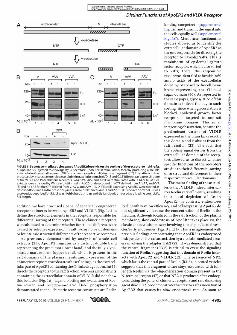
nystatin (Fig. 7C ) did not inhibit this process.Another aspect of the Reelin signaling cascade is the Reelin-induced cleavage of ApoER2, which produces a soluble extra-cellular ( NTF ) and a soluble intracellular fragment (intracellu-lar domain; ICD) by the sequential action of ␣- and␥ -secretase,respectively (33, 34) (Fig. 8 A). Because the production of theintracellular domain cannot be monitored directly, most likely due to its inherent instability (34), we followed the productionof the membrane-bound precursor CTF by Western blotting.
Incubation of 3T3 cells expressing WT ApoER2 with Reelinresulted in a significant increase of the 25-kDa CTF (supple-mental Fig. 4, lane 1), which was not observed upon the addi-tion of receptor-associated protein (lane 3). Again, CTF pro-
duction could be triggered by thebivalentagent Ab 186 (lane5),and this was blocked by the addition of soluble receptor frag-ment (lane6 ). For controls, thecells were incubatedwith mock-conditioned medium (lane 2) and Ab 20 (directed against theintracellular domain; lane 4). Under these conditions, smallamounts of CTF were detected, apparently produced even
when the cells are not stimulated. The properties of the chi-meric receptors in regard to the specific fragmentation process(Fig. 8 B) were more complex than those described above forreceptor degradation and endocytosis. Although VLDLR doesnot undergo fragmentation at all (Fig. 8C , lane 1), chimeras
containing the extracellular domain of ApoER2 produced sig-nificant amounts of CTF upon Reelin treatment (Fig. 8C , lanes3 and 5). It should be noted that the antibody used (6A6) todetect the CTF containing the respective domains of VLDLRconsistently gives weaker signals than Ab 20 directed against
the intracellular domain of ApoER2. Chimeras containing thetransmembrane plus intracellular domains (VAA) or only theintracellular domain of ApoER2 (VVA) are processed to a smallextent, but this was not enhanced by Reelin (Fig. 8 B, lanes 3–6 ).Disruption of rafts using CDX significantly blocked the Reelin-
induced production of CTF from WT ApoER2 (Fig. 8 D).
DISCUSSION
Inactivation of the genes for both Reelin receptors, ApoER2
and VLDLR, leads to a reeler phenotype in mice (25). Singleknock-out mice lacking the gene either for ApoER2 or forVLDLR display only subtle abnormalities in their brain archi-tectures. This suggests that ApoER2 and VLDLR can at leastpartially compensate for each other, at least in respect to their
function in establishing laminated brain structures. This wasconfirmed in primary neurons and in a fibroblast cell model by
FIGURE 4. ApoER2 and VLDLR internalize Reelin via the early endosomal compartment. A, Reelin co-localizes with ApoER2 and VLDLR at 4 °C. 3T3 cellsexpressing ApoER2 (upper panel ) or VLDLR (lower panel ) were incubated with RCM at 4 °C to allow binding of Reelin to the receptors. Cells were fixed,permeabilized,and stained using Ab186 fordetection of ApoER2, Ab 187 forVLDLR, andAb G10 forReelin. B and C , internalized Reelin co-localizes with EEA1.3T3 cells expressing ApoER2(B) orVLDLR(C ) wereincubated with RCM at4 °Cfor 1 h,washed, and incubated withOpti-MEMfor 10min at 4 °C (upper panels)or37 °C (lowerpanels), respectively. Cellswere fixed, permeabilized,and stained usingAb G10for detectionof Reelin andan antibodyagainst EEA1for stainingof the early endosomal compartment. Scale bars, 10 m.
FIGURE 5. Reelin internalization and degradation rates of ApoER2 and
VLDLR depend on their sorting within the plasma membrane. 3T3 cellsexpressing ApoER2or oneof the chimeric receptors containing theextracel-lular domain of ApoER2 ( A) or VLDLR or one of the chimeric receptorscontaining the extracellular domain of VLDLR (B) were incubated with RCM at4 °C to allow binding of Reelin to the respective receptors. Cells were thenshifted to 37 °C for the indicated time periods to allow internalization anddegradationof theligand.After washing thecells, extractswere preparedandanalyzed forcell-associated Reelinby Western blotting using Ab G10in com-binationwith an HRP-coupledgoat anti-mouse antibody.C , 3T3cells express-ing ApoER2 were treated as described for A in the presence (lower panel ) orabsence (upper panel ) of the raft-disrupting agent CDX (5 mM). Cell extractswere analyzed as described for A.
Distinct Functionsof ApoER2andVLDL Receptor
FEBRUARY 12, 2010 • VOLUME 285• NUMBER 7 JOURNAL OF BIOLOGICAL CHEMISTRY 4903
http://www.jbc.org/content/suppl/2009/11/30/M109.025973.DC1.htmlSupplemental Material can be found at:

7/28/2019 Paper ReelinDifferential Functions of ApoER2 and Very Low Density Lipoprotein Receptor in Reelin Signaling Depe…
http://slidepdf.com/reader/full/paper-reelindifferential-functions-of-apoer2-and-very-low-density-lipoprotein 9/13
demonstrating that the initial event of the Reelin signalingpathway (i.e. phosphorylation of Dab1) is equally well sup-ported by ApoER2 and VLDLR (11, 23). Divergent roles forApoER2 and VLDLR have been corroborated recently. In neu-
ronal migration, VLDLR mediates a stop signal for migratingneurons, whereas ApoER2 plays a distinct role in the migrationof neocortical neurons generated late (26). The specific pheno-types of the single knock-out animals and the observed distinct
roles for both receptors might be inpart caused by selective expression
of the receptors in distinct areas of the brain (35). On the other hand,ApoER2 and VLDLR may have dis-tinct functions in the Reelin path-way, apart from the primary phos-
phorylation event. Other processesin the central nervous system seemto depend on specific variants of ApoER2 only and involve the mod-ulation of synaptic plasticity and
memory (36), the control of neuro-nal survival (37), and seleniumuptake (38, 39). One reason for dif-ferential functions of these recep-tors is structural differences in the
respective intracellular domains(40) linking ApoER2 to adaptersthat do not interact with VLDLR
(41). Other features could be selec-tive expression in distinct subdo-
mains of the cell membrane and/ortheir divergent endocytic compe-tence. As shown in our laboratory,ApoER2 is prevalently present inraft domains, whereas VLDLR is not
(23). The avian ortholog of VLDLRis expressed on growing oocyteswhere it efficiently mediates recep-tor-mediated endocytosis of yolkprecursors, thus playing a pivotal
role in follicle development (42),whereas ApoER2 was demonstratedto exert an extremely slow endocy-tosis rate in comparison with othermembers of the LDL receptor fam-
ily (43). As recently postulated,VLDLR and/or ApoER2-mediatedendocytosis of full-length Reelin orits central fragment produced by
metalloproteinases might play animportant role in the Reelin signal-ing pathway by controlling thestrength of the signal via modulat-
ing the availability of the ligand (44).Thus, we set out to study structuralfeatures of the receptors that mightbe responsible for different fatesand/or functions of the receptors in
terms of cellular sorting, endocytosis, and receptor trafficking.
For most of the present studies, we used a recently estab-lished fibroblast-based cell system in which theReelin signalingpathway has been partially reconstituted by expressing Dab1and either ApoER2 or VLDLR (23). These cells respond to Ree-lin stimulation with Dab1 phosphorylation and phosphatidyl-
inositol 3-kinase activation, leading to protein kinase B/Aktphosphorylation undistinguishable from primary neurons. In
FIGURE 6. ApoER2 degradation is induced by multivalent ligands and is mediated by the lysosomalpathway. Primary ratneurons( A) and3T3 cells expressing ApoER2 (B) wereincubatedfor5 h withRCM, MCM,Ab 186(1:100; targets ApoER2 ligand binding domain), Ab 20 (1:100; targets ApoER2 intracellular domain), Ab186 and recombinant ApoER2 N-terminal fragment (MBP-ApoER2), or 20 g/ml recombinant receptor-associ-ated protein (RAP ). Total cell extracts were analyzed by Western blotting using Ab 20 (ApoER2) and Ab 6A6(VLDLR) andan antibodyagainstLis1 as a loading control in combination withthe correspondingHRP-coupled
secondary antibodies. C , ApoER2-expressing 3T3 cellswere incubated with RCM or MCM. Lysosomal degrada-tion wasblockedby theaddition of10 mM NH4Clor 25M chloroquine; proteasomal degradation was blockedusing 25 M MG132. Cell extracts were analyzed as described for A and B, and results were quantified bydensitometry.IOD values of ApoER2 bands werenormalized to the density of Lis1bands. Error bars,S.E.(nϭ3).
FIGURE 7. The extracellular domain of ApoER2 but not its sorting to lipid rafts is required for Reelin-induced lysosomal receptor degradation. A, 3T3 cells expressing one of the wild type ( A and V ) or chimericreceptors( AVV , AAV , VAA,and VVA) were stimulatedwith RCM(lanes 1, 3, 5, 7 , 9, and 11)orMCM(lanes 2, 4, 6, 8,10,and 12) for5 h andanalyzedforreceptordegradation byWesternblottingusing Ab20 forApoER2, VAA, andVVA; Ab 220 forAAV andAVV;and Ab 74 forVLDLR. Lis1 washighlightedusingan anti-Lis1antibodyand usedas a loading control. B, ApoER2-expressing 3T3 fibroblasts were incubated with RCM ( lanes 1 and 3) or MCM(lanes 2 and 4) and the raft-disrupting agent CDX (5 mM; lanes 3 and 4) for 5 h. Cell extracts were analyzed forApoER2 degradation by Western blotting using Ab 20 in combination with an HRP-coupled goat-anti-rabbitantibody.Lis1 washighlighted using an anti-Lis1antibodyand used as a loading control. C , cells were treatedand analyzed as described for B, except that 15 g/ml nystatin was used for disruption of rafts.
Distinct Functionsof ApoER2andVLDL Receptor
4904 JOURNAL OF BIOLOGICAL CHEMISTRY VOLUME 285•NUMBER 7• FEBRUARY 12, 2010
http://www.jbc.org/content/suppl/2009/11/30/M109.025973.DC1.htmlSupplemental Material can be found at:
7/28/2019 Paper ReelinDifferential Functions of ApoER2 and Very Low Density Lipoprotein Receptor in Reelin Signaling Depe…
http://slidepdf.com/reader/full/paper-reelindifferential-functions-of-apoer2-and-very-low-density-lipoprotein 10/13
addition, we have now used a panel of genetically engineered
receptor chimeras between ApoER2 and VLDLR (Fig. 1 A) todefine the structural elements in the receptors responsible fordifferential sorting of the receptors. These chimeric receptorswere also used to determine whether functional differences arecaused by selective expression in raft versus non-raft domains
or by intrinsic structural differences of therespective receptors.As previously demonstrated by analysis of whole cell
extracts (23), ApoER2 migrates as a distinct double bandrepresenting the precursor (lower band) and the fully glyco-
sylated mature form (upper band), which is present in theraft domains of the plasma membrane. Expression of thechimeric receptors corroboratesthese findings, as the extracel-lular part of ApoER2containing the O-linkedsugar domain(45)directs the receptors to the raft fraction, whereas all constructs
containing the extracellular domain of VLDLR did not show this behavior (Fig. 1 B). Ligand blotting and evaluation of Ree-lin-induced and receptor-mediated Dab1 phosphorylationdemonstrated that all chimeric receptor constructs are Reelin
binding-competent (supplementalFig. 1 B) and transmit the signal intothe cells equally well (supplemental
Fig. 1C ). Membrane fractionationstudies allowed us to identify theextracellular domain of ApoER2 asthe one responsible for directing the
receptor to caveolae/rafts. This isreminiscent of epidermal growthfactor receptor, which is also sortedto rafts; there, the responsibleregion wasidentified to be within 60amino acids of the extracellular
domain juxtaposed to the cell mem-brane representing the O-linkedsugar domain (46). As reported inthe same paper, glycosylation of thisdomain is indeed the key to such
sorting, since when glycosylation isblocked, epidermal growth factor
receptor is targeted to non-raftmembrane domains. This is aninteresting observation, because the
predominant variant of VLDLRexpressed in the brain lacks exactly this domain and is absent from theraft fraction (23). The fact that
the sorting signal derives from theextracellular domain of the recep-tors allowed us to dissect whetherspecific functions of the receptorsare due to their differential sorting
or to structural differences in theirrespective intracellular domains.
The major finding of these stud-ies is that VLDLR indeed internal-izes Reelin very efficiently, resulting
in a local depletion of Reelin.ApoER2, in contrast, endocytoses
Reelin with very low efficiency, and cells expressing ApoER2 donot significantly decrease the concentration of Reelin in themedium. Although localized in the raft fraction of the plasma
membrane, slow endocytosis of ApoER2 takes place via theclassic endocytosis pathway involving coated pits/coated vesi-cles/early endosomes (Figs. 3 and 4). This is in agreement with
previous findings demonstrating that ApoER2 is endocytosedindependent of its raft association by a clathrin-mediated proc-
ess involving the adapter Dab2 (22). It was demonstrated thatthe central fragment (R3-6) is critical to exert the signalingfunction of Reelin, suggesting that this domain of Reelin inter-acts with ApoER2 and VLDLR (12). The presence of NR2,which lacks the central part of Reelin (R3-6), in coated vesicles
suggests that this fragment either stays associated with full-length Reelin via the oligomerization domain present in theN-terminal region (47) or that NR2 is produced after endocy-tosis. Using the panel of chimeric receptors and raft-dissolvingagentslike CDX, we demonstrate that it is theraft association of
ApoER2 that causes its slow endocytosis rate. As soon as
FIGURE 8. Secretase-mediatedcleavageof ApoER2depends on the sorting of thereceptorto lipid rafts. A, ApoER2 is subjected to cleavage by ␣-secretase upon Reelin stimulation, thereby producing a solubleextracellularN -terminalfragment(NTF ) anda membrane-bound C-terminalfragment (CTF ).The latteris further
processedby ␣-secretaseto release a soluble intracellular domain (ICD). B and C , 3T3fibroblasts expressingoneof the WT ( A and V ) or chimeric receptors (VAA, VVA, AVV , and AAV ) were stimulated with RCM or MCM. Cellextracts were analyzedby Western blotting using Ab 20for detectionof theCTF derived from A, VAA, andVVA(B) and Ab 6A6 for the CTF derived from V, AVV, and AAV ( C ). D, 3T3 cells expressing ApoER2 were treated asdescribedfor B and C inthepresence(lanes3 and 4)orabsence(lanes1 and 2)ofCDX.ProductionoftheCTFwasanalyzed as describedfor B. Lis1 washighlightedusingan anti-Lis1antibody andusedas a loading control. FL,full-length.
Distinct Functionsof ApoER2andVLDL Receptor
FEBRUARY 12, 2010 • VOLUME 285• NUMBER 7 JOURNAL OF BIOLOGICAL CHEMISTRY 4905
http://www.jbc.org/content/suppl/2009/11/30/M109.025973.DC1.htmlSupplemental Material can be found at:

7/28/2019 Paper ReelinDifferential Functions of ApoER2 and Very Low Density Lipoprotein Receptor in Reelin Signaling Depe…
http://slidepdf.com/reader/full/paper-reelindifferential-functions-of-apoer2-and-very-low-density-lipoprotein 11/13
ApoER2 loses its association with rafts, the endocytosis rate of the receptor increases significantly. This effect might even beunderestimated by the experiment using CDX to dissolve therafts because this agent also blocks clathrin-dependent endocy-
tosis by generally depleting the membrane of cholesterol (31).
Thus, slow translocation from rafts to non-raft domains of themembrane might be the rate-limiting step of ApoER2-medi-ated endocytosis.
The second significant difference between ApoER2 and
VLDLR is the fact that the levels of ApoER2, but not of VLDLR,significantly drop in the presence of Reelin (23). As demon-strated here, Reelin-induced degradation of ApoER2 occurs viathe lysosomal pathway and depends on the presence of recep-tor-clustering ligands. Again, the extracellular domain of
ApoER2 determines the fate of the receptor upon binding of Reelin. Disruption of rafts by CDX or nystatin had no influenceon receptor degradation, demonstrating that neither the caveo-lin-mediated pathway originating from raft structures nor the
targeting of raft-associated receptors to endosomes (48) isresponsible for receptor degradation. Thus, ApoER2 degrada-tion most likely occurs by clathrin-mediated endocytosis andsubsequent sorting to lysosomes. Structural differences in theextracellular domains of ApoER2 and VLDLR must be respon-
sible for directing ApoER2 predominantly to lysosomes andVLDLR into the recycling pathway back to the plasma mem-brane. For the LDL receptor, it was shown that a distinct regionof the extracellular domain (i.e. the cysteine-rich growth factorrepeats (epidermal growth factor repeats)) is responsible for
uncoupling of receptor and ligand within the endocytic path-way (49). When this domain is deleted, the receptor no longerreleases itsligandand becomes degraded in thelysosome. Thus,we speculate that differences in the epidermal growth factorrepeats between ApoER2 and VLDLR are responsible for the
distinct behavior of the receptors.Parallel to lysosomal degradation of ApoER2, specific break-
down products of this receptor are produced upon Reelin stim-
FIGURE 9. Model of the intracellular fates of ApoER2 and VLDLR upon Reelin stimulation. Upon binding of Reelin, both ApoER2 and VLDLR mediatephosphorylation of Dab1(step 1). VLDLR internalizes Reelin rapidly via clathrin-mediated endocytosis (step2) andis separatedfromReelin inthe compartmentof uncoupling of receptor and ligand (step 3). VLDLR then recycles back to the plasma membrane (step 4), whereas Reelin is delivered to the lysosome fordegradation (step 5). ApoER2internalizes Reelinvia thesamepathway(step2), although thereceptor originally resides in lipid rafts andendocytoses itsligandat a much slower rate. In contrast to VLDLR, ApoER2 is not recycled but ends up in the lysosome together with Reelin ( step 6). As an additional feedback mechanism, Reelin stimulationinduces secretase-mediatedcleavageof ApoER2,therebygenerating a soluble intracellularfragment(ICD) (step7 ),thefunctionof which is not defined yet, and a soluble extracellular fragment containing the ligand binding domain (step 8). This fragment can, together with anotherN-terminal fragment produced from an ApoER2 isoform by furin cleavage (step 9), inhibit theReelin signalby sequestering free Reelin in the surroundings of the cell (step 10).
Distinct Functionsof ApoER2andVLDL Receptor
4906 JOURNAL OF BIOLOGICAL CHEMISTRY VOLUME 285•NUMBER 7• FEBRUARY 12, 2010
http://www.jbc.org/content/suppl/2009/11/30/M109.025973.DC1.htmlSupplemental Material can be found at:

7/28/2019 Paper ReelinDifferential Functions of ApoER2 and Very Low Density Lipoprotein Receptor in Reelin Signaling Depe…
http://slidepdf.com/reader/full/paper-reelindifferential-functions-of-apoer2-and-very-low-density-lipoprotein 12/13
ulation (33, 34). Here we demonstrate that this effect (i) is notsimply induced by binding of a ligand but requires the presence
of a clustering ligand, such as Reelin, or antibodies against theextracellular domain, whichalso induce Dab1 phosphorylation;(ii) is dependent on the presence of the extracellular domain of ApoER2; and (iii) is dependent on the presence of rafts. VLDLR variants carrying the extracellular domain of ApoER2 are
cleaved like ApoER2 itself, whereas intact VLDLR completely escapes this process. This is in agreement with previous find-ings that the presence of the O-linked sugar domain pro-motes ␥ -secretase-mediated cleavage of the receptor (34)
and that the ␥ -secretase complex is associated with rafts (50,51). Thus, most likely, the entire processing (␣- and ␥ -secre-tase-mediated) takes place within the raftdomain of the plasmamembrane. Whether clustering of the receptors or Dab1 phos-phorylation or both actually triggers the induction of the cleav-
age is still an open question.These results allow us to propose a model for describing both
the interrelated and independent functions of ApoER2 andVLDLR in Reelin signaling (Fig. 9). VLDLR present in non-raft
domains of theplasmamembrane binds Reelin, which results inDab1 phosphorylation (Fig. 9, step 1). The Reelin-receptorcomplex is rapidly internalized by clathrin-mediated endocyto-sis (Fig. 9, step 2), and Reelin is uncoupled from the receptor(Fig. 9, step 3) and sorted to lysosomes (Fig. 9, step 5), whereasVLDLR recycles back to the cell membrane (Fig. 9, step 4). This
reduces the amount of extracellular Reelin significantly, thusshutting off the persistence of the signal without rendering thecell refractory to VLDLR-mediated actions. Binding of Reelin toApoER2 present in rafts also leads to immediate phosphoryla-tion of Dab1 (Fig. 9, step 1) but not to a significant reduction of
extracellular Reelin, because ApoER2-mediated endocytosis isslow. The Reelin signal itself can be turned off via degradationof phosphorylated Dab1, which occurs independently of thesignaling receptor (52). Selective Reelin-mediated loss of ApoER2 depends on clathrin-mediated endocytosis and lysoso-
mal degradation of the receptor (Fig. 9, step 6 ). As shown here,this process is much slower than for VLDLR but in the long runrenders the target cell refractory to further Reelin stimulationuntil new receptor is synthesized. In parallel, specific fragmen-tation by ␣- and ␥ -secretases leads to the production of solubleintracellular receptor fragment (Fig. 9, step 7 ), which mightexert its action within the nucleus (34), and a soluble extracel-lular fragment (Fig. 9, step 8). The extracellular fragment con-
taining the ligand binding domain, together with secreted sol-
uble ApoER2 fragments (Fig. 9, step 9) produced by the actionof furin from certain splice variants of the receptor (28), bindReelin andthereby attenuatethe entirepathway (Fig. 9, step 10).Thus, in regions of the brain where mostly ApoER2 is
expressed, the Reelin-induced activation of target cells is notaccompanied by a reduction of Reelin. Thus, Reelin might keepexerting other functions without further inducing the canoni-cal Reelin signal, which is efficiently turned off by Dab1 degra-dation and production of dominant negative receptor frag-
ments. Whether the production of soluble intracellularfragments derived from ApoER2 by ␥ -secretase cleavage repre-sents an independent signal within the cell as proposed (34)awaits clarification.
Acknowledgment—We appreciate the technical assistance of Harald
Rumpler.
REFERENCES
1. Rice, D. S., Curran, T. (2001) Annu. Rev. Neurosci. 24, 1005–1039
2. Lambert de Rouvroit, C., and Goffinet, A. M. (1998) Adv. Anat. Embryol.
Cell Biol. 150, 1–106
3. Cooper, J. A. (2008) Trends Neurosci. 31, 113–119
4. Forster, E.,Jossin, Y.,Zhao,S., Chai, X.,Frotscher, M.,and Goffinet,A. M.
(2006) Eur. J. Neurosci. 23, 901–909
5. Tissir, F., and Goffinet, A. M. (2003) Nat. Rev. Neurosci. 4, 496–505
6. Niu, S., Renfro, A., Quattrocchi, C. C., Sheldon, M., and D’Arcangelo, G.
(2004) Neuron 41, 71–84
7. Olson, E. C., Kim, S., and Walsh, C. A. (2006) J. Neurosci. 26, 1767–1775
8. Assadi, A. H., Zhang, G., Beffert, U., McNeil, R. S., Renfro, A. L., Niu, S.,
Quattrocchi, C. C., Antalffy, B. A., Sheldon, M., Armstrong, D. D., Wyn-
shaw-Boris, A., Herz, J., D’Arcangelo, G., and Clark, G. D. (2003) Nat.
Genet. 35, 270–276
9. Pramatarova, A., Ochalski, P. G., Chen, K., Gropman, A., Myers, S., Min,
K. T., and Howell, B. W. (2003) Mol. Cell. Biol. 23, 7210–7221
10. Jossin, Y., and Goffinet, A. M. (2007) Mol. Cell. Biol. 27, 7113–7124
11. Strasser, V., Fasching, D., Hauser, C., Mayer, H., Bock, H. H., Hiesberger,T.,Herz, J.,Weeber,E. J.,Sweatt,J. D.,Pramatarova,A., Howell, B.,Schnei-
der, W. J., and Nimpf, J. (2004) Mol. Cell. Biol. 24, 1378–1386
12. Jossin, Y., Ignatova, N., Hiesberger, T., Herz, J., Lambert de Rouvroit, C.,
and Goffinet, A. M. (2004) J. Neurosci. 24, 514–521
13. Blake, S. M., Strasser, V., Andrade, N., Duit, S., Hofbauer, R., Schneider,
W. J., and Nimpf, J. (2008) EMBO J. 27, 3069–3080
14. Arnaud, L.,Ballif, B. A.,Forster, E.,and Cooper, J. A. (2003) Curr. Biol. 13,
9–17
15. Howell, B.W., Gertler,F. B.,andCooper,J. A.(1997) EMBO J. 16, 121–132
16. Jossin, Y., Ogawa, M., Metin, C., Tissir, F., and Goffinet, A. M. (2003)
J. Neurosci. 23, 9953–9959
17. Kuo, G., Arnaud, L.,Kronstad-O’Brien, P., andCooper,J. A. (2005) J. Neu-
rosci. 25, 8578–8586
18. Howell, B. W., Lanier, L. M., Frank, R., Gertler, F. B., and Cooper, J. A.
(1999) Mol. Cell. Biol. 19, 5179–518819. Beffert, U., Durudas, A., Weeber, E. J., Stolt, P. C., Giehl, K. M., Sweatt,
J. D., Hammer, R. E., and Herz, J. (2006) J. Neurosci. 26, 2041–2052
20. Schneider, W. J., and Nimpf, J. (2003) CMLS 60, 892–903
21. Gotthardt, M., Trommsdorff, M., Nevitt, M. F., Shelton, J., Richardson,
J. A., Stockinger, W., Nimpf, J., and Herz, J. (2000) J. Biol. Chem. 275,
25616–25624
22. Cuitino, L., Matute, R., Retamal, C., Bu, G., Inestrosa, N. C., and Marzolo,
M. P. (2005) Traffic 6, 820–838
23. Mayer, H., Duit, S., Hauser, C., Schneider, W. J., and Nimpf, J. (2006) Mol.
Cell. Biol. 26, 19–27
24. Riddell, D. R., Sun, X. M., Stannard, A. K., Soutar, A. K., and Owen, J. S.
(2001) J. Lipid Res. 42, 998–1002
25. Trommsdorff, M., Gotthardt, M., Hiesberger, T., Shelton, J., Stockinger,
W.,Nimpf,J., Hammer, R. E.,Richardson, J.A., andHerz, J. (1999) Cell 97,
689–70126. Hack, I., Hellwig, S., Junghans, D., Brunne, B., Bock, H. H., Zhao, S., and
Frotscher, M. (2007) Development 134, 3883–3891
27. Brandes, C.,Kahr,L., Stockinger, W.,Hiesberger,T., Schneider, W. J.,and
Nimpf, J. (2001) J. Biol. Chem. 276, 22160–22169
28. Koch, S., Strasser, V., Hauser, C., Fasching, D., Brandes, C., Bajari, T. M.,
Schneider, W. J., and Nimpf, J. (2002) EMBO J. 21, 5996–6004
29. Stockinger, W., Hengstschlager-Ottnad, E., Novak, S., Matus, A., Hut-
tinger, M., Bauer, J., Lassmann, H., Schneider, W. J., and Nimpf, J. (1998)
J. Biol. Chem. 273, 32213–32221
30. Nandi,P. K., Irace,G.,Van Jaarsveld, P.P., Lippoldt,R. E.,andEdelhoch, H.
(1982) Proc. Natl. Acad. Sci. U.S.A. 79, 5881–5885
31. Rodal, S. K., Skretting, G., Garred, O., Vilhardt, F., van Deurs, B., and
Sandvig, K. (1999) Mol. Biol. Cell 10, 961–974
32. Howell, B. W., Herrick, T. M., Hildebrand, J. D., Zhang, Y., and Cooper,
Distinct Functionsof ApoER2andVLDL Receptor
FEBRUARY 12, 2010 • VOLUME 285• NUMBER 7 JOURNAL OF BIOLOGICAL CHEMISTRY 4907
http://www.jbc.org/content/suppl/2009/11/30/M109.025973.DC1.htmlSupplemental Material can be found at:

7/28/2019 Paper ReelinDifferential Functions of ApoER2 and Very Low Density Lipoprotein Receptor in Reelin Signaling Depe…
http://slidepdf.com/reader/full/paper-reelindifferential-functions-of-apoer2-and-very-low-density-lipoprotein 13/13
J. A. (2000) Curr. Biol. 10, 877–885
33. Hoe, H. S., and Rebeck, G. W. (2005) Brain Res. Mol. Brain Res. 137,
31–39
34. May, P., Bock, H. H., Nimpf, J., and Herz, J. (2003) J. Biol. Chem. 278,
37386–37392
35. Perez-Garcia, C. G., Tissir, F., Goffinet, A. M., and Meyer, G. (2004) Eur.
J. Neurosci. 20, 2827–2832
36. Beffert, U., Weeber, E. J., Durudas, A., Qiu, S., Masiulis, I., Sweatt, J. D., Li,
W. P., Adelmann, G., Frotscher, M., Hammer, R. E., and Herz, J. (2005) Neuron 47, 567–579
37. Beffert,U., NematollahFarsian, F.,Masiulis, I.,Hammer,R. E.,Yoon, S. O.,
Giehl, K. M., and Herz, J. (2006) Curr. Biol. 16, 2446–2452
38. Burk, R. F., Hill, K. E., Olson, G. E., Weeber, E. J., Motley, A. K., Winfrey,
V. P., and Austin, L. M. (2007) J. Neurosci. 27, 6207–6211
39. Masiulis, I., Quill, T. A., Burk, R. F., and Herz, J. (2009) Biol. Chem. 390,
67–73
40. Brandes, C., Novak, S., Stockinger, W., Herz, J., Schneider, W. J., and
Nimpf, J. (1997) Genomics 42, 185–191
41. Stockinger, W., Brandes, C., Fasching, D., Hermann, M., Gotthardt, M.,
Herz, J., Schneider, W. J., and Nimpf, J. (2000) J. Biol. Chem. 275,
25625–25632
42. Bujo, H., Hermann, M., Kaderli, M. O., Jacobsen, L., Sugawara, S., Nimpf,
J., Yamamoto, T., and Schneider, W. J. (1994) EMBO J. 13, 5165–5175
43. Li, Y., Lu, W., Marzolo, M. P., and Bu, G. (2001) J. Biol. Chem. 276,
18000–18006
44. Jossin, Y., Gui, L., and Goffinet, A. M. (2007) J. Neurosci. 27, 4243–4252
45. Novak, S., Hiesberger, T., Schneider, W. J., and Nimpf, J. (1996) J. Biol.
Chem. 271, 11732–11736
46. Yamabhai, M., and Anderson, R. G. (2002) J. Biol. Chem. 277,
24843–24846
47. Utsunomiya-Tate, N., Kubo, K., Tate, S., Kainosho, M., Katayama, E.,
Nakajima, K., and Mikoshiba, K. (2000) Proc. Natl. Acad. Sci. U.S.A. 97,9729–9734
48. Pol, A.,Lu, A.,Pons, M.,Peiro, S.,and Enrich, C. (2000) J. Biol. Chem. 275,
30566–30572
49. Davis,C. G.,Goldstein,J. L.,Sudhof, T. C., Anderson,R. G., Russell, D. W.,
and Brown, M. S. (1987) Nature 326, 760–765
50. Cheng, H., Vetrivel, K. S., Drisdel, R. C., Meckler, X., Gong, P., Leem, J. Y.,
Li, T., Carter, M., Chen, Y., Nguyen, P., Iwatsubo, T., Tomita, T., Wong,
P. C., Green, W. N., Kounnas, M. Z., and Thinakaran, G. (2009) J. Biol.
Chem. 284, 1373–1384
51. Vetrivel, K. S., Cheng, H., Lin, W., Sakurai, T., Li, T., Nukina, N., Wong,
P. C., Xu, H., and Thinakaran, G. (2004) J. Biol. Chem. 279, 44945–44954
52. Feng, L., Allen, N. S., Simo, S., and Cooper, J. A. (2007) Genes Dev. 21,
2717–2730
Distinct Functionsof ApoER2andVLDL Receptor
http://www.jbc.org/content/suppl/2009/11/30/M109.025973.DC1.htmlSupplemental Material can be found at: