parasite component community of gulf killifish, fundulus grandis, in
TRANSCRIPT

Parasite component community of Gulf killifish, Fundulus grandis , in an oiled
Louisiana saltmarsh
by
Carlos Fernando Ruiz
A thesis submitted to the Graduate Faculty of
Auburn University
in partial fulfillment of the
requirements for the Degree of
Master of Science
Auburn, Alabama
August 3rd
2013
Keywords: biodiversity, component community, ectoparasite, endoparasite,
Gulf of Mexico, oil spill
Copyright 2013 by Carlos Fernando Ruiz
Approved by
Stephen A. Bullard, Chair, Assistant Professor of Fisheries and Allied Aquacultures
Covadonga R. Arias, Associate Professor of Fisheries and Allied Aquacultures
Christopher J. Anderson, Assistant Professor of Forestry and Wildlife Sciences

Abstract
Fish parasites comprise a large portion of marine biodiversity but so far have been
underutilized as Gulf of Mexico bioindicators. Shifts in parasite diversity, prevalence, and
intensity resulting from the 2010 BP Deepwater Horizon Oil Spill (DHOS) could indicate spill-
related changes to water quality, abundances and immunological health of free-living organisms,
or the Gulf of Mexico food web. Ectoparasites with direct life cycles (no intermediate host or
food-web mediated transmission) may be sentinels for acute spill effects, as they are typically
small, have high surface area to volume ratios, and remain immersed in seawater. Endoparasites
with indirect life cycles (intermediate host[s] required) involving food-web mediated
transmission may be sentinels for detecting chronic spill effects, as they reside in a host where
they are less vulnerable to toxins and have larvae requiring predator/prey transmission. To test
these ideas, one must have a species-level understanding of the parasite component community
in a geographic area. Chapter 1 enumerates the parasite component community of Gulf killifish,
Fundulus grandis (Cyprinidontiformes: Fundulidae) in Barataria Bay, LA. Using those data as a
critical baseline and for the units of analysis, Chapter 2 statistically tests for differences in the
structure of the parasite component community across 4 oiled sites and 4 reference (non-oiled)
sites in Barataria Bay. Regarding the survey and inventory of parasites, the parasite component
community includes 44 species (31 endoparasites; 13 ectoparasites) infecting 23 fish tissues. Of
these parasite species, 10 are putatively new to science, 24 constitute new host records to F.
grandis, and nearly all, 42 of 44 (95%), are putatively new locality records in LA. Regarding the
ii

use of those taxonomic units to test hypotheses concerning ecosystem functioning, no significant
differences were detected between prevalence (aggregated) of ectoparasites (Monogenoidea,
Hirudinida, Copepoda, Branchiura, Isopoda) and endoparasites (Myxozoa, Digenea, Cestoda,
Nematoda, Acanthocephala) in oiled and non-oiled sites nor in mean intensity and prevalence
(aggregated) of ectoparasites and endoparasites in oiled and non-oiled sites. However, significant
differences were detected in species prevalence of acanthocephalans (19.6% in non-oiled sites vs.
2.9% in oiled sites), copepods (21.7% vs. 34.2%), and branchiurans (13.8% vs. 28.3). Seasonal
effects were statistically detected in myxozoans (highest intensity of infection during May 2011)
and digeneans (highest intensity in August 2011), and those seasonal patterns were not
significantly different between oiled and non-oiled sites. Digenean metacercariae infecting gill
and heart, monogenoideans infecting skin, and nematodes infecting body cavity each have a
significantly higher log mean intensity in oiled sites. Species richness of ectoparasites and
endoparasites was not significantly different between oiled and non-oiled sites and across
seasons. Condition factor of F. grandis was not significantly different between oiled and non-
oiled sites and across seasons.!
iii

Acknowledgements
I thank my major professor, Dr. Ash Bullard (Assistant Professor, Auburn University,
Department of Fisheries, Auburn, AL), for his encouragement and support and for
enthusiastically sharing his knowledge and expertise with me. I thank my committee members,
Dr. Cova Arias (Associate Professor, Auburn University, Department of Fisheries, Auburn, AL),
and Dr. Chris Anderson (Assistant Professor, Auburn University, Department of Forestry and
Wildlife, Auburn, AL) for providing comments concerning my graduate career at Auburn and
resulting thesis. This work has been funded by grants from the Marine Environmental Science
Consortium of Alabama, Gulf of Mexico Research Institute, and National Science Foundation.
I thank Dr. George Benz (Professor, Middle Tennessee State University, Department of
Biology, Murfreesboro, TN) and Mr. Eric Salmon (MSc student, Middle Tennessee State
University, Department of Biology, Murfreesboro, TN) for their assistance with field collections.
I thank Myron Fischer (Director, Grand Isle Marine Laboratory, LA Department of Wildlife and
Fisheries, Grand Isle, LA) and Clint Edds (Biologist, Grand Isle Marine Laboratory, LA
Department of Wildlife and Fisheries, Grand Isle, LA) for providing logistical support for field
collections.
I thank Andrew Austin, Elizabeth Cameron, Elliot Broder, Raquel Patella, and Stephanie Cox
for assisting me with fish necropsies and data collection at Auburn University. I thank my lab
mates, Matthew Womble and Andrew McElwain for providing me with transportation during a
critical time of need that resulted in the completion of this thesis, and also Raphael Orelis-
iv

Ribiero for his assistance with field collections. I thank Maria Ruiz (mom), Fernando Ruiz (dad),
David and Dominick Ruiz (brothers), and Cameron Ruiz (sister), for their love and support.!
v

Table of Contents
Abstract ......................................................................................................................................... ii
Acknowledgments ........................................................................................................................ iv
List of Tables ............................................................................................................................... ix
List of Plates ................................................................................................................................. x
Chapter 1. Biodiversity survey and inventory of metazoan parasites of Fundulus grandis, Baird
and Girard, 1853, (Cyprinodontiformes: Fundulidae) in Barataria Bay, LA .......................... 1
1.1. Abstract ............................................................................................................................... 2
1.2. Introduction ......................................................................................................................... 2
1.2.1. Barataria Bay and the 2010 BP Deepwater Horizon Oil Spill ............................................ 2
1.2.2. The Gulf killifish, Fundulus grandis ................................................................................... 4
1.2.3. Parasite component community characterization ............................................................... 5
1.3. Justification ......................................................................................................................... 6
1.4. Objective and hypothesis .................................................................................................... 6
1.5. Materials and methods ........................................................................................................ 6
1.5.1. Site selection ....................................................................................................................... 6
1.5.2. Fish collection ..................................................................................................................... 7
1.5.3. Tissue fixation ................................................................................................................... 10
1.5.4. Laboratory necropsy ......................................................................................................... 10
1.5.5. Parasite specimen selection and preparation ..................................................................... 12
1.5.6. Parasite identification ........................................................................................................ 12
vi

1.5.7. Reporting of taxonomic authorities and putatively new species ...................................... 15
1.6. Results ............................................................................................................................... 16
1.6.1. Myxozoa ........................................................................................................................... 16
1.6.2. Platyhelminthes ................................................................................................................. 22
1.6.3. Nematoda .......................................................................................................................... 40
1.6.4. Acanthocephala ................................................................................................................. 43
1.6.5. Annelida ............................................................................................................................ 46
1.6.6. Arthropoda ........................................................................................................................ 46
1.6.7. Protozoa ............................................................................................................................ 54
1.7. Discussion ......................................................................................................................... 57
1.7.1. Myxozoa ........................................................................................................................... 57
1.7.2. Platyhelminthes ................................................................................................................. 69
1.7.3. Nematoda .......................................................................................................................... 78
1.7.4. Acanthocephala ................................................................................................................. 44
1.7.5. Annelida ............................................................................................................................ 80
1.7.6. Arthropoda ........................................................................................................................ 80
1.8. Conclusions ....................................................................................................................... 84
Chapter 2. Parasites as bioindicators of ecosystem functioning in Barataria Bay, LA, after the
2010 BP Deepwater Horizon Oil Spill .................................................................................. 86
2.1. Abstract ............................................................................................................................. 87
2.2. Introduction ....................................................................................................................... 88
2.3. Justification ....................................................................................................................... 92
2.4. Objective and Hypothesis ................................................................................................. 92
2.5. Materials and Methods ...................................................................................................... 93
vii

2.5.1. Calculating prevalence and mean intensity ....................................................................... 93
2.5.2. Statistical testing ............................................................................................................... 94
2.5.3. Estimating species richness between sites and seasons .................................................... 94
2.5.4. Condition factor analysis .................................................................................................. 95
2.6. Results ............................................................................................................................... 95
2.6.1. Prevalence of parasite infections ...................................................................................... 95
2.6.2. Mean intensity of infections .............................................................................................. 95
2.6.3. Species richness of ectoparasites and endoparasites ......................................................... 96
2.6.4. Seasonality of gill parasites .............................................................................................. 96
2.6.5. Condition factor analysis .................................................................................................. 96
2.7. Discussion ....................................................................................................................... 122
2.7.1. Prevalence of parasite infections .................................................................................... 122
2.7.2. Mean intensity of infections ............................................................................................ 123
2.7.3. Species richness of ectoparasites and endoparasites ....................................................... 125
2.7.4. Seasonality of gill parasites ............................................................................................ 125
2.8. Conclusions ..................................................................................................................... 126
Literature Cited ......................................................................................................................... 129
Appendices ................................................................................................................................ 154
!
viii
List of Tables
Table 1. Systematic listing of the species members of the parasite (metazoans only) component
community of Gulf killifish, Fundulus grandis Baird and Girard, 1853 in Barataria Bay and
from previously-published records ....................................................................................... 58
Table 2. Myxobolus infecting species of Fundulus in North America ..................................... 155
Table 3. Summary of statistical tests and conclusions for the parasite component community of
Fundulus grandis ................................................................................................................... 97
Table 4. Significant results across major metazoan parasite groups ........................................ 128
Table 5. Raw parasite prevalence and intensity data for oiled sites ......................................... 157
Table 6. Raw parasite prevalence and intensity data for non-oiled sites .................................. 164
!
ix

List of Plates
Plate 1. Oiled (O1!O4) and non-oiled (N1!N4) study sites in Barataria Bay, LA ...................... 8
Plate 2. Kudoa cf. funduli (Hahn, 1915) Meglitsch, 1947 (Myxozoa: Multivalvuvida: Kudoidae)
infecting Fundulus grandis Baird and Girard, 1853 from Barataria Bay, LA ...................... 17
Plate 3. Myxobolus spp. (Myxozoa: Bivalvuvida: Myxobolidae) infecting Fundulus grandis
Baird and Girard, 1853 from Barataria Bay, LA .................................................................. 20
Plate 4. Non-heterophyid metacercariae (Platyhelminthes: Digenea) encysted in tissues of
Fundulus grandis Baird and Girard, 1853 from Barataria Bay, LA ...................................... 23
Plate 5. Heterophyidae metacercariae (Platyhelminthes: Digenea) encysted in tissues of Fundulus
grandis Baird and Girard, 1853 from Barataria Bay, LA ..................................................... 25
Plate 6. Heterophyidae metacercariae (Platyhelminthes, Digenea) infecting Fundulus grandis
Baird and Girard, 1853 from Barataria Bay, LA .................................................................. 29
Plate 7. Salsuginus spp. (Platyhelminthes: Monogenoidea: Ancyrocephalidae) infecting Fundulus
grandis Baird and Girard, 1853 from Barataria Bay, LA ..................................................... 33
Plate 8. Gyrodactylidae (Platyhelminthes: Monogenoidea) infecting Fundulus grandis Baird and
Girard, 1853 collected from Barataria Bay, LA ................................................................... 35
Plate 9. Larval cestodes (Platyhelminthes: Cestoda) infecting Fundulus grandis Baird and Girard,
1853 collected from Barataria Bay, LA ................................................................................ 38
Plate 10. Nematode larvae infecting Fundulus grandis Baird and Girard, 1853 from Barataria
Bay, LA ................................................................................................................................. 41
Plate 11. Acanthocephala infecting Fundulus grandis Baird and Girard, 1853 from Barataria Bay,
LA ......................................................................................................................................... 44
Plate 12. Hirudinida (Annelida: Piscicolidae) infecting Fundulus grandis Baird and Girard, 1853
from Barataria Bay, LA ........................................................................................................ 47
Plate 13. Copepoda (Arthropoda: Ergasilidae) infecting Fundulus grandis Baird and Girard, 1853
from Barataria Bay, LA ........................................................................................................ 50
x

Plate 14. Branchiura (Arthropoda, Argulidae) infecting Fundulus grandis Baird and Girard, 1853
from Barataria Bay, LA ........................................................................................................ 52
Plate 15. Lironeca ovalis Say 1818 (Arthropoda, Isopoda, Cymthoidae) infecting buccal cavity of
Fundulus grandis Baird and Girard, 1853 from Barataria Bay, LA ...................................... 55
Plate 16. Parasite component communities as indicators of ecosystem functioning and food webs
in oiled and non-oiled sites ................................................................................................... 90
Plate 17. Log mean intensity of metacercariae infecting gill lamellae of Fundulus grandis Baird
and Girard, 1853 in oiled and non-oiled sites ....................................................................... 98
Plate 18. Log mean intensity of Monogenoidea infecting skin of Fundulus grandis Baird and
Girard, 1853 in oiled and non-oiled sites ............................................................................ 100
Plate 19. Prevalence of ectoparasites (direct life cycles) and endoparasites (indirect life cycles)
infecting Fundulus grandis Baird and Girard, 1853 in oiled and non-oiled sites .............. 102
Plate 20. Prevalence of major metazoan parasite groups infecting Fundulus grandis Baird and
Girard, 1853 in oiled and non-oiled sites ............................................................................ 104
Plate 21. Mean intensity of ectoparasites (direct life cycles) and endoparasites (indirect life
cycles) infecting Fundulus grandis Baird and Girard, 1853 in oiled and non-oiled sites ... 106
Plate 22. Mean intensity of major metazoan parasite groups infecting Fundulus grandis Baird and
Girard, 1853 in oiled and non-oiled reference sites ............................................................ 108
Plate 23. Seasonal species richness of ectoparasites and endoparasites infecting Fundulus grandis
Baird and Girard, 1853 in oiled and non-oiled sites ........................................................... 110
Plate 24. Seasonal mean intensity of myxozoans infecting gill lamellae of Fundulus grandis
Baird and Girard, 1853 in all oiled sites ............................................................................. 112
Plate 25. Seasonal mean intensity of myxozoans infecting gill lamellae of Fundulus grandis
Baird and Girard, 1853 in all non-oiled sites ...................................................................... 114
Plate 26. Seasonal mean intensity of metacercariae infecting gill lamellae of Fundulus grandis
Baird and Girard, 1853 in all oiled sites ............................................................................. 116
Plate 27. Seasonal mean intensity of metacercariae infecting gill of Fundulus grandis Baird and
Girard, 1853 in all non-oiled sites ....................................................................................... 118
Plate 28. Seasonal condition factor of Fundulus grandis Baird and Girard, 1853 in oiled and non-
oiled sites ............................................................................................................................ 120
!
xi

Chapter 1
BIODIVERSITY SURVEY AND INVENTORY OF METAZOAN PARASITES
OF FUNDULUS GRA NDIS , BAIRD AND GIRARD, 1853,
(CYPRINODONTIFORMES: FUNDULIDAE) IN BARATARIA BAY,
LOUISIANA
1.1. Abstract
1.2. Introduction
1.2.1. Barataria Bay and the 2010 BP Deepwater Horizon Oil Spill
1.2.2. The Gulf killifish, Fundulus grandis
1.2.3. Parasite component community characterization
1.3. Justification
1.4. Objective and hypothesis
1.5. Materials and Methods
1.5.1. Site selection
1.5.2. Fish collection
1.5.3. Tissue fixation
1.5.4. Laboratory necropsy
1.5.5. Parasite specimen selection and preparation
1.5.6. Parasite identification
1.5.7. Reporting of taxonomic authorities and putatively new species
1.6. Results
1.6.1. Myxozoa
1.6.2. Platyhelminthes
1.6.3. Nematoda
1.6.4. Acanthocephala
1.6.5. Annelida
1.6.6. Arthropoda
1.6.7. Protozoa
1.7. Discussion
1.7.1. Myxozoa
1.7.2. Platyhelminthes
1.7.3. Nematoda
1.7.4. Acanthocephala
1.7.5. Annelida
1.7.6. Arthropoda
1.8. Conclusions
1

1.1. Abstract
No systematic survey of the parasite community infecting Gulf killifish, Fundulus grandis,
has been published to date. Herein, I present a biodiversity survey and inventory of the parasite
component community that infects F. grandis in Barataria Bay, LA. Parasites were collected
from 480 individual F. grandis and identified from four seasonal collection events in order to
capture as much parasite biodiversity as possible, while also accounting for seasonal fluctuations
in parasite biodiversity. The parasite component community of F. grandis documented from
Barataria Bay consisted of 44 species (31 endoparasites; 13 ectoparasites) that infected 23 fish
tissues. Of these parasite species, 10 are putatively new to science and are not presently named in
the primary literature, 24 constitute new host records to F. grandis, and nearly all, 42 of 44
(95%), are putatively new locality records for Louisiana, north-central Gulf of Mexico west of
Mobile Bay. The present study documents how poorly understood the parasite component
community of F. grandis was previous to the 2010 BP Deepwater Horizon Oil Spill. In addition
to enhancing our knowledge of the parasite biodiversity that exploits F. grandis, the present data
comprise a critical baseline for future studies that document potential changes to the parasite
community in Barataria Bay and, by extension, the Gulf of Mexico food web subsequent to an
anthropogenic disturbance.
1.2. Introduction
1.2.1. Barataria Bay and the 2010 BP Deepw ater Horiz on Oil Spill
My study employs a systematic survey of the parasite component community that infects
Gulf killifish, Fundulus grandis Baird and Girard, 1853, (Cyprinodontiformes: Fundulidae) in
Barataria Bay, LA in order to test ecosystem functioning (food-web health) after the 2010 BP
Deepwater Horizon Oil Spill (DHOS). Approximately 206 million gallons (Ramseur and
2

Hagerty, 2013) of light-sweet crude oil flowed from Canyon Block 252 of the Macondo Well
from April 20th
to July 15th
2010 (Liu et al., 2011). Media coverage of oil-laden and purportedly
diseased wildlife exacerbated concerns that Gulf of Mexico resources were negatively affected
by the spill, which impacted the socioeconomics of coastal communities (Safford, 2012) and
prompted immediate action by state and federal agencies and scientists along the northern Gulf
of Mexico. In Louisiana, Bay Jimmy, the eastern portion of Barataria Bay, was one of the most
heavily oiled regions of the Gulf of Mexico after the DHOS (Lin and Mendelssohn, 2012) (see
Chapter 1.5.1.) while the western portion, Hackberry Bay, was relatively untouched by surface
oil. The unique distribution of surface oil in Barataria Bay has allowed researchers to use this
region of the Gulf of Mexico as a center for ecological hypothesis testing after the DHOS.
The dominant species of marshgrass in Barataria Bay is smooth cordgrass, Spartina
alterniflora Loisel, (Plantae: Poaceae) followed by needlegrass rush, Juncus roemerianus
Scheele, (Plantae: Juncaceae) (see Lin and Mendelssohn, 2012). Recent data have been
published by Lin and Mendelssohn (2012) showing that oil persisted in Bay Jimmy sediments
seven months after oil hit the shoreline and caused acute mortality (kill-backs) of both
marshgrass species in heavily oiled sites compared to reference sites in Hackberry Bay.
However, the majority of mortality was documented on fringing marsh that also showed signs of
recovery after seven months, i.e., increased growth/shoot biomass. Another study, also utilizing
oiled sites in Bay Jimmy and reference sites in Hackberry Bay, similarly documented acute
mortality and resilience of S. alterniflora and reported but did not quantify mortality of
invertebrates including snails and oysters (Silliman et al., 2012). (Noteworthy but beyond the
scope of this thesis is that Silliman et al. [2012] showed that the loss of saltmarsh habitat was not
only a result of oiled conditions but was often irreversibly exacerbated by the effects of
3

subsidence and substrate erosion resulting from channelization of the Mississippi River Delta).
McCall and Pennings (2012), however, showed that abundances of a common marsh snail,
Littoraria Griffith and Pidgeon, 1834, (Gastropoda: Littorinidae) in heavily oiled sites in
Barataria Bay were not significantly different from those in reference sites. The results of these
studies are significant with regard to the ecology and subsequent health of F. grandis, which
utilizes the saltmarsh habitat for its entire life history and is sympatric with other organisms like
snails that are all part of the Gulf of Mexico food web.
1.2.2. The Gulf killifish, Fundulus grandis
The Gulf killifish is an abundant and non-vagile fish species that is a year-round resident of
saltmarshes in the Gulf of Mexico (Lotrich, 1975; Subrahmanyam and Coultas, 1980; Rozas and
Reed, 1993; Rozas and Zimmerman, 2000; Teo and Able, 2003). The species is commercially
valued as bait for artisanal fishers, and is a keystone species that serves as an important food item
for piscivorous birds and fishes that utilize saltmarsh habitats (Tatum et al., 1982). Eco-
toxicological studies have been conducted using a cognate species, Fundulus heteroclitus
(Linnaeus, 1776), (Cyprinodontiformes: Fundulidae) on the east coast of the United States. F.
heteroclitus has been shown to serve as a model vertebrate sentinel in eco-toxicological studies
(Burnett, et al., 2007; Van and Nacci, 2008). A single study documenting the putative genomic
and physiological effects of the DHOS has been published for F. grandis in Barataria Bay
(Whitehead, 2010). This study concluded that divergent gene expression was predictive of
hydrocarbon exposure and indicated physiological and reproductive impairment in F. grandis.
Since F. grandis and F. heteroclitus share a recent ancestry and have similar physiologies and
life history (Weisburg and Lotrich, 1982; Able and Hata, 1984; Rozas and Laselle, 1990; Kneib,
1997; Nordlie, 2006; Whitehead, 2010), F. grandis can serve as a model vertebrate sentinel for
4

ecosystem health in the Gulf of Mexico. Furthermore, F. heteroclitus apparently harbors a rich
parasite community compared to what has been reported to infect F. grandis in the Gulf of
Mexico (Harris and Vogelbein, 2006). Previous to this study, the parasites that exploit F. grandis
were poorly known.
1.2.3. Parasite component community characterization
A “parasite component community” is all parasite species that infect a host population (Bush
et al., 1997). The taxonomic identity and respective abundances of all parasite species are
required to sufficiently characterize a parasite component community. A parasite component
community is distinct from “species richness”, which has a broader definition as the total number
of parasite species in a sample (Bush et al., 1997). Parasite component communities can be
further characterized by recording intensity of infection, which is the number of individuals of a
parasite species infecting a single host (Margolis et al., 1982; Bush et al., 1997). "Mean
intensity” is the average number of a parasite species divided by the number of infected hosts
(Bush eta l., 1997). Mean intensity is a parasitological measurement that demonstrates
abundance of a parasite species in a sample of infected hosts only; this measurement does not
inform how often a parasite infects a conspecific host population.
Prevalence is the number of individual hosts within a susceptible population infected by a
parasite species divided by the host sample size (Bush et al., 1997). Prevalence tells us what
percentage of a host population is infected by a parasite species; this is a proportion of infected
to non-infected hosts and measures how often a parasite species infects a host population.
A parasite component community is also distinct from but is linked to parasite “biodiversity”,
which is a concept used by ecologists to describe community composition in terms of the number
and “types” of species that are present in an ecosystem (Bush et al., 1997). With regard to
5

parasite life cycles, there are two “types” of parasites that can comprise a component
community: parasites having direct life cycles (requiring a single host to produce offspring;
typically ectoparasites that are transmitted in the water column or by direct contact between
hosts) and those having indirect life cycles (requiring multiple hosts to produce offspring;
typically endoparasites that are trophically transmitted). In this study, parasite types were
identified based on morphology and also delineated based on sites of infection, which is
indirectly related to life cycle and thereby facilitated identification of species that comprised the
parasite component community of F. grandis in Barataria Bay, LA.
1.3. Justification
The proper taxonomic identification and parasite community characterization (Chapter 1) is
the foundation of all subsequent ecological hypotheses concerning the DHOS (Chapter 2). The
parasite component community of F. grandis in the Gulf of Mexico, in addition Barataria Bay,
has yet to be systematically surveyed such that the result is a species-specific taxonomic listing
of parasites.
1.4. Objective and hypothesis
Objective I: Survey (over 12-months) the metazoan parasite component community of Fundulus
grandis in Barataria Bay, Louisiana.
Hypothesis I: The parasite component community has been completely characterized in the
published literature, i.e., no novel infections are present nor will be detected.
1.5. Materials and methods
1.5.1. Site selection
Barataria Bay is located 170 km W/NW of the MC-252 Macondo well. As previously stated,
the eastern portion, Bay Jimmy, was heavily oiled by the DHOS while the western portion,
6

Northern Hackberry Bay, was relatively untouched by surface oil. Observations of the presence
of surface oil in Barataria Bay have been continually monitored by Louisiana Department of
Wildlife and Fisheries and logged in the government regulated public web-page known as the
Geospatial Platform) (http://www.geoplatform.gov/gulfresponse/). Based on these data, four BP
Deepwater Horizon oiled sites (O1: Wilkonson Canal, 29°27'33.1"N / 89°57'03.9"W; O2:
Wilkonson Bay, 29°28'09.2"N / 89°56'17.6"W; O3: Wilkonson Bayou, 29°27'16.0"N /
89°53'50.6"W; O4: Northern Bay Jimmy, 29°27'28.0"N / 89°53'27.3"W) and four non-oiled
reference sites (Northern Hackberry Bay, N1: 29°27'56.2"N / 90°02'07.9"W; N2: 29°26'17.5"N /
90°02'30.8"W; N3: 29°25'35.8"N / 90°01'54.0"W; N4: 29°26'35.7"N / 90°00'20.9"W) were
designated for the collection of F. grandis in Barataria Bay (Plate 1). Clint Edds, Fisheries
Biologist of LDWF, aided with navigation to and between sites.
To further validate sites, three collaborative collections (20!21 Aug 2011; 28!29 Oct 2011;
9!10 May 2012), made possible by GoMRI-RFPIII funding, permitted the sampling of sediment
that was subsequently analyzed for polyaromatic hydrocarbon (PAH) concentrations in one
heavily oiled (O4) and one non-oiled (N1) reference site. Dr. Mike Unger (Associate Professor,
Virginia Institute of Marine Science, Gloucester Point, VA) obtained preliminary results using
gas chromatography-mass spectrometry analysis and showed a significantly higher PAH
concentration in the O4-heavily oiled site. These data are unpublished and were obtained by my
advisor, Dr. Bullard, through correspondence with Dr. Unger.
1.5.2. Fish collection
100 individuals of F. grandis were captured per site over four collection events (n = 3,200)
(Collection event 1: 16!19 Oct 2010; Collection event II: 25!27 Feb 2011; Collection event III:
10!11 May 2011; Collection event IV: 20!21 Aug 2011) using up to 15 baited minnow traps
7

PLATE 1. Oiled (O1!O4) and non-oiled (N1!N4) study sites in Barataria Bay, Louisiana.
!
8

Fig. 1. Barataria Bay showing our already-established/monitored non-oiled (N1!N4) & oiled (O1!O4) study sites.
9

(per site) that were set closely apposed to the edge of marsh grass dominated by S. alterniflora.
Traps were set in 1-hr intervals at approximate depths of 12!22 cm. Catch from each site was
separated into aerated and site-labeled buckets before transporting to the Grand Isle Marine
Laboratory (LDWF) for subsequent tissue fixation.
1.5.3. Tissue fixation
Each fish was abdominally injected with 10% formalin and submersed in a whirl-pak filled
with 10% formalin and labeled with the collection site and collection date before subsequent
necropsy at the Aquatic Parasitology Laboratory in Auburn, Alabama.
1.5.4. Laboratory Necrospy
Fifteen fish per site per collection event (480 total fish, 240 oiled and 240 non-oiled)
were randomly selected (blindly hand-drawn from a shaken bucket) for necropsy and parasite
collection. The wet weight (g), standard length (mm), and total length (mm) were recorded for
each fish. A host number corresponding to the collection site and collection event was assigned
to each fish.
A stereo dissecting microscope (Meiji RZ 3288) fitted with a digital camera and a fiber-optic
light source was used to examine external infection sites (buccal cavity, eye, fins, gill filaments,
skin) and internal sites (body cavity, brain, fat, fin rays, gall bladder, gill lamellae, gonad, heart,
intestine, kidney, liver, mesentery, peritoneum, pseudobranch, somatic muscle, stomach, swim
bladder, and urinary bladder) for the presence of major metazoan ectoparasite (Monogenoidea,
Copepoda, Branchiura, and Hirudinida) and endoparasite (Myxozoa, Digenea, Acanthocephala,
Cestoda, and Nematoda) groups. Major parasite groups were identified based on external
morphology and life history information found in Roberts and Janovy (2005) (see Chapter 1.5.6.
below).
10

To facilitate parasite species identifications, infections were photographed in situ before
parasites were excised from tissues, wet-mounted, and photographed at high magnification using
a compound light microscope (Leica DM 2500) fitted with a digital camera and equipped with
differential interference contrast (DIC) optics. Parasites and infected tissues were collected and
stored in cryovials filled with 10% neutral buffered formalin for species identification. All
collected parasites and infected tissues were assigned a unique accession number that
corresponded with the host number and collection event. Intensity data for each parasite and sites
of infection were recorded on a standardized data sheet, photocopied, and inventoried in a host-
parasite matrix (Microsoft Excel).
Examination of all external infection sites preceded examination of internal sites.
All fins were removed from the body and examined individually. To better inspect the buccal
cavity, each operculum including all components of the syncranium was removed to expose the
entire branchial basket. All 8 gill arches were excised as a single unit and the gill lamellae,
filaments, and arches were grossly examined for ectoparasites before isolating the second gill
arch for quantification of Myxozoa, Digenea (metacercariae), and Monogenoidea.
The viscera of each fish was grossly examined for the presence of endoparasites by first
exposing the body cavity from the sinistral side of the fish. The visceral mass was removed and
all organs were excised and examined separately from the digestive system, mesentery, and fat.
Using a scalpel, a thin section of somatic muscle was sampled from the dextral side of the body.
When infections were not grossly conspicuous, entire liver, head kidney, gonad, pseudobranch,
gill, spleen, somatic muscle, and brain were compressed and examined using glass plates (0.64
cm thickness) and a dissecting microscope. The heart, including the bulbous arteriosus, was
removed from the pericardial sac and the epicardium and compact myocardium were extensively
11

searched for endoparasites including blood flukes. Gall bladder, urinary bladder, swim bladder,
and peritoneum of the body cavity were also excised and examined separately.
1.5.5. Parasite specimen selection and preparation
Inventoried parasites were sorted using Microsoft Excel according to major metazoan
parasite groups and sites of infection. At least 3 representative specimens of each major
metazoan parasite group from 23 sites of infection were selected across collection events from
both oiled and non-oiled reference sites for identification. Parasite specimens were prepared for
subsequent identification as follows: Myxozoan plasmodia were cleaned of host tissue, rinsed in
deionized water, dehydrated with a graded ethanol-series, cleared in clove oil, and whole-
mounted in Canada balsam. Digenean metacercariae were cleaned of host tissue, excysted, and
rinsed in deionized water before wet-mounting. Specimens of Monogenoidea were rinsed in
deionized water, cleared, and whole-mounted under slight cover-slip pressure directly in Gray
and Wess medium from 10% nbf without dehydration. Cestode larvae were cleaned of host
tissues, excysted, and rinsed in deionized water before wet-mounting. Nematode larvae were
cleaned and rinsed of encapsulations in deionized before wet-mounting. Acanthocephalans were
cleaned of host tissue before wet-mounting under slight coverslip pressure. Hirudinida,
Copepoda, Branchiura, and Isopoda were rinsed in deionized water before wet-mounting.
1.5.6. Parasite identification
Parasites were identified to the lowest taxonomic level by comparing representative
specimens to published species descriptions of parasites that infect species of Fundulus (Hahn,
1915, 1917a, 1917b; Davis, 1917; Kudo, 1918, 1920; Van Cleave, 1947; Chandler, 1935; Bond,
1937, 1938; Mueller, 1937; Bangham, 1940; Fantham, et al., 1940; Meglitsch, 1947; Martin,
1950; Martin, 1953; Hargis, 1955; Rigdon and Hendricks, 1955; Bullock, 1957; Sogandares-
12

Bernal and Lumsden, 1963; Sparks, 1960; Bullock, 1966; Mizelle and Kritsky, 1967; Abbott,
1968; Golvan, 1969; Rogers, 1969; Roberts, 1970; Williams and Rogers, 1971; Schmidt, 1973;
Billeter, 1974; Kinsella and Heard, 1974; Williams and Gaines, 1974; Williams, 1980; Wiles,
1975; Fusco and Overstreet, 1978; Williams, 1980; Murith and Beverley-Burton, 1985;
Overstreet et al., 1985; Kabata, 1986; Dyková et al., 1994; Billeter, et al., 2000; Akaishi et al.,
2004; Eiras, Molnár, and Lu, 2005; King and Cone, 2009). Key diagnostic characters that
matched those described in published literature were documented in order to demonstrate each
putative species. This is distinct from a taxonomic description, which requires replicated
morphometrics of all external and internal anatomical structures that are used to diagnose a
genus plus a description of features that are unique to the species. A taxonomic description also
requires that redescribed or newly described species are deposited as museum type material.
Ideally, redescribed or newly described species are compared to museum type material of all
species within the genus. These collective requirements for taxonomy result in a differential
diagnosis that is often termed “Remarks” in published species descriptions. I did not deposit
specimens and did not compare any specimens identified in this thesis to museum type material,
thus, an in-depth differential diagnosis of species is beyond the scope of this thesis.
Alternatively, I provided remarks that diagnose each putative species and highlighted those
species that are putatively new to science or are putatively new host or locality records. Species
new to science are those parasite specimens that could not be matched with any other species
description and may represent undiagnosed and unnamed species. Parasite specimens with three
or more apparent morphological or morphometric differences not comparable to published
species descriptions were also designated as putatively new species. Some species herein have
been designated with the genus followed by “cf.” followed by the specific epithet. This means,
13

“compare to” and corresponds to a species description that closely matches the specimen, but the
specimen bears one or two features that the taxonomic authority does not mention.
The major metazoan parasite groups infecting F. grandis where of the following phyla:
Myxozoa (cnidarian endoparasites; see Siddall et al., 1995), Platyhelminthes including Digenea
(endoparasitic flatworms), Monogenoidea (ectoparasitic flatworms), Cestoda (tapeworms),
Phylum Nematoda (roundworms), Phylum Acanthocephala (spiny-headed worms), Phylum
Annelida (leeches), and Phylum Arthropoda including Copepoda, Branchiura, Isopoda
(ectoparasitic crustaceans). Myxozoan spores were identified based on spore shape, number of
filament coils in polar capsules, presence or absence of a vacuole or dense bodies within the
sporoplasm, and external spore features. Spores were bio-illustrated at high magnification using
a compound light microscope equipped with a drawing tube (10 ocular units, 100X objective, 2X
magnifier, DIC). Digenea were identified using body shape and morphology of oral suckers.
Photomicrographs of isolated cysts, excysted whole-body specimens, and armed or unarmed oral
suckers facilitated digenean identifications. Current digenean taxonomy is primarily based on
adult specimens and thus the identification of most digeneans to a named species was not
possible. Monogenoidea were identified according to the morphology of calcified structures
associated with the haptor (posterior attachment organ). Haptors of each species were bio-
illustrated using a compound light microscope equipped with DIC and a drawing tube. Cestode
larvae were identified according to external morphology of the scolex (anterior attachment
organ). Host records and sites of infection mostly facilitated nematode larvae identifications.
Since key morphological characters for fish cestodes and nematodes are mainly described from
adult specimens in the primary literature, the identification of cestode and nematode larvae to the
level of species was not possible in this study. Acanthocephala were identified according to
14

morphology of the proboscis (anterior attachment organ). This feature was bio-illustrated using a
compound light microscope equipped with a drawing tube. Hirudinida were identified based on
body shape and morphology of caudal and oral suckers. Copepoda were identified by studying
the morphology of second antennae. Branchiura were identified based on body shape and
morphology of the base of the second maxilla (ventral). Bio-illustrations of copepod second
antennae and branchiuran second maxillae were facilitated with a compound light microscope
equipped with a drawing tube. A single isopod specimen was identified according to dimensions
of thoracic segments.
1.5.7. Reporting of taxonomic authorities and putatively new species
Wherever discussed in this thesis and as outlined in the “International Code of Zoological
Nomenclature” the taxonomic authority of parasites will follow immediately after the italicized
genus and specific epithet; e.g., Fundulotrema prolongis (Hargis, 1955) Kritsky and Thatcher,
1977, (Monogenoidea: Gyrodactylidae). The latter is an example of a species that was described
by Hargis (1955) as a species of Gyrodactylus, but was then reassigned to Fundulotrema by
Kritsky and Thatcher (1977). Similarly, the taxonomic authority of fish hosts will follow
immediately after the italicized genus and specific epithet; e.g., Fundulus grandis Baird and
Girard, 1853, (Cyprinodontiformes: Fundulidae). Some host species will bear taxonomic
authorities that are enclosed in parentheses, which signifies that the species was originally
described under another genus and the original authority and year of publication have not been
determined from the literature (see Nelson et al., 2004); e.g. Fundulus heteroclitus (Linnaeus,
1766), (Cyprinodontiformes: Fundulidae). For both parasites and fishes, the taxonomic authority
will follow the species when first mentioned in the text in each chapter of this thesis. To mitigate
confusion in the text, I have included the word “see” following any literature reference that is not
15

a taxonomic authority; e.g., Kudoa cf. funduli (see Dyková et al., 1994).
Wherever discussed or demonstrated in this thesis, putatively new species are denoted with
“n. sp.” following the taxon (putative genus). Parasites that could not be identified to the level of
species and may represent species that have already been described are denoted by “sp.”
followed by a sequential number if multiple species were identified to the same taxonomic level
(e.g. Bucephalidae sp. 1, Bucephalidae sp. 2, etc.).
1.6. Results
Diagnostic characters, sites of infection, and literature references regarding the taxonomy of
7 species of Myxozoa, 15 species of Digenea, 5 species of Monogenoidea, 2 species of Cestoda,
4 species of Nematoda, 3 species of Acanthocephala, 2 species of Hirudinida, 3 species of
Copepoda, 2 species of Branchiura, and 1 species of Isopoda are presented below (Table 1).
1.6.1. Myxozoa
Multivalvuvida
Kudoidae
K udoa Meglitsch, 1947
K udoa cf. funduli (Hahn, 1915) Meglitsch, 1947 (Plate 2)
SITE(S) OF INFECTION: Somatic musculature of flank (Fig. 2.1, 2.2).
DIAGNOSTIC CHARACTER(S): Spore masses elongate. Spores quadrate with anterior and
posterior ends slightly pointed in sutural view, with 4 polar capsules. Polar capsules with
unknown number of filament coils (Fig. 2.3, 2.4).
REFERENCE(S): Meglitsch, 1947; Meglitsch et al., 1948; Dykova et al., 1994; Moran et al.,
1999; Akaishi et al., 2004; Blaylock et al., 2004.
Bivalvuvida
16

PLATE 2. Figures 2.1!2.4. Kudoa cf. funduli (Hahn, 1915) Meglitsch, 1947 (Myxozoa,
Multivalvuvida, Kudoidae) infecting Fundulus grandis Baird and Girard, 1853,
(Cyprinodontiformes: Fundulidae) collected from Barataria Bay, Louisiana. Fig. 2.1. Low
magnification view of elongated spore mass encysted in somatic muscle (75X). Fig. 2.2. High
magnification view of spore mass (10 ocular units, 20X objective). Fig. 2.3. Wet-mounted
quadrate spores from ruptured spore mass (10 ocular units, 100X oil immersion objective, DIC).
Fig. 2.4. High magnification view of wet-mounted spores (10 ocular units, 100X oil immersion
objective, 2X magnifier, DIC).
17

2.2.
2.3.
2.4.
2.1.
18

Myxobolidae
My xobolus Bütschli, 1882
My xobolus n. sp. 1 (Plate 3)
SITE(S) OF INFECTION: Gill lamellae (Fig. 3.1).
DIAGNOSTIC CHARACTER(S): Spores pyriform, rarely with caudal process, with 3!6
posterior-sutural bumps, variably with 2 posterior ridges. Polar capsules with 7!8 filament coils
(Fig. 3.2).
REFERENCE(S): Hahn, 1915; Kudo, 1920; Bond, 1938; Fantham et al., 1940; Landsberg and
Lom, 1991.
My xobolus n. sp. 2 (Plate 3)
SITE(S) OF INFECTION: Skin beneath scales (Fig. 3.3).
DIAGNOSTIC CHARACTER(S): Spores pyriform with 3 posterior-sutural bumbs. Polar
capsules with 5 filament coils. Sporoplasm with dense bodies at posterior ends of polar capsules
(Fig. 3.4).
REFERENCE(S): Hahn, 1915; Kudo, 1920; Bond, 1938; Fantham et al., 1940; Landsberg and
Lom, 1991.
My xobolus n. sp. 3 (Plate 3)
SITE(S) OF INFECTION: Branchiostegal skin (Fig. 3.5).
DIAGNOSTIC CHARACTER(S): Spores pyriform. Polar capsules with 7!8 filament coils.
Sporoplasm with conspicuous vacuole and 2 dense bodies (Fig. 3.6).
REFERENCE(S): Hahn, 1915; Kudo, 1920; Bond, 1938; Fantham, Porter, and Richardson,
1940; Landsberg and Lom, 1991.
My xobolus n. sp. 4 (Plate 3)
19

PLATE 3. Figures 3.1!3.12. Myxobolus spp. (Myxozoa: Bivalvuvida: Myxobolidae) infecting
Fundulus grandis Baird and Girard, 1853, (Cyprinodontiformes: Fundulidae) collected from
Barataria Bay, Louisiana. All scale bars equivalent to 5 µm. Fig. 3.1. Low magnification view of
Myxobolus sp. 1 spore masses in gill lamellae (20X). Fig. 3.2. Myxobolus sp. 1 spore types
(n=3). Fig. 3.3. Low magnification view of Myxobolus sp. 2 spore masses in skin beneath scales
(7.5X). Fig. 3.4. Myxobolus sp. 2 spore. Fig. 3.5. Low magnification view of Myxobolus sp. 3
spore masses in branchiostegal skin (15X). Fig. 3.6. Myxobolus sp. 3 spore types (n=2). Fig. 3.7.
Low magnification view of Myxobolus sp. 4 in skin of eye (15X). Fig. 3.8. Myxobolus sp. 4
spore types (n=2). Fig. 3.9. Low magnification view of Myxobolus sp. 5 embedded at base of
pectoral fin (7.5X). Fig. 3.10. Myxobolus sp. 5 spore. Fig. 3.11. Low magnification view of
Myxobolus sp. 6 in swim bladder (25X). Fig. 3.12. Myxobolus sp. 6 spore.
20

3.1 3.2
3.3 3.4 3.5 3.6
3.7 3.8
3.9 3.10 3.123.11
21

SITE(S) OF INFECTION: Skin of eye (Fig 3.7).
DIAGNOSTIC CHARACTER(S): Spores ovoid or pyriform with 1!2 posterior ridges. Polar
capsules with 6!7 filament coils (Fig. 3.8).
REFERENCE(S): Hahn, 1915; Kudo, 1920; Bond, 1938; Fantham et al., 1940; Landsberg and
Lom, 1991.
My xobolus n. sp. 5 (Plate 3)
SITE(S) OF INFECTION: Base of pectoral fins (Fig. 3.9).
DIAGNOSTIC CHARACTER(S): Spore masses robust and spherical. Spores sub-circular. Polar
capsules with 5 filament coils. Sporoplasm without vacuole (Fig. 3.10).
REFERENCE(S): Hahn, 1915; Kudo, 1920; Bond, 1938; Fantham et al., 1940; Landsberg and
Lom, 1991.
My xobolus n. sp. 6 (Plate 3)
SITE(S) OF INFECTION: Swim bladder (Fig. 3.11).
DIAGNOSTIC CHARACTER(S): Spore masses diffusely rounded in tissue. Spores sub-circular.
Polar capsules with 4 coils. Sporoplasm with conspicuous vacuole (Fig. 3.12).
REFERENCE(S): Hahn, 1915; Kudo, 1920; Bond, 1938; Fantham et al., 1940; Landsberg and
Lom, 1991.
1.6.2. Platyhelminthes
Digenea
The majority of species within this group were metacercariae that utilize F. grandis as a
second intermediate host. Species in this group infected a variety of tissue sites (Plates 4, 5).
Two adult species of Digenea infected the stomach and swim bladder of F. grandis. These two
species were not identified to any other taxonomic level other than Digenea. Diagnostic!
22

PLATE 4. Figures 4.1!412. Non-heterophyid metacercariae (Platyhelminthes: Digenea)
encysted in Fundulus grandis Baird and Girard, 1853, (Cyprinodontiformes: Fundulidae)
collected from Barataria Bay, Louisiana. Fig. 4.1. Bucephalidae sp. 1 metacercariae infecting
ventricle myocardium (20X). Fig. 4.2. Bucephalidae sp. 1 excysted from ventricle myocardium
(10 ocular units, 10X objective, DIC). Fig. 4.3. Bucephalidae sp. 2 metacercariae infecting head
kidney (50X). Fig. 4.4. Bucephalidae sp. 2 excysted from head kidney (10 ocular units, 10X
objective, DIC). Fig. 4.5. Bucephalidae sp. 3 metacercaria infecting somatic muscle of flank
(20X). Fig. 4.6. Bucephalidae sp. 3 excysted from head kidney (10 ocular units, 10X objective,
DIC). Fig. 4.7. Bucephalidae sp. 4 metacercariae infecting lumen of swim bladder (75X). Fig.
4.8. Bucephalidae sp. 4 excysted from swim bladder (10 ocular units, 10X objective, DIC). Fig.
4.9. Diplostomidae sp. metacercariae infecting swim bladder (7.5X). Fig. 4.10. High
magnification view of Diplostomidae sp. 1 metacercaria excysted from swim bladder (50X). Fig.
4.11. Strigeidae sp. infecting fat (30X). Fig. 4.12. Strigeidae sp. excysted from fat (50X).
23

4.1 4.2 4.3 4.4
4.5 4.6 4.7 4.8
4.9 4.10 4.11 4.12
24

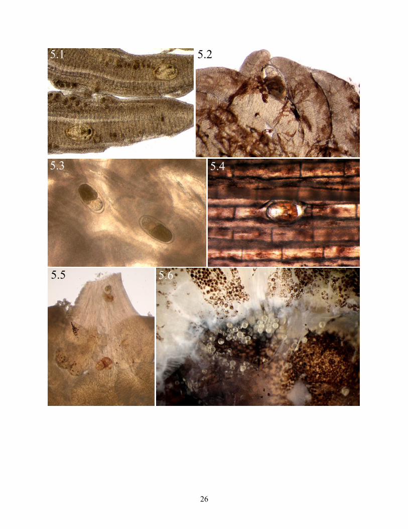
PLATE 5. Figures 5.1!5.6. Heterophyidae metacercariae (Platyhelminthes: Digenea) encysted
in tissues of Fundulus grandis Baird and Girard, 1853, (Cyprinodontiformes: Fundulidae)
collected from Barataria Bay, Louisiana. Fig. 5.1. Ascocotyle diminuta Stunkard and Haviland,
1924 infecting gill (75X). Fig. 5.2. Ascocotyle sp. infecting pseudobranch (75X). Fig. 5.3.
Heterophyidae sp. infecting serosa of intestine (75X). Fig. 5.4. Heterophyidae sp. infecting fin
ray (75X). Fig. 5.5. Ascocotyle tenuicolis Price, 1935 infecting bulbous arteriosus (75X). Fig.
5.6. Euhaplorchis sp. infecting neurocranium (75X).
!
25
5.1 5.2
5.3 5.4
5.5 5.6
26

characters included relatively large oral and ventral suckers. These species are not demonstrated
as bio-illustrations or photomicrographs in this thesis. However, these are inventoried as part of
the parasite component community (Digenea sp. 1 and Digenea sp. 2; see Table 1) of F. grandis.
Bucephalidae
Bucephalidae sp. 1 (Plate 4)
SITE(S) OF INFECTION: Ventricle myocardium (Fig. 4.1).
DIAGNOSTIC CHARACTER(S): Oral sucker hemi-spherical with flattened anterior edge (Fig.
4.2).
REFERENCE(S): Hopkins, 1954; Kinsella and Heard, 1974; Stunkard, 1976; Curran and
Overstreet, 2009.
Bucephalidae sp. 2 (Plate 4)
SITE(S) OF INFECTION: Head kidney (Fig. 4.3).
DIAGNOSTIC CHARACTER(S): Oral sucker inflated, well-demarcated, hemi-spherical, with
rounded anterior edge (Fig. 4.4.).
REFERENCE(S): Hopkins, 1954; Kinsella and Heard, 1974; Stunkard, 1976; Curran and
Overstreet, 2009.
Bucephalidae sp. 3 (Plate 4)
SITE(S) OF INFECTION: Somatic musculature (Fig. 4.5).
DIAGNOSTIC CHARACTER(S): Oral sucker uninflated, hemi-spherical, with rounded anterior
edge (Fig. 4.6).
REFERENCE(S): Hopkins, 1954; Kinsella and Heard, 1974; Stunkard, 1976; Curran and
Overstreet, 2009.
Bucephalidae sp. 4 (Plate 4)
27

SITE(S) OF INFECTION: Swim bladder (Fig. 4.7).
DIAGNOSTIC CHARACTER(S): Oral sucker nearly spherical with 4 papillae-like structures
(Fig. 4.8).
REFERENCE(S): Hopkins, 1954; Kinsella and Heard, 1974; Stunkard, 1976; Curran and
Overstreet, 2009.
Diplostomidae
Diplostomidae sp. (Plate 4)
SITE(S) OF INFECTION: Swim bladder (Fig. 4.9).
DIAGNOSTIC CHARACTER(S): Metacercariae with bulbous posterior end (Fig. 4.10).
REFERENCE(S): Hoffman, 1958; Bullard and Overstreet, 2008.
Heterophyidae
A scocoty le Looss, 1899
A scocoty le diminuta Stunkard and Haviland, 1924 (Plate 5, 6)
SITE(S) OF INFECTION: Gill lamellae (Fig. 5.1).
DIAGNOSTIC CHARACTER(S): Oral sucker with 16 oral spines plus two shorter dorsal
accessory spines (Fig. 6.1).
REFERENCE(S): Stunkard and Haviland, 1924; Chandler, 1941; Martin, 1953; Sogandares
Bernal and Lumsden, 1963; Ostrowski de Núñez, 1993.
A scocoty le tenuicolis Price, 1935 (Plate 5, 6)
SITE(S) OF INFECTION: Bulbous arteriosus (Fig. 5.5).
DIAGNOSTIC CHARACTER(S): Oral sucker with two rows of 16 spines (Fig. 6.2).
REFERENCE(S): Brock and Font, 2009; Scholz et al., 1995.
A scocoty le sp. (Plate 5, 6)!
28

PLATE 6. Figures 6.1!6.6. Heterophyidae metacercariae (Platyhelminthes, Digenea) infecting
Fundulus grandis Baird and Girard, 1853, (Cyprinodontiformes: Fundulidae) collected from
Barataria Bay, Louisiana. Fig. 6.1. Ascocotyle diminuta Stunkard and Haviland, 1924 excised
from gill (10 ocular units, 40X objective, DIC). Fig. 6.2. Ascocotyle tenuicolis Price, 1935
excised from bulbous arteriosus (10 ocular units, 20X objective, DIC). Fig. 6.3. Ascocotyle sp.
excised from pseudobranch (10 ocular units, 20X objective, DIC). Fig. 6.4. Oral spines of
Heterophyidae sp. 1 from fin ray (10 ocular units, 40X objective, 2X magnifier, DIC). Fig. 6.5.
Oral spines of Heterophyidae sp. 2 from fin ray (10 ocular units, 40X objective, 2X magnifier,
DIC). Fig. 6.6. Oral spines of Heterophyidae sp. 3 from fin ray (10 ocular units, 40X objective,
2X magnifier, DIC).
29

6.1 6.2
6.3
6.4
6.5
6.6
30

SITE(S) OF INFECTION: Pseudobranch (Fig. 5.2).
DIAGNOSTIC CHARACTER(S): Oral sucker with two rows of 18 spines (Fig. 6.3).
REFERENCE(S): Sogandares-Bernal and Bridgman, 1960; Sogandares-Bernal and Lumsden,
1963; Font et al., 1984.
Euhaplorchis Martin, 1950
Euhaplorchis sp. (Plate 5)
SITE(S) OF INFECTION: Neurocranium (Fig. 5.6).
DIAGNOSTIC CHARACTER(S): Metacercariae developing and with high intensity of infection
(Fig. 5.6).
REFERENCE(S): Martin, 1950; Abbott, 1968; Bullard and Overstreet, 2008.
Heterophyidae sp. 1 (Plate 5, 6)
SITE(S) OF INFECTION: Fin rays (Fig. 5.4).
DIAGNOSTIC CHARACTER(S): Oral sucker with two rows of 24 spines (Fig. 6.4).
REFERENCE(S): None recovered.
Heterophyidae sp. 2 (Plate 5, 6)
SITE(S) OF INFECTION: Fin rays (Fig. 5.4).
DIAGNOSTIC CHARACTER(S): Oral sucker with one rows of 28 spines (Fig. 6.5).
REFERENCE(S): None recovered.
Heterophyidae sp. 3 (Plate 5, 6)
SITE(S) OF INFECTION: Fin rays (Fig. 5.4).
DIAGNOSTIC CHARACTER(S): Oral sucker with one row of 24 spines (Fig. 6.6).
REFERENCE(S): None recovered.
Strigeidae
31

Strigeidae sp. (Plate 4)
SITE(S) OF INFECTION (Fig. 4.11): Fat.
DIAGNOSTIC CHARACTER(S): Body with tapered anterior and posterior ends (Fig. 4.12).
REFERENCE(S): Bullard and Overstreet, 2008.
Monogenoidea
Ancyrocephalidae
Salsuginus Bevelery-Burton, 1984
Salsuginus n. sp.1 (Plate 7)
SITE(S) OF INFECTION: Gill lamellae (Figs. 7.1, 7.3).
DIAGNOSTIC CHARACTER(S): Transverse dorsal and ventral bars highly arched. Hamuli
sharply recurved (Fig. 7.2).
REFERENCE(S): Williams, 1980; Beverley-Burton, 1984; Murith and Beverley-Burton, 1985.
Salsuginus n. sp. 2 (Plate 7)
SITE(S) OF INFECTION: Gill lamellae (Figs. 7.1, 7.3).
DIAGNOSTIC CHARACTER(S): Transverse ventral bar with deep ridges (Fig. 7.4).
REFERENCE(S): Williams, 1980; Beverley-Burton, 1984; Murith and Beverley-Burton, 1985.
Gyrodactylidae
Gy rodacty lus Nordmann, 1832
Gy rodacty lus stephanus Mueller, 1937 (Plate 8)
SITE(S) OF INFECTION: Buccal cavity (Fig. 8.1) and skin of body.
DIAGNOSTIC CHARACTER(S): Haptor with sixteen marginal hooks and single pair of hamuli
with ventro-mesial knobs articulating with ventral bar (Fig. 8.2).
REFERENCE(S): Mueller, 1937; Hargis, 1955; Mizelle and Kritsky, 1967 (key to North!
32

PLATE 7. Figures 7.1!7.4. Salsuginus spp. (Platyhelminthes: Monogenoidea:
Ancyrocephalidae) infecting Fundulus grandis Baird and Girard, 1853, (Cyprinodontiformes:
Fundulidae) collected from Barataria Bay, Louisiana. Fig. 7.1. Low magnification view of
Salsuginus infecting gill lamellae (25X). Fig. 7.2. Light micrograph of Salsuginus sp. 1 haptor
with transverse bars and hamuli. Fig. 7.3. Higher magnification view of Salsuginus infecting gill
lamellae (50X). Fig. 7.4. Salsuginus sp. 2 transverse bars and hamuli. Scale bar equal to 15 µm.
!
33

7.1 7.2
7.3 7.4
34

PLATE 8. Figures 8.1!8.6. Gyrodactylidae (Platyhelminthes: Monogenoidea) infecting
Fundulus grandis Baird and Girard, 1853, (Cyprinodontiformes: Fundulidae) collected from
Barataria Bay, Louisiana. All scale bars equal 20 µm. Fig. 8.1. Gyrodactylus stephanus Mueller,
1937 infecting buccal cavity (15X). Fig. 8.2. Haptor of G. stephanus. Fig. 8.3. Fundulotrema cf.
prolongis (Hargis, 1955) Kritsky and Thatcher, 1977 infecting skin of fins (30X). Fig. 8.4.
Haptor of F. cf. prolongis. Fig. 8.5. Swingleus polyclithroides Rogers, 1969 infecting skin of eye
(75X). Fig. 8.6. Haptor of S. polyclithroides.
!
35

8.1
8.5
8.3
8.6
8.2
8.4
36

American species of Gyrodactylus); Hendrix, 1994; King and Cone, 2009.
Fundulotrema Kritsky and Thatcher, 1977
Fundulotrema cf. prolongis (Hargis, 1955) Kritsky and Thatcher, 1977
(Plate 8)
SITE(S) OF INFECTION: Skin of buccal cavity (Fig. 8.2) and fins (Fig. 8.3).
DIAGNOSTIC CHARACTER(S): Haptor with sixteen marginal hooks and single pair of
relatively long hamuli supported by dorsal and ventral bars and lacking lateral wing-like bars
(Fig. 8.4).
REFERENCE(S): Kritsky and Thatcher, 1977 (generic diagnosis); Cone and Odense, 1988
(emended generic diagnosis); Beverley-Burton, 1984; Hendrix, 1994.
Sw ingleus Rogers, 1969
Sw ingleus poly clithroides Rogers, 1969 (Plate 8)
SITE(S) OF INFECTION: Skin of eye (Fig. 8.3), body, and fins (Fig. 8.5).
DIAGNOSTIC CHARACTER(S): Haptor with sixteen marginal hooks and pair of relatively
long hamuli supported by ventral bar with shield, with lateral wing-like bars, and without dorsal
bar (Fig. 8.6).
REFERENCE(S): Rogers, 1969 (generic diagnosis); Hendrix, 1994; Hoffman, 1999.
Cestoda
Gryporhynchidae
Gryporhynchidae sp. 1 (Plate 9)
SITE(S) OF INFECTION: Body cavity (Fig. 9.1), gonad, liver, mesentery.
DIAGNOSTIC CHARACTER(S): Metacestode with armed scolex (Fig. 9.2).
REFERENCE(S): Chandler, 1935; Scholz and Salgado-Maldonado, 2001; Scholz et al. 2002;!
37

PLATE 9. Figures 9.1!9.4. Larval cestodes (Platyhelminthes: Cestoda) infecting Fundulus
grandis Baird and Girard, 1853, (Cyprinodontiformes: Fundulidae) collected from Barataria Bay,
Louisiana. Fig. 9.1. Gryporhynchidae sp. metacestode larvae infecting serosa of testes (25X).
Fig. 9.2. Excysted Gryporhynchidae sp. metacestode larva (40X). Fig. 9.3. Proteocephalidae sp.
pleurocercoid infecting mucosa of stomach (75X). Fig. 9.4. Heat-killed Proteocephalidae sp.
(pleurocercoid) larva (10 ocular units, 20X objective).
!
38

9.49.3
9.1 9.2
39

Scholz et al., 2004; Scholz and Harris, 2006.
Proteocephalidae
Proteocephalidae sp. 1 (Plate 9)
SITES OF INFECTION: Mucosa of stomach (Fig. 9.3).
DIAGNOSTIC CHARACTER(S): Pleurocercoid with 4 unarmed suckers and apical organ (Fig.
9.4).
REFERENCE(S): Freze, 1969; Hoffman, 1999.
1.6.3. Nematoda
Two adult forms are previously reported to infect F. grandis. These are Capillaria
cyprinodonticola Huffman and Bullock, 1973 (in liver) and Spirocamallanus cricotus Fusco and
Overstreet, 1978 (in intestine). During this study, only two specimens of adult nematodes were
collected from the intestine of two individuals of F. grandis. These specimens have not been
studied and require further taxonomic comparison with the aforementioned adult forms.
Therefore, adult nematodes are not demonstrated in this thesis but are included as part of the
parasite component community infecting F. grandis.
Eustrongy lides Jägerskiöld, 1909 (Plate 10)
SITE(S) OF INFECTION: Atrium of heart (Fig. 10.1), pericardium, body cavity, caudal kidney,
liver (Fig. 10.2).
DIAGNOSTIC CHARACTER(S): Body relatively large, opaque, grossly visible with naked eye,
red in color when alive.
REFERENCE(S): Lichtenfels and Pilitt, 1986; Hoffman, 1999; Overstreet, 2003.
Spiuridae
Spiuridae sp. (Plate 10)!
40

PLATE 10. Figures 10.1!10.3. Nematode larvae infecting Fundulus grandis Baird and
Girard, 1853, (Cyprinodontiformes: Fundulidae) collected from Barataria Bay, Louisiana. Fig.
10.1. Eustrongylides sp. (L3) infecting atrium of heart (15X). Fig. 10.2. Eustrongylides sp. (L3)
infecting liver (15X). Fig. 10.3. Spiuridae sp. (L3) excised from mucosa of digestive system (10
ocular units, 40X objective).
!
41

10.1
10.2
10.3
42

SITE(S) OF INFECTION: Mucosa of intestine.
DIAGNOSTIC CHARACTER(S): L3 with tapered posterior end (Fig. 10.3).
REFERENCE(S): Hoffman, 1999.
1.6.4. Acanthocephala
Two adult species of acanthocephala primarily infected the mucosa of the intestine of F.
grandis and their key diagnostic features are demonstrated herein. An additional species infected
the body cavity of F. grandis and was identified as an encysted cystacanth larva. This larval form
was identified based on the presence of a thick cyst wall that surrounded retracted specimens.
Proboscis hooks were visible through both the cyst and the tegument. Heat-killed specimens with
a fully everted proboscis were not collected or extensively studied and photographs and drawings
of this rare species are not demonstrated in this thesis. However, this species is included as part
of the parasite component community of F. grandis (Table 1).
Echinorhynchida
Illiosentidae
Dollfusentis Golvan, 1969
Dollfusentis cf. chandleri Golvan, 1969 (Plate 11)
SITE(S) OF INFECTION: Mucosa of intestine (Fig. 11.1).
DIAGNOSTIC CHARACTER(S): Proboscis with 6 elongated hooks at base. Neck with cuticular
spines (Fig. 11.2).
REFERENCE(S): Golvan, 1969.
Neoechinorhynchida
Neoechinorhynchidae
Octospiniferoides Bullock, 1957 !
43

PLATE 11. Figures 11.1!11.4. Acanthocephala infecting Fundulus grandis Baird and
Girard, 1853, (Cyprinodontiformes: Fundulidae) collected from Barataria Bay, Louisiana. Scale
bars equal to 50 µm. Fig.11.1. Dollfusentis cf. chandleri Golvan, 1969 (Echinorhynchida;
Illiosentidae) in sediment after scraping mucosa of intestine (25X). Fig. 11.2. Proboscis and neck
armature of D. cf. chandleri. Fig. 11.3. Octospiniferoides chandleri Bullock, 1957
(Neoechinorhynchida: Neoechinorhynchidae) removed from mucosa of intestine (100X). Fig.
11.4. Proboscis armature of O. chandleri.
!
44

11.1 11.2
11.3
11.4
45

Octospiniferoides chandleri Bullock, 1957 (Plate 11)
SITE(S) OF INFECTION: Mucosa of intestine.
DIAGNOSTIC CHARACTER(S): Proboscis short and with 3 rows of 8 slender hooks (Figs.
11.3!11.4).
REFERENCE(S): Bullock, 1957; Bullock, 1966; Salgado-Maldonado et al., 1992; Salgado-
Maldonado et al., 1997.
1.6.5. Annelida
Hirudinida
Piscicolidae
Malmiana Strand, 1942
Malmiana philotherma Sawyer, Lawler, and Overstreet, 1975 (Plate 12)
SITE(S) OF INFECTION: Skin of fins (Fig. 12.1) and branchiostegal membrane.
DIAGNOSTIC CHARACTER(S): Body flattened. Caudal sucker large and well-demarcated
from posterior end of body (urosome) (Fig. 12.2).
REFERENCE(S): Sawyer et al., 1975; Sawyer and Kinard 1980; Burreson and Kalman, 2006.
My z obdella Leidy, 1851
My z obdella lugubris Leidy, 1851 (Plate 12)
SITE(S) OF INFECTION: Skin of fins and branchiostegal membrane (Fig. 12.3).
DIAGNOSTIC CHARACTER(S): Caudal sucker confluent with urosome (Fig. 12.4).
REFERENCE(S): Sawer et al., 1975; Appy and Dadswell, 1980.
1.6.6. Arthropoda
Copepoda
Two species of adult copepods primarily infected the gill filaments of F. grandis. Another
46

PLATE 12. Figures 12.1!12.4. Hirudinida (Annelida: Piscicolidae) infecting Fundulus grandis
Baird and Girard, 1853, (Cyprinodontiformes: Fundulidae) collected from Barataria Bay,
Louisiana. Fig. 12.1. Malmiana philotherma Sawyer, Lawler, and Overstreet, 1975 infecting anal
fin (40X). Fig. 12.2. Heat-killed M. philotherma from fin (30X). Fig. 12.3. Myzobdella lugubris
Leidy, 1851 infecting pectoral fin (7.5X). Fig. 12.4. Heat-killed M. lugubris from fin (25X).
!
47

12.1 12.2
12.3 12.4
48

juvenile species, a copepodid of Caligidae (Cressey, 1991), infected the fins of F. grandis. This
species was not identified to any other level other than family and is not demonstrated in this
thesis. However, this species is included as part of the parasite component community of F.
grandis (Table 1).
Ergasilidae
Ergasilus Nordman, 1832
Ergasilus cf. arthrosis Roberts, 1969 (Plate 13)
SITE(S) OF INFECTION: Gill filaments (Fig. 13.1, 13.3).
DIAGNOSTIC CHARACTER(S): Second antennae with 4th
segment 80% as long as 3rd
segment
(Fig. 13.2).
REFERENCES: Roberts, 1970; Kabata, 1986.
Ergasilus funduli Krøyer, 1863 (Plate 13)
SITE(S) OF INFECTION: Gill filaments (Fig. 13.1, 13.3).
DIAGNOSTIC CHARACTER(S): Second antennae with inflated 2nd
segment of second antenna
and single tubercle on inner margin of 4th
segment (Fig. 13.4).
REFERENCE(S): Roberts, 1970; Kabata, 1986.
Branchiura
Argulidae
A rgulus Müller
A rgulus n. sp. 1 (Plate 14)
SITE(S) OF INFECTION: Buccal cavity (Fig. 14.1).
DIAGNOSTIC CHARACTER(S): Base of second maxilla with large pad of cycloid-like scales,
three elongated basal teeth, with 5 ventral and 2 dorsal setae (Fig. 14.2).!
49
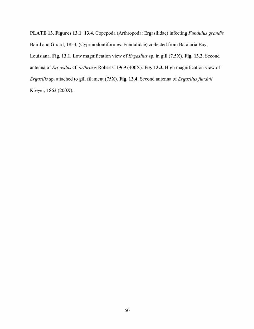
PLATE 13. Figures 13.1!13.4. Copepoda (Arthropoda: Ergasilidae) infecting Fundulus grandis
Baird and Girard, 1853, (Cyprinodontiformes: Fundulidae) collected from Barataria Bay,
Louisiana. Fig. 13.1. Low magnification view of Ergasilus sp. in gill (7.5X). Fig. 13.2. Second
antenna of Ergasilus cf. arthrosis Roberts, 1969 (400X). Fig. 13.3. High magnification view of
Ergasilis sp. attached to gill filament (75X). Fig. 13.4. Second antenna of Ergasilus funduli
Krøyer, 1863 (200X).
!
50

13.1 13.2
13.3 13.4
51

PLATE 14. Figures 14.1!14.4. Branchiura (Arthropoda, Argulidae) infecting Fundulus grandis
Baird and Girard, 1853, (Cyprinodontiformes: Fundulidae) collected from Barataria Bay,
Louisiana. Scale bars equal to 30 µm. Fig. 14.1. Dorsal view of Argulus sp. infecting buccal
cavity (20X). Fig. 14.2. Base of second maxilla of Argulus n. sp. Fig. 14.3. Ventral view of
Argulus cf. funduli Krøyer, 1863 (40X). Fig. 14.4. Base of second maxilla of A. cf. funduli.
!
52

14.1
14.3
14.2
14.4
53

REFERENCE(S): Wilson, 1905; Cressy, 1978; Kabata, 1988.
A rgulus n. sp. 2 (Plate 14)
SITE(S) OF INFECTION: Buccal cavity (Fig. 14.1).
DIAGNOSTIC CHARACTER(S): Base of second maxilla with 3 short basal teeth, four ventral
setae (Fig. 14.4).
REFERENCE(S): Wilson, 1905; Cressy, 1978; Kabata, 1988.
Isopoda
Cymthoidae
Lironeca Leach, 1818
Lironeca ov alis Say, 1818 (Plate 15)
SITE(S) OF INFECTION: Buccal cavity (Figs. 15.1!15.2).
DIAGNOSTIC CHARACTER(S): 5th
thoracic segment wider than 3rd
thoracic segment (Fig.
15.2).
REFERENCE(S): Price and Schlueter, 1980; Williams and Bowman, 1994; Hoffman, 1999.
1.6.7. Protozoa
Calyptospora cf. funduli (Duszynski, Solangi, and Overstreet, 1979) Overstreet, Hawkins,
and Fournie, 1984, (Apicomlexa: Calyptosporidae) primarily infected the liver of F. grandis
collected from Barataria Bay, LA. Other infection sites included fat, mesentery, and peritoneum
of the body cavity. C. cf. funduli also infected the cysts of digenean metacercariae, metacestodes,
and encapsulations of nematodes. Solangi and Overstreet (1980), Upton and Duszynski (1982),
Hawkins et al. (1983), Fournie and Overstreet (1993), and Fournie et al. (2000) have published
information regarding the taxonomy and biology of this parasite.
Other morphologically distinct coccidia were occasionally viewed infecting the apical!
54

PLATE 15. Figure 15.1!15.2. Lironeca ovalis Say 1818 (Arthropoda, Isopoda, Cymthoidae)
infecting buccal cavity of Fundulus grandis Baird and Girard, 1853, (Cyprinodontiformes:
Fundulidae) collected from Barataria Bay, Louisiana. Fig. 15.1. Lateral view in buccal cavity
(7.5X). Fig. 15.2. Dorsal view in exposed buccal cavity (10X).
55

15.1
15.2
56

surface of the urinary bladder. As this work mainly deals with major metazoan parasite groups,
no plates were constructed for protozoan parasites of F. grandis in this thesis.
1.7. Discussion
The parasite component community of F. grandis collected from Barataria Bay consisted of
44 species (31 endoparasites and 13 ectoparasites) that infected 23 tissue types. Of these, 10 are
putatively new to science (Table 1), 24 constitute new host records, and 42 are putatively new
locality records in Louisiana. Including 29 previous records, there are 60 total metazoan parasite
species reported to infect F. grandis. Only 7 nominal accepted species have been originally
described from F. grandis (Table 1), which underscores the probability of discovering species
that are new to science.
Herein, I present information regarding the taxonomy and systematics of each major
metazoan parasite group wherever applicable. Additional remarks are provided for each putative
species including new species and taxa that could not be identified to the level of species.
1.7.1. Myxozoa (Plates 2!3)
The Myxozoa are histozoic and coelozoic endoparasites of freshwater and marine fishes
and mature in invertebrates but infect fishes (Kent et al., 1994), including chondrichthyans, as
second intermediate hosts (Benz and Bullard, 2004; Blaylock et al., 2004). Initially considered to
be Protozoa, Myxozoans have since been classified as Metazoa but the systematics of this group
is still debated today. In this thesis, I followed current classification and regarded the Myxozoa
as a major metazoan parasite phylum (Dyková and Lom, 2007).
K udoa Meglitsch, 1947
As of 2004, Kudoa consisted of 49 named species with distributions in the northern Gulf of
Mexico, Western Atlantic Ocean, and Western Pacific Ocean (Blaylock et al., 2004). Three
57

Tab
le 1
. Sys
tem
atic
list
ing
of th
e sp
ecie
s mem
bers
of t
he p
aras
ite (m
etaz
oans
onl
y) c
ompo
nent
com
mun
ity o
f Gul
f kill
ifish
,
Fund
ulus
gra
ndis
Bai
rd a
nd G
irar
d, 1
853
in B
arat
aria
Bay
and
from
pre
viou
sly-
publ
ishe
d re
cord
s. Ta
xa w
ith (*
) com
pris
e th
e
para
site
com
pone
nt c
omm
unity
of F
. gra
ndis
in B
arat
aria
Bay
. Abb
r. Lo
calit
y: M
issi
ssip
pi (M
S), L
ouis
iana
(LA
), Te
xas (
TX),
Flor
ida
(FL)
, Ala
bam
a (A
L), n
ot re
porte
d (N
R).
Life
his
tory
: adu
lt (a
), as
exua
l myx
ospo
re (a
m),
cope
podi
d (c
o), c
ysta
cant
h (c
y), t
hird
stag
e
larv
a (L
3), l
arva
l for
m (l
f), m
etac
erca
ria (m
e), m
etac
esto
de (m
s), p
leur
ocer
coid
larv
a (p
); Li
fe c
ycle
type
: dire
ct,
ecto
para
site
(D),
indi
rect
, end
opar
asite
(I).
Taxo
n Si
te o
f In
fect
ion
Loca
lity
Life
H
isto
ry
Life
Cyc
le
New
H
ost
Rec
ord
New
Lo
calit
y R
ecor
d
Ref
eren
ce(s
)
MY
XO
ZO
A
Kud
oida
e
Kud
oa sp
. sm
M
S am
I
D
ykov
á et
al.,
19
94
Kud
oa c
f. fu
ndul
i*
sm
LA
am
I X
X
pr
esen
t stu
dy
Myx
obol
idae
Myx
obol
us n
. sp.
1*
g LA
am
I
X
X
pres
ent s
tudy
M
yxob
olus
n. s
p. 2
* sk
LA
am
I
X
X
pres
ent s
tudy
M
yxob
olus
n. s
p. 3
* bs
LA
am
I
X
X
pres
ent s
tudy
M
yxob
olus
n. s
p. 4
* e
LA
am
I X
X
pr
esen
t stu
dy
Myx
obol
us n
. sp.
5 *
bf
LA
am
I X
X
pr
esen
t stu
dy
Myx
obol
us n
. sp.
6 *
sb
LA
am
I X
X
pr
esen
t stu
dy
DIG
EN
EA
D
igen
ea sp
. 1*
st
LA
a I
X
X
pres
ent s
tudy
D
igen
ea sp
. 2*
sb
LA
a I
X
X
pres
ent s
tudy
58

Buc
epha
lidae
Buc
epha
lidae
sp. 1
* vm
LA
m
e I
X
pr
esen
t stu
dy
Buc
epha
lidae
sp. 2
* k
LA
me
I
X
pres
ent s
tudy
B
ucep
halid
ae sp
. 3*
sm
LA
me
I
X
pres
ent s
tudy
B
ucep
halid
ae sp
. 4*
sb
LA
me
I
X
pres
ent s
tudy
Pr
osor
hync
hoid
es
stro
ngyl
urae
nr
TX
m
e I
Spar
ks, 1
960
Rhip
idoc
otyl
e lin
toni
nr
TX
m
e I
Spar
ks, 1
960
Dip
lost
omid
ae
Po
stho
dipl
osto
mum
min
imum
nr
FL
m
e I
Ban
gham
, 194
0 Po
stho
dipl
osto
mum
sp.*
f,
ms,
sb
LA
me
I X
X
pr
esen
t stu
dy
Het
erop
hyid
ae
As
coco
tyle
ang
rens
e g
LA
me
I
So
gand
ares
-Ber
nal
& L
umsd
en, 1
963
Asco
coty
le d
imin
uta*
g
LA
me
I
So
gand
ares
-Ber
nal
& B
ridgm
an, 1
960;
pr
esen
t stu
dy
Asco
coty
le la
geni
form
is
g TX
m
e I
Mar
tin, 1
953
Asco
coty
le te
nuic
olis*
ba
LA
m
e I
X
pr
esen
t stu
dy
Asco
coty
le sp
.*
p LA
m
e I
X
X
pres
ent s
tudy
Eu
hapl
orch
is sp
.*
n N
R; L
A
me
I
X
Bul
lard
&
Ove
rstre
et, 2
008;
pr
esen
t stu
dy
Het
erop
hyid
ae sp
. 1*
fr
LA
me
I
X
pres
ent s
tudy
H
eter
ophy
idae
sp. 2
* fr
LA
m
e I
X
X
pres
ent s
tudy
H
eter
ophy
idae
sp. 3
* fr
LA
m
e I
X
X
pres
ent s
tudy
St
icto
dora
cur
sita
ns
sm
FL
me
K
inse
lla &
Hea
rd,
1964
59

Stri
geid
ae
St
rigei
dae
sp.*
f
LA
me
I X
X
pr
esen
t stu
dy
MO
NO
GE
NO
IDE
A
Anc
ycro
cpeh
alid
ae
Sals
ugin
us n
. sp.
1*
g LA
a
D
X
X
pres
ent s
tudy
Sa
lsug
inus
n. s
p. 2
* g
LA
a D
X
X
pr
esen
t stu
dy
Sals
ugin
us sp
irae
g
AL
a D
W
illia
ms,
1980
G
yrod
acty
lidae
Gyr
odac
tylu
s cf.
step
hanu
s*
g, sk
FL
; LA
a
D
X
H
argi
s, 19
55; p
rese
nt
stud
y Fu
ndul
otre
ma
prol
ongi
s*
sk, g
FL
; AL
a
X
H
argi
s, 19
55;
Rog
ers,
1969
Sw
ingl
eus p
olyc
lithr
oide
s*
sk
AL
a D
X
Rog
ers,
1969
C
EST
OD
A
Bot
hrio
ceph
alid
ae
Both
rioc
epha
lus s
p.
nr
FL
p I
Ban
gham
, 194
0 G
rypo
rhyn
chid
ae
G
rypo
rhyn
chid
ae sp
.*
bc, m
s, sd
, l,
go
LA
ms
I X
X
pr
esen
t stu
dy
Prot
eoce
phal
idae
Prot
eoce
phal
idae
sp.*
LA
p I
X
X
pres
ent s
tudy
Pr
oteo
ceph
alus
sp.
FL
p
I
B
angh
am, 1
940
NE
MA
TO
DA
Nem
atod
a sp
. 1*
i LA
a
I
X
pres
ent s
tudy
N
emat
oda
sp. 2
* i
LA
a I
X
pr
esen
t stu
dy
Cam
alla
nida
e
Spir
ocam
alla
nus c
rico
tus
i M
S a
I
Fu
sco
& O
vers
treet
, 19
78
60

Dio
ctop
hym
atoi
dae
Eu
stro
ngyl
ides
igno
tus
bc
NR
L3
I
Ove
rstre
et, 2
003
Eust
rong
ylid
es sp
.*
bc, h
, k, l
, m
s, sb
LA
L3
I
X
pr
esen
t stu
dy
Spiu
rida
e
Spiu
ridae
sp.*
i
LA
L3
I
X
pres
ent s
tudy
T
rich
inel
lidae
Cap
illar
ia c
ypri
nodo
ntic
ola
l FL
a
I
Huf
fman
& B
ullo
ck,
1973
A
CA
NT
HO
CE
PHA
LA
A
cant
hoce
phal
a sp
.*
bc
LA
cy
I
X
pr
esen
t stu
dy
Illio
sent
idae
Dol
lfuse
ntis
cf.
chan
dler
i*
i TX
; LA
a
I
X
B
ullo
ck, 1
957;
pre
sent
st
udy
Neo
echi
norh
ynch
idae
Oct
ospi
nife
roid
es c
hand
leri
* i
TX; F
L;
LA
a
I
X
Bul
lock
, 195
7;
Bul
lock
, 196
6; p
rese
nt
stud
y Po
lym
orph
idae
Sout
hwel
lina
hisp
ida
ms
TX
cy
I
Bul
lock
, 195
7 R
hadi
norh
ynch
idae
Lept
orhy
ncho
ides
thec
atus
i,
ms
FL; A
L cy
I
B
angh
am, 1
940;
W
illia
ms &
Gai
ns,
1974
H
IRU
DIN
IDA
Pisc
icol
idae
Mal
mia
na p
hilo
ther
ma*
sk
A
L; L
A
a
D
X
W
illia
ms,
1979
; pr
esen
t stu
dy
61

Myz
obde
lla lu
gubr
is*
sk
MS;
LA
a
D
X
Saw
yer e
t al.,
197
5;
pres
ent s
tudy
PE
NT
AST
OM
IDA
Sebe
kia
mis
siss
ippi
ensi
s ct
M
S lf
I
O
vers
treet
et a
l., 1
985
CO
PEPO
DA
Cal
igid
ae
C
alig
idae
sp.*
f
LA
co
D
X
X
pres
ent s
tudy
E
rgas
ilida
e
Erga
silu
s cf.
arth
rosi
s*
g LA
a
D
X
X
pres
ent s
tudy
Er
gasi
lus f
undu
li*
g A
L, F
L,
MS;
LA
a
D
X
Jo
hnso
n &
Rog
ers,
1973
; pr
esen
t stu
dy
Erga
silu
s liz
ae
g FL
; AL
a D
B
ere,
193
6; Jo
hnso
n &
R
oger
s, 19
73
BR
AN
CH
IUR
A
A
rgul
idae
Argu
lus n
. sp.
1*
bc
LA
a D
X
X
pr
esen
t stu
dy
Argu
lus n
. sp.
2*
bc
LA
a D
X
X
pr
esen
t stu
dy
ISO
POD
A
C
ymth
oida
e
Liro
neca
ova
lis*
bc
FL, L
A
a D
X
Pric
e &
Sch
luet
er, 1
980;
pr
esen
t stu
dy
62

unnamed congeners have been reported from fundulids that were not identified to species, the
western mosquitofish, Gambusia affinis (Baird and Girard, 1853), (Cyprinodontiformes:
Poeciliidae), and the inland silverside, Menidia beryllina (Cope, 1867), (Atheriniformes:
Atherinopsidae) in the northern Gulf of Mexico (Dykova et al., 1994). It is supported that
molecular tools should be used when describing species of Kudoa because workers have shown
that spore morphometrics cannot differentiate all species in the genus (Blaylock et al., 2004).
K udoa cf. funduli (Hahn, 1915) Meglitsch, 1947 (Plate 2)
Specimens examined in this study most closely resemble Kudoa funduli (Hahn, 1915)
Meglitsch, 1947, (Multivalvuvida: Kudoidae). Spores of K. funduli are described as rounded
posteriorly and “sharply attenuated” anteriorly in sutural view (Meglitsch, 1948). However,
Akaishi et al. (2004) used scanning electron microscopy to also show that the posterior end of
spores of K. funduli is slightly pointed in sutural view but also showed that this feature was not
readily apparent in formalin fixed wet-mounted spores. In contrast, wet-mounted spores of K. cf.
funduli studied herein contained conspicuously but not sharply pointed posterior and anterior
ends in sutural view (Figs. 2.3!2.4). Also in agreement with supplemental descriptions of K.
funduli, (Meglitsch et al., 1948; Moran et al., 1999; Akaishi et al., 2004) spores studied herein
were contained in elongated cysts (Figs. 2.1!2.2) in somatic muscle and possessed apical
projections on spore valves (Figs. 2.3!2.4).
My xobolus Bütschli, 1882
A total of 6 species of Myxobolus were identified during this study (Table 2, Plate 3). These
are putatively new species and would be the first records of Myxobolus infecting F. grandis in
the Gulf of Mexico. An attempt was made to scrutinize the morphological and morphometric
characteristics of spores based on sites of infection. I have included a table showing
63

morphometric and morphological data from species of Myxobolus observed herein along with
published data from species of Myxobolus infecting other species of Fundulus (Table 2). This
table is provided to demonstrate morphometric and morphological differences between described
species of Myxobolus and the congeners studied herein.
The presence or absence of a vacuole within the sporoplasm of myxospores was once used to
diagnose species of Myxobolus and Myxosoma (see Landsberg and Lom, 1991). Because of the
reported unreliability of this feature (Lom and Noble, 1984), all species of Myxosoma (those
species without an iodinophilous vacuole [Kudo, 1918]), were assigned to Myxobolus
(Landsberg and Lom, 1991). The unreliability of this feature was evident in specimens studied
herein, but the beginners mind requires consideration of this feature, and thus, the presence or
absence of a conspicuous unstained vacuole in the sporoplasm of myxospores is noted (Table 2).
Myxobolus spp. infected gill lamellae (Fig. 3.1), skin beneath scales (Fig. 3.3),
branchiostegal skin (Fig. 3.5), skin of eye (Fig. 3.7), the base of pectoral fins (Fig. 3.9; putatively
dermis or somatic muscle), and the swim bladder (Fig. 3.11) of F. grandis. Myxozoans also
infected the serosa of the intestine, kidney, spleen, branchial vessels of gill arches, and the
musculature of the head. Myxozoans infecting these sites were not studied and are not included
in this thesis.
The high number of infection sites observed in F. grandis poses interesting taxonomic
questions. While some myxozoan species are regarded as highly host specific and infect specific
infection sites across a limited number of ecologically and phylogenetically related hosts
(Ferguson et al., 2008), others infect a high number of distantly related hosts (Bond, 1937; Eiras
et al., 2005). Myxozoans that infect F. heteroclitus have been experimentally shown to only
infect other species of Fundulus collected from the same habitat (Bond, 1937). Noteworthy is
64

that the type species, Myxobolus muelleri Bütshchli, 1882, (Bivalvuvida: Myxobolidae) has been
reported from more than 80 hosts, most of which are Eurasian cyprinid hosts (Lom and Dyková,
2006).
Myxobolus spp. are generally histozoic parasites that infect freshwater, estuarine, and marine
fishes including species of Fundulus in Canada (Wiles, 1975), the eastern United States (Hahn,
1915, 1917a, 1917b), and the northern Gulf of Mexico; i.e., Fundulus similis (Baird and Girard,
1853), (Cyprinodontiformes: Fundulidae) (see Rigdon and Hendricks, 1955). Approximately 792
named species of Myxobolus exist in the literature including 7 that infect amphibian hosts (Lom
and Dyková, 2006). However, many species of Myxobolus that infect fishes may be
synonymous, which has partly resulted from difficulty in differentiating species based on
morphology and morphometrics (Lom and Dyková, 2006), confusion in the taxonomy of the
Myxobolus/ Myxosoma group (Landsberg and Lom, 1991), and the lack of studies utilizing
molecular tools (SSU rRNA gene sequences for only 170 species of Myxobolus exist in
GenBank, see Ferguson et al., 2008) to diagnose and identify species (Cone and Easy, 2005).
Furthermore, it is suggested by some workers that marine species, (30 of approximately 792
[3.8%]) may belong in another genus due to the predominance of freshwater species (Lom and
Dyková, 2006), however, studies utilizing molecular tools are lacking to support this idea.
Species of Myxobolus have pyriform, ellipsoid, ovoid, or sub-circular spores in valvular
view, and appear biconvex in sutural view. In addition, spores consist of two anteriorly-
positioned pyriform or rounded polar capsules bearing a coiled filament. Posterior ridges are
present in some species (Bond, 1938) along with posterior sutural markings (Kudo, 1918); herein
referred to as posterior sutural bumps. In addition, the sporoplasm of spores is with or without an
iodinophilous vacuole (Landsberg and Lom, 1991).
65

My xobolus n. sp. 1 (Fig. 3.1!3.2)
The three spore types of Myxobolus sp. 1 studied herein have overlapping spore lengths with
Myxobolus bilineatum Bond, 1938, (Bivalvuvida: Myxobolidae) but have significantly different
widths (Table 2). Myxobolus n. sp. 1 is differentiated from Myxobolus diaphanus (Fantham,
Porter, and Richardson, 1940) Landsberg and Lom, 1991, (Bivalvuvida: Myxobolidae),
Myxobolus hudsonis Bond, 1938, (Bivalvuvida: Myxobolidae), Myxobolus subtecalis (Bond,
1938) Landsberg and Lom, 1991, (Bivalvuvida: Myxobolidae), and Myxobolus funduli (Hahn,
1915) Kudo, 1920, (Bivalvuvida: Myxobolidae), by having spores less than 12 µm in length and
polar filaments with no more than 8 coils. Myxobolus n. sp. 1 is differentiated from Myxobolus
n. sp. 2 by rarely possessing dense bodies in the sporoplasm (relatively common in Myxobolus n.
sp. 2) and by sometimes bearing posterior crescentic ridges (Fig. 3.2) (absent in Myxobolus sp.
2). Myxobolus n. sp. 1 is differentiated from Myxobolus n. sp. 3 by having a significantly shorter
spore length, rarely containing a vacuole within the sporoplasm (relatively common in
Myxobolus n. sp. 3), and by sometimes bearing posterior crescentic ledges (absent in Myxobolus
sp. 3). Myxobolus n. sp. 4 spores were significantly longer and wider than Myxobolus n. sp. 1
spores and lacked a vacuole and dense bodies within the sporoplasm and lacked posterior sutural
bumps.
This thesis includes 3 spore types that were drawn from spores occupying the same
plasmodium. The presence of a caudal process was rarely viewed in spores occupying the same
plasmodium. Posterior sutural bumps were viewed on all spores except those bearing a caudal
process, perhaps indicating these spores (with sutural bumps) are more developed forms.
My xobolus n. sp. 2 (Fig. 3.3!3.4)
Myxobolus n. sp. 2 is a putatively distinct from M. diaphanus, M. subtecalis, and M.
66

funduli by having a significantly shorter spore length. The number of filament coils in these
species are far more than that observed for Myxobolus n. sp. 2 while M. bilineatum and M.
hudsonis have overlapping filament coils. Myxobolus n. sp. 2 differs from M. bilineatum by
having pyriform spores compared to ovate or nearly rounded spores. M. hudsonis differs from
Myxobolus n. sp. 2 mainly by bearing polar capsules with anterior ends that are slightly offset
relative to the bilateral plane in valvular view (parallel to the bilateral plane in Myxobolus n. sp.
2). The pyriform spores of Myxobolus n. sp. 2 differ from the ovate to rounded spores of
Myxobolus n. sp. 5 and 6. Myxobolus n. sp. 3 and 4 are significantly longer than those of
Myxobolus n. sp. 1 (Table 2).
My xobolus n. sp. 3 (Figs. 3.5!3.6)
This species is most morphometrically (overlapping spore length and width and polar capsule
length and width) and morphologically (both pyriform spores with prominent vacuole in
sporoplasm) similar to M. funduli. The species demonstrated herein, however, has fewer coils
contained within polar capsules, i.e. 7!9 in Myxobolus n. sp. 3, and 11!14 in M. funduli.
M. subtecalis and M. diaphanus also possess a greater number of filament coils per polar
capsule compared to Myxobolus n. sp. 3. The spore shape of M. bilineatum is ovate to rounded
and not pyriform as in Myxobolus n. sp. 3. M. hudsonis differs from Myxobolus n. sp. 3 by
bearing polar capsules with anterior ends that are slightly offset relative to the bilateral plane.
The spore length, width, and number of filament coils overlaps with Myxobolus n. sp. 4,
however, these two putative species are differentiated by the complete lack of a vacuole and
dense bodies in the sporoplasm and absence of posterior sutural bumbs in Myxobolus n. sp. 4.
I identified two spore types that will tentatively be regarded as conspecific. These spores
occupied the same plasmodium (cyst) with one spore type bearing 2 dense bodies within the
67

sporoplasm.
My xobolus n. sp. 4 (Figs. 3.7!3.8)
This species is most morphometrically similar to M. funduli. They are morphologically
distinct in that M. funduli is reported to have 11!14 coils per polar capsule, whereas this species
from the eye (putatively the sclera of the eye) of F. grandis only has 6!7 visible coils. In
addition, and in contrast to M. funduli and Myxobolus n. sp. 3, this species lacks a vacuole
within the sporoplasm.
Another spore type was observed to originate from the same plasmodium as Myxobolus n.
sp. 4 perhaps indicating different stages of development. The primary feature that differed
between the two types was the overall shape where the probable immature spore was ovoid
compared to the mature, pyriform-shaped spore. I assigned these as a single species based on
multiple observations of the 2 spore types from different plasmodia collected from the same
infection site (sclera of eye). Furthermore, the presence of posterior crescentic ridges in both
morphotypes, along with polar capsules loosely containing a filament with 6!7 coils, led me to
consider these types as conspecifics.
My xobolus n. sp. 5 (Figs. 3.9!3.10)
This species is morphologically and morphometrically similar to Myxobolus n. sp. 6 and M.
bilineatum. Myxobolus n. sp. 5 differs from M. bilineatum by lacking a vacuole within the
sporoplasm, and by having no more than five filament coils within polar capsules. The primary
difference between Myxobolus n. sp. 5 and 6 besides sites of infection is the presence of a
vacuole in Myxobolus sp. 6.
My xobolus sp. 6 (Figs. 3.11!3.12)
Myxobolus n. sp. 6 most closely resembles M. bilineatum, which was originally described as
68

having an ovate to rounded spore with an iodinophilus vacuole. The following prompted me to
question these as conspecifics: M. bilineatum possesses wider spores and the congener identified
herein possesses no more than 5 coils within polar capsules whereas M. bilineatum possesses
7!9 coils. All other spore types reported from Fundulus, excluding Myxobolus n. sp. 5, are
distinctly pyriform in shape.
1.7.2. Platyhelminthes
Digenea (Plates 4!6)
Bucephalidae
At present, the four putative species listed below have not been confidently identified to
genus or species. However, the relative abundance of these metacercariae in their respective
infection sites leads me to question them as conspecifics. Species descriptions of genera reported
from Fundulus, i.e., Rhipidocotyle Diesing, 1858 and Prosorhynchoides Dollfus, 1929, include
descriptions of stained internal structures. Stained taxonomic vouchers have not been prepared,
thus, these metacercariae are identified to the Bucephalidae based on Hopkins (1954), Kinsella
and Heard (1974), Stunkard (1976), Hoffman, 1999, and Curran and Overstreet (2009).
Bucephalidae sp. 1 (Figs. 4.1!4.2)
This putative species differs from all other bucephalids presented herein by having a hemi-
spherical anterior sucker with a distinct, flattened anterior edge.
Bucephalidae sp. 2 (Figs. 4.3!4.4)
This putative species differs from all other bucephalids presented herein by having an
inflated hemi-spherical and well-demarcated anterior sucker with a rounded anterior edge.
Bucephalidae sp. 3 (Figs. 4.5!4.6)
This putative species differs from all other bucephalids presented herein by having an non
69
inflated hemi-spherical anterior sucker with a rounded anterior edge that is not well demarcated
from the rest of the body.
Bucephalidae sp. 4 (Figs. 4.7!4.8)
This putative species differs from all others presented herein by having a nearly spherical
anterior sucker with 4 prominent papillae-like structures.
Diplostomidae
As with the bucephalids, diplostomid metacercariae have not been stained and permanently
mounted. The diplostomid reported herein may belong to Posthodiplostomum Dubois, 1936.
Members of this genus are endoparasites that infect freshwater and estuarine fishes including F.
grandis (Hoffman, 1958). Posthodiplostomum minimum (MacCallum, 1921) Hoffman, 1958
metacercariae have been reported from 97 species of primarily freshwater fish across 18 families,
(Hoffman, 1958). P. minimum metacercariae have been reported from kidney, spleen, liver, and
pericardium (Hoffman, 1958). Tentative identification of specimens of Posthodiplostomum is
based on the aforementioned references and Bullard and Overstreet (2008).
Posthodiplostomum sp. (Figs. 4.9!4.10)
This species is morphologically distinct from all other digenea reported herein by having a
bulbous anterior end.
A scocoty le Looss, 1899
Species of Ascocotyle are constituents of the “Ascocotyle complex”, which is a series of
proposed genera and subgenera that have undergone much debate (Sogandares-Bernal and
Bridgman, 1960). Ascocotyle is limited with regard to life cycle research that links metacercarial
stages with adult forms. Even so, some species in this genus have been described on the basis of
life cycle information (experimental) and morphology of cercaria and metacercaria (Ostrowski
70

de Núñez, 2001). Since species in this genus are primarily described on the basis of adult
features (Scholz et al., 1997) and since descriptions of metacercariae are poor and depauperate in
the literature, the armature of the oral sucker was used as a primary diagnostic feature (i.e.
number and length of 1 or 2 rows of oral spines), which has been shown to coincide with that of
adults (Sogandares-Bernal and Bridgman, 1960; Sogandares-Bernal and Lumsden, 1963).
Caution was taken when describing the oral spines of species because these structures are
vulnerable to fixation artifact after storage in formalin (Font et al., 1984).
A total of 6 putative species of Ascocotyle (Heterophyidae) are presented herein. Members of
this genus possess round, ellipsoid, or ovoid cysts that are with or without melanin-like pigment
on the apical surface of cyst walls. The cuticle of species are characteristically spinose and the
oral sucker is with or without oral (crown) spines.
A scocoty le diminuta Stunkard and Haviland, 1924 (Figs. 5.1, 6.1)
Supplemental diagnosis of the metacercarial stage of this species can be found in Martin
(1953). In this work, metacercaria of Ascocotyle lageniformis (Chandler, 1941) Martin, 1953
were described before Sogandares-Bernal and Lumsden (1963) synonymized this species with A.
diminuta. The status of A. diminuta as a valid species is confusing since specimens of this
species were originally reported to have a single row of 16 oral spines (Stunkard and Haviland,
1924) and apparently were without two dorsal accessory spines. I lack a complete understanding
of synonymies that have been proposed in the literature for A. diminuta, however, it should be
noted that variation in oral spine counts has been reported for metacercariae and adult specimens
of this species (Ostrowski de Núñez, 1993). Furthermore, I have learned that congeneric
metacercariae and adult heterophyids cannot be solely identified with oral spine counts since
there are species with overlapping or exact counts (Ostrowski de Núñez, 2001). In this thesis, I
71

maintained identification of specimens of A. diminuta from Barataria Bay based on the
aforementioned publications and a key provided by Scholz et al. (1997), which affirms that this
species has an oral sucker with 16 circumoral spines plus 2 accessory (dorsal) oral spines.
A scocoty le tenuicolis Price, 1935 (Figs 5.5, 6.2)
This is a putatively new host record for A. tenuicolis in Louisiana. As per Scholz et al. (1995)
and Brock and Font (2009), specimens of this species possess 2 complete rows of 16 circumoral
spines (=32 total; no figure illustrating oral spines is provided in this thesis).
A scocoty le sp. (Figs. 5.2, 6.3)
This species has 2 complete rows of 18 circumoral spines (=36 total; no figure illustrating
oral spines is provided in this thesis). This oral spine count does not coincide with any references
listed above and may constitute a new species pending acquisition of more properly fixed and
mounted material and additional references pertaining to Ascocotyle in Fundulus or any other
cyprinodontiform host.
Euhaplorchis Martin, 1950 (Fig. 5.6)
Species of Euhaplorchis encyst in the neurocranium, i.e., beneath the meningeal layer and
surface of the brain, of fundulids (Martin, 1950; Abbott, 1968; Bullard and Overstreet, 2008;
Shaw et al., 2010). The life cycle involves three hosts, i.e., a snail, a fish host, and piscivorous
birds (Martin, 1950). Cercariae are shed from the snail host and directly penetrate the skin of the
fish host before migrating to the braincase and developing into metacercariae (references in
Shaw et al., 2010).
Heterophyidae sp. 1 (Figs. 5.4, 6.4)
Specimens of this species and the two other heterophyids that follow (bellow) may belong to
a genus other than Ascocotyle. Currently, no records of morphologically similar heterophyids
72

infecting species of Fundulus have been obtained and these species certainly are not reported
from F. grandis. Heterophyidae sp. 1 possesses a broad (wide) oral sucker with 2 rows of 24 oral
spines (=48 total oral spines) with the anterior row of spines much longer than the posterior row .
Heterophyidae sp. 2 (Figs. 5.4, 6.5)
Heterophyidae sp. 2 possesses a broad oral sucker with 1 row of 28 oral spines that are in
length.
Heterophyidae sp. 3 (Figs. 5.4, 6.6)
Heterophyid sp. 3 possesses a broad oral sucker with 1 row of 24 oral spines.
Strigeidae sp. (Figs. 4.11!4.12)
This species is morphologically distinct from all other digenean metacercariae reported
herein by having slightly tapered anterior and posterior ends.
Monogenoidea (Plates 7!8)
The Monogenoidea, also referred to as Monogenea in the primary literature, are ectoparasitic
flatworms that infect the skin, gill epithelia, and olfactory organs of fishes. A few species are
reported from the skin of amphibians and reptiles (Benz and Bullard, 2004). This group is
characterized as having simple, direct-life cycles. Many species lay eggs on the host, wherein an
oncomiracidia larva emerges and either develops and matures on the same host or within the
water column where infection (colonization) on another host may occur. The group is generally
regarded as host specific; however, the lifecycles of the majority of species are unknown (Benz
and Bullard, 2004).
Salsuginus Beverley-Burton, 1984
Before the diagnosis of this genus, several species were assigned to Ancyrocephalus
(Beverley-Burton, 1984; Murith and Beverley-Burton, 1985) including Salsuginus spirae
73

(Williams, 1980) Murith and Beverley-Burton, 1985, (Monogenoidea: Ancyrocephalidae). This
species was originally described from the gill lamellae of F. similis collected from Dauphin
Island, AL. In the description, F. grandis is documented as an additional host, however, Murith
and Beverley Burton (1985) suggested that specimens collected from F. grandis should be re-
examined. Specimens of Salsuginus studied herein bear distinct morphological characteristics
from other species that infect Fundulus in the United States and Canada (see Murith and
Beverley-Burton, 1985). As with the gyrodactylids, members of this genus are primarily
described on the basis of haptoral sclerite morphology and morphometrics. Morphology of
copulatory sclerites is also an important diagnostic feature. Herein, I have identified two
putatively new species of Salsuginus collected from F. grandis.
Salsuginus n. sp. 1 (Figs. 7.1!7.3)
The primary distinguishing characteristics of Salsuginus n. sp. 1 studied herein include
highly arched transverse dorsal and ventral bars and sharply recurved hamuli that differs from
both Salsuginus angularis (Mueller, 1934) Beverley-Burton, 1984, (Monogenoidea:
Ancyrocephalidae) and Salsuginus heterocliti Murith and Beverley-Burton, 1985,
(Monogenoidea: Ancyrocephalidae). Salsuginus n. sp. 1 also has ventral hamuli that are
consistently longer than dorsal hamuli, which differs from both Salsuginus umbraensis (Mizelle,
1938) Murith and Beverley-Burton, 1985, (Monogenoidea: Ancyrocephalidae) and Salsuginus
fundulus (Mueller, 1934) Beverley-Burton, 1984, (Monogenoidea: Ancyrocephalidae), which
bear dorsal and ventral hamuli that are consistently of the same length. S. spirae possesses
hamuli with relatively long and slender roots that are very distinct from the broad roots of
Salsuginus n. sp. 1 studied herein.
Salsuginus n. sp. 2 (Figs. 7.1, 7.3!7.4)
74

The primary distinguishing characteristic of Salsuginus n. sp. 2 studied herein that differs
from Salsuginus n. sp. 1 is the morphology of the ventral bar. This structure bears three
prominent transverse ridges that are absent in all previously discussed species of Salsuginus. The
hamuli of this species are most identical to S. heterocliti (cf. Fig. 18 in Murith and Beverley-
Burton) but in addition to the presence of ridges on the ventral bar, shorter (non-overlapping)
ventral bar lengths were recorded from specimens studied herein (29!31 µm in Salsuginus n. sp.
2 and 32!36 µm in S. heterocliti.
Genus Gy rodacty lus Nordmann, 1832
Over 400 species of Gyrodactylus infect the external surfaces of fishes worldwide (Boeger et
al., 2003; Harris et al., 2004). All gyrodactylids are ectoparasites with direct life cycles and many
species are regarded as highly host specific (Boeger et al., 2003). Although there are
inconsistencies in the published literature regarding morphometrics, diagnosis of this genus and
subsequent genera within Gyrodactylidae are based on morphology of sclerotized structures
associated with the haptor (Vignon, 2011). Members of this genus have a haptor with 16
marginal hooks and a single pair of hamuli that articulate with dorsal and ventral bars.
Gy rodacty lus stephanus Meuller, 1937 (Figs. 8.1!8.2)
The specimens observed herein are most likely G. stephanus, however, additional specimens
should be mounted, measured, and compared to available type material. The supplemental
description provided by King and Cone (2009) described the dorsal bar of this species as
possessing a median notch that was not mentioned in the original description (Mueller, 1937),
which also lacks morphometric data concerning the dorsal bar. The redescription provided by
Hargis (1955) coincided with my observations of the lack of a median notch in the dorsal bar of
this species.
75

Genus Fundulotrema Kritsky and Thatcher, 1977
Members of this genus bear a haptor with 16 marginal hooks arranged in two groups (three
pairs of anterolateral hooks and five pairs of posterior hooks) and a single pair of relatively long
anchors (hamuli) supported by a dorsal and ventral bar.
Fundulotrema cf. prolongis (Hargis, 1955) Kritsky and Thatcher, 1977 (Figs. 8.2,
8.4)
The putative specimens of F. cf. prolongis observed herein possess a dorsal bar that appears
morphologically distinct from the original description along with the re-description of the
species. According to the original description (Hargis, 1955), the dorsal bar of F. prolongis is
“stout, butterfly-shaped” while no such comparison was made for the re-described species, which
appears to have a thin, fragile, medially constricted, and steeply arched (anteriorly-directed)
dorsal bar. Our specimens differ from specimens described in both of the aforementioned
accounts by having an arched (also anteriorly directed) dorsal bar that is uniform in width, not as
steep as that reported in the re-description, and certainly not butterfly-shaped as in the original
description. Additional morphometric data and the acquisition of type material are required to
solidify these observations. Furthermore, specimens from Barataria Bay should be compared to
other published descriptions of species within Fundulotrema; i.e., Fundulotrema foxi (Rawson,
1973) Kritsky and Thatcher, 1977, (Monogenoidea: Gyrodactylidae), Fundulotrema
megacanthus (Wellborn and Rogers, 1967) Kritsky and Thatcher, 1977, (Monogenoidea:
Gyrodactylidae), Fundulotrema stableri (Hathaway and Herlevich, 1973) Kritsky and Thatcher,
1977, (Monogenoidea: Gyrodactylidae), and Fundulotrema trematoclithrus (Rogers, 1967)
Kritsky and Thatcher, 1977, (Monogenoidea: Gyrodactylidae).
Genus Sw ingleus Rogers, 1969
76

Species within this genus bear a haptor with 16 marginal hooks arranged in two groups (three
pairs of anterolateral hooks and five pairs of posterior hooks), a single pair of relatively long
hamuli bearing dense caps of tissues anteriorly and supported by a ventral bar, and have lateral
wing-like bars instead of a dorsal bar.
Sw ingleus poly clithroides Rogers, 1969 (Figs. 8.2, 8.6)
Specimens of S. polyclithroides identified herein coincide with the original description
(Rogers, 1969). This species was compared with the species descriptions for Swingleus ancistrus
Billeter, Klink, and Maugel, 2000, (Monogenoidea: Gyrodactylidae) a congener that infects F.
heteroclitus on the east coast of the United States (Billeter et al., 2000), and Swingleus sp. of
Billeter (1974) infecting F. heteroclitus and Fundulus majalis (Walbaum, 1972),
(Cyprinodontiformes: Fundulidae) on the east coast of the United States. S. ancistrus and
Swingleus sp. both bear a conspicuous mid-sagittal notch on the anterior margin of the haptor
and have digitiform extensions on the dense cap of tissue on the anterior ends of hamuli. These
features were absent in specimens of S. polyclithroides identified herein.
Cestoda (Plate 9)
At least two species of tapeworms were collected from F. grandis. However, since these
species are larval (metacestode and plerocercoid) forms, identification to species is not currently
possible. However, I have provided information below regarding possible genera that may be
infecting F. grandis from Barataria Bay based on host records from other species of Fundulus;
e.g. Fundulus grandissimus Hubbs, 1936, (Cyprinodontiformes: Fundulidae), F. heteroclitus, F.
majalis, and Fundulus persimilis Miller, 1955, (Cyprinodontiformes: Fundulidae).
Gryporhynchidae
Gryporhynchidae sp. 1 (Figs. 9.1!9.2)
77

Members of Gryporhynchidae were once placed within the Dilepididae (Scholz et al., 2004).
Phylogenetic studies based on comparative morphology (Hoberg et al., 1999) and molecular data
(Mariaux, 1998) have shown that Dilepididae is a distinct lineage from that of species within the
Gryporhynchidae. Species of Gryporhynchidae mature in piscivorous birds and metacestodes
only infect fish second intermediate hosts that include 110 fish species across 27 families and 12
orders (Scholz et al., 2004). Identification of this group requires examination of the number,
arrangement, size, and shape rostellar hooks (Scholz and Salgado-Maldonado, 2001). The
metacestodes observed herein are possibly species of Glossocercus Chandler, 1935 or Cyclustera
Fuhrmann, 1901 (see Scholz et al., 2004). These genera have been reported from fundulids in the
United States and Mexico (Chandler, 1935; Scholz and Salgado-Maldonado, 2001; Scholz et al.
2002; Scholz and Harris, 2006).
Proteocephalidae sp. (Figs. 9.3!9.4)
Proteocephalid pleurocercoids infect the stomach mucosa of fishes and are nearly impossible
to identify as immature forms (Hoffman, 1999) especially when specimens are cold-fixed in
formalin. Preliminary identification to family is based on the lack of a rostellum and the presence
of four unarmed suckers and an apical organ (Freze, 1969; Hoffman, 1999). Specimens studied
herein may be species of Proteocephalus, which has been reported from F. diaphanus, F. grandis,
F. heteroclitus and F. similis (see Harris and Vogelbein, 2006 for host records).
1.7.3. Nematoda (Plate 10)
Eustrongy lides sp. Jägerskiöld, 1909 (Figs. 10.1!10.2)
Identification of Eustrongylides spp. is based on descriptions of adult specimens (Lichtenfels
and Pilitt, 1986). Specimens of this putative genus infecting F. grandis (mainly determined from
published host records) are third stage larvae that cannot be identified to species at this time.
78

Spiuridae (Fig. 10.3)
Larval nematodes infected the gut mucosa of F. grandis but were not identified to species.
This species was relatively abundant (compared to putative Eustrongylides sp.) and was mostly
viewed in sediment after washing and scraping the gut mucosa of individuals of F. grandis.
Preliminary observations of the sharply tapered posterior end of specimens suggest that this may
be a species of Spiuridae (Hoffman, 1999).
1.7.4. Acanthocephala (Plate 11)
Dollfusentis cf. chandleri , 1969 (Figs. 11.1!11.2)
D. chandleri is reported from a diversity of fishes (Salgado-Maldonado, 1976; Kohn and
Macedo, 1984; Noronha et al., 1986; Vicente et al., 1989; Luque et al. 1996a, 1996b; Alves and
Luque 2001; Santos et al., 2008), which has resulted in confusion and subsequent synonymy of
D. chandleri with other congeners in the northern Gulf of Mexico (Buckner et al., 1978). While
specimens of D. cf. chandleri from Barataria Bay identified herein most likely are species of
Dollfusentis, visualization of only 6 enlarged hooks (8 in D. chandleri; see Golvan, 1969) at the
base of the proboscis and the inability to visualize the position of sensory papillae and the
number of hooks per longitudinal row limited my identification.
Octospiniferoides chandleri Bullock, 1957 (Figs. 11.3!11.4)
Specimens of O. chandleri identified herein matched the original description of this species
(Bullock, 1957). This species is differentiated from Octospinferoides incognita Schmidt and
Hugghins, 1973, (Neoechinorhynchida: Neoechinorhynchidae) by having markedly shorter
hooks (O. chandleri hook length= 12!24 apical, 10!19 medial, 8!15 basal; O. incognita= 80!90
apical, 60 medial, 46!50 basal). Furthermore, O. incognita has 10 hooks in each circular row on
the proboscis, whereas O. chandleri only has 8 per row. Octospiniferoides australis Schmidt and
79

Hugghins, 1973, (Neoechinorhynchida: Neoechinorhynchidae) (hook length 28!32 apical,
22!26 medial, 12!14 basal) also possesses longer hook armature than O. chandleri (Schmidt and
Hugghins, 1973).
1.7.5. Annelida (Plate 12)
Malmiana philotherma Sawyer, Lawler, and Overstreet, 1975 (Figs. 12.1!12.2)
Specimens of Malmiana philotherma studied herein were identified using keys provided by
Sawyer et al. (1975) and Sawyer and Kinard (1980). Key characters include a flattened body and
a large disc-like caudal sucker that is well demarcated and slightly larger or equal to the
maximum body width. Because this species has a single pair of eyes on the oral sucker, it has
been suggested that this species may belong to another genus; i.e., other species of Malmiana
have at least 2 pairs of eyes on the oral sucker (Sawyer et al., 1975; Sawyer, 1986; Burreson and
Kalman, 2006). The species is reported in this thesis as M. philotherma because no record of its
reassignment to another genus has been published. This is a new locality record for this parasite
in the Gulf of Mexico; a previous locality (Dauphin Island, AL) and host record for this parasite
infecting F. grandis is published by Williams, 1979.
My z obdella lugubris Leidy, 1851 (Figs. 12.3!12.4)
Specimens of this species were identified using keys provided by Sawer et al. (1975) and
Appy and Dadswell (1980). Key characters include a single pair of eye spots (this feature was
variably present in conspecifics studied herein but may be a result of fixation [Appy and
Dadswell, 1980]) and a terminal caudal sucker that is not distinct from the posterior end
(urosome) of the body.
1.7.6. Arthropoda
Copepoda (Plate 13)
80

Ergasilus Nordman, 1832
As of 1999, Ergasilus Nordman, 1832 was comprised of about 33 freshwater or estuarine
species in North America (Hoffman, 1999). This group of ectoparasites has a direct life-cycle
where only females are parasitic. Although life cycle studies are generally lacking, it is accepted
that eggs hatch while the female is infecting the host (primarily gill) and juveniles undergo up to
seven morphologically distinct free-living stages before reaching reproductive maturity (Paperna
and Zwerner, 1975). To my knowledge, no checklist or keys to species specifically regarding
species of Ergasilus in the Gulf of Mexico exists today.
Ergasilus cf. arthrosis Roberts, 1969 (Figs. 13.1!13.2)
Ergasilus cf. arthrosis Roberts, 1969, (Copepoda: Ergasilidae) is morphologically similar to
Ergasilus lizae Krøyer, 1863, (Copepoda: Ergasilidae). In the key provided by Kabata (1988), E.
arthrosis is not included and by using the morphological feature in couplet 2 that states “no
inflation between first and second segments of second antenna,” I initially identified one
specimen as E. lizae. However, this species, like E. arthrosis, has a slender second antenna with a
small spine-like protuberance on the inner margin of the second segment (Roberts, 1970).
Furthermore, and also similar to E. lizae, the specimen studied herein possesses a proximal and
distal setule on the inner margin of the 3rd
segment of the second antenna. The final identification
is based on the length of the 4th
segment of the second antenna relative to the third segment. The
4th antennal segment in E. lizae is reported to be 50-60% of the length of the 3rd
segment,
however, there is variability that has been reported for this character (Roberts, 1970). The 4th
antennal segment in E. arthrosis is 80-90% of the length of the third segment, which coincides
with specimens identified herein.
Ergasilus funduli Krøyer, 1863 (Figs. 13.3!13.4)
81

According to Kabata (1986) the type material for Ergasilus funduli Krøyer, 1863 has been
destroyed. In this work, the bio-illustrations in Figs.1!4 now serve as the lectotype for the
species. This re-description was also based on weathered and partially compressed specimens.
Nonetheless, preliminary identification of our specimens of this species was supplemented with
the key and diagnosis provided by Roberts (1970).
Branchiura (Plate 14)
A rgulus Müller, 1785
Of the four genera comprising the Branchiura, Argulus is the largest and most diverse group
that infect the external skin surfaces of fishes (Møller, 2009). Approximately 129 nominal
accepted species of Argulus exist in the literature today, 44 of which are estuarine and marine
species, with the remaining 85 considered freshwater species (Poly, 2008). There are
approximately ten species of Argulus that range in the Gulf of Mexico (Poly, 2009). The
specimens of Argulus observed herein were compared to descriptions of 5 of those species; i.e.,
Argulus alosae Gould, 1841, (Branchiura: Argulidae), Argulus floridensis Meehean, 1940,
(Branchiura: Argulidae), Argulus funduli Krøyer, 1863, (Branchiura: Argulidae), Argulus
megalops Smith, 1873, (Branchiura: Argulidae), and Argulus laticauda Smith, 1873 (Branchiura:
Argulidae). To better scrutinize specimens collected in this study, descriptions must be acquired
for the remaining species that occur in the Gulf of Mexico and eastern Atlantic Ocean; i.e.,
Argulus bicolor Bere, 1936, (Branchiura: Argulidae), Argulus fuscus Bere, 1936, (Branchiura:
Argulidae), Argulus rotundus Wilson, 1944, (Branchiura: Argulidae), Argulus varians Bere,
1936, (Branchiura: Argulidae), and Argulus yucatanus Poly, 2005 (Branchiura: Argulidae).
Argulus is a group of dioecious fish ectoparasites that utilize the habitat of the host to lay
eggs after mating (Benz and Bullard 2005; Poly, 2008). Argulus exhibit a number of sexual
82

dimorphisms (Poly, 2008) that must be considered in the taxonomy of this group. Knowledge of
life history characteristics associated with molting stages is sparse in the literature (Shimura,
1981; Møller, 2009), however, such information could prove valuable to taxonomy of Argulus in
Barataria Bay.
During this study, larval forms of Argulus sp. were observed infecting fins and skin of the
body but are not demonstrated or bioillustrated in this thesis. Species of Argulus undergo nine
molting stages throughout development (Shimura, 1981; Benz and Bullard, 2005) and each stage
can possess or lack certain morphological characteristics that can make identification of species
rather difficult. Our putative larval specimens from Barataria Bay contained the following larval
characteristics deduced from Wilson (1907) and Shimura (1981): carapace margins with minute
cilia-like structures, numerous posteriorly projecting spine-like structures on ventral surface of
carapace, and under-developed first maxillary suckers (sucker replaced by claw-like structure
and lacking maxillary support rods).
A rgulus n. sp. 1 (Figs. 14.1!14.2)
Argulus n. sp. 1 is most morphologically similar to A. funduli. I do not regard the specimens
of Argulus n. sp. 1 observed herein to be conspecific with A. funduli due to the following
distinctions: Argulus n. sp. 1 possesses a relatively large pad of cycloid-like vs. ctenoid-like
scales (deduced from bio-illustrations in Kabata, 1988) that covers a larger area on the ventral
base of the second maxilla compared to A. funduli. Also, the three basal teeth of the second
maxilla in specimens studied herein are longer that those of A. funduli illustrated in Kabata
(1988). Furthermore, the presence of 5 ventral and 2 dorsal setae at the base of the second
maxilla in our specimens is not mentioned in any description or key concerning A. funduli. I
have not encountered any statements about morphological variation or sexual dimorphism of the
83

structures associated with the base of second maxilla in adults, but I acknowledge that such an
account may be in existence for other species of Argulus.
A rgulus n. sp. 2 (Figs. 14.3!14.4)
Specimens of Argulus n. sp. 2 also possess three basal teeth on the second maxilla that are
distinctly shorter than those of Argulus n. sp. 1 studied herein. The presence of four ventral setae
at the base of the second maxilla further differentiates Argulus n. sp. 2 from Argulus n. sp.1.
Isopoda (Plate 15)
Cymthoidae
Lironeca ov alis Say, 1818 (Fig. 15.1!15.2)
A single specimen of this species has been collected in this study perhaps due to the relative
ease of escaping capture and swimming ability. This species has been identified from F. grandis
(Price and Schlueter, 1980) in Florida, therefore, this may be a new locality record for the
species in the Gulf of Mexico. However, the primary literature must be further investigated to
confirm this suspicion. This specimen was identified using a key provided in Hoffman (1999)
and is characterized by having a fifth thoracic segment that is distinctly wider than the third
thoracic segment.
1.8. Conclusions
The species-level identification of the parasites infecting F. grandis requires complete
taxonomic exploration. This entails comparing voucher specimens from this study to available
museum type material. Certain parasite groups including the Myxozoa and Digenea may also
require molecular differentiation due to the lack of type material (for putatively new species) and
the lack of species descriptions that describe immature and intermediate forms (e.g.
metacercariae).
84

Although 44 species were putatively identified herein, I suspect that more species could be
discovered to infect F. grandis upon examination of additional inventoried parasite material not
included in this thesis. I did not construct species accumulation (rarefaction) curves in this study,
however, with this large host sample size I expect that a rarefaction curve would likely show a
distinct plateau. The parasite component community of F. grandis in Barataria Bay could
potentially be compared to other parasite surveys conducted in other localities. For instance, the
parasite component community of F. grandis in Perdido Bay, AL, could be characterized and the
results from such a survey could be compared to my study in terms of both biodiversity and the
potential use of parasites as bioindicators of environmental alterations (e.g. the effects of land-
use and urbanization).
Another area of exploration would be to characterize the protozoan parasites that infect F.
grandis, which could be facilitated with histopathology of infected tissues.
85

Chapter 2
PARASITES AS BIOINDICATORS OF ECOSYSTEM FUNCTIONING IN
BARATARIA BAY, LA, AFTER THE 2010 BP DEEPW A TER HORIZON OIL
SPILL
2.1. Abstract
2.2. Introduction
2.3. Justification
2.4. Objectives and hypotheses
2.5. Materials and Methods
2.5.1. Calculating mean intensity and prevalence
2.5.2. Statistical testing
2.5.3. Estimating species richness between sites and seasons
2.5.4. Condition factor analysis
2.6. Results
2.6.1. Prevalence of parasite infections
2.6.2. Mean intensity of infections
2.6.3. Seasonality of gill parasites
2.6.4. Species richness of ectoparasites and endoparasites
2.6.5. Condition factor analysis
2.7. Discussion
2.7.1. Prevalence of parasite infections
2.7.2. Mean intensity of infections
2.7.3. Seasonality of gill parasites
2.7.4. Species richness of ectoparasites and endoparasites
2.8. Conclusions
86

2.1. Abstract
The use of parasites as ecosystem indicators is common in the literature, but no such study
has been conducted in the Gulf of Mexico nor subsequent to the 2010 BP Deepwater Horizon Oil
Spill. I used the foundational knowledge of the parasite component community (Chapter 1) to
test the hypothesis that decreased parasite species richness or changes in mean intensity or
prevalence indicate an ecosystem disturbance. The analysis considers all metazoan parasites of
Gulf killifish, Fundulus grandis, including ectoparasites and endoparasites. Within Barataria Bay,
4 reference sites (non-oiled) and 4 oiled sites were sampled continuously for 12 months,
accounting for seasonal variation in parasite life cycle and subsequent abundance and intensity.
No significant differences were detected between prevalence (aggregated) of ectoparasites
(Monogenoidea, Hirudinida, Copepoda, Branchiura, Isopoda) and endoparasites (Myxozoa,
Digenea, Cestoda, Nematoda, Acanthocephala) in oiled and non-oiled sites nor in mean intensity
across parasite groups in oiled and non-oiled sites. However, significant differences were
detected in species prevalence of acanthocephalans (21.4% in non-oiled sites vs. 3.2% in oiled
sites), copepods (20.5% vs. 33.6%), and branchiurans (13.6% vs. 27.3). Seasonal effects were
statistically detected in myxozoans (highest intensity of infection during May 2011) and
digeneans (highest intensity in August 2011), and those seasonal patterns were not significantly
different between oiled and non-oiled sites. Digenean metacercariae infecting gill and heart,
monogenoideans infecting skin, and nematodes infecting body cavity each have a significantly
higher log mean intensity in oiled sites. No significant differences were detected for species
richness of ectoparasites and endoparasites in oiled and non-oiled sites and across seasons.
Condition factor of F. grandis was not significantly different between oiled and non-oiled sites
and across seasons.
87

2.2. Introduction to parasites as bioindicators of ecosystem functioning
Endoparasites and ectoparasites greatly outnumber free-living organisms in freshwater and
marine ecosystems (Benz, 1995), and have thus far been under-utilized as bioindicators in
community-level eco-toxicological studies (Sures, 2004). Aquatic contaminants can change
parasite component communities, e.g., decrease parasite species richness (Diamant, 1999; Pech,
2009), result in heavy infections (Paperna, 1975; Paperna and Overstreet, 1981; Broeg, 1999;
Khan, 1990; Williams and MacKenzie, 2003; Pérez-del Olmo et al., 2007), lower prevalence
(Khan and Payne, 1997; Halmetoja et al., 2000), or skew parasite diversity indices (Du"ek et al.,
1998; Dzikowski et al., 2003; Schmidt et al., 2003; Rueckert et al., 2009). Some published works
regarding parasites as bioindicators utilize metadata to draw generalized conclusions about the
possible effects of environmental toxins (including crude oil, eutrophication, heavy metals,
pesticides, PCBs, and pulp mill effluents) on major parasite lineages (Sures et al., 1999; Vidal-
Martinez et al., 2009). Experimental exposure studies have also provided evidence of aromatic
hydrocarbon toxicity effects on parasite prevalence and intensity and host immunity (Moles,
1980; Khan and Kiceniuk, 1983; Khan, 1987; Khan and Kiceniuk, 1988; Khan, 1991; Khan et
al., 1994; Marcogliese et al., 1998; Moles and Norcross, 1998; Moles and Wade, 2001; Khan and
Payne, 2004). For instance, prevalence and intensity of infection has been shown to increase for
Gyrodactylus spp. infecting the gill of Atlantic cod, Gadus morhua Linnaeus, 1758, (Gadiformes:
Gadidae) throughout chronic exposure to petroleum hydrocarbons (Khan and Kiceniuk, 1988).
Marcogliese et al. (1998) reported similar results with regard to higher intensity of Gyrodactylus
sp. infecting the skin of the body and fins of American plaice, Hippoglossoides platessoides
(Fabricius, 1780), (Pleuronectiformes: Pleuronectidae) after exposure to oil contaminated
sediments.
88

Parasites can inform ecosystem functioning at various levels. This is because a given parasite
species has a specific host or hosts that it requires to complete its life cycle (Plate 16).
Endoparasites have a complex indirect life cycle involving trophic interactions. Hence, the
presence of one parasite species in one host is an indicator that all other required hosts such as
snails, shrimp, annelids, crabs, amphipods, birds, mammals, and fishes are present in that
ecosystem (Hechinger et al., 2007). Endoparasites are less vulnerable because they are insulated
by their host’s tissues yet over a longer temporal scale are imperiled if required host populations
decrease or are extirpated (Möller, 1987; MacKenzie et al., 1995; Lafferty, 1997; MacKenzie,
1999; Khan, 2003; Hudson et al., 2006; Hechinger et al., 2007). This makes endoparasites
potential bioindicators of chronic environmental perturbations; generally, high endoparasite
richness correlates with pristine habitat (Hudson et al. 2006) and proxies for ecosystem
functioning. Toxic events, like an oil spill, that decrease the abundance of or eliminate free-
living invertebrates and/or vertebrates therefore have great potential to severely impact parasite
community structure.
Not all parasites require a host other than that of its parent(s). These parasites have simple,
direct life cycles because they infect a single host species. Direct life cycle parasites are
primarily ectoparasites that are vulnerable to water-borne toxins because they are bathed in
ambient water (MacKenzie, 1999; Sasal et al., 2007). Their life cycles are generally regarded as
simple and involve a single host with no trophic interaction or intermediate hosts. These
ecological qualities theoretically make ectoparasites susceptible to aquatic pollution and
bioindicators of acute environmental perturbation, i.e., ectoparasites can die soon after a toxic
event. High biodiversity or increasing intensity or prevalence of ectoparasites can also indicate
that a host population is immunocompromised (Moles et al., 1993; Valtonen et al., 1997;
89

PLATE 16. Parasite communities as indicators of ecosystem functioning and food webs in oiled
(right) and non-oiled (left) sites. Parasites are extirpated (X) and component communities change
as their hosts’ populations are reduced/eliminated.
!
90

OILEDectoparasites endoparasites
para
site
life
cyc
les
endoparasitesectoparasites
1st host 2nd host 3rd host 1st host 2nd host 3rd host
NON-OILEDF.
gra
ndis
par
asite
com
mun
ity
Fig. 2. Parasite communities reflect ecosystem functioning & food webs in non-oiled (left) & oiled (right) sites. Parasites are extirpated (X) & component communities change as their hosts’ populations are reduced/eliminated.
91

Steyermark et al., 1999) or stimulated by anthropogenic eutrophication (Overstreet and Howse,
1977; Lafferty, 2008) or that parasites are evolutionarily adapted to toxic events (Lafferty and
Kuris, 1999).
Herein, characterization of the parasite component community of F. grandis (Chapter 1) was
used as a foundation for analyzing mean intensity, prevalence, and species richness of parasites
in oiled and non-oiled reference sites across four collection events in order to (1) evaluate
ecosystem functioning and (2) to identify and document seasonality in the parasite component
community of F. grandis.
2.3. Justification
The use of parasites as ecosystem indicators is common in the literature, but no such study
has been conducted in the Gulf of Mexico nor subsequent to the 2010 BP Deepwater Horizon Oil
Spill. Furthermore, shifts in parasite diversity, prevalence, and intensity resulting from the 2010
DHOS could indicate spill related changes to water quality, abundances and immunological
health of free-living organisms, or the Gulf of Mexico food web.
2.4. Objectives and Hypotheses
Objective II: Test for differences in prevalence, mean intensity and species richness of
ectoparasites and endoparasites between oiled and non-oiled study sites, thereby testing their
use as biological indicators of acute and chronic perturbations resulting from the 2010 BP
Deepwater Horizon Oil Spill.
Hypothesis II: Prevalence, mean intensity, and species richness of ectoparasites and
endoparasites infecting Gulf killifish collected from four oiled and four non-oiled sites in
Barataria Bay, LA are not significantly different.
Objective III: Document seasonality of infections by members of the parasite component
92

community.
Hypothesis III: The parasite component community is not seasonal.
2.5. Materials and methods
The sampling design employed in Chapter 1 of my thesis corresponds to the ecological
analysis presented herein. Briefly, 100 Gulf killifish were collected from four oiled and four non-
oiled reference sites in Barataria Bay, LA (see Chapter 1.5.1.). Each fish was abdominally
injected in the field with 10% formalin and immersed in whirl-paks filled with 10% formalin.
480 fish were necropsied at Auburn University and their parasites were collected and tentatively
identified to one of ten major metazoan parasite groups (Myxozoa, Digenea, Mongenoidea,
Cestoda, Nematoda, Acanthocephala, Hirudinida, Branchiura, Copepoda, Isopoda). Their
respective sites of infection and intensity of infection were recorded on standardized data sheets
that were entered in a host-parasite matrix using Microsoft Excel. At least three specimens of
each major metazoan parasite group infecting twenty-three tissue sites were identified to the
lowest taxonomic level by comparing them to published species descriptions. These species
comprised the parasite component community of F. grandis in Barataria Bay, LA and provided
the foundation for hypothesis testing of ecosystem functioning after the DHOS.
2.5.1. Calculating prevalence and mean intensity
Mean intensity is the average number of a parasite species in a sample of infected hosts
divided by the total number of infected hosts while prevalence is the total number of infected
hosts divided by the total number of examined hosts in a sample (Bush et al., 1997) (see Chapter
1.2.3.). Prevalence (aggregated) and mean intensity (aggregated) of ectoparasites
(Monogenoidea, Hirudinida, Copepoda, Branchiura) and endoparasites (Digenea, Cestoda,
Nematoda, Acanthocephala) and each major metazoan parasite group were calculated (see
93

Tables 5!6 in Appendices) and graphically represented for oiled and non-oiled reference sites
using Microsoft Excel. (Note: Since intensity values from host to host vary with sample size,
there is an associated estimate of the standard error of the mean [Norman and Streiner, 1986].
Prevalence is not an average [usually expressed as a percentage, see Bush et al., 1997] and
therefore has no mean and no estimate of the standard error of the mean).
2.5.2. Statistical testing
Operational taxonomic units (OTUs) for statistical analysis were established based on
identification of major metazoan parasite groups and their respective sites of infection (see
Chapter 1.5.). Histograms of mean intensity data for all OTUs were used to assess normal
distributions. Intensity distributions were generally skewed non-normal distributions that were
variably fixed with log-transformation. Thus, OTUs infecting tissue sites with >40% prevalence
were statistically tested using non-parametric tests. The Wilcoxon rank-sum test was used for
differences between oiled and non-oiled reference sites (OTUs= digenean metacercariae
infecting gill, bulbous arteriosus, and fin rays; monogenoids infecting skin and gill; nematodes
infecting body cavity). The Kruskal-Wallis test was used for differences in seasonality of gill
parasites (OTU's= Myxozoa, Digenea, Monogenoidea). Tests were run using SAS 9.0 software.
All significance values were set at " =0.05.
2.5.3. Estimating species richness between sites and seasons
Using the putative parasite species identified in Chapter 1, species richness was estimated
across four collection events for aggregated ectoparasites (Monogenoidea, Hirudinida,
Copepoda, Branchiura, Isopoda) and endoparasites (Myxozoa, Digenea, Cestoda, Nematoda,
Acanthocephala) in four oiled and four non-oiled reference sites. Total species richness estimates
across four collection events were graphically represented using Microsoft Excel.
94

2.5.4. Condition factor analysis
Host condition factor (K=100#weight/length3) was calculated for 480 individuals of F.
grandis randomly selected for necropsy and parasite component community characterization
(Chapter 1.5.4.; see Table 5, 6). A t-test for significant differences in mean condition factor of
necropsied fish in oiled (n=240) and non-oiled (240) reference sites was run using Microsoft
Excel with significance set at " =0.05. Seasonal mean condition factor of F. grandis was also
calculated and tested for significant differences (ttest). Seasonal mean condition factor was
graphically represented using Microsoft Excel.
2.6. Results
2.6.1. Prevalence of parasite infections
No significant differences were detected between aggregated prevalence of ectoparasite
(Monogenoidea, Hirudinida, Copepoda, Branchiura, Isopoda) and aggregated prevalence of
endoparasite (Myxozoa, Digenea, Cestoda, Nematoda, Acanthocephala) groups in oiled and non-
oiled sites (Plate 19). Significant differences were detected in aggregated species prevalence of
Acanthocephala, Copepoda, and Branchiura (Plate 20). Acanthocephalans infected F. grandis
with 19.6% prevalence in non-oiled sites vs. 2.9% in oiled sites. Copepods infected F. grandis
with 21.7% prevalence in non-oiled sites vs. 34.2% in oiled sites. Branchiurans infected F.
grandis with 13.8% prevalence in non-oiled sites vs. 28.3% in oiled sites.
2.6.2. Mean intensity of infections
No significant differences were detected between aggregated mean intensity of ectoparasite
(Monogenoidea, Hirudinida, Copepoda, Branchiura, Isopoda) and aggregated mean intensity of
endoparasite (Myxozoa, Digenea, Cestoda, Nematoda, Acanthocephala) groups in oiled and non-
oiled sites (Plate 21). Furthermore, no significant differences in mean intensity were detected for
95

any major metazoan parasite group (Plate 22). However, statistical testing of putative species-
level OTU's showed some significant differences (Table 3!4). Digenean metacercariae of the gill
and heart had a significantly higher log mean intensity in oiled sites (p-value= 0.0007 for gill
metacercariae [see Plate 17]; p-value= 0.0025 for heart metacercariae). Monogenoidea infecting
skin showed a higher log mean intensity of infection in oiled sites (p-value= 0.0001 [see Plate
18]). Nematoda (L3) infecting the body cavity of F. grandis had a greater mean intensity in oiled
sites (p-value= 0.01).
2.6.3. Species richness of ectoparasites and endoparasites
No significant differences were detected for seasonal species richness of ectoparasites and
endoparasites and no significant differences were detected between oiled and non-oiled sites
(Plate 23).
2.6.4. Seasonality of gill parasites
Myxozoa infecting gill showed a seasonal trend where the greatest intensities of infection
were during May 2011 (Plates 24!25). This trend was consistent and not significantly different
between oiled and non-oiled sites. Digenean metacercariae infecting gill also showed a seasonal
trend where the greatest intensities of infection were during August 2011 (Plates 26!27). This
trend was also consistent and not significantly different between oiled and non-oiled sites.
2.6.5. Condition factor analysis
No significant differences were detected for mean condition factor (Plate 28) of F. grandis
between oiled and non-oiled sites (p-value= 0.37) and across seasons (October 2010 p-value=
0.13; February 2011= 0.45; May 2011= 0.45; August 2011= 0.05). The greatest difference was
6.7% in August 2011 (condition=1.5 [oiled], 1.4 [non-oiled]).
!
96

Tab
le 3
. S
um
mary
of
stati
stic
al
test
s an
d c
on
clu
sion
s fo
r th
e p
ara
site
com
pon
ent
com
mu
nit
y o
f F
un
du
lus
gra
ndis
.
Tax
on
Sit
e of
Infe
ctio
n
Null
hypoth
esis
T
est
P-v
alue(
s)
Concl
usi
on
Myxozo
a
gil
l N
o s
easo
nal
dif
fere
nce
in m
ean i
nte
nsi
ty
Kru
skal
-
Wal
lis
0.0
001 (
O);
0.0
01 (
NO
)
Rej
ect
null
; M
ean i
nte
nsi
ty s
ignif
ican
tly
gre
ater
in M
ay f
or
both
(O
) an
d (
NO
) si
tes
Dig
enea
gil
l N
o s
easo
nal
dif
fere
nce
in m
ean i
nte
nsi
ty
Kru
skal
-
Wal
lis
0.0
001 (
O);
0.0
01 (
NO
)
Rej
ect
null
; M
ean i
nte
nsi
ty s
ignif
ican
tly
gre
ater
in M
ay f
or
both
(O
) an
d (
NO
) si
tes
gil
l N
o d
iffe
rence
in m
ean
inte
nsi
ty b
etw
een s
ites
Wil
coxon
0.0
007
Rej
ect
null
; M
ean i
nte
nsi
ty s
ignif
ican
tly
gre
ater
in (
O)
site
s
bulb
ous
arte
riosu
s
No d
iffe
rence
in m
ean
inte
nsi
ty b
etw
een s
ites
Wil
coxon
0.0
025
Rej
ect
null
; M
ean i
nte
nsi
ty s
ignif
ican
tly
gre
ater
in (
O)
site
s
fi
n r
ays
No d
iffe
rence
in m
ean
inte
nsi
ty b
etw
een s
ites
Wil
coxon
0.2
623
Fai
l to
rej
ect
null
; M
ean i
nte
nsi
ty n
ot
signif
ican
tly d
iffe
rent
bet
wee
n s
ites
Mon
ogen
oid
ea
gil
l N
o s
easo
nal
dif
fere
nce
in m
ean i
nte
nsi
ty
Kru
skal
-
Wal
lis
0.7
529 (
O);
0.0
563 (
NO
)
Fai
l to
rej
ect
null
; M
ean i
nte
nsi
ty n
ot
signif
ican
tly d
iffe
rent
bet
wee
n s
easo
ns
gil
l N
o d
iffe
rence
in m
ean
inte
nsi
ty b
etw
een s
ites
Wil
coxon
0.8
506
Fai
l to
rej
ect
null
; M
ean i
nte
nsi
ty n
ot
signif
ican
tly d
iffe
rent
bet
wee
n s
ites
sk
in
No d
iffe
rence
in m
ean
inte
nsi
ty b
etw
een s
ites
Wil
coxon
0.0
001
Rej
ect
null
; M
ean i
nte
nsi
ty s
ignif
ican
tly
gre
ater
in (
O)
site
s
Ces
tod
a
!
!
!
!
!
Nem
ato
da
body
cavit
y
No d
iffe
rence
in m
ean
inte
nsi
ty b
etw
een s
ites
Wil
coxon
0.0
1
Rej
ect
null
; M
ean i
nte
nsi
ty s
ignif
ican
tly
gre
ater
in (
O)
site
s
Aca
nth
oce
ph
ala
!
!
!
!
!
Hir
ud
inid
a
!
!
!
!
!
Cop
epod
a
!
!
!
!
!
Bra
nch
iura
!
!
!
!
!
Isop
od
a
!
!
!
!
!
97

PLATE 17. Log mean intensity of metacercariae infecting gill lamellae of Fundulus grandis
Baird and Girard, 1853, (Cyprinodontiformes: Fundulidae) in oiled and non-oiled sites.
!
98

99

PLATE 18. Log mean intensity of Monogenoidea infecting skin of Fundulus grandis Baird
and Girard, 1853, (Cyprinodontiformes: Fundulidae) in oiled and non-oiled sites.
!
100

101

PLATE 19. Prevalence of ectoparasites (direct life cycles) and endoparasites (indirect life
cycles) infecting Fundulus grandis Baird and Girard, 1853, (Cyprinodontiformes: Fundulidae) in
oiled and non-oiled sites. Spotted bars indicate oiled sites and open bars indicate non-oiled sites.
!
!
!
!
!
!
!
!
!
!
!
!
!
!
!
102

!
!
!
!
! !
103

PLATE 20. Prevalence of major metazoan parasite groups infecting Fundulus grandis Baird
and Girard, 1853, (Cyprinodontiformes: Fundulidae) in oiled and non-oiled reference sites.
Prevalence of Protozoa (Apicomplexa and Ciliophora) and adult digeneans are included. Spotted
bars indicate oiled sites and open bars indicate non-oiled sites. Significantly different prevalence
values above bars.
!
!
!
!
!
!
!
!
!
!
!
!
!
!
104

!
!
!
!
! !
105

PLATE 21. Mean intensity (+S.E.) of ectoparasites (direct life cycles) and endoparasites
(indirect life cycles) infecting Fundulus grandis Baird and Girard, 1853, (Cyprinodontiformes:
Fundulidae) in oiled and non-oiled reference sites. Spotted bars indicate oiled sites and open bars
indicate non-oiled sites.
!
!
!
!
!
!
!
!
!
!
!
!
!
!
!
!
!
106

!
!
!
!
! !
107

PLATE 22. Mean intensity (+S.E.) of major metazoan parasite groups infecting Fundulus
grandis Baird and Girard, 1853, (Cyprinodontiformes: Fundulidae) in oiled and non-oiled sites.
Mean intensity of Ciliophora (Protozoa) is included. Spotted bars indicate oiled sites and open
bars indicate non-oiled sites.
!
!
!
!
!
!
!
!
!
!
!
!
!
!
!
108

!
!
!
!
!
!
109

PLATE 23. Seasonal species richness of ectoparasites and endoparasites infecting Fundulus
grandis Baird and Girard, 1853, (Cyprinodontiformes: Fundulidae) in oiled and non-oiled
reference sites. Spotted bars indicate oiled sites and open bars indicate non-oiled sites.
!
!
!
!
!
!
!
!
!
!
!
!
!
!
!
!
!
!
!
!
!
!
110

!
!
!
!
!
!
!
! !
111

PLATE 24. Seasonal mean intensity of myxozoans infecting gill lamellae of Fundulus grandis
Baird and Girard, 1853, (Cyprinodontiformes: Fundulidae) in all oiled sites. Outliers represented
by circles above error bars. Dotted lines within box-plots represent median values. Asterisks
represent mean values.
112

113

PLATE 25. Seasonal mean intensity of myxozoans infecting gill lamellae of Fundulus grandis
Baird and Girard, 1853, (Cyprinodontiformes: Fundulidae) in all non-oiled sites. Outliers
represented by circles above error bars. Dotted lines within box-plots represent median values.
Asterisks represent mean values.
114

115

PLATE 26. Seasonal mean intensity of metacercariae infecting gill lamellae of Fundulus
grandis Baird and Girard, 1853, (Cyprinodontiformes: Fundulidae) in all oiled sites. Outliers
represented by circles above error bars. Dotted lines within box-plots represent median values.
Asterisks represent mean values.
116

117

PLATE 27. Seasonal mean intensity of metacercariae infecting gill of Fundulus grandis Baird
and Girard, 1853, (Cyprinodontiformes: Fundulidae) in all non-oiled sites. Outliers represented
by circles above error bars. Dotted lines within box-plots represent median values. Asterisks
represent mean values.
118

119

PLATE 28. Seasonal condition factor (+S.E.) of Fundulus grandis Baird and Girard, 1853,
(Cyprinodontiformes: Fundulidae) in oiled and non-oiled sites. Spotted bars indicate oiled sites
and open bars indicate non-oiled sites.
!
!
!
!
!
!
!
!
!
!
!
!
!
!
!
!
!
!
120

!
!
121

2.7. Discussion
2.7.1. Prevalence of parasite infections
No significant differences were detected for aggregated prevalence of ectoparasite and
endoparasite groups in oiled and non-oiled reference sites (Plate 19). My results differ from
those reported by Dzikowski et al. (2003) for the parasite community infecting Liza aurata
(Risso, 1810), (Mugiliformes: Mugilidae) and Liza ramada (Risso, 1810), (Mugiliformes:
Mugilidae) from the Mediterranean Sea. These authors showed that prevalence of aggregated
endoparasites was significantly higher at a non-polluted site while prevalence of ectoparasites
was greater in an anthropogenically-polluted site. They stated that high concentrations of heavy
metals and nutrients were previously reported in their polluted site and concluded that these
conditions may be toxic to endoparasites including their free-swimming larval stages but
beneficial to ectoparasites infecting an immunocompromised population of Liza spp.
Higher prevalence was detected for acanthocephalans infecting the intestine of F. grandis in
non-oiled reference sites (Plate 20, Table 4). These differences in prevalence can indicate two
ecological perturbations: (1) populations of an intermediate host, likely an ostracod (DeMont and
Corkum, 1982), in Barataria Bay were being reduced or extirpated through direct toxicity in
oiled sites, or (2) the adult form itself was directly killed because fishes in a saltmarsh drink
seawater in order to osmoregulate. Khan and Kiceniuk (1983) experimentally showed that
prevalence of the acanthocephalan, Echinorhynchus gadi Zoega, 1776, (Palaeacanthocephala:
Echinorhynchidae) was significantly greater in G. morhua control groups vs. those exposed to
crude oil fractions. Furthermore, Dzikowski et al. (2003) showed a complete absence of gut
helminths infecting Liza spp. in one anthropogenically polluted site compared to a pristine
reference site along the Mediterranean coast. Both the latter and former study speculated that
122

aquatic pollutants may have had a direct toxic effect on parasites inhabiting the digestive system
of fishes.
Statistically, the Acanthocephala requires a larger host sample size before tests for significant
differences (with an associated p-value) can be applied. However, continued longterm sampling
of the parasite component community of F. grandis in Barataria Bay could reveal noteworthy
results especially if coupled with population assessments and parasite surveys of ostracod
intermediate hosts in Barataria Bay.
Herein, Copepoda and Branchiura had greater prevalences in oiled sites (Plate 20, Table 4).
These results also coincide with Dzikowski et al. (2003) who showed increased prevalence in
copepods infecting Liza spp. from an anthropogenically-polluted site in the Mediterranean. The
authors suggested that increased copepod populations could have been facilitated by immune
suppression of hosts in the polluted site. The authors also showed that fishes examined from the
polluted site had a significantly lower mean condition factor than fishes from the reference site.
My study showed no significant differences in condition factor that could suggest impaired host
immunity and the subsequent increase in prevalence of arthropod ectoparasites.
2.7.2. Mean intensity of infections
Metacercariae infecting the bulbous arteriosus of F. grandis had a significantly higher mean
intensity in oiled sites (Table 4). In contrast, Pech et al. (2009) showed that the mean intensities
of two species of digenean metacercariae, Apharyngostrigea sp. and Stephanostomum sp.
infecting the brain and kidney, respectively, of checkered puffer, Sphoeroides testudineus
(Linnaeus, 1758), (Tetraodontiformes: Tetraodontidae) were negatively correlated with
increasing levels of pollution including PAH concentrations in coastal lagoons of the Yucatan.
The authors suggested that certain abundant parasite species may respond differently to varying
123

degrees of pollution. Herein, I tentatively agree with these authors and speculate that the species
of metacercariae infecting the bulbous arteriosus of F. grandis, putatively identified as
Ascocotyle tenuicolis Price, 1935, (Digenea: Heterophyidae) has an indirect life cycle mediated
by a specific intermediate host population that is potentially pre-adapted to oiled conditions in
Barataria Bay.
Differences in mean intensity were observed in monogenoids infecting skin of the body and
fins of F. grandis (Plate 18, Table 4). Putative skin monogenoids identified in Chapter 1 include
Gyrodactylus stephanus Mueller, 1937, (Monogenoidea: Gyrodactylidae), Fundulotrema
prolongis (Hargis, 1955) Kritsky and Thatcher, 1977, (Monogenoidea: Gyrodactylidae) and
Swingleus polyclithroides Rogers, 1969, (Monogenoidea: Gyrodactylidae) (see Plate 8). My
results show that mean intensity of skin monogenoid infection in oiled sites is greater than non-
oiled reference sites. Similar results by Pérez-del Olmo et al. (2006) showed greater intensities of
two species of Monogenoidea, Cyclocotyla bellones (Otto, 1821), (Monogenoidea:
Diclidophoridae) and Microcotyle erythrini Van Beneden and Hesse, 1863 (Monogenoidea:
Microcotylidae) in two oiled sites after the 2002 Prestige Oil Spill off the Galician coast of
Spain. These differences were recorded in 2004 and 2005 and compared to historical data from
the same sites in 2001 (survey data prior to the Prestige spill). The authors of the latter study
stated that these differences might have been attributed to chronic toxicant exposure effects on
final host immunology and greater adaptability of ectoparasites to polluted environments.
Nematoda (L3) putatively identified as Eustrongylides sp. infected the body cavity of F.
grandis with a significantly greater mean intensity in oiled sites. This is in contrast to a
laboratory exposure study conducted for Anisakis simplex larvae infecting the body cavity and
mesentery of Pacific herring Clupea pallasii pallasii Valenciennes, 1847 (Clupeiformes:
124

Clupeidae) (see Moles et al., 1993). A field component of this study accompanied laboratory
results showing that intensities of this nematode were significantly lower in two oil-polluted sites
in Prince William Sound, Alaska, two weeks after the Exxon Valdez Oil Spill. The authors
attributed decreased intensities of Anisakis larvae to direct oil toxicity and noted that larvae also
infected somatic muscle more often in hosts sampled from oil-contaminated sites.
2.7.3. Species richness of ectoparasites and endoparasites
No significant differences were detected for species richness of ectoparasite and endoparasite
groups in oiled and non-oiled reference sites (Plate 23). Herein, endoparasite species richness
was consistently greater than ectoparasite species richness across the study period. A survey by
Rueckert et al. (2009) comparing parasite species richness of Mugil cephalus (n=70) and
Scatophagus argus (n=70) between an oil-polluted mangrove site located near an oil processing
plant and a reference site in Indonesia showed that total species richness of ectoparasites was
greater than that of endoparasites. The authors did not speculate on these results but perhaps
biogeographical differences between an Asian-Pacific mangrove and the Gulf of Mexico can
account for differences in parasite biodiversity composition. Furthermore, Rueckert et al. (2009)
showed that endoparasite richness was greater in their reference site. The authors concluded that
the absence of endoparasites in their polluted site was likely a result of industrialization effluents
that prevented the completion of multiple-host life cycles.
2.7.4. Seasonality of gill parasites
My results showed that mean intensity of Myxozoa, putatively identified as Myxobolus n. sp.
1, infecting the gill lamellae of F. grandis was greatest in May 2011 (Plates 24!25, Table 4),
which could indicate a seasonal effect that is mediated through some abiotic factor such as water
temperature. Hiner and Moffitt (2002) showed that significant differences in seasonal intensity of
125

Myxobolus cerebralis Hofer, 1903, (Bivalvuvida: Myxobolidae) were positively correlated with
water temperature and abundance of an oligochaete definitive host, Tubifex tubifex. The authors
also showed that population abundances of T. tubifex were correlated with water temperature. T.
tubifex were examined from three river drainages in Idaho and had a lower prevalence of M.
cerebralis infection compared to the fish intermediate hosts, rainbow trout, Oncorhynchus
mykiss (Walbaum, 1792), (Salmoniformes: Salmonidae) and cut-throat trout, Oncorhynchus
clarkia clarkii (Richardson, 1836), (Salmoniformes: Salmonidae). Furthermore, the abundance of
T. tubifex was positively correlated with habitat containing heavy silt and organic debris. My
results showed no significant differences between oiled and non-oiled reference sites. This could
indicate that oil-laden sediments may not have affected populations of the appropriate definitive
host (likely an annelid) up to a year after the DHOS. My results do not allow me to conclude that
oil from the DHOS provided an organically enriched habitat correlating with abundance of a
definitive host.
Metacercariae infecting gill of F. grandis also showed a seasonal difference with the highest
intensities recorded in August 2011 (Plates 26!27, Table 4). There were no significant
differences detected between oiled and non-oiled reference sites. In contrast to my results, a five
year field study by Khan (2004) showed significantly higher intensities of metacercariae
infecting the gill of Pleuronectes americanus (Walbaum, 1792), (Pleuronectiformes:
Pleuronectidae) collected from five polluted sites on the coast of Newfoundland. Khan (2004)
attributed these differences to chronic nutrient loading in polluted sites that proliferated
intermediate host populations.
2.8. Conclusions
The process of testing the use of parasites as biological indicators of ecosystem functioning is
126

not simple. It requires species-level identification, long-term sampling that allows one to obtain
data with which to test natural seasonal cycles of each parasite taxon, and a large sample size to
account for rare infections. Acute or chronic perturbations resulting from the DHOS were not
apparent, especially when assessing parasite species richness and even condition factor of F.
grandis across seasons. My results showed that generally comparing ecto- and endoparasites did
not differentiate oiled and non-oiled study sites; however, particular taxonomic groups showed
significant differences in prevalence and intensity between the oiled and non-oiled study sites.
Acanthocephalans decreased in prevalence in the oiled sites, perhaps indicating the extirpation of
a required invertebrate intermediate host (likely an ostracod of the benthos).
Copepods and branchiurans were more frequently encountered on Gulf killifish from oiled sites,
perhaps indicating that the fish were immunocompromised.
Definitively stating the biological process that explains these statistically significant
differences requires at least laboratory experiments and field trials involving caged or tagged
fish. In the strict sense, I cannot address why these differences in prevalence or intensity came
about; however, the differences observed could indicate habitat integrity differences in the oiled
and non-oiled sites.
As previously stated, seasonality of infections among some taxa was detected, indicating that
my sampling design indeed could differentiate between the natural processes that lead to
fluctuations in parasite abundance, prevalence, and intensity from an oil spill.
!
127

Table 4. Significant results across major metazoan parasite groups.
Taxon Non-oiled references sites Oiled sites
Myxozoa
Intensity highest in May (gill)
Digenea
Intensity higher (gill and heart)
Intensity highest in August (gill)
Monogenoidea Intensity higher (skin)
Cestoda ! !
Nematoda Intensity higher (body cavity)
Acanthocephala Prevalence higher (intestine)
Hirudinida ! !
Copepoda ! Prevalence higher (gill)
Branchiura ! Prevalence higher (skin)
Isopoda ! !
128

Literature Cited
Abbott, F.S. 1968. Metacercariae of a trematode in the brain of Fundulus heteroclitus L.
Canadian Journal of Zoology 46:1205!1206.
Able, K.W. and D. Hata. 1984. Reproductive behavior in the Fundulus heteroclitus-F.
grandis complex. Copeia 4:820!825.
Akaishi, F., R. Easy, S. St-Jean, S. Courtenay, C.A. de Oliveira Ribiero, and D.
Cone. 2004. Supplemental diagnosis of Kudoa funduli (Myxozoa) parasitizing Fundulus
heteroclitus (Cyprinodontidae) from coastal northeastern North America. Journal of
Parasitology 90:477!480.
Alves, D.R. and J.L. Luque. 2001. Community ecology of the metazoan parasites of the
white croaker Micropogonias furnieri (Osteichthyes: Sciaenidae) from the coastal zone of the
state of Rio de Janeiro, Brazil. Memórias do Instituto Oswaldo Cruz 96:145!153.
Amin, O.A. 1985. Classification. In Biology of the Acanthocephala. Crompton, D. W. T. and
B. B. Nickol (Eds.). Cambridge University Press, Cambridge. p. 27!72.
Appy, R. G. and M.J. Dadswell. (1981). Marine and estuarine piscicolid leeches
(Hirudinea) of the Bay of Fundy and adjacent waters with a key to species. Canadian Journal
of Zoology 59: 183!192.
Argáez-García, N., S. Guillén-Hernández, and M. Leopoldina Aguirre-Macedo.
2010. Intestinal helminthes of Lutjanus griseus (Perciformes: Lutjanidae) from three
environments in Yucatán (Mexico), with a checklist of its parasites in the Gulf of Mexico
129

and Caribbean region. Revista Mexicana de Biodiversidad 81:903!912.
Baker, T. G., S. Morand, C.A. Wenner, W.A. Roumillat, and I.D. Buron. 2007.
Stock identification of the sciaenid fish Micropogonias undulatus in the western North
Atlantic Ocean using parasites as biological tags. Journal of Helminthology 81:155!167.
Bangham, R.V. 1940. Parasites of fresh-water fish of southern Florida. Proceedings of the
Florida Academy of Sciences 5:289!307.
Benz, G.W. 1995. Biodiversity: are we neglecting half of Earth’s fauna? In: C. Barrett (ed.).
Proceedings of the Third International Aquarium Congr. Boyd Press, Inc., Wheeling, WV.
Benz, G.W. and S.A. Bullard. 2004. Metazoan parasites and associates of
chondrichthyans with emphasis on taxa harmful to captive hosts. In The Elasmobranch
Husbandry Manual: Captive Care of Sharks, Rays, and their Relatives. Ohio Biological
Survey, Columbus, Ohio. pp. 325!416.
Bere, R. 1936. Parasitic copepods from Gulf of Mexico fish. American Midland Naturalist
17:577!625.
Beverley-Burton, M. 1984. Monogenea and Turbellaria. In Guide to the parasites of fishes
of Canada. Part 1. L. Margolis and Z. Kabata (eds.) Canadian Special Publication of
Fisheries and Aquatic Sciences 74:5!209.
Billeter, P.A. 1974. New host and locality records for the genus Swingleus Rogers, 1969.
Journal of Parasitology 60:1041.
Billeter, P.A., M.M. Klink, T.K. Maugel. 2000. A new species of Swingleus
(Monogenea: Gyrodactylidae) from the Mummichog Fundulus heteroclitus, in the Delaware
Bay 86:1219!1222.
Blaylock, R.B., S.A. Bullard, and C.M. Whipps. 2004. Kudoa hypoepicardialis n. sp.
130

(Myxozoa: Kudoidae) and associated lesions from the heart of seven perciform fishes in the
northern Gulf of Mexico. Journal of Parasitology 90:584!593.
Boeger, W.A., D.C. Kritsky, and M.R. Pie. 2003. Context of diversification of the
viviparous Gyrodactylidae (Platyhelminthes, Monogenoidea). Zoologica Scripta
32:437!448.
Bond, F.F. 1937. Host specificity of the myxosporidia of Fundulus heteroclitus (Linn).
Journal of Parasitology 23:540!542.
Bond, F.F. 1938. Cnidosporidia from Fundulus heteroclitus Lin. American Microscopical
Society 57:107!122.
Brock, S. and W.F. Font. 2009. Helminths of the western mosquitofish (Gambusia affinis)
in Bayou Traverse, Louisiana, U.S.A. Comparative Parasitology 76:210!221.
Broeg, K., S. Zander, A. Diamant, W. Korting, G. Kruner, I. Paperna, and H.v.
Westernhagen. 1999. The use of fish metabolic, pathological and parasitological indices
in pollution monitoring. I. North Sea. Helgoland Marine Research 53:171!194.
Buckner, R.L., R.M. Overstreet, and R.W. Heard. 1978. Intermediate hosts for
Tegorhynchus furcatus and Dollfusentis chandleri (Acanthocephala). Proceedings of the
Helminthological Society of Washington 45:195–201.
Bullard, S. A. and R. M. Overstreet. 2008. Digeneans as enemies of fishes. In Fish
Diseases. Jorge Eiras, Helmut Segner, Thomas Wahil, and B. G. Kapoor (eds.). Science
Publishers, US, p. 817–976.
Bullock, W.L. 1957. Octospiniferoides chandleri n. gen., n. sp., a neoechinorhynchid
acanthocephalan from Fundulus grandis Baird and Girard on the Texas coast. Journal of
Parasitology 43:97!100.
131

Bullock, W.L. 1966. A redescription of Octospiniferoides chandleri Bullock, 1957. Journal
of Parasitology. 52:735!738.
Burnett, K.G., L.J. Bain, W.S. Bladwin, G.V. Callard, S. Cohen, R.T. Di Giulio,
D.E. Evans, M. Gómez-Chiarri, M.E. Hahn, C.I. Hoover, S.I. Karchner, F.
Katoh, D.L. MacLatchey, W.S. Marshall, J. N. Meyer, D.E. Nacci, M.F.
Oleksiak, B.B. Rees, T.D. Singer, J.J. Stegeman, D.W. Towle, P.A. Van
Veld, W.K. Vogelbein, A. Whitehead, R.N. Winn, and D.L. Crawford. 2007.
Fundulus as the premiere teleost model in environmental biology: Opportunities for new
insights using genomics. Comparative Biochemistry and Physiology Part D2:257!286.
Burreson, E.M. and J.E. Kalman. 2006. A new species of Malmiana (Oligochaeta:
Hirudinida: Piscicolidae) from tidepool fishes in northern California. Journal of Parasitology
92:89!92.
Bush, A.O., K.D. Lafferty, J.M. Lotz, and A.W. Shostak. 1997. Parasitology meets
ecology on its own terms: Margolis et al. revisited. Journal of Parasitology 83:575!583.
Chandler, A.C. 1935. Parasites of fishes in Galveston Bay. Proceedings of the United States
National Museum 83:123!157.
Chandler, A.C. 1941. Helminths of muskrats in southeast Texas. Journal of Parasitology
27:175!181.
Cone, D.K. and R.H. Wasy. 2005. Supplemental diagnosis and molecular taxonomy of
Myxobolus diaphanus (Fantham, Porter et Richardson, 1940) (Myxozoa) parasitizing
Fundulus diaphanus (Cyprinodontiformes) in Nova Scotia, Canada. Folia Parasitologica
52:217!222.
Cone, D.K. and P.H. Odense. 1988. Light and scanning electron microscope studies of
132

Fundulotrema prolongis (Monogenea: Gyrodactylidea) parasitizing Fundulus diaphanous
(Cyprinodontidae) in Nova Scotia, Canada, with an emended diagnosis of Fundulotrema.
Proceedings of the Helminthological Society of Washington 55:224!228.
Cressy, R.F. 1978. Marine flora and fauna of the northeastern United States: Crustacea:
National Marine Fisheries Service.
Cressey, R.F. 1991. Parasitic copepods from the Gulf of Mexico and Carribean Sea. III.
Caligus. Smithsonian Contributions to Zoology 497:1!53.
Curran, S.S. and R.M. Overstreet. 2009. Rhipidocotyle tridecapapillata n. sp. and
Prosorhynchoides potamoensis n. sp. (Digenea: Bucephalidae) from inland fishes in
Mississippi, U.S.A. Comparative Parasitology 76:24!33.
Daniels, B.A. and R.T. Sawyer. 1975. The biology of the leech Myzobdella lugubris
infesting blue crabs and catfish. Biological Bulletin 148:193!198.
Davis, H.S. 1917. The myxosporidia of the Beaufort Region, a systematic and biologic study.
Bulletin of the Bureau of Fisheries. 35:201!243.
DeGiusti, D.L. 1949. The life cycle of Leptorhynchoides thecatus (Linton), an
acanthocephalan of fish. Journal of Parasitology 35:437!460.
DeMont, D.J. and K.C. Corkum. 1982. The life cycle of Octospiniferoides chandleri
Bullock, 1957 (Acanthocephala: Neoechinorhynchidae) with some observations on parasite-
induced, photophilic behavior in ostracods. Journal of Parasitology 68:125!130.
Diamant, A., A. Banet, I. Paperna, H.v. Westernhagen, K. Broeg, G., Kruener,
W. Koerting, and S. Zander. 1999. The use of fish metabolic, pathological and
parasitological indices in pollution monitoring. II. The Red Sea and Mediterranean.
Helgoland Marine Research 53:195!208.
133

Du!ek, L., M. Gelnar, and " . "ebelová. 1998. Biodiversity of parasites in a freshwater
environment with respect to pollution: metazoan parasites of chub (Leuciscus cephalus L.) as
a model for statistical evaluation. International Journal for Parasitology 28:1555!1571.
Duszynski, D.W., M.A. Solangi, and R.M. Overstreet. 1979. A new and unusual
eimerian (Protozoa: Eimeriidae) from the liver of the gulf killifish, Fundulus grandis. Journal
of Wildlife Diseases 15:543!552.
Dyková, I. , J. Lom, and R.M. Overstreet. 1994. Myxosporean parasites of the genus
Kudoa Meglitsch, 1947 from some Gulf of Mexico fishes: description of two new species
and notes on their ultrastructure. European Journal of Protistology 30:316!323.
Dzikowski, R., I. Paperna, and A. Diamant. 2003. Use of fish parasite species richness
indices in analyzing anthropogenically impacted coastal marine ecosystems. Helgoland
Marine Research 57:220!227.
Eiras, J.C., K. Molnar, and Y.S. Lu. 2005. Synopsis of the species of Myxobolus
Bütschli, 1882 (Myxozoa: Myxosporea: Myxobolidae). Systematic Parasitology 61:1!46.
Fantham, H.B., A., Porter, and L.B. Richardson. 1940. Some more Myxosporidia
observed in Canadian fishes. Parasitology 32:333–353.
Ferguson, J.A., S.D. Atkinson, C.W. Whipps, and M.L. Kent. 2008. Molecular and
morphological analysis of Myxobolus spp. of salmonid fishes with the description of a new
Myxobolus species. Journal of Parasitology 94:1322!1334.
Fodrie, F.J. and K.L. Heck Jr. 2011. Response of coastal fishes to the Gulf of Mexico oil
disaster. PLoS ONE 6:1!8.
Font, W.F., R.M. Overstreet, and R.W. Heard. 1984. Taxonomy and biology of
Phagicola nana (Digenea: Heterophyidae). Transactions of the American Microscopical
134

Society 103:408!422.
Fournie, J.W. and R.M. Overstreet. 1993. Host specificity of Calyptospora funduli
(Apicomplexa: Calyptosporidae) in Atheriniform fishes. Journal of Parasitology
79:720!727.
Fournie, J.W., W.K. Vogelbein, R.M. Overstreet, and W.E. Hawkins. 2000. Life
cycle of Calyptospora funduli (Apicomplexa: Calyptosporidae). Journal of Parasitology
86:501!505.
Freze, V.I. 1969. Essentials of cestodology. Proteocephalata in fish, amphibians, and reptiles.
K.I. Skrjabin (ed.). English translation from Russian. Academy of Science of the USSR
Helminthological Laboratory, Israel Program for Scientific Translations, Jerusalem. p. 597.
Fusco, A.L., and R.M. Overstreet. 1978. Spirocamallanus cricotus sp. n. and S.
halitrophus sp. n. (Nematoda: Camallanidea) from fishes in the northern Gulf of Mexico.
Journal of Parasitology 64:239!244.
Galbraith, M. 2004. Identification of larval stages of Caligus clemensi and Lepeophtheirus
salmonis from the Broughton Archipelago. Canadian Technical Report of Fisheries and
Aquatic Sciences 2548:21 p.
Golvan, Y.J. 1969. Systematique des Acanthocephales (Acanthocephala Rudolphi, 1801).
L’Ordre des Paleacanthocephala Meyer, 1931. La super-famille des Echinorhynchoidea
(Cobbold, 1876) Golvan et Houin, 1963. Mem. Mus. Nat. Hist. Nat. Paris, ser. A. Zool.,
57:1–373.
Graham, W.M., R.H. Condon, R.H. Carmichael, I. D’Ambra, H.K. Patterson,
L.J. Linn, and F.J. Hernandez, Jr. 2010. Oil carbon entered the coastal planktonic
food web during the Deepwater Horizon oil spill. Environmental Research Letters 5:1!6.
135

Hahn, C.W. 1915. Sporozoön parasites of certain fishes in the vicinity of Woods Hole,
Massachusetts. Bulletin of the Unites States Bureau of Fisheries 33:193!214.
Hahn, C.W. 1917a. On the sporozoon parasites of the fishes of woods hole and vicinity. I.
Further observations on Myxobolus musculi from Fundulus. Journal of Parasitology
3:91!104.
Hahn, C.W. 1917b. On the sporozoon parasites of the fishes of woods hole and vicinity.
II.Additional observations upon Myxobolus musculi of Fundulus and nearly related species,
M. pleuronectidae of Pseudopleuronectes americanus. Journal of Parasitology 3:150!162.
Halmetoja, A., E.T. Valtonen, E. Koskenniemi. 2000. Perch (Perca fluviatilis)
parasites reflect ecosystem conditions: a comparison of a natural lake and two acidic
reservoirs in Finland. International Journal for Parasitology 30 :1437!1444.
Hargis, W.J. 1955. Monogenetic trematodes of Gulf of Mexico fishes. Part. I. The
Superfamily Gyrodactyloidea. Biological Bulletin 108:125!137.
Harms, C.E. 1965. The life cycle and larval development of Octospinifer macilentis
(Acanthocephala: Neoechinorhynchidae). Journal of Parasitology 51:286!293.
Harris, C.E. and W.K. Vogelbein. 2006. Parasites of mummichogs, Fundulus
heteroclitus from New York River, Virginia, U.S.A., with a checklist of parasites of Atlantic
coast Fundulus spp. Comparative Parasitology 73:72!110.
Harris, P.D., A.P. Shinn, J. Cable, and T.A. Bakke. 2004. Nominal species of the
genus Gyrodactylus von Nordmann 1832 (Monogenea: Gyrodactylidae), with a list of
principal host species. Systematic Parasitology 59:1!27.
Hawkins, W.E., M.A. Solangi, and R.M. Overstreet. 1983. Ultrastructure of the
microgamont and microgamete of Eimeria funduli, a coccidium parasitizing killifishes.
136

Journal of Fish Diseases 6:45!57.
Hawkins, W.E., J.W. Fournie, and R.M. Overstreet. 1984. Intrahepatic stages of
Eimeria funduli (Protista: Apicomplexa) in the longnose killifish, Fundulus similis.
Transactions of the American Microscopical Society 103:185!194.
Hechinger, R.F., K.D. Lafferty, T.C. Huspeni, A.J. Brooks, and A.M. Kuris.
2007. Can parasites be indicators of free-living diversity? Relationships between the species
richness and abundance of larval trematodes with that of local fishes and benthos. Oecologia
151:82!92.
Hendrix, S.S. 1994. Marine flora and fauna of the eastern United States: Platyhelminthes:
Monogenea. NOAA Technical Report NMFS 121. U.S. Department of Commerce, Seattle,
Washington: p. 108.
Hiner, M. and C.M. Moffitt. 2002. Epidemiological modeling of Myxobolus cerebralis
infections in trout: associations with habitat variables. In 2001 Whirling Disease Symposium.
107 p.
Hoberg, E.P., A. Jones, and R.A. Bray. 1999. Phylogenetic analysis among the families
of the Cyclophyllidea (Eucestoda) based on comparative morphology, with new hypotheses
for co evolution in vertebrates. Systematic Parasitology 42:51!73.
Hoffman, G.L. 1958. Experimental studies on the cercaria and metacercaria of a strigeoid
trematode, Posthodiplostomum minimum. Experimental parasitology 7:23!50.
Hoffman, G.L. 1999. Parasites of North American Freshwater Fishes. Second Edition.
Cornell University Press, Ithaca, New York. 539 p.
Hopkins, S. H. 1954. The American species of trematode confused with Bucephalus
(Bucephalopsis) haimeanus. Parasitology 44:353!370.
137

Hudson, P.J., A.P. Dobson, and K.D. Lafferty. 2006. Is a healthy ecosystem one that
is rich in parasites? Trends in Ecology and Evolution 21:381!385.
Huffman, D.G. and W.L. Bullock. 1973. Capillaria cyprinodonticola sp. n. (Nematoda:
Trichinellida) from the livers of cyprinodontiform fishes of the Florida Keys. Journal of
Parasitology 59:260!263.
Johnson, S.K. and W.A. Rogers. 1973. Distribution of the genus Ergasilus in several Gulf
of Mexico drainage basins. Agricultural Experiment Station Bulletin 445:1!74.
Kabata, Z. 1986. Type specimens of Ergasilus funduli Krøyer, 1863 (Crustacea: Copepoda)
re- examined. Zoological Museum, University of Copenhagen 12:153!156.
Kabata, Z. 1988. Copepoda and Branchiura. In L. Margolis and Z. Kabata [ed.] Guide to the
parasites of fishes of Canada. Part II-Crustacea. Canadian Special Publication of Fisheries
and Aquatic Sciences 101:184 p.
Kent, M.L., L. Margolis, and J.O. Corliss. 1994. The demise of a class of protists:
taxonomic and nomenclatural revisions proposed for the protist phylum Myxozoa Grassé,
1970. Canadian Journal of Zoology 72:932!937.
Khan, R.A. 1987. Effects of chronic exposure to petroleum hydrocarbons on two species of
marine fish infected with a hemoprotozoan, Trypanosoma murmanensis. Canadian Journal of
Zoology. 65:2703!2709.
Khan, R.A. 1990. Parasitism in marine fish after chronic exposure to petroleum hydrocarbons
in the laboratory and to the Exxon Valdez oil spill. Bulletin of Environmental Contamination
and Toxicology 44:759!763.
Khan, R.A. 1991. Influence of concurrent exposure to crude oil and infection with
Trypanosoma murmanensis (Protozoa: Mastigophora) on mortality in winter flounder,
138

Pseudopleuronectes americanus. Canadian Journal of Zoology 69:876!880.
Khan, R.A. 2003. Health of flatfish from localities in Placentia Bay, Newfoundland,
contaminated with petroleum and PCBs. Archives of Environmental Contamination and
Toxicology 44:485!492.
Khan, R.A. 2004. Parasites of fish as biomarkers of environmental degradation: A field
study. Bulletin of Environmental Contamination and Toxicology 72:394!400.
Khan, R.A and J. Kiceniuk. 1983. Effects of crude oils on the gastrointestinal parasites of
two species of marine fish. Journal of Wildlife Diseases 19:253!258.
Khan, R.A. and J.W. Kiceniuk. 1988. Effect of petroleum aromatic hydrocarbons on
monogeneids parasitizing Atlantic cod, Gadus morhus L. Bulletin of Environmental
Contaminants and Toxicology 41:94!100.
Khan, R.A. and J.F. Payne. 1997. A multidisciplinary approach using several biomarkers,
including a parasite, as indicators of pollution: a case history from a paper mill in
Newfoundland. Parasitologia 39:183!188.
Khan, R.A. and J.F. Payne. 2004. Comparative study of oil well drill cuttings and
polycyclic aromatic hydrocarbons on parasitism in winter flounder: A dose-response study.
Bulletin of Environmental Contamination and Toxicology 73:652!658.
Khan, R.A. and J. Thulin. 1991. Influence of pollution on parasites of aquatic animals.
Advances in Parasitology 30:201!238.
Khan, R.A., D.E. Barker, K. Williams-Ryan, & R.G. Hooper. 1994. Influence of
crude oil and pulp and paper mill effluent on mixed infections of Trichodina cottidarium and
T. saintjohnsi (Ciliophora) parasitizing Myoxocephalus octodecemspinosus and M. scorpius.
Canadian Journal of Zoology 72:247!251.
139

King, S.D. and D.K. Cone. 2009. Morphological and molecular taxonomy of a new
species of Fundulotrema and comments on Gyrodactylus stephanus (Monogenea:
Gyrodactylidae) from Fundulus heteroclitus (Actinopterygii: Cyprinodontiformes) in Nova
Scotia, Canada. Journal of Parasitology 95:846!849.
Kinsella, J.M. and R.W. Heard, III. 1974. Morphology and life cycle of Stictodora
cursitans n. comb. (Tematoda: Heterophyidae) from mammals in Florida salt marshes.
Transactions of the American Microscopical Society 93:408!412.
Kneib, R.T. 1997. The role of tidal marshes in the ecology of estuarine nekton.
Oceanography and Marine Biology: An Annual Review 35:163!220.
Kohn, A. and B. Macedo. 1984. First record of Aspiculuris tetraptera (Nitzch, 1821)
Nematoda: Oxyuroidea) and Dollfusentis chandleri (Golvan 1969) (Acanthocephala:
Illioentidae) in Haemulon scirius (shaw 1803) (Pisces: Pomadasyidae). Annales de
Parasitologie Humaine et Comparée 59:477!482.
Kudo, R. 1918. Contributions to the study of parasitic Protozoa. IV. Note on some
myxosporidia from certain fish in the vicinity of Woods Hole. Journal of Parasitology
5:11!16.
Kudo, R. 1920. Studies on myxosporidia. A synopsis of genera and species of Myxosporidia.
Illinois Biological Monographs, University of Illinois, Illinois, USA. p. 265.
Lafferty, K.D. 1997. Environmental parasitology: What can parasites tell us about human
impacts on the environment? Parasitology Today 13:251!255.
Lafferty, K.D. 2008. Ecosystem consequences of parasites. Journal of Fish Biology
73:2083! 2093.
Lafferty, K.D. and A.M. Kuris. 1999. How environmental stress affects the impacts of
140

parasites. Limnology and Oceanography 44:925!931.
Landsberg, J.H. and J. Lom. 1991. Taxonomy of the genera of the Myxobolus/Myxosoma
group (Myxobolidae: Myxosporea), current listing of species and revision of synonyms.
Systematic Parasitology 18:165!186.
Lichtenfels, J.R. and P.A. Pilitt. 1986. Eustrongylides sp. (Nematoda:
Dictophymatoidea): Differentiation of third- and fourth-stage larvae from killifish, Fundulus
sp., collected in Chesapeake Bay Area, U.S.A. Proceedings of the Helminthological Society
of Washington 53:144!148.
Lin, Q. and I.A. Mendelssohn. 2012. Impacts and recovery of the Deepwater Horizon Oil
Spill onvegetative structure and function of coastal salt marsh in the northern Gulf of
Mexico. Environmental Science and Technology. 20 pp.
Liu, Y. , R.H. Weisberg, C.C. Hu, and L.L. Zheng. 2011. Trajectory forecast as a rapid
response to the Deepwater Horizon Oil Spill. Monitoring and Modeling the Deepwater
Horizon Oil Spill: A Record-Breaking Enterprise. 153!165.
Lom, J. and I. Dyková. 2006. Myxozoan genera: definition and notes on taxonomy, life-
cycle terminology and pathogenic species. Folia Parasitologia 53:1!36.
Lotrich, V.A. 1975. Summer home range and movements of Fundulus heteroclitus (Pisces:
Cyprinodontidae) in a tidal creek. Ecology 56:191!198.
Luque, J.L., J.F.R. Amato, and R.M. Takemoto. 1996a. Comparative analysis of the
communities of metazoan parasites of Orthopristis ruber and Haemulon steindachneri
(Osteichthyes: Haemulidae) from the southeastern Brazilian littoral: I. Structure and
influence of the size and sex of hosts. Revista Brasileira de Biologia 56:279!292.
Luque, J.L., J.F.R. Amato, and R.M. Takemoto. 1996b. Comparative analysis of the
141

communities of metazoan parasites of Orthopristis ruber and Haemulon steindachneri
(Osteichthyes: Haemulidae) from the southeastern Brazilian littoral. II. Diversity,
interspecific associations, and distribution. Revista Brasileira de Biologia 56:292!302.
MacKenzie, K. 1999. Parasites as pollution indicators in marine ecosystems: a proposed
early warning system. Marine Pollution Bulletin 38:955!959.
MacKenzie, K., H.H. Williams, A.H. McVicar, and R. Siddal. 1995. Parasites as
indicators of water quality and the potential use of helminth transmission in marine pollution
studies. Advances in Parasitology 35:86–144.
Marcogliese, D.J., J.J. Nagler, and D.G. Cyr. 1998. Effects of exposure to
contaminated sediments on the parasite fauna of American plaice (Hippoglossoides
platessoides). Bulletin of Environmental Contamination and Toxicology 61:88–95.
Margolis, L., G.W. Esch, J.C. Holmes, A.M. Kuris, and G.A. Schad. 1982. The
use of ecological terms in parasitology (Report of an ad hoc committee of the American
Society of Parasitologists). Journal of Parasitology 68:131!233.
Mariaux, J. 1998. A molecular phylogeny of the Eucestoda. Journal of Parasitology
84:114!124.
Martin, W.E. 1950. Euhaplorchis californiensis n. g., n. sp., Heterophyidae, Trematoda, with
notes on its life-cycle. Transactions of the American Microscopical Society 64:194!209.
Martin, W.E. 1953. Redescription of Phagicola lageniformis Chandler (Trematoda:
Heterophyidae) and observations on part of its life cycle. in J. Dayal & K.S. Singh, eds.
Thapar Commemoration Volume. University of Lucknow, Lucknow, India. p. 201!208.
McCall, B.D. and S.C. Pennings. 2012. Disturbance and recovery of saltmarsh arthropod
communities following BP Deepwater Horizon Oil Spill. PLoS ONE 7:1!7.
142

Meehean, O.L. 1940. A review of the parasitic Crustacea of the genus Argulus in the
collections of the United States National Museum. Proceedings of the United States National
Museum 88:459!522.
Meglitsch, P.A. 1947. Studies on myxosporidia of the Beaufort Region. I. Observations on
Chloromyxum renalis, n. sp., and Chloromyxum granulosum Davis. Journal of Parasitology
33:265!270.
Meglitsch, P.A. 1948. On Kudoa funduli (Hahn). Transactions of the American
Microscopical Society 67:272!274.
Mizelle, J.D. and D.C. Kritsky. 1967. Studies on monogenetic trematodes. XXXIII. New
species of Gyrodactylus and a key to the North American species. Transactions of the
American Microscopical Society 86:390!401.
Moles, A. 1980. Sensitivity of parasitized coho salmon fry to crude oil, toluene, and
naphthalene. Transactions of the American Fisheries Society 109:293!297.
Moles, A. and B.L. Norcross. 1998. Effects of oil-laden sediments on growth and health
of juvenile flatfishes. Canada Journal of Fisheries and Aquatic Sciences 55:605!610.
Moles, A. and T.L. Wade. 2001. Parasitism and phagocytic function among sand lance
Ammodytes hexapterus Pallas exposed to crude oil-laden sediments. Bulletin of
Environmental Contamination and Toxicology 66:528!535.
Moles, A.D., S.D. Rice, and M.S. Okihiro. 1993. Herring parasite and tissue alterations
following the Exxon Valdez oil spill. In Proceedings of the 1993 Oil Spill Conference,
American Petroleum Institute (API), Washington, D.C. 20005. API Publication No. 4580. pp.
325–328.
Möller, H. 1987. Pollution and parasitism in the aquatic environment. International Journal for
143

Parasitology 17:353!362.
Moran, J.D.W., D.J. Whitaker, and M.L. Kent. 1999. A review of the myxosporean
genus Kudoa Meglitsch, 1947, and its impact on the international aquaculture industry and
commercial fisheries. Aquaculture 172:163!196.
Mueller, J.F. 1937. Further studies on North American Gyrodactyloidea. The American
Midland Naturalist 18:207!219.
Murith, D. and M. Beverley-Burton. 1985. Salsuginus Beverley-Burton, 1984
(Monogenea: Ancyrocephalidae) from Cyprinodontoidei (Atheriniformes) in North America
with descriptions of Salsuginus angularis (Mueller, 1934) Beverley-Burton, 1984 from
Fundulus diaphanus and Salsuginus heterocliti n. sp. from F. heteroclitus. Canadian Journal
of Zoology 63:703!714.
Nelson, J.S., E.J. Crossman, H. Espinosa-Pérez, L.T. Findley, C.R. Gilbert, R.N.
Lea, and J.D. Williams. 2004. Common and scientific names of fishes from the United
States, Canada, and Mexico. American Fisheries Society, Special Publication 29, Bethesda,
Maryland.
Nordlie, F.G. 2006. Physicochemical environments and tolerances of cyprinodontoid fishes
found in estuaries and salt marshes of eastern North America. Reviews in Fish Biology and
Fisheries 16:51!106.
Norman, G. R. and D. H. Streiner. 1986. Statistics PDQ. BC Decker, Toronto. 176 pp.
Ostrowski de Núñez, M. 1993. Life-history studies of heterophyid trematodes in the
Neotropical region: Ascocotyle (Phagicola) diminuta (Stunkard & Haviland, 1924) and A.
(P.) angrense Travassos, 1916. Systematic Parasitology 24:191!199.
Ostrowski de Núñez, M. 2001. Life cycles of two new sibling species of Ascocotyle
144

(Ascocotyle) (Digenea, Heterophyidae) in the Neotropical Region. Acta Parasitologica
46:119!129.
Overstreet, R.M. 1988. Aquatic pollution problems, Southeastern U.S. coasts: histopathological
indicators. Aquatic Toxicology 11:213!239.
Overstreet, R.M. 2003. Presidential address. Flavor buds and other delights. Journal of
Parasitology 89:1093!1107.
Overstreet, R.M. and H.D. Howse. 1977. Some parasites and diseases of estuarine fishes in
polluted habitats of Mississippi. Annals of the New York Academy of Sciences
298:427!462.
Overstreet, R.M., J.T. Self, and K.A. Vilet. 1985. The pentastomid Sebekia
mississippiensis sp. n. in the American alligator and other hosts. Proceedings of the
Helminthological Society of Washington 52:266!277.
Paperna, I. 1975. Parasites and diseases of Grey mullet (Mugilidae) with special reference to
the seas of the Near East. Aquaculture 5:65!80.
Paperna, I. and D.E. Zwerner. 1976. Studies on Ergasilus labracis Krøyer (Cyclopidea:
Ergasilidae) parasitic on striped bass, Morone saxatilis, from the lower Chesapeake Bay. I.
Distribution, life cycle, and seasonal abundance. Canadian Journal of Zoology 54:449!462.
Paperna, I. and R.M. Overstreet. 1981. Parasites and diseases of mullets (Mugilidae). In
Aquaculture of Grey Mullets. Cambridge University Press. p. 411!493.
Pech, D., V.M. Vidal-Martinez, M. L. Aguirre-Macedo, G. Gold-Bouchot, J.
Herrera Silveira, O. Zapata-Perez, and D.J. Marcogliese. 2009. The checkered
puffer (Sphoeroides testudineus) and its helminths as biological indicators of chemical
pollution in Yucatan coastal lagoons. Science of the Total Environment 407:2315!2324.
145

Pérez-del Olmo, A., J.A. Raga, A. Kostodinova, and M. Fernandez. 2007. Parasite
communities in Boops boops (L.) (Sparidae) after the Prestige oil-spill: detectable
alterations. Marine Pollution Bulletin 54:266!276.
Poly, W.J. 2008. Global diversity of fishlike (Crustacea: Branchiura: Argulidae) in
freshwater. Hydrobiologia 595:209!212.
Poly, W.J. 2009. Branchiura (Crustacea) of the Gulf of Mexico. In Gulf of Mexico origin,
waters, and biota. D.L. Felder and D.K. Camp (eds.). Texas A&M University Press, College
Station, Texas, p. 837!840.
Poulin, R. 1999. The functional importance of parasites in animal communities: many roles at
many levels? International Journal for Parasitology 29:903!914.
Price, W.W., and R.A. Schlueter. 1980. Occurrence of the parasite, Lironeca ovalis
(Isopoda: Cymothoidae) on the fishes of McKay Bay, Tampa Bay System, FL. Florida
Scientist 45:26.
Ramseur, J.L. and C.L. Hagerty. 2013. Deepwater Horizon Oil Spill: Recent activities
and ongoing developments. Congressional Research Service. 15 pp.
Rigdon, R.H. and J.W. Hendricks. 1955. Myxosporidia in fish in waters emptying into
the Gulf of Mexico 41:511!518.
Roberts, L. S. and J. Janovy Jr. 2005. Foundations of parasitology. 7th edn. McGraw-
Hill Companies, New York, NY. 702 pp.
Roberts, L.S. 1970. Ergasilus (Copepoda: Cyclopoida): Revision and key to species in North
America. Transactions of the American Microscopical Society 89:134!161.
Roche, D.G., B. Leung, E.F. Mendoza Franco, M.E. Torchin. 2010. Higher parasite
richness, abundance, and impact in native versus introduced cichlid fishes. International
146

Journal for Parasitology 40:1525-1530.
Rogers, W.A. 1969. Swingleus polyclithroides gen. et. sp. n. (Monogenea: Gyrodactylidae)
from Fundulus grandis Baird and Girard. Tulane Studies in Zoology and Botany 16:22!25.
Rozas, L.P. and D.J. Reed. 1993. Nekton use of marsh-surface habitats in Louisiana
(USA) deltaic salt marches undergoing submergence. Marine Ecological Progress Series
96:147!157.
Rozas, L.P. and M.W. Laselle. 1990. A comparison of the diets of Gulf killifish,
Fundulus grandis Baird and Girard, entering and leaving a Mississippi brackish marsh.
Estuaries 13:332!336.
Rozas, L.P. and R.J. Zimmerman. 2000. Small-scale patterns of nekton use among marsh
and adjacent shallow nonvegitated areas of the Galveston Bay Estuary, Texas (USA). Marine
Ecological Progress Series 193:217!239.
Rózsa, L., J. Reiczigel, and G. Majoros. 2000. Quantifying parasites in samples of
hosts. Journal of Parasitology 86:228!232.
Rueckert, S., W. Hagen, A.T. Yuniar, and H.W. Palm. 2009. Metazoan fish parasites
of Segara Anakan Lagoon, Indonesia, and their potential use as biological indicators.
Regional Environmental Change 9:315!328.
Safford, T.G., J.D. Ulrich, and L.C. Hamilton. 2011. Public perceptions of the
response to the Deepwater Horizon Oil Spill: Personal experiences, information sources, and
social context. Journal of Environmental Management 113:31!39.
Salgado-Maldonado, G., R. Pineda-López, V.M. Vidal-Martínez, and C.R.
Kennedy. 1997. A checklist of metazoan parasites of cichlid fish from Mexico.
Proceedings of the Helminthological Society of Washington 64:195!207.
147

Salgado-Maldonado, G. 1976. Acantocéfalos de peces III. Redescripción de Dollfusentis
chandleri Golvan, 1969 (Acanthocephala: Illiosentidae) y descripción de una nueva especie
del mismo genero. An. Inst. Biol. Nal. Autón. México 47:19!34.
Santos, C. P., D.I. Gibson, L.E. Tavares, & J.L. Luque, J. L. 2008. Checklist of
Acanthocephala associated with the fishes of Brazil. Zootaxa 1938:1!22.
Sasal, P., D. Mouillot, R. Fichez, S. Chifflet, and M. Kulbicki. 2007. The use of
fish parasites as biological indicators of anthropogenic influences in coral-reef lagoons: A
case study of Apogonidae parasites in New-Caledonia. Marine Pollution Bulletin
54:1697!1706.
Sawyer, R. T. 1986. Leech biology and behaviour. Vol. 2. Feeding biology, ecology, and
systematics. Clarendon Press, Oxford, U.K., 374 pp.
Sawyer, R.T., A.R. Lawler, and R.M. Overstreet. 1975. Marine leeches of the eastern
United States and the Gulf of Mexico with a key to the species. Journal of Natural History
9:633!667.
Sawyer, R. T., and W.F. Kinard. 1980. A checklist and key to the marine and freshwater
leeches (Annelida: Hirudinea) of Puerto Rico and other Caribbean Islands. Caribbean Journal
of Science, 15:83!85.
Schmidt, G. D., and E.J. Hugghins. 1973. Acanthocephala of South American Fishes.
Part I, Eoacanthocephala. Journal of Parasitology 59:829!835.
Schmidt, V., S. Zander, W. Korting, K. Broeg, H. von Westernhagen, H. Dizer,
P.D. Hansen, A. Skouras, and D. Steinhagen. 2003. Parasites of flounder
(Platichthys flesus L.) from the German Bight, North Sea, and their potential use in
biological effects monitoring. Helgoland Marine Research 57:262!271.
148

Scholz, T., J. Vargas-Vázquez, F. Moravec, C. Vivas-Rodríguez, and E.
Mendoza-Franco. 1995. Metacercariae of trematodes of fishes from cenotes (=sinkholes)
of the Yucatan Peninsula, Mexico. Folia Parasitologica 42:173!192.
Scholz, T., J. Vargas-Vasquez, L. Aguirre-Macedo, and V.M. Vidal-Martinez.
1997. Speciesof Ascocotyle Looss, 1899 (Digenea: Heterophyidae) of the Yucatan
Peninsula, Mexico, and notes on their life cycles. Systematic Parasitology 36:161!181.
Scholz, T. and C.E. Harris. 2006. First report of metacestodes of Cyclustera rally
(Underwood and Dronen, 1986) (Cestoda: Cyclophyllidea) from the fish intermediate host
Fundulus heteroclitus (Linnaeus, 1766) (Cyprinodontiformes: Fundulidae) from Virginia,
U.S.A. Comparative Parasitology. 73:130!133.
Scholz, T. and G. Salgado-Maldonado. 2001. Metacestodes of the family Dilepididae
(Cestoda: Cyclophyllidea) parasitising fishes in Mexico. Systematic Parasitology 49:23!40.
Scholz, T., E. Steele, M. Beckham, and R.A. Bray. 2002. Larval tapeworms
(Cestoda: Dilepididae) from the Mummichog Fundulus heteroclitus (Linnaeus, 1766) and
striped killifish Fundulus majalis (Walbaum, 1792) from South Carolina, U.S.A.
Comparative Parasitology 69:104!108.
Scholz, T., R.A. Bray, R. Kuchta, and R. #epová. 2004. Larvae of gryporhynchid
cestodes (Cyclophyllidea) from fish: a review. Folia Parasitologica 51:131!152.
Shaw, J.C., R.F. Hechinger, K.D. Lafferty, and A.M. Kuris. 2010. Ecology of the
brain trematode Euhaplorchis californiensis and its host, the California killifish (Fundulus
parvipinnis). Journal of Parasitology 96:482!490.
Shimura, S. 1981. The larval development of Argulus coregoni Thorell (Crustacea:
Branchiura). Journal of Natural History 15:331!348.
149

Siddall, M.E., D.S. Martin, D. Bridge, S.S. Desser, and D.K. Cone. 1995. The
demise of a phylum of protists: Phylogeny of myxozoa and other parasitic cnidarian. Journal
of Parasitology 81:961!967.
Silliman, B.R., J. van de Koppel, M.W. McCoy, J. Diller, G.N. Kasozi, K. Earl,
P.N. Adams, and A.R. Zimmerman. 2012. Degradation and resilience in Louisiana
salt marshes after the BP–Deepwater Horizon oil spill. Proceedings of the National Academy
of Sciences 109:11234-11239.
Sogandares-Bernal, F. and J.F. Bridgman. 1960. Three Ascocotyle complex trematodes
(Heterophyidae) encysted in fishes from Louisiana, including the description of a new genus.
Tulane Studies in Zoology 8:31!39.
Sogandares-Bernal, F. and R.D. Lumsden. 1963. The generic status of the heterophyid
trematodes of the Ascocotyle complex, including notes on the systematics and biology of
Ascocotyle angrense Travassos, 1916. Journal of Parasitology 49:264!274.
Solangi, M.A. and R.M. Overstreet. 1980. Biology and pathogenesis of the coccidium
Eimeria funduli infecting killifishes. Journal of Parasitology 66:513!526.
Sparks, A. K. 1960. Some aspects of the zoogeography of the digenetic trematodes of shallow
water fishes of the Gulf of Mexico. Libro Homenaje al Dr. Eduardo Caballero y Caballero,
Jubileo 1930-1960:285!298.
Steyermark, A.C., J.R. Spotila, D. Gillette, and H. Isseroff. 1999. Biomarkers
indicate health problems in brown bullheads from the industrialized Schuylkill River,
Philadelphia. Transactions of the American Fisheries Society 128:328!338.
Stunkard, H.W. & C.B. Haviland. 1924. Trematodes from the rat. American Museum
Novitates 126:1!10.
150

Subrahmanyam, C.B. and C.L. Coultas. 1980. Studies on the animal communities in
two north Florida salt marshes. 3. Seasonal fluctuations of fish and macroinvertebrates.
Bulletin of Marine Science 30:790!818.
Sures, B. 2004. Environmental parasitology: relevancy of parasites in monitoring
environmental pollution. Trends in Parasitology 20:170!177.
Sures, B., R. Siddal, and H. Taraschewski. 1999. Parasites as accumulation indicators
of heavy metal pollution. Parasitology Today 15:46!51.
Tatum, W.M., J.P. Hawke, R.V. Minton, and W.C. Trimble. 1982. Production of
bull minnows (Fundulus grandis) for the live bait market in coastal Alabama. Alabama
Marine Resources Bulletin 13:1!35.
Teo, S.L.H., and K.W. Able. 2003. Habitat use and movement of the mummichog,
(Fundulus heteroclitus) in a restored salt marsh. Estuaries 26:720!730.
Upton, S.J. and D.W. Duszynski. 1982. Development of Eimeria funduli in Fundulus
heteroclitus. Journal of Protozoology 29:66!71.
Valtonen, E.T., J.C. Holmes, and M. Koskivaara. 1997. Eutrophication, pollution, and
fragmentation: effects on parasite communities in roach (Rutilus rutilus) and perch (Perca
fluviatlis) in four lakes in central Finland. Canada Journal of Fisheries and Aquatic Sciences
54:572!585.
Valtonen, E.T., J.C. Holmes, J. Aronen, and I. Rautalahti. 2003. Parasite
communities as indicators of recovery from pollution: parasites of roach (Rutilus rutilus) and
perch (Perca fluviatilis) in central Finland. Parasitology 126:S43!S52.
Van Cleave, H.J. 1947. On the occurrence of the acanthocephalan genus Telosentis in North
America. Journal of Parasitology 33:126!133.
151

Van Cleave, H.J. and E.C. Haderlie. 1950. A new species of the Acanthocephalan genus
Octospinifer from California. Journal of Parasitology 36:169!173.
Van Veld, P.A. and D.E. Nacci. 2008. Toxicity resistance. In The Toxicology of Fishes.
R.T. Di Giulio, D.E. Hinton (eds.). Taylor and Francis, Boca Raton, FL. p. 597!641.
Vicente, J.J., R.M. Pinto, and O. Aguilera. 1989. On Dichelyne (Cucullanellus)
elongates (Tornquist, 1931) Petter, 1974: South American correlated species (Nematoda,
Cucullanidae) and some other helminthes of Micropogonias furnieri (Desmarest, 1823)
(Pisces, Sciaenidae). Memórias do Instituto Oswaldo Cruz 84:357!361.
Vidal-Martinez, V.M., D. Pech, B. Sures, S.T. Purucker and R. Poulin. 2009. Can
parasites really reveal environmental impact? Trends in Parasitology 26:44!51.
Vignon, M. 2011. Putting in shape – towards a unified approach for the taxonomic
description of monogenean haptoral hard parts. Systematic Parasitology 79:161!174.
Weisburg, S.B. and V.A. Lotrich. 1982. The importance of an infrequently flooded
intertidal marsh surface as an energy source for the mummichog Fundulus heteroclitus: an
experimental approach. Marine Biology 66:307!310.
Whitehead, A., B. Duansky, C. Bodinier, T.I. Garcia, S. Miles, C. Pilley, V.
Raghunathan, J.L. Roach, N. Walker, R.B. Walter, C.D. Rice, and F. Galvez.
2012. Genomic and physiological footprint of the Deepwater Horizon oil spill on resident
marsh fishes. Proceedings of the National Academy of Sciences 109:20298!20302.
Wiles, 1975. Parasites of Fundulus diaphanus (LeSueur) (Pisces: Cyprinodontidae) in certain
Nova Scotian freshwaters. Canadian Journal of Zoology 53:1578!1580.
Williams, E.H., Jr. 1979. Leeches of some fishes of the Mobile Bay region. Northeast Gulf
Science 3:47!49.
152

Williams, E.H., Jr. 1980. Two new species of Ancyrocephalus (Monogenea:
Dactylogyridae) from marine fishes in the northern Gulf of Mexico. Proceedings of the
Biological Society of Washington 93:383!387.
Williams, E.H. and J.L. Gaines, Jr. 1974. Acanthocephala of fishes from marine and
brackish waters of the Mobile Bay region. Journal of Marine Sciences of Alabama 2:135–
148.
Williams, E. H., Jr. and T.E. Bowman. 1994. Case 2915. Lironeca Leach, 1818
(Crustacea, Isopoda): proposed conservation as the correct original spelling. Bulletin of
Zoological Nomenclature 51:224!225.
Williams, H.H. and K. MacKenzie. 2003. Marine parasites as pollution indicators: an
update. Parasitology 126:S27!S41.
Williams, E.H. and W.A. Rogers. 1971. Two new species of Gyrodactylus (Trematoda:
Monogenea) and a redescription and new host record for G. prolongis Hargis, 1955. Journal
of Parasitology 57:845!847.
Wilson, C.B. 1905. The fish parasites of the genus Argulus found in the Woods Hole region.
Bulletin of the Bureau of Fisheries 115:117!131.
Wilson, C.B. 1907. Additional notes on the development of the Argulidae with description of
a new species. Proceedings of the United States National Museum 32:411!424.
Yamaguti, S. 1963. Parasitic Copepoda and Branchiura of fishes. New York: Interscience
Publishers, 1104 p.
!
153

Appendices
!
154

Tab
le 2
. M
yxobolu
s in
fect
ing F
un
du
lus
in N
ort
h A
mer
ica (
ord
ered
by d
ecre
asi
ng s
pore
len
gth
). A
bbr.
Len
gth
! w
idth
(L
! W
);
fila
men
t co
ils
(FC
); s
pore
shap
e (S
S):
pyri
form
(py),
ovat
e-ro
und (
ov);
vac
uole
(V
): a
bse
nt
(ab),
com
mon (
co),
pre
sent
(pr)
, ra
re (
ra);
post
erio
r bum
ps
(PB
); p
ost
erio
r ri
dges
(P
R);
pola
r ca
psu
le (
PC
); i
nfe
ctio
n s
ite:
tes
tes
(t),
ct
(connec
tive
tiss
ue)
, fi
n (
f), fa
t of
bra
in (
fb),
skin
of
eye
(se)
, in
tegum
ent
(in),
musc
le (
mu),
gil
l (g
), b
ranch
iost
egal
skin
(bs)
, bas
e of
fin (
bs)
, sk
in (
sk),
bra
in (
b),
kid
ney
(k),
uri
nar
y b
ladder
(ub);
Loca
lity
: N
ova
Sco
tia
(NV
); M
aryla
nd (
MD
), L
ouis
iana
(LA
), M
assa
chuse
tts
(MA
), N
ew Y
ork
(N
Y).
Taxon
S
pore
(L !
W)
FC
S
S
V
DB
P
B
PL
P
C
(L !
W)
Host
spec
ies
Infe
ctio
n
site
Loca
lity
R
efer
ence
(s)
Myx
obolu
s
dia
phanus
16–20
! 5
–8
11–15
py
ab
ab
ab
ab
7–10 !
2
dia
phanus
t B
C
Fan
tham
et
al.,
1940;
Lan
dsb
erg a
nd
Lom
, 1991
Myx
obolu
s
subte
cali
s
15–18
! 7
–8
11–12
py
ab
pr
ab
1
7–8 !
2
het
erocl
itus
ct, f,
fb
M
D
Bond, 1938;
Lan
dsb
erg a
nd
Lom
, 1991
Myx
obolu
s
n. sp
. 4
15 (
14–15)
! 8
(8–9)
7 (
6–7)
py
ab
ab
ab
1–2
9 (
8–10)
! 2
(1–2)
gra
ndis
se
L
A
pre
sent
study
Myx
obolu
s
funduli
14 !
8
11–14
py
pr
ab
7–10
ab
8 !
2
het
erocl
itus,
maja
lis,
dia
phanus
in, m
u, g
MA
H
ahn, 1915;
Kudo, 1920
Myx
obolu
s
n. sp
. 3
14 (
12–15)
! 8
(7–8)
9 (
7–9)
py
co
ra
3–5
ab
8 (
6–10)
! 2
(1–2)
gra
ndis
bs,
fr
LA
pre
sent
study
Myx
obolu
s
hudso
nis
12–13
! 7
7–9
py
ab
pr
ab
ab
4–5
! 2
–3
het
erocl
itus
bf
NY
B
ond, 1938
Myx
obolu
s
n. sp
. 2
11 (
10–11)
! 7
(7–8)
7 (
5–7)
py
ra
co
3–5
ab
6 (
5–6)
! 2
(2–3)
gra
ndis
sk
L
A
curr
ent
study
Myx
obolu
s
n. sp
. 1
10 (
10–12)
! 6
(5–7)
8 (
7–8)
py
ra
ra
3–10
1–2
6 (
5–6)
! 2
(1–2)
gra
ndis
g
LA
pre
sent
study
Myx
obolu
s 10 (
9–11)
5 (
4–5)
ov
ab
ab
ab
ab
4 (
3–4)
gra
ndis
bf
LA
pre
sent
study
155

n. sp
. 5
! 9
(8–10)
!
2
Myx
obolu
s
n. sp
. 6
10 (
9–10)
! 9
(8–10)
4 (
3–5)
ov
co
ab
ab
ab
4 (
3–4)
! 2
gra
ndis
sb
L
A
pre
sent
study
156
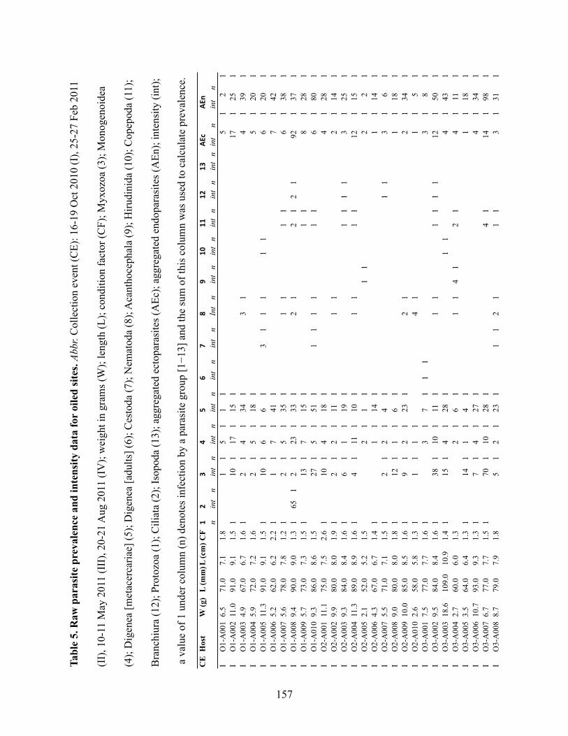
Tabl
e 5.
Raw
par
asite
pre
vale
nce
and
inte
nsity
dat
a fo
r oi
led
site
s. Ab
br. C
olle
ctio
n ev
ent (
CE)
: 16-
19 O
ct 2
010
(I),
25-2
7 Fe
b 20
11
(II)
, 10-
11 M
ay 2
011
(III
), 20
-21
Aug
201
1 (I
V);
wei
ght i
n gr
ams (
W);
leng
th (L
); co
nditi
on fa
ctor
(CF)
; Myx
ozoa
(3);
Mon
ogen
oide
a
(4);
Dig
enea
[met
acer
caria
e] (5
); D
igen
ea [a
dults
] (6)
; Ces
toda
(7);
Nem
atod
a (8
); A
cant
hoce
phal
a (9
); H
irudi
nida
(10)
; Cop
epod
a (1
1);
Bra
nchi
ura
(12)
; Pro
tozo
a (1
); C
iliat
a (2
); Is
opod
a (1
3); a
ggre
gate
d ec
topa
rasi
tes (
AEc
); ag
greg
ated
end
opar
asite
s (A
En);
inte
nsity
(int
);
a v
alue
of 1
und
er c
olum
n (n
) den
otes
infe
ctio
n by
a p
aras
ite g
roup
[1!1
3] a
nd th
e su
m o
f thi
s col
umn
was
use
d to
cal
cula
te p
reva
lenc
e.
CE
Hos
tW
(g)
L (m
m)L
(cm
)CF
!"
#$
%&
'(
)!*
!!!"
!#+,
-+,
.n
int
nin
tn
int
nin
tn
int
nin
tn
Int
nin
tn
int
nin
tn
int
nin
tn
int
nin
tn
IO
1-A
001
6.5
71.0
7.1
1.8
11
51
11
51
21
IO
1-A
002
11.0
91.0
9.1
1.5
110
117
115
117
125
1I
O1-
A00
34.
967
.06.
71.
61
21
41
341
31
41
391
IO
1-A
004
5.9
72.0
7.2
1.6
21
51
181
51
201
IO
1-A
005
11.3
91.0
9.1
1.5
110
16
16
13
11
11
16
120
1I
O1-
A00
65.
262
.06.
22.
21
11
71
411
71
421
IO
1-A
007
5.6
78.0
7.8
1.2
12
15
135
11
11
16
138
1I
O1-
A00
89.
490
.09.
01.
31
651
21
231
331
21
21
21
921
371
IO
1-A
009
5.7
73.0
7.3
1.5
113
17
115
11
18
128
1I
O1-
A01
09.
386
.08.
61.
527
15
151
11
11
11
16
180
1I
O2-
A00
111
.175
.07.
52.
61
101
41
181
41
281
IO
2-A
002
9.9
80.0
8.0
1.9
12
12
111
11
12
114
1I
O2-
A00
39.
384
.08.
41.
61
61
11
191
11
11
31
251
IO
2-A
004
11.3
89.0
8.9
1.6
14
111
110
11
11
112
115
1I
O2-
A00
52.
152
.05.
21.
52
11
11
12
12
1I
O2-
A00
64.
367
.06.
71.
41
11
141
11
141
IO
2-A
007
5.5
71.0
7.1
1.5
12
12
14
11
13
16
1I
O2-
A00
89.
080
.08.
01.
81
121
11
61
11
181
IO
2-A
009
10.0
85.0
8.5
1.6
19
12
123
12
12
134
1I
O2-
A01
02.
658
.05.
81.
31
11
11
41
11
51
IO
3-A
001
7.5
77.0
7.7
1.6
13
17
11
13
18
1I
O3-
A00
29.
584
.08.
41.
61
381
101
111
11
11
11
121
501
IO
3-A
003
18.6
109.
010
.91.
41
151
41
281
11
41
431
IO
3-A
004
2.7
60.0
6.0
1.3
21
61
11
41
21
41
111
IO
3-A
005
3.5
64.0
6.4
1.3
114
11
14
11
118
1I
O3-
A00
610
.793
.09.
31.
31
71
41
271
41
341
IO
3-A
007
6.7
77.0
7.7
1.5
170
110
128
14
114
198
1I
O3-
A00
88.
779
.07.
91.
85
12
123
11
12
11
13
131
1
157

IO
3-A
009
9.2
87.0
8.7
1.4
12
110
138
11
111
140
1I
O3-
A01
04.
870
.07.
01.
41
51
31
41
21
31
111
IO
4-A
001
8.6
82.0
8.2
1.6
12
18
15
139
11
18
11
18
156
1I
O4-
A00
24.
471
.07.
11.
21
21
31
161
31
31
211
IO
4-A
003
16.1
102.
010
.21.
51
61
11
151
31
11
21
241
IO
4-A
004
3.4
64.0
6.4
1.3
14
11
131
11
135
1I
O4-
A00
55.
772
.07.
21.
51
31
31
11
31
41
IO
4-A
006
6.8
79.0
7.9
1.4
14
13
116
12
14
11
14
126
1I
O4-
A00
75.
973
.07.
31.
51
61
91
11
11
11
161
IO
4-A
008
11.1
93.0
9.3
1.4
16
125
118
113
162
1I
O4-
A00
910
.187
.08.
71.
51
61
371
151
11
11
581
IO
4-A
010
3.9
76.0
7.6
0.9
120
120
1I
O1-
A04
15.
370
.07.
01.
51
81
41
251
11
41
341
IO
1-A
042
8.4
80.0
8.0
1.6
61
31
451
11
51
81
521
IO
1-A
043
3.1
60.0
6.0
1.4
71
41
191
21
11
51
281
IO
1-A
044
2.7
59.0
5.9
1.3
31
31
121
11
31
161
IO
1-A
045
2.0
52.0
5.2
1.4
81
31
111
11
41
191
IO
2-A
041
5.8
72.0
7.2
1.5
15
118
123
1I
O2-
A04
28.
180
.08.
01.
61
41
241
51
331
IO
2-A
043
1.2
48.0
4.8
1.1
21
11
121
11
11
151
IO
2-A
044
17.0
97.0
9.7
1.9
110
111
11
121
1I
O2-
A04
59.
183
.08.
31.
61
11
91
21
21
101
IO
3-A
041
3.5
65.0
6.5
1.3
13
12
118
12
121
1I
O3-
A04
25.
870
.07.
01.
72
13
14
11
13
16
1I
O3-
A04
32.
055
.05.
51.
22
14
15
14
17
1I
O3-
A04
43.
666
.06.
61.
32
14
12
12
16
14
1I
O3-
A04
53.
967
.06.
71.
31
41
61
11
111
IO
4-A
041
20.7
110.
011
.01.
61
461
51
281
51
741
IO
4-A
042
4.3
67.0
6.7
1.4
14
15
12
11
11
111
1I
O4-
A04
311
.686
.08.
61.
81
111
31
191
111
31
411
IO
4-A
044
4.7
70.0
7.0
1.4
13
16
13
16
1I
O4-
A04
54.
069
.06.
91.
21
11
131
11
11
11
151
IIO
1-A
011
5.6
69.0
6.9
1.7
11
141
51
11
141
61
IIO
1-A
012
12.3
89.0
8.9
1.7
19
113
132
113
113
154
1II
O1-
A01
35.
169
.06.
91.
52
17
115
13
11
18
120
1II
O1-
A01
44.
970
.07.
01.
41
21
141
131
11
11
161
151
IIO
1-A
015
12.0
92.0
9.2
1.5
110
125
120
19
110
154
1II
O1-
A01
64.
670
.07.
01.
31
17
17
12
17
110
1II
O1-
A01
76.
775
.07.
51.
66
121
12
13
19
123
1II
O1-
A01
82.
959
.05.
91.
44
110
114
11
12
112
118
1II
O1-
A01
98.
986
.08.
61.
41
81
191
281
11
41
231
371
IIO
1-A
020
15.4
102.
010
.21.
51
41
171
361
21
51
71
241
471
158

IIO
2-A
011
7.8
84.0
8.4
1.3
15
117
118
114
11
11
119
137
1II
O2-
A01
217
.296
.09.
61.
91
61
181
231
231
21
201
521
IIO
2-A
013
3.8
64.0
6.4
1.4
16
12
11
12
17
1II
O2-
A01
42.
962
.06.
21.
21
11
131
161
11
51
131
231
IIO
2-A
015
4.0
64.0
6.4
1.5
51
61
51
11
11
71
11
71
191
IIO
2-A
016
5.9
73.0
7.3
1.5
16
17
16
15
11
18
117
1II
O2-
A01
74.
972
.07.
21.
31
81
41
11
11
101
41
IIO
2-A
018
14.5
110.
011
.01.
11
31
81
281
561
11
91
871
IIO
2-A
019
4.0
64.0
6.4
1.5
113
19
12
113
111
1II
O2-
A02
04.
362
.06.
21.
81
51
41
31
11
51
81
IIO
3-A
011
3.6
67.0
6.7
1.2
116
150
11
11
117
151
1II
O3-
A01
22.
654
.05.
41.
71
11
201
51
11
11
11
221
71
IIO
3-A
013
2.9
62.0
6.2
1.2
19
18
11
15
114
19
1II
O3-
A01
44.
666
.06.
61.
61
21
411
61
11
21
431
81
IIO
3-A
015
4.1
63.0
6.3
1.6
115
19
12
11
116
111
1II
O3-
A01
63.
157
.05.
71.
74
17
13
13
17
110
1II
O3-
A01
72.
458
.05.
81.
33
13
12
15
13
1II
O3-
A01
83.
361
.06.
11.
47
111
17
111
114
1II
O3-
A01
92.
056
.05.
61.
21
15
110
15
111
1II
O3-
A02
02.
457
.05.
71.
324
111
11
124
112
1II
O4-
A01
13.
059
.05.
91.
51
171
121
21
11
171
151
IIO
4-A
012
5.6
75.0
7.5
1.3
12
156
16
12
11
159
18
1II
O4-
A01
33.
863
.06.
31.
51
41
81
171
11
11
21
21
121
231
IIO
4-A
014
4.8
67.0
6.7
1.6
119
112
11
12
121
113
1II
O4-
A01
53.
961
.06.
11.
71
51
81
11
61
81
IIO
4-A
016
10.9
89.0
8.9
1.5
119
113
11
14
11
119
118
1II
O4-
A01
74.
866
.06.
61.
71
21
151
161
11
31
191
181
IIO
4-A
018
3.1
60.0
6.0
1.4
132
18
11
133
18
1II
O4-
A01
95.
971
.07.
11.
61
21
141
231
11
21
161
261
IIO
4-A
020
2.1
55.0
5.5
1.2
12
18
13
11
11
18
17
1II
O1-
A04
613
.095
.09.
51.
51
61
461
81
11
61
541
IIO
1-A
047
3.4
69.0
6.9
1.0
21
21
191
21
211
IIO
1-A
048
5.1
70.0
7.0
1.5
110
137
19
11
11
12
114
91
111
IIO
1-A
049
7.8
80.0
8.0
1.5
11
391
11
11
421
IIO
1-A
050
8.0
80.0
8.0
1.6
18
113
11
11
19
114
1II
O2-
A04
64.
871
.07.
11.
31
71
31
71
31
IIO
2-A
047
5.7
75.0
7.5
1.4
11
101
101
51
11
111
161
IIO
2-A
048
5.9
70.0
7.0
1.7
118
13
116
118
119
1II
O2-
A04
914
.211
0.0
11.0
1.1
121
91
61
121
151
IIO
2-A
050
16.1
107.
010
.71.
31
71
171
71
171
IIO
3-A
046
8.3
84.0
8.4
1.4
21
251
61
251
81
IIO
3-A
047
4.0
60.0
6.0
1.9
21
31
291
11
31
321
159

IIO
3-A
048
6.6
73.0
7.3
1.7
15
15
15
15
1II
O3-
A04
917
.010
2.0
10.2
1.6
11
16
113
11
16
115
1II
O3-
A05
04.
769
.06.
91.
410
13
11
11
110
15
1II
O4-
A04
62.
559
.05.
91.
21
11
11
11
11
21
IIO
4-A
047
9.4
88.0
8.8
1.4
150
16
11
11
14
154
18
1II
O4-
A04
83.
867
.06.
71.
32
112
11
113
12
1II
O4-
A04
94.
974
.07.
41.
211
12
11
11
113
12
1II
O4-
A05
03.
868
.06.
81.
24
12
11
14
13
1II
IO
1-A
021
5.0
65.0
6.5
1.8
112
31
81
471
51
11
91
175
1II
IO
1-A
022
9.3
89.0
8.9
1.3
114
41
31
161
11
81
111
160
1II
IO
1-A
023
6.3
71.0
7.1
1.8
137
14
135
11
13
14
176
1II
IO
1-A
024
18.3
109.
010
.91.
41
155
113
151
11
13
11
12
115
121
01
III
O1-
A02
57.
273
.07.
31.
91
111
112
14
141
13
11
11
116
115
71
III
O1-
A02
65.
571
.07.
11.
51
188
13
141
12
112
12
117
123
11
III
O1-
A02
73.
361
.06.
11.
51
771
141
121
21
11
151
911
III
O1-
A02
85.
070
.07.
01.
517
21
491
21
31
21
31
51
226
1II
IO
1-A
029
20.2
102.
010
.21.
91
441
131
351
111
111
261
21
411
101
1II
IO
1-A
030
10.5
85.0
8.5
1.7
111
128
12
12
11
114
128
1II
IO
2-A
021
7.2
74.0
7.4
1.8
156
118
111
12
22
16
11
125
117
11
III
O2-
A02
26.
375
.07.
51.
51
921
181
91
51
11
191
106
1II
IO
2-A
023
8.4
79.0
7.9
1.7
112
61
251
151
91
251
150
1II
IO
2-A
024
5.6
67.0
6.7
1.9
114
71
181
191
21
11
191
168
1II
IO
2-A
025
4.5
64.0
6.4
1.7
118
117
118
11
14
14
121
141
1II
IO
2-A
026
5.1
71.0
7.1
1.4
178
123
124
11
12
16
12
131
120
31
III
O2-
A02
73.
863
.06.
31.
512
31
81
91
131
11
91
145
1II
IO
2-A
028
6.6
72.0
7.2
1.8
116
110
113
11
110
113
01
III
O2-
A02
94.
463
.06.
31.
81
180
17
112
11
18
119
21
III
O2-
A03
04.
071
.07.
11.
114
51
11
21
11
51
61
148
1II
IO
3-A
021
7.8
75.0
7.5
1.8
231
181
11
11
21
411
III
O3-
A02
28.
075
.07.
51.
910
19
114
11
13
19
128
1II
IO
3-A
023
3.9
62.0
6.2
1.6
112
21
21
61
11
41
11
51
71
133
1II
IO
3-A
024
5.0
66.0
6.6
1.8
11
81
41
41
21
71
121
III
O3-
A02
53.
359
.05.
91.
61
167
15
110
11
11
12
17
117
91
III
O3-
A02
626
.711
1.0
11.1
2.0
149
117
121
117
13
13
11
121
118
71
III
O3-
A02
74.
671
.07.
11.
356
17
116
11
18
172
1II
IO
3-A
028
6.7
71.0
7.1
1.9
145
18
18
18
14
14
12
11
115
165
1II
IO
3-A
029
4.6
64.0
6.4
1.8
331
31
11
101
361
21
11
61
801
III
O3-
A03
011
.075
.07.
52.
61
123
120
110
15
14
129
113
31
III
O4-
A02
15.
670
.07.
01.
655
15
14
12
111
11
11
11
17
172
1II
IO
4-A
022
8.1
75.0
7.5
1.9
631
51
81
11
51
31
11
91
771
III
O4-
A02
37.
176
.07.
61.
61
651
51
151
21
71
801
III
O4-
A02
47.
372
.07.
21.
91
41
101
21
81
41
31
111
201
160

III
O4-
A02
54.
465
.06.
51.
61
881
31
51
71
31
100
1II
IO
4-A
026
6.2
72.0
7.2
1.7
182
13
16
15
11
11
15
119
31
III
O4-
A02
74.
162
.06.
21.
766
12
15
11
113
12
11
15
185
1II
IO
4-A
028
3.3
61.0
6.1
1.5
921
21
111
21
221
51
71
127
1II
IO
4-A
029
3.4
62.0
6.2
1.4
151
12
14
18
18
12
11
15
117
11
III
O4-
A03
04.
364
.06.
41.
713
41
11
111
21
321
11
21
179
1II
IO
1-A
051
7.9
76.0
7.6
1.8
112
01
141
291
11
21
51
191
150
1II
IO
1-A
052
7.8
77.0
7.7
1.7
130
116
120
12
118
150
1II
IO
1-A
053
5.6
71.0
7.1
1.6
721
31
281
131
41
71
113
1II
IO
1-A
054
4.6
67.0
6.7
1.5
751
511
101
321
71
41
11
901
901
III
O1-
A05
55.
168
.06.
81.
61
401
41
281
171
41
851
III
O2-
A05
16.
374
.07.
41.
61
451
91
161
51
121
11
101
781
III
O2-
A05
215
.210
2.0
10.2
1.4
12
114
111
11
11
11
14
118
115
1II
IO
2-A
053
5.3
71.0
7.1
1.5
371
101
91
11
11
111
471
III
O2-
A05
43.
962
.06.
21.
618
281
21
121
11
21
201
431
III
O2-
A05
54.
771
.07.
11.
331
19
19
13
11
110
143
1II
IO
3-A
051
4.6
65.0
6.5
1.7
301
21
71
11
31
371
III
O3-
A05
25.
070
.07.
01.
58
18
1II
IO
3-A
053
8.8
99.0
9.9
0.9
11
11
11
III
O3-
A05
45.
871
.07.
11.
63
11
11
11
14
1II
IO
3-A
055
7.8
72.0
7.2
2.1
301
51
221
51
521
III
O4-
A05
18.
583
.08.
31.
51
141
110
119
110
116
01
III
O4-
A05
24.
166
.06.
61.
41
191
19
18
11
11
120
81
III
O4-
A05
37.
382
.08.
21.
31
51
121
11
31
11
91
131
III
O4-
A05
44.
466
.06.
61.
510
71
61
11
11
115
1II
IO
4-A
055
9.9
88.0
8.8
1.5
15
122
11
15
123
1IV
O1-
A03
11.
248
.04.
81.
11
14
141
13
11
14
146
1IV
O1-
A03
21.
044
.04.
41.
23
12
116
12
12
121
1IV
O1-
A03
31.
147
.04.
71.
04
13
17
111
13
122
1IV
O1-
A03
41.
448
.04.
81.
28
12
115
12
123
1IV
O1-
A03
51.
346
.04.
61.
32
114
15
121
1IV
O1-
A03
61.
342
.04.
21.
715
12
139
12
12
156
1IV
O1-
A03
71.
041
.04.
11.
41
41
191
231
IVO
1-A
038
3.6
59.0
5.9
1.7
12
130
12
130
1IV
O1-
A03
90.
940
.04.
01.
458
11
11
158
1IV
O1-
A04
00.
840
.04.
01.
218
13
122
11
13
141
1IV
O2-
A03
117
.710
2.0
10.2
1.7
124
14
123
15
11
12
16
152
1IV
O2-
A03
23.
160
.06.
01.
42
13
114
16
13
122
1IV
O2-
A03
310
.685
.08.
51.
751
17
112
11
12
19
164
1IV
O2-
A03
47.
373
.07.
31.
91
71
11
501
11
11
11
591
IVO
2-A
035
7.1
76.0
7.6
1.6
301
21
841
11
11
31
115
1IV
O2-
A03
610
.376
.07.
62.
31
291
61
231
11
11
71
521
161

IVO
2-A
037
9.7
84.0
8.4
1.6
18
12
121
11
13
129
1IV
O2-
A03
816
.295
.09.
51.
91
341
11
221
21
41
21
31
621
IVO
2-A
039
11.7
76.0
7.6
2.7
154
14
118
11
15
11
14
179
1IV
O2-
A04
02.
054
.05.
41.
21
31
11
161
11
21
191
IVO
3-A
031
8.4
80.0
8.0
1.6
128
15
157
15
15
190
1IV
O3-
A03
25.
568
.06.
81.
81
381
31
101
31
481
IVO
3-A
033
2.3
54.0
5.4
1.4
110
139
149
1IV
O3-
A03
416
.997
.09.
71.
91
136
12
166
11
13
120
21
IVO
3-A
035
2.0
54.0
5.4
1.3
101
21
391
21
491
IVO
3-A
036
2.6
55.0
5.5
1.6
71
41
151
41
221
IVO
3-A
037
12.1
85.0
8.5
2.0
124
17
160
17
184
1IV
O3-
A03
813
.490
.09.
01.
81
61
561
11
61
571
IVO
3-A
039
2.9
55.0
5.5
1.7
14
12
117
12
121
1IV
O3-
A04
02.
053
.05.
31.
31
11
11
201
11
211
IVO
4-A
031
2.3
55.0
5.5
1.4
113
118
131
1IV
O4-
A03
21.
958
.05.
81.
03
15
124
11
15
128
1IV
O4-
A03
31.
750
.05.
01.
42
11
132
11
12
134
1IV
O4-
A03
42.
355
.05.
51.
41
11
18
13
14
19
1IV
O4-
A03
52.
255
.05.
51.
310
17
11
118
1IV
O4-
A03
61.
650
.05.
01.
31
91
11
311
11
21
401
IVO
4-A
037
3.0
59.0
5.9
1.4
14
13
128
14
13
136
1IV
O4-
A03
810
.079
.07.
92.
01
61
11
481
11
541
IVO
4-A
039
1.4
53.0
5.3
0.9
91
21
561
11
11
31
651
IVO
4-A
040
2.2
54.0
5.4
1.4
21
341
21
381
IVO
1-A
056
19.2
100.
010
.01.
91
961
61
581
31
11
61
157
1IV
O1-
A05
72.
961
.06.
11.
31
111
41
271
11
41
391
IVO
1-A
058
1.8
54.0
5.4
1.1
12
116
118
1IV
O1-
A05
93.
060
.06.
01.
41
14
123
124
1IV
O1-
A06
02.
051
.05.
11.
51
21
11
161
11
11
191
IVO
2-A
056
6.2
79.0
7.9
1.3
11
81
91
IVO
2-A
057
8.9
71.0
7.1
2.5
191
112
110
31
IVO
2-A
058
6.4
72.0
7.2
1.7
110
11
111
1IV
O2-
A05
97.
976
.07.
61.
811
120
131
1IV
O2-
A06
014
.693
.09.
31.
819
18
127
1IV
O3-
A05
65.
673
.07.
31.
428
128
1IV
O3-
A05
75.
459
.05.
92.
61
61
251
311
IVO
3-A
058
9.5
86.0
8.6
1.5
31
261
11
11
291
IVO
3-A
059
6.7
62.0
6.2
2.8
61
271
331
IVO
3-A
060
6.5
74.0
7.4
1.6
171
112
12
183
1IV
O4-
A05
62.
354
.05.
41.
51
41
11
351
41
431
IVO
4-A
057
2.5
57.0
5.7
1.3
138
12
118
11
157
1IV
O4-
A05
82.
454
.05.
41.
51
71
11
281
11
351
162

IVO
4-A
059
1.9
50.0
5.0
1.5
13
12
146
149
1IV
O4-
A06
01.
751
.05.
11.
31
41
11
471
11
521
ME
AN
6.4
71.5
7.1
1.5
4733
7.7
192.
73.
35.
12
13
11
10.3
49.3
TOTA
L IN
FEC
TE
D14
85
195
203
234
761
120
724
8268
120
923
9PR
EVA
LE
NC
E62
2.1
8185
982.
925
503
1034
280
87.1
99.6
STA
ND
AR
D E
RR
OR
INT
EN
SIT
Y19
3.6
0.6
0.9
1.7
0.6
0.7
0.4
0.1
0.4
0.1
1.07
3.36
163

Ta
ble
6.
Ra
w p
ara
site
pre
va
len
ce a
nd
in
ten
sity
da
ta f
or
no
n-o
iled
ref
eren
ce s
ites
. A
bb
r. C
oll
ecti
on
ev
ent
(CE
); w
eig
ht
in g
ram
s
(W);
len
gth
(L
); c
on
dit
ion
fac
tor
(CF
); P
roto
zoa
(1);
Cil
iata
(2
); M
yx
ozo
a (3
); M
on
og
eno
idea
(4
); D
igen
ea [
met
acer
cari
ae]
(5);
Dig
enea
[ad
ult
s] (
6);
Ces
tod
a (7
); N
emat
od
a (8
); A
can
tho
cep
hal
a (9
); H
iru
din
ida
(10
); C
op
epo
da
(11
); B
ran
chiu
ra (
12
); I
sop
od
a (1
3);
agg
reg
ated
ect
op
aras
ites
(A
Ec)
; ag
gre
gat
ed e
nd
op
aras
ites
(A
En
); i
nte
nsi
ty (
int)
; a
val
ue
of
1 u
nd
er c
olu
mn
(n
) d
eno
tes
infe
ctio
n b
y a
par
asit
e g
rou
p [
1!
13
] an
d t
he
sum
of
this
co
lum
n w
as u
sed
to
cal
cula
te p
rev
alen
ce.
CE
Host
W (
g)
L (
mm
)L
(cm
)C
F1
111
21
3A
Ec
nA
En
n
nin
tn
int
nin
tn
int
nin
tn
int
nIn
tn
int
nin
tn
int
nin
tn
int
n
IN
1-A
001
5.1
74
7.4
1.3
12
12
11
81
21
11
21
23
1
IN
1-A
002
17
.69
59
.52
.11
41
21
21
11
31
61
IN
1-A
003
10
.28
88
.81
.51
11
13
18
13
13
12
21
IN
1-A
004
3.6
50
52
.91
11
11
31
11
41
IN
1-A
005
13
.79
59
.51
.61
15
11
12
11
11
71
IN
1-A
006
7.8
75
7.5
1.9
111
11
12
81
21
11
41
1
IN
1-A
007
5.0
70
71
.41
11
10
11
11
111
1
IN
1-A
008
5.8
73
7.3
1.5
11
11
21
11
21
12
1
IN
1-A
009
11
.39
29
.21
.54
61
11
10
11
12
15
61
IN
1-A
010
5.0
72
7.2
1.3
13
16
13
16
1
IN
2-A
001
9.0
80
81
.81
21
44
11
12
12
14
71
IN
2-A
002
4.6
70
71
.31
16
11
51
11
61
17
1
IN
2-A
003
7.4
75
7.5
1.8
11
14
11
21
41
13
1
IN
2-A
004
24
.5111
11
.11
.81
49
13
11
51
12
14
12
14
19
18
01
IN
2-A
005
3.3
66
6.6
1.1
18
31
31
45
12
15
11
28
1
IN
2-A
006
19
.81
07
10
.71
.61
64
13
12
51
21
31
21
81
91
1
IN
2-A
007
4.8
70
71
.41
14
11
01
11
25
1
IN
2-A
008
4.9
62
6.2
2.1
12
91
29
1
IN
2-A
009
4.6
68
6.8
1.5
12
15
31
11
11
67
1
IN
2-A
010
21
.01
05
10
.51
.81
11
12
01
11
11
11
31
41
33
1
IN
3-A
001
4.8
70
71
.41
81
11
91
11
17
1
IN
3-A
002
11
.08
98
.91
.61
23
14
12
11
21
11
11
61
46
1
IN
3-A
003
2.2
56
5.6
1.2
12
11
19
11
11
11
12
12
31
IN
3-A
004
15
.99
99
.91
.61
10
16
14
12
01
IN
3-A
005
6.2
77
7.7
1.4
13
13
11
01
31
13
1
IN
3-A
006
9.4
89
8.9
1.3
12
14
111
12
14
11
51
IN
3-A
007
3.4
65
6.5
1.2
12
15
15
31
51
11
61
60
1
IN
3-A
008
13
.09
59
.51
.51
31
19
12
51
11
10
15
61
89
10
23
45
67
164

IN
3-A
009
5.9
74
7.4
1.5
12
61
41
27
11
14
15
41
IN
3-A
010
5.0
68
6.8
1.6
18
13
11
81
11
31
27
1
IN
4-A
001
6.4
75
7.5
1.5
111
14
13
31
36
11
11
15
18
11
IN
4-A
002
4.3
70
71
.31
11
11
34
11
11
13
61
IN
4-A
003
4.4
68
6.8
1.4
11
51
41
35
14
15
01
IN
4-A
004
7.0
78
7.8
1.5
12
51
11
23
11
11
11
12
14
15
01
IN
4-A
005
14
.69
59
.51
.71
19
17
16
71
51
11
71
92
1
IN
4-A
006
2.7
60
61
.31
41
51
17
11
11
16
12
21
IN
4-A
007
5.7
70
71
.61
38
19
12
11
81
11
11
40
12
91
IN
4-A
008
2.2
54
5.4
1.4
41
51
15
12
15
12
11
IN
4-A
009
11
.08
38
.31
.91
24
12
61
21
32
11
11
11
12
81
59
1
IN
4-A
010
10
.79
19
.11
.41
12
17
14
11
21
11
21
91
56
1
IN
1-A
041
5.4
73
7.3
1.4
11
12
41
11
11
11
26
1
IN
1-A
042
2.4
56
5.6
1.3
11
10
12
11
31
IN
1-A
043
7.3
77
7.7
1.6
12
15
11
91
21
51
23
1
IN
1-A
044
17
.08
18
.13
.21
10
14
11
81
21
41
30
1
IN
1-A
045
2.5
56
5.6
1.4
15
71
11
22
13
11
18
21
IN
2-A
041
5.3
70
71
.55
12
16
11
12
11
21
IN
2-A
042
3.0
62
6.2
1.2
41
31
11
11
91
IN
2-A
043
13
.89
59
.51
.61
11
30
13
11
IN
2-A
044
3.1
61
6.1
1.4
61
15
11
13
12
51
IN
2-A
045
20
.41
09
10
.91
.61
21
56
11
13
11
11
16
21
IN
3-A
041
10
.48
58
.51
.71
20
16
12
31
11
61
44
1
IN
3-A
042
14
.89
79
.71
.61
21
27
11
12
12
13
01
IN
3-A
043
12
.89
29
.21
.61
27
11
13
51
11
11
11
64
1
IN
3-A
044
3.5
64
6.4
1.3
12
11
11
11
12
13
1
IN
3-A
045
2.2
53
5.3
1.5
12
11
12
41
11
26
1
IN
4-A
041
5.8
73
7.3
1.5
11
17
01
11
70
1
IN
4-A
042
3.3
67
6.7
1.1
25
12
51
IN
4-A
043
4.3
65
6.5
1.6
31
27
11
12
13
13
01
IN
4-A
044
2.4
57
5.7
1.3
11
11
51
16
1
IN
4-A
045
8.5
75
7.5
2.0
51
11
18
11
12
31
IIN
1-A
011
1.2
47
4.7
1.1
11
11
12
11
13
1
IIN
1-A
012
3.9
56
5.6
2.2
12
11
21
11
11
11
31
14
1
IIN
1-A
013
8.1
80
81
.61
31
21
29
19
12
14
11
IIN
1-A
014
1.5
50
51
.22
11
01
61
21
16
1
IIN
1-A
015
14
.39
99
.91
.51
51
31
44
11
18
11
19
12
11
51
58
1
IIN
1-A
016
5.6
70
71
.61
21
12
11
61
41
31
21
44
1
IIN
1-A
017
6.0
70
71
.81
11
11
51
13
11
11
91
IIN
1-A
018
2.5
56
5.6
1.4
14
11
12
11
16
1
IIN
1-A
019
1.9
55
5.5
1.1
11
81
41
11
12
1
IIN
1-A
020
1.2
47
4.7
1.1
11
16
11
12
16
1
165

IIN
2-A
011
3.1
60
61
.41
13
14
12
11
13
18
1
IIN
2-A
012
8.0
77
7.7
1.8
41
24
11
01
11
25
11
41
IIN
2-A
013
4.7
67
6.7
1.6
11
16
11
41
21
16
11
71
IIN
2-A
014
4.3
62
6.2
1.8
12
15
12
11
11
12
19
1
IIN
2-A
015
5.2
69
6.9
1.6
11
14
111
11
11
12
16
11
41
IIN
2-A
016
5.1
70
71
.55
19
18
12
11
11
111
11
51
IIN
2-A
017
4.8
68
6.8
1.5
11
41
81
11
51
91
IIN
2-A
018
1.8
52
5.2
1.3
12
14
14
12
11
15
18
1
IIN
2-A
019
8.7
86
8.6
1.4
11
71
41
21
21
31
11
81
21
1
IIN
2-A
020
3.6
64
6.4
1.4
11
11
15
11
11
11
12
18
1
IIN
3-A
011
4.4
66
6.6
1.5
13
11
71
21
11
30
11
11
11
91
36
1
IIN
3-A
012
13
.08
28
.22
.41
16
12
91
16
12
91
IIN
3-A
013
6.6
73
7.3
1.7
11
31
11
10
11
12
11
61
11
1
IIN
3-A
014
6.1
75
7.5
1.4
11
51
71
31
61
19
11
12
31
28
1
IIN
3-A
015
7.0
74
7.4
1.7
11
29
11
01
21
41
21
31
11
71
IIN
3-A
016
2.3
56
5.6
1.3
41
13
11
15
11
15
11
91
IIN
3-A
017
4.5
64
6.4
1.7
16
71
11
15
13
16
71
19
1
IIN
3-A
018
5.9
70
71
.71
14
11
12
14
1
IIN
3-A
019
2.1
54
5.4
1.3
11
13
111
11
11
14
11
31
IIN
3-A
020
6.3
73
7.3
1.6
13
11
41
13
12
01
31
11
11
19
13
61
IIN
4-A
011
3.2
61
6.1
1.4
12
18
14
91
81
21
11
11
15
91
IIN
4-A
012
3.2
58
5.8
1.6
41
28
14
12
81
IIN
4-A
013
4.8
65
6.5
1.7
15
18
11
12
11
71
91
IIN
4-A
014
3.8
63
6.3
1.5
14
12
91
31
17
12
91
IIN
4-A
015
2.7
59
5.9
1.3
61
41
12
11
11
11
15
12
01
IIN
4-A
016
2.7
58
5.8
1.4
17
11
31
11
41
11
21
10
11
81
IIN
4-A
017
2.6
60
61
.21
21
31
28
11
11
61
11
11
41
48
1
IIN
4-A
018
2.7
59
5.9
1.3
61
45
11
14
16
15
01
IIN
4-A
019
2.2
56
5.6
1.2
41
31
11
11
51
41
IIN
4-A
020
3.1
62
6.2
1.3
63
15
12
11
11
21
70
12
21
IIN
1-A
046
1.8
53
5.3
1.2
21
21
21
21
IIN
1-A
047
3.2
61
6.1
1.4
71
11
51
21
11
14
1
IIN
1-A
048
1.9
54
5.4
1.2
12
81
11
71
11
35
1
IIN
1-A
049
2.1
52
5.2
1.5
11
21
81
11
21
10
1
IIN
1-A
050
1.3
45
4.5
1.4
31
31
61
IIN
2-A
046
3.8
67
6.7
1.3
11
41
21
21
11
81
IIN
2-A
047
2.7
67
6.7
0.9
11
11
11
11
21
IIN
2-A
048
4.4
67
6.7
1.5
12
81
13
12
81
13
1
IIN
2-A
049
3.5
69
6.9
1.1
31
31
31
31
IIN
2-A
050
6.0
75
7.5
1.4
51
81
11
11
19
11
61
IIN
3-A
046
6.2
73
7.3
1.6
41
14
18
11
41
12
1
IIN
3-A
047
6.3
77
7.7
1.4
15
17
11
11
15
19
1
166

IIN
3-A
048
8.4
90
91
.15
12
15
12
1
IIN
3-A
049
8.0
82
8.2
1.4
51
11
51
11
IIN
3-A
050
6.7
75
7.5
1.6
61
41
11
61
51
IIN
4-A
046
5.9
71
7.1
1.7
41
41
41
41
IIN
4-A
047
2.8
60
61
.34
11
51
11
51
15
1
IIN
4-A
048
3.8
65
6.5
1.4
31
91
31
11
41
12
1
IIN
4-A
049
4.9
69
6.9
1.5
81
71
11
11
91
81
IIN
4-A
050
17
.211
011
1.3
81
44
11
11
11
01
44
1
III
N1-A
021
3.1
60
61
.41
12
01
11
71
11
11
12
81
III
N1-A
022
3.8
61
6.1
1.7
16
41
51
24
14
11
11
17
11
92
1
III
N1-A
023
3.8
61
6.1
1.7
11
75
11
11
61
21
11
19
31
III
N1-A
024
3.2
60
61
.51
40
14
51
21
91
61
11
43
16
01
III
N1-A
025
12
.38
78
.71
.91
68
17
12
21
11
11
21
91
92
1
III
N1-A
026
4.1
61
6.1
1.8
11
38
14
12
21
11
51
61
1
III
N1-A
027
14
.78
88
.82
.21
37
12
12
51
21
11
21
41
65
1
III
N1-A
028
2.5
58
5.8
1.3
15
91
31
21
11
13
18
11
III
N1-A
029
10
.88
28
.22
.01
78
13
19
13
11
14
19
01
III
N1-A
030
4.2
65
6.5
1.5
18
21
41
19
11
14
11
02
1
III
N2-A
021
2.0
53
5.3
1.3
93
13
13
13
19
61
III
N2-A
022
3.4
63
6.3
1.4
11
58
12
19
13
11
13
11
70
1
III
N2-A
023
3.4
62
6.2
1.4
13
71
16
71
41
51
71
41
11
79
1
III
N2-A
024
7.0
72
7.2
1.9
19
01
31
31
11
41
93
1
III
N2-A
025
6.0
72
7.2
1.6
17
11
11
41
81
11
83
1
III
N2-A
026
6.9
76
7.6
1.6
14
51
21
20
12
11
11
12
16
91
III
N2-A
027
4.6
64
6.4
1.8
18
91
31
51
10
12
13
11
06
1
III
N2-A
028
5.4
65
6.5
2.0
12
71
31
71
31
34
1
III
N2-A
029
7.9
77
7.7
1.7
14
61
41
19
11
11
11
12
12
18
16
81
III
N2-A
030
4.1
66
6.6
1.4
11
96
18
14
01
11
11
11
10
12
37
1
III
N3-A
021
3.4
56
5.6
1.9
30
13
12
11
11
14
13
31
III
N3-A
022
8.3
78
7.8
1.7
18
51
21
41
21
41
61
91
1
III
N3-A
023
4.5
66
6.6
1.5
15
51
51
19
13
12
15
17
91
III
N3-A
024
7.9
80
81
.51
88
11
31
13
14
11
31
10
51
III
N3-A
025
3.8
63
6.3
1.5
14
11
15
11
61
11
25
1
III
N3-A
026
6.5
73
7.3
1.7
19
71
51
12
11
11
11
16
12
11
1
III
N3-A
027
2.5
55
5.5
1.5
15
11
01
21
71
10
1
III
N3-A
028
3.9
66
6.6
1.4
11
12
12
41
31
33
11
13
16
11
III
N3-A
029
3.2
61
6.1
1.4
17
31
31
22
13
12
11
14
11
00
1
III
N3-A
030
3.4
64
6.4
1.3
18
13
13
11
16
11
51
61
III
N4-A
021
4.2
64
6.4
1.6
41
14
12
91
11
11
31
71
72
1
III
N4-A
022
4.4
66
6.6
1.5
13
41
41
47
12
111
14
11
94
1
III
N4-A
023
4.4
67
6.7
1.5
14
21
11
31
11
11
73
1
III
N4-A
024
3.9
66
6.6
1.4
37
13
12
61
41
21
31
61
69
1
167

III
N4-A
025
5.8
71
7.1
1.6
16
91
31
47
12
11
12
16
12
18
1
III
N4-A
026
6.6
73
7.3
1.7
13
91
50
11
11
11
12
19
01
III
N4-A
027
11
.08
78
.71
.73
41
41
87
12
13
11
18
11
23
1
III
N4-A
028
5.4
70
71
.67
71
41
60
11
31
11
51
15
01
III
N4-A
029
4.4
65
6.5
1.6
16
71
61
35
11
31
31
91
21
51
III
N4-A
030
6.0
71
7.1
1.7
19
31
31
32
11
15
18
12
26
1
III
N1-A
051
15
.29
99
.91
.61
41
59
12
16
15
91
III
N1-A
052
5.4
66
6.6
1.9
18
01
91
21
14
16
18
21
29
1
III
N1-A
053
5.6
70
71
.61
41
11
13
11
15
11
41
III
N1-A
054
5.9
72
7.2
1.6
81
12
11
81
31
14
11
61
10
21
III
N1-A
055
2.5
56
5.6
1.4
51
41
26
111
16
11
01
42
1
III
N2-A
051
4.0
70
71
.21
11
31
11
21
31
41
III
N2-A
052
8.8
70
72
.61
11
1
III
N2-A
053
6.4
74
7.4
1.6
13
13
11
14
13
1
III
N2-A
054
12
.58
38
.32
.21
19
51
41
91
21
41
81
20
61
III
N2-A
055
19
.79
99
.92
.01
21
81
19
12
11
01
31
1
III
N3-A
051
3.1
63
6.3
1.2
16
21
61
26
13
11
71
11
71
10
81
III
N3-A
052
3.1
60
61
.41
84
16
11
01
11
61
95
1
III
N3-A
053
4.3
65
6.5
1.6
13
11
61
11
39
13
15
61
III
N3-A
054
7.9
80
81
.51
22
11
01
61
31
11
11
41
14
13
31
III
N3-A
055
2.3
58
5.8
1.2
11
61
51
19
12
31
21
41
51
16
41
III
N4-A
051
5.4
68
6.8
1.7
11
44
13
14
71
21
31
19
31
III
N4-A
052
8.5
78
7.8
1.8
11
56
13
17
31
11
11
31
23
11
III
N4-A
053
15
.19
99
.91
.64
81
11
14
71
11
31
41
15
19
91
III
N4-A
054
4.4
67
6.7
1.5
14
91
31
53
15
11
14
11
07
1
III
N4-A
055
5.6
66
6.6
1.9
13
14
71
31
47
1
IVN
1-A
031
3.2
59
5.9
1.6
37
14
13
61
41
73
1
IVN
1-A
032
1.7
51
5.1
1.3
17
13
11
38
1
IVN
1-A
033
1.7
49
4.9
1.4
11
31
27
13
11
13
13
21
IVN
1-A
034
2.8
56
5.6
1.6
17
13
14
01
11
11
11
31
50
1
IVN
1-A
035
1.0
43
4.3
1.2
21
17
12
51
71
46
1
IVN
1-A
036
4.0
61
6.1
1.8
14
15
11
31
51
27
1
IVN
1-A
037
2.5
58
5.8
1.3
15
15
13
11
11
51
47
1
IVN
1-A
038
1.0
44
4.4
1.2
12
11
13
41
11
46
1
IVN
1-A
039
2.5
56
5.6
1.4
41
30
11
14
13
11
IVN
1-A
040
1.4
48
4.8
1.3
71
11
22
13
11
13
21
IVN
1-A
056
1.6
51
5.1
1.2
11
01
11
17
11
12
71
IVN
1-A
057
2.0
52
5.2
1.5
11
51
21
44
12
15
91
IVN
1-A
058
5.6
70
71
.61
72
11
01
11
73
1
IVN
1-A
059
2.1
54
5.4
1.3
13
12
14
11
11
21
55
1
IVN
1-A
060
1.5
50
51
.21
41
11
33
11
11
13
81
IVN
2-A
031
2.7
56
5.6
1.5
16
17
12
41
21
71
32
1
168

IVN
2-A
032
3.7
63
6.3
1.5
11
01
71
19
12
17
13
11
IVN
2-A
033
3.4
60
61
.61
41
10
11
15
11
01
IVN
2-A
034
2.7
56
5.6
1.6
31
61
41
12
16
14
61
IVN
2-A
035
15
.61
01
10
.11
.51
22
11
21
84
12
11
11
21
10
91
IVN
2-A
036
9.3
80
81
.81
42
11
15
71
11
11
10
01
IVN
2-A
037
13
.08
78
.72
.03
61
71
29
19
17
17
41
IVN
2-A
038
1.9
54
5.4
1.2
41
51
23
15
12
71
IVN
2-A
039
13
.98
98
.92
.01
31
16
14
61
71
56
18
91
IVN
2-A
040
17
.59
89
.81
.91
20
11
51
13
13
11
11
51
37
1
IVN
2-A
056
1.4
48
4.8
1.3
15
14
11
11
19
1
IVN
2-A
057
2.9
56
5.6
1.6
15
12
11
31
21
18
1
IVN
2-A
058
2.0
51
5.1
1.5
14
11
12
31
31
11
30
1
IVN
2-A
059
2.3
56
5.6
1.3
11
91
11
19
11
12
13
81
IVN
2-A
060
2.1
51
5.1
1.6
13
15
12
61
51
39
1
IVN
3-A
031
3.4
63
6.3
1.4
13
12
11
21
41
11
11
31
20
1
IVN
3-A
032
3.1
63
6.3
1.2
19
19
12
12
11
11
12
21
IVN
3-A
033
2.8
59
5.9
1.4
13
13
12
41
41
31
31
1
IVN
3-A
034
3.3
57
5.7
1.8
11
21
14
11
12
71
IVN
3-A
035
11
.78
58
.51
.91
21
11
16
01
11
11
11
11
31
83
1
IVN
3-A
036
3.7
67
6.7
1.2
19
17
13
11
17
11
31
IVN
3-A
037
4.1
62
6.2
1.7
31
31
61
IVN
3-A
038
5.2
69
6.9
1.6
13
01
41
15
11
11
01
41
56
1
IVN
3-A
039
2.9
57
5.7
1.6
11
31
11
51
11
18
1
IVN
3-A
040
2.2
54
5.4
1.4
31
41
11
11
31
61
IVN
3-A
056
1.6
50
51
.38
18
1
IVN
3-A
057
2.8
60
61
.31
34
11
11
91
11
53
1
IVN
3-A
058
2.2
54
5.4
1.4
17
13
13
11
11
41
IVN
3-A
059
1.9
53
5.3
1.3
13
111
13
11
11
12
11
71
IVN
3-A
060
1.8
52
5.2
1.3
12
12
1
IVN
4-A
031
2.7
58
5.8
1.4
11
11
03
13
11
11
08
1
IVN
4-A
032
1.9
52
5.2
1.3
11
01
50
11
16
11
IVN
4-A
033
1.9
53
5.3
1.2
49
15
15
41
IVN
4-A
034
4.5
67
6.7
1.5
15
61
56
1
IVN
4-A
035
2.7
57
5.7
1.4
111
11
17
31
71
11
91
1
IVN
4-A
036
1.9
51
5.1
1.4
41
59
16
31
IVN
4-A
037
1.8
51
5.1
1.3
11
12
12
71
11
21
29
1
IVN
4-A
038
12
.99
19
.11
.71
11
21
11
99
11
11
12
12
1
IVN
4-A
039
3.6
63
6.3
1.4
14
14
31
41
43
1
IVN
4-A
040
3.0
60
61
.41
41
11
64
16
11
17
41
IVN
4-A
056
3.2
61
6.1
1.4
17
11
71
IVN
4-A
057
3.9
62
6.2
1.6
12
11
21
IVN
4-A
058
2.4
55
5.5
1.5
71
66
11
11
17
31
169

IVN
4-A
059
12
.29
29
.21
.69
31
42
11
11
11
35
1
IVN
4-A
060
13
.89
39
.31
.79
31
60
11
53
1
ME
AN
5.8
68
.43
36
.84
31
.53
33
54
.72
21
23
.84
.21
.61
.62
.31
.16
.86
51
.5
TO
TA
L I
NF
EC
TE
D1
51
91
77
20
22
36
26
31
29
47
36
52
33
02
13
24
0
PR
EV
AL
EN
CE
63
47
48
49
81
26
54
20
15
22
14
08
91
00
ST
AN
DA
RD
ER
RO
R I
NT
EN
SIT
Y8
.83
.60
.41
.311
10
.50
.10
.20
.30
.10
.71
3.3
6
170