part 1: wound healingswainathletictrainer.weebly.com/.../wound_care.pdf · wound care part 1: wound...
TRANSCRIPT

Wound Care
PART 1: Wound Healing
EXTERNAL WOUNDS
A wound is a damaged area of the body. Because this course, addresses external wounds—damage
that includes the skin—we begin with a review of the anatomy of skin.
The Structure of Skin
Skin varies in thickness from less than one millimeter in the eyelids to greater than four millimeters
on the soles of the feet, but everywhere, skin is composed of two layers, the epidermis and the
dermis, underlain by a sheet of subcutaneous tissue (Figure 1) (Habif, 2004).
Figure 1. The skin has two layers, the epidermis and the dermis, below which lies subcutaneous
tissue. (National Cancer Institute, n.d.)
EPIDERMIS
The outer layer of the skin is the epidermis. The deepest part of the epidermis is a row of
germinative cells. Germinative cells are specialized stem cells that continually divide to give off
keratinocytes, the main cells in the remainder of the epidermis. As they age, the new keratinocytes
fill with keratin (a tough fibrous protein) and are pushed to the surface, where they die; thus, the
outermost layer of the epidermis is made of flat, dead keratinocytes. The epidermis also contains

melanocytes (pigment-containing cells) and immune system cells. The epidermis, a protective layer
that is normally impermeable to water, does not have sufficient strength to hold sutures or staples.
There are no blood vessels in the epidermis, and it receives its oxygen and nutrients by diffusion
from blood circulating in the underlying dermis. Hair, nails, sweat glands, and sebaceous glands are
sunken epidermal appendages that lie in deep valleys in the dermis surrounded by a row of
germinative epidermal cells.
During normal healing, the epidermis re-grows from germinative cells left in the skin at the edges of
the wound. The growing cells are called epithelial cells, and the regrowth of the epidermis is called
re-epithelialization.
Some wounds, such as surface abrasions (scrapes), are confined mostly to the epidermis. In
epidermal wounds, a new epidermis grows from the germinative cells that surround the bottoms of
epidermal appendages deep in the dermis. Epidermal wounds usually heal quickly with little or no
scarring.
Partial-thickness wounds, such as deep abrasions, destroy or remove the epidermis and the upper
portion of the dermis. When there are still some germinative cells left in the remaining dermis, they
will re-grow a new epidermis. Partial-thickness wounds usually heal with scarring.
DERMIS
The layer of skin directly beneath the epidermis is the dermis. A basement membrane separates these
two layers. The dermis is mainly connective tissue and is therefore much stronger than the
epidermis. The dermis varies in thickness across the surface of the body, but everywhere it is
significantly thicker than the overlying epidermis.
The connective tissue of the dermis contains small blood vessels, lymph vessels, nerves, nerve
endings, and in a few places, muscles. The dermis is also populated by a variety of individual cells
including macrophages, fibroblasts (which synthesize the extracellular connective tissue
components such as collagen), and mast cells (which release histamine and other molecules that
increase inflammation).
The dermis is loosely stratified. The upper (most superficial) layer contains capillaries and sensory
endings of nerves. The deepest layer has thick interlacing collagen and elastic fibers arranged in
parallel rows. The extracellular fibers in the deep dermis are responsible for the strength and
toughness of the skin. When closing a wound with sutures, they must be anchored in the strong
connective tissue of the lower layer of the dermis.

Wounds that penetrate the dermis are true breaks in the skin. For the skin to regain its strength, new
fibrous connective tissue must bridge these wounds; however, the new fibrous tissue—the scar—is
never as strong as the original dermis. In full-thickness wounds, both the epidermis and the dermis
are destroyed or removed. These wounds always heal with a scar.
SUBCUTANEOUS TISSUE
Beneath the dermis is a layer of subcutaneous tissue containing fat. The thickness of the
subcutaneous layer varies throughout the body. It is thickest along the anterior thigh and thinnest on
the back of the hands.
Besides fat cells, subcutaneous tissue contains blood vessels, lymph vessels, and nerves. The
subcutaneous layer is held together by a continuous sheet of fibrous membrane that runs parallel to
the surface of the skin. This membrane is called the superficial fascia.
Beneath the subcutaneous tissue layer, structures (such as muscles and organs) are enclosed in their
own separate connective tissue sheaths. The generic name for these sheaths is deep fasciae. Deep
fasciae generally look off-white in fresh wounds. When treating a wound, tears in the deep fasciae
are repaired whenever possible.
The subcutaneous tissue is a loosely organized compartment. When skin wounds extend deeper than
the dermis, dirt is easily pushed into and spread within the subcutaneous tissue. When cleansing a
wound that has penetrated deeper than the dermis, remove any loose fat and wash out the
subcutaneous compartment thoroughly to reduce the risk of infection.
Types of Wounds
External wounds are named by the type of force that caused them. There are seven basic wound
types: abrasions, lacerations, crushes or contusions, punctures, avulsions, burns, and ulcers. A
traumatic wound is often a mixture of types.
Abrasions are scrapes. Mild abrasions remove epidermis; serious abrasions also remove the dermis and, sometimes,
subcutaneous tissue. An abrasion is usually a broad, shallow wound with irregular edges.

Figure 2. A typical small abrasion on the upper forearm. (Courtesy of Richard Thompson,
SelfCareNet, n.d.)
Lacerations are cuts. When made by a knife-like object, a laceration is a narrow, deep wound with sharp edges.
When made by a blunt object, a laceration is a rip with jagged edges.
Crushes or contusions are compression wounds. A crush wound bruises and damages the skin and the underlying
tissue, although the skin can remain closed in some crush wounds.
Punctures are narrow, deep wounds. Typically, punctures have small openings with sharp edges. Puncture wounds
have a relatively high risk of infection.
Avulsions are wounds in which tissue has been torn out. Sometimes, the avulsed tissue remains partly connected to
its normal surroundings.
Burns are wounds made by external destructive energy (eg, heat) or by external chemicals (eg, acid). First-degree
burns are superficial and red. Second-degree wounds include damage to the dermis and produce blisters (see
photograph of second-degree wound below). Third-degree wounds go deeper than the dermis and produce dry, dead
tissue.
Ulcers are usually made by innate destructive processes, such as ischemia. Ulcers often have destruction of tissue in
a broad, roughly circular area (Shai & Maibach, 2005). (See the photograph of an inflamed diabetic foot ulcer
below.)
Scars
Many parts of the human body can heal after being wounded, but few wounds heal seamlessly. The
new seam, or patch, is called a scar. Scars are mainly connective tissue and cannot replicate the
specialized functions of the original injured tissue. In the skin, scars are covered by a layer of
epidermis (Habif, 2004).
NORMAL SCARS
Scars are imperfect replacements for damaged tissue, but scars are a natural result of healthy healing.
Large wounds, wounds that heal slowly, and wounds involving extensive destruction of the
surrounding tissues heal with large scars; nonetheless, these scars are not necessarily abnormal.
PROBLEM SCARS

Normal scars can lead to problems. Even under the best healing conditions, some normal scars may
end up interfering with the movement of the skin and the underlying tissue. In addition, some normal
scars are unsightly.
When the healing situation is not ideal, however, scars are more likely to become problems. After
poor healing, some scars become unnecessarily large or unnecessarily weak. For example,
infections, tissue necrosis, sebaceous skin, and wounds perpendicular to natural lines of minimal
skin tension will all lead to scars that are larger than normal. If a wound separates (process of
dehiscence*) before it is effectively sealed, the scar will be wider and, usually, weaker. If too few
capillaries grow into the forming scar tissue, leading to ischemia, the scar will be very weak and may
develop into an ulcer.
* Dehiscence is the spontaneous re-opening of a closed wound before it has fully healed.
At the other end of the spectrum, the wound patching process may go overboard and generate too
many new cells or, more commonly, too much collagen in the scar. Such scars will enlarge and
bulge from the wound. Scars built of too many cells (mainly fibroblasts) are called desmoids, or
aggressive fibromatoses. Scars built from too much collagen are either hypertrophic scars or keloids.
When excessive scars form tight ridges along the skin and permanently interfere with normal
movement (such as bending a joint), they are called contractures.
HYPERTROPHIC SCARS
Hypertrophic scars are caused by excess deposition of collagen fibers in a healing wound. This
overactive scar-making process is usually triggered by a prolonged regrowth (proliferative) phase
during healing. This happens in burns, infected wounds, and wounds healing under tension. In
hypertrophic scars, the excessive formation of collagen usually stops within a few weeks. The result
is a scar that is thicker than normal and is raised above the plane of the skin, but unlike a keloid, a
hypertrophic scar does not expand out beyond the actual wound. Hypertrophic scars, which usually
get smaller spontaneously, can occur anywhere on the body.
KELOIDS
Keloids are also caused by the excess deposition of collagen in a healing wound. Keloids, however,
are benign tumors, and the tendency to develop keloids is inherited, African Americans being
particularly susceptible. Unlike hypertrophic scars, keloids develop late in the healing process; they
can show up months or even years after the injury. Keloids bulge out beyond the edges of the
wound, and some keloids can get sizeable. Keloids, which do not regress spontaneously, are usually
found on the upper half of the body.
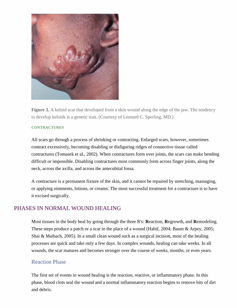
Figure 3. A keloid scar that developed from a skin wound along the edge of the jaw. The tendency
to develop keloids is a genetic trait. (Courtesy of Leonard C. Sperling, MD.)
CONTRACTURES
All scars go through a process of shrinking or contracting. Enlarged scars, however, sometimes
contract excessively, becoming disabling or disfiguring ridges of connective tissue called
contractures (Tomasek et al., 2002). When contractures form over joints, the scars can make bending
difficult or impossible. Disabling contractures most commonly form across finger joints, along the
neck, across the axilla, and across the antecubital fossa.
A contracture is a permanent fixture of the skin, and it cannot be repaired by stretching, massaging,
or applying ointments, lotions, or creams. The most successful treatment for a contracture is to have
it excised surgically.
PHASES IN NORMAL WOUND HEALING
Most tissues in the body heal by going through the three R's: Reaction, Regrowth, and Remodeling.
These steps produce a patch or a scar in the place of a wound (Habif, 2004; Baum & Arpey, 2005;
Shai & Maibach, 2005). In a small clean wound such as a surgical incision, most of the healing
processes are quick and take only a few days. In complex wounds, healing can take weeks. In all
wounds, the scar matures and becomes stronger over the course of weeks, months, or even years.
Reaction Phase
The first set of events in wound healing is the reaction, reactive, or inflammatory phase. In this
phase, blood clots seal the wound and a normal inflammatory reaction begins to remove bits of dirt
and debris.

The reaction phase begins immediately after an injury, as blood vessels constrict temporarily and
blood clotting begins. Soon, the local capillaries become excessively permeable, fluid flows out, and
the tissues swell, producing edema. The blood coagulation process releases chemical activators from
inside entrapped blood platelets; these activators increase the capillary permeability and attract
wandering tissue cells (macrophages) and white blood cells.
The first white blood cells on the scene—polymorphonuclear cells, also called neutrophils—chew up
debris and release chemicals that attract more white blood cells. The various biologically active
molecules being released into the wound also hypersensitize the endings of local pain nerves,
making them react to smaller amounts of chemical and mechanical irritation, making the wound site
tender. Together, the processes in the reaction phase produce local inflammation.
Large wounds, such as ulcerative pressure sores or burns, do not seal during this phase. Instead, the
accumulating fluid, cells, and clotting materials form a pale yellowish viscous exudate, an eschar.
As they age, the coagulant proteins of the exudate link together and dry, making the wound bed
crusty.
During the reaction phase, neutrophils remove bacteria and debris. If the wound does not become
colonized with bacteria, neutrophils stop entering the wound by about day 2 following the injury.
Neutrophils live for less than 24 hours, so in a healthy wound most neutrophils are gone by about
day 3. In infected wounds, however, neutrophils continue to pour in and, as they die, they
accumulate to form pus.
Under healthy conditions, most of the new cells entering the wound after day 2 are mononuclear
cells (monocytes), which are the second wave of white blood cells to migrate into a wound.
Monocytes transform into macrophages. Macrophages are scavengers that continue to debride the
wound biologically by removing dead and dying bits of tissue, dirt, and bacteria. Macrophages also
release growth factors, chemicals that stimulate the growth of fibroblasts, endothelial cells, and
epithelial cells, all of which are players in the next phase of wound healing.
Regrowth Phase
The second set of events in wound healing is the regrowth, reparative, or proliferative phase. In this
phase, new cells grow into the wound and begin to lay down the collagen and other extracellular
fibers that will give strength to the scar. At the same time, new blood vessels are growing into the
wound. Together, the newly forming cells, blood vessels, and loose extracellular matrix are called
granulation tissue. Granulation tissue fills the base of an open wound (eg, a pressure ulcer) during
the regrowth phase of wound healing.

The phases of wound healing overlap. Even as white blood cells are cleansing the wound area in the
reaction phase, epithelial cells are moving over the granulation tissue from the cut edges of the
wound to begin the regrowth phase. These epithelial cells come from germinative cells in the
adjacent skin, and the new epithelial cells will eventually give rise to the epidermis covering the
scar.
If the granulation tissue is moist, the epithelial cells can move quickly. In contrast, if the granulation
tissue is covered with a dry, scabby exudate, the epithelial cells migrate slowly. For this reason,
wounds that are kept moist heal more quickly than those that dry out.
When the wound area is not too large, epithelial cells repopulate the entire surface and generate a
new epidermal covering; this process is called re-epithelialization. A healthy wound that has been
closed (eg, with sutures) has only a small area to be covered with epidermis, and it will re-
epithelialize in less than two days.
When a wound has been re-covered with epithelium, it is impermeable to water. Over the next few
days, the new epithelium continues to deepen and differentiate, and eventually, it becomes a typical
epidermal layer. In the process, the new epithelium grows along the top of the granulation tissue but
it grows under the crust from the wound exudate and under any remaining blood clots. This dried
matter forms the scab, and as the underlying epithelium turns into an epidermis, it loosens the scab,
which eventually crumbles off the top of the scar.
Underneath the growing epithelial layer, the granulation tissue is thickening and solidifying. Within
48 hours after the injury, fibroblasts are filling the granulation tissue and laying down collagen and
elastin fibers. Collagen is the principal structural protein of the body, and healthy tissue repair
requires that new collagen be synthesized, deposited, and cross-linked (ie, strengthened). Besides
making collagen, fibroblasts also secrete sticky amorphous extracellular matrix molecules, the
glycoproteins.
In a healthy wound, fibroblasts begin to fill the wound during days 2 to 4 after an injury. Fibroblasts
grow especially well in the low oxygen/high lactate environment of a healing wound, when it is still
covered by an exudate or a scab.
Remodeling Phase
The final set of events in wound healing is the remodeling, or maturational, phase. In this phase, the
number of fibroblasts in the new scar decreases and the temporary dense capillary network thins.
The scar tissue contracts, edema disappears, and the wounded region continues to strengthen and to
adjust to the tensions applied during day-to-day life. This remodeling continues for 6 to 12 months.

As the wound heals, a special class of cells, the myofibroblasts, begins to pull the edges of the
wound toward one another. Myofibroblasts are modified fibroblasts. Like fibroblasts, myofibroblasts
secrete extracellular molecules. Unlike fibroblasts, however, myofibroblasts can contract like
smooth muscle cells. Over a period of 3 to 4 days, the myofibroblasts in the scar contract and slowly
shrink the wound (Tomasek et al., 2002).
Wound contraction usually begins after about a week of healing. The contraction is not only a
surface phenomenon: the whole thickness of the wound edge is gradually pulled toward the center of
the wound. Significant contraction occurs mainly in large wounds, such as ulcers, that are not yet
entirely covered by a regrown epithelium.
The new scar is weak for the first five days. Its strength increases markedly over the next month, as
new collagen is laid down and then cross-linked. Nonetheless, most scars will never be as strong as
the original tissues they replace. Scar strengthening and remodeling taper off after about a year.
IMPEDIMENTS TO WOUND HEALING
The steps in the formation of a normal scar offer many opportunities for the process of wound
healing to become sidetracked (Habif, 2004; Baum & Arpey, 2005; Shai & Maibach, 2005). Even
when all the steps do eventually occur, delays can cause abnormal healing.
Large wounds and wounds in which much tissue has been lost heal slowly and produce larger scars.
Wounds containing dirt and debris have more problems healing than cleaner wounds. Poor blood
supply to the injured area can slow or even stop the healing process. Of all problems, however,
infection is the most common impediment to wound healing.
Infection
Infections always obstruct wound healing. Wounds that have been contaminated with significant
numbers of bacteria and other foreign material are at risk for developing infections, because such
wounds are not easily cleansed by the natural scavenging processes of the reaction (inflammatory)
phase of healing.
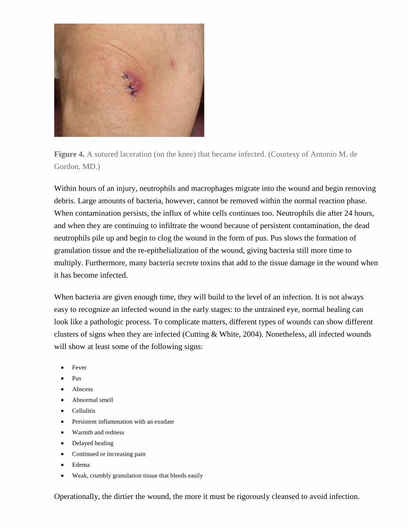
Figure 4. A sutured laceration (on the knee) that became infected. (Courtesy of Antonio M. de
Gordon, MD.)
Within hours of an injury, neutrophils and macrophages migrate into the wound and begin removing
debris. Large amounts of bacteria, however, cannot be removed within the normal reaction phase.
When contamination persists, the influx of white cells continues too. Neutrophils die after 24 hours,
and when they are continuing to infiltrate the wound because of persistent contamination, the dead
neutrophils pile up and begin to clog the wound in the form of pus. Pus slows the formation of
granulation tissue and the re-epithelialization of the wound, giving bacteria still more time to
multiply. Furthermore, many bacteria secrete toxins that add to the tissue damage in the wound when
it has become infected.
When bacteria are given enough time, they will build to the level of an infection. It is not always
easy to recognize an infected wound in the early stages: to the untrained eye, normal healing can
look like a pathologic process. To complicate matters, different types of wounds can show different
clusters of signs when they are infected (Cutting & White, 2004). Nonetheless, all infected wounds
will show at least some of the following signs:
Fever
Pus
Abscess
Abnormal smell
Cellulitis
Persistent inflammation with an exudate
Warmth and redness
Delayed healing
Continued or increasing pain
Edema
Weak, crumbly granulation tissue that bleeds easily
Operationally, the dirtier the wound, the more it must be rigorously cleansed to avoid infection.

Re-Injury
Re-injury can slow or stop wound healing. A new scar is weaker than the adjacent tissue, and the
newest scars are the weakest. Pushes and pulls that would have no effect on healthy parts of the body
can reopen a healing wound, even when it is protected by a well-made dressing. Similarly, if there is
significant skin tension surrounding the wound (eg, over a bent knee), the healing wound will not be
able to seal tightly.
Ischemia/Hypoxia
During normal healing, the granulation tissue develops a temporary dense capillary bed to provide
sufficient fluid, nutrients, and oxygen to the growing cells. After the reaction (inflammatory) phase,
oxygen is especially important for strengthening (ie, cross-linking) the collagen in the developing
connective tissue (Habif, 2004; Baum & Arpey; 2005, Shai & Maibach, 2005). Anything that
decreases the effectiveness of the local circulation will impede wound healing and weaken the scar.
Ischemia of a wound can arise from too much physical tension across the wound, ineffective
oxygenation of the blood (anemia, lung problems, smoking), or reduced circulation (atherosclerosis,
heart failure, kidney failure, vasoconstriction, too much pressure on the wound). Differences in the
available blood supplies account, in part, for the fact that facial wounds tend to heal better than foot
wounds. The importance of local circulation to wound healing is reflected in the healthcare maxim
"Wounds that don't bleed don't heal."
Local Skin Tension
Skin and its underlying tissues are normally under tension. Skin tension is negligible along skin
creases, moderate over relaxed joints and muscles, and high over bent joints (knees and elbows) and
over the skull. During a cutting, ripping, or puncturing injury, the tension from the adjacent intact
skin pulls the free edges of the wound apart. In places where the wounded skin is under greater
tension, the wound gapes more widely and heals more slowly, and the resulting scar is relatively
large.

Figure 5. Lines of least skin tension on the body. The drawing also indicates the areas of the body
where skin wounds have the highest risk of infection. (Courtesy of Scott Moses, MD.)
Patient Factors
DISEASES
Certain diseases are noted for causing poor wound healing. The most common of the problem
diseases is diabetes mellitus. Scars formed by diabetics have less collagen, and the collagen that is
laid down is more brittle than normal.
Diabetes also damages blood vessels and makes the skin more prone to ischemia. The reduced
circulation is especially notable in the feet, and foot wounds are notorious for not healing well in
diabetic patients. (See photograph of diabetic foot ulcer below.)
To make matters worse, diabetes leads to peripheral neuropathy. Diabetic patients lose sensation in
their fingers and toes, so diabetic injuries tend to go unnoticed in the extremities. Finally, diabetics
have a weakened inflammatory response, and they are more susceptible than other people to
developing tissue infections.
MALNUTRITION
Malnourished people begin to break down their proteins as a source of energy, and this slows
healing. Specific vitamin deficiencies also lead to poor wound healing. Vitamin A deficiency
impedes the transformation of monocytes into macrophages, which can slow or halt healing. Vitamin
C deficiency leads to weak collagen, which is the basis of scurvy. Vitamin K deficiency impairs
blood clotting.
OLD AGE
As people age, they heal more slowly. In older people, scars form with less and poorer-quality
collagen, and older adults are more likely than the young to have wounds reopen (dehiscence).
SMOKING
Patients who smoke have poor wounding healing—in addition to suffering a number of other skin
problems that include wrinkling, premature skin aging, higher risks of squamous cell carcinoma,
psoriasis, and hair loss (Freiman et al., 2004).
Healthcare Impediments
Medical care of wounds is an attempt to overcome obstacles to natural healing. In the course of
managing a wound, we reduce the amount of contamination, minimize the area that must be filled by
new tissue, keep the granulation tissue moist, and protect the healing area. However, our efforts at
facilitating wound healing sometimes introduce new impediments.
WOUND CLOSURE MISSTEPS
Wounds must be clean before they can safely seal themselves. In an attempt to close wounds
quickly, doctors sometimes suture together insufficiently cleansed tissues. This leads to an infection
and then the dehiscence of the closure.
Another problem in wound closure is the use of suture material that is too thin and subsequently
breaks. In addition, sutures that are too thin or that are tied too tightly can tear through the weakened
skin at the edges of the wound.
Finally, if sutures, staples, or tapes are removed too early, the wound edges will not have developed
sufficient adhesion and the wound will reopen.
PROBLEMATIC DRUGS, SOLUTIONS, AND OINTMENTS
Certain drugs, solutions, and ointments slow wound healing. Doxorubicin (Adriamycin) given
preoperatively inhibits postoperative wound healing. Glucocorticoids (eg, prednisone) limit the
proliferation of fibroblasts and the production of collagen, and thus steroids make scars relatively
weak. Some antiseptic solutions (eg, 10% povidone-iodine, 3% hydrogen peroxide, 0.5%
chlorhexidine) can slow wound healing. Hemostatic solutions (e.g., ferric subsulfate, 30% aluminum
chloride, silver nitrate) slow the healing of large wounds.
Some topical ointments also slow wound healing; these include triamcinolone acetonide ointment
(0.1%), Furacin, and USP petrolatum. In contrast, other ointments speed wound healing; these
include Neosporin ointment, Silvadene cream, Benoxyl peroxide preparations, and Eucerin.
X-RAYS
Ionizing radiation damages actively dividing cells. In wounds, the regrowing epithelium, the newly
growing blood vessels, and the fibroblasts that form new connective tissue are likely to be damaged
by a large dose of ionizing radiation. Normal x-ray imaging is usually not a problem. Cancer
therapies, however, give relatively high doses of ionizing radiation and, in areas of the body exposed
to radiation therapy, wounds heal poorly and infections are more common.
PART 2: Primary Wound Care
PROTECTION OF CAREGIVERS
People who take care of patients, especially patients with open wounds, should not be allowed to
pose a risk of infecting the patients. At the same time, caregivers should be protected from acquiring
infections from patients. Both goals can be met through the same set of precautions.
General Precautions
Healthcare workers who will have contact with patients should be screened for infectious diseases
when they are hired. They should be up to date on their immunizations; this includes vaccination
against measles, rubella, varicella, and hepatitis B, and ongoing yearly influenza immunizations.
A health clinic, office, or hospital will have written safety rules and procedures for controlling
infections, and these should be reviewed regularly with staff. The rules should include a list of steps
to be taken by healthcare workers who inadvertently come in direct contact with a patient's blood or
body fluids—for example, by an accidental needle stick.
Specific Precautions
Any patient can potentially carry the human immunodeficiency virus (HIV). Medical staffers
working on the first stage of wound care—inspecting, cleansing, closing, and covering—should
wear protective eyewear, a surgical mask, and gloves that are not easily ripped. Although they will
be wearing face masks, medical personnel who have upper respiratory infections should further
protect their patients by not talking or coughing while leaning over wounds.
PATIENT CARE

Primary wound care means acute wound care—managing a wound the first time it is presented to a
healthcare professional. Currently, we do not have the technology to repair a wound. Nonetheless,
we can remove many obstacles that inhibit the body's innate wound repair mechanisms.
Before modern medicine, many wounds did not heal. These wounds may have been too big, have
involved too great a loss of blood, or have became infected. Today, we can give natural healing
mechanisms a much better chance of success because we have the technology to stop serious
bleeding, to cleanse the wound well, and to close surgically (or otherwise effectively protect) large
wounds. The elements of modern wound care are inspecting, cleansing, closing, and covering.
Basic Plan
Before going into the details of how to inspect, cleanse, close, and cover a wound, here is the basic
plan for treating acute wounds (Auerbach, 2001; Kroot & Hurst, 2004; Simon & Hern, 2004;
Lammers, 2006b).
STABILIZE THE PATIENT
Wounds can be dramatic, but treat any life-threatening conditions first. After the patient has been
stabilized, take an inventory of his injuries and plan the order in which they are to be treated.
TAKE A HISTORY
Follow the order of your treatment plan. When it is time to treat an external wound, get a history of
the cause of the wound and get a description of the environment in which it occurred. Also, find out
the patient's chronic illnesses, medical conditions, current medicines, and allergies. Ask about the
immunization history for tetanus. While taking the history, explain to the patient what to expect
during the wound care procedures.
EXAMINE AND ANESTHETIZE THE INJURY
Inspect the wound. Estimate its depth, the degree of contamination, and the internal tissues that are
injured, if any. When there are injuries to internal structures (nerves, tendons, bones, muscles, ducts,
organs), call in a surgical specialist.
Check for significant neural or vascular damage. Caring for a wound will cause the patient pain;
nevertheless, before giving an anesthetic you must check to see if major nerves or blood vessels have
been injured. Test the injured area and the more distal areas of the body:
Is there full sensation?
Can all muscles be moved?
Is there adequate circulation?

If you suspect major neural or vascular problems, call in a surgical specialist.
Now, anesthetize the injured area and create a bloodless field. You will already have stopped any
major bleeding. At this point, stop any continuing bleeding. For wounds on the extremities, consider
using an inflated blood pressure cuff as a controllable tourniquet proximal to the wound.
CLEANSE THE WOUND
Prepare the wound field by cleansing the area surrounding the wound, clipping any interfering hair,
and washing all the adjacent regions with an antiseptic solution. Drape the areas surrounding the
wound.
With instruments (not gloved fingers), remove visible debris. If there is a possibility that you have
missed contaminants or pieces of broken bone, have the wound imaged to search for debris. Debride
the wound, removing devitalized or shredded tissue. Then, irrigate the wound with a cleansing
solution at high pressure
CLOSE THE WOUND
Stop any bleeding that the cleansing process has restarted. If you are going to close the wound
immediately, now suture, staple, tape, or glue the edges of the wound together.
COVER THE WOUND
Apply topical antibiotics or ointments if appropriate. If the wound is complex, large, or highly
contaminated, you may decide to delay wound closure, instead packing the wound with saline-
moistened sterile gauze. Whether open or closed, cover the wound with a dressing. Immobilize the
injured part of the body as needed.
MEDICATIONS AND CARE INSTRUCTIONS
Give tetanus prophylaxis, rabies prophylaxis, or systemic antibiotics as needed. If you are sending
the patient home, give wound care instructions. Describe the signs and symptoms of infection.
Schedule a follow-up visit for a wound inspection and/or suture or staple removal.
Detailed Plan
This section spells out the details for carrying out the basic primary treatment of an external body
wound (Auerbach, 2001; Kroot & Hurst, 2004; Simon & Hern, 2004; Lammers, 2006b).
STABILIZE THE PATIENT
A bleeding wound will grab your attention, but you must always assess the whole patient first and
immediately begin treating any life-threatening problems. Check the patient's breathing and heart
function. Look for signs of shock, for neck, back, or skull injuries, and for indications of internal
bleeding.
In the beginning, establish the ABCs of trauma care: Airway, Breathing, and Circulation. Of prime
importance is a clear airway; if necessary, intubate or perform a tracheotomy. If the patient is not
breathing spontaneously, the lungs must be artificially inflated at regular intervals. If the heart is not
beating, perform cardiac resuscitation immediately.
At the same time, serious bleeding must be stopped by pressure or clamping, and hypovolemic shock
needs to be treated with replacement fluids. All these procedures should be carried out without
moving the patient's head or cervical spine until it is clear that there are no injuries that threaten the
spinal cord.
TAKE A HISTORY
After ensuring the stability of the patient and assessing his overall condition, take a history. Find out
the cause of the injury (eg, "hit with a hammer," "fell down the steps"). The cause can warn you of
internal damage, as happens, for example, in crush injuries. The cause will also alert you to the
possibility of hidden internal debris, such as bullet fragments, glass, metal shards, gravel, or wood
chips. Finally, the cause will be a guide to prophylaxis, especially if there is the possibility of tetanus
or rabies infection.
Ascertain the time of the injury. The longer a wound remains unclean, the more likely it is to be
infected. A wound that is 12 or more hours old may need to be observed for a few days before it is
directly closed. Determine the environment in which the injury occurred (eg, "on my back lawn,"
"in my car"). The surroundings at the time of injury can suggest the degree to which the wound may
be contaminated.
Has the patient had a recent tetanus immunization? What are the patient's current diseases and
medications? For example, does the patient have diabetes or other diseases such as anemia or
atherosclerosis that may compromise the effectiveness of local circulation? Does the patient have
clotting disorders, drug allergies, or heart disease? Is the patient taking anti-coagulants,
immunosuppressive drugs, or glucocorticoids?
Next, explain the procedures and assure the patient that you will be keeping pain to a minimum.
Your calmness will reduce the patient's fear and stress.
If the patient wants emotional support, limit the friends or family to one person. Warn this person
that there will be needles, scalpels, and blood, and to sit during the procedures. Then, have the
patient lie supine because fainting is common. Wound care for children requires special patient and
family preparation.

EXAMINE AND ANESTHETIZE THE INJURY
While talking to the patient, completely expose and examine the wound area. Remove jewelry and
constricting clothing both proximal and distal to the wound area. Wound inspection and physical
examination are important, but they must be done carefully to avoid worsening the injuries.
Try to determine the depth of the wound, the degree of contamination, and the internal tissues (if
any) that have been injured. When nerves, tendons, bones, joints, muscles, ducts, or organs have
been injured, call in the appropriate surgical specialist. If there is the possibility of a fracture, get x-
rays.
Before administering anesthetics, you must determine any significant nerve, blood vessel, or
tendon damage. Test the sensory perception and muscle movements in the wound area and more
distally. Check that there is appropriate perfusion distal to the wound area. When the wound area
involves joints or tendons, be certain that there is still full range of motion. If there are any deficits,
get the advice of a surgeon of the appropriate specialty.
At this point, the patient should be stable and lying on the back (supine). Bleeding should be
minimal, and you should know whether any major arteries or nerves have been damaged. You
should also have a medical history of the injury and of the patient.
Your goal, now, is to cleanse the wound of bacteria, dirt, and debris. This can be painful for the
patient, so first provide anesthesia. In an outpatient setting, you will be using a local anesthetic. Most
anesthetics used for wound care are synthetic relatives of cocaine; they include:
Benzocaine (Americaine)
Bupivacaine (Marcaine)
Chloroprocaine (Nesacaine)
Dibucaine (Nupercaine)
Lidocaine (Xylocaine)
Mepivacaine (Carbocaine)
Prilocaine (Citanest)
Procaine (Novocain)
Ropivacaine (Naropin)
Tetracaine (Pontocaine) (Strichartz & Berde, 2005)
Local anesthetics can be applied topically, infiltrated locally, or injected at a distance from the
wound to produce a regional block. Anesthetics applied directly to the wound may slow the healing
process, and injections in or near the wound site will temporarily distort the anatomy. For these
reasons, regional anesthesia is often preferable to local anesthesia, but regional nerve blocks require
a trained operator, such as an anesthesiologist, surgeon, or experienced trauma specialist (Strichartz
& Berde, 2005).

Various topical anesthetic solutions are used for shallow wounds. Common solutions include:
EMLA (lidocaine-adrenaline-prilocaine)
LAT (lidocaine-adrenaline-tetracaine)
TAC (tetracaine-adrenaline-cocaine) (TAC is not used for highly contaminated wounds or wounds in or near
mucous membranes.)
Note that adrenaline is commonly known today as epinephrine.
Topical anesthetics are applied by holding cotton balls or sterile gauze soaked in the drug against the
wound for between 10 and 60 minutes, depending on the drug and the wound.
Local infiltration anesthetics are safe and fast-acting. The commonly used anesthetics differ mainly
in their duration of action. Duration of numbness produced by three common infiltration anesthetics
is:
Procaine (Novocaine) 20–30 minutes
Lidocaine (Xylocaine) 30–60 minutes
Bupivacaine (Marcaine) 120–240 minutes
The dose and concentration of the local anesthetic depends upon the weight of the patient and the
area that needs to be anesthetized.
To infiltrate a local anesthetic, begin injecting at the periphery of the wound. Make successive small
injections moving toward the center of the wound, with each needle stick in an area that is already
becoming numb.
Infiltrating the skin with an anesthetic produces a sharp pain. This pain can be reduced by using a
small (25- to 30-gauge) needle, inserting the needle perpendicular to the skin rather than at an angle,
and injecting the anesthetic slowly. To further minimize the pain, the anesthetic should be warm and
buffered with sodium bicarbonate.
Figure 6. Lidocaine (Xylocaine) is a local anesthetic commonly used to infiltrate an injury in
preparation for debridement and washing. (Courtesy of John L. Bezzant, MD.)

Injecting a local anesthetic proximal to the wound and alongside the appropriate nerve produces
anesthesia in the entire sensory region of the nerve. This is effective, and especially useful for
wounds in the extremities. In such a regional block, the onset of anesthesia takes longer (typically,
10–20 min) but it lasts 3 to 4 times longer than when the same drug is used locally (Thomson &
Lalonde, 2006). Administering a regional nerve block requires special training and a detailed
knowledge of anatomy.
Before proceeding, it is important to create a bloodless field. Direct pressure is an effective way to
reduce rapid bleeding. Using gauze sponges, sterile packing material, a pressure dressing, or simply
a gloved hand, press down on the wound and maintain the pressure for at least 10 minutes. A simple
compression dressing can be made by putting a stack of absorptive sponges on the bleed and then
wrapping the stack (not too tightly) with elastic tape or an elastic wrap.
If the wound is in a limb, elevate it above the level of the heart. If direct elevation and sustained
pressure for 15 to 20 minutes are not sufficient to slow the bleeding in an extremity, temporarily
inflate a blood pressure cuff around the limb proximal to (on the heart side of) the wound; the cuff
should left on no longer than 45 minutes.
When wounds continue to bleed, severed small vessels can be clamped and then ligated individually
with absorbable suture material. (Arteries can be tied off, but veins need to be sutured.) There is an
art to clamping vessels while avoiding adjacent structures such as nerves. Electrocautery at
minimum power can be used for the tinier vessels. Closing large vessels is a job for a vascular
surgeon. Controlling bleeding in highly vascularized areas, such as the scalp, may require other
specialized techniques.
CLEANSE THE WOUND
Traumatic wounds must be cleared of dirt and debris. Before cleansing, debridement, and wound
closure, the wound area must be prepared and draped. When hair hangs into the wound or makes it
hard to cleanse the regions around a wound, it is better to cut the hair rather than to shave it with a
razor. Shaved wounds have a higher rate of infection.
Put on rip-resistant gloves, protective eyewear, and a surgical mask. Begin by cleansing the surfaces
of skin adjacent to the wound, but avoid getting antiseptic into the wound itself. Wipe off all the
particulate matter, and then scrub the skin with 10% povidone-iodine (Betadine scrub), which
effectively reduces viruses, fungi, and gram-positive and gram-negative bacteria. Chlorhexidine
gluconate (Hibiclens) solution is a commonly used and effective alternative skin antiseptic.
After cleansing a wide area, paint the antiseptic broadly around the wound. Cover the painted areas
with drapes, leaving an open window over the wound. When the wounded area is on a hand, paint
the hand with an antiseptic. Then, put the hand in a sterile glove, and cut a window in the glove over
the wound.
After draping the area, put on clean, rip-resistant gloves to protect both you and the patient. The
gloves need not be sterile, but if they have any talc or powder, then they should be rinsed before you
touch the wound. Be sure to wear eye protection and a face mask.
Foreign material increases the risk for infection, so examine the wound for debris. Use good lighting
and try to create a bloodless field. With a metal probe look methodically through the whole wound –
do not use your fingers, because sharp wood, bone, glass, or metal can rip your glove, cut your skin,
and inoculate you with the patient's blood. With a hemostat or forceps, remove all visible debris.
Sometimes it is necessary to extend the wound by separating or even removing tissues to explore the
injured area thoroughly. Fatty tissues are especially difficult to search. If there is any question that
you may have missed debris, then use imaging studies (x-rays, ultrasound, CT scan) to survey the
wound.
After carefully picking visible foreign material from a wound, you must remove tissue that is
seriously injured or that is contaminated by bacteria and other debris. Contaminated and dying
tissues provide an ideal growth environment for bacteria, and thorough debridement (removing
seriously damaged and contaminated tissue) will dramatically decrease the risk of infection.
Debride the wound with forceps and scalpel or scissors. (This is called sharp or surgical
debridement.) Sharp debridement is one of the arts of trauma management, and it is best left to an
experienced physician, but the following are some of the principles of debridement.
In a few special cases, an entire wound area can be excised. In this way, an irregular, contaminated
wound can be converted into a clean, smooth-edged surgical wound. This can sometimes be done for
small wounds when (a) there are no vital structures—eg, major blood vessels, nerves, tendons—in
the wound, and (b) the skin tension in the area is not too great. Complete excision is an ideal way to
deal with highly contaminated wounds, such as animal bites, and with certain puncture wounds,
from which debris can be difficult to remove.
Surgical debridement is known as sharp debridement when it is done in an office, clinic, or
emergency room. In most cases, the entire wound cannot be excised. Instead, through use of a
scalpel or scissors, the wound is examined inch by inch, cutting off bits of devitalized and
contaminated tissue and attempting to smooth extremely ragged or shredded edges. The severely
injured or necrotic tissue is grasped with a forceps or a small hemostat and cut out along the healthy
edge. The art of sharp debridement is in distinguishing viable and nonviable areas of the tissue (Shai
& Maibach, 2005).

When a wound is contaminated, all the exposed fat should be removed during debridement. Care
must be taken not to spread contamination deeper into the wound. Structural tissues, such as fascia,
tendons, and bones, are cleansed and left in place. Nerves, too, are very gently cleansed and left in
place.
For the protection of healthcare workers and other patients, wounds should be treated as if they
contain contagious bacteria and viruses. Therefore, debrided material must be put in
decontamination containers. Sharp debridement is done with an eye to the geometry of the eventual
wound closure. On the face and the hands, sharp debridement is an especially challenging task.
Wounds should be washed both before and after debridement.
You have stabilized the patient, assessed and anesthetized the injured area, cleansed and draped the
adjacent skin surfaces, and debrided the wound. The clock is still ticking, however, because the
longer you wait before cleansing the wound, the greater the chance that bacterial growth will reach
infective levels.
Wounds are cleansed by irrigation, and when irrigation is insufficient, wounds are directly scrubbed
and then re-irrigated. Soaking a wound (in saline or in an antiseptic solution) is not an effective
substitute for irrigation.
Irrigation means continuously pressure-washing a wound. When done with sufficient force,
irrigation effectively removes bacteria and debris from the wound and from the crevices extending
into the edges and bottom of the wound. Proper irrigation reduces wound infection rates
dramatically.
Pressure-washing is critical – simply flooding the wound or rinsing it by pouring on cleansing
solution will not reduce the chance of infection. Effective irrigation requires that the washing
solution be squirted into the wound at pressures greater than 7 pounds per square inch (psi).
Commercial irrigating machines, such as Water Pik, Canyons Wound Irrigation System, Irrijet, and
the Travenol Pressure Irrigation Set, all deliver irrigating solution at effective pressures. A 19-gauge
needle attached to a 35-ml syringe will also squirt solution at sufficiently high pressures if you push
the plunger with both hands (Auerbach, 2001).
When washing a small wound, try to irrigate it with at least 1/3 liter of cleansing solution. When
irrigating larger wounds (or if you are using lower than optimal pressure), flush with more than 1/3
liter of solution. Let the continuous drainage of solution run off into a basin. Be sure to carry out the
irrigation behind a protective cover or splash shield to keep droplets of the patient's blood from
spraying the surroundings. ZeroWet Inc. makes a protective device (ZeroWet Splashshield) that fits
on the end of a syringe to contain splashes from the wound.
Scrubbing a wound by hand is called mechanical scrubbing. Mechanical scrubbing with an antiseptic
sponge is effective at removing bacteria and debris, but it is also damaging to the wound tissues.
When a wound is highly contaminated, mechanical scrubbing may be the only way to fully cleanse
the wound. In this case, you minimize the abrasive damage by using a nonionic detergent solution
and a fine-pore sponge (eg, Optipore sponges). Irrigate the wound after the mechanical scrubbing.
Many cleansing solutions are available for wound irrigation. One of the best is plain tap water,
which is safe, inexpensive, and as good as or better than sterile saline (Anglen, 2005; Beam, 2006).
In the past, sterile 0.9% saline was the standard irrigating solution. Another commonly used solution
is 1% povidone-iodine (Betadine preparation, not the 10% Betadine scrub), which is then followed
by flushing with sterile 0.9% saline solution. In addition, nonionic detergents, such as Poloxamer
188 (Pluronic F-68, Shur-Clens, Pharma Clens), are safe for wound irrigation.
Other cleansing solutions are not recommended for irrigation. For example, some antiseptic
solutions [eg, 5%, 7.5%, or 10% povidone-iodine; chlorhexidine gluconate (Hibiclens); 3%
hydrogen peroxide; hexachlorophene (pHisoHex); alcohols] that are very effective on intact skin can
be too harsh for wounds when used in a pressure irrigator. Antibiotics are not needed in the irrigation
solution, even for deep or complex wounds (Anglen, 2005).
CLOSE THE WOUND
After washing the wound, gently pat it dry with a sterile pad and then wipe the skin around the
edges. Now, decide whether to close the wound directly and, if so, what method to use. Your
decision can minimize the risk of infection and will also determine the size of the scar.
When deciding about wound closure, take into account the local skin tensions in the area of the
wound (Habif, 2004). Most skin in the body is being stretched, at least slightly, by the adjacent skin
and the underlying structures, but the actual tension at any one location varies along the surface of
the body. Movement changes skin tension: bending a joint stretches the overlying skin, while
contracting a muscle tends to reduce tension in the overlying skin. Skin creases and skin wrinkles are
indications of lines of least tension; on the face, the lines of facial expression are also lines of least
tension. As a rule, the lines of least skin tension are perpendicular to the long axis of underlying
muscles.
In areas of high skin tension (eg, on the scalp, along the front of the tibia) the edges of a wound are
pulled apart, and the wound gaps in the direction of the greatest tension (perpendicular to the lines of
least tension). Skin tension will strain a healing wound and it will widen the final scar. To achieve
the thinnest scars, surgeons make elective incisions in or parallel to skin creases and perpendicular to
underlying muscles. If you decide to close the wound that you have just cleansed, try to make the
long axis of the wound seam parallel to skin creases, perpendicular to underlying muscles, and along
the local line of least skin tension.
There are three general plans for wound closures: immediate direct closure, indirect closure, and
delayed direct closure (Lammers, 2006a).
Immediate direct closure (by suturing, stapling, gluing, or taping) is called healing by first intention or primary
wound repair. The wound closure at the end of a typical surgical operation is an immediate direct closure.
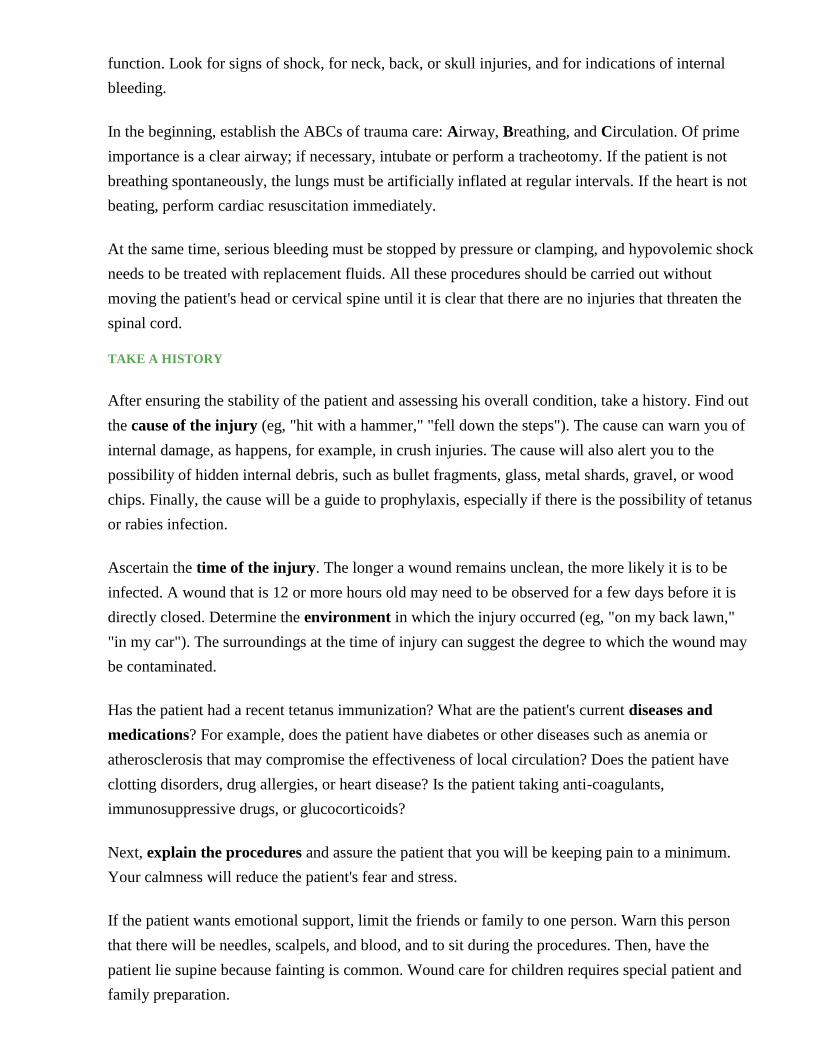
Indirect closure allows the wound to contract and to re-epithelialize on its own. This is called healing by second
intention or secondary wound repair. Indirect closures tend to leave a larger scar, but they avoid protecting bacteria
inside a warm moist tissue environment. For highly contaminated wounds, indirect closure significantly reduces the
risk of infection.
Figure 7. A pressure sore healing by indirect closure. Periosteum of bone is visible in the top
picture. Healthy granulation tissue covers the wound in the two middle pictures. Healing took
several months. (Courtesy of Charlie Goldberg, MD. Copyright © Regents of the University of
California.)
Delayed direct closure lets the wound remain open initially and later closes it with sutures or staples. This two-part
closure plan is called healing by third intention or tertiary wound repair. Delayed direct closure is used for highly
contaminated wounds, which may need repeated debridement or may need to be treated with antibiotics before
being closed.
Choosing among the three closure plans is a balance between protection, risk of infection, and size
of the eventual scar. The immediate direct closure of a well-cleansed wound protects it from new
contamination and allows the most control over the size and appearance of the final scar. In addition,
immediate direct closure protects from drying any exposed deep tissues and structures, such as
nerves, blood vessels, tendons, or bones.
On the other hand, immediate direct closure of an unclean wound encourages the development of
infection. Besides providing a protected environment for bacteria, wounds closed with sutures add
new foci for infection, namely, the suture holes, the sutures themselves, and the tissue damaged by
the sutures. Clean unsutured wounds are less likely to become infected than clean sutured wounds.
Indirect closures heal more slowly than direct closures. A healthy indirect closure provides a longer
reaction (inflammatory) phase and a more thorough natural debridement. Moreover, if infection does
develop in an indirect closure, you have direct access to the inside of the wound, so you can debride,
irrigate, and apply antibiotic. Indirect closure (ie, helping a wound to grow closed naturally) is also
called open wound management.
Open wound management should always be considered for wounds that are:
Already infected
Very dirty (especially when contaminated by organic matter)
Made by animal or human bites
Many hours old at the time of treatment
Made by crushes, explosions, or other forces causing extensive tissue damage
On the bottom of a foot, especially wounds made when stepping in organic matter, such as found in fields, woods,
streams, or garbage
While managing an open wound, you usually have the option of closing the wound directly during
the first five days. Delayed direct closure is sometimes the best compromise between immediately
suturing a wound to prevent a large scar and leaving the wound open to prevent the development of
infection. Waiting and watching is often the wisest course.
In wounds with exposed or injured internal structures, such as nerves, joints, or bones, consult a
surgical specialist before deciding on a closure plan.
When you decide on indirect closure, pack the wound and cover it with a dressing. It is usually
unnecessary to apply antibiotics. The packing material should be sterile fine-mesh gauze moistened
with sterile 0.9% saline. The cover dressing should be a dry, thick absorbent sterile pad or pack of
gauze pads. When the wound is near a joint, the joint should be splinted to keep the injured area
immobile.
From this point on, open wound management can often be an outpatient procedure. The patient, a
household member, or a visiting nurse is enlisted to repack the wound with saline-moistened sterile
gauze and to re-cover it with a dry dressing once each day. (See instructions below.) Although
doctors frequently send a patient home with oral antibiotics, this is usually not necessary.
The wound should be evaluated professionally in 3 to 4 days, which is approximately the time when
the healing process in an open wound is making the transition from the reaction (inflammatory)
phase to the regrowth (proliferative) phase. If there is no evidence of infection and if the edges of the
wound can be pulled together without too much tension, you can suture the wound closed.
The delay will have allowed time for nonviable tissue to become apparent, and you should debride
the wound and irrigate it again before closing it. A direct closure that has been delayed only a few
days will produce a scar not much larger than if the wound had been directly closed immediately.
If a hematoma forms in a closed wound, it will push the edges apart, slow the healing processes, and
increase the chance of infection. Even though you stopped the bleeding earlier, the subsequent
debridement and cleansing may restart it; therefore, before you directly close a wound, make certain
that all bleeding is stopped.
To hold the edges of a wound together in a direct closure, your arsenal includes sutures, staples,
tape, and adhesives. Sutures are the best choice for wounds that are being pulled apart by tension
from the surrounding tissues and for wounds that require detailed matching of the opposing edges.
Suturing is probably the most widely used direct closure technique, but stapling and gluing are better
methods for closing wounds in the field, away from a medical facility (Auerbach, 2001). When
closing a wound with sutures, you can align the edges of the wound carefully and hold them together
strongly. The detailed nature of each wound dictates its particular suturing requirements.
Suturing takes skill and experience. In broad terms, the first step is closing the major deep tissues.
When closing an acute traumatic wound, you typically use an absorbable suture, such as chromic gut
or polyglactin (eg, Vicryl) for the deep tissues.
The second step is closing the skin. Typically, you use nonabsorbable sutures, such as nylon (eg,
Ethilon) or polypropylene (e.g., Prolene) for the skin. To close the skin, push the needle
perpendicularly through the full skin so that when you pull the edges together they will be everted.
Carefully match jagged wound edges as long as the skin along the edges is still viable. The goal is to
align the upper surface of the skin along the scar, so try to insert the needle through equal depths of
the opposing edges of the wound. Tighten each stitch gently so that the edges are everted and
touching but are not crushed together.
After closure, there should be minimal tension across the wound. Semi-permeable tape strips (e.g.,
SteriStrips, Clearon Skin Closures) can help to further reduce tension across a sutured wound. Leave
spaces between the strips of tape so fluid and exudate can escape and be absorbed by the overlying
dressing. When the wound is large, tension across the sutured edges can be reduced by first stitching
together individual deeper layers of tissue or by carefully undercutting and removing some of the
deeper tissue. Undercutting wound edges should be left to an experienced clinician.
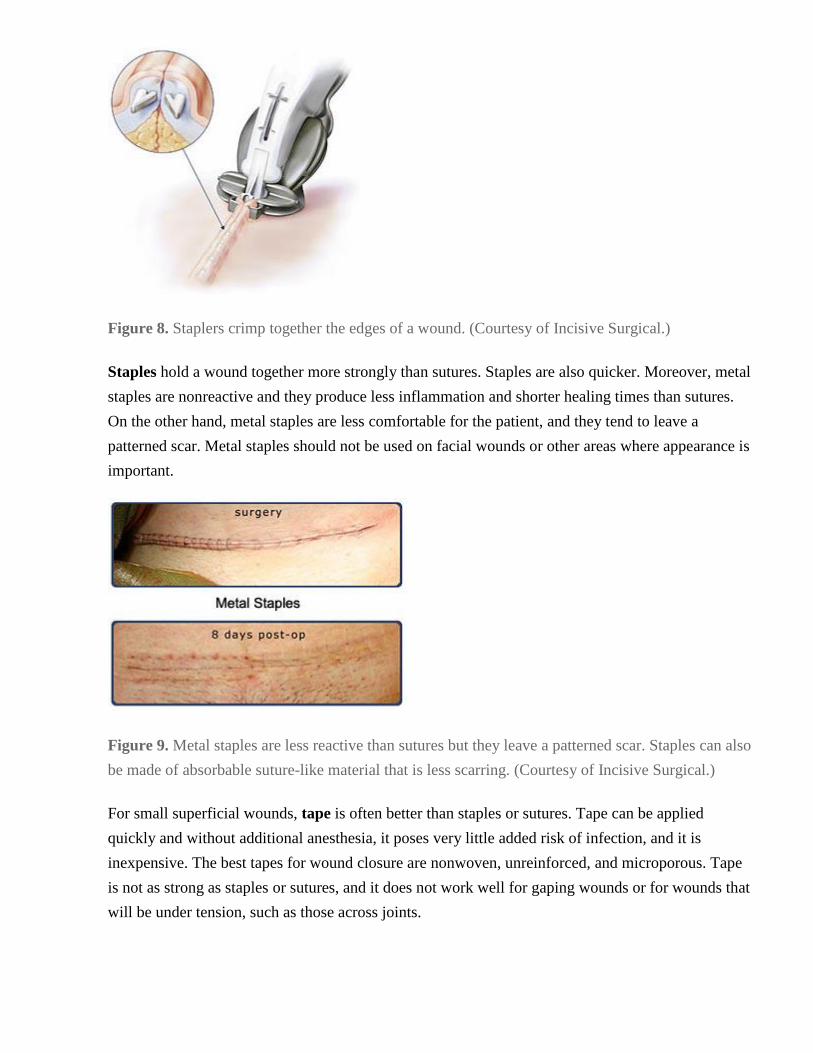
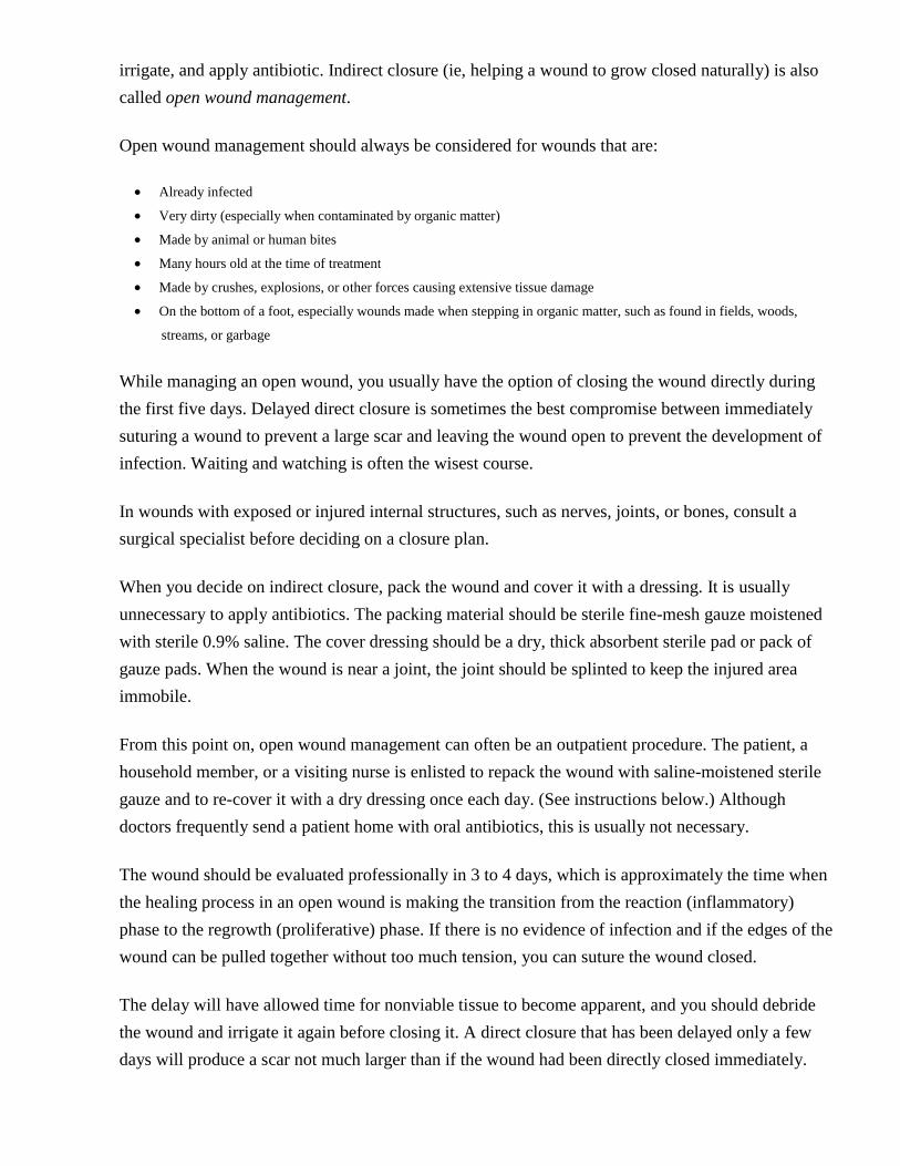
Figure 8. Staplers crimp together the edges of a wound. (Courtesy of Incisive Surgical.)
Staples hold a wound together more strongly than sutures. Staples are also quicker. Moreover, metal
staples are nonreactive and they produce less inflammation and shorter healing times than sutures.
On the other hand, metal staples are less comfortable for the patient, and they tend to leave a
patterned scar. Metal staples should not be used on facial wounds or other areas where appearance is
important.
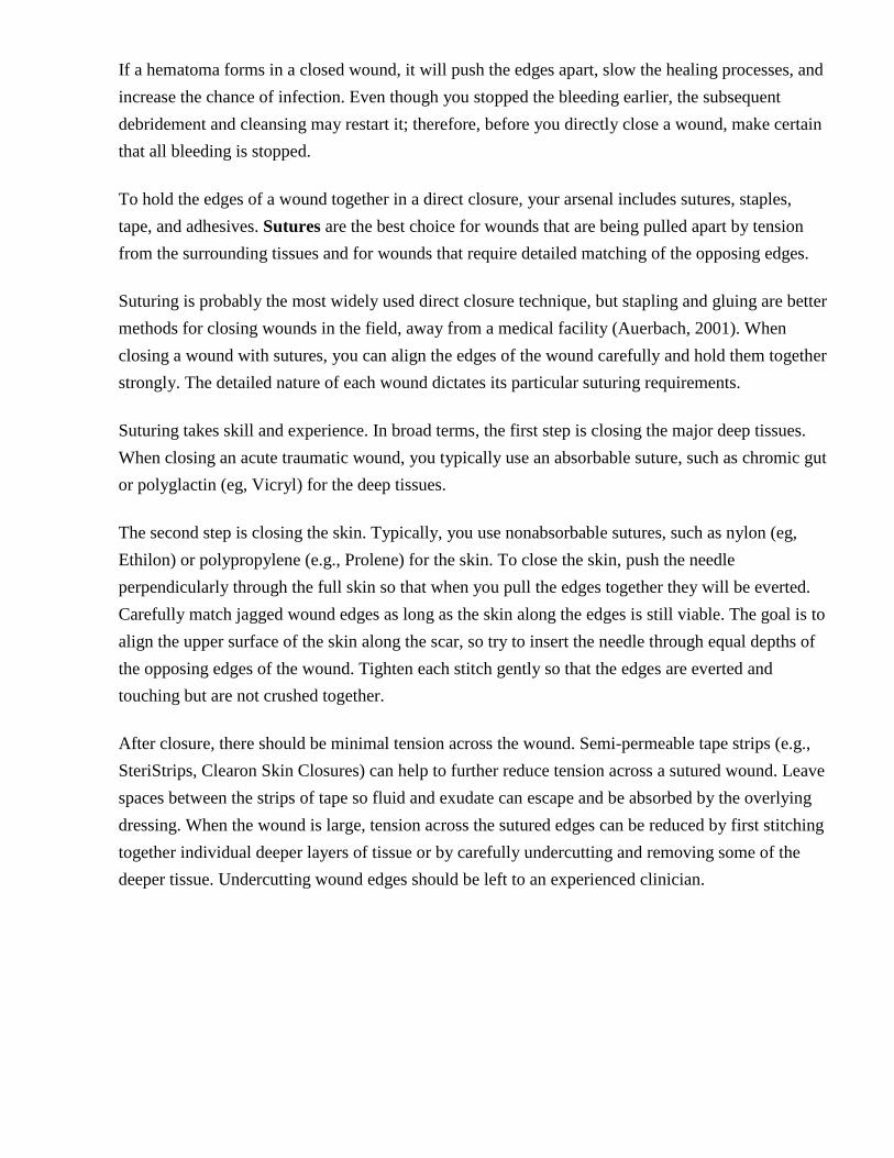
Figure 9. Metal staples are less reactive than sutures but they leave a patterned scar. Staples can also
be made of absorbable suture-like material that is less scarring. (Courtesy of Incisive Surgical.)
For small superficial wounds, tape is often better than staples or sutures. Tape can be applied
quickly and without additional anesthesia, it poses very little added risk of infection, and it is
inexpensive. The best tapes for wound closure are nonwoven, unreinforced, and microporous. Tape
is not as strong as staples or sutures, and it does not work well for gaping wounds or for wounds that
will be under tension, such as those across joints.

To tape a wound closed, first stop all the bleeding and dry the edges of the wound. If the skin is oily
or sweaty, wipe it thoroughly; you can even use a solvent such as acetone to wipe the adjacent skin.
Closely crop any hair around the wound.
When the adjacent skin is clean and dry, increase its adhesiveness by painting tincture of benzoin,
Matisol, or cyanoacrylate glue on the skin alongside the wound. (Let the benzoin or Matisol become
tacky before putting on the tape. Glue can be taped when it is still wet.)
Use a pre-cut tape (eg, SteriStrips) or cut a piece of sterile adhesive tape to a width of 1/4 inch (0.5
cm). Each strip should be long enough to extend beyond the wound about 1 inch (2.5 cm) on either
side. Fix an end of each tape strip onto the skin at one side of the wound. Use your fingers to hold
the edges of the wound closed, and fix the other end of the tape strip to the skin along the opposed
edge of the wound. Put the tape strips in parallel, like railroad ties, along the wound. Space the strips
1/8 inch (0.3 cm) apart to allow for wound drainage. After putting strips across the full length of the
wound, lay a single long thin strip of tape along the ends of the cross strips on either side of the
wound, like a railroad track on railroad ties. The long strips will help to keep the cross strips from
peeling off the skin.
Tissue glues—butyl-2-cyanoacrylate (Histoacryl) or octyl-2-cyanoacrylate (Dermabond), which is
the stronger of the two—are good for closing shallow, sharp-edged wounds. Like tape, tissue glue
can be applied quickly and easily without additional anesthesia. Glues not only pose no additional
infection risk, they actually decrease the rate of wound infections. Tissue glues are ideal for closing
small lacerations in the field, away from medical facilities (Auerbach, 2001). Glues, however, are
not as strong as sutures or staples.
When closing a wound with tissue glue, first stop the bleeding. Then cleanse and dry the skin along
the edges of the wound. Hold the wound edges together with your fingers, and paint a thin layer of
glue along the cut. Allow the glue to dry (approximately 2 minutes), and paint on another layer.
Repeat the process until there are 3 or 4 layers of glue coating the wound.
Glued wounds need extra care: they cannot be immersed in water, and they can only be rubbed
gently. Glues are also disrupted by petroleum-based ointments and salves, which should not be used
on glued wounds. Sometimes tissue glues cause a mild local inflammatory reaction.
Tissue glues slough off spontaneously after about 4 days, by which time the wound has usually
healed sufficiently to remain sealed without the glue.
COVER THE WOUND
After closing the wound, gently cleanse the surface with moistened gauze, and cover the wound—
typically with an ointment, a dressing, and a protective bandage.

Ointments help to keep wounds moist, and they reduce the crust that can form on the surface.
Ointments also keep dressings from sticking to the wound. On the other hand, ointments will
dissolve tissue glues, so ointments should not be put on wounds that have been closed with
adhesives. For any wound, do not apply ointments containing corticosteroids, which impede wound
healing.
Triple antibiotic ointments with neomycin, bacitracin, and polymyxin are commonly used on open
wounds. Most wound ointments contain antibiotics, but it is currently unclear whether the
antimicrobial action plays any useful role in the healing of uninfected wounds. Neosporin ointment
and Silvadene cream have been shown to improve wound healing.
Dressings keep a healing wound warm and protected. They also keep the wound from drying out,
while at the same time absorbing excess fluid and exudate, both of which can slow healing. On the
other hand, once a wound is infected, a thick dressing will encourage bacterial growth; therefore,
thick or impermeable dressings are not put over infected wounds.
A wound dressing usually has two layers. The primary dressing is put directly on the wound surface,
and it is used to keep the wound moist. The secondary dressing is the outer layer, and it is used to
absorb excess drainage and to protect the wound.
Ideally, the full dressing should protect the wound from bacteria and dirt while allowing oxygen to
diffuse into and water vapor to diffuse away from the wound. The ideal dressing should keep the
wound moist and it should absorb excess fluid. In addition, in most cases, the primary layer of the
dressing should not stick to the healing surfaces.
A simple traditional dressing begins with a primary layer of petrolatum gauze (Adaptic, Aquaflo,
Betadine, Xeroform) applied to an ointment-covered wound. Over this, there is a secondary layer of
thick, dry gauze. The whole dressing is held in place by tape (Auerbach, 2001).
Today, a wide variety of primary dressings is available beyond petrolatum gauze. If you have a
choice, tailor the primary dressing to the amount of drainage you expect from the wound. A non-
draining wound can be covered with an occlusive (impermeable) or semi-occlusive (semi-
permeable) dressing such as a wound film. (See the illustration below of wound film applied to a
burn.) A wound draining 1 to 2 ml fluid/day needs a semi-occlusive or an absorbent (nonadherent)
dressing. A wound draining >3 ml fluid/day should have a very absorbent dressing.
In general, changing a dressing daily is frequent enough to avoid infections. Therefore, wounds with
significant drainage should have secondary dressings sufficiently absorbent to soak up a day's worth
of wound fluid. This may require extra gauze pads, sponges, or cotton.

A wide variety of primary dressings is available, and in a well-stocked clinic or hospital you can
tailor the primary dressing to the specific needs of each particular wound. Briefly, the major types of
specialty primary dressings include (Shai & Maibach, 2005):
Nonocclusive dressings
o Activated charcoal—absorbent (Carboflex, Carbonet, Kaltocarb)
o Alginate dressings—very absorbent (Algiderm, Curasorb, Hyperion, Kaltostat, Maxorb, Nu-derm
alginate, Nutrastat, Orisorb, Seasorb, Tegagen alginate)
o Dextranomer hydrophilic granules—very absorbent (Debrisan, Iodosorb)
Occlusive dressings
o Water-retentive foams—absorbent (3M Foam, Biatain, Curafoam plus, Hydrasorb, Lyofoam,
Orifoam, Sof-foam, Tielle, Vigifoam)
o Hydrocolloids—absorbent (Comfeel, Dermacol, DuoDerm, Exuderm, Granuflex, Hydrocol, Nu-derm,
Oriderm, Tegasorb, Ultec)
o Hydrogel—nonabsorbent, moistening (Aquaflo, Curafil, Dermagran hydrogel zinc-saline, Duoderm
hydroactive gel, Granugel, Hydrosorb, Purilon gel, Vigilon)
o Thin films—nonabsorbent (Bioclusive, Blisterfilm transparent, Dermafilm, Epiview, Opsite, Orifilm,
Polyskin, Tegaderm)
A bandage—an outer layer of dressing—is used to mechanically protect a wound. Bandages help
hold the wound closure in place and can reduce tension across the healing scar. In addition, the
compression provided by a bandage will reduce the open space (dead space) in a wound and thus
discourage hematomas and edema. Bandages also protect against injuries to the healing wound by
providing an additional layer of padding and by reducing the mobility of the wound area.
Bandages can be made of pads or of cotton overlaid with tape. On the limbs, bandages can be made
using an elastic wrap. All bandages should be smooth and unwrinkled and should apply pressure
equally across a wound. Fix the bandage in place with tape, and make sure it feels firm, but do not
make the bandage so tight that it impedes circulation.
Repeatedly moving a wound by contracting nearby muscles will slow wound healing and increase
the size of the eventual scar, so immobilize any nontrivial wound that is in a part of the body near a
joint. On the extremities, you can immobilize an injured area by splinting the nearby joints. Plastic
or aluminum splints can sometimes be added to the outer bandages of a wound. Otherwise, put a
separate splint along the joint. At times, a plaster cast may be needed.
Give any necessary prophylactic medications. For nonsurgical wounds, you must always consider
protection against tetanus. For wounds caused by mammal bites, rabies is a consideration. In
addition, certain, but not all, wounds should be treated prophylactically with systemic antibiotics.

Tetanus is a neurologic disease resulting from the poison produced by Clostridium tetani bacteria.
This toxin causes uncontrollable, continuous muscle contractions. Even in the best hospital settings,
tetanus has a fatality rate of 10% or more (Bleck, 2005).
In the United States there are approximately 50 cases of tetanus reported each year, mainly in older
adults. Even this small number could be reduced by a more comprehensive immunization program.
For adults, the CDC recommends a routine booster dose of tetanus toxoid–containing vaccine
every 10 years. For adults who do not know if they have had a primary set of vaccinations, the CDC
recommends that they begin with a three-dose primary series. Detailed up-to-date recommendations
for wound prophylaxis can be found at http://www.cdc.gov/mmwr/preview/mmwrhtml/.
Adults who have completed the three-dose primary tetanus vaccination series and who have received
a tetanus toxoid-containing vaccine <5 years ago are protected against tetanus and do not require
tetanus prophylaxis as part of their wound care. For other injured patients, the treatment
recommendations depend on the known history of tetanus vaccinations and the category of the
wound.
For tetanus prophylaxis, wounds are divided into two categories: clean-minor wounds, and major
and/or tetanus-prone wounds. Clean-minor wounds are small open lacerations made by clean
objects in clean environments (eg, an accidental cut with a clean scalpel). Major and/or tetanus-
prone wounds include:
Wounds contaminated with dirt, saliva, or feces
Wounds untreated for >6 hours
Puncture wounds (including nonsterile injections)
Bullet wounds
Burns
Frostbite
Avulsions
Crushes
TETANUS PROPHYLAXIS DURING WOUND CARE (adults aged 19–64 years)
If the patient has had a three-dose primary series of tetanus immunizations and has had:
A booster within the last 5 years—no tetanus prophylaxis is needed for any wound.
A booster within the last 10 years—tetanus toxoid–containing vaccine should be given only for major and tetanus-prone
wounds.
No booster within the last 10 years—tetanus toxoid–containing vaccine should be given for all wounds.

If the patient has NOT had a complete three-dose primary series of tetanus immunizations (or if primary
immunization status is unknown):
Tetanus toxoid–containing vaccine should be given for clean wounds.
Tetanus toxoid–containing vaccine and tetanus immune globulin should be given for major and tetanus-prone wounds.
All bites by mammals should be considered for rabies prophylaxis. Rabies is a viral disease with a
typical incubation period of 1 to 3 months. Once symptoms appear, the disease is almost 100% fatal;
therefore, prophylactic treatment of bites from potentially rabid animals is essential (Bleck &
Rupprecht, 2005).
Rabies is most common in bats, raccoons, and skunks, and the disease is transmitted in saliva. Most
human cases of rabies have come from bat bites. Some increased risk factors:
Bites on bare skin are more likely to develop rabies than bites through clothes.
Multiple bites are more likely to lead to rabies than a single bite.
Bites on the face are more likely to transmit rabies than bites on the extremities.
Unprovoked animal bites are more likely to develop rabies than bites from animals biting because were disturbed or
frightened.
The decision to begin rabies prophylaxis depends mainly on the type of animal that caused the bite.
Most patients will be able to tell you what bit them and why, and by contacting your local public
health officials or the CDC 24-hour rabies hotline (404-332-4555), you will be advised about the risk
in your location of the biting animal having rabies. Your public health officials will also try to find
the animal if there is any chance that it might be rabid. Rabies prophylaxis can be begun after the
wound has been cared for, so there is time to consult and to make a well-informed decision.
A few general rules can help you in your initial decisions:
All mammals can potentially be infected with rabies.
Rabies virus is inactivated by drying and by ultraviolet irradiation.
Rabies is transmitted only in saliva, but saliva can get into existing cuts or abrasions even when the animal does not
bite a person.
Blood, urine, and feces will not transmit rabies.
Petting a rabid animal will not transmit rabies.
The risk for developing rabies from an animal bite depends on the prevalence of the disease in your
locale. The following table presents some general guidelines on prophylactic treatment for a variety
of mammal bites.
RABIES TREATMENT FOR MAMMAL BITES

Animal Relative Rabies
Risk
Typical Treatment
PETS
Small rodent Very low None
Rabbit Very low None
Cat, dog, ferret Low None, unless animal's behavior changes within 10
days
FARM
Livestock Low None, unless animal's behavior changes within 10
days
STRAY OR WILD
Small rodent Very low None, but check with public health officials
Rabbit Very low None, but check with public health officials
Cat, dog, ferret Medium Consult public health officials
Raccoons,
skunks
High Begin treatment and consult public health officials
Foxes, coyotes High Begin treatment and consult public health officials
Bats Very high Begin treatment and consult public health officials
People who may have been infected with rabies virus need both active and passive immunization.
Active immunization comes from a five-dose course of rabies vaccine injections; the effect begins
within 7 to 10 days and lasts at least two years. Passive (direct) immunization comes from an
injection of anti-rabies immune globulin; the effect begins immediately and lasts for a few weeks
(Rupprecht, 2004). Before administering rabies prophylaxis, consult with local public officials and
review the latest CDC recommendations. Detailed up-to-date recommendations can be found at
http://www.cdc.gov/mmwr/preview/mmwrhtml/00056176.htm.
Err on the side of caution. If prophylaxis has been started and subsequent tests find that the
offending animal did not have rabies, the treatment regimen can always be stopped safely.
Although systemic antibiotics are often given to patients with acute wounds, most clinicians argue
that antibiotics should only be given with a specific purpose in mind.
Surgically, all open, fresh, accidental, or penetrating wounds are considered to be potentially
contaminated wounds, and some physicians use this as a reason for giving anti–skin flora antibiotics
such as Cefazolin.

On the other hand, emergency department physicians usually flag only certain wounds for systemic
antibiotic prophylaxis (Kroot & Hurst, 2004; Simon & Hern, 2004; Lammers 2006b). These wounds
include:
Bite wounds
Overwhelmingly contaminated wounds
Sutured intraoral lacerations
Orocutaneous wounds (mouth wounds that open out into the skin)
Crush wounds
Wounds involving tendons, bones, or joints
Delayed-treatment wounds
Wounds in people at risk for developing infections
Systemic antibiotics are best given as early as possible during wound treatment, and the first dose
should be administered intravenously or intramuscularly. There is no one standard antibiotic regimen
for wounds at high risk of infection, and there is no universal agreement on how long the antibiotic
prophylaxis should last.
For minimizing the risk of infection, systemic antibiotics cannot replace debridement and irrigation.
Remember that when a wound needs systemic antibiotic prophylaxis it probably also needs indirect
or delayed direct wound closure.
Specific Types of Acute Wounds
The basic wound care techniques detailed above need to be tailored to the particulars of each
individual wound. Here are some cautions, changes, and additional techniques for specific types of
wounds (Auerbach 2001, Kroot & Hurst 2004, Simon & Hern 2004, Lammers 2006b).
MINOR BURNS
Serious burns require treatment in a specialized burn center. The following discussion is about the
treatment of minor burns (Kumar et al., 2004; Edlich et al., 2006). To be considered a minor burn,
the area injured must be a single small patch of the body outside of the hands, face, feet, armpits,
popliteal region, or perineum. The burn cannot be across a major joint, and it cannot be in a band
extending around any part of the body. Chemical and electrical burns should be treated in a burn
center. Infants, older adults, and patients with major medical problems may need to be hospitalized
for burns that would otherwise seem minor.
Minor burns are limited to first- and second-degree burns. First-degree burns, such as sunburns, are
called epidermal burns because the damage is mainly to the outermost layer of the skin. Epidermal
burns are red and painful but they do not blister. After about 2 days, the injured epidermis in a first-
degree burn sloughs off ("peels") as new epithelium grows underneath the damaged tissue.

Second-degree burns are called partial-thickness burns because they involve injury to the epidermis
and part of the dermis. Partial-thickness burns form blisters, usually beginning a few hours after the
injury. The tissue under these blisters is moist and pink, and it is extremely sensitive; even air
currents can be painful. Superficial partial-thickness burns will heal in 2 to 3 weeks, leaving minor
scarring with a lighter pigmentation than the surrounding skin. Deep partial-thickness burns will heal
in 3 to 6 weeks and will leave significant scars.
Full-thickness burns are third-degree burns. They go all the way through the dermis, and they require
treatment in a specialized burn center. In the emergency room, it can be difficult to distinguish
between partial-thickness burns and full-thickness burns. Here are some general rules (Alsbjorn,
2007):
Partial-thickness burns look red, white, or pink, and they tend to be wet, painful, and blistering.
Full-thickness burns look grey, white, or brownish, and they tend to be dry, painless, and without blisters. Full-
thickness burns can give the appearance of intact skin, but burn areas lack sensation.
Removing Debris and Debriding
In second-degree wounds, collapsed burn blisters can turn into protected areas of infection. The
decision to drain burn blisters or to leave them intact is a matter of experience. Small burn blisters
and burn blisters on the palms or soles are usually left intact. Large burn blisters and those over
mobile joints are usually opened, and the blister roof is entirely removed. The open surface is then
re-irrigated.
Cleansed
A new burn should be cleansed immediately with cold tap water. A strong stream of cold water will
irrigate the injury, reduce the pain, minimize edema, and slow some of the developing damage. Do
not use ice or ice water, however (Alsbjorn 2007).
Closing and Covering
Dressings for burns should keep the wound moist and absorb excess exuded fluids. When the
dressing is of a type that must be changed regularly, the dressing should not be allowed to stick to
the wound.
First-degree wounds are irrigated and then overlaid with a nonadherent dressing, such as petrolatum-
impregnated gauze. Second-degree wounds are covered in one of three ways:
Bulky gauze dressing. Strips of sterile fine-mesh (type 1) gauze are soaked in sterile saline and laid over the wound.
Fluffed coarse-mesh (type 6) gauze pads are layered over the strips and are held in place with an inelastic roller-
gauze bandage.
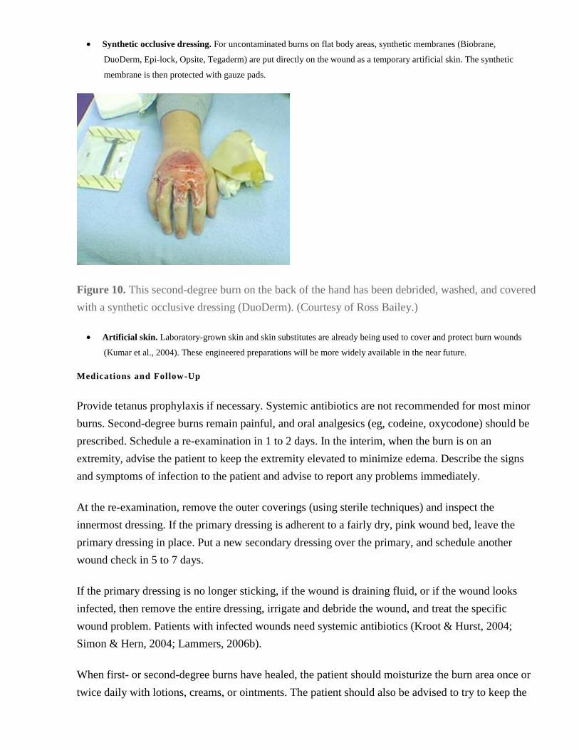
Synthetic occlusive dressing. For uncontaminated burns on flat body areas, synthetic membranes (Biobrane,
DuoDerm, Epi-lock, Opsite, Tegaderm) are put directly on the wound as a temporary artificial skin. The synthetic
membrane is then protected with gauze pads.
Figure 10. This second-degree burn on the back of the hand has been debrided, washed, and covered
with a synthetic occlusive dressing (DuoDerm). (Courtesy of Ross Bailey.)
Artificial skin. Laboratory-grown skin and skin substitutes are already being used to cover and protect burn wounds
(Kumar et al., 2004). These engineered preparations will be more widely available in the near future.
Medications and Follow-Up
Provide tetanus prophylaxis if necessary. Systemic antibiotics are not recommended for most minor
burns. Second-degree burns remain painful, and oral analgesics (eg, codeine, oxycodone) should be
prescribed. Schedule a re-examination in 1 to 2 days. In the interim, when the burn is on an
extremity, advise the patient to keep the extremity elevated to minimize edema. Describe the signs
and symptoms of infection to the patient and advise to report any problems immediately.
At the re-examination, remove the outer coverings (using sterile techniques) and inspect the
innermost dressing. If the primary dressing is adherent to a fairly dry, pink wound bed, leave the
primary dressing in place. Put a new secondary dressing over the primary, and schedule another
wound check in 5 to 7 days.
If the primary dressing is no longer sticking, if the wound is draining fluid, or if the wound looks
infected, then remove the entire dressing, irrigate and debride the wound, and treat the specific
wound problem. Patients with infected wounds need systemic antibiotics (Kroot & Hurst, 2004;
Simon & Hern, 2004; Lammers, 2006b).
When first- or second-degree burns have healed, the patient should moisturize the burn area once or
twice daily with lotions, creams, or ointments. The patient should also be advised to try to keep the

burn area shaded when he is outdoors and to apply sunscreen of >25 SPF before exposing the burn
area to sunlight.
BITES
All animal bites pose a risk of infection (Rupprecht, 2004; Taplitz, 2004). The most critical piece of
information about a bite wound is the type of animal that inflicted it. This will guide your choice of
prophylactic medicines.
Bites by adult humans are some of the most serious. Human bites can transmit HIV and hepatitis
along with a variety of necrotizing bacteria. In addition, human bites are both crushing and tearing
and are often made to the hand, where they injure tendons and joints.
Anesthetizing and Examining
When examining a bite wound, look for underlying fractures and damage to tendons, ligaments, or
joints. Also search for tooth fragments.
Patients do not always get immediate medical care for bites. Those who present after a delay often
do so because they already have signs and symptoms of infection. If an old bite wound shows
evidence of a necrotizing infection—signs such as progressing redness, blistering, or blackened
dying tissue—the patient should be hospitalized for extensive debridement and IV antibiotics.
Removing Debris and Debriding
Dog bites are typically a mix of lacerations and crushes, producing significant destruction to the
tissues. These bites can require sharp debridement, ideally within a few hours of the injury.
Cleansing
The best way to prevent infection in bite wounds is aggressive cleansing. Cleanse, debride, and
irrigate the wound thoroughly.
Closing and Covering
Whether to suture a bite wound is frequently a judgment call. Other than human and monkey bites,
most bite wounds are minor, and of these, only about 10% will need suturing.
Of the serious bites, those with a high risk of infection should be left open, packed, and watched.
Human, monkey, and cat bites are always cleansed and left open. Dog bites to the hands, dog bite
puncture wounds, and dog bites that crush tissues are usually left open. All bite wounds older than 6
to 12 hours are left open. In addition, bites to the hand, ear, or other areas in which tendon, joints, or
cartilage are likely to have been injured are left open.

Other large bites should be cleansed vigorously and sutured to prevent new microbes from invading
the deeper tissues.
Medications and Follow-Up
For a bite wound, always consider tetanus prophylaxis. For mammalian bites, also consider rabies
prophylaxis.
For a human bite, victims should be considered for prophylactic treatments for HIV and for viral
hepatitis if there is a chance the attacker was a carrier. Human bites do not transmit HIV through
saliva alone—blood inoculation is necessary. Details of recent CDC recommendations for HIV
prophylaxis may be found at http://www.cdc.gov/mmwr/preview/mmwrhtml/rr5402a1.htm.
Hepatitis B has been transmitted by a human bite, possibly through infected saliva. For details of
recent CDC recommendations for hepatitis B prophylaxis, go to
http://www.cdc.gov/mmwr/preview/mmwrhtml/rr5516a3.htm.
Consult an infectious disease specialist to decide on the proper prophylaxis for your specific case.
As to prophylaxis for other microbes, systemic antibiotics are recommended for bite wounds that are
not medically treated until 6 or more hours after the injury, for bites to the face, and for bites that
have injured deep tissues such as tendons, joints, and bones. Antimicrobial prophylaxis is also
recommended for all human bites. Bites by cats are deep puncture wounds and carry an especially
high risk of infection; therefore, antimicrobial prophylaxis is recommended for most cat bites. Dog
bites to the hands, dog bite puncture wounds, and dog bites that have crushed tissue are also given
prophylactic antibiotics.
A culture of the wound (unless it is already infected) will not identify an incipient wound infection.
The best way to choose the appropriate antibiotic is by the type of animal that made the bite. Many
bites will include various species of staphylococcus, streptococcus, and corynebacterium. Dog bites
are also associated with Capnocytophaga canimorsus, cat bites with Bartonella henselae and
Pasteurella multocida, and rat bites with Streptobacillus moniliformis and Spirillum minus. A recent
discussion of systemic antibiotic prophylaxis for various animal bites may be found in Taplitz
(2004). If possible, consult an infectious disease specialist when choosing the antibiotics for your
specific case.
Patients who present with an infected bite wound, especially when there is a necrotizing infection,
need to be hospitalized for treatment, which should include parenteral antibiotics. Likewise, bite
wounds that include tendon, joint, cartilage, or bone injuries should be treated in a hospital.

If a bite wound victim is treated and sent home, the patient should be told to keep the injured part
elevated and immobilized, and to monitor the wound for signs of infection. The wound should be
professionally examined daily for the first 3 days and then every second day for two more visits.
PUNCTURE WOUNDS
Puncture wounds, especially of the foot, are commonly seen in emergency departments. Puncture
wounds are difficult to cleanse and are prone to infection (Auerbach, 2001).
Anesthetizing and Examining
Begin by infiltrating the tissue with 1% lidocaine.
Removing Debris and Debridement
It is usually not possible to cleanse a deep puncture wound fully. Use a forceps to probe gently and
then remove debris. Puncture wounds in the hands, wrists, feet, or ankles can be near critical
tendons, nerves, or muscles; to avoid damaging these structures, do not probe the wounds
extensively. In other parts of the body, you can sometimes carefully open the top of ("unroof") the
wound to cleanse it more thoroughly. If the history suggests there may be internal debris, such as
shattered glass or bits of metal, order radiologic images to identify foreign objects.
Coring out (excising) or other forms of major debridement are only used when a puncture wound has
a great deal of contamination or an existing infection.
Cleansing
Irrigate the wound under low pressure. Too much direct pressure will force contamination deeper
into the tissues.
Closing and Covering
If the wound is wide enough (eg, if it has been unroofed) pack it open with moist sterile gauze.
Otherwise, simply put on a protective dressing. In all cases, plan to have the wound professionally
re-examined in 2 to 3 days.
Medications and Follow-Up
Be sure the patient is up-to-date with tetanus immunizations. (Tetanus prophylaxis is discussed in
more detail in the Basic Wound Care section above.)
Even medically treated puncture wounds have a high rate of bacterial infection, frequently by
pseudomonas species. For this reason, anti-pseudomonas cephalosporins, such as ceftazidime
(intramuscular) or ciprofloxacin (oral), are often prescribed prophylactically.

It is hard to predict how well a puncture wound will heal, so schedule a professional examination of
the wound in 2 to 3 days. Give the patient a list of the signs and symptoms of infection and instruct
to report to a medical professional if any problems develop.
The patient should keep wounded extremities elevated and should not put pressure on the wound
(e.g., walking on an injured foot) until after the first follow-up examination.
If you find tenderness or infection in a puncture wound during a follow-up visit, consider cellulitis,
septic arthritis, abscess, and osteomyelitis. Imaging, including local bone scans, may be needed for a
proper diagnosis of an infected puncture wound, and consulting a specialist is the best course of
action.
GUNSHOTS
High-velocity military weapons and shotguns produce severe blast injuries and extensive tissue
damage. Such wounds require surgical debridement, irrigation, and sometimes fracture treatment.
These patients need to be hospitalized and given at least 24 to 48 hours of IV antibiotics. On the
other hand, low-velocity minor gunshot wounds can often be treated as outpatient cases, even when
the wound includes minor fractures and retained bullet fragments (Hollerman & Fackler, 2004). In
general, minor gunshot wounds are treated as puncture wounds.
Anesthetizing and Examining
Infiltrate the tissue with 1% lidocaine, and image the wound for bullet fragments and for evidence of
internal structural damage.
Removing Debris and Debridement
Debride the surface tissue of powder burns. Unless radiographic images show bullet fragments that
are easily accessible, do not explore the wound deeply for debris. Be especially careful exploring
wounds in the hands, wrists, feet, or ankles. Excising the wound is rarely a good plan for gunshot
injuries.
Cleansing
When a gunshot wound creates a complete tunnel through the body, irrigate the wound with high
pressure. If the wound does not have an exit, irrigate the wound under low pressure; too much direct
pressure can force contamination deeper into the tissues.
Closing and Covering
If the wound includes bone fractures, consult an orthopedic surgeon. Otherwise, pack the wound,
and leave it open under a protective dressing.

Medications and Follow-Up
Unlike other types of puncture wounds, minor gunshot wounds have a low infection rate. In general,
antibiotic prophylaxis is not needed.
The patient should keep a wounded extremity elevated and should not put pressure on the wound
(eg, walking on an injured foot) until after the first follow-up examination. Give the patient a list of
the signs and symptoms of infection and instruct to report to a medical professional if any problems
develop. Schedule a professional examination of the wound in 1 to 2 days.
ORAL WOUNDS
Small wounds of the tongue and the mucosa of the mouth heal quickly. Larger wounds may require
the attention of an oral surgeon.
Anesthetizing and Examining
Be sure to examine the teeth near the wound because they may be loose or fractured.
Closing and Covering
Small lacerations will probably not need closing. Large wounds and wounds involving underlying
structures need to be repaired. After thorough irrigation with saline, re-approximate muscles with
absorbable sutures (eg, 5-0 Vicryl). Then, with a minimum number of absorbable sutures, close the
overlying mucosa. For wounds with extensive damage, consult an oral surgeon.
Medications and Follow-Up
Wounds limited to the inside of the mouth are slightly more likely to become infected than similar
external wounds. When the wound penetrates through to the outside skin, however, the infection rate
doubles. Prophylactic antibiotics, such as penicillin, are recommended for large or complex wounds
inside the mouth and for penetrating (through-and-through) wounds.
Have the patient rinse the wound with warm salt water 2 to 3 times daily.
PART 3: Secondary Wound Care
After their primary treatment, most patients with external wounds can be sent home. At that point
secondary wound care begins, and its success depends on the cooperation of the patient.
Instructions to Patient
Be sure to take time and explain what the patient can expect over the next few days and what things
the patient should do.
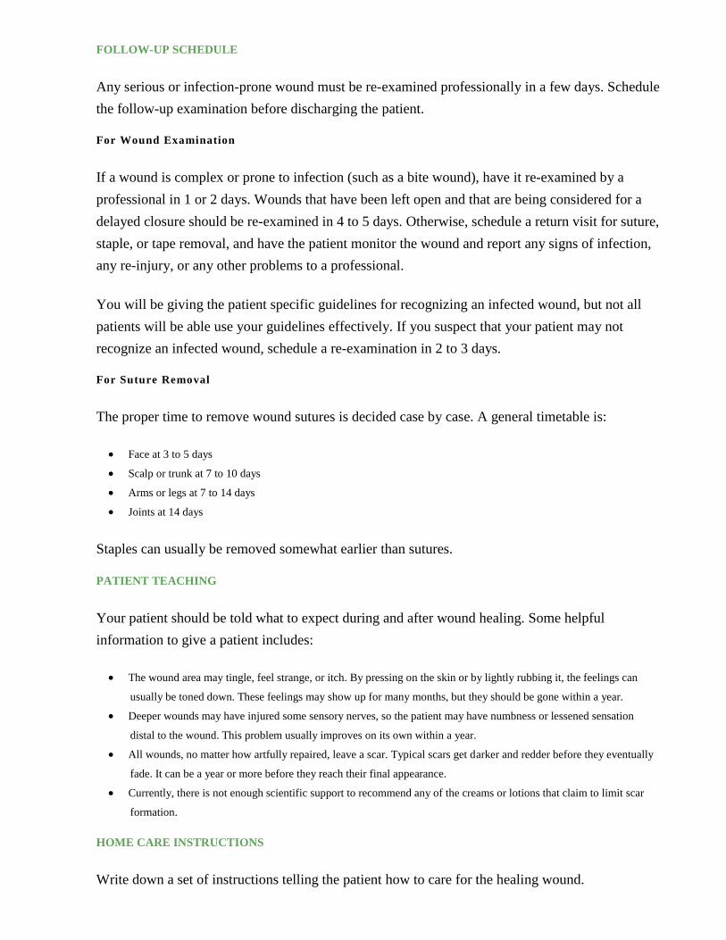
FOLLOW-UP SCHEDULE
Any serious or infection-prone wound must be re-examined professionally in a few days. Schedule
the follow-up examination before discharging the patient.
For Wound Examination
If a wound is complex or prone to infection (such as a bite wound), have it re-examined by a
professional in 1 or 2 days. Wounds that have been left open and that are being considered for a
delayed closure should be re-examined in 4 to 5 days. Otherwise, schedule a return visit for suture,
staple, or tape removal, and have the patient monitor the wound and report any signs of infection,
any re-injury, or any other problems to a professional.
You will be giving the patient specific guidelines for recognizing an infected wound, but not all
patients will be able use your guidelines effectively. If you suspect that your patient may not
recognize an infected wound, schedule a re-examination in 2 to 3 days.
For Suture Removal
The proper time to remove wound sutures is decided case by case. A general timetable is:
Face at 3 to 5 days
Scalp or trunk at 7 to 10 days
Arms or legs at 7 to 14 days
Joints at 14 days
Staples can usually be removed somewhat earlier than sutures.
PATIENT TEACHING
Your patient should be told what to expect during and after wound healing. Some helpful
information to give a patient includes:
The wound area may tingle, feel strange, or itch. By pressing on the skin or by lightly rubbing it, the feelings can
usually be toned down. These feelings may show up for many months, but they should be gone within a year.
Deeper wounds may have injured some sensory nerves, so the patient may have numbness or lessened sensation
distal to the wound. This problem usually improves on its own within a year.
All wounds, no matter how artfully repaired, leave a scar. Typical scars get darker and redder before they eventually
fade. It can be a year or more before they reach their final appearance.
Currently, there is not enough scientific support to recommend any of the creams or lotions that claim to limit scar
formation.
HOME CARE INSTRUCTIONS
Write down a set of instructions telling the patient how to care for the healing wound.

Check for Signs of Infection
Include a written list of the basic signs and symptoms of infection. Tell patients that if they are
worried about something seen or felt in the wound or if they develop any of the signs and symptoms
of an infection, they should report it to a healthcare professional. Be sure to tell your patient whom
to contact.
SIGNS AND SYMPTOMS OF AN INFECTED WOUND
Pus or yellow, greenish, or thick whitish fluid in the wound
Increased redness in the wound
Redness radiating out into the skin around the wound
Red lines progressing up an extremity
Increasing pain or tenderness (note: uninfected sutures do not cause pain)
Swelling
Wound getting warmer than normal skin
Fever
Source: Cutting & White, 2004.
Elevate the Wound
Elevating the injured area will minimize swelling, reduce any throbbing pain, and speed up healing.
If the wound is on an arm or a leg, tell the patient to try to keep the injured area elevated during the
first 2 days. For injuries to the hand or forearm, the patient should consider wearing a sling.
Protect the Wound
Tell the patient:
When it is healing normally, most wounds that have been directly closed will become impermeable to bacteria and
water within two days.
The edges of a directly closed healing wound are held together only weakly for the first 5 days; therefore, be
especially careful with the wound for the first week. The new scar will then strengthen rapidly over the next month.
A wound that has been splinted should remain immobilized until the sutures or staples have been removed.
There is no general reason that most healing wounds need to be kept dry. After day 2, patients with sutured or
stapled wounds can bathe and shower. Swimming in uncontaminated water is usually safe after the wound begins to
strengthen (i.e., after about 5 days).
Cleanse the Wound
You will probably send all wounds home with a protective dressing. Minor wounds and many
sutured or stapled wounds will not need these coverings after a day. The coverings can then be
discarded and the wound left uncovered.

In most cases, you can tell your patient that, when treating the wound, cleanliness is needed but
sterility is not.
A sutured or stapled wound without a dressing can be cleansed gently twice a day with soap and
water, beginning 1 day after the suturing or stapling. A major goal of these washings is to remove
the crusting that develops from the wound exudate. Such a wound can remain uncovered from day 2
on.
When the wound is inside the mouth, have the patient rinse the injured area at least three times daily
with warm salt water.
For wounds that need continued covering, for the first 2 days, the patient should keep the wound
dressing clean and dry, and change the dressing only if it gets dirty or becomes saturated with
exudate. Beginning on day 2, the wound can be cleansed and the dressing changed daily. After a
week, most wounds can be left uncovered.
To change a dressing:
Gently remove the dressings.
Wash the wound with soap and tap water (Anglen, 2005)
Pat the wound dry.
Apply a moistening ointment, such as a triple antibiotic ointment.
Put on a fresh dressing.
Medicines to Avoid
In the early healing stages, patients should not take aspirin, because it decreases the strength of the
scar and it increases the risk of developing a hematoma. Patients should also not drink alcohol
during the hours after the initial wound treatment.
Professional Care
For serious, infected, and nonhealing wounds, a professional should handle the secondary care.
DRESSING CHANGES AND WOUND CLEANSING
As a general rule, wait 48 hours before the first dressing change, unless the dressing becomes
saturated with wound exudate. After the first dressing change, change the dressing only when it
becomes dirty or saturated with wound drainage or other fluid. When a dressing becomes filled with
fluid, microbes can diffuse through it and into the underlying wound. Therefore, any wound
coverings that become saturated with drainage need to be changed. Dressings over heavily draining

wounds may have to be changed frequently. Sometimes, you can reduce the number of dressing
changes by aspirating the fluid (eg, with a sterile needle and syringe) from the dressing.
A dressing change includes gently removing the dressings, washing the wound with soap and tap
water, patting the wound dry, applying a moistening ointment, and putting on a fresh dressing. If the
dressing sticks to the wound during removal, replace it with a less adherent type of primary dressing.
EXCESS GRANULATION TISSUE
Granulation tissue is the loose collection of fibroblasts, inflammatory cells, and new blood vessels
that forms in the base of open wounds during the regrowth (proliferative) phase of healing. Epithelial
cells use granulation tissue as a surface to move along as they re-cover the wound.
When wounds are left open to heal by indirect closure, the regrowth phase is prolonged. Sometimes,
the granulation tissue has sufficient time to overgrow the top of the wound and become a barrier to
the growing epithelial cells. Excess granulation tissue (also called exuberant granulation tissue)
forms most often in scalp, temple, and lower leg wounds.
For a wound to heal properly, excess granulation tissue should be removed. To clear the wound,
scrape out the granulation tissue down to the base of the wound. Then, irrigate the wound. Regrowth
of the granulation tissue can be inhibited by treating the wound base with silver nitrate or by
covering the wound base with Scarlet Red–impregnated gauze (Scarlet Red Ointment Dressing).
Finally, protect the wound with sterile gauze (Habif, 2004).
INFLAMED WOUNDS
Infected wounds become red, warm, tender, and swollen; they may ooze pus and have a disagreeable
smell (Cutting & White, 2004). If a closed wound becomes infected, remove the sutures, staples,
tape, or glue. Re-examine the wound for debris, then debride, irrigate, and pack the wound with
moist sterile gauze. Apply warm compresses over the packing, and give the appropriate antibiotics.
Re-cleanse and repack the wound at least once daily, and plan to let the wound heal by secondary
intention, without closure. If patients develops sepsis, they must be hospitalized.
Some wounds that have been sutured closed over extensive subcutaneous tissue dissection and
debridement can develop a temporary inflammatory reaction in which they become red and
edematous, although they are not infected. If you suspect this problem, remove one or two stitches to
lessen the tension, and apply warm compresses. Then, cleanse the wound daily with soap and water.
This type of inflammatory reaction will decrease within 48 hours.
CHRONIC NONHEALING WOUNDS

Chronic wounds are those that fail to heal when expected. Large wounds heal slowly, but you would
expect even large wounds to be healing within 3 to 4 months.
Figure 11. A chronic nonhealing wound—an inflamed ulcer on the foot of a diabetic patient.
(Courtesy of Frank DiMauro.)
Treat the Whole Person
Wounds do not heal well if they are infected, edematous, or ischemic. In chronic nonhealing
wounds, these problems are often secondary to broader health disorders, such as diabetes, venous
blockage, arterial insufficiency, malnutrition, cigarette smoking, or excess alcohol consumption.
Therefore, when dealing with a chronic wound, begin by assessing the entire person, and try to
improve overall health. Meanwhile, find and work on the specific direct impediments to the healing
of the wound (Shai & Maibach, 2005; Kravitz et al., 2007).
Debride, Irrigate, Pack, and Leave Open
All open wounds contain some bacteria, but ulcers and other chronic wounds frequently have an
excess bacterial burden, even when the surrounding tissue has not developed overt infection
(cellulitis). Moreover, given sufficient time, chronic wound bacteria become coated with
polysaccharides that form a biofilm that keeps antibiotics at bay. Therefore, many chronic wounds
need a new debridement followed by thorough irrigation (Falanga, 2006; Shai & Maibach, 2005).
Debridement appears to help any chronic wound that has not produced much granulation tissue
(Sherman, 2002; Williams et al., 2005), and the classic treatment for most chronic wounds is
debride, irrigate, pack with moist gauze, and leave open for natural (indirect) closure.
Pressure Ulcers
Pressure ulcers (formerly, decubitus ulcers) are the most common type of chronic wound (Shai &
Maibach, 2005). In a pressure ulcer, the injury has been caused by local ischemia from the

continuous compression of capillary beds. Often, this compression happens when a patient lies
unmoving on a hard mattress.
A pressure ulcer is usually worse than it appears from the surface. Skin is more resistant to ischemia
than is underlying tissue, and in a pressure ulcer a small skin wound can be the top of a larger injury
in the subcutaneous tissue and muscle below. As the deeper tissues break down hidden pockets form,
and these spaces will shelter bacteria from superficial cleansings (Diegelmann, 2003).
Pressure ulcers usually need debridement, irrigation, and packing with moist gauze. As with other
ischemic wounds, pressure ulcers can benefit from treatment with hyperbaric oxygen (Mathieu et al.,
2006; Niinikoski, 2006).
Negative-Pressure Dressings
Some chronic wounds became stalled in the reaction (inflammatory) phase of healing (Shai &
Maibach, 2005). Typically, these wounds exude fluid that is low in constructive molecules, such as
growth factors, and high in destructive molecules, such as proteases (Diegelmann, 2003). The
destructive factors constantly impede the construction of the extracellular matrix that is the
foundation of healthy granulation tissue. To get healing back on track, the excess fluid produced by
these wounds must be wicked away.
Negative-pressure dressings (also called vacuum-assisted closure devices or sub-atmospheric
pressure dressings) are labor-saving devices for removing excess fluid from wounds (Wolvos, 2004;
Shirakawa & Isseroff, 2005; Segre et al., 2006). A negative-pressure dressing is made of an
absorbent pad fitted into the wound and covered by a plastic film. A suction tube is buried in the
pad, and when vacuum is applied to the tube it continuously sucks fluid from the wound. The
vacuum also pulls the plastic film tightly over the top of the wound, sealing the wound from the
environment. In addition to removing excess wound fluid, a negative-pressure dressing dilates
capillary beds around the wound and increases local circulation. For complex wounds, it may be
necessary to apply a negative-pressure dressing continuously for as long as 3 weeks (Herscovici et
al., 2003).
Negative-pressure dressings remove fluid while maintaining moisture in the wound. This is
important, because drying a wound will slow its healing.
Culture and Biopsy
Wounds that refuse to heal after special care may have unusual infections such as fungi, or have
developed destructive processes such as carcinomas. When a wound resists healing, consider having
it cultured and biopsied (Shai & Maibach, 2005).
Future Aids

Skin grafts, even when they are not permanent, can help some recalcitrant wounds to heal. In the
near future, artificial skin, laboratory-grown skin, and skin substitutes will be more commonly
available to cover and protect wounds that are not able to epithelialize themselves (Clark et al.,
2007). In addition, the application of constructive biochemical molecules, such as growth factors,
has proved useful in inducing healing in chronic wounds (Cianfarani et al., 2006; Faler et al., 2006;
Inokuma et al., 2006; Deshane et al., 2007). Solutions of these molecules will be more widely
available in the future.
Suture and Staple Removal
The edges of a closed wound must be held together long enough for a strong natural seal to form. If
sutures or staples are removed too early, the wound will reopen (dehisce) and the final scar will be
unnecessarily wide. When planning to remove staples or sutures, first remove one or two and check
the strength of the developing seal between the skin edges. If the seal seems too weak to stay closed
without help, replace the removed sutures or staples with tape strips.
When preparing to remove sutures or staples:
1. Wash the wound and gently remove crust around the sutures or staples.
2. Wipe the skin with an alcohol swab.
To remove sutures:
3. Cut the stitches one by one with the tip of a scissors (or a No. 11 scalpel blade) close to the skin on one side of the wound.
Then grasp the suture near the skin on the opposite side of the wound and pull the suture out through the wound.
4. After removing sutures from the face or from wounds that may be weak, replace the sutures with tape strips (eg,
SteriStrips) to ease the tension on the wound.
If sutures have been left in place too long (usually >10 days), they will leave holes (epithelial tracts)
that can become infected and that may heal with an unsightly pattern of scars. If small abscesses
develop in the epithelial tracts, apply warm moist compresses after the sutures have been removed.
The abscesses should clear quickly and antibiotics are rarely needed.
When the suturing has been complicated—such as when nonabsorbable subcuticular sutures have
been used or when there are special cosmetic requirements—an experienced professional should
decide on the removal schedule and should then carry out the removal.
Staples can usually be removed somewhat sooner than sutures. When staples are removed from any
part of the body, they are often replaced with tape strips to continue protecting the healing seam.
Minimizing Scars

Scars are the natural patches produced in a healing wound. In the skin, scars are made of
unspecialized fibrous tissue covered by a layer of epidermis (Habif, 2004).
In the first few days after an injury, closed skin wounds are being knit weakly together by the
forming scar tissue. By about day 5, the basic architecture of the wound patch has been established,
and from then on, the healing process consists largely of strengthening and remodeling the scar.
Scars can take 6 to 9 months to mature. New scars tend to be red and thick for a month or two before
gradually become less vascular (i.e., paler), less bulky, and flat. It can take as long as 5 years for a
scar to reach its final color.
The width of the scar can be minimized by thorough debridement, by careful suturing (avoiding
inversion of the skin edges), by removing excess granulation tissue, by good secondary wound care
(especially by keeping the wound from becoming infected), and by removing sutures promptly.
Some maturing scars manufacture too much collagen. These scars are either keloids or hypertrophic
scars. Keloids are benign tumors that grow beyond the bounds of the wound and do not regress. The
tendency to form keloids is genetic, and there are, at present, no preventive measures. (See earlier
photograph of a keloid scar.)
In contrast, hypertrophic scars, which are thick but do not grow outside the edges of the wound,
usually get smaller spontaneously. Hypertrophic scars are produced in wounds that are under
tension, such as those parallel to underlying muscles. By reducing the tension on a wound,
hypertrophic scars can often be prevented or minimized.
Hypertrophic scars are also produced in wounds that have a long reaction (inflammatory) healing
phase, where re-epithelialization has been delayed, such as is the case in many burn wounds. For
burn patients, continuous pressure (constant pressure lasting 6 to 12 months) can help to reshape and
flatten hypertrophic scars.
PART 4: Telephone Counseling
Nurses and other health professionals who advise patients over the telephone should know
straightforward answers to basic questions. Here are a few important questions and answers about
taking care of external wounds.
Advice and Triage Questions
When do I need to get medical treatment for a wound?
Shallow cuts and scrapes that stop bleeding on their own can be treated at home.
Wounds that should be checked by a doctor are:
Still bleeding after 5 minutes of direct pressure.
From bites by people or animals.
Old and have not healed.
Still quite painful after a day.
Dirty with debris that you cannot remove.
More than 1/3 inch wide.
On the face.
Deep.
Gaping open.
Cuts over joints.
Scrapes all the way through the skin.
Accompanied by the inability to move muscles or joints.
Caused by a crush from a heavy object.
Swelling.
In a person who has not had a tetanus vaccination in more than 5 years.
My little boy fell off his bicycle. He doesn't have any cuts, but he has scrapes on his leg
and a big bruise that is swollen. How should I take care of it?
Wash the scrape well with soap and water. Ice packs will reduce the swelling. Even when there
are no cuts, a bruise tells you that there is some injury inside the leg. A big bruise can indicate
damage to a bone or a joint, especially when the area is swelling. Therefore, take your child to a
doctor, clinic, or hospital as soon as possible.
What should I do if I am bitten?
You should see a doctor immediately. In addition, notify the police so that the animal can be
watched for signs of disease. Bites by people can be extremely serious, so don't delay seeing a
doctor.
I have a cut, but it doesn't seem too deep. How should I take care of it?
First, stop the bleeding. Press directly on the cut with your hand for 5 or 6 minutes. Try to keep
the wounded part elevated; rest it on a table or a chair and don't let it hang down. If after 6 minutes
the wound is still bleeding freely, see a doctor.
When the bleeding has stopped, wash the wound thoroughly with mild soap and a strong stream of
tap water. Let the water flood the wound as rapidly as possible for at least 3 minutes. Pat the area
dry. Look to see if there is any dirt or debris inside. If you see anything in the wound, continue

trying to wash it out with tap water. If you cannot get the wound clean, try scrubbing it gently and
then wash it again under strong tap water. If the wound remains dirty, see a doctor.
At this point, if you have a clean wound with only a very small amount of bleeding, coat the wound
with an antibiotic ointment, such as Neosporin or Triple Antibiotic Ointment. Then put a sterile
gauze pad or a large bandage over the ointment.
Keep the injured area elevated as much as possible and do not put pressure on it for the first day. If
the wound begins bleeding again, see a doctor.
Over the next few days, watch for these signs and symptoms of infection:
Fever
Pus in the wound
Increasing redness of the wound or the area around it
Swelling
Persistent or increasing pain
Redness or red streaks spreading out from the wound
If these or any other unusual things happen, see a doctor. Otherwise, you can take the bandage off
after 2 days. The healing wound will not be very strong for about 5 days, so protect it for the rest of
the week.
What should I do for a burn?
A serious burn needs immediate medical care. Electrical and chemical burns always need
medical attention. Burns that injure more than one place on your body and burns on your hands,
face, feet, armpits, or groin need to be seen immediately. Any burns to babies or children or to older
adults need medical attention. Finally, if you have any medical conditions (such as diabetes) or if
you taken any drugs (such as prednisone) that make it harder for your body to heal, then get medical
help for your burn.
If you have a small burn, immediately put the wound under a strong stream of running cold water for
a minute or two; then run cold water on the injury for another 10 minutes at a slower speed. Cold
water will reduce the damage of a burn, and it will keep down the swelling, lessen the pain, and
wash the wound. Do not use ice or ice water, which can cause further damage. After washing the
wound, put cool, wet compresses on it. To ease the pain, apply an over-the-counter sunburn ointment
that contains the anesthetic lidocaine; avoid ointments with benzocaine.

Minor burns, such as sunburn, will make your skin red but will not cause blisters. After a day or two,
the injured skin will peel off and there will be new healthy skin underneath without a noticeable scar.
If blisters develop on a burn wound, you need to see a doctor.
Informational Questions
How do I know if a wound is infected?
See a doctor if you have any of these signs and symptoms of an infection:
Fever
Pus in the wound
Increasing redness of the wound or the area around it
Swelling
Persistent or increasing pain
Redness or red streaks spreading out from the wound
How long should a wound hurt?
This depends on the size and type of wound, but a wound that is healing should hurt less and
less each day.
I have been bitten by an animal. Will I get rabies?
If you are bitten by an unknown or sick animal, see a doctor immediately.
In most places in the United States, rabies is uncommon. In theory, though, any bite by a mammal
(cat, dog, bat, raccoon, skunk, rat, and so on) can transmit rabies to a person. Small pets, such as
mice, gerbils, hamsters, and guinea pigs, almost never have rabies. Pet dogs, cats, and rabbits rarely
have rabies.
Wild animals and strays have a higher chance of having rabies, especially if the animal attacks you
for no reason. It is important to describe to the doctor and to the police what kind of animal bit you
and what was happening when you were bitten. The doctor can then decide whether you need
protective medicines. People who get protective medicines will not develop rabies even when they
have been bitten by an animal with the disease.
What kind of treatment will I get in an emergency room for a wound?
The doctor or nurse will examine your injury. They will numb the pain and then cleanse the
wound of debris and injured tissue. They may take x-rays to search for any debris they could not see
and to check for broken bones. Cleansing solution will be sprayed into the wound to wash it
thoroughly. Then, either the wound will be stitched closed or it will be filled with moist sterile gauze
and covered with a bandage.
You may be given antibiotics to protect against infection, and you may get a shot to protect against
tetanus. Except for very serious injuries, you will probably be sent home, with instructions for taking
care of your wound. You may also be scheduled for a wound check-up in a few days.
Does aloe lotion help a wound to heal?
Rubbing herbal medicines made from the aloe vera plant on wounds is a common home
remedy. Scientific studies show that aloe preparations do not help infected wounds to heal. On the
other hand, keeping a wound moist with creams, ointments, or lotions will speed up healing, and
aloe preparations can be effective moisturizers.
Where can I get accurate information about wound care on the Internet?
Three good websites are:
American Academy of Dermatology—http://www.aad.org.
American College of Emergency Physicians—http://www.acep.org.
Merck Manual: Home Edition, Section on Accidents and Injuries—http://www.merck.com.