partitioning of bacterial communities between travertine ... · partitioning of bacterial...
TRANSCRIPT

Partitioning of bacterial communities betweentravertine depositional facies at Mammoth HotSprings, Yellowstone National Park, U.S.A.1
Bruce W. Fouke, George T. Bonheyo, Beth Sanzenbacher, and Jorge Frias-Lopez
Abstract: A culture-independent molecular survey indicates that the composition of bacterial communities is distinctlypartitioned between travertine depositional facies in the surface drainage system of Spring AT-1 at Angel Terrace,Mammoth Hot Springs, Yellowstone National Park. PCR (polymerase chain reaction) amplification and sequencing of16S rRNA genes with universally conserved bacterial primers has identified over 553 unique partial and 104 completegene sequences (derived from more than 14 000 clones), affiliated with 221 unique species that represent 21 bacterialdivisions. These sequences exhibited < 12% similarity in bacterial community composition between each of the travertinedepositional facies. This implies that relatively little downstream bacterial transport and colonization took place despitethe rapid and continuous flow of spring water from the high-temperature to low-temperature facies. These results suggestthat travertine depositional facies, which are independently determined by the physical and chemical conditions of thehot spring drainage system, effectively predict bacterial community composition as well as the morphology and chemistryof travertine precipitation.
Résumé : Un relevé moléculaire, indépendant des cultures, indique que la composition des communautés bactériennesest répartie de façon distincte entre les faciès de déposition de travertin dans le système de drainage de surface de lasource AT-1 à Angel Terrace, Mammoth Hot Springs, du parc national Yellowstone. Une amplification PCR et leséquençage des gènes 16S rARN avec des amorces bactériennes universellement conservées a permis l’identification deplus de 553 séquences partielles uniques et 104 séquences complètes de gènes (dérivées de plus de 14 000 clones) affiliées à221 espèces uniques qui représentent 21 divisions bactériennes. Dans la composition de la communauté bactérienne, lesséquences montrent moins de 12 % de similitude entre chacun des faciès de déposition du travertin. Cela impliquequ’il s’effectue peu de transport bactérien vers l’aval et qu’une colonisation a lieu malgré l’écoulement rapide et continueldes eaux de source du faciès de haute température vers le faciès de basse température. Ces résultats suggèrent que lesfaciès de déposition du travertin, qui sont définis de façon indépendante par les conditions physiques et chimiques dusystème de drainage des sources thermales, prédisent effectivement la composition de la communauté bactérienne et lamorphologie et la chimie de la précipitation du travertin.
[Traduit par la Rédaction] Fouke et al. 1548
Introduction
A fundamental scientific endeavor unique to the geosciencesover the last 170 years has been the reconstruction of ancientearth surface environmental conditions from the analysis ofancient sedimentary rocks (Blatt et al. 1980; Reading 1996;Boggs 2002). To accomplish this, workers have studied theinteraction of physical, chemical, and biological processes inmodern-day sedimentary environments that deposit analogoussedimentary rocks. This work has resulted in the developmentof depositional facies models that correlate (1) the depth,velocity, temperature, and chemistry of water in the environ-ment; (2) the grain size, sorting, mineralogy, structure, and
geometry of sediments being deposited; and (3) the animaland plant communities living in each community (e.g., Walkerand James 1992).
Sedimentary facies models provide an essentialprocess-oriented conceptual tool to systematically break downlarger complex depositional environments into smallersub-environments, each of which are linked along continu-ous hydrologic environmental gradients. Facies modelsare independent of the relative size and specific location ofthe environment being studied, and thus have nearly univer-sal applicability in settings that range from terrestrial gla-ciers to marine coral reefs (Blatt et al. 1980; Reading 1996;Boggs 2002). Of equal importance, facies models create sys-
Can. J. Earth Sci. 40: 1531–1548 (2003) doi: 10.1139/E03-067 © 2003 NRC Canada
1531
Received 5 February 2003. Accepted 11 June 2003. Published on the NRC Research Press Web site at http://cjes.nrc.ca on25 November 2003.
Paper handled by Associate Editor B. Chatterton.
B.W. Fouke,2 G.T. Bonheyo, B. Sanzenbacher, and J. Frias-Lopez. Department of Geology, University of IllinoisUrbana-Champaign, Urbana, IL 61801, U.S.A.
1This article is one of a selection of papers published in this Special Issue on Sedimentology of hot spring systems.2Corresponding author (e-mail: [email protected]).
J:\cjes\cjes4011\E03-067.vpNovember 18, 2003 2:01:05 PM
Color profile: DisabledComposite Default screen

© 2003 NRC Canada
1532 Can. J. Earth Sci. Vol. 40, 2003
tematic spatial and temporal frameworks within which detailedanalyses of geochemistry and macroorganisms (plants andanimals) can be holistically linked to system-scale environ-mental processes (Wilson 1975; Flügel 1982; Walker andJames 1992). However, with the notable exception ofstromatolites (Walker and James 1992), depositional faciesmodels have lacked detailed evaluation of the pivotal rolethat may be played by microorganisms during sediment de-position and associated water–rock reactions. This is pre-sumably because no previous study of modern sedimentaryfacies models has included culture-independent molecularanalyses (see summary in Pace 1997) of the bacteria inhabit-ing each facies.
The purpose of the present study is to complete the firstcomprehensive survey of bacterial 16S rRNA gene sequencesin the context of the depositional facies that the bacteria inhabit.The study site for this analysis is the surface drainage systemof the terrestrial carbonate hot spring called Spring AT-1 onAngel Terrace in the Mammoth Hot Springs complex of Yellow-stone National Park (Fouke et al. 2000). This location waschosen because the physical, chemical, and biological conditionsof the spring water change drastically as it flows away fromthe vent, and these conditions are associated with the precip-itation of carbonate mineral deposits called travertine (sensustrictu Pentecost and Viles 1994; Ford and Pedley 1996).These environmental changes create a systematic series offive travertine depositional facies along the Spring AT-1 drainagesystem, each of which have previously been defined by theiraqueous chemistry and travertine morphology and chemistry(Fouke et al. 2000). Results are presented in the presentstudy that indicate that the composition of the bacterialcommunity inhabiting each travertine facies is more than87% unique from the next directly adjoining down flow facies.This suggests that the travertine facies model is an accuratepredictive tool for bacterial community composition in additionto the morphology and chemistry of travertine deposition.
Geological setting
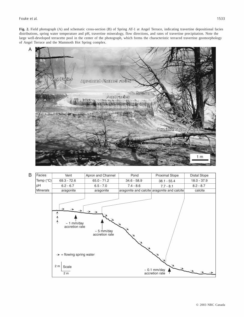
Geothermal groundwater erupts at a temperature of 73 °Cfrom subsurface conduits at Spring AT-1 on Angel Terrace inthe Mammoth Hot Springs complex (Fig. 1), creating a seriesof terraced travertine deposits (Allen and Day 1935; Bargar1978; Fig. 2). As the Spring AT-1 groundwater cools, degasses,and flows along surface drainage channels, travertine composedof aragonite and calcite (CaCO3) is precipitated at rates ashigh as 5 mm/day (Friedman 1970; Pentecost 1990; Fouke etal. 2000). The travertine depositional facies described fromthe Spring AT-1 drainage system in Fouke et al. (2000) isbriefly summarized in the following.
Angel Terrace Spring AT-1 is composed of a series ofshallow-water environments extending from the spring ventto the distal parts of the system. These aqueous environmentsand their associated travertine deposits are divisible into fivedepositional facies: (1) vent facies, (2) apron and channel facies,(3) pond facies, (4) proximal-slope facies, and (5) distal-slopefacies (Fig. 2). The boundaries of the facies are based onsystematic changes in travertine crystal morphology andchemistry and associated changes in water chemistry. Thecomposition and relative sequence of the facies has beenobserved in other springs and is consistently re-established
as the springs shift their position due to changes in springwater flow velocity and the opening of new vents (Fouke etal. 2000; Fouke 2001). Travertine precipitated in each of theSpring AT-1 facies exhibits distinct growth forms and chem-istries, which are accompanied by a general transition inmineralogy from aragonite in the high-temparture waters tocalcite in the low-temperature waters (Fig. 2). The water inthe drainage system ranges from depths of approximately 1to 30 cm and flows over substrates composed of activelyprecipitating travertine and living microbial mats. Watertemperature decreases from 73 to 18 °C. Vent water temper-atures are invariant throughout the year, while the distal-slopewaters reach their minimum temperatures in the winter(Fouke et al. 2000; Fig. 2). The inconsistent drops in watertemperature observed along the drainage system (Fig. 2) arecaused by lateral diversions in flow and the variability inwater depth between each facies. Spring water pH increasesfrom 6.2 at the vent to 8.7 in the distal-slope (Fig. 2). This isaccompanied by large magnitude changes in the chemistry
Fig. 1. Location of Spring AT-1 at Angel Terrace in the Mam-moth Hot Springs complex of Yellowstone National Park. Shad-ing depicts the surface area covered by the Spring AT-1 watersas the flow away from the vent (shown by a �).
J:\cjes\cjes4011\E03-067.vpNovember 18, 2003 2:01:05 PM
Color profile: DisabledComposite Default screen
© 2003 NRC Canada
Fouke et al. 1533
Fig. 2. Field photograph (A) and schematic cross-section (B) of Spring AT-1 at Angel Terrace, indicating travertine depositional faciesdistributions, spring water temperature and pH, travertine mineralogy, flow directions, and rates of travertine precipitation. Note thelarge well-developed terracette pool in the center of the photograph, which forms the characteristic terraced travertine geomorphologyof Angel Terrace and the Mammoth Hot Spring complex.
J:\cjes\cjes4011\E03-067.vpNovember 18, 2003 2:01:05 PM
Color profile: DisabledComposite Default screen

© 2003 NRC Canada
of the spring water (total dissolved inorganic carbon, δ13C,δ18O, 87Sr/86Sr, dissolved sulfate, δ34S) and travertine (δ13C,δ18O, 87Sr/86Sr, δ34S; Fouke et al. 2000).
The vent facies (5–30 cm water depth) contains moundedtravertine composed of aragonite needle botryoids (Figs. 3A–3C,4A, 4B). The vent facies transitions into the apron and channelfacies (≤5 cm water depth), which is floored by hollowtravertine tubes composed of aragonite needle botryoids thatencrust filamentous thermophilic bacteria (Figs. 3A–3D, 4C,4D). The transition into the pond facies is an abrupt contact,with the pooled pond waters reaching depths of 30 cm.Travertine in the pond facies forms large step-like morphologiescalled terracettes that include aragonite needle shrubs at highertemperatures and ridged networks of calcite and aragonite atlower temperatures (Farmer 2000). In addition, calcite “icesheets,” calcified bubbles, and aggregates of aragonite needles(“fuzzy dumbbells”) precipitate at the air–water interfaceand settle to pond floors (Figs. 3D, 3E, 4D, 4E; Fouke et al.2000). An abrupt facies transition is exhibited at the margins(or lips) of the pond pools. The proximal-slope facies(≤3 cm water depth), which is composed of arcuate aragoniteneedle clusters that create small fluted microterracettes onthe steep slope face (Figs. 3F, 3G, 4F, 4G). Finally, a gradualtransition takes place into the distal-slope facies (≤2 cm waterdepth), where travertine forms broad low-relief microterracettesthat are composed entirely of calcite spherules and “feather”calcite crystals (Figs. 3H, 4H).
The aqueous chemistry of the hot spring drainage systemis strongly influenced by CO2 degassing, as indicated byRayleigh-type fractionation calculations of spring water δ13Cversus dissolved inorganic carbon concentrations (Fouke etal. 2000). However, while the physical factors of temperaturedecrease and degassing are significant in helping to drive therapid precipitation of travertine, strong bacterial influenceson travertine crystal form and isotope composition have alsobeen observed throughout the Spring AT-1 drainage system.One important example is aragonite crystals that encrust andthus preserve the shape of filamentous bacteria as hollowtravertine “streamers” in the vent, as well as apron and channelfacies (Farmer and Des Marais 1994; Fouke et al. 2000;Farmer 2000). A chemical expression of biological influenceis the disequilibrium fractionation observed in the δ13C, δ18O,and δ34S composition of the travertine, which can be detectedonly after the fractionation effects of CO2 degassing andtemperature drop have been quantitatively subtracted (Foukeet al. 2000).
Materials and methods
Field sampling of spring water, microbial mats, and freshtravertine precipitated on the floor of the Spring AT-1 drainage
system was completed during daylight hours in January 1999,as well as January and May 2000. Fifty samples were collectedfor analysis over this time period from the five travertinedepositional facies (Fig. 2). Use of the travertine faciesmodel permitted sampling in equivalent spring environmentsin 2000 after the drainage system had shifted laterally fromits original position in 1999 due to temporal changes in theflow velocity and travertine precipitation of hot groundwateremerging from the Spring AT-1 vent.
Sample collectionSpring water was sampled by collecting 2 L from each facies
in acid-cleaned 1 L Nalgene HDPE bottles. The water washand-pumped through a sterile 0.45 µm filter-loaded cup(Pall/Gelman). All filters were then immediately frozen at–20 °C, transported to Illinois on dry ice and stored at –40to –80 °C. Travertine was collected by removing a 2 cm2
portion of the uppermost 0.25–1 cm of the floor of the drainageflow path with a cleaned spatula and placing the sample in asterile disposable 50 mL polypropylene centrifuge tube. Portionsof the microbial mats growing on the travertine substrateswere also physically peeled off and collected using forceps.Microbial mat samples were immersed in 80% ethanol insterile 15 mL polypropylene centrifuge tubes. Travertinesubstrate samples were crushed and homogenized in sterile15 mL polypropylene centrifuge tubes with sterile blades,creating a slurry of ethanol, microbial mats, loose micro-organisms, and travertine crystals.
DNA extractionSeveral methods of DNA extraction were used with each
sample to increase the likelihood that no microorganismwould escape detection. Filters were sectioned using flamesterilized scissors and forceps and placed in sterile disposable50 mL polypropylene centrifuge tubes with 3 mL of sterileultra pure water. Cells were then washed off the filter duringthree minutes of vigorous agitation on a vortex and storedfrozen at –80 °C.
Bead beating (Hugenholtz et al. 1998), freeze–thaw cycling,and chemical lysis protocols (Sambrook et al. 1989) wereused to extract community genomic DNA from the cellscollected on the filters, the crushed travertine slurries, andthe microbial mat samples. For bead beating, 300 µL of samplewas added to a 2 mL screw-capped microcentrifuge tubecontaining 600 µL of sterile ultra pure water and 800 µL of0.1 mm zirconia–silica beads (BioSpec Products, Bartlesville,Okla.). The beads were cleaned and sterilized beforehandwith a series of HCl acid and bleach washes. The tube wasthen filled to capacity with sterile ultra pure water and shakenon a reciprocating Mini-BeadBeater-8 (BioSpec Products,Bartlesville, Okla.) for 2.5 min at the homogenize (highest)
1534 Can. J. Earth Sci. Vol. 40, 2003
Fig. 3. Field photographs of the travertine depositional facies composing the Spring AT-1 drainage system at Angel Terrace. (A) Over-view of the vent facies and apron and channel facies showing trees that have been inundated by travertine deposition. (B) View of thevent within the vent facies, where spring water is 73 °C with water depths of 30 cm. (C) Gradational contact of the vent facies andapron and channel facies, where water depths shallow from 30 cm to 1 cm. (D) Abrupt contact between the apron and channel faciesand the pond facies. (E) Enlargement of the sharp facies contact shown in D. (F) Microterracettes forming at the sharp contact be-tween the pond facies (pooled water lapping against the lip of the terracette at the top of the photograph) and the nearly vertical proxi-mal-slope facies. (G) Small pools and microterracettes composing the proximal-slope facies. (H) Broad and shallow microterracettepools composing the distal-slope facies.
J:\cjes\cjes4011\E03-067.vpNovember 18, 2003 2:01:06 PM
Color profile: DisabledComposite Default screen

© 2003 NRC Canada
Fouke et al. 1535
J:\cjes\cjes4011\E03-067.vpNovember 18, 2003 2:01:07 PM
Color profile: DisabledComposite Default screen

© 2003 NRC Canada
speed setting. This protocol has been optimized using severalsamples, as well as E. coli positive controls. For thefreeze–thaw procedure, samples were added to sterile 2 mLO-ring screw-capped microcentrifuge tubes containing 1 mLof sterile ultra pure water. The tubes were frozen at –80 °Cand rapidly thawed by plunging the tubes into a 65 °C waterbath. The freeze–thaw cycle was repeated three times, withthe tubes vigorously agitated on a vortex for � 1 min aftereach thaw cycle. In some instances, samples were treatedwith an alkaline lysis step using NaOH (0.13 M final (1 M =2 mol/L)), sodium dodecyl sulfate (SDS, 0.3%), and incubationat 25 to 100 °C for 2 min (Sambrook et al. 1989).
For both the bead beating and freeze–thaw techniques, a400 µL aliquot of the lysate was used for additional DNAprecipitation using two volumes absolute ethanol, followedby a series of washing steps using 70% EtOH (Sambrook etal. 1989). The ethanol precipitated lysates, untreated lysates,and untreated samples were used in subsequent PCRs (poly-merase chain reactions). Each protocol routinely included300 µL of the sterile ultra pure water as both a negative controland contamination screen, and 50 µL of an overnight Luriabroth E. coli culture as a positive control. The control prepa-rations were included with their simultaneously preparedenvironmental samples each time a PCR reaction wasperformed. In no case were nucleic acids detected in thenegative controls.
PCR AmplificationTotal environmental chromosomal DNA was used as tem-
plate for PCR amplification of 16S rRNA genes using aMastercycler Gradient thermocycler (Eppendorf, Westbury,N.Y.) and universal 16S rRNA primers for Bacteria. Primersused in the PCRs were obtained from Operon Technologies,Inc. (Alemeda, Calif.). B. Paster (personal communication,1998) provided the sequence of each primer: forward primer:28F (5′-GAGTTTGATYMTGGCTC); reverse primer: 1492R(5′-GYTACCTTGTTACGACTT). Reaction mixtures includeda final concentration of: 1X TaqMaster buffer (Eppendorf),1x TaqM enhancer (Eppendorf), 0.2 mM each dNTP(Gibco/BRL, Rockville, Md.), 200 ng each of forward andreverse primers, 5–30 µL of the sample preparation, and waterto bring the total volume to 100 µL. Reactions were layeredbeneath 50 µL of mineral oil (Sigma, St. Louis, Mo.). Aninitial denaturation – hot start of 5 min at 95 °C was followedby the addition of 0.5 µL (�2 U) of MasterTaq polymerase(Eppendorf) or Taq DNA polymerase (Gibco/BRL, Rockville,Md.). The hot start was followed by 30 cycles of the followingincubation pattern: 94 °C for 1 min, 55 °C for 1 min, and72 °C for 2 min. A final soak at 72 °C for 5 min concluded
the reaction. All PCRs were mixed and run in an enclosedPCR work station to prevent contamination.
Cloning and sequencingPCR products were purified by electrophoresis through a
1.0% low-melting agarose gel (SeaPlaque GTG; BioWhittakerMolecular Applications, Rockland, Maine), stained withethidium bromide and visualized on a UV (ultraviolet)trans-illuminator. The �1500 bp heterologous 16S rRNAgene product was excised from the gel, and the DNA waspurified from the gel slice using the Wizard PCR Prep kit(Promega, Madison, Wis.). The gel-purified PCR productwas cloned into the pGEM-T Easy vector (Promega) andtransformed into calcium chloride competent DH5αMCRE. coli cells (Gibco/BRL) using standard techniques (Sambrooket al. 1989). Clone libraries were created by patchingtransformants in 6 × 8 grids on standard Luria broth agar(100 µg/mL of Ampicilin and 40 µg/mL X-Gal) petri dishes.The clone libraries were then screened to verify the presenceof the appropriate sized insert to identify unique clones forsequencing. Petri dish libraries were inoculated using a48-pin replicator (Midwest Scientific, St. Louis, Mo.) into96-well culture blocks (2 mL well capacity) containingLuria broth (15% glycerol and 100 µg/mL of Ampicilin).Culture blocks were sealed with AirPore tape (Qiagen, Va-lencia, Calif.) and shaken overnight at 37 °C. On the follow-ing day, 3 µL from each well was transferred to a 96-wellflex-plate (GeneMate, Kaysville, Utah) containing 27 µL ofPCR mixture (0.2 mM dNTP (Gibco/BRL, Rockville, Md.),1x PCR Buffer (Gibco/BRL), 0.8U Taq (Gibco/BRL),1.5 mM MgCl2, and 200 ng each of T7(-26) forward and M13(-48) reverse primers). The culture block was then sealedwith plastic tape (Qiagen) and the remaining culture wasfrozen at –80 °C. The flex-plates were sealed with tape(Qiagen) and a 30-cycle PCR amplification was carried outas previously described. Because the forward and reverseprimer sequences are located on the pGEM-T vector flank-ing the cloning site, only insert DNA is amplified. After am-plification, 8 µL from each flex-plate well was transferred toa new well of a 96-well microtiter plate containing 32 µL ofrestriction digest mixture consisting of the 4-base recogni-tion site enzymes MspI and HinP1 I in 1x NEB Buffer 2(New England Biolabs, Beverly, Mass.). The digest productswere then separated by electrophoresis on a 3.0% agarosegel (MetaPhor; BioWhittaker Molecular Applications, Rockland,Maine) stained with ethidium bromide, and the RFLP(restriction fragment length polymorhism) patterns used toidentify unique clones to be submitted for sequence analysis.Three to five samples with seemingly identical RFLP patterns
1536 Can. J. Earth Sci. Vol. 40, 2003
Fig. 4. Close-up field photographs of the travertine depositional facies comprising the Spring AT-1 drainage system at Angel Terrace.(A) The vent within the vent facies (white box shows the sample collection position of the photograph in B). (B) Handsample ofmounded travertine deposits from the vent facies. (C) Floor of the apron and channel facies composed of hollow sinuous aragonitictravertine tubes viewed through < 2 cm of swiftly flowing spring water. (D) Enlargement of the sharp contact between the apron andchannel facies and the pond facies, showing long filamentous strands of Aquificales pBB that project out into the pond and are beingencrusted by aragonite needles to form slender hollow tubes of travertine. (E) Floor of the pond facies composed of spherical clustersof shrub-like aragonite travertine. (F) View looking directly down on the sharp lip of the pond, illustrating the abrupt transition fromthe aragonite shrubs on the floor of the pond facies to the aragonitic microterracettes covering the face of the proximal-slope facies.(G) Aragonite microterracettes forming in the central portion of the proximal-slope facies submerged under < 1 cm of swiftly flowingspring water. (H) A broad and shallow (<1 cm water depth) travertine microterracette composed of calcite in the distal-slope facies.
J:\cjes\cjes4011\E03-067.vpNovember 18, 2003 2:01:07 PM
Color profile: DisabledComposite Default screen

© 2003 NRC Canada
Fouke et al. 1537
J:\cjes\cjes4011\E03-067.vpNovember 18, 2003 2:01:08 PM
Color profile: DisabledComposite Default screen

© 2003 NRC Canada
were selected for sequence analysis in an effort to capturedifferent sequences with similar RFLP patterns. Clones selectedfor sequence analysis were patched onto Luria broth agarpetri dishes supplemented with 200 µg/mL ampicillin (RocheMolecular Biochemicals, Indianapolis, Ind.) and incubatedovernight at 37 °C.
Inoculation, cell culturing, template preparation, andsequencing were performed in the High Throughput Laboratoryof the University of Illinois Urbana-Champaign W. M. KeckCenter for Comparative and Functional Genomics. The petridish cultures were used to inoculate 2 mL 96-well cultureblocks containing Circle Grow media (Bio100) supplementedwith ampicillin (100 µm/mL). Plasmid template DNA waspurified from the cultures using an automated system andthe QIAwell 96 Turbo prep BioRobot Kit (Qiagen, Valencia,Calif.). Sequencing was completed using the T7(-26) primersynthesized in-house (Studier and Dunn 1983). Sequencereactions were performed on the plasmid templates using aQiagen Bio Robot 9600 and Big Dye Terminator chemistry(v.2.0) from ABI. Sequencing was performed on an ABI3700 capillary sequencer, then processed in the BioinformaticsUnit of the W. M. Keck Center.
To generate nearly complete sequences, unique clones wereselected based on the sequences generated from the T7(-26)primer (Studier and Dunn 1983), and the remainder of the16S sequence was determined using either the M13(-24) orM13(-48) primer (Viera and Messing 1982). Two other primerpairs located within the 16S rRNA gene sequence were alsoused. The first pair consisted of Bact343Fwd. 5′-TACGGRAGGCAGCAG and Bact 1115Rev. 5′-AGGGTTGCGCTCGTTRC (Wilmotte et al. 1993) and the second pair consistedof 805aF 5′-ATTAGATACCCYGGTAGTC and 926/20 5′-CCGTCAATTYYTTTRAGTTT (Wilmotte et al. 1993; Hugenholtzet al. 1998). Contiguous sequences were assembled manuallywith the DNA analysis software Sequencher 4.1 (Gene CodesCorp., Ann Arbor, Mich.).
Sequence analysesThe 16S rRNA gene sequences, determined with a 3%
similarity cut-off (Martin 2002), were first compared withthe GenBank database using the basic local alignment searchtool (BLAST) network service (Altschul et al. 1990). Fromthe alignments created by this search, the orientation of eachcloned 16S rRNA gene could be determined and an approximatedivision-level association established. Each sequence was
analyzed using the CHIMERA_CHECK version 2.7 programavailable at the Ribosomal Database Project web site (Maidiket al. 1997). Those sequences deemed to be chimeric wereculled from the data set. A 97% to 100% match of the unknownclone with the GenBank data set was considered an approximateidentification to the species level, 93% to 96% similaritywas accepted as a genus-level identification, and an 86% to92% match was considered a more distant yet related organism(Goebel and Stackebrandt 1994).
Nucleotide sequence accession numbersThe GenBank accession numbers for each 16S rRNA gene
sequence generated in this study are listed in Tables 1 through 5.
Results
Bacterial clone 16S rRNA gene sequence libraries wereconstructed for each of the five travertine depositional faciesat Spring AT-1. The following is a summary of the distributionof these sequences and their species-level and division-levelaffiliations. The number of base pairs of each 16S rRNAgene sequence, the species-level and division-level percentmatch with affiliated microoganisms in GenBank, and theoccurrence or absence of each sequnce in each travertinedepositional facies is presented in Tables 1 through 5.
16S rRNA gene sequence clone librariesMore than 14 000 clones were generated in 65 clone libraries
(one per PCR reaction) from the vent, apron and channel,pond, proximal-slope, and distal-slope travertine depositionalfacies (Fig. 3; Tables 1–5). From this large pool, 1050 cloneswere selected based on their RFLP patterns and submitted tobe sequenced. Ultimately, 657 clones were successfullysequenced, yielding 221 unique gene sequence types (16SrRNA gene species-level affiliations). The remaining 436sequences were duplications of the 221 unique 16S rRNAgene sequence types. Only 6% of the bacterial gene sequencesdetected in the Spring AT-1 drainage system could not beassigned to a particular division (i.e., these particular 16SrRNA gene sequences differ significantly in their gene sequencefrom genes previously detected and recorded in the GenBankdatabase; Tables 1–5). This similarity has permitted inferrenceof bacterial affiliation and associated ecology from the SpringAT-1 sequences.
The molecular identification technique applied in this study
1538 Can. J. Earth Sci. Vol. 40, 2003
ID% Bp Access. Best match organism Division facies V AC P PS DS
96–98 1484 AF445739 Uncultured Aquificales pBB Aquificales 45 2 17 34 197–99 1483 AF445734 Unidentified Aquificales OPB13 Aquificales 17 2 — 1 —99 1469 AF445735 Unidentified Green Non-sulfur OPB65 Green Non-Sulfur Bacteria 2 1 — — —97 533 AF446260 Dictyologlomas thermophilum Firmicutes (Low G + C) 1 — — — —97 662 AF446242 Cyanobacterium sp. OS type B Cyanobacteria 1 — — — —87 700 AF446244 Flectobacillus major BCF 1 — — — —98 1500 AF445689 Unidentified bacterium OPB30 β-Proteobacteria 1 3 45 51 7
Total no. of clones in facies 68 8 62 86 8
Note: Table includes the percent similarity to previously sequenced organisms (ID%), the number of base pairs sequenced (Bp), the sequence accessionnumber (Access.), and the best match organism in Genbank and its division. The number of 16S rRNA genes detected in each facies is indicated (V, ventfacies; AC, apron and channel facies; P, pond facies; PS, proximal-slope facies; DS, distal-slope facies).
Table 1. 16S rRNA gene sequencing results for bacteria inhabiting the Spring AT-1 vent facies.
J:\cjes\cjes4011\E03-067.vpNovember 18, 2003 2:01:09 PM
Color profile: DisabledComposite Default screen

© 2003 NRC Canada
has several potential biases (Sambrook et al. 1989; Hurst etal. 2002). These vary from the possibility of generatingmultiple clones of a 16S rRNA gene from any one bacterialcell in the environmental sample to failing to detect 16SrRNA gene sequences from bacteria whose cell walls aredifficult to lyse for DNA extraction. In addition to this, thenumber of samples analyzed and PCR analyses completed inthis study have been concentrated on the pond facies (Fig. 5)to eventually determine the bacterial communities associatedwith formation of the pond lip, which is the hallmark structuralcomponent of hot spring travertine terracette morphology.Furthermore, environmental 16S rRNA gene surveying onthe scale at which it was applied in this study is time consumingand expensive. As a result of these multiple interrelated factors,the five travertine facies have not been fully molecularlysaturated (i.e., the “true” total bacterial phylogenetic diversityhas not been analytically determined), as can be shown bystandard statistical analyses (data not shown; Hughes et al.2001; Martin 2002). However, this is a problem that facesall molecular studies of bacterial diversity in the environment,and no study proposes to have determined the entire spectrumof bacterial diversity in any given natural environmental sample(Hurst et al. 2002). Therefore, the approach adopted in thepresent study, as in many previous studies of hot springmicrobiology (e.g., Hugenholtz et al. 1996; Blank et al. 2002),has been to conduct a minimum number of PCRs to establisha reliable first-order baseline estimate of the bacterialcommunities inhabiting the Spring AT-1 drainage system.However, the present study has the important advantage thatthe analyses were conducted within the environmental frame-work of an independently established travertine facies model.Therefore, the resulting information on bacterial communitycomposition has a direct physical and chemical environmentalcontext that has not previously been known for hot springmicrobial communities.
A pie chart graphical format has been chosen to depict the
sequencing data in this study because phylogenetic trees arenot effective in illustrating the environmental context providedby the travertine facies model (Hillis et al. 1996). Therefore,the division-level phylogenetic diversity of bacteria affiliatedwith the 16S rRNA gene sequences in each facies is presentedin two types of pie diagrams (Fig. 4). The first type dividesthe number of 16S rRNA genes cloned from each bacterialdivision by the total number of sequences in each faciesclone library (Figs. 4A, 4C, 4E, 4G, 4I). The second typedivides the total number of division-level affiliations observedin the clone library by the total number of affiliated speciesidentified in each facies (Figs. 4B, 4D, 4F, 4H, 4J). Use ofthese graphs permits a comparative evaluation of bacterialcommunity composition from the total proportion of 16SrRNA gene sequences (the raw data) and the proportions ofdivision-level identifications (the interpreted data). Bothapproaches are useful because neither the proportions of 16SrRNA gene sequence or species-level sequence affiliationsare necessarily accurate estimates of bacterial communitystructure due to potential biases during PCR amplificationand other laboratory manipulations (Hurst et al. 2002).Determination of the bacterial community structure andfunctional metabolic diversity in each facies will be completedin future studies by applying optical and molecular techniquesthat build directly upon the 16S rRNA gene sequence clonelibraries constructed in this study (Tables 1–5).
Microbial communities inhabiting the vent faciesThe vent facies clone libraries were dominated by bacterial
sequences affiliated with Aquificales (91% sequences, 29%16S rRNA gene types; Table 1; Figs. 6A, 6B). The observeddiscrepancy between total number of gene sequences and thetotal number of unique affiliated 16S rRNA gene types(Figs. 6A, 6B) presumably represents the preferential ampli-fication of Aquificales 16S rRNA gene sequences duringPCR. More than 40% of the total 16S rRNA gene types detected
Fouke et al. 1539
ID% Bp Access. Best match organism Division facies V AC P PS DS
98 1446 AF445697 Uncultured eubacterium env.OPS 1 Aquificales — 3 4 — —94 424 AF446247 Planctomyces sp. (strain 599) Planctomycetales — 2 — — —87 654 AF446248 Uncultured bacterium mle 1-41 Planctomycetales — 1 — — —87–88 1439 AF445645 Uncultured bacterium #0319-7F4 Planctomycetales — 1 — 1 —90–94 559 AF446256 Candidate division OP11 clone OPB92 OP11 — 1 14 1 —91 610 AF446272 Uncultured bacterium, clone GR-WP33-58 Chloroplasts — 1 — — —92–94 619 AF446276 Chlorobium ferroxidans Green Sulfur Bacteria — 1 1 1 194 606 AF446280 Cytophaga sp. KT02ds22 BCF — 1 — — —89 1453 AF445737 Sphingobacterium-like sp., strain PC1.9 BCF — 1 — — —94 640 AF446285 Uncultured celluphaga SIC.B8113 BCF — 1 — — —91 593 AF446287 Unidentified cytophagales clone LD2 BCF — 1 — — —90 634 AF446291 Ultramicrobacterium str. VeSm13 Verrucomicrobium Group — 1 — — —97 600 AF446303 Erythromonas ursinicola α-Proteobacteria — 1 — — —90 598 AF446311 a-Proteobacteria A0904 α-Proteobacteria — 1 — — —92 581 AF446328 Unidentified bacterium strain BD7-11 Unknown division — 1 — — —98 550 AF446331 Uncultured bacterium BHA9 Unknown division — 1 — — —94 622 AF446345 Unidentified bacterium clone 49524 Unknown division — 1 — — —
Total no. clones in facies 0 20 19 3 1
Note: See Table 1 for abbreviations.
Table 2. 16S rRNA gene sequencing results for bacteria inhabiting the Spring AT-1 apron and channel facies.
J:\cjes\cjes4011\E03-067.vpNovember 18, 2003 2:01:09 PM
Color profile: DisabledComposite Default screen

© 2003 NRC Canada
1540 Can. J. Earth Sci. Vol. 40, 2003
ID% Bp Access. Best Match Organism Division facies V AC P PS DS
92 607 AF446246 Candidate division OP8 clone OPB23 OP 8 — — 1 — —89–93 1479 AF445727 Pirellula staleyi (strain DSM 6068T) Planctomycetales — — 7 — —92 267 AF446249 Chloroflexus sp., HS-7 Green Non-Sulfur Bacteria — — 1 — —93 564 AF446250 Chloroflexus aurantiacus Green Non-Sulfur Bacteria — — 1 — —94 1484 AF445701 Uncultured bacterium sp. oral clone BE109 Candidate division TM7 — — 1 — —84 263 AF446253 Candidate division OP11 clone OPd29 OP11 — — 1 — —98 1474 AF445690 Candidate division OP11 clone NTd42 OP11 — — 1 1 —94–95 527 AF446254 Candidate division OP11 clone OPd47 OP11 — — 2 — —95–98 1476 AF445687 Candidate division OP11 Clone OPB92 OP11 — — 9 — —93–97 606 AF446257 Candidate division OP11 clone OPB92 OP11 — — 8 — 198–99 1473 AF445644 Thermus YSPID A.1 Thermus-Deinococcus Group — — 4 1 —94 1500 AF445685 Paenibacillus larvae subsp. pulvifaciens Firmicutes (Low G + C) — — 1 — —99 632 AF446261 Paenibacillus sp. Isolate TOD45 Firmicutes (Low G + C) — — 1 — —93 612 AF446262 Uncultured eubacterium OPI-6 Firmicutes (Low G + C) — — 1 — —96–99 1484 AF445745 Eubacterium sp. (OS type L) Firmicutes (Low G + C) — — 4 — —97 1486 AF445720 Eubacterium strain OS type L Firmicutes — — 1 — —97 592 AF446263 Oscillatoria neglecta Cyanobacteria — — 1 — —98–99 545 AF446264 Planktothrix sp. FP1 Cyanobacteria — — 3 — —95 480 AF446265 Planktothrix sp. FP1 Cyanobacteria — — 2 — —97–99 1448 AF445707 Spiruilina sp., strain CCC Snake P. Y-85 Cyanobacteria — — 1 7 299 1432 AF445722 Synechococcus sp. ATCC 700246 Cyanobacteria — — 16 12 389 1446 AF445691 Synechococcus sp. PCC 7902 Cyanobacteria — — 1 7 —97 600 AF446243 Cyanobacterium sp. OS type B Cyanobacteria — — 1 — —92–93 379 AF487343 Mesostigma viride chloroplast Chloroplasts — — 1 — —87–91 600 AF446269 Cyanophora paradoxa cyanelle Chloroplasts — — 7 — —87–90 216 AF446270 Nephroselmis olivacea chloroplast Chloroplasts — — 3 — —92 486 AF446271 Unidentified eukaryote OM81 Chloroplasts — — 1 — —88 1436 AF445704 Prosthecochloris aestuarii Green Sulfur Bacteria — — 1 — —99 1507 AF445702 Unidentified green sulfur bacterium OPS77 Green Sulfur Bacteria — — 1 — —91–92 620 AF446290 Dyadobacter fermentens BCF — — 2 — —84 659 AF446282 Flectobacillus sp., strain MWH38 BCF — — 2 — —90 1483 AF445721 Haliscomenobacter hydrossis ATCC27775 BCF — — 1 — —95 424 AF446283 Lewinella nigricans BCF — — 1 — —93 1468 AF445698 Spirosoma-like sp. (strain PC5.1A) BCF — — 1 — —94 1483 AF445684 Uncultured Cytophagales bacterium clone 13 BCF — — 3 1 —84 266 AF446286 uncultured cytophaga 67C12 BCF — — 1 — —99 637 AF446288 Unidentified Cytophagales OPB73 BCF — — 1 — —95–97 687 AF446289 Uncultured bacterium CLEAR-1 BCF — — 3 — —90 1482 AF445681 Uncultured bacterium EKHO-12 BCF — — 2 — —92–93 563 AF446290 Uncultured bacterium GKS2–164 BCF — — 3 — —90 1422 AF445718 Uncultured Verrucomicrobium DEV008 Verrucomicrobium Group — — 1 — —86 424 AF446292 Bdellovibrio bacteriovorus (Strain SRA9) δ-Proteobacteria — — 1 — —90 1471 AF445705 Bdellovibrio bacteriovorus strain BRP4 δ-Proteobacteria — — 1 1 —91 1460 AF445695 Bdellovibrio bacteriovorus strain BRP4 δ-Proteobacteria — — 1 — —92 415 AF446294 Polyangium vitellinum δ-Proteobacteria — — 1 — —99 1447 AF445742 Blastobacter denitrificans α-Proteobacteria — — 1 — —99 596 AF446296 Brevundimonas sp., strain FWC04 α-Proteobacteria — — 1 — —98 600 AF446297 Brevundimonas sp., strain FWC43 α-Proteobacteria — — 2 — —98 516 AF446299 Brevundimonas alba, strain CB88 α-Proteobacteria — — 1 — —99–100 409 AF446300 Brevundimonas bacteroides, strain CB7 α-Proteobacteria — — 1 2 —99 611 AF487342 Brevundimonas variablis α-Proteobacteria — — 11 — —97 1430 AF445680 Devosia riboflavina (strain:IFO 13584) α-Proteobacteria — — 1 — —95 1414 AF445717 Hyphomonas jannaschiana, strain ATCC 33883 (T) α-Proteobacteria — — 1 — —99 566 AF446304 Hyphomonas rosenbergii α-Proteobacteria — — 1 — —95 610 AF446305 Mesorhizobium sp. USDA 3466 α-Proteobacteria — — 1 — —99 654 AF446307 Porphyrobacter sp. KK351 α-Proteobacteria — — 1 — —96 565 AF446309 Rhodobacter sphaeroides α-Proteobacteria — — 1 — —95 1412 AF445668 Rubrimonas cliftonensis (strain: OCh317) α-Proteobacteria — — 4 2 —96 558 AF446313 Alpha-proteobacterium TV6–2b α-Proteobacteria — — 1 — —94 1498 AF445686 Lysobacter antibioticus DSM 2044 γ-Proteobacteria — — 1 — —95–96 537 AF446314 Gamma-proteobacteria MBIC 3957 γ-Proteobacteria — — 2 — —95 1504 AF445683 Uncultured bacterium, clone BIsiii14 γ-Proteobacteria — — 6 — —94 1505 AF445723 unidentified bacterium (strain: rJ15) γ-Proteobacteria — — 3 — —
Table 3. 16S rRNA gene sequencing results for bacteria inhabiting the Spring AT-1 pond facies.
J:\cjes\cjes4011\E03-067.vpNovember 18, 2003 2:01:10 PM
Color profile: DisabledComposite Default screen
© 2003 NRC Canada
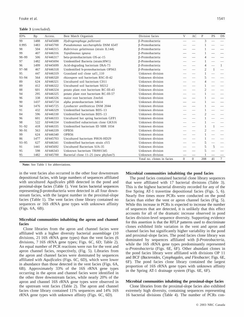
Fouke et al. 1541
in the vent facies also occurred in the other four downstreamdepositional facies, with large numbers of sequences affiliatedwith uncultured Aquificales pBB detected in the pond andproximal-slope facies (Table 1). Vent facies bacterial sequencesrepresenting β-proteobacteria were detected in all four down-stream facies, with the largest number in the proximal-slopefacies (Table 1). The vent facies clone library contained nosequences or 16S rRNA gene types with unknown affinity(Figs. 6A, 6B).
Microbial communities inhabiting the apron and channelfacies
Clone libraries from the apron and channel facies wereaffiliated with a higher diversity bacterial assemblage (10divisions, 21 16S rRNA gene types) than the vent facies (6divisions, 7 16S rRNA gene types; Figs. 6C, 6D; Table 2).An equal number of PCR reactions were run for the vent andapron channel facies, respectively, (Fig. 5). Libraries fromthe apron and channel facies were dominated by sequencesaffiliated with Aquificales (Figs. 6C, 6D), which were lowerin abundance than those detected in the vent facies (Figs. 6A,6B). Approximately 33% of the 16S rRNA gene typesoccurring in the apron and channel facies were identified inthe other three downstream facies, while nearly 20% of theapron and channel 16S rRNA gene types were observed inthe upstream vent facies (Table 2). The apron and channelfacies clone library contained 11% sequences and 14% 16SrRNA gene types with unknown affinity (Figs. 6C, 6D).
Microbial communities inhabiting the pond faciesThe pond facies contained bacterial clone library sequences
that were affiliated with 17 bacterial divisions (Table 3).This is the highest bacterial diversity recorded for any of thefive Spring AT-1 travertine depositional facies (Figs. 5, 6).Nearly five times more PCRs were conducted on the pondfacies than either the vent or apron channel facies (Fig. 5).While this increase in PCRs is expected to increase the numberof sequences that are detected, it is unlikely that this effectaccounts for all of the dramatic increase observed in pondfacies division-level sequence diversity. Supporting evidencefor this assertion is that the RFLP patterns used to discriminateclones exhibited little variation in the vent and apron andchannel facies but significantly higher variability in the pondand proximal-slope facies. The pond facies clone library wasdominated by sequences affiliated with β-Proteobacteria,while the 16S rRNA gene types predominantly representedα-Proteobacteria (Figs. 6E, 6F). Other abundant clones inthe pond facies library were affiliated with divisions OP 11and BCF (Bacteroides, Cytophagales, and Flexibacter; Figs. 6E,6F). The pond facies clone library contained the largestproportion of 16S rRNA gene types with unknown affinityin the Spring AT-1 drainage system (Figs. 6E, 6F).
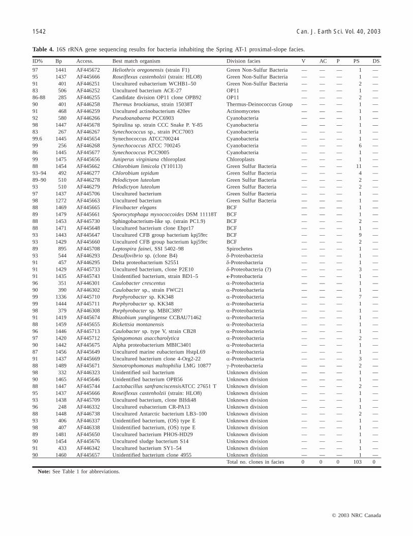
Microbial communities inhabiting the proximal-slope faciesClone libraries from the proximal-slope facies also exhibited
extremely high diversity, containing sequences representing16 bacterial divisions (Table 4). The number of PCRs con-
ID% Bp Access. Best Match Organism Division facies V AC P PS DS
99 1488 AF445688 Hydrogenophaga palleronii β-Proteobacteria — — 3 — —0.995 1483 AF445700 Pseudomonas saccharophila DSM 654T β-Proteobacteria — — 1 — —98 504 AF446315 Rubrivivax gelatinosus (strain IL144) β-Proteobacteria — — 1 — —99 407 AF446316 Tepidimonas ignava β-Proteobacteria — — 1 — —98–99 506 AF446317 beta-proteobacterium OS-ac-15 β-Proteobacteria — — 4 — —97 1482 AF445694 Unidentified Bacteria (strain:HW1) β-Proteobacteria — — 1 — —96 1499 AF445699 Acid-degrading bacterium DhA-73 β-Proteobacteria — — 4 — 197–98 467 AF446318 Unidentified b-proteobacterium OPS63 β-Proteobacteria — — 2 1 —95 447 AF446319 Grassland soil clone saf1_110 Unknown division — — 1 — —93–94 564 AF446320 rhizospere soil bacterium RSC-II-42 Unknown division — — 5 — —89 624 AF446321 Uncultured soil bacterium C011 Unknown division — — 4 — —89 412 AF446322 Uncultured soil bacterium S0212 Unknown division — — 1 — —88 601 AF446324 potato plant root bacterium RC-III-43 Unknown division — — 2 — —94 295 AF446325 potato plant root bacterium RC-III-57 Unknown division — — 1 — —96 338 AF446326 maize root bacterium ZmrIs6 Unknown division — — 1 — —90 1437 AF445724 alpha proteobacterium 34614 Unknown division — — 1 — —94 1476 AF445725 Lysobacter antibioticus DSM 2044 Unknown division — — 1 — —91 432 AF446329 Unidentified bacterium BD5–13 Unknown division — — 1 — —84 596 AF446330 Unidentified bacterium BD5–13 Unknown division — — 2 — —96 601 AF446333 Uncultured hot spring bacterium GFP1 Unknown division — — 1 — —98 522 AF446334 Unidentified eubacterium clone GKS16 Unknown division — — 1 — —91–92 418 AF446335 Unidentified bacterium ID SBR 1034 Unknown division — — 1 1 —90–91 563 AF446339 OPB56 Unknown division — — 2 — —99 624 AF446340 OPB56 Unknown division — — 1 — —86 1477 AF445703 Uncultured bacterium PHOS-HD29 Unknown division — — 1 — —93–95 627 AF446341 Unidentified bacterium strain rJ15 Unknown division — — 5 — —91 1441 AF445692 Uncultured Bacterium SJA-35 Unknown division — — 5 5 —95 598 AF446343 Unknown bacterium TBJ001345 Unknown division — — 5 — —95 1482 AF445700 Bacterial clone 11–25 (new phylum?) Unknown division — — 1 — —
Total no. clones in facies 0 0 208 41 7
Note: See Table 1 for abbreviations.
Table 3 (concluded).
J:\cjes\cjes4011\E03-067.vpNovember 18, 2003 2:01:10 PM
Color profile: DisabledComposite Default screen
© 2003 NRC Canada
1542 Can. J. Earth Sci. Vol. 40, 2003
ID% Bp Access. Best match organism Division facies V AC P PS DS
97 1441 AF445672 Heliothrix oregonensis (strain F1) Green Non-Sulfur Bacteria — — — 1 —95 1437 AF445666 Roseiflexus castenholzii (strain: HLO8) Green Non-Sulfur Bacteria — — — 1 —91 401 AF446251 Uncultured eubacterium WCHB1–50 Green Non-Sulfur Bacteria — — — 2 —83 506 AF446252 Uncultured bacterium ACE-27 OP11 — — — 1 —86-88 285 AF446255 Candidate division OP11 clone OPB92 OP11 — — — 2 —90 401 AF446258 Thermus brockianus, strain 15038T Thermus-Deinococcus Group — — — 1 —91 468 AF446259 Uncultured actinobacterium 420ev Actinomycetes — — — 1 —92 580 AF446266 Pseudoanabaena PCC6903 Cyanobacteria — — — 1 —98 1447 AF445678 Spirulina sp. strain CCC Snake P. Y-85 Cyanobacteria — — — 1 —83 267 AF446267 Synechococcus sp., strain PCC7003 Cyanobacteria — — — 1 —99.6 1445 AF445654 Synechococcus ATCC700244 Cyanobacteria — — — 1 —99 256 AF446268 Synechococcus ATCC 700245 Cyanobacteria — — — 6 —86 1445 AF445677 Synechococcus PCC9005 Cyanobacteria — — — 1 —99 1475 AF445656 Juniperus virginiana chloroplast Chloroplasts — — — 1 —88 1454 AF445662 Chlorobium limicola (Y10113) Green Sulfur Bacteria — — — 11 —93–94 492 AF446277 Chlorobium tepidum Green Sulfur Bacteria — — — 4 —89–90 510 AF446278 Pelodictyon luteolum Green Sulfur Bacteria — — — 2 —93 510 AF446279 Pelodictyon luteolum Green Sulfur Bacteria — — — 2 —97 1437 AF445706 Uncultured bacterium Green Sulfur Bacteria — — — 1 —98 1272 AF445663 Uncultured bacterium Green Sulfur Bacteria — — — 1 —88 1469 AF445665 Flexibacter elegans BCF — — — 1 —89 1479 AF445661 Sporocytophaga myxococcoides DSM 11118T BCF — — — 1 —88 1453 AF445730 Sphingobacterium-like sp. (strain PC1.9) BCF — — — 2 —88 1471 AF445648 Uncultured bacterium clone Ebpr17 BCF — — — 1 —93 1443 AF445647 Uncultured CFB group bacterium kpj59rc BCF — — — 9 —93 1429 AF445660 Uncultured CFB group bacterium kpj59rc BCF — — — 2 —89 895 AF445708 Leptospira fainei, SSI 5402–98 Spirochetes — — — 1 —93 544 AF446293 Desulfovibrio sp. (clone B4) δ-Proteobacteria — — — 1 —91 457 AF446295 Delta proteobacterium S2551 δ-Proteobacteria — — — 1 —91 1429 AF445733 Uncultured bacterium, clone P2E10 δ-Proteobacteria (?) — — — 3 —91 1435 AF445743 Unidentified bacterium, strain BD1–5 �-Proteobacteria — — — 1 —96 351 AF446301 Caulobacter crescentus α-Proteobacteria — — — 1 —90 390 AF446302 Caulobacter sp., strain FWC21 α-Proteobacteria — — — 1 —99 1336 AF445710 Porphyrobacter sp. KK348 α-Proteobacteria — — — 7 —99 1444 AF445711 Porphyrobacter sp. KK348 α-Proteobacteria — — — 1 —98 379 AF446308 Porphyrobacter sp. MBIC3897 α-Proteobacteria — — — 1 —91 1419 AF445674 Rhizobium yanglingense CCBAU71462 α-Proteobacteria — — — 1 —88 1459 AF445655 Rickettsia montanensis α-Proteobacteria — — — 1 —96 1446 AF445713 Caulobacter sp. type V, strain CB28 α-Proteobacteria — — — 1 —97 1420 AF445712 Spingomonas asaccharolytica α-Proteobacteria — — — 2 —90 1442 AF445675 Alpha proteobacterium MBIC3401 α-Proteobacteria — — — 1 —87 1456 AF445649 Uncultured marine eubacterium HstpL69 α-Proteobacteria — — — 1 —91 1437 AF445669 Uncultured bacterium clone 4-Org2-22 α-Proteobacteria — — — 3 —88 1489 AF445671 Stenotrophomonas maltophilia LMG 10877 γ-Proteobacteria — — — 2 —98 332 AF446323 Unidentified soil bacterium Unknown division — — — 1 —90 1465 AF445646 Unidentified bacterium OPB56 Unknown division — — — 1 —88 1447 AF445744 Lactobacillus sanfranciscensisATCC 27651 T Unknown division — — — 2 —95 1437 AF445666 Roseiflexus castenholzii (strain: HLO8) Unknown division — — — 1 —93 1438 AF445709 Uncultured bacterium, clone BIfdi48 Unknown division — — — 1 —96 248 AF446332 Uncultured eubacterium CR-PA13 Unknown division — — — 1 —88 1448 AF446738 Uncultured Antarctic bacterium LB3–100 Unknown division — — — 2 —93 406 AF446337 Unidentified bacterium, (OS) type E Unknown division — — — 1 —98 407 AF446338 Unidentified bacterium, (OS) type E Unknown division — — — 1 —89 1481 AF445650 Uncultured bacterium PHOS-HD29 Unknown division — — — 1 —90 1454 AF445676 Uncultured sludge bacterium S14 Unknown division — — — 1 —91 433 AF446342 Uncultured bacterium SY1–54 Unknown division — — — 1 —90 1460 AF445657 Unidentified bacterium clone 4955 Unknown division — — — 1 —
Total no. clones in facies 0 0 0 103 0
Note: See Table 1 for abbreviations.
Table 4. 16S rRNA gene sequencing results for bacteria inhabiting the Spring AT-1 proximal-slope facies.
J:\cjes\cjes4011\E03-067.vpNovember 18, 2003 2:01:11 PM
Color profile: DisabledComposite Default screen

© 2003 NRC Canada
Fouke et al. 1543
ducted on the proximal-slope facies is comparable to thenumber of reactions run on the pond facies (Fig. 5). Therefore,the affiliated bacterial diversity in the proximal-slope faciescan be directly compared with the pond facies. As was thecase in the pond facies, the proximal-slope facies clone librarywas dominated by sequences affiliated with β-Proteobacteria,while the 16S rRNA gene types predominantly representedα-Proteobacteria (Figs. 6G, 6H). Other important clones inthe proximal-slope facies library were affiliated with Cyano-bacteria, Aquificales, and BCF (Figs. 6G, 6H). The proximal-slope facies clone library contained the largest proportion ofsequences with unknown affinity (Figs. 6G, 6H).
Microbial communities inhabiting the distal-slope faciesOnly three PCRs were run on samples collected from the
distal-slope facies (Fig. 5). Therefore, although it is knownthat the bacterial diversity in the distal-slope facies has beensignificantly underestimated due to the small number of PCRs,this initial molecular survey has provided a useful evaluationof the extent of bacterial community partitioning and apreliminary estimate of the phylogentic diversity in the facies.Fewer analyses were performed in the distal-slope faciesbecause it was assumed that (1) the bacterial phylogeneticdiversity would be significantly greater at lower watertemperatures; and (2) “wash through” of bacteria from thehigher temperature facies would be significant and thereforecomplicate interpretations of the bacterial communities. Resultssuggest that neither assumption was accurate for the SpringAT-1 drainage system. The distal-slope facies clone librarycontained 11% sequences and 18% 16S rRNA gene typeswith unknown affinity (Table 3; Figs. 5, 6I, 6J). Whiledominated by sequences affiliated with α-Proteobacteria,the libraries also contained clones affiliated with 8 bacterialdivisions including β-Proteobacteria and Cyanobacteria(Figs. 6I, 6J).
Discussion
The distribution of 16S rRNA gene sequences detected inthis study indicate that bacteria are distinctly partitioned amongthe five travertine depositional facies composing the SpringAT-1 drainage system (Fig. 2). The percentage of 16S rRNAgene types found exclusively within each facies are vent facies43%, apron and channel facies 64%, pond facies 80%, proximal-slope facies 79%, and distal-slope facies 79% (Tables 1–5).For the Spring AT-1 drainage system as a whole, 88% of the657 gene sequences and 77% of the 221 16S rRNA genetypes were found in only one of the five facies (Tables 1–5).The following discussion evaluates the implications of thisbacterial partitioning with respect to downstream transport,microbial ecology, and travertine precipitation.
Downstream transportThe strong facies-specific partitioning of bacterial gene
sequences implies that relatively little (≤�25%) downstreamtransport of bacterial cells occurs despite the constant flowof water across the Spring AT-1 drainage system (Fouke etal. 2000). The high level of partitioning also argues againstthe uncertainties created by not knowing (1) the completebacterial phylogenetic diversity of each facies; (2) whetherthe bacteria affiliated with the 16S rRNA sequences detectedin this study were alive or dead at the moment they werecollected; and (3) exactly what proportion of the bacterialcells may have been washed downstream from upstream facies(Fig. 2). Therefore, the bacteria affiliated with gene sequencesdetected in this study are interpreted to have been normal insitu inhabitants of each facies. Downstream cell transportmay take place via cell movement and gliding within substratebiofilms and microbial mats, cell suspension in the watercolumn, and cell attachment to H2O molecules that rise assteam from the water–air interface (aerosolization; Bonheyoet al. 2000). It is possible that higher proportions of down-
ID% Bp Access. Best match organism Division facies V AC P PS DS
96 560 AF446241 Uncultured eubacterium env. OPS 7 Aquificales — — — — 195 1467 AF445715 Uncultured bacterium clone SL35 Planctomycetales — — — — 190 1442 AF445719 Synechococcus elongatus Cyanobacteria — — — — 294 1427 AF445714 Unidentified rhodophyte PRD01a010B Chloroplasts — — — — 284 405 AF446273 Chrysodidymus synuroideus mitochondria Eukaryota, Mitochondria — — — — 188–90 409 AF446274 Chrysodidymus synuroideus mitochondria Eukaryota, Mitochondria — — — — 292 311 AF446275 Chrysodidymus synuroideus mitochondria Eukaryota, Mitochondria — — — — 195 221 AF446284 Runella slithyformis BCF — — — — 190 1461 AF445716 Acidomonas methanolica IMET10945 α-Proteobacteria — — — — 298 405 AF446298 Brevundimonas alba, strain CB88 α-Proteobacteria — — — — 193 405 AF446306 Paracoccus, isolate B8B1 α-Proteobacteria — — — — 195 570 AF446245 Rhodobacter sp. (strain TCRI 3) α-Proteobacteria — — — — 188 611 AF446310 Rickettsia massiliae α-Proteobacteria — — — — 195 338 AF446312 Alpha-proteobacterium A0904 α-Proteobacteria — — — — 195 539 AF446346 Grassland soil clone saf2_421 Unknown division — — — — 192 339 AF446327 Uncultured maize root bacterium Zmrc235 Unknown division — — — — 193 1479 AF445728 Uncultured bacterium, clone BIfdi48 Unknown division (GS, BCF) — — — — 195 453 AF446344 Uncultured bacterium TRA1–20 Unknown division — — — — 1
Total no. clones in facies 0 0 0 0 22
Note: See Table 1 for abbreviations.
Table 5. 16S rRNA gene sequencing results for bacteria inhabiting the Spring AT-1 distal-slope facies.
J:\cjes\cjes4011\E03-067.vpNovember 18, 2003 2:01:11 PM
Color profile: DisabledComposite Default screen

© 2003 NRC Canada
1544 Can. J. Earth Sci. Vol. 40, 2003
stream bacterial transport are occurring, but at levels belowdetection.
Seasonal monitoring of the Spring AT-1 drainage systemindicates that the travertine depositional facies and theirassociated bacterial communities are systematically reestab-lished in days or weeks over lateral distances of up to a fewmetres (Fouke et al. 2000). This occurs in response to shiftsin vent position, fluctuations in water discharge and flowrate, and (or) the emergence of new spring vents. The successfulcolonization of new substrate after each shift of the springdrainage system requires that the bacterial cells in the newflow paths are viable and actively multiply. An alternative tobacterial transport is the hypothesis that “everything iseverywhere and the environment selects” (Brock et al.2001). This concept was first introduced by Bass-Becking in1935, who concluded that the energetics of phototrophy andchemolithotrophy require that the distribution of bacteriautilizing these metabolisms are controlled by the geochemicalcomposition of the water in which they live (Brock et al.2001). If bacterial cells are everywhere and in all environments,then the observed facies-specific partitioning of bacteria inSpring AT-1 is triggered by, and therefore even more directlyreflects, the systematic changes in water physical and chemical
conditions that take place along the drainage system (Fig. 2Aand B; Fouke et al. 2000).
Microbial ecologyThe presence or absence of bacteria is directly controlled
by hot spring physical and chemical environmental conditions,which include parameters such as water temperature, flowrate, pH, light, and nutrient availability (Pace 1997; Brock etal. 2001). In turn, bacteria manipulate the surrounding physicaland chemical aqueous environment, to varying degrees, viatheir metabolic activity and physical presence (Pace 1997;Brock et al. 2001). From a geological perspective, this feedbacksystem ultimately controls the ability of bacteria to influencecarbonate crystal precipitation. Therefore, tracking theseinteractions in modern hot spring travertine facies may even-tually permit, in future studies, the reconstruction of theecology and diversity of ancient bacteria from fossilizedterrestrial hot spring sedimentary deposits.
The present approach of conducting molecular analyseswithin a travertine facies model is a first step toward a process-oriented understanding of these bacteria, water, and carbonatecrystal interactions. Previous microbiological work in terrestrialand marine hot spring systems has focused on the phylogenetic
Fig. 5. Histogram summary of the molecular microbiology analyses completed in the Spring AT-1 travertine depositional facies, includ-ing number of gene sequences, inferred 16S rRNA gene types, polymerase chain reactions (PCRs), and affiliated divisions. The totalnumber of physical samples and PCRs leading to clone libraries is indicated for each facies. Each physical sample from the spring wastreated using multiple DNA extraction methods and multiple PCRs (described in the Methods section).
J:\cjes\cjes4011\E03-067.vpNovember 18, 2003 2:01:12 PM
Color profile: DisabledComposite Default screen

© 2003 NRC Canada
Fouke et al. 1545
Fig. 6. Pie diagrams illustrating the division-level diversity of the partial 16S rRNA bacterial sequences composing the clone librariesderived from each facies. The clone library data is presented in two different ways for each facies. The first is a pie diagram showingthe division-level proportion of the total number of gene sequences (raw data) representing each division. The second is a pie diagramshowing the division-level proportion of 16S rRNA gene types (interpreted data) representing each division.
J:\cjes\cjes4011\E03-067.vpNovember 18, 2003 2:01:13 PM
Color profile: DisabledComposite Default screen

© 2003 NRC Canada
1546 Can. J. Earth Sci. Vol. 40, 2003
diversity of thermophilic bacteria and archea (Stahl et al.1985; Ward et al. 1998; Hugenholtz et al. 1998; Reysenbachet al. 1999; Huber et al. 2002; Blank et al. 2002). Thesepioneering studies have revolutionized our knowledge of thediversity of microbial life on this planet (Woese 1987; Pace1997; Whitman 1998). However, because much of this workhas focused exclusively on demonstrating microbial diversity,the molecular analyses have generally not been linked withrigorous environmental analyses. Therefore, relatively littleis known of the ecological and geological context of thermo-philic and mesophilic bacterial life in hot springs (Skirnisdottiret al. 2000).
The extremely high facies-specific partitioning of bacterial16S rRNA gene sequences observed in this study indicatesthat bacterial communities are themselves a sensitive biologicalindicator of environmental physical and chemical conditions.Specific environmental conditions, called niches, are requiredfor a bacterium to grow. These include the availability ofnutrients, interactions with other organisms, and the physicaland chemical conditions of the water in which they live(Brock et al. 2001). While every bacterium inhabits a primeniche in which it is most successful, most bacteria can alsoinhabit other niches in which they are less successful(Brock et al. 2001). Sequences affiliated with Aquificales,β-Proteobacteria, and Green Non-Sulfur bacteria from thehigh-temperature vent facies were detected in downstreamlower temperature facies, as well as in the vent (Table 1).This particular suite of autotrophic thermophiles may beable to tolerate extreme changes in temperature, pH, pCO2,nutrient availability, and flow rate conditions that are outsideof their prime niche. It is also possible that at least some ofthe detected gene sequences were washed down from upstreamfacies. However, the high proportion of facies-specificsequences detected in this study indicates that the majorityof bacteria inhabiting the Spring AT-1 outflow have a primeniche that is equivalent to the nutrient availability and envi-ronmental conditions present in each travertine depositionalfacies.
An example of a bacterium with a broad niche is AquificalespBB, which was the most abundant bacterial sequence in thevent facies clone library (Table 1). This unique unculturedstrain occurred in all downstream depositional facies andthus may have an extremely wide range of environmentaltolerance if the downstream facies contained living viablecells. It has previously been observed in deep-sea hydrothermalvents on the Mid-Atlantic Ridge (112 °C and pH 6, Reysenbachet al. 2000a). Closely related aquificales sequences have alsobeen reported from Calcite Springs in Yellowstone NationalPark (83 °C and pH 7.6, Reysenbach et al. 2000b) and terrestrialsiliceous hot springs in Iceland (65–70 °C and pH 8.3,Skirnisdottir et al. 2000; 79–83 °C and pH 8.8, Takacs et al.2002). Vent facies sequences affiliated with AquificalesOPB13, Green Non-Sulfur OPB65, and β-ProteobacteriumOPB30 (Table 1) have been reported from Obsidian Pool onthe northern flank of the Yellowstone caldera (Hugenholtz etal. 1998). Obsidian Pool contains 75–95 °C siliceous watersthat are rich in reduced iron, sulfide, CO2, and hydrogen(Hugenholtz et al. 1998). These conditions are significantlydifferent from the physical and chemical environment ofSpring AT-1 (Fouke et al. 2000). The cyanobacterium OStype B was detected in Octopus Spring, which is also a
48–72 °C siliceous pool in the lower Yellowstone NationalPark geyser basin (Ferris et al. 1996). The BCF bacteriumFlectobacillus major in the Spring AT-1 vent facies hasbeen reported from siliceous hot springs in Antarctic lakesand Japan (Suzuki et al. 2001).
Travertine precipitationThe partitioning of microbes within depositional facies
along the Spring AT-1 drainage system (Tables 1–5) indicatesthat there are systematic correlations between the compositionof bacterial communities and the water chemistry, travertinechemistry, and travertine morphology described in Fouke etal. (2000). The facies-specific 16S rRNA gene libraries identifiedin the present study set the stage for future studies that willuse integrated molecular 16S rRNA gene and optical analyticaltools to correlate the presence and activity of individual bacterialspecies with specific travertine crystal growth forms andchemistries.
Although the potential results of these types of studies areextremely promising, specific interactions between bacteriaand carbonate crystal precipitation are extremely complicatedand will be challenging to identify. As an example, it isgenerally hypothesized that the consumption of CO2 byphotosynthetic autotrophs is capable of driving extracellularcarbonate precipitation. However, recent work indicates thatthe PxcA protein in the plasma membrane of photosystem IIcyanobacteria may significantly lower the pH of the outercell membrane (Zak et al. 2001). The activity of this proteincould, therefore, prevent extracellular carbonate mineralizationeven during active photosynthesis and CO2 consumption.Therefore, a biofilm or mat of cyanobacteria could eitherpositively, neutrally, or negatively affect travertine crystalnucleation and growth (Beveridge 1988; Vasconcelos et al.1997; Newman et al. 1997). Furthermore, the thickestcyanbacterial mats occur in the pond and proximal-slope facieswhere travertine precipitation is the most rapid. This mayimply that bacteria somehow attenuate or limit crystal nucleationon their outer cell walls.
A unique environmental context of the microbes inhabitingSpring AT-1 is that they must cope with extremely high ratesof travertine precipitation, which reach 5 mm/day at the marginof the pond facies (Fig. 2; Fouke et al. 2000). To survive thisrapid crystallization, microbes must restrict precipitation astheir cells migrate to the top of the encrusting crystals orrapidly divide and laterally colonize new substrates to remaina step ahead of crystal entombment and resulting cell death.For the bacteria whose cell walls form crystallization substrates,the race to escape crystal entombment will have significantinfluence on the shape and perhaps chemical composition ofthe associated travertine deposits. This is consistent withprevious observations in eutrophic lakes indicating thatbacterial filament formation is a growth rate-controlleddefense mechanism against grazing by eukaryotes (Hahn etal. 1999). In the case of Spring AT-1, the high rates of travertinecrystallization may be an analogous environmental pressureto that of predation with respect to filamentous growth mor-phology. A striking example of this process is the formationof hollow aragonite “streamers” in the vent and apron andchannel facies (Farmer and Des Marais 1994 Fouke et al.2000; Farmer 2000). Aquificales cells move or divide laterallywithin filamentous sheaths at rates slightly higher than the
J:\cjes\cjes4011\E03-067.vpNovember 18, 2003 2:01:13 PM
Color profile: DisabledComposite Default screen
© 2003 NRC Canada
Fouke et al. 1547
rate of crystal precipitation, forming the aragonite streamercrystalline morphology.
Conclusions
The 16S rRNA diversity of terrestrial hot spring bacteriahas been mapped within travertine depositional faciescomprising the Spring AT-1 drainage system at Angel Terrace,Mammoth Hot Springs, Yellowstone National Park. This haspermitted direct correlation of the distribution of bacterialcommunities with systematic changes in spring water conditionsand travertine crystal morphology and chemistry. For theSpring AT-1 drainage system as a whole, a remarkable 88%of 657 gene sequences and 77% of 221 16S rRNA genetypes were found in only one of the five travertine depositionalfacies. Therefore, relatively little (≤~25%) downstream transportof bacterial cells occurs despite the constant flow of wateracross the surface of the Spring AT-1 drainage system. Theseresults indicate that the aqueous environmental conditionsdefining each travertine depositional facies are also the dominantcontrols on microbial ecology and distribution. The correlationof community-based bacterial diversity with the morphologyand chemistry of travertine in each facies is a first step towardusing carbonate crystal shape and composition as a sensitiveindicator of bacterial diversity and activity during carbonatecrystal precipitation.
Acknowledgments
This work was supported by research awards from theNational Science Foundation Biocomplexity in the EnvironmentCoupled Biogeochemical Cycles Program (EAR 0221743),the National Science Foundation Geosciences PostdoctoralResearch Fellowship Program (EAR-0000501), the PetroleumResearch Fund of the American Chemical Society StarterGrant Program (34549-G2), and the University of IllinoisUrbana-Champaign Critical Research Initiative. Conclusionsin this study are those of the authors and do not necessarilyreflect those of the funding agencies. Discussions with A.Salyers, C. Woese, N. Goldenfeld, J. Veysey, and H. Garciathroughout the project were essential to improving all aspectsof the data collection and interpretation. Thorough reviewsprovided by B. Chatterton, C. Blank, and J. Thompson servedto significantly improve the manuscript.
References
Allen, E.T., and Day, A.L. 1935. Hot Springs of the YellowstoneNational Park. Carnegie Institution of Washington, Publication466.
Altschul, S.F., Gish, W., Miller, W., Meyers, E.W., and Lipman,D.J. 1990. Basic local allignment search tool. Journal of MolecularBiology, 59: 143–169.
Bargar, K.E. 1978. Geology and thermal history of Mammoth HotSprings, Yellowstone National Park, Wyoming. United StatesGeological Survey Bulletin, 1444, 54 p.
Beveridge, T. 1988. Role of cellular design in bacterial metal accu-mulation and mineralization. Annual Review of Microbiology,43: 147–171.
Blank, C.E., Cady, S.L., and Pace, N.R. 2002. Microbial compositionof near-boiling silica-depositing thermal springs throughout Yellow-
stone National Park. Applied and Environmental Microbiology,68: 5123–5135.
Blatt, H., Middleton, G.V., and Murray, R.C. 1980. Origin ofsedimentary rocks. Prentice-Hall, Englewood Cliffs, N.J.
Boggs, S.J. 2002. Principles of sedimentology and stratigraphy. 3rded. Prentice Hall, Upper Saddle River, N.J.
Bonheyo, G.T., Fouke, B.W., Sanzebacher, B., and Salyers, A.A.2000. By land, sea, or air? microbial transport at the MammothHot Springs complex. GSA Abstracts with Programs, 7(. 7): A-492.
Brock, T.D., Madigan, M.T., Martinko, J.M., and Parker, J. 2001.Biology of microorganisms. Prentice Hall, Upper Saddle River,N.J.
Farmer, J.D. 2000. Hydrothermal systems: doorways to early biosphereevolution. GSA Today, 10: 1–8.
Farmer, J.D., and Des Marais, D.J. 1994. Biological versus inorganicprocesses in stromatolite morphogenesis: observations frommineralizing sedimentary systems. In Microbial mats: structure,development, and environmental significance. Edited by L.J.Stal and P. Caumette. NATO ASI Series in Ecological Sciences.Springer-Verlag, Berlin and Heidelberg, Germany, pp. 61–68.
Ferris, M.J., Muyzer, G., and Ward, D.M. 1996. Denaturing gradientgel electrophoresis profiles of 16S rRNA-defined populationsinhabiting a hot spring microbial mat community. Applied andEnvironmental Microbiology, 62: 340–346.
Flügel, E. 1982. Microfacies analysis of limestones. Springer-Verlag,Berlin, Germany.
Ford, T.D., and Pedley, H.M. 1996. A review of tufa and travertinedeposits of the world. Earth-Science Reviews, 41: 117–175.
Fouke, B.W. 2001. Depositional facies and aqueous-solid geochemistryof travertine-depositing hot springs (Angel Terrace, MammothHot Springs, Yellowstone National Park, USA). Journal ofSedimentary Research, 71: 497–500.
Fouke, B.W., Farmer, J.D., Des Marais, D.J., Pratt, L., Sturchio,N.C., Burns, P.C., and Discipulo, M.K. 2000. Depositional faciesand aqueous-solid geochemistry of travertine-depositing hot springs(Angel Terrace, Mammoth Hot Springs, Yellowstone NationalPark, USA). Journal of Sedimentary Research, 70: 265–285.
Friedman, I. 1970. Some investigations of the deposition of travertinefrom hot springs: I. The isotope chemistry of a travertine-depositingspring. Geochimica et Cosmochimica Acta, 34: 1303–1315.
Goebel, B.M., and Stackebrandt, E. 1994. Cultural and phylogeneticanalysis of mixed microbial populations found in natural andcommercial bioleaching environments. Applied and EnvironmentalMicrobiology, 60: 1614–21.
Hahn, M.W., Moore, E.R.B., and Hofle, M.G. 1999. Bacterial filamentformation, a defense mechanism against flagellate grazing, isgrowth rate controlled in bacteria of different phyla. Appliedand Environmental Microbiology, 65: 25–35.
Hillis, D.M., Moritz, C., and Mable, B.K. 1996. Molecular systematics.Sinauer Associates, Sunderland, Mass.
Huber, J.A., Butterfield, D.A., and Baross, J.A. 2002. Temporalchanges in archael diversity and chemistry in a mid-ocean ridgeseafloor habitat. Applied and Environmental Microbiology, 68:1585–1594.
Hugenholtz, P., Pitulle, C., Hershberger, K.L., and Pace, N.R. 1998a.Novel division level diversity in a Yellowstone hot spring. Journalof Bacteriology, 180: 366–376.
Hugenholtz, P., Pitulle, C., Hershberger, K.L., and Pace, N.R. 1998b.Novel division level bacterial diversity in a Yellowstone hotspring. Journal of Bacteriology, 180: 366–376.
Hughes, J.B., Hellman, J.J., Ricketts, T.H., and Bohannon, B.J.M.2001. Mini-Review—Counting the Uncountable: Statistical ap-proaches to estimating microbial diversity. Applied and Environ-mental Microbiology, 67: 4399–4406.
J:\cjes\cjes4011\E03-067.vpNovember 18, 2003 2:01:13 PM
Color profile: DisabledComposite Default screen

© 2003 NRC Canada
1548 Can. J. Earth Sci. Vol. 40, 2003
Hurst, C.J., Crawford, R.L., Knudsen, G.R., McInernery, M.J., andStetzenbach, L.D. 2002. Manual of environmental microbiology.ASM Press, Washington, D.C.
Maidik, B.L., Olsen, G.J., Larsen, N., Overbeek, R., Mccaughey,M.J., and Woese, C.R. 1997. The RDP (Ribosomal DatabaseProject). Nucleic Acids, 25: 109–110.
Martin, A.P. 2002. Phylogenetic approaches for describing andcomparing the diversity of microbial communities: Applied andEnvironmental Microbiology, 68:. 3673–3682.
Messing, J. 1983. New M13 cloning vectors. Methods in Enzymology,101: 20–78.
Newman, D.K., Beveridge, T.J., and Morell, F.M.M. 1997. Precipitationof arsenic trisulfide by Desulfotomacuum auripigmentum. Appliedand Environmental Microbiology, 63: 2022–2028.
Pace, N.R. 1997. A molecular view of microbial diversity and thebiosphere. Science (Washington, D.C.), 276: 734–740.
Pentecost, A. 1990. The formation of travertine shrubs: MammothHot Springs, Wyoming. Geological Magazine, 127: 159–168.
Pentecost, A., and Viles, H.A. 1994. A review and reassessment oftravertine classification. Geographie Physique et Quaternaire, 48:305–314.
Reading, H.G. 1996. Sedimentary environments: processes, faciesand stratigraphy. Blackwell Science, London, England.
Reysenbach, A.L., Setzinger, S., Kirshtein, J., and Mclaughlin, E.1999. Molecular constraints on a high-temperature evolution ofearly life. Biological Bulletin, 196: 367–372.
Reysenbach, A.L., Ehringer, M., and Hershberger, K.L. 2000a.Microbial diversity at 83 °C in Calcite Springs, YellowstoneNational Park: another environment where Aquificales and“Korarchaeota” coexist. Extremophiles, 4: 61–67.
Reysenbach, A.L., Banta, A., Boone, D., Cary, S., and Luther, G.2000b. Microbial essentials at hydrothermal vents. Nature (London),404: 835.
Sambrook, J., Fritsch, E.F., and Maniatis, T. 1989. Molecular cloning.A laboratory manual, Cold Spring Harbor Laboratory Press.
Skirnisdottir, S., Hreggvidsson, G.O., Hjorleifsdottir, S., Marteinsson,V.T., Petursdottir, S.K., Holst, O., and Kristjansson, J.K. 2000.Influence of sulfide and temperature on species composition andcommunity structure of hot spring microbial mats. Applied andEnvironmental Microbiology, 66: 2835–2841.
Stahl, D.A., Lane, D.J., Olsen, G.J., and Pace, N.R. 1985. Charac-terization of a Yellowstone hot spring microbial community.Applied Environmental Microbiology, 49: 1379–1384.
Studier, W.F., and Dunn, J.J. 1983. Complete nucleotide sequenceof bacteriophage T7 DNA and the location of T7 genetic elements.Journal of Molecular Biology, 166: 477–535.
Suzuki, M., Nakagawa, Y., Harayama, S., and Yamamoto, S. 2001.Phylogenetic analysis and taxonoimic study of marineCytophaga-like bacteria. Journal of Systematic Evolutionary Mi-crobiology, 5: 1639–1652.
Takacs, C.D., Ehringer, M., Favre, R., Cermola, M., Eggertson, G.,Palsdottir, A., and Reysenbach, A.L. 2002. Phylogeneticchracaterization of the blue green filamentous bacterial communityfrom an Icelandic geothermal spring. FEMS Microbiology andEcology, 35: 123–128.
Vasconcelos, C., and Mckenzie, J.A. 1997. Microbial Meditation ofModern Dolomite Precipitation and Diagenesis Under AnoxicConditions (Lagoa Vermelha, Rio De Janero, Brazil). Journal ofSedimentary Research, 67: 378–390.
Vieira, J., and Messing, J. 1982. The pUC plasmids, anM13mp7-derived system for insertion mutagenesis and sequenc-ing with synthetic universal primers. Gene (Amsterdam), 19:259–268.
Walker, R.G., and James, N.P. 1992. Facies Models: response tosea level change. GeoText 1, Geological Association of Canada.
Ward, D.M., Ferris, M.J., Nold, S.C., and Bateson, M.M. 1998. Anatural view of microbial biodiversity within hot springcyanobacterial mat communities. Microbiology and MoelcularBiology Reviews, 62: 1353–1370.
Whitman, W.B. 1998. Prokaryotes: the unseen majority. Proceedingsof the National Academy of Science, 95: 6578–6583.
Wilmotte, A., Van der Auwera, G., and DeWachter, R. 1993. Structureof the 16S ribosomal RNA of the thermophilic cyanobacteriumChlorogloeopsis HTF (Mastigocladus laminosus HTF) strainPCC7518, and phylogenetic analysis. FEBS Letters, 317: 96–100.
Wilson, J.L. 1975. Carbonate facies in geologic history.Springer-Verlag, New York.
Woese, C.R. 1987. Bacterial evolution. Microbiology Reviews, 51:221–271.
Zak, E., Norling, B., Maitra, R., Huang, F., Anderson, B., andPakrasi, H. 2001. The initial steps of biogenesis of cyanobacterialphotosystems occur in plasma membranes. Proceeding of theNational Academies of Science, 98: 13 443 – 13 448.
J:\cjes\cjes4011\E03-067.vpNovember 18, 2003 2:01:14 PM
Color profile: DisabledComposite Default screen