patellofemoral instability lab -...
TRANSCRIPT

1
Patellofemoral Instability Lab
Friday November 27, 201511 - 5 pm
Skills Centre for Health Science

32
LearningObjectives
4. 14.8. 16. 20.ANATOMY CLINICAL
FINDINGSBIOMECHANICS DIAGNOSTIC
IMAGINGTREATMENT
Faculty11 am Lunch & Introduction
11:15-11:45 Anatomy and Pathophysiology of Patellar Instability - Dr. Nathan Urquhart
11:45-12:15 Acute and Chronic Patellar Instability - Dr. Catherine Coady
12:15 -12:45 Patellar Instability in the Paediatric Patient - Dr. Karl Logan
12:45-1:15 Diagnostic Imaging in Patellar Instability - Dr. Gord Boyd
1:15-1:45 Surgical Treatment of Recurrent Patellar Instability -Dr. Laurie Hiemstra
1:45-2:15 Surgical Demo - Dr. Laurie Hiemstra
2:15 - 5 pm Hands On Cadaver Workshop Arthroscopy - examination of the patellofemoral joint, arthroscopic/open medial imbrication, lateral release MPFL Surgery - MPFL reconstruction, MQTFL reconstruction Medial and Lateral Knee Dissection Tibial Tubercle Transfers Trocheoplasty Surgery
Patellofemoral Instability Knee Lab Schedule
Many thanks to ConMed Linvatec for sponsoring the hands-on skills lab and the Skills Centre for Health Sciences for hosting the session.
Dr. Laurie HiemstraBanff Sports Med
Dr. Karl LoganIWK Health Centre
Dr. Gord BoydQEII Health Sciences Centre
Dr. Nathan UrquhartDartmouth General Hospital
Dr. Catherine CoadyQEII Health Sciences Centre
At the completion of this session participants will be able to:
General:1. Demonstrate a competent understanding of the anatomy and biomechanics of the patello-femoral joint.2. Describe the patho-anatomy of patellofemoral joint instability. 3. List the pertinent steps of arthroscopic assessment of patellofemoral instability
Specific:1 Review and apply patellofemoral surgical anatomy and biomechanics. 2 Differentiate patellofemoral treatments and comprehend logical approaches to treating patients with patellofemoral pain and instability. 3 Identify the newest concepts in patellofemoral joint preservation including how to optimize patellofemoral stability as well as patellofemoral articular loading & balance with the appropriate surgical technique(s)

54
Patellofemoral instability is a commong disorder affecting the knee joint. Young athletes suffer pa-
tellar dislocations more commonly than any other age group. Patients most at risk for patellar instability are 12 - 20 years of age. Females are also at higher risk for patellar instability.
Traumatic dislocations may occur from both contact and noncontact injuries to the knee. An athlete can dislocate his/her patella when the foot is planted and a rapid change of direction or twisting occurs. This may happen in sports such as soccer, gymnastics or basket-ball. Direct blows to a knee can also cause dislocations. This type of injury may occur in sports such as hockey or football. The force of these types of injuries is typ-ically much greater and usually causes more severe damage to the knee especially to the restraining
ligaments. Patients with traumat-ic dislocations also have a higher incidence of associated fractures in the patellofemoral joint.
Some patients have “predispos-ing factors” which increase their chance of having patellar instability such as trochlear dysplasia, patella alta, lower extremity malalign-ment, quadriceps atrophy and an increased tibial tubercle offset. In these patients, the patella can sublux or dislocate with seemingly minor activites such as going up stairs or even kneeling. ANATOMY:
Instability of the patellofemoral articulation can occur when the bony anatomy of the patella, and/or the femoral trochlea are abnormal. The patella is the largest sesamoid bone in the body. It has multifac-eted articular surface with medial
and lateral facets separated by a central ridge and a much smaller odd facet located far medially. The articular cartilage of the patella is the thickest in the body designed to withstand joint reactive forces up to 10 times body weight with daily activiies and 20 times body weight with sporting activities. The patella is a fulcrum for the extensor mech-anism. It increases the distance of the line of action of the extensor mechanism from the center of rotation of the knee -- increasing the force that can be generated by the contraction of the quadriceps. The medial and lateral facets of the patella articulate with the medial and lateral facets of the femoral trochlea.
The patellofemoral joint contains both static and dynamic constraints on its dislocation. The static con-straints include the osseous anat-omy of the patellofemoral joint (particularly its trochlear geome-try), the medial patellar retinacu-lum, and the medial patellofemoral
ligament (MPFL). The static con-straints provide a constant check-rein to patellar dislocation, and when the trochlea can no longer provide stability, as it normally does in greater degrees of knee flexion, the soft-tissue restraints become the sole providers of patellar stability from 0° to 20° of flexion. The main dynamic restraint to patellar dislo-cation is the vastus medialis muscle, which neutralizes the lateralizing force of the vastus lateralis muscle during muscle contraction and activity. When there is disruption of any of these constraints either through direct trauma (eg, a tear in the MPFL) or indirectly through pathologic alignment or a fault in osseous anatomy (eg, an altered Q angle, external tibial torsion, femoral anteversion, foot prona-tion, genu valgum, patella alta, or trochlear dysplasia), subluxation or dislocation of the patella can occur.
Articulation between the inferior margin of the patella and the femur begins at approximately 10 – 20 degrees of knee flexion. The patella does not articulate with the troch-lea near terminal knee extension.
As the knee proceeds into greater degrees of knee flexion, the contact area of the patellofemoral joint moves proximally along the patella and posterior along the condyles.
Static stabilizers provide fixed inhibition to lateral translation of the patella. Absence has been demon-strated to cause up to 49% reduction in lateral stability at zero degrees of flexion. In addi-tion, lying beneath are three liga-ments; the patella-femoral (MPFL), patella-meniscal (MPML) and patella-tibial (MPTL). These are the primary ligamentous structures constraining lateral patellar motion, of which the MPFL is the most notable. The MPFL is a continua-tion of the deep retinacular surface of the vastus medialis obliquus (VMO). It runs transversely be-tween the proximal half of the medial border of the patella to the femur between the medial epicon-dyle and the adductor tubercle, forming the second layer between the superficial medial retinaculum
and the capsule. The anatomy of the lateral side is more complex. The superficial layer consists anteri-orly of the fibrous expansion of the vastus lateralis and the superficial oblique retinaculum further pos-teriorly. The tightness of the ITB (dynamic stabilizer) will influence the lateral stability force inferred by the lateral retinacular structures.
“The patella is the largest
sesamoid bone in the human
body”.
Patellofemoral Instability
PATEllOfEMOrAl CONTACT ArEA
wITH PrOgrESSIvE kNEE flExION
4

76
TrOCHlEAr gEOMETrY The femoral trochlea anatomy typically closely matches the ar-ticular shape of the patella with a longer and higehr lateral wall that serves as the most important bony restraint to lateral tranlation. Once the patella enters the confines of the trochlea, the bony anatomy allows for inherent stability of the PFJ. The normal shape of the trochlea is a concave trough, with a normal sul-cus angle of 138 ± 6° (on Merchant view axial radiographs). The lateral femoral trochlea extends further anteriorly than the medial side, providing a buttress to lateral patel-lar subluxation thereby maintaining the patella’s centered position in the trochlea. The lateral extremity of the trochlea extends further prox-
imally than it does on the medial side, providing a mechanism for engaging the patella in early knee flexion and deflecting it medially to the center. Trochlear dysplasia is an important risk factor for recurrent patellar dislocation (classification by Dejour et. al.).
PATEllA gEOMETrY The patella has a convex articular surface and this congruity between the patella and the trochlea pro-vides some constraint to the PFJ. When the knee begins to bend, the initial contact area is the distal and lateral patella facet. With further flexion the contact area on the pa-tella articular surface moves more proximally until in deep flexion where the medial facet has then
made contact. PATEllA HEIgHT Patellar height also contributes to PFJ stability. Trochlear geometry is such that it encourages early engagement of the patella during the flexion arc. Engagement of the patella depends entirely on patella height. With patella alta, engage-ment into the trochlear does not occur in the early phase of knee flexion, thus potentiating instability at the PFJ. Once the patella enters the trochlea, dysplasia of the patel-la, the trochlea, or both, can con-tribute to instability by decreasing the bony restraint and consequently the amount of energy required to dislocate. Patellofemoral dysplasia has been classified by Dejour et al. Dysplasia typically occurs on both sides of the joint, with congruous articulation between the two bones, although incongruous articulation can also occur, and leads to some of the worstinstability
lIMB AlIgNMENTQ angle is an angle formed by the line of pull of the quadriceps mech-anism and that of the patellar ten-don as they intersect at the center of the patella. Clinically, this angle is represented by the intersection of a line drawn from the anterior superior iliac spine to the center of the patella with a second line drawn from the center of the tibial tuber-osity to the center of the patella. In
StaticStabilizers
males, the Q angle is 8-10° and in females it is 15±5°. An increase in the Q angle results in an increased valgus vector to the PFJ. Genu val-gum, increased femoral ante-ver-sion, external tibial torsion and/or a lateralized tibial tuberosity can all ultimately affect patella tracking.
SOfT-TISSuE STruCTurES Important structure for PFJ stabili-ty include:1. medial retinaculum2. vastus medialis oblique muscle3. medial patellofemoral ligament4. lateral retinaculum5. iliotibial band6. vastus lateralis muscle
Normally, these all work in con-cert to provide proper stability and tracking of the patellofemoralarticulation. When medial stabiliz-ers are weakened or disrupted, the typical lateral instability may occur. Tightness or excessive force by the lateral stabilizers typically does notcause actual instability, as long as the medial structures are normal,
but may cause symptomaticabnormalities in patella tilt and tracking.Soft tissue static restraints include the quadriceps and patellar ten-dons, the lateral retinaculum, the medial retinaculum, and the medial patellofemoral ligament (MPFL). Warren and Marshall6 divided the Soft tissue static restraints include the quadriceps and patellar ten-dons, the lateral retinaculum, the medial retinaculum, and the medial patellofemoral ligament (MPFL).
Warren and Marshall divided the medial soft tissue restraints into 3 layers:1. the superficial layer consists of
the fasciae of the vastus media-lis and sartorius
2. the second layer includes the MPFL and superficial medial collateral ligament (MCL)
3. the deepest layer consists of the joint capsule.
Recent studies have documented the anatomy and function of the MPFL and identified it as the pri-
mary soft tissue restraint to lateral instability.
MPfl ANATOMY:Length: 55 mmFemoral origin width: 15 mmPatella insertion width: 17 mm
rESTrAINT TO lATErAl SuBluxATION IN ExTENSION & EArlY flExION:MPFL: 60%Lateral Retinaculum: 10%Medial Patellomeniscal Ligament: 13%
Dejour Classification
3 Layers of Medial Soft Tissue Restraints
“Injury to the medial retinaculum and MPFL has been reported to be as high as 94% to 98% after acute dislocation of the patella”.

98
DynamicStabilizersThe quadriceps muscles are the principal dynamic restraint. Of the 4 heads of the quadriceps, the vastus medialis obliquus (VMO) is best positioned to resist lateral subluxation, due to its approxi-mately 60-degree angle of insertion on the patella. The function of the VMO as a restraint to subluxation is greatest in extension and early flexion because bony restraint at these positions is minimal. An underdeveloped VMO is a predis-posing factor for recurrent instabil-ity and is a major focus of rehabil-itative efforts during conservative management.As well as influencing the patella in a lateral-medial direction, the vastii muscles also exert a posterior force vector. This force stabilizes the pa-tella within the trochlear groove.
The VMO overlies and merges with the MPFL, acting together to provide both active and passive stabilization of the patella.
sion, the patella is not engaged in the trochlear groove, and it is de-pendent on soft-tissue restraints for stability. The medial knee retinacu-lar structures, including the VMO, MPFL and the medial patellotibial ligament, provide static restraint to lateral patellar translation. Correct patellar centering within the groove during early knee flexion is essen-tial to maintaining patella-femoral stability and is largely determined, again, by the dynamic and static soft-tissue structures. The MPFL is the primary soft tissue restraint to lateral translation of the patella during the initial 20 to 30 degrees of knee flexion. This ligament is most taut in full extension, with the quadriceps contracted, and as-sists in guiding the patella into the trochela during the early stages of flexion. Once engaged in the troch-lea, the patellofemoral joint com-pression provided by the increasing force vectors of the quadricpes and patellar tendons, compbined with PFJ geometry, provides the major effect on stability as knee flexion progresses. At 30° of knee flexion, the patella should be centered in the trochlear groove, with <1 cm
lateral translation. The depth and slope of the trochlea influence pa-tellar stability as the knee continues to flex. The role of soft tissue con-straints is minimized with flexion >30°, at which point the trochlea becomes the most important patel-lar stabilizer. The height and slope of the lateral trochlear facet provide the primary resistance to lateral patellar translation as knee flexion progresses. The lateral facet of the trochlear groove is most prominent proximally and decreases in height distally and posteriorly. Once flexion is initiated, the soft-tissue constraints of the quadriceps and patellar tendons place a posteriorly
directed force on the patella and provide increased patellar stabili-ty. As the knee bends, the contact pressure across the chondral sur-face of the patella moves from the lateral facet to the medial facet and from distal to proximal along the chondral surface
The patella can be thought of as a sliding tongue-in-groove joint that has soft-tissue restraints
that guide motion within the groove. Variation in the depth of the troclear groove, the size and sloped of the patellar facets as well
as the length and integrity of the soft-tissue restrainst all play roles in establishing and maintaining patellar stability. The patellofem-oral joint withstands the highest joint-reactive forces in the body. These forces are least when the knee is in extension and increase
with flexion to approximately 7 to 8 times a person’s body weight at 130 degrees. PFJ stability is dependent on coordinated interaction between static, dynamic, and osseous struc-tures to keep the patella centered within the trochlea during knee range of motion. During full exten-
Contact points between patella and femur move from proximal to distal on the femur and from distal to proximal on the patel-la during knee flexion.
Bifurcation of patello-femoral contact area beyond 100° of knee flexion. the odd facet of the patella only makes contact with the medial femoral condyle beyond 125° to 135° of knee flexion.
BIOMECHANICS

1110
rISk fACTOrS fOr PATEllAr INSTABIlITY
1. Trochlear dysplasia2. paTella alTa3. increased (TT-TG)4. increased ‘‘Q’’ anGle5. Medial paTellofeMoral liGaMenT injury / insufficiency6. Quadriceps aTrophy/dysplasia7. hypoplasTic laTeral feMoral condyle8. paTellar TilT9. Miserable MalaliGnMenT feMoral anTeversion exTernal Tibial Torsion fooT pronaTion10. younG aGe aT TiMe of dislocaTion11. faMily hisTory12. feMale GenGer13. Generalized liGaMenTous laxiTy
Ligament Stabilizers: Medial Patella-Femoral Ligament (MPFL) Medial Patella-Meniscal Ligament (MPML) Medial Patella-tibial Ligament (MPTL) Medial Retinaculum
Bone Stabilizers: Deep femoral sulcus High lateral trochlea
PASSIvE STruCTurES
STATIC
STATIC
StabilizerS
Stabilizers Quadriceps Muscles Predominantly the vastus medialis obliquus muscle
The rectus femoris, vastus lateralis, vastus intermedius and vastus medialis insert in a layered arrangement on to the proximal patella, creating different vectors of
DYNAMIC STruCTurESDYNAMICStabilizerS
The peak of instability occurs with the knee in extension and lesser degrees of flexion and is due in part to the fact that the tongue-in-groove relationship is not engaged until approximately 20 degrees of flexion. In associated condi-tions such as patella alta, the trochlea is not engaged by the patella until higher degrees of flexion and is thought to contribute to the patellar instability
Normal patellofemoral function during strenuous physical activity requires a combination of stabilizing forces af-forded by bony, dynamic, and static soft tissue restraints. The bony elements of the generally congruent PFJ contribute significantly to its stability. The patel-la is proximal to the femoral trochlea when the knee is in extension and does not enter the sulcus until about 20 to 30 degrees of knee flexion. The lateral trochlear ridge acts as a buttress to help resist lat- eral translation of the patella. If the lateral ridge is hypo- plastic, the re-straint to lateral subluxation is decreased.
The static constraints provide a constant check-rein to patellar dislocation, and when the troch-lea can no longer provide stability, as it normal-ly does in greater degrees of knee flexion, the soft-tissue restraints become the sole providers of patellar stability from 0° to 20° of flexion.
“When there is disruption of dynamic +/- static con-straints either through direct trauma (eg, a tear in the MPFL) or indirectly through pathologic align-ment or a fault in osseous anatomy (eg, an altered Q angle, external tibial torsion, femoral anteversion, foot pronation, genu valgum, patella alta, or trochlear dysplasia), dislocation can occur”.

1312
Risk FactoRs FoR patellaR instability
The normal trochlea is concave. In the presence of dysplasia the intercondylar groove may be flat-tened or convex. Troclear dyspla-sia does not allow the patella to fit into the troclea during range of motion. Trochlear dysplasia is one of the most improtant anatomical abnormailites in patellar instabil-ity. Trochelar dysplasia does not allow the patella to fit into the trochela during range of motion.This is riskiest in the first degrees of flexion as the lateral structures can easily overtake the medial ones.
TT- TG distance is the distance measured on superimposed axial cuts on CT or MRI of the apex of the tibial tubercle (TT) and the deepest part of the trochlear groove (TG). A TT-TG distance of greater than 20 is pathologic.
Patella alta is characterized by a more proximal position of the patella (high riding). When the patellar tendon is longer than normal, during quadriceps con-traction, the patella goes proximal and above the femoral trochlear groove. Consequently there is no lateral bony support that would typically prevent lateral disloca-tion. Patella alta also modifies the lever arm between the quadri-ceps and patellar tendons thereby increasing the compression forces in the PF joint which may lead to early PF degenerative changes.
The MPFL is a very important stat-ic stabilizer that helps prevent the patella from moving too far later-ally out of the trochlear groove. The MPFL is almost always injured in traumatic patellar dislocations.
The Q angle is the angle formed by the line of pull of the quadri-ceps and the patellar tendon as they intersect at the centre of the patella. An increase in the Q an-gle results in an increased valgus vector to the PF joint.
The Q angle is largest in exten-sion.
Males: Q angle: 8-10 degreesFemales: Q angle 15 +/- 5 degrees
Rotational and axial deformity of the entire leg can play a role in patellar instability. Increased fem-oral anteversion and/or increased tibial torsion are often associated with patellar instability.
TROCHLEAR DYSPLASIA
INCREASED TT-TG
PATELLA ALTA
MPFL INJURY
INCREASED Q ANGLE
MALALIGNMENT
MPFLMPFL is the main static stabilizer and it provides 50–60 % of the total restraining force against lateral patellar displacement.
MPFL: 55 mm in length
fEMOrAl OrIgIN:• 10.6 mm proximal & 8.8 mm posterior to
medial epicondyle• 1.9 mm anterior and 3.8 mm distal to adduc-
tor tubercle
Schottle’S point:LateraL X-ray
• 1 mm anterior to extension of posterior cortical line• 2.5mm distal to posterior origin of the medial
femoral condyle • Proximal to level of Posterior Point of blumensaat’s
line
PATEllAr INSErTION: • Broad insertion on the patella• 7.4 mm anterior to articular surface & 5.4
mm distal to the proximal edge of the artic-ular surface.
• Insertion spans 20 mm • Centered at the junction of the proximal 1/3
and distal 2/3 of the patella

1514
ClinicalFindings
with gentle extension of the knee with a medially directed force on the pa-tella. Sedation may be re-quired. Many dislocated patellas do spontaneously reduce. Patients typically have tenderness over the medial structures where the medial retinaculum and MPFL are locat-ed. Traumatic first time dislocators often develop a large hemarthrosis. Quadriceps muscle atro-phy may be noted early on due to the knee injury.
Patients with recurrent patellar instability should have a more thorough physical examiantion performed looking for identifying pre-existing risk factors for dislo-cation. These include generalized ligamen-tous laxity, pronation of the foot, genu valgum, femoral anterversion, external tibial torsion.
The Q angle should be measured. Many have chronic quadriceps weakness. The boney structures shoulde be assessed for tenderness. Range of motion testing should be performed with special attention to the position of the patella as it approaches terminal extension. In patients with recurrent patellar instability, it may be observed that the patella moves from a central position within the trochlear groove to a more lateral terminal position as the knee tran-sitions from flexion into extension. As the patella passes the lateral condy-lar ridge, this subluxation event might be recog-nized and is referred to as the J sign. Additionally, when the patient’s knee is flexed to 20–30 degrees, a laterally directed force to the patella may induce
Refers to lateral patellar deviation with terminal
extension of the knee
J SIgN
kNEE EffuSION
QuADrICEPS wASTINg
APPrEHENSIONTEST
ASSESSACl
a reflexive protective quadriceps contraction, observed clinically as a positive apprehension test. The moving patellar apprehension test, which has 100% sensitivity and 88% specificity, involves applying a later-ally directed force on the patella to a knee that is being flexed concurrent-ly by the examiner. The ligaments of the knee should be assessed espe-cially the ACL as the mechanism of injury, history and clinical findings can be very similar.
Known predisposing fac-tors for patellar instability (especially in patients with
recurrent patellar instability) are often assessed such as generalized joint hypermobility, weakness in the muscles around the knee as well as the alignment of the lower extremity. Both varus and valgus stress exam-ination of the knee also should be performed as there are fibers of the medial patellofemoral ligament that may be avulsed from the superficial medial attachment to the medial collateral ligament of the knee.
An inspection of the patient’s stance and gait may reveal genu valgum, and additional measurement of the patient’s Q angle should be performed with the knee in slight flexion to reduce tibial external rotation that occurs in terminal knee extension. The examiner must also remember that an unstable patella may naturally subluxate laterally resulting in an underestimation of the actual Q angle.
Additional special testing can in-clude patellar mobility to assess patellar tilt and patellar displace-ment. Patellar tilt testing assesses the amount of lateral tightness that exists while attempting to lift the lateral facet of the patella off the lateral femoral trochlea. Patellar
displacement is recorded in terms of number of quadrants of displace-ment produced, with two quadrants representing roughly half the width of the patella. Finally, it is important to examine the knee for crepitus throughout the arc of motion. Early crepitus may indicate an inferior pole of the patellar chondral defect, whereas later crepitus may indicate a proximal lesion.
As many patients with patellar instability have underlying joint hypermobility, a Beighton score should be performed to document the extent of generalized ligamen-tous laxity.
PATEllAr TIlT
PATEllAr glIDE
Physical examina-tion of a patient with a patellar
dislocation with vary depending on the timing of presentation. In the acute setting, a thorough neurovascular and liga-
mentous examination of the knee should be per-formed. Gross deformity should be noted. The entire knee region should be palpated. If the patella is still dislocated, reduc-tion can be performed
14

1716
A standard series of radio-graphs of the knee should be obtained including:
AP Lateral Merchant
Initial assessment of these radiographs should include confirmation of a concentri-cally reduced patella as well as examination for the presence of fracture fragments. Later-al radiographs after an acute subluxation event often reveals
an effusion. Evidence of a fluid/fluid level on lateral radio-graphs can serve as a clue that a lipohemarthrosis exists, which may indicate an associated osteochondral fracture.
DIAGNOSTICIMAGING
Lateral radiographs with symmetric overlap of the medial and lateral poste-rior femoral condyles allow for assess-ment of patella alta.
There are several methods of measuring patella alta on the lateral radiograph including:
1. Insall-Salvati Index2. Modified Insall Salvati Index3. Caton-Deschamps Index4. Blackburne-Peel Index
STANDARD RADIOGRAPHS
The Merchant view is performed with the knee in 40 degrees of flexion and the beam is inclined 30 degrees caudad.
AP
LAT
MERCHANT
Type A DYSPLASIA: charac-terized by a crossing sign on the lateral view and a shallow trochlea on the axial view.
Type B DYSPLASIA: charac-terized by a crossing sign and supratrochlear spur on the lateral view and a flattened trochlea on the axial view.
Type C DYSPLASIA: char-acterized by the crossing sign with a double contour on the lateral view, and medial hypo-plasia on the axial view.
Type D DYSPLASIA: char-acterized by the crossing sign, double contour, with supra-trochlear spur on the lateral view, and asymmetry of the trochlear facets on the axial view.
TrOCHlEAr DYSPlASIA can also be visualized on the lateral radiograph via the crossing sign, supratrochlear spur-ring, or a double contour. In a normal trochlea, the trochlear groove is poste-rior to the femoral condyles. When the trochlea is flat, the trochlear groove is in the same plane as or anterior to the femoral condyles (the crossing sign). Alternatively, a prominent superolateral spur may develop (supratrochlear spur) to prevent lateral displacement of the patella. A double line at the anterior aspect of the femoral condyles (double contour) represents the subchondral bone of a hypoplastic medial condyle.
PATEllA AlTA ASSESSMENT
16
1918
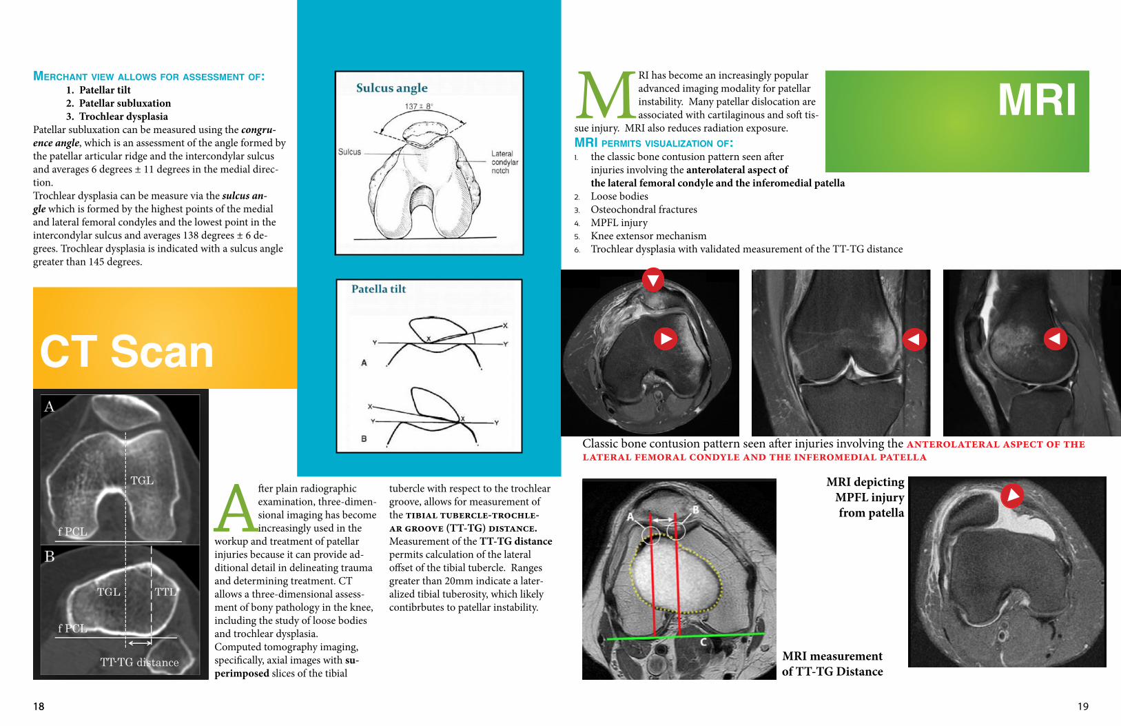
MErCHANT vIEw AllOwS fOr ASSESSMENT Of: 1. Patellar tilt 2. Patellar subluxation
3. Trochlear dysplasiaPatellar subluxation can be measured using the congru-ence angle, which is an assessment of the angle formed by the patellar articular ridge and the intercondylar sulcus and averages 6 degrees ± 11 degrees in the medial direc-tion. Trochlear dysplasia can be measure via the sulcus an-gle which is formed by the highest points of the medial and lateral femoral condyles and the lowest point in the intercondylar sulcus and averages 138 degrees ± 6 de-grees. Trochlear dysplasia is indicated with a sulcus angle greater than 145 degrees.
After plain radiographic examination, three-dimen-sional imaging has become increasingly used in the
workup and treatment of patellar injuries because it can provide ad-ditional detail in delineating trauma and determining treatment. CT allows a three-dimensional assess-ment of bony pathology in the knee, including the study of loose bodies and trochlear dysplasia. Computed tomography imaging, specifically, axial images with su-perimposed slices of the tibial
tubercle with respect to the trochlear groove, allows for measurement of the tibial tubercle-trochle-ar groove (TT-TG) distance. Measurement of the TT-TG distance permits calculation of the lateral offset of the tibial tubercle. Ranges greater than 20mm indicate a later-alized tibial tuberosity, which likely contibrbutes to patellar instability.
CT Scan
MRIMRI has become an increasingly popular advanced imaging modality for patellar instability. Many patellar dislocation are associated with cartilaginous and soft tis-
sue injury. MRI also reduces radiation exposure. MrI PErMITS vISuAlIzATION Of:1. the classic bone contusion pattern seen after
injuries involving the anterolateral aspect of the lateral femoral condyle and the inferomedial patella
2. Loose bodies3. Osteochondral fractures4. MPFL injury5. Knee extensor mechanism 6. Trochlear dysplasia with validated measurement of the TT-TG distance
Classic bone contusion pattern seen after injuries involving the anterolateral aspect of the lateral femoral condyle and the inferomedial patella
MRI measurement of TT-TG Distance
MRI depicting MPFL injury from patella
18
2120
The treatment goals are to prevent further episodes of patella instability, restore normal strength
and function in the knee joint and to prevent further damage to the articular cartilage of the knee joint.
Most first-time patellar dislocations are treated non-operatively unless there is evidence of an osteochon-dral fracture +/- significant MPFL injury. Patients are often immobi-lized for a short period of time after an acute dislocation. Rehabilita-tion in patellar dislocation should emphasize the recovery of full range of motion (ROM), strength, and proprioception. Gluteal muscle weakness should be addressed in addition to quadriceps function. Weight-bearing or closed-chain ex-ercises have been shown to produce a more rapid rehabilitative response than open-chain extension exer-cise. Patients can return to sports when they have regained full range of motion, have no effusion in the knee joint and have 80% quadriceps strenght. This typically takes at least 3 months.
Operative intervention is appropri-ate for those recurrentpatellar subluxations or disloca-tions, and in those whereconservative treatment has failed. More than 100 different operations have been described for thetreatment of patellar instability, and
these procedures typicallyinvolve a combination of lateral release, medial imbrication,distal realignment, and antero-medialization of the tibial tuber-cle. Determining the appropriate intervention for recurrent patellar dislocation requires locating the causative pathology as:1. Trochlear Dysplasia2. Proximal SoftTisue Compro-
mise3. Lateralized tibial Tuberlcle4. Patella alta5. Malalignment6. Generalized Ligamentous Lax-
ityIf multiple pathologies exist, the patient may require multiple proce-dures.
lATErAl rElEASEHistorically, a lateral retinacular release was a commonly performed procedure with the belief that a tight lateral retinaculum predis-posed patients to lateral patellar subluxation and dislocation. Iso-lated lateral releases for patellar instability can be complicated by medial patellar instability if the re-lease extends into, and detaches, the vastus lateralis obliquus. The lateral retinaculum contributes to 10% of medial stability. The addition of a lateral release to medialsoft-tissue repairs has been shown to actually decrease the force reaquired to dislocate the patella, compared to medial repair alone. At this time, lateral releases are recommended for treating patients with a stable patella and excessive lateral pressure associated with an increase in lateral patellar tilt. Furthermore, lateral release may
be performed in combination with a medial-sided procedure such as a medial plication or a reconstruc-tion of the medial patellofemoral ligament and/or if there is osseous malalignment. They are often re-quired in congenital patellar dislo-cation surgery due to the extent of the pathology often present in these patients.
MEDIAl rEPAIr:There are a multitude of surgical options that have been described to stabilize the medial side of the knee including direct repair, imbrication or plication of the medial retinac-ular structures. These procedures can be performed as an open +/- arthroscopic technique.
MPfl rECONSTruCTION:Since the recognition of the impor-tance of the MPFL, there has been increasing interest in treating this important medial patellar stabilizer.There are a number of surgical techniques described to reconstruct the MPFL as well as a number of graft options including auto-graft hamstrings, allograt tissue and synthetic grafts. The varying techniques all offer satisfactory results. The most important factor to ensure a good outcome includes anatomic reconstruction and en-suring that care is taken to tension the MPFL properly.
TIBIAl TuBErClE TrANSfEr:There are several described tech-niques to realign the tibial tuberle including Elmslie Trillat, Fulkerson, and distalization. These procedures are geared toward correcting a large Q angle or an increased tibial tu-
Treatmentntbercle to trochlear groove (TT-TG)offset. The classic Elmslie Trillat proce-dure involves a lateral retinacular release, emdial capsule reefing and medial transposition of the anterior tibial tubercle hinged on a distal periosteal attachment. One poten-tial complication of a classic Elms-lie-Trillat procedure is overloading the medial patellar articular surgace which may lead to patellofemoral arthritis.Another popular tibial tubercle transfer is the Fulkerson procedure. This procedure involves medializa-tion of the tibial tubercle to correct the Q angle and anteriorization of the tuberlce which elevates the dis-tal pole of the patella. By elevating
the distal pole of the patella, there is a reduction in the contact on the distal patella during early knee flexion.Finally, distalization of the tibial tu-bercle is performed in patients with patella alta and instability. Care must be taken not to overly medial-ize the tubercle.
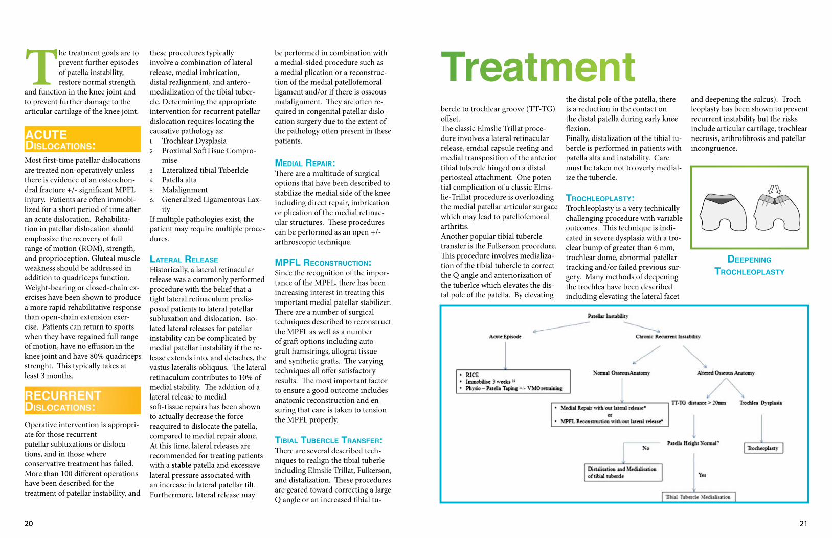
TrOCHlEOPlASTY:Trochleoplasty is a very technically challenging procedure with variable outcomes. This technique is indi-cated in severe dysplasia with a tro-clear bump of greater than 6 mm, trochlear dome, abnormal patellar tracking and/or failed previous sur-gery. Many methods of deepening the trochlea have been described including elevating the lateral facet
and deepening the sulcus). Troch-leoplasty has been shown to prevent recurrent instability but the risks include articular cartilage, trochlear necrosis, arthrofibrosis and patellar incongruence.
DEEPENINgTrOCHlEOPlASTY
ACuTEDISlOCATIONS:
rECurrENTDISlOCATIONS:
20

2322
2524

2726
2928

3130
3332

3534

3736

3938

4140

42