pathogenesis,immunology,anddiagnosisof...
TRANSCRIPT

Hindawi Publishing CorporationClinical and Developmental ImmunologyVolume 2011, Article ID 814943, 17 pagesdoi:10.1155/2011/814943
Review Article
Pathogenesis, Immunology, and Diagnosis ofLatent Mycobacterium tuberculosis Infection
Suhail Ahmad
Department of Microbiology, Faculty of Medicine, Kuwait University, P.O. Box 24923, Safat 13110, Kuwait
Correspondence should be addressed to Suhail Ahmad, suhail [email protected]
Received 6 September 2010; Accepted 28 October 2010
Academic Editor: James Triccas
Copyright © 2011 Suhail Ahmad. This is an open access article distributed under the Creative Commons Attribution License,which permits unrestricted use, distribution, and reproduction in any medium, provided the original work is properly cited.
Phagocytosis of tubercle bacilli by antigen-presenting cells in human lung alveoli initiates a complex infection process byMycobacterium tuberculosis and a potentially protective immune response by the host. M. tuberculosis has devoted a large partof its genome towards functions that allow it to successfully establish latent or progressive infection in the majority of infectedindividuals. The failure of immune-mediated clearance is due to multiple strategies adopted by M. tuberculosis that blunt themicrobicidal mechanisms of infected immune cells and formation of distinct granulomatous lesions that differ in their ability tosupport or suppress the persistence of viable M. tuberculosis. In this paper, current understanding of various immune processesthat lead to the establishment of latent M. tuberculosis infection, bacterial spreading, persistence, reactivation, and waning orelimination of latent infection as well as new diagnostic approaches being used for identification of latently infected individualsfor possible control of tuberculosis epidemic are described.
1. Introduction
Tuberculosis (TB) has afflicted mankind from the timeimmemorial. Evidence of spinal disease has been foundin Egyptian mummies of several thousand years BC andreferences to TB are found in ancient Babylonian andChinese writings. Recent molecular genetic studies haveshown that Mycobacterium tuberculosis, the most commoncause of TB in humans worldwide, has a progenitor ∼3million years old [1]. Mycobacterium tuberculosis is a memberof the M. tuberculosis complex (MTBC) which includes sixother closely related species: M. bovis, M. africanum, M.microti, M. pinnipedii, M. caprae, and M. canetti. Although allMTBC members are obligate pathogens and cause TB, theyexhibit distinct phenotypic properties and host range. TheMTBC members are genetically extremely closely related, thegenome of M. tuberculosis shows <0.05% difference with M.bovis, the latter species primarily infects cattle but can alsocause TB in other mammals including humans [2, 3].
Tuberculosis is one of the most prevalent infections ofhuman beings and a formidable public health challenge thatshows little sign of abating. The disease contributes consider-ably to illness and death around the world, exacting a heavytoll on the world’s most vulnerable citizens. The current TB
epidemic is being sustained and fuelled by two importantfactors: the human immunodeficiency virus (HIV) infectionand its association with active TB disease and increasingresistance of Mycobacterium tuberculosis strains to the mosteffective (first-line) anti-TB drugs [4]. Other contributingfactors include population expansion, poor case detectionand cure rates in impoverished countries, active transmissionin overcrowded hospitals, prisons, and other public places,migration of individuals from high-incidence countriesdue to wars or famine, drug abuse, social decay, andhomelessness. Active disease patients with sputum smear-positive pulmonary TB are the main source of infection ina community. Primary infection with M. tuberculosis leads toclinical disease in only∼10% of individuals. In the remainingcases, the ensuing immune response arrests further growthof M. tuberculosis. However, the pathogen is completelyeradicated in only∼10% people, while the immune responsein the remaining∼90% individuals only succeeds in contain-ment of infection as some bacilli escape killing by bluntingthe microbicidal mechanisms of immune cells (such asphagosome-lysosome fusion, antigen presentation by MHCclass I, class II, and CD1 molecules, production of nitricoxide, and other reactive nitrogen intermediates) and remainin nonreplicating (dormant or latent) state in old lesions.

2 Clinical and Developmental Immunology
The process is termed as latent tuberculosis infection (LTBI),and the dormant bacilli retain the ability to resuscitate andto cause active TB if a disruption of immune response (asin HIV infection) occurs. The World Health Organization(WHO) has estimated that one-third of the total worldpopulation is latently infected with M. tuberculosis and5%–10% of the infected individuals will develop active TBdisease during their life time [4, 5]. However, the risk ofdeveloping active disease is 5%–15% every year and lifetimerisk is∼50% in HIV coinfected individuals [4, 6]. Most of theactive disease cases in low TB incidence countries arise fromthis pool of latently infected individuals.
According to WHO estimates, 9.27 million new activedisease cases corresponding to an estimated incidence of139 per 100,000 population occurred throughout the worldin 2007 [4]. Only 5.5 million of 9.27 million cases of TB(new cases and relapse cases) were notified to nationaltuberculosis programs of various countries, while the restwere based on assessments of effectiveness of surveillancesystems. The highest number of TB cases occurred in Asia(55%) followed by Africa (31%). The highest incidence rate(363 per 100,000 population) was recorded for the Africanregion, mainly due to high prevalence of HIV infection. Thesix most populous countries of Asia (China, India, Indonesia,Pakistan, Bangladesh, and Philippines) accounted for >50%of all TB cases worldwide. An estimated 1.37 million (15%)of incident TB cases in 2007 were coinfected with HIV.Nearly 80% of the HIV-infected TB patients were living inthe African region [4]. Globally, 13.7 million total prevalentTB cases were recorded in 2007 corresponding to 206 casesper 100 000 population that resulted in 1.756 million deaths(including 456 000 among TB patients coinfected with HIV)[4]. Nearly 500 000 cases of multidrug-resistant TB (MDR-TB, defined as infection with M. tuberculosis strains resistantat least to the two most important first-line drugs, rifampinand isoniazid) occurred in 2007 [4]. By the end of 2008,extensively drug-resistant TB (XDR-TB; defined as MDR-TB strains additionally resistant to a fluoroquinolone and aninjectable agent such as kanamycin, amikacin, viomycin, orcapreomycin) has been found in 55 countries and territoriesof the world [4]. While MDR-TB is difficult and expensive totreat, XDR-TB is virtually an untreatable disease in most ofthe developing countries [7].
Population-based studies have shown that some indi-viduals are more at risk of acquiring infection and devel-oping active disease than others. Active transmission alsooccurs more frequently in small households and crowdedplaces in countries with a high incidence of TB [8, 9].Molecular epidemiological studies have shown that thereare distinct differences in the disease presentation andpopulation demographics in low TB incidence and high TBincidence countries. In several African and Asian countries,where the transmission of M. tuberculosis has been stable orincreased in the last few years, the incidence rate is highestamong young adults with most cases resulting from recentepisodes of infection or reinfection. On the contrary, inlow TB incidence countries of Western Europe and NorthAmerica, a higher proportion of cases occur in older patientsor among immigrants from high TB incidence countries
[8, 10]. Pulmonary TB accounts for >85% of active TBcases in high TB incidence countries due to higher ratesof active transmission, while extrapulmonary TB is alsocommon in low TB incidence countries of the developedworld, particularly among HIV-infected individuals andimmigrants originating from TB endemic countries [11, 12].
2. Transmission of M. tuberculosis Infection
Tuberculosis is a communicable disease and patients withpulmonary TB are the most important source of infection.Infection is initiated by inhalation of droplet nuclei, whichare particles of 1–5 μm in diameter containing M. tuberculo-sis, expectorated by patients with active pulmonary TB (openTB), typically when the patient coughs. The droplet nuclei,due to their small size, can remain suspended in the air forseveral minutes to hours. The risk of infection (Figure 1)is dependant on several factors such as the infectiousnessof the source case, the closeness of contact, the bacillaryload inhaled, and the immune status of the potential host[8–10]. The primary route of infection involves the lungs.Inhaled droplet nuclei avoid the defenses of the bronchidue to their small size and penetrate into the terminalalveoli where they are engulfed by phagocytic immunecells (macrophages and dendritic cells). M. tuberculosiscan also infect nonphagocytic cells in the alveolar spaceincluding M cells, alveolar endothelial, and type 1 andtype 2 epithelial cells (pneumocytes) [13–19]. In the earlyphase of infection, M. tuberculosis, internalized by phagocyticimmune cells, replicates intracellularly, and the bacteria-laden immune cells may cross the alveolar barrier to causesystemic dissemination [14, 15]. The intracellular replicationand simultaneous dissemination of the pathogen to the pul-monary lymph nodes and to various other extrapulmonarysites occur prior to the development of the adaptive immuneresponses. This exemplifies the extraordinary ability of M.tuberculosis to establish a protected niche where they canavoid elimination by the immune system and to persistindefinitely [20, 21].
In the vast majority of the infected individuals, aneffective cell-mediated immune response develops 2–8 weeksafter infection that stops further multiplication of thetubercle bacilli (Figure 1). The activated T lymphocytes,macrophages, and other immune cells form granulomasthat wall off the growing necrotic tissue limiting furtherreplication and spread of the tubercle bacilli. Most of theM. tuberculosis are killed in the caseating granulomas, anddisease progression is arrested. However, the pathogen is notcompletely eradicated in some individuals as M. tuberculosishas evolved effective strategies to evade the immune responseresulting in survival and persistence of some bacilli in anonreplicating state in the host (LTBI) [8, 21, 22]. In supportof this hypothesis, M. tuberculosis has been cultured andpresence of M. tuberculosis DNA has been demonstratedfrom lung tissues of individuals who died from other diseasesand who did not exhibit any pathological sign of TB disease[23, 24]. Furthermore, a recent report showing transmissionof infection from father to son in Denmark in 1961 and
Clinical and Developmental Immunology 3
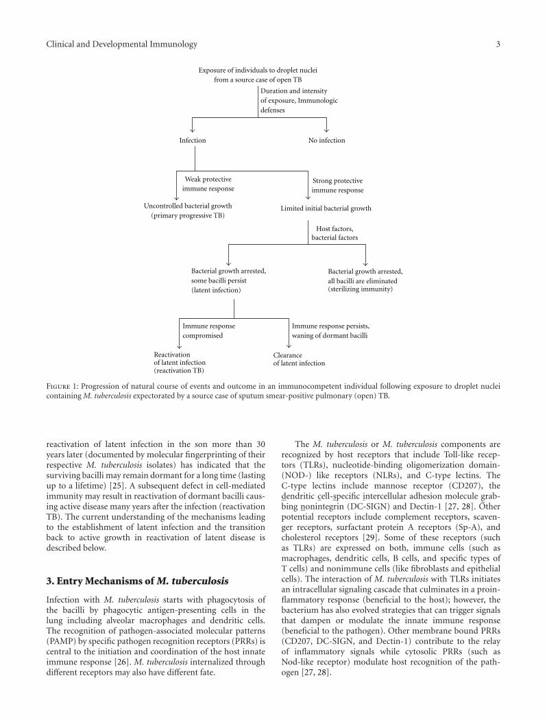
Exposure of individuals to droplet nucleifrom a source case of open TB
Infection No infection
Duration and intensityof exposure, Immunologicdefenses
Weak protectiveimmune response
Uncontrolled bacterial growth(primary progressive TB)
Strong protectiveimmune response
Limited initial bacterial growth
Bacterial growth arrested,some bacilli persist(latent infection)
Host factors,bacterial factors
Immune response persists,waning of dormant bacilli
Immune responsecompromised
Clearanceof latent infection
Reactivationof latent infection(reactivation TB)
Bacterial growth arrested,all bacilli are eliminated(sterilizing immunity)
Figure 1: Progression of natural course of events and outcome in an immunocompetent individual following exposure to droplet nucleicontaining M. tuberculosis expectorated by a source case of sputum smear-positive pulmonary (open) TB.
reactivation of latent infection in the son more than 30years later (documented by molecular fingerprinting of theirrespective M. tuberculosis isolates) has indicated that thesurviving bacilli may remain dormant for a long time (lastingup to a lifetime) [25]. A subsequent defect in cell-mediatedimmunity may result in reactivation of dormant bacilli caus-ing active disease many years after the infection (reactivationTB). The current understanding of the mechanisms leadingto the establishment of latent infection and the transitionback to active growth in reactivation of latent disease isdescribed below.
3. Entry Mechanisms of M. tuberculosis
Infection with M. tuberculosis starts with phagocytosis ofthe bacilli by phagocytic antigen-presenting cells in thelung including alveolar macrophages and dendritic cells.The recognition of pathogen-associated molecular patterns(PAMP) by specific pathogen recognition receptors (PRRs) iscentral to the initiation and coordination of the host innateimmune response [26]. M. tuberculosis internalized throughdifferent receptors may also have different fate.
The M. tuberculosis or M. tuberculosis components arerecognized by host receptors that include Toll-like recep-tors (TLRs), nucleotide-binding oligomerization domain-(NOD-) like receptors (NLRs), and C-type lectins. TheC-type lectins include mannose receptor (CD207), thedendritic cell-specific intercellular adhesion molecule grab-bing nonintegrin (DC-SIGN) and Dectin-1 [27, 28]. Otherpotential receptors include complement receptors, scaven-ger receptors, surfactant protein A receptors (Sp-A), andcholesterol receptors [29]. Some of these receptors (suchas TLRs) are expressed on both, immune cells (such asmacrophages, dendritic cells, B cells, and specific types ofT cells) and nonimmune cells (like fibroblasts and epithelialcells). The interaction of M. tuberculosis with TLRs initiatesan intracellular signaling cascade that culminates in a proin-flammatory response (beneficial to the host); however, thebacterium has also evolved strategies that can trigger signalsthat dampen or modulate the innate immune response(beneficial to the pathogen). Other membrane bound PRRs(CD207, DC-SIGN, and Dectin-1) contribute to the relayof inflammatory signals while cytosolic PRRs (such asNod-like receptor) modulate host recognition of the path-ogen [27, 28].
4 Clinical and Developmental Immunology
The TLR engagement, particularly TLR2 and TLR4,with M. tuberculosis/M. tuberculosis component is an earlyevent in the interaction of the pathogen with host cellsand TLR signaling is the main arm of the innate immuneresponse during M. tuberculosis infection [27, 28, 30]. TheTLR polymorphisms regulate the innate immune responseto mycobacterial lipopeptides and clinical susceptibility topathogens [31]. Typically, signals generated by the inter-actions of TLRs with ligands on M. tuberculosis inducethe activation of proinflammatory and antimicrobial innateimmune response.
The M. tuberculosis cell envelope is composed of a cellwall that is covered with a thick waxy mixture of lipidsand polysaccharides and is characterized by a high contentof mycolic acids. The most important M. tuberculosis cellsurface ligands that interact with TLRs and other receptorsinclude the 19 and 27 kDa lipoproteins, 38 kDa glycol-ipoprotein, the lipomannan (LM) and mannose-capped li-poarabinomannan (ManLAM) [32–34]. Other ligandsinclude LprA and LprG lipoproteins [35, 36] and, perhapsalso, surface-exposed mammalian cell entry (Mce) proteinsencoded by mce1 and mce3 operons [37–39]. The interactionof M. tuberculosis ligand(s) with TLRs eventually resultsin activation of nuclear transcription factor (NF)-κB andproduction of proinflammatory cytokines such as tumornecrosis factor (TNF)-α, interleukin (IL)-1, IL-12, chemok-ines, and nitric oxide through either myeloid differentiationprimary response protein 88 (MyD88)-dependant orMyD88-independent pathway [27, 34, 40, 41].
Restricting TLR-induced proinflammatory signals isessential to avoid the risk of producing excessive inflamma-tion that could damage host tissues. A family of receptortyrosine kinases termed Tyro3/Axl/Mer (TAM) provide anegative feedback mechanism to both TLR-mediated andcytokine-driven proinflammatory immune responses [42].This property has been exploited by M. tuberculosis to itsadvantage. The 19 kDa lipoprotein of M. tuberculosis is anagonist of the TLR2 and modulates the innate immunity andantigen presenting cell function [32]. Studies have shownthat prolonged TLR2 signaling by lipoproteins of M. tuber-culosis inhibits major histocompatibility complex (MHC)-IIexpression and processing of antigens by macrophages [43,44]. Thus, a subset of infected macrophages with decreasedantigen-presenting cell function may be unable to present M.tuberculosis antigens to CD4+ T cells resulting in insufficientactivation of effector T cells leading to evasion of immunesurveillance and creation of niches where M. tuberculosissurvives and persists [27, 32].
The mannose receptors interact with ManLAM presentin the cell envelop of M. tuberculosis. The phagocytosis oftubercle bacilli by macrophages through mannose receptor isassociated with an anti-inflammatory response as ManLAMinhibits mannose receptor-dependant IL-12 production.This inhibition of macrophage response to M. tuberculosispromotes infection and subsequent survival of M. tuber-culosis in macrophages. The ManLAM exerts its effectson phagolysosome maturation by limiting phagosome-lysosome fusion [45, 46].
4. Immune Response of the Host toM. tuberculosis
The alveolar macrophages, after entry of M. tuberculo-sis, produce inflammatory cytokines and chemokines thatserve as a signal for infection. The monocytes, neutrophils,and lymphocytes migrate to the focal site of infection,but they are unable to kill the bacteria efficiently. Dur-ing this time, the bacilli resist the bactericidal mecha-nisms of the macrophage (phagolysosome) by prevent-ing phagosome-lysosome fusion, multiply in the phago-some, and cause macrophage necrosis [47]. The releasedbacilli multiply extracellularly, are phagocytosed by anothermacrophage that also fails to control the growth of M.tuberculosis, and likewise are destroyed. In the meantime,dendritic cells with engulfed bacilli mature, migrate tothe regional lymph node, and prime T cells (both CD4+
and CD8+) against mycobacterial antigens [48]. The spe-cific immune response produces primed T cells whichmigrate back to the focus of infection, guided by thechemokines produced by the infected cells. The accumula-tion of macrophages, T cells, and other host cells (dendriticcells, fibroblasts, endothelial cells, and stromal cells) leadsto the formation of granuloma at the site of infection[49].
The granuloma formation walls off tubercle bacillifrom the rest of the lung tissue, limits bacterial spread,and provides microenvironment for interactions amongmacrophages and other cells of the immune system and thecytokines produced by these cells. The CD4+ T cells pro-ducing interferon-γ (IFN-γ) recognize infected macrophagespresenting antigens from M. tuberculosis and kill them [50].The infection progression is halted; however, some resistantbacilli capable of surviving under the stressful conditionsgenerated by the host escape killing and enter a stateof dormancy and persist by avoiding elimination by theimmune system [22, 51, 52]. Recent studies have shown thatdifferences exist in the immunological response mountedby different individuals that lead to the formation ofphysiologically distinct granulomatous lesions in individualsexposed to M. tuberculosis. Some of these lesions suppress(sterilizing immunity) while others promote the persistenceof viable M. tuberculosis in the microenvironment [53].Low-dose infection of cynomolgus macaques that reproducethe clinical characteristics of human latent TB leads to theformation of at least two types of tuberculous granuloma [54,55]. Histopathological studies have shown that the classiccaseous granuloma are composed of epithelial macrophages,neutrophils, and other immune cells surrounded by fibrob-lasts. The central caseous necrotic region in this type ofgranuloma consists of dead macrophages/other cells and ishypoxic with M. tuberculosis residing inside macrophages inthe hypoxic center [55, 56]. The other kind of granulomasseen in latent tuberculosis in both humans and cynomolgusmacaques are fibrotic lesions, composed almost exclusivelyof fibroblasts that contain very few macrophages [55].However, it is not known at present whether M. tuberculosisis located inside macrophages or in the fibrotic area in theselesions.

Clinical and Developmental Immunology 5
The microenvironment of the granuloma (hypoxia, lowpH, presence of nitric oxide and carbon monoxide, etc.)increases the expression of several M. tuberculosis genesinvolved in dormancy induction [57–60]. Recent findingsof formation of spore-like structures in M. bovis BCG, M.marinum, and M. smegmatis in response to prolonged sta-tionary phase or nutrient starvation suggest that sporulationmay be a general mechanism for mycobacterial dormancy[60–62]. The dormant bacilli can inhabit the granulomaduring the lifetime of the host, but are able to resuscitate(or germinate) in the event of local immunodepression. Thelatent infection in a person without overt signs of the diseaseis indicated by the delayed-type hypersensitivity (DTH)response to purified protein derivative (PPD) prepared fromculture filtrates of M. tuberculosis (tuberculin skin test)[8].
5. Specific Roles of Immune Cells andCytokines in M. tuberculosis Infection
Studies in animal models and in humans have demonstratedthat a wide range of immune components are involved inan effective immune response against M. tuberculosis. Theseinclude, beside macrophages and dendritic cells, αβ-T cells(both CD4+ and CD8+), CD1 restricted T cells, γδ-T cells,and cytotoxic T cells, as well as the cytokines produced bythese immune cells [22, 63, 64]. The most important amongthese are CD4+ T cells and the cytokine IFN-γ. AlthoughCD4+ T cells along with CD8+ T cells and the naturalkiller (NK) cells are the major producers of IFN-γ, studiescarried out in CD4+ deficient mice have shown that it is theearly production of IFN-γ by CD4+ T cells and subsequentactivation of macrophages that determine the outcome ofinfection [65, 66]. The CD4+ T cells also play other roles inthe defense against infection that is independent of IFN-γproduction. Depletion of CD4+ T cells was associated withthe reactivation of infection in a chronically infected miceand resulted in increasing pathological features and death,even though IFN-γ levels were still high due to a strongresponse from CD8+ T cells and normal levels of induciblenitric oxide synthase (iNOS) [67].
The CD4+ T cells carry out several functions thatare important to control infection in the granuloma.These include apoptosis of infected macrophages throughFas/Fas ligand interaction, production of other cytokines(such as IL-2 and TNF-α), induction of other immunecells (macrophages or dendritic cells) to produce otherimmunoregulatory cytokines such as IL-10, IL-12, and IL-15, and activation of macrophages through direct contact viaCD40 ligand [63, 66, 68, 69]. The CD4+ T cells also appear tobe critical for the cytotoxic function of CD8+ T cells that ismediated by IL-15 [66, 70]. It has also been shown that CD4+
T cells can control the intracellular growth of M. tuberculosisby a nitric oxide-dependent mechanism that is independentof IFN-γ production [66, 71]. Thus, CD4+ T cells, in additionto early production of IFN-γ appear to have several othersecondary functions that are critical in the control of M.tuberculosis infection.
The CD8+ T-cells, in addition to producing IFN-γ andother cytokines, may also be cytotoxic for M. tuberculosis-infected macrophages, and thus play an important role inproviding immunity to TB. The CD8+ T-cells can directlykill M. tuberculosis via granulysin, and facilitate the controlof both the acute as well as chronic infection [66, 72]. Theabundant presence of M. tuberculosis-specific CD8+ T cellsin latently infected individuals shows that the CD8+ T cellsalso have a role in the control of latent infection. This isalso supported by reactivation of latent infection followingdepletion of CD8+ T cells in the Cornell model of latent TB[73].
Studies in primate models of TB have shown thatunconventional T cells such as CD1 restricted T cells, and γδ-T cells also contribute to the protection against TB [64, 74,75]. The CD1 restricted T cells recognize the glycolipids suchas LAM that are abundant in the mycobacterial cell wall whileγδ-T cells recognize small metabolites containing phosphate(phospholigands) [74]. Although it is well established thatmycobacterial antigens in the phagosome of macrophages ordendritic cells are picked up by the MHC class II moleculesand presented to CD4+ T cells, studies have shown that thephagosomal membrane is also equipped with the MHC classI processing machinery [76, 77]. Also, CD1 proteins havethe capability to present lipid antigens and lipopeptides to Tcells, and thus play important roles in the immune responseagainst lipid-rich M. tuberculosis [75, 78, 79]. Further, thevesicles formed due to apoptosis of M. tuberculosis-infectedmacrophages and containing mycobacterial antigens suchas ManLAM, lipoproteins, and so forth are taken up bydendritic cells and presented to the T cells through the MHCclass I and CD1 molecules [75, 78, 80].
The IFN-γ is the key cytokine for a protective immuneresponse against M. tuberculosis. Humans and mice defectivein IFN-γ or IFN-γ receptor genes are more susceptible toM. tuberculosis infection [63, 66, 81]. The IFN-γ, producedmainly by CD4+, CD8+ T cells, and the NK cells, synergizeswith TNF-α and activates macrophages to kill intracellularbacilli. The IFN-γ also augments antigen presentation,leading to recruitment of CD4+ T-cells and/or cytotoxicCD8+ T-cells, which participate in mycobacterial killingand also prevents exhaustion of memory T cells [63, 82].Furthermore, IFN-γ induces the transcription of more than200 genes in macrophages including the upregulation ofMHC class II expression and the production of antimicrobialeffectors such as oxygen radicals and nitric oxide. A majoreffector mechanism responsible for the antimicrobial activityof IFN-γ in association with TNF-α is the induction ofthe production of nitric oxide and other reactive nitrogenintermediates (RNI) by macrophages via iNOS [63, 66, 83].However, some M. tuberculosis factor(s), such as the 19-kDalipoprotein, have the potential to attenuate the response ofmacrophages to IFN-γ by blocking the transcription of asubset of IFN-γ-responsive genes (Table 1) [44, 84, 85].
TNF-α, produced by macrophages, dendritic cells, andT-cells, is another cytokine that has a major protective roleagainst M. tuberculosis infection both in mice and humans[104, 105]. Paradoxically, TNF-α also contributes signifi-cantly to the development of immunopathology associated
6 Clinical and Developmental Immunology
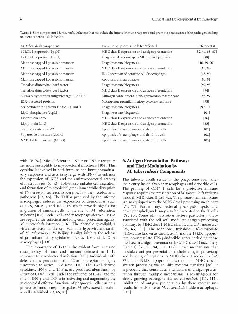
Table 1: Some important M. tuberculosis factors that modulate the innate immune response and promote persistence of the pathogen leadingto latent tuberculosis infection.
M. tuberculosis component Immune cell process inhibited/affected Reference(s)
19 kDa Lipoprotein (LpqH) MHC class II expression and antigen presentation [32, 44, 85–87]
19 kDa Lipoprotein (LpqH) Phagosomal processing by MHC class I pathway [88]
Mannose capped lipoarabinomannan Phagolysosome biogenesis [46, 89, 90]
Mannose capped lipoarabinomannan MHC class II expression and antigen presentation [85, 90]
Mannose capped lipoarabinomannan IL-12 secretion of dentritic cells/macrophages [45, 90]
Mannose capped lipoarabinomannan Apoptosis of macrophages [90, 91]
Trehalose dimycolate (cord factor) Phagolysosome biogenesis [92, 93]
Trehalose dimycolate (cord factor) MHC class II expression and antigen presentation [94]
6-kDa early secreted antigenic target (ESAT-6) Pathogen containment in phagolysosome/macrophage [95–97]
ESX-1 secreted proteins Macrophage proinflammatory cytokine response [98]
Serine/threonine protein kinase G (PknG) Phagolysosome biogenesis [99, 100]
Lipid phosphatase (SapM) Phagolysosome biogenesis [101]
Lipoprotein LprA MHC class II expression and antigen presentation [36]
Lipoprotein LprG MHC class II expression and antigen presentation [35]
Secretion system SecA2 Apoptosis of macrophages and dendritic cells [102]
Superoxide dismutase (SodA) Apoptosis of macrophages and dendritic cells [102]
NADH dehydrogenase (NuoG) Apoptosis of macrophages and dendritic cells [103]
with TB [52]. Mice deficient in TNF-α or TNF-α receptorsare more susceptible to mycobacterial infections [104]. Thiscytokine is involved in both immune and immunomodula-tory responses and acts in synergy with IFN-γ to enhancethe expression of iNOS and the antimycobacterial activityof macrophages [63, 83]. TNF-α also initiates cell migrationand formation of microbicidal granulomas while disruptionof TNF-α responses leads to overgrowth of the mycobacterialpathogens [63, 66]. The TNF-α produced by the infectedmacrophages induces the expression of chemokines, suchas IL-8, MCP-1, and RANTES which provide signals formigration of immune cells to the sites of M. tuberculosisinfection [106]. Both T cell- and macrophage-derived TNF-αare required for sufficient and long-term protection againstM. tuberculosis infection [107]. The phenolic glycolipid, avirulence factor in the cell wall of a hypervirulent strainof M. tuberculosis (W-Beijing family) inhibits the releaseof pro-inflammatory cytokines TNF-α, IL-6 and IL-12 bymacrophages [108].
The importance of IL-12 is also evident from increasedsusceptibility of mice and humans deficient in IL-12responses to mycobacterial infections [109]. Individuals withdefects in the production of IL-12 or its receptor are highlysusceptible to active TB disease [110]. The T-cell-derivedcytokines, IFN-γ and TNF-α, are produced abundantly byactivated CD4+ T cells under the influence of IL-12, and therole of IFN-γ and TNF-α in activating and augmenting themicrobicidal effector functions of phagocytic cells during aprotective immune response against M. tuberculosis infectionis well established [63, 66, 83].
6. Antigen Presentation Pathwaysand Their Modulation byM. tuberculosis Components
The tubercle bacilli reside in the phagosome soon aftertheir entry inside alveolar macrophages and dendritic cells.The priming of CD4+ T cells for a protective immuneresponse requires the presentation of M. tuberculosis antigensthrough MHC class II pathway. The phagosomal membraneis also equipped with the MHC class I processing machinery[74, 77]. Further, mycobacterial glycolipids, lipids, andother phospholigands may also be presented to the T cells[78, 80]. Some M. tuberculosis factors particularly thoseassociated with the cell wall modulate antigen-processingpathways by MHC class I, MHC class II, and CD1 molecules[28, 63, 111]. The ManLAM, trehalose 6, 6′-dimycolate(TDM, also known as cord factor), and the 19 kDa lipopro-tein downregulate IFN-γ-inducible genes including thoseinvolved in antigen presentation by MHC class II machinery(Table 1) [32, 86, 94, 111, 112]. Other mechanisms thatmodulate antigen presentation include antigen processingand binding of peptides to MHC class II molecules [32,87]. The 19 kDa lipoprotein also inhibits MHC class Iantigen processing via Toll-like receptor signaling [88]. Itis probable that continuous attenuation of antigen presen-tation through multiple mechanisms is advantageous forslowly growing pathogens like M. tuberculosis [111, 112].Inhibition of antigen presentation by these mechanismsresults in persistence of M. tuberculosis inside macrophages[112].
Clinical and Developmental Immunology 7
7. Dampening of Other MacrophageFunctions by M. tuberculosis Components
The two major antimycobacterial mechanisms of macropha-ges include the generation of nitric oxide and other RNIwhich exert toxic effects on the bacilli and fusion of thephagosomes containing mycobacteria with lysosomes thatis bactericidal [83, 92, 113]. The T cell-derived cytokines,mainly IFN-γ and TNF-α, activate macrophages, whichthen generate nitric oxide and other RNI by iNOS andare mycobactericidal [83]. Direct demonstration of thepresence of nitrotyrosine, an RNI derived from tyrosine andperoxynitrite in the lungs of infected mice, has shown thatRNI are formed in tuberculous granuloma. Furthermore,inhibition of iNOS activity or disruption of iNOS gene,required for the production of RNI, not only abolished theprotective effect of RNI but also led to reactivation of latentinfection in mice [83]. Although these studies point towardsan essential role for iNOS in the control of both acuteand chronic persistent infection, the RNI generated throughthese mechanisms is not sufficient to eliminate the bacteriumcompletely.
The protective role of RNI in human TB has also beensuggested [92, 114]. Studies have shown that M. tuberculosishas evolved several strategies to evade the RNI toxicity. Ithas been shown that iNOS, a cytoplasmic protein, may berecruited to the phagosomes and this recruitment may beinhibited by M. tuberculosis [115]. The M. tuberculosis gene,alkyl hydroperoxide reductase subunit C (ahpC), detoxifies,in conjunction with some other proteins, the highly reactiveperoxynitrite anion (formed by the reaction of nitric oxidewith superoxide) [116]. Another potential mechanism forblunting the toxic effects of RNI is the presence of twohaemoglobin-like proteins encoded by glbN and glbO in M.tuberculosis. The glbN knockout mutant of M. bovis BCG washighly attenuated, and its growth, in vitro, was also inhibitedby nitric oxide under aerobic conditions [117]. Microarrayanalyses have shown that more than 30 M. tuberculosis genesare induced by RNI and hypoxia [59, 118]. Furthermore,hypoxia and inhibition of respiration by nitric oxide induce adormancy program in M. tuberculosis that leads to increasedsurvival and persistence of the pathogen in immune cells[59, 119].
8. Phagolysosome Maturation and ItsInhibition by M. tuberculosis Components
The phagocytosis of M. tuberculosis by macrophages isfollowed by the maturation of phagosomes containing thepathogen through a series of fusion and fission events withseveral endocytic vesicles that culminate in a phagolysosome[120, 121]. The fission-fusion events remodel the phago-somal membrane, and the recruitment of vacuolar-protontransporting ATPase (vH+-ATPase) lowers the internal pHthat allows lysosome-derived acid hydrolases to function effi-ciently for their microbicidal effect [122, 123]. Furthermore,phagosome maturation is dependant on Ca+2 signalingcascade that begins with phosphorylation of sphingosine
to sphingosine 1-phosphate by sphingosine kinase resultingin elevation of cytosolic [Ca+2] inside macrophages due torelease of Ca+2 from intracellular stores in the endoplasmicreticulum and continues through Ca+2-calmodulin complex-dependant activation of protein kinase II and phosphatidylinositol 3-kinase (PI-3K). The cascade culminates in phos-phorylation of phosphatidyl inositol to phosphatidyl inositol3-phosphate (PI-3P) by PI-3K in the phagosome membraneand maturation of phagosome to an acidic bactericidal com-partment (phagolysosome) after binding of early endosomalantigen-1 (EEA-1) to PI-3P [89, 124–126].
M. tuberculosis has also evolved several strategies toavoid the destruction by lysosomal enzymes by disrupt-ing the maturation of bacilli-containing phagosomes intophagolysosmes [46, 90, 92, 127, 128]. Exclusion of vH+-ATPase during maturation of phagosomes contributes to theacidification defect that prevents the fusion of phagosomeswith lysosomes [122]. Similarly, modulation of Ca+2 signal-ing cascade, such as SapM-mediated hydrolysis and inacti-vation of PI-3P, inhibits phagosome maturation leading toenhanced intracellular survival of M. tuberculosis (Table 1)[101, 126–128].
Other M. tuberculosis components such as ManLAM andTDM (Table 1) also affect phagosome maturation by inter-fering with the tethering and fusion machinery of vesiculartransport in mammalian cells and promote persistence ofthe bacterium inside macrophages [46, 89, 90, 93, 101].The targets include the soluble N-ethylmaleimide-sensitivefactor-attachment protein receptors (SNAREs), the tetheringproteins (such as EEA-1), and the Rab family of GTPases [89,120, 126, 128]. Some of the membrane trafficking processesaffected by mycobacterial factors are also affected by HIVduring viral budding and this overlap partially contributestowards the synergism observed between AIDS and active TB[129]. Another mechanism by which mycobacteria interferewith phagosome maturation is by retention of the hosttryptophan aspartate rich coat protein (TACO) (homolog ofcoronin-1) on the cytoplasmic side of their phagosomes thatlikely inhibits the normal process of phagosome-lysosomefusion [99]. The serine/threonine protein kinase G encodedby pknG of M. tuberculosis (Table 1) is implicated as thepotential effector of the inhibition of phagosome-lysosomefusion [100, 111].
Another component of the antimicrobial repertoire ofmacrophages includes lysosomal killing of M. tuberculosismediated by ubiquitin-derived peptides [130]. The ubiquiti-nation destroys tubercle bacilli by autophagy as a ubiquitin-derived peptide impairs the membrane integrity of M.tuberculosis that allows nitric oxide to kill more efficiently.On the contrary, decreased outer membrane permeabilityprotects M. tuberculosis from killing by ubiquitin-derivedpeptides [131].
9. Apoptosis of Infected Macrophages and ItsInhibition by M. tuberculosis Components
The apoptosis of infected macrophages participates in hostdefense against infection as apoptotic vesicles containing
8 Clinical and Developmental Immunology
mycobacterial antigens are taken up by dendritic cells forCD8+ T cell activation by phagosome-enclosed antigens [79,80]. The CD8+ T cells activated by apoptotic vesicles fromM. tuberculosis-infected cells produce IFN-γ, which causesuninfected macrophages to produce RNI to effectively killintracellular M. tuberculosis. Several M. tuberculosis-derivedfactors are capable of modulating (activating as well asinhibiting) the apoptosis of infected macrophages throughdifferential expression of proapoptotic and antiapoptoticgenes [90]. The mycobacterial components modulatingapoptosis of macrophages usually target the caspase cascadeor the one involving TLRs. The M. tuberculosis componentsthat inhibit apoptosis include cell wall components, Man-LAM, virulence-related secretion system encoded by secA2that transports superoxide dismutase (encoded by sodA) tocontrol reactive oxygen intermediates, and NADH dehydro-genase (encoded by nuoG) (Table 1) [91, 102, 103]. Twosecretory proteins of M. tuberculosis encoded by Rv3654cand Rv3655c that inhibit apoptosis of macrophages havealso been identified recently [132]. By inhibiting apoptosisof macrophages, M. tuberculosis avoids host defenses andescapes from infected cells by causing necrotic cell death[133].
10. Escape of M. tuberculosis fromPhagosome/Phagolysosome
Although it has been known for quite some time thatM. tuberculosis survives in the phagosome by blocking(or slowing down) its maturation into phagolysosome andpersists, one of the mechanism by which it escapes fromphagosome/phagolysosome to infect other macrophages andother immune/alveolar cells has been elucidated recently.Initial subtractive hybridization-based studies identified agenomic region, termed region of difference 1 (RD1), thatwas present in all virulent M. tuberculosis and M. bovisstrains but was absent in the vaccine strain M. bovis BCG[2, 134, 135]. Subsequently, it was shown that RD1 is crucialfor the virulence of M. tuberculosis as it encoded proteinsthat formed a novel protein secretion system (ESX-1). ESX-1 (type VII secretion system) is involved in the export ofseveral M. tuberculosis proteins including two potent T cellantigens encoded by RD1 itself, the 6-kDa early secretedantigenic target (ESAT-6) (encoded by esxA) and 10-kDaculture filtrate protein (CFP-10) (encoded by esxB) that lacksignal sequences for their export [2, 134, 136–140].
The importance of ESX-1 secreted proteins in virulenceof M. tuberculosis has been shown by deletion of RD1 ordisruption of ESAT-6 from M. tuberculosis genome thatresulted in reduced virulence (spreading) both, in culturedmacrophages and in mice [140, 141]. Furthermore, theintroduction of RD1 genes in M. bovis BCG resulted inaltered colonial morphology, increased virulence in severelycombined immune deficient mice including the formationof granuloma, and longer persistence in immunocompetentmice [142]. In M. tuberculosis, ESAT-6 complexes withCFP-10 in 1 : 1 ratio before its export outside the cellbut can dissociate from its partner (CFP-10) at lower pH.
Individually, ESAT-6, but not CFP-10, can cause disruptionof artificial membranes as well as cytolysis [143–145]. ESAT-6 alone has also been shown to associate strongly withliposomes containing dimyristoylphosphatidylcholine andcholesterol (constituents of mammalian cell membranes)and causing destabilization and lysis of liposomes [95, 96,144].
The studies carried out by de Jonge et al. [96] have shownthat ESAT-6:CFP-10 complex secreted by live M. tuberculosisinside phagosome splits apart when tubercle bacilli arestressed following acidification of phagosome, and ESAT-6inserts itself into lipid bilayer, causing lysis and escape ofM. tuberculosis from phagosome. Further studies have shownthat ESAT-6 also induces apoptosis of macrophages via theextrinsic (caspase-dependent) pathway by formation of poresin cell membrane [146] and contributes (or helps) in thetranslocation of M. tuberculosis from the phagolysosomes tothe cytoplasm in myeloid cells [97]. More recently, ESAT-6has also been shown to cause cytolysis of type 1 and type2 alveolar epithelial cells. This ESAT-6-mediated cytolysiswas shown to help in the dissemination of M. tuberculosisthrough alveolar wall [19]. The ESAT-6 homolog fromMycobacterium marinum, the bacterium that causes tubercu-lous granuloma in zebrafish, has also been demonstrated tocause lysis of red blood cells and macrophages by formingpores in their membranes [147, 148]. The presence of acapsular layer containing high amounts of proteins that aresecreted via the ESX-1 secretion system including ESAT-6 has also been demonstrated in pathogenic mycobacteriarecently [98]. Furthermore, ESX-1-associated proteins in thecapsular layer enhanced the interaction of mycobacteria withmacrophages and also dampened proinflammatory cytokineresponse of macrophage. These studies have establishedthe role of ESX-1 secretion system and ESAT-6 proteinof M. tuberculosis in facilitating macrophage infection andsubsequent bacterial escape to infect other nearby cells(Table 1).
11. Persistence and Reactivation ofLatent TB Infection
The hallmark of M. tuberculosis infection in humans isthe inability of an otherwise effective immune responseto completely eliminate the pathogen. The tubercle bacillihave evolved multiple strategies to manipulate infected hostcells in order to evade or modify the ensuing immuneresponse so as to avoid elimination and thus persist in thehost. As described above, several M. tuberculosis factors,ManLAM and 19-kDa lipoprotein notable among them, havebeen identified that modulate antigen presentation pathwaysand either blunt the microbicidal functions of macrophagesand other immune cells (such as RNI) or prevent theirmaturation (phagolysosome) (Table 1).
Two experimental strategies have been employed to iden-tify other M. tuberculosis factors, which promote persistenceof the pathogen in immune cells including macrophages.One approach involves cloning of M. tuberculosis genes innonpathogenic mycobacteria and studying their increased

Clinical and Developmental Immunology 9
survival in macrophages or other mammalian cells whilethe other approach uses knockout mutants of M. tubercu-losis for selected genes for persistence of the pathogen inmacrophages and other immune cells. Several additionalM. tuberculosis factors promoting persistence or increasedsurvival in mammalian cells have been identified. Some ofthese factors include phospholipases encoded by plcA, plcB,plcC, and plcD [149], the two PhoP and PhoQ regulatoryproteins [21], phosphate-binding proteins PstS1 and PstS2[150], and proteins encoded by mce operons [151]. Thus,M. tuberculosis has devoted a large part of its genometowards functions that promote its intracellular survival inmammalian cells including macrophages.
Reactivation of latent infection requires latent M. tuber-culosis cells to exit dormancy. Several factors can triggerthe development of active disease from the reactivation ofremote infection, and this typically involve the weakeningof the immune system. HIV infection is the most importantsingle risk factor for progression to active disease in adultsas it causes depletion of CD4+ T cells and functionalabnormalities of CD4+ and CD8+ T-cells which play animportant role in providing protection against active TBdisease [4, 6]. Likewise, M. tuberculosis infection acceleratesthe progression of asymptomatic HIV infection to acquiredimmunodeficiency syndrome (AIDS) and eventually todeath. This is recognized in the current AIDS case definitionas pulmonary or extrapulmonary TB in HIV-infected patientis sufficient for the diagnosis of AIDS. Old age, malnutrition,and medical conditions that compromise the immune systemsuch as poorly controlled diabetes mellitus, renal failure, andtherapy with immunosuppressive drugs are other factors thatlead to immunodepression and reactivation of a dormantinfection [6, 8, 152, 153]. The reactivation TB can occur inany organ system in which the tubercle bacilli were seededduring the primary infection; however, in immunocompe-tent individuals, the reactivation usually occurs in the upperlobes, where higher oxygen pressure supports good bacillarygrowth. The lytic transglycosylases known as resuscitationpromoting factors (RPFs) and an endopeptidase (RipA)of M. tuberculosis have recently been recognized as vitalcomponents for revival from latency [154–156].
12. Current Dynamic Model of Latent Infection
The LTBI has traditionally been defined as infection withM. tuberculosis in foci within granuloma that remain innonreplicating state but retain their ability to come outof latency and cause active TB if and when a disruptionof the immune response occurs [57]. However, recentexperimental data supports a dynamic model of LTBI whereendogenous reactivation as well as damage response occursconstantly in immunocompetent individuals [157]. Themodel suggests that during infection, M. tuberculosis growswell inside phagosomes; however, some bacilli released fromnecrotic macrophages in extracellular milieu in developinggranulomas stop replicating. The arrest of bacterial growthoccurs even before an effective immune response has fullybeen developed due to hypoxic and acidic environment
(conditions that mimic stationary bacterial cultures) inthe extracellular milieu and release of bactericidal enzymesfrom dead macrophages and neutrophils. The activelygrowing bacillary population is eventually killed due to thedevelopment of an effective immune response; however,nonreplicating bacilli resist killing and survive [158, 159].
The model further suggests that some macrophages(foamy macrophages) also emerge during the chronicinflammatory process, as they have phagocytosed the cellulardebris rich in fatty acids and cholesterol derived fromcellular membranes [160, 161]. The foamy macrophages alsophagocytose extracellular nonreplicating lipid-rich M. tuber-culosis; however, the bacilli do not grow in the intracellularenvironment of activated macrophages but are also not killeddue to the nonreplicating state of the bacilli [162]. Thenonreplicating bacilli-laden foamy macrophages drain fromlung granuloma towards the bronchial tree and return to adifferent region of lung parenchyma due to aerosols gener-ated by inspired air and begin the infection process at thisnew location once again [157, 160, 161, 163]. In this dynamicprocess, reinfection in the upper lobe may have the chance tocause cavitary lesion. This is aided by higher oxygen pressurein the upper lobes that can support rapid extracellularbacillary growth resulting in bacillary concentration that cannot be controlled by the optimum immune response by thehost. The subsequent much stronger inflammatory responseleads to tissue destruction, liquefaction, and extracellularbacillary growth which amplifies the response further andcauses cavitation [157, 158].
The dynamic infection process leading to active dis-ease in the upper lobes has some parallels with immunereconstitution inflammatory syndrome and the active TBdisease that occurs in HIV-infected patients. The presenceof bacilli is tolerated by the HIV-infected patients withlow CD4+ cell counts as the host is unable to mountan inflammatory response needed to control their growth.However, the sudden increase in CD4+ T cells in AIDSpatients receiving highly active antiretroviral treatmentcauses an aggressive granulamatous response and activeTB disease [164, 165]. The possibility of slow clearance oflatent infection proposed by the dynamic infection modelhas also been supported by a recent study from Norwaycomprising a population of individuals exposed to a minimalrisk of active transmission of infection. Cohort analysisof data from National Tuberculosis Registry to calculaterates and changes in rates of active TB disease, amongpatients previously exposed to M. tuberculosis, has shownthat the rate of reactivated TB has progressively decreasedover time [166]. The study further suggested that the numberof individuals with latent infection could be reduced inhalf in approximately 9 years in populations in which newinfections are effectively prevented. The dynamic infectionmodel also explains how therapy for a relatively shorttime (9 months) with a single drug (isoniazid), active onlyagainst actively dividing bacilli [167], is highly effective fora latent infection that can possibly remain dormant forthe entire lifetime of the host. As isoniazid will preventepisodes of reinfection by bacilli resuscitated from dormancy,slow drainage and destruction of nonreplicating bacilli in
10 Clinical and Developmental Immunology
the stomach will eventually lead to clearance of latentinfection [157, 166].
13. Diagnosis of LatentM. tuberculosis Infection
The persons infected with M. tuberculosis may be identifiedby tuberculin skin test six to eight weeks after exposure to thebacilli. The test is based on a delayed-type hypersensitivity(DTH) response to a complex cocktail of M. tuberculosisantigens, known as purified protein derivative (PPD). Theinduration of more than 5 mm, recorded 48 to 72 hoursafter injection of PPD, is considered as positive. Surveysconducted with PPD skin test suggest that nearly a thirdof the world’s and half of Asia’s population is infected withM. tuberculosis [5]. Skin test reaction over 20 mm is usuallydue to active disease; however, a negative skin test in anactive TB patient may also result from anergy or incorrectadministration of the test. The tuberculin skin test lackssensitivity and specificity as it can not differentiate betweeninfection with M. tuberculosis and sensitization with otherenvironmental mycobacteria [5, 8]. Also, BCG vaccinationmay cause false-positive reactions, but these generally lastonly a few years after vaccination and are in the moderaterange (5 to 10 mm).
More sensitive and specific tests such as cell-mediatedimmunity-based interferon-gamma (IFN-γ) release assays(IGRAs) have also been developed that detect T cell responsesafter stimulation by two M. tuberculosis-specific antigens,early secreted antigenic target-6 (ESAT-6) and culture filtrateprotein-10 (CFP-10) [168–173]. The IGRAs have excellentspecificity as the antigens (ESAT-6 and CFP-10) used in theseassays are encoded by genes deleted in the vaccine strainM. bovis BCG and majority of environmental mycobacteriaof clinical relevance [134, 135, 170]. Another variationof conventional IGRAs has also been developed by usingflow cytometry [174]. Although flow cytometric approachhas an advantage over conventional IGRAs as a smallerblood volume (<1 ml) is required for testing, the assay haslimited utility in much of the developing world due to therequirement of technical expertise and the high cost of flowcytometers. The detection of significant levels of antibodiesto some M. tuberculosis-specific proteins has also been notedin latently infected individuals as well as in patients withactive TB disease but not in healthy subjects [175–178].However, antibody-based tests have not been used so far forthe detection of LTBI.
Two commercial IGRAs, whole blood, ELISA-based Quan-tiFERON (QFN)-TB Gold assay (Cellestis Ltd., Carnegie,Australia) and peripheral blood mononuclear cell (PBMC)and enzyme-linked immunospot (ELISPOT) technology-based T SPOT-TB (Oxford Immunotec, Oxford, UK) testhave also been developed and approved by Food and DrugAdministration (FDA) for detecting LTBI. The tests wereinitially based on stimulation of T lymphocytes with ESAT-6and CFP-10 proteins and measurement of IFN-γ production(QFN-TB Gold) or detection of T-cells themselves (T SPOT-TB). These tests have undergone further improvement. The
newer version of the QFN-TB Gold is called QuantiFERON-TB-Gold-In-Tube (QFT-G-IT) (Cellestis Ltd., Carnegie,Australia) that uses ESAT-6 and CFP-10 and TB7.7 (corre-sponding to Rv2654 [2]) peptides as antigens. The newerversion of T-Spot-TB also uses peptides of ESAT-6 and CFP-10 instead of whole ESAT-6 and CFP-10 proteins as antigens(Oxford Immunotec, Oxford, UK). The performance ofboth QFT-G-IT and T-Spot-TB tests have recently beenevaluated extensively, and several systematic reviews areavailable for a more detailed description [173, 179–182].Although IGRAs can not distinguish between LTBI andactive TB disease in immunocompetent adults [173], in high-risk individuals with immunosuppressive conditions andchildren, IGRAs may help in the diagnosis of active diseaseas adjunctive diagnostic tests, particularly if specimens fromthe suspected site of infection (such as bronchoalveolarlavage, cerebrospinal fluid) rather than blood is used [183,184]. While the results of IGRAs exhibit better correlationwith surrogate measures of exposure to M. tuberculosis inlow TB incidence countries; however, their performance issuboptimal in countries with a high TB incidence [173, 180–182, 185].
In low TB incidence countries of North America andWestern Europe, the majority of active TB disease cases occurin foreign-born persons. Previous studies have shown thatmajority of active disease cases in immigrants/expatriatesoriginating from TB endemic countries occur as a result ofreactivation of previously acquired infection mostly withintwo years of their migration [8, 12, 171, 172]. Applicationof IGRAs to identify latently infected individuals and theirtreatment for LTBI has greatly helped in lowering the inci-dence of TB in rich, advanced countries [171, 172, 182, 186,187]. Some other low-intermediate TB incidence countrieswhich contain large expatriate populations originating fromTB endemic countries [188–192] are also evolving similarstrategies for controlling TB [12, 193].
14. Conclusions
With nearly 9 million new active TB cases and 2 milliondeaths occurring every year, TB remains a major infectiousdisease of global proportion. Active disease patients withsputum smear-positive pulmonary TB are the main sourceof infection. Primary infection with M. tuberculosis leads toclinical disease in ∼10% of individuals. In the remainingcases, the ensuing immune response arrests further growthof M. tuberculosis. However, the pathogen is eradicated com-pletely in ∼10% people while the immune response in theremaining ∼90% individuals only succeeds in containmentof infection as some bacilli escape killing by blunting themicrobicidal mechanisms of immune cells and remain innonreplicating (dormant or latent) state in old lesions. Thedormant bacilli retain their ability to induce reactivationand to cause active TB if a disruption of immune responseoccurs. While active transmission is a significant contributorof active disease cases in high TB burden countries, mostcases in low TB incidence countries arise from this poolof latently infected individuals. The positive tuberculin skin

Clinical and Developmental Immunology 11
test or more recent and specific T cell-based IGRAs in aperson without overt signs of the disease indicates LTBI.Two commercial IGRAs, QFT-G-IT and T-Spot-TB, arealso available. Application of IGRAs to identify latentlyinfected individuals and their treatment for LTBI has greatlyhelped in lowering the incidence of TB in rich, advancedcountries. Similar approaches also hold great promise forother countries with low-intermediate rates of TB incidence.
Acknowledgment
S. Ahmad was supported by Kuwait University Research Ad-ministration Grant MI 05/00.
References
[1] M. G. Cristina, S. Brisse, R. Brosch et al., “Ancient originand gene mosaicism of the progenitor of Mycobacteriumtuberculosis,” PLoS Pathogens, vol. 1, no. 1, article e5, 2005.
[2] S. T. Cole, R. Brosch, J. Parkhill et al., “Deciphering thebiology of Mycobacterium tuberculosis from the completegenome sequence,” Nature, vol. 393, no. 6685, pp. 537–544,1998.
[3] T. Garnier, K. Eiglmeier, J.-C. Camus et al., “The completegenome sequence of Mycobacterium bovis,” Proceedings of theNational Academy of Sciences of the United States of America,vol. 100, no. 13, pp. 7877–7882, 2003.
[4] World Health Organization, “Global tuberculosis con-trol: surveillance, planning and financing,” WHO/HTM/TB/2009.411, WHO, Geneva, Switzerland, 2009.
[5] C. Dye, S. Scheele, P. Dolin, V. Pathania, and M. C.Raviglione, “Consensus statement. Global burden of tuber-culosis: estimated incidence, prevalence, and mortality bycountry. WHO Global Surveillance and Monitoring Project,”JAMA, vol. 282, no. 7, pp. 677–686, 1999.
[6] C. D. Wells, J. P. Cegielski, L. J. Nelson et al., “HIV infectionand multidrug-resistant tuberculosis—the perfect storm,”Journal of Infectious Diseases, vol. 196, no. 1, pp. S86–S107,2007.
[7] S. Ahmad and E. Mokaddas, “Recent advances in the diag-nosis and treatment of multidrug-resistant tuberculosis,”Respiratory Medicine, vol. 103, no. 12, pp. 1777–1790, 2009.
[8] T. R. Frieden, T. R. Sterling, S. S. Munsiff, C. J. Watt, and C.Dye, “Tuberculosis,” The Lancet, vol. 362, no. 9387, pp. 887–899, 2003.
[9] P. C. Hill, R. H. Brookes, A. Fox et al., “Large-scaleevaluation of enzyme-linked immunospot assay and skin testfor diagnosis of Mycobacterium tuberculosis infection againsta gradient of exposure in The Gambia,” Clinical InfectiousDiseases, vol. 38, no. 7, pp. 966–973, 2004.
[10] B. Mathema, N. Kurepina, D. Fallows, and B. N. Kreiswirth,“Lessons from molecular epidemiology and compara-tive genomics,” Seminars in Respiratory and Critical CareMedicine, vol. 29, no. 5, pp. 467–480, 2008.
[11] M. P. Golden and H. R. Vikram, “Extrapulmonary tubercu-losis: an overview,” American Family Physician, vol. 72, no. 9,pp. 1761–1768, 2005.
[12] E. Mokaddas, S. Ahmad, and I. Samir, “Secular trends insusceptibility patterns of Mycobacterium tuberculosis isolatesin Kuwait, 1996-2005,” International Journal of Tuberculosisand Lung Disease, vol. 12, no. 3, pp. 319–325, 2008.
[13] L. E. Bermudez and J. Goodman, “Mycobacterium tuber-culosis invades and replicates within type II alveolar cells,”Infection and Immunity, vol. 64, no. 4, pp. 1400–1406, 1996.
[14] R. Teitelbaum, W. Schubert, L. Gunther et al., “The M cellas a portal of entry to the lung for the bacterial pathogenMycobacterium tuberculosis,” Immunity, vol. 10, no. 6, pp.641–650, 1999.
[15] L. E. Bermudez, F. J. Sangari, P. Kolonoski, M. Petrofsky,and J. Goodman, “The efficiency of the translocation ofMycobacterium tuberculosis across a bilayer of epithelial andendothelial cells as a model of the alveolar wall is a con-sequence of transport within mononuclear phagocytes andinvasion of alveolar epithelial cells,” Infection and Immunity,vol. 70, no. 1, pp. 140–146, 2002.
[16] L. Danelishvili, J. McGarvey, Y.-J. Li, and L. E. Bermudez,“Mycobacterium tuberculosis infection causes different levelsof apoptosis and necrosis in human macrophages andalveolar epithelial cells,” Cellular Microbiology, vol. 5, no. 9,pp. 649–660, 2003.
[17] B. E. Garcıa-Perez, R. Mondragon-Flores, and J. Luna-Herrera, “Internalization of Mycobacterium tuberculosis bymacropinocytosis in non-phagocytic cells,” Microbial Patho-genesis, vol. 35, no. 2, pp. 49–55, 2003.
[18] P. K. Mehta, R. K. Karls, E. H. White, E. W. Ades, and F. D.Quinn, “Entry and intracellular replication of Mycobacteriumtuberculosis in cultured human microvascular endothelialcells,” Microbial Pathogenesis, vol. 41, no. 2-3, pp. 119–124,2006.
[19] A. G. Kinhikar, I. Verma, D. Chandra et al., “Potential role forESAT6 in dissemination of M. tuberculosis via human lungepithelial cells,” Molecular Microbiology, vol. 75, no. 1, pp. 92–106, 2010.
[20] A. A. Chackerian, J. M. Alt, T. V. Perera, C. C. Dascher, andS. M. Behar, “Dissemination of Mycobacterium tuberculosisis influenced by host factors and precedes the initiation ofT-cell immunity,” Infection and Immunity, vol. 70, no. 8, pp.4501–4509, 2002.
[21] S. M. Hingley-Wilson, V. K. Sambandamurthy, and W. R.Jacobs Jr., “Survival perspectives from the world’s mostsuccessful pathogen, Mycobacterium tuberculosis,” NatureImmunology, vol. 4, no. 10, pp. 949–955, 2003.
[22] J. M. Tufariello, J. Chan, and J. L. Flynn, “Latent tuberculosis:mechanisms of host and bacillus that contribute to persistentinfection,” The Lancet Infectious Diseases, vol. 3, no. 9, pp.578–590, 2003.
[23] E. L. Opie and J. D. Anderson, “Tubercle bacilli in latenttuberculosis lesions and in lung tissue without tuberculosislesions,” Archives of Pathology and Laboratory Medicine, vol.4, no. 1, pp. 1–21, 1927.
[24] R. Hernandez-Pando, M. Jeyanathan, G. Mengistu et al.,“Persistence of DNA from Mycobacterium tuberculosis insuperficially normal lung tissue during latent infection,” TheLancet, vol. 356, pp. 2133–2138, 2000.
[25] T. Lillebaek, A. Dirksen, I. Baess, B. Strunge, V. ∅. Thomsen,and A. B. Andersen, “Molecular evidence of endogenousreactivation of Mycobacterium tuberculosis after 33 years oflatent infection,” Journal of Infectious Diseases, vol. 185, no. 3,pp. 401–404, 2002.
[26] S. Akira, S. Uematsu, and O. Takeuchi, “Pathogen recogni-tion and innate immunity,” Cell, vol. 124, no. 4, pp. 783–801,2006.
[27] E.-K. Jo, “Mycobacterial interaction with innate receptors:TLRs, C-type lectins, and NLRs,” Current Opinion in Infec-tious Diseases, vol. 21, no. 3, pp. 279–286, 2008.
12 Clinical and Developmental Immunology
[28] C. V. Harding and W. H. Boom, “Regulation of anti-gen presentation by Mycobacterium tuberculosis: a role forToll-like receptors,” Nature Reviews Microbiology, vol. 8,no. 4, pp. 296–307, 2010.
[29] S. H. El-Etr and J. D. Cirillo, “Entry mechanisms ofmycobacteria,” Front Biosci, vol. 6, pp. D737–747, 2001.
[30] C.-S. Yang, J.-S. Lee, C.-H. Song et al., “Protein kinase Czeta plays an essential role for Mycobacterium tuberculosis-induced extracellular signal-regulated kinase 1/2 activationin monocytes/macrophages via Toll-like receptor 2,” CellularMicrobiology, vol. 9, no. 2, pp. 382–396, 2007.
[31] T. R. Hawn, E. A. Misch, S. J. Dunstan et al., “A commonhuman TLR1 polymorphism regulates the innate immuneresponse to lipopeptides,” European Journal of Immunology,vol. 37, no. 8, pp. 2280–2289, 2007.
[32] E. H. Noss, R. K. Pai, T. J. Sellati et al., “Toll-like recep-tor 2-dependent inhibition of macrophage class II MHCexpression and antigen processing by 19-kDa lipoprotein ofMycobacterium tuberculosis,” The Journal of Immunology, vol.167, no. 2, pp. 910–918, 2001.
[33] S.-B. Jung, C.-S. Yang, J.-S. Lee et al., “The mycobacterial 38-kilodalton glycolipoprotein antigen activates the mitogen-activated protein kinase pathway and release of proinflam-matory cytokines through Toll-like receptors 2 and 4 inhuman monocytes,” Infection and Immunity, vol. 74, no. 5,pp. 2686–2696, 2006.
[34] E.-K. Jo, C.-S. Yang, C. H. Choi, and C. V. Harding,“Intracellular signalling cascades regulating innate immuneresponses to mycobacteria: branching out from Toll-likereceptors,” Cellular Microbiology, vol. 9, no. 5, pp. 1087–1098,2007.
[35] A. J. Gehring, K. M. Dobos, J. T. Belisle, C. V. Harding, andW. H. Boom, “Mycobacterium tuberculosis LprG (Rv1411c):a novel TLR-2 ligand that inhibits human macrophage classII MHC antigen processing,” The Journal of Immunology, vol.173, no. 4, pp. 2660–2668, 2004.
[36] N. D. Pecora, A. J. Gehring, D. H. Canaday, W. H. Boom,and C. V. Harding, “Mycobacterium tuberculosis LprA is alipoprotein agonist of TLR2 that regulates innate immunityand APC function,” The Journal of Immunology, vol. 177, no.1, pp. 422–429, 2006.
[37] S. Chitale, S. Ehrt, I. Kawamura et al., “Recombinant My-cobacterium tuberculosis protein associated with mammaliancell entry,” Cellular Microbiology, vol. 3, no. 4, pp. 247–254,2001.
[38] S. Ahmad, S. El-Shazly, A. S. Mustafa, and R. Al-Attiyah,“The six mammalian cell entry proteins (Mce3A-F) encodedby the mce3 operon are expressed during in vitro growthof Mycobacterium tuberculosis,” Scandinavian Journal ofImmunology, vol. 62, no. 1, pp. 16–24, 2005.
[39] S. El-Shazly, S. Ahmad, A. S. Mustafa, R. Al-Attiyah, andD. Krajci, “Internalization by HeLa cells of latex beadscoated with mammalian cell entry (Mce) proteins encodedby the mce3 operon of Mycobacterium tuberculosis,” Journalof Medical Microbiology, vol. 56, no. 9, pp. 1145–1151, 2007.
[40] M. Yamamoto, S. Sato, H. Hemmi et al., “Role of adaptorTRIF in the MyD88-independent toll-like receptor signalingpathway,” Science, vol. 301, no. 5633, pp. 640–643, 2003.
[41] Y. Xu, C. Jagannath, X.-D. Liu, A. Sharafkhaneh, K. E.Kolodziejska, and N. T. Eissa, “Toll-like receptor 4 Is a sensorfor autophagy associated with innate immunity,” Immunity,vol. 27, no. 1, pp. 135–144, 2007.
[42] C. V. Rothlin, S. Ghosh, E. I. Zuniga, M. B. A. Oldstone, andG. Lemke, “TAM receptors are pleiotropic inhibitors of the
innate immune Response,” Cell, vol. 131, no. 6, pp. 1124–1136, 2007.
[43] S. A. Fulton, S. M. Reba, R. K. Pai et al., “Inhibition of majorhistocompatibility complex II expression and antigen pro-cessing in murine alveolar macrophages by mycobacteriumbovis BCG and the 19-kilodalton mycobacterial lipoprotein,”Infection and Immunity, vol. 72, no. 4, pp. 2101–2110, 2004.
[44] R. K. Pai, M. E. Pennini, A. A. R. Tobian, D. H. Canaday, W.H. Boom, and C. V. Harding, “Prolonged toll-like receptorsignaling by Mycobacterium tuberculosis and its 19-kilodaltonlipoprotein inhibits gamma interferon-induced regulation ofselected genes in macrophages,” Infection and Immunity, vol.72, no. 11, pp. 6603–6614, 2004.
[45] J. Nigou, C. Zelle-Rieser, M. Gilleron, M. Thurnher, andG. Puzo, “Mannosylated lipoarabinomannans inhibit IL-12production by human dendritic cells: evidence for a negativesignal delivered through the mannose receptor,” The Journalof Immunology, vol. 166, no. 12, pp. 7477–7485, 2001.
[46] P. B. Kang, A. K. Azad, J. B. Torrelles et al., “The humanmacrophage mannose receptor directs Mycobacterium tuber-culosis lipoarabinomannan-mediated phagosome biogene-sis,” Journal of Experimental Medicine, vol. 202, no. 7, pp.987–999, 2005.
[47] M. Chen, H. Gan, and H. G. Remold, “A mechanism ofvirulence: virulent Mycobacterium tuberculosis strain H37Rv,but not attenuated H37Ra, causes significant mitochondrialinner membrane disruption in macrophages leading tonecrosis,” The Journal of Immunology, vol. 176, no. 6, pp.3707–3716, 2006.
[48] K. A. Bodnar, N. V. Serbina, and J. L. Flynn, “Fate ofMycobacterium tuberculosis within murine dendritic cells,”Infection and Immunity, vol. 69, no. 2, pp. 800–809, 2001.
[49] M. Gonzalez-Juarrero, O. C. Turner, J. Turner, P. Marietta, J.V. Brooks, and I. M. Orme, “Temporal and spatial arrange-ment of lymphocytes within lung granulomas induced byaerosol infection with Mycobacterium tuberculosis,” Infectionand Immunity, vol. 69, no. 3, pp. 1722–1728, 2001.
[50] A. J. Wolf, L. Desvignes, B. Linas et al., “Initiation of theadaptive immune response to Mycobacterium tuberculosisdepends on antigen production in the local lymph node, notthe lungs,” Journal of Experimental Medicine, vol. 205, no. 1,pp. 105–115, 2008.
[51] J. L. Flynn and J. Chan, “Immune evasion by Mycobacteriumtuberculosis: living with the enemy,” Current Opinion inImmunology, vol. 15, no. 4, pp. 450–455, 2003.
[52] J. L. Flynn and J. Chan, “What’s good for the host is good forthe bug,” Trends in Microbiology, vol. 13, no. 3, pp. 98–102,2005.
[53] D. B. Young, H. P. Gideon, and R. J. Wilkinson, “Eliminatinglatent tuberculosis,” Trends in Microbiology, vol. 17, no. 5, pp.183–188, 2009.
[54] P. L. Lin, M. Rodgers, L. Smith et al., “Quantitative com-parison of active and latent tuberculosis in the cynomolgusmacaque model,” Infection and Immunity, vol. 77, no. 10, pp.4631–4642, 2009.
[55] C. E. Barry III, H. I. Boshoff, V. Dartois et al., “Thespectrum of latent tuberculosis: rethinking the biology andintervention strategies,” Nature Reviews Microbiology, vol. 7,no. 12, pp. 845–855, 2009.
[56] L. E. Via, P. L. Lin, S. M. Ray et al., “Tuberculous granulomasare hypoxic in guinea pigs, rabbits, and nonhuman primates,”Infection and Immunity, vol. 76, no. 6, pp. 2333–2340, 2008.
Clinical and Developmental Immunology 13
[57] T. Ulrichs and S. H. E. Kaufmann, “New insights into thefunction of granulomas in human tuberculosis,” Journal ofPathology, vol. 208, no. 2, pp. 261–269, 2006.
[58] A. Kumar, J. S. Deshane, D. K. Crossman et al., “Hemeoxygenase-1-derived carbon monoxide induces the Mycobac-terium tuberculosis dormancy regulon,” The Journal of Biolog-ical Chemistry, vol. 283, no. 26, pp. 18032–18039, 2008.
[59] T. R. Rustad, A. M. Sherrid, K. J. Minch, and D. R. Sher-man, “Hypoxia: a window into Mycobacterium tuberculosislatency,” Cellular Microbiology, vol. 11, no. 8, pp. 1151–1159,2009.
[60] M. U. Shiloh and P. A. DiGiuseppe Champion, “To catcha killer. What can mycobacterial models teach us aboutMycobacterium tuberculosis pathogenesis?” Current Opinionin Microbiology, vol. 13, no. 1, pp. 86–92, 2010.
[61] J. Ghosh, P. Larsson, B. Singh et al., “Sporulation in mycobac-teria,” Proceedings of the National Academy of Sciences of theUnited States of America, vol. 106, no. 26, pp. 10781–10786,2009.
[62] A. M. Anuchin, A. L. Mulyukin, N. E. Suzina, V. I. Duda,G. I. El-Registan, and A. S. Kaprelyants, “Dormant formsof Mycobacterium smegmatis with distinct morphology,”Microbiology, vol. 155, no. 4, pp. 1071–1079, 2009.
[63] J. Chan and J. Flynn, “The immunological aspects of latencyin tuberculosis,” Clinical Immunology, vol. 110, no. 1, pp. 2–12, 2004.
[64] S. Beetz, D. Wesch, L. Marischen, S. Welte, H.-H. Oberg, andD. Kabelitz, “Innate immune functions of human γδ T cells,”Immunobiology, vol. 213, no. 3-4, pp. 173–182, 2008.
[65] A. M. Caruso, N. Serbina, E. Klein, K. Triebold, B. R.Bloom, and J. L. Flynn, “Mice deficient in CD4 T cells haveonly transiently diminished levels of IFN-γ, yet succumb totuberculosis,” The Journal of Immunology, vol. 162, no. 9, pp.5407–5416, 1999.
[66] A. M. Cooper, “Cell-mediated immune responses in tuber-culosis,” Annual Review of Immunology, vol. 27, pp. 393–422,2009.
[67] C. A. Scanga, V. P. Mohan, K. Yu et al., “Depletion of CD4+T cells causes reactivation of murine persistent tuberculosisdespite continued expression of interferon γ and nitric oxidesynthase 2,” Journal of Experimental Medicine, vol. 192, no. 3,pp. 347–358, 2000.
[68] M. Cella, D. Scheidegger, K. Palmer-Lehmann, P. Lane, A.Lanzavecchia, and G. Alber, “Ligation of CD40 on dendriticcells triggers production of high levels of interleukin-12 andenhances T cell stimulatory capacity: T-T help via APCactivation,” Journal of Experimental Medicine, vol. 184, no. 2,pp. 747–752, 1996.
[69] M. Oddo, T. Renno, A. Attinger, T. Bakker, H. R. Mac-Donald, and P. R. A. Meylan, “Fas ligand-induced apoptosisof infected human macrophages reduces the viability ofintracellular Mycobacterium tuberculosis,” The Journal ofImmunology, vol. 160, no. 11, pp. 5448–5454, 1998.
[70] N. V. Serbina, V. Lazarevic, and J. L. Flynn, “CD4+ T cellsare required for the development of cytotoxic CD8+ T cellsduring Mycobacterium tuberculosis infection,” The Journal ofImmunology, vol. 167, no. 12, pp. 6991–7000, 2001.
[71] S. C. Cowley and K. L. Elkins, “Ca4+ T Cells Mediate IFN-γ-Independent Control of Mycobacterium tuberculosis InfectionBoth In Vitro and In Vivo,” The Journal of Immunology, vol.171, no. 9, pp. 4689–4699, 2003.
[72] J. E. Grotzke and D. M. Lewinsohn, “Role of Ca8+ T lym-phocytes in control of Mycobacterium tuberculosis infection,”Microbes and Infection, vol. 7, no. 4, pp. 776–788, 2005.
[73] L. A. H. Van Pinxteren, J. P. Cassidy, B. H. C. Smedegaard, E.M. Agger, and P. Andersen, “Control of latent Mycobacteriumtuberculosis infection is dependent on CD8 T cells,” EuropeanJournal of Immunology, vol. 30, no. 12, pp. 3689–3698, 2000.
[74] S. H. E. Kaufmann, “New issues in tuberculosis,” Annals ofthe Rheumatic Diseases, vol. 63, no. 2, pp. ii50–ii56, 2004.
[75] D. C. Barral and M. B. Brenner, “CD1 antigen presentation:how it works,” Nature Reviews Immunology, vol. 7, no. 12, pp.929–941, 2007.
[76] P. Guermonprez, L. Saveanu, M. Kleijmeer, J. Davoust, P. VanEndert, and S. Amigorena, “ER-phagosome fusion defines anMHC class I cross-presentation compartment in dendriticcells,” Nature, vol. 425, no. 6956, pp. 397–402, 2003.
[77] M. Houde, S. Bertholet, E. Gagnon et al., “Phagosomesare competent organelles for antigen cross-presentation,”Nature, vol. 425, no. 6956, pp. 402–406, 2003.
[78] D. B. Moody, D. C. Young, T.-Y. Cheng et al., “T cell activa-tion by lipopeptide antigens,” Science, vol. 303, no. 5657, pp.527–531, 2004.
[79] F. Winau, S. Weber, S. Sad et al., “Apoptotic vesicles cross-prime CD8 T cells and protect against tuberculosis,” Immu-nity, vol. 24, no. 1, pp. 105–117, 2006.
[80] U. E. Schaible, F. Winau, P. A. Sieling et al., “Apoptosisfacilitates antigen presentation to T lymphocytes throughMHC-I and CD1 in tuberculosis,” Nature Medicine, vol. 9,no. 8, pp. 1039–1046, 2003.
[81] J. L. Flynn, J. Chan, K. J. Triebold, D. K. Dalton, T. A.Stewart, and B. R. Bloom, “An essential role for interferon γin resistance to Mycobacterium tuberculosis infection,” Journalof Experimental Medicine, vol. 178, no. 6, pp. 2249–2254,1993.
[82] M. S. Russell, R. Dudani, L. Krishnan, and S. Sad, “IFN-β expressed by T cells regulates the persistence of antigenpresentation by limiting the survival of dendritic cells,” TheJournal of Immunology, vol. 183, no. 12, pp. 7710–7718, 2009.
[83] C. A. Scanga, V. P. Mohan, K. Tanaka, D. Alland, J. L.Flynn, and J. Chan, “The inducible nitric oxide synthaselocus confers protection against aerogenic challenge of bothclinical and laboratory strains of Mycobacterium tuberculosisin mice,” Infection and Immunity, vol. 69, no. 12, pp. 7711–7717, 2001.
[84] A. J. Gehring, R. E. Rojas, D. H. Canaday, D. L. Lakey, C.V. Harding, and W. H. Boom, “The Mycobacterium tuber-culosis 19-kilodalton lipoprotein inhibits gamma interferon-regulated HLA-DR and FcγR1 on human macrophagesthrough toll-like receptor 2,” Infection and Immunity, vol. 71,no. 8, pp. 4487–4497, 2003.
[85] R. K. Pai, M. Convery, T. A. Hamilton, W. Henry Boom, andC. V. Harding, “Inhibition of IFN-γ-induced class II trans-activator expression by a 19-kDa lipoprotein from Mycobac-terium tuberculosis: a potential mechanism for immuneevasion,” The Journal of Immunology, vol. 171, no. 1, pp. 175–184, 2003.
[86] M. E. Pennini, R. K. Pai, D. C. Schultz, W. H. Boom, and C.V. Harding, “Mycobacterium tuberculosis 19-kDa lipoproteininhibits IFN-γ-induced chromatin remodeling of MHC2TAby TLR2 and MAPK signaling,” The Journal of Immunology,vol. 176, no. 7, pp. 4323–4330, 2006.
[87] M. Torres, L. Ramachandra, R. E. Rojas et al., “Role ofphagosomes and major histocompatibility complex class II(MHC-II) compartment in MHC-II antigen processing ofMycobacterium tuberculosis in human macrophages,” Infec-tion and Immunity, vol. 74, no. 3, pp. 1621–1630, 2006.

14 Clinical and Developmental Immunology
[88] A. A. R. Tobian, N. S. Potter, L. Ramachandra et al., “Alternateclass I MHC antigen processing is inhibited by Toll-likereceptor signaling pathogen-associated molecular patterns:Mycobacterium tuberculosis 19-kDa lipoprotein, CpG DNA,and lipopolysaccharide,” The Journal of Immunology, vol. 171,no. 3, pp. 1413–1422, 2003.
[89] R. A. Fratti, J. Chua, and V. Deretic, “Induction of p38mitogen-activated protein Kinase reduces early endosomeautoantigen 1 (EEA1) recruitment to phagosomal mem-branes,” The Journal of Biological Chemistry, vol. 278, no. 47,pp. 46961–46967, 2003.
[90] V. Briken, S. A. Porcelli, G. S. Besra, and L. Kremer,“Mycobacterial lipoarabinomannan and related lipoglycans:from biogenesis to modulation of the immune response,”Molecular Microbiology, vol. 53, no. 2, pp. 391–403, 2004.
[91] M. Rojas, L. F. Garcia, J. Nigou, G. Puzo, and M. Olivier,“Mannosylated lipoarabinomannan antagonizes Mycobac-terium tuberculosis- induced macrophage apoptosis by alter-ing CA+2-dependent cell signaling,” Journal of InfectiousDiseases, vol. 182, no. 1, pp. 240–251, 2000.
[92] D. M. E. Bowdish, K. Sakamoto, M.-J. Kim et al., “MARCO,TLR2, and CD14 are required for macrophage cytokineresponses to mycobacterial trehalose dimycolate and Myco-bacterium tuberculosis,” PLoS Pathogens, vol. 5, no. 6, articlee1000474, pp. 1–14, 2009.
[93] S. Axelrod, H. Oschkinat, J. Enders et al., “Delay of phago-some maturation by a mycobacterial lipid is reversed by nitricoxide,” Cellular Microbiology, vol. 10, no. 7, pp. 1530–1545,2008.
[94] C. Kan-Sutton, C. Jagannath, and R. L. Hunter Jr., “Trehalose6,6′-dimycolate on the surface of Mycobacterium tuberculosismodulates surface marker expression for antigen presenta-tion and costimulation in murine macrophages,” Microbesand Infection, vol. 11, no. 1, pp. 40–48, 2009.
[95] L.-Y. Gao, S. Guo, B. McLaughlin, H. Morisaki, J. N. Engel,and E. J. Brown, “A mycobacterial virulence gene clusterextending RD1 is required for cytolysis, bacterial spreadingand ESAT-6 secretion,” Molecular Microbiology, vol. 53, no. 6,pp. 1677–1693, 2004.
[96] M. I. de Jonge, G. Pehau-Arnaudet, M. M. Fretz et al.,“ESAT-6 from Mycobacterium tuberculosis dissociates fromits putative chaperone CFP-10 under acidic conditions andexhibits membrane-lysing activity,” Journal of Bacteriology,vol. 189, no. 16, pp. 6028–6034, 2007.
[97] N. van der Wel, D. Hava, D. Houben et al., “M. tuberculosisand M. leprae translocate from the phagolysosome to thecytosol in myeloid cells,” Cell, vol. 129, no. 7, pp. 1287–1298,2007.
[98] M. Sani, E. N.G. Houben, J. Geurtsen et al., “Directvisualization by Cryo-EM of the mycobacterial capsularlayer: a labile structure containing ESX-1-secreted proteins,”PLoS Pathogens, vol. 6, no. 3, article e1000794, pp. 1–10, 2010.
[99] R. Jayachandran, V. Sundaramurthy, B. Combaluzier et al.,“Survival of mycobacteria in macrophages is mediated bycoronin 1-dependent activation of calcineurin,” Cell, vol. 130,no. 1, pp. 37–50, 2007.
[100] A. Walburger, A. Koul, G. Ferrari et al., “Protein kinaseG from pathogenic mycobacteria promotes survival withinmacrophages,” Science, vol. 304, no. 5678, pp. 1800–1804,2004.
[101] I. Vergne, J. Chua, H.-H. Lee, M. Lucas, J. Belisle, and V.Deretic, “Mechanism of phagolysosome biogenesis blockby viable Mycobacterium tuberculosis,” Proceedings of the
National Academy of Sciences of the United States of America,vol. 102, no. 11, pp. 4033–4038, 2005.
[102] J. Hinchey, S. Lee, B. Y. Jeon et al., “Enhanced priming ofadaptive immunity by a proapoptotic mutant of Mycobac-terium tuberculosis,” The Journal of Clinical Investigation, vol.117, no. 8, pp. 2279–2288, 2007.
[103] K. Velmurugan, B. Chen, J. L. Miller et al., “Mycobacteriumtuberculosis nuoG is a virulence gene that inhibits apoptosisof infected host cells,” PLoS Pathogens, vol. 3, no. 7, articlee110, 2007.
[104] A. G. D. Bean, D. R. Roach, H. Briscoe et al., “Structural defi-ciencies in granuloma formation in TNF gene-targeted miceunderlie the heightened susceptibility to aerosol Mycobac-terium tuberculosis infection, which is not compensated forby lymphotoxin,” The Journal of Immunology, vol. 162, no. 6,pp. 3504–3511, 1999.
[105] J. Keane, “TNF-blocking agents and tuberculosis: new drugsilluminate an old topic,” Rheumatology, vol. 44, no. 6, pp.714–720, 2005.
[106] H. M. S. Algood, J. Chan, and J. L. Flynn, “Chemokines andtuberculosis,” Cytokine and Growth Factor Reviews, vol. 14,no. 6, pp. 467–477, 2003.
[107] B. M. Saunders, S. Tran, S. Ruuls, J. D. Sedgwick, H. Britton,and B. J. Warwick, “Transmembrane TNF is sufficientto initiate cell migration and granuloma formation andprovide acute, but not long-term, control of Mycobacteriumtuberculosis infection,” The Journal of Immunology, vol. 174,no. 8, pp. 4852–4859, 2005.
[108] M. B. Reed, P. Domenech, C. Manca et al., “A glycolipidof hypervirulent tuberculosis strains that inhibits the innateimmune response,” Nature, vol. 430, no. 7004, pp. 84–87,2004.
[109] E. Jouanguy, R. Doffinger, S. Dupuis, A. Pallier, F. Altare,and J.-L. Casanova, “IL-12 and IFN-γ in host defense againstmycobacteria and salmonella in mice and men,” CurrentOpinion in Immunology, vol. 11, no. 3, pp. 346–351, 1999.
[110] E. G. R. Lichtenauer-Kaligis, T. de Boer, F. A. W. Verreck et al.,“Severe Mycobacterium bovis BCG infections in a large seriesof novel IL-12 receptor β1 deficient patients and evidencefor the existence of partial IL-12 receptor β1 deficiency,”European Journal of Immunology, vol. 33, no. 1, pp. 59–69,2003.
[111] A. Baena and S. A. Porcelli, “Evasion and subversion ofantigen presentation by Mycobacterium tuberculosis,” TissueAntigens, vol. 74, no. 3, pp. 189–204, 2009.
[112] S. T. Chang, J. J. Linderman, and D. E. Kirschner, “Multiplemechanisms allow Mycobacterium tuberculosis to continu-ously inhibit MHC class II-mediated antigen presentationby macrophages,” Proceedings of the National Academy ofSciences of the United States of America, vol. 102, no. 12, pp.4530–4535, 2005.
[113] J. Chan, Y. Xing, R. S. Magliozzo, and B. R. Bloom, “Killingof virulent Mycobacterium tuberculosis by reactive nitrogenintermediates produced by activated murine macrophages,”Journal of Experimental Medicine, vol. 175, no. 4, pp. 1111–1122, 1992.
[114] H.-S. Choi, P. R. Rai, H. W. Chu, C. Cool, and E. D.Chan, “Analysis of nitric oxide synthase and nitrotyrosineexpression in human pulmonary tuberculosis,” AmericanJournal of Respiratory and Critical Care Medicine, vol. 166, no.2, pp. 178–186, 2002.
[115] B. H. Miller, R. A. Fratti, J. F. Poschet et al., “Mycobacteriainhibit nitric oxide synthase recruitment to phagosomes
Clinical and Developmental Immunology 15
during macrophage infection,” Infection and Immunity, vol.72, no. 5, pp. 2872–2878, 2004.
[116] R. Bryk, C. D. Lima, H. Erdjument-Bromage, P. Tempst, andC. Nathan, “Metabolic enzymes of mycobacteria linked toantioxidant defense by a thioredoxin-like protein,” Science,vol. 295, no. 5557, pp. 1073–1077, 2002.
[117] H. Ouellet, Y. Ouellet, C. Richard et al., “Truncated hemo-globin HbN protects Mycobacterium bovis from nitric oxide,”Proceedings of the National Academy of Sciences of the UnitedStates of America, vol. 99, no. 9, pp. 5902–5907, 2002.
[118] H. Ohno, G. Zhu, V. P. Mohan et al., “The effects of reactivenitrogen intermediates on gene expression in Mycobacteriumtuberculosis,” Cellular Microbiology, vol. 5, no. 9, pp. 637–648,2003.
[119] M. I. Voskuil, D. Schnappinger, K. C. Visconti et al., “Inhibi-tion of respiration by nitric oxide induces a Mycobacteriumtuberculosis dormancy program,” Journal of ExperimentalMedicine, vol. 198, no. 5, pp. 705–713, 2003.
[120] O. V. Vieira, R. J. Botelho, and S. Grinstein, “Phagosomematuration: aging gracefully,” Biochemical Journal, vol. 366,no. 3, pp. 689–704, 2002.
[121] K. Rhode, R. M. Yates, G. E. Purdy, and D. G. Russell,“Mycobactrium tuberculosis and the environment within thephagosome,” Immunological Reviews, vol. 219, pp. 37–54,2007.
[122] S. Sturgill-Koszycki, P. H. Schlesinger, P. Chakraborty etal., “Lack of acidification in Mycobacterium phagosomesproduced by exclusion vesicular proton-ATPase,” Science, vol.263, no. 5147, pp. 678–681, 1994.
[123] R. M. Yates, A. Hermetter, and D. G. Russell, “The kinet-ics of phagosome maturation as a function of phago-some/lysosome fusion and acquisition of hydrolytic activity,”Traffic, vol. 6, no. 5, pp. 413–420, 2005.
[124] D. L. Clemens and M. A. Horwitz, “Characterization of theMycobacterium tuberculosis phagosome and evidence thatphagosomal maturation is inhibited,” Journal of ExperimentalMedicine, vol. 181, no. 1, pp. 257–270, 1995.
[125] J. Chua and V. Deretic, “Mycobacterium tuberculosis repro-grams waves of phosphatidylinositol 3-phosphate on phago-somal organelles,” The Journal of Biological Chemistry, vol.279, no. 35, pp. 36982–36992, 2004.
[126] D. J. Kusner, “Mechanisms of mycobacterial persistence intuberculosis,” Clinical Immunology, vol. 114, no. 3, pp. 239–247, 2005.
[127] Z. A. Malik, C. R. Thompson, S. Hashimi, B. Porter, S. S. Iyer,and D. J. Kusner, “Cutting edge: Mycobacterium tuberculosisblocks Ca2+ signaling and phagosome maturation in humanmacrophages via specific inhibition of sphingosine kinase,”The Journal of Immunology, vol. 170, no. 6, pp. 2811–2815,2003.
[128] G. B. Kyei, I. Vergne, J. Chua et al., “Rab14 is critical formaintenance of Mycobacterium tuberculosis phagosome mat-uration arrest,” The EMBO Journal, vol. 25, no. 22, pp. 5250–5259, 2006.
[129] V. Deretic, I. Vergne, J. Chua et al., “Endosomal membranetraffic: convergence point targeted by Mycobacterium tuber-culosis and HIV,” Cellular Microbiology, vol. 6, no. 11, pp.999–1009, 2004.
[130] S. Alonso, K. Pethe, D. G. Russell, and G. E. Purdy, “Lyso-somal killing of Mycobacterium mediated by ubiquitin-derived peptides is enhanced by autophagy,” Proceedings ofthe National Academy of Sciences of the United States ofAmerica, vol. 104, no. 14, pp. 6031–6036, 2007.
[131] G. E. Purdy, M. Niederweis, and D. G. Russell, “Decreasedouter membrane permeability protects mycobacteria fromkilling by ubiquitin-derived peptides,” Molecular Microbiol-ogy, vol. 73, no. 5, pp. 844–857, 2009.
[132] L. Danelishvili, Y. Yamazaki, J. Selker, and L. E. Bermudez,“Secreted Mycobacterium tuberculosis Rv3654c and Rv3655cproteins participate in the suppression of macrophage apop-tosis,” PloS ONE, vol. 5, no. 5, article e10474, 2010.
[133] H. Gan, J. Lee, F. Ren, M. Chen, H. Kornfeld, and H. G.Remold, “Mycobacterium tuberculosis blocks crosslinking ofannexin-1 and apoptotic envelope formation on infectedmacrophages to maintain virulence,” Nature Immunology,vol. 9, no. 10, pp. 1189–1197, 2008.
[134] G. G. Mahairas, P. J. Sabo, M. J. Hickey, D. C. Singh, and C.K. Stover, “Molecular analysis of genetic differences betweenMycobacterium bovis BCG and virulent M. bovis,” Journal ofBacteriology, vol. 178, no. 5, pp. 1274–1282, 1996.
[135] M. A. Behr, M. A. Wilson, W. P. Gill et al., “Comparativegenomics of BCG vaccines by whole-genome DNA microar-ray,” Science, vol. 284, no. 5419, pp. 1520–1523, 1999.
[136] M. Harboe, T. Oettinger, H. G. Wiker, I. Rosenkrands, andP. Andersen, “Evidence for occurrence of the ESAT-6 proteinin Mycobacterium tuberculosis and virulent Mycobacteriumbovis and for its absence in Mycobacterium bovis BCG,”Infection and Immunity, vol. 64, no. 1, pp. 16–22, 1996.
[137] F.-X. Berthet, P. B. Rasmussen, I. Rosenkrands, P. Andersen,and B. Gicquel, “A Mycobacterium tuberculosis operon encod-ing ESAT-6 and a novel low-molecular-mass culture filtrateprotein (CFP-10),” Microbiology, vol. 144, no. 11, pp. 3195–3203, 1998.
[138] R. L. V. Skjøt, T. Oettinger, I. Rosenkrands et al., “Com-parative evaluation of low-molecular-mass proteins fromMycobacterium tuberculosis identifies members of the ESAT-6 family as immunodominant T-cell antigens,” Infection andImmunity, vol. 68, no. 1, pp. 214–220, 2000.
[139] A. S. Mustafa, “Development of new vaccines and diagnosticreagents against tuberculosis,” Molecular Immunology, vol.39, no. 1-2, pp. 113–119, 2002.
[140] K. M. Guinn, M. J. Hickey, S. K. Mathur et al., “IndividualRD1—region genes are required for export of ESAT-6/CFP-10 and for virulence of Mycobacterium tuberculosis,” Molecu-lar Microbiology, vol. 51, no. 2, pp. 359–370, 2004.
[141] K. N. Lewis, R. Liao, K. M. Guinn et al., “Deletion of RD1from Mycobacterium tuberculosis mimics bacille Calmette-Guerin attenuation,” Journal of Infectious Diseases, vol. 187,no. 1, pp. 117–123, 2003.
[142] A. S. Pym, P. Brodin, L. Majlessi et al., “RecombinantBCG exporting ESAT-6 confers enhanced protection againsttuberculosis,” Nature Medicine, vol. 9, no. 5, pp. 533–539,2003.
[143] P. S. Renshaw, P. Panagiotidou, A. Whelan et al., “Conclusiveevidence that the major T-cell antigens of the Mycobacteriumtuberculosis complex ESAT-6 and CFP-10 form a tight, 1:1complex and characterization of the structural propertiesof ESAT-6, CFP-10, and the ESAT-6·CFP-10 complex.Implications for pathogenesis and virulence,” The Journal ofBiological Chemistry, vol. 277, no. 24, pp. 21598–21603, 2002.
[144] T. Hsu, S. M. Hingley-Wilson, B. Chen et al., “The primarymechanism of attenuation of bacillus Calmette-Guerin is aloss of secreted lytic function required for invasion of lunginterstitial tissue,” Proceedings of the National Academy ofSciences of the United States of America, vol. 100, no. 21, pp.12420–12425, 2003.

16 Clinical and Developmental Immunology
[145] P. Brodin, I. Rosenkrands, P. Andersen, S. T. Cole, and R.Brosch, “ESAT-6 proteins: protective antigens and virulencefactors?” Trends in Microbiology, vol. 12, no. 11, pp. 500–508,2004.
[146] S. C. Derrick and S. L. Morris, “The ESAT6 proteinof Mycobacterium tuberculosis induces apoptosis of macro-phages by activating caspase expression,” Cellular Microbiol-ogy, vol. 9, no. 6, pp. 1547–1555, 2007.
[147] J. Smith, J. Manoranjan, M. Pan et al., “Evidence for poreformation in host cell membranes by ESX-1-secreted ESAT-6 and its role in Mycobacterium marinum escape from thevacuole,” Infection and Immunity, vol. 76, no. 12, pp. 5478–5487, 2008.
[148] H. E. Volkman, T. C. Pozos, J. Zheng, J. M. Davis, J. F. Rawls,and L. Ramakrishnan, “Tuberculous granuloma inductionvia interaction of a bacterial secreted protein with hostepithelium,” Science, vol. 327, no. 5964, pp. 466–469, 2010.
[149] C. Raynaud, C. Guilhot, J. Rauzier et al., “Phospholipases Care involved in the virulence of Mycobacterium tuberculosis,”Molecular Microbiology, vol. 45, no. 1, pp. 203–217, 2002.
[150] P. Peirs, P. Lefevre, S. Boarbi et al., “Mycobacterium tuberculo-sis with disruption in genes encoding the phosphate bindingproteins PstS1 and PstS2 is deficient in phosphate uptakeand demonstrates reduced in vivo virulence,” Infection andImmunity, vol. 73, no. 3, pp. 1898–1902, 2005.
[151] K. Y. Dunphy, R. H. Senaratne, M. Masuzawa, L. V. Kendall,and L. W. Riley, “Attenuation of Mycobacterium tuberculosisfunctionally disrupted in a fatty acyl-coenzyme a synthetasegene fadD5,” Journal of Infectious Diseases, vol. 201, no. 8, pp.1232–1239, 2010.
[152] E. L. Corbett, C. J. Watt, N. Walker et al., “The growingburden of tuberculosis: global trends and interactions withthe HIV epidemic,” Archives of Internal Medicine, vol. 163,no. 9, pp. 1009–1021, 2003.
[153] K. E. Dooley and R. E. Chaisson, “Tuberculosis and diabetesmellitus: convergence of two epidemics,” The Lancet Infec-tious Diseases, vol. 9, no. 12, pp. 737–746, 2009.
[154] E. C. Hett, M. C. Chao, A. J. Steyn, S. M. Fortune, L. L.Deng, and E. J. Rubin, “A partner for the resuscitation-promoting factors of Mycobacterium tuberculosis,” MolecularMicrobiology, vol. 66, no. 3, pp. 658–668, 2007.
[155] B. D. Kana, B. G. Gordhan, K. J. Downing et al., “The resus-citation-promoting factors of Mycobacterium tuberculosis arerequired for virulence and resuscitation from dormancy butare collectively dispensable for growth in vitro,” MolecularMicrobiology, vol. 67, no. 3, pp. 672–684, 2008.
[156] E. Russell-Goldman, J. Xu, X. Wang, J. Chan, and J. M. Tu-fariello, “A Mycobacterium tuberculosis Rpf double-knockoutstrain exhibits profound defects in reactivation from chronictuberculosis and innate immunity phenotypes,” Infection andImmunity, vol. 76, no. 9, pp. 4269–4281, 2008.
[157] P.-J. Cardona, “A dynamic reinfection hypothesis of latenttuberculosis infection,” Infection, vol. 37, no. 2, pp. 80–86,2009.
[158] P.-J. Cardona, “New insights on the nature of latent tubercu-losis infection and its treatment,” Inflammation and Allergy—Drug Targets, vol. 6, no. 1, pp. 27–39, 2007.
[159] D. Martınez, M. Vermeulen, E. Von Euw et al., “Extracellularacidosis triggers the maturation of human dendritic cells andthe production of IL-12,” The Journal of Immunology, vol.179, no. 3, pp. 1950–1959, 2007.
[160] P. Peyron, J. Vaubourgeix, Y. Poquet et al., “Foamy ma-crophages from tuberculous patients granulomas constitute
a nutrient-rich reservoir for M. tuberculosis persistence,”PLoS Pathogens, vol. 4, no. 11, article e1000204, pp. 1–14,2008.
[161] N. Caceres, G. Tapia, I. Ojanguren et al., “Evolution of foamymacrophages in the pulmonary granulomas of experimentaltuberculosis models,” Tuberculosis, vol. 89, no. 2, pp. 175–182, 2009.
[162] N. J. Garton, H. Christensen, D. E. Minnikin, R. A. Adegbola,and M. R. Barer, “Intracellular lipophilic inclusions ofmycobacteria in vitro and in sputum,” Microbiology, vol. 148,no. 10, pp. 2951–2958, 2002.
[163] P. J. Cardona, R. Llatjos, S. Gordillo et al., “Evolution of gran-ulomas in lungs of mice infected aerogenically with Mycobac-terium tuberculosis,” Scandinavian Journal of Immunology,vol. 52, no. 2, pp. 156–163, 2000.
[164] M. Lipman and R. Breen, “Immune reconstitution inflam-matory syndrome in HIV,” Current Opinion in InfectiousDiseases, vol. 19, no. 1, pp. 20–25, 2006.
[165] G. Meintjes, S. D. Lawn, F. Scano et al., “Tuberculosis-associated immune reconstitution inflammatory syndrome:case definitions for use in resource-limited settings,” TheLancet Infectious Diseases, vol. 8, no. 8, pp. 516–523, 2008.
[166] H. G. Wiker, T. Mustafa, G. A. Bjune, and M. Harboe,“Evidence for waning of latency in a cohort study oftuberculosis,” BMC Infectious Diseases, vol. 10, article 37, pp.1–10, 2010.
[167] D. A. Mitchison, “Basic mechanisms of chemotherapy,”Chest, vol. 76, no. 6, pp. 771–781, 1979.
[168] I. Brock, M. E. Munk, A. Kok-Jensen, and P. Andersen,“Performance of whole blood IFN-γ test for tuberculosisdiagnosis based on PPD or the specific antigens ESAT-6and CFP-10,” International Journal of Tuberculosis and LungDisease, vol. 5, no. 5, pp. 462–467, 2001.
[169] A. Lalvani, A. A. Pathan, H. Durkan et al., “Enhanced contacttracing and spatial tracking of Mycobacterium tuberculosisinfection by enumeration of antigen-specific T cells,” TheLancet, vol. 357, no. 9273, pp. 2017–2021, 2001.
[170] M. Pai, L. W. Riley, and J. M. Colford Jr., “Interferon-γ assaysin the immunodiagnosis of tuberculosis: a systematic review,”The Lancet Infectious Diseases, vol. 4, no. 12, pp. 761–766,2004.
[171] C. C. Whalen, “Diagnosis of latent tuberculosis infectionmeasure for measure,” Journal of the American MedicalAssociation, vol. 293, no. 22, pp. 2785–2787, 2005.
[172] P. Nahid, M. Pai, and P. C. Hopewell, “Advances in thediagnosis and treatment of tuberculosis,” Proceedings of theAmerican Thoracic Society, vol. 3, no. 1, pp. 103–110, 2006.
[173] M. Pai, A. Zwerling, and D. Menzies, “Systematic review:T-cell-based assays for the diagnosis of latent tuberculosisinfection: an update,” Annals of Internal Medicine, vol. 149,no. 3, pp. 177–184, 2008.
[174] S. Fuhrmann, M. Streitz, and F. Kern, “How flow cytometryis changing the study of TB immunology and clinicaldiagnosis,” Cytometry Part A, vol. 73, no. 11, pp. 1100–1106,2008.
[175] S. Ahmad, H. A. Amoudy, J. E. R. Thole, D. B. Young, and A.S. Mustafa, “Identification of a novel protein antigen encodedby a Mycobacterium tuberculosis-specific RD1 region gene,”Scandinavian Journal of Immunology, vol. 49, no. 5, pp. 515–522, 1999.
[176] S. Ahmad, S. El-Shazly, A. S. Mustafa, and R. Al-Attiyah,“Mammalian cell-entry proteins encoded by the mce3 operonof Mycobacterium tuberculosis are expressed during natural

Clinical and Developmental Immunology 17
infection in humans,” Scandinavian Journal of Immunology,vol. 60, no. 4, pp. 382–391, 2004.
[177] S. El-Shazly, A. S. Mustafa, S. Ahmad, and R. Al-Attiyah,“Utility of three mammalian cell entry proteins of Mycobac-terium tuberculosis in the serodiagnosis of tuberculosis,”International Journal of Tuberculosis and Lung Disease, vol. 11,no. 6, pp. 676–682, 2007.
[178] K. K. Singh, Y. Dong, S. A. Patibandla, D. N. McMurray, V. K.Arora, and S. Laal, “Immunogenicity of the Mycobacteriumtuberculosis PPE55 (Rv3347c) protein during incipient andclinical tuberculosis,” Infection and Immunity, vol. 73, no. 8,pp. 5004–5014, 2005.
[179] A. Lalvani, “Diagnosing tuberculosis infection in the 21stcentury: new tools to tackle an old enemy,” Chest, vol. 131,no. 6, pp. 1898–1906, 2007.
[180] D. Menzies, M. Pai, and G. Comstock, “Meta-analysis: newtests for the diagnosis of latent tuberculosis infection: areasof uncertainty and recommendations for research,” Annals ofInternal Medicine, vol. 146, no. 5, pp. 340–354, 2007.
[181] M. Pai and R. O’Brien, “New diagnostics for latent and activeTuberculosis: state of the art and future prospects,” Seminarsin Respiratory and Critical Care Medicine, vol. 29, no. 5, pp.560–568, 2008.
[182] M. Pai, J. Minion, K. Steingart, and A. Ramsay, “New andimproved tuberculosis diagnostics: evidence, policy, practice,and impact,” Current Opinion in Pulmonary Medicine, vol. 16,no. 3, pp. 271–284, 2010.
[183] A. Strassburg, C. Jafari, M. Ernst, W. Lotz, and C. Lange,“Rapid diagnosis of pulmonary TB by BAL enzyme-linkedimmunospot assay in an immunocompromised host,” Euro-pean Respiratory Journal, vol. 31, no. 5, pp. 1132–1135, 2008.
[184] S.-H. Kim, K. Chu, S.-J. Choi et al., “Diagnosis of central ner-vous system tuberculosis by T-cell-based assays on peripheralblood and cerebrospinal fluid mononuclear cells,” Clinicaland Vaccine Immunology, vol. 15, no. 9, pp. 1356–1362, 2008.
[185] R. N. van Zyl-Smit, A. Zwerling, K. Dheda, and M. Pai,“Within-subject variability of interferon-g assay results fortuberculosis and boosting effect of tuberculin skin testing: asystematic review,” PLoS ONE, vol. 4, no. 12, article e8517,pp. 1–8, 2009.
[186] R. M. Jasmer, P. Nahid, and P. C. Hopewell, “Latent tuber-culosis infection,” The New England Journal of Medicine, vol.347, no. 23, pp. 1860–1866, 2002.
[187] Centers for Disease Control and Prevention, “Guidelines forthe investigation of contacts with infectious tuberculosis: rec-ommendations from the National Tuberculosis ControllersAssociation and CDC,” Morbidity and Mortality WeeklyReport Recommendation Report, vol. 54, no. RR-15, pp. 1–47,2005.
[188] A. T. Abal, S. Ahmad, and E. Mokaddas, “Variations in theoccurrence of the S315T mutation within the katG genein isoniazid-resistant clinical Mycobacterium tuberculosis iso-lates from Kuwait,” Microbial Drug Resistance, vol. 8, no. 2,pp. 99–105, 2002.
[189] S. Ahmad, E. Mokaddas, and E. Fares, “Characterization ofrpoB mutations in rifampin-resistant clinical Mycobacteriumtuberculosis isolates from Kuwait and Dubai,” DiagnosticMicrobiology and Infectious Disease, vol. 44, no. 3, pp. 245–252, 2002.
[190] S. Ahmad, E. Fares, G. F. Araj, T. D. Chugh, and A. S.Mustafa, “Prevalence of S315T mutation within the katGgene in isoniazid-resistant clinical Mycobacterium tuberculo-sis isolates from Dubai and Beirut,” International Journal of
Tuberculosis and Lung Disease, vol. 6, no. 10, pp. 920–926,2002.
[191] S. Ahmad and E. Mokaddas, “Contribution of AGC to ACCand other mutations at codon 315 of the katG gene inisoniazid-resistant Mycobacterium tuberculosis isolates fromthe Middle East,” International Journal of AntimicrobialAgents, vol. 23, no. 5, pp. 473–479, 2004.
[192] S. Ahmad, A.-A. Jaber, and E. Mokaddas, “Frequencyof embB codon 306 mutations in ethambutol-susceptibleand—resistant clinical Mycobacterium tuberculosis isolates inKuwait,” Tuberculosis, vol. 87, no. 2, pp. 123–129, 2007.
[193] M. Al-Zarouni, N. Dash, M. Al Ali, F. Al-Shehhi, andD. Panigrahi, “Tuberculosis and MDR-TB in the northernEmirates of United Arab emirates: a 5-year study,” SoutheastAsian Journal of Tropical Medicine and Public Health, vol. 41,no. 1, pp. 163–168, 2010.

Submit your manuscripts athttp://www.hindawi.com
Stem CellsInternational
Hindawi Publishing Corporationhttp://www.hindawi.com Volume 2014
Hindawi Publishing Corporationhttp://www.hindawi.com Volume 2014
MEDIATORSINFLAMMATION
of
Hindawi Publishing Corporationhttp://www.hindawi.com Volume 2014
Behavioural Neurology
EndocrinologyInternational Journal of
Hindawi Publishing Corporationhttp://www.hindawi.com Volume 2014
Hindawi Publishing Corporationhttp://www.hindawi.com Volume 2014
Disease Markers
Hindawi Publishing Corporationhttp://www.hindawi.com Volume 2014
BioMed Research International
OncologyJournal of
Hindawi Publishing Corporationhttp://www.hindawi.com Volume 2014
Hindawi Publishing Corporationhttp://www.hindawi.com Volume 2014
Oxidative Medicine and Cellular Longevity
Hindawi Publishing Corporationhttp://www.hindawi.com Volume 2014
PPAR Research
The Scientific World JournalHindawi Publishing Corporation http://www.hindawi.com Volume 2014
Immunology ResearchHindawi Publishing Corporationhttp://www.hindawi.com Volume 2014
Journal of
ObesityJournal of
Hindawi Publishing Corporationhttp://www.hindawi.com Volume 2014
Hindawi Publishing Corporationhttp://www.hindawi.com Volume 2014
Computational and Mathematical Methods in Medicine
OphthalmologyJournal of
Hindawi Publishing Corporationhttp://www.hindawi.com Volume 2014
Diabetes ResearchJournal of
Hindawi Publishing Corporationhttp://www.hindawi.com Volume 2014
Hindawi Publishing Corporationhttp://www.hindawi.com Volume 2014
Research and TreatmentAIDS
Hindawi Publishing Corporationhttp://www.hindawi.com Volume 2014
Gastroenterology Research and Practice
Hindawi Publishing Corporationhttp://www.hindawi.com Volume 2014
Parkinson’s Disease
Evidence-Based Complementary and Alternative Medicine
Volume 2014Hindawi Publishing Corporationhttp://www.hindawi.com