pathological and molecular characterization of ... · bream fish osamples were collected from...
TRANSCRIPT

SCVMJ, XXIII (2) 2018 109
Pathological and Molecular Characterization of
Lymphocystis Disease Virus (LCDV) In Sea Bream Fish in
Egypt
Aly S. M.(1), Shimaa M. Mansour (2), Randa Y. Thabet (3)
(1) Department of Pathology, Faculty of veterinary Medicine, Suez Canal
University. (2) Department of Virology, Faculty of Veterinary Medicine,
Zagazig University. (3) Department of Aquaculture Diseases Control, Fish
Farming and Technology Institute, Suez Canal University.
Abstract
Hypertrophied nodules or papilloma like lesions were detected in the
skin and fin of Sea bream farms (at Mothalath El Deba and
Bardaweil Lake) in Egypt with morbidity rate reached to 70% and
mortality rate up to 40%. A total of 20 affected cultured sea bream
samples were collected for laboratory examinations.
Histopathological examination revealed intracytoplasmic basophilic
inclusion bodies in the skin and fins lesions. In addition, a thick
hyaline capsule surrounding the hypertrophied fibroblast and contain
heavily enlarged cells (lymphocystis) with necrosis and
inflammatory response. PCR results with primers specific for the
gene encoding major capsid protein (MCP) gave a predicted
amplified product at 405 bp fragment by agarose gel electrophoresis.
The current results encourage further investigations for this virus in
Egyptian farms and recommend the development of prevention
strategies against LCDV in Egypt.
Key words: Sea bream, Egypt, Iridovirus, Lymphocystis disease
virus, Histopathology, Molecular detection, PCR, phylogenetic
analysis.
Introduction
Aquaculture has been practiced in
Egypt for millennia, but modern
approaches have recently been
adopted to maximize its output.
Today, aquaculture production in
Egypt is the largest in Africa with
about one million tonnes per annum
(Shaalan 2018). Fish has become
an important resource in Egypt to
meet the food and nutrition security
needs for the rapidly expanding
human population (Aly, 2013). Fish
is a food of excellent nutritional
value, providing high quality
protein, a wide variety of vitamins
and minerals, a range of
micronutrients, and fatty acids
particularly the omega-3 fatty acids
essential for human brain

110 Aly et al.
development and reproduction
(Tacon and Metian, 2013) because
they are required for fetal and infant
growth, maturation and cognitive
development (Michaelsen et al.,
2011). They are also often the
cheapest and most frequently
consumed animal-source food in
low income food deficit countries
(World Bank, 2006), making an
important contribution to diversity
in otherwise monotonous diets
dominated by starchy staples
(Thilsted, 2013). In developing
countries, fish contribute about 30%
of the total consumption of animal
protein per capita (Wang et al.,
2015). Marine fish production in
Egypt contributes to about 70% of
the total production of marine fish
from North Africa as most of the
North African countries depend on
fishery catches rather than
aquaculture (Rodger and Davies,
2000; Mustapha et al., 2014).
Many challenges face the
development of mariculture in
Egypt and it is important to
determine the constraints and
limitations facing the development
of this industry and plan to alleviate
these issues wherever possible. The
most important obstacles hindering
sustainable aquaculture
development in Egypt are viral
diseases which are incriminated for
high economic losses and
mortalities in fish farms. Besides
mortalities, fish viral pathogens
have a negative impact on feed
conversion rates and total body
weights of recovered fish post
infection (Shaalan et al., 2018).
Viral infections are more difficult to
control due to the high
susceptibility of aquatic animals to
them at an early age, limited
availability of therapeutics,
insufficient knowledge about
pathogenesis of virus infections and
limited knowledge about natural
resistance mechanisms in aquatic
animals. In Egypt, there is not
enough information about the real
map of viral infections and
distribution in fish due to lack of
surveillance program for monitoring
of viral infections in fish (Shaalan
et al., 2018). Major disease
problems affecting gilthead
seabream farms is lymphocystis
disease, the most frequently
reported viral infection in farms in
the South Atlantic and
Mediterranean areas (Borrego et
al., 2001; Colorni & Padros, 2011). Lymphocystis disease (LCD) is a
self-limiting condition characterized
by the appearance of hypertrophied
fibroblasts in the connective tissue
of fish, primarily in the skin and
fins. The causative agent of the
disease is the Lymphocystis disease
virus (LCDV), a member of the
Iridoviridae family, belonging to
Lymphocystivirus genus. LCDV
genome is a single linear double-
stranded DNA molecule
(Jancovich et al., 2012). LCDV is a
large icosahedral non-enveloped
viral particle may vary in size
from 120 to 340 nm in diameter
(Tidona & Darai 1999; Paperna et
al., 2001). Development of the

SCVMJ, XXIII (2) 2018 111
disease is usually associated with
several environmental factors and,
more frequently, with stress
conditions. In gilthead seabream,
LCD-associated lesions have been
described only in the fish skin and
fins, and usually disappear after 20-
45 days depending on water
temperature (Paperna et al., 1982;
Gonzalez de Canales et al., 1996;
Kvitt et al., 2008). In Egypt, few
published studies addressed the
marine fish diseases. Accordingly,
it is important to investigate the
current situation of the emerging
viral diseases among cultured
marine fish in Egypt. The present
study aimed to diagnose LCDV
among cultured gilthead seabream
(Sparus aurata) through clinical
signs, postmortem lesions and
histopathologically as well as
molecular biology investigation.
Materials and methods
A total of 20 clinically infected sea
bream fish samples were collected
from Egyptian farms at Mothalath
El-Deba, Damietta Province and
Bardaweil Lake, North Sinai
Province (10 fish each) during the
period from January to April 2017.
The collected fish showed
multifocal to diffuse white, round,
firm, papilloma or tumor like
nodules on the skin of the body,
fins, eyes and mouth easily detected
by naked eye with mortalities up to
40%. The collected tissue
specimens of fish (skin, muscle, fin)
were used for histopathological
examination, molecular detection
and sequencing.
Histopathological examination
All collected sea bream fish were
cleaned, washed three times in
sterile distilled water and dried
thoroughly with sterile towels. The
collected tissue specimens (skin,
fins, hepatopancrease) were fixed in
10% neutral buffered formalin for
48 hours, then washed under
running water and dehydrated by
using increased graded
concentrations of ethyl alcohol. The
specimens were cleared by xylene
then blocked in paraffin. Five
micron thick paraffin sections were
prepared and stained with
hematoxylin and eosin. The
sectioned were mounted in Canada
balsam and covered with cover
slips, then examined
microscopically (Suvarna et al.,
2013).
Fish samples for PCR
DNA extraction kits
A part of skin, muscle and fin are
kept in separate containers at -20oC
for DNA extraction and diagnosis
of Lymphocystis disease virus with
PCR. Total DNA were extracted by
following the manufacturer
instructions of the extraction Kits
GeneJET Genomic DNA
Purification Kit (Thermo scientific,
UK).
Gene amplification reaction
conditions for LCDV MCP gene Oligonucleotide Primers according
to Kvitt et al., (2008):
F 5’ TTTGAATGGGAGGATCAC
3’; R 5’
TCCGTAAATGCTGTTAGC 3’.
that were targeting (405 bp) the

112 Aly et al.
complete ORFs of structural major
capsid protein of LCDV. The PCR
was performed in a total volume of
50 µl in a sterile 0.2 ml RNase free
PCR tube using 2X DreamTaq™
PCR Master Mix (Thermo
scientific, UK). The solution phase
PCRs contain the following
contents: Master mix (including
DNA polymerase + 10mM dNTP
mix) 25 µl, Forward primer (50
pmole) 1 µl , Reverse primer (50
pmole) 1 µl , DNase free water 20
µl , Template (cDNA) 3 µl with a
total 50 µl.
The optimized PCR cyclic reaction
conditions were performed
according to Poulos and Lightner
(2006) in Creacon, Thermo cycler,
Holand and described as follow: 1
cycle of an initial denaturation at
95°C for 2 minutes and followed by
39 cycles of denaturation at 95°C
for 30 seconds, annealing at 65°C
for 30 seconds and elongation at
72°C for 30 seconds. The PCR
amplification was completed using
1 cycle of final elongation step at
72°C for 10 minutes then cool
incubation at 4°C.
Gel electrophoresis
Fifty ml from 1.5% agarose was
prepared in 1x TAE buffer by
heating in microwave. The melted
agarose was left to cool to about
45oC. 50µl from ethidium bromide
(stock=0.5 mg/ml) was added to
give a 0.5 µg/ml as final
concentration. The gel was poured.
After complete solidification of
agarose, the comb was removed
then 1X TAE buffer was added.
Five µl of the PCR products and 5
µl molecular weight marker were
added into the marked wells formed
in gel. Electrophoresis was done at
100 volts for 40 min then the gel
was viewed and photographed on
the UV transilluminator. Gel
documentation system (Geldoc-it,
UVP, England), was applied for
data analysis using Totallab
analysis software Ver.1.0.1
(www.totallab.com).
Partial sequencing:
Purification of specific PCR
amplicons from agarose gel:
The PCR amplicons (405 bp) was
considered specific bands for
LCDV. The amplified PCR
products were purified using
QIAEX II Gel Extraction Kit
(QIAGEN, USA) following the
manufacturer’s instruction.
Specific bands were excised using a
clean scalpel. The gel slices were
weighed. Three volumes of Buffer
QX1 were added to 1 volume of gel
for DNA fragments. (For example,
300 µl of Buffer QX1 was added to
each 100 mg of gel). A 30 µl of
QIAEX II was added to the sample.
The tubes were incubated at 50°C
for 10 min to solubilize the agarose
and bind the DNA. The samples
were centrifuged for 30 seconds at
13000 rpm. The supernatants were
carefully removed with a pipette. A
500 µl of Buffer QX1 were added
to the pellets. The pellets were
resuspended by vortexing. The
tubes were centrifuged for 30
seconds at 13000 rpm. The
supernatants were carefully

SCVMJ, XXIII (2) 2018 113
removed with a pipette. A 500 μl of
Buffer PE were added to the pellets.
The pellets were resuspended by
vortexing. The tubes were
centrifuged for 30 seconds at 13000
rpm. The supernatants were
carefully removed with a pipette.
The washing process with Buffer
PE was repeated twice to remove
residual salt contaminants. The
pellets were kept for 30 min for air-
dryness.
The DNA was eluted by addition of
20 μl of Tris buffer. The pellets
were resuspended by vortexing. The
tubes were incubated at room temp
for 5 min, centrifuged for 30
seconds at 13000 rpm. The
supernatants containing the purified
DNA were carefully transferred to a
clean tube.
Sanger sequencing:
The purified PCR products were
sequenced using the same forward
and reverse primers used in PCR
(Delta Scientific
Consultancy Center, Alexandria,
Egypt). Forward and reverse
sequences were aligned together to
generate a consensus sequence
using DNA Baser Sequence
Assembler version 4.36
(http://www.dnabaser.com/). The
obtained sequences were further
identified by nucleotide BLAST
(http://www.ncbi.nlm.nih.gov/BLA
ST).
Sequence and Phylogenetic
analysis:
The nucleotide sequences obtained
from the PCR products were
aligned with other viral sequences
available in GenBank
(http://www.ncbi.nlm.nih.gov/) of
the National Centre for
Biotechnology Information (NCBI).
Comparative alignment of
nucleotide sequences was
performed using ClustalW using
MegAlign module of DNAStar
software (Lasergene version 7.2,
USA). The phylogenetic tree was
generated using neighbour-joining
method employing the Kimura 2-
parameter correction in MEGA
version 5 (www.megasoftware.net)
by aligning the nucleotide sequence
of virus genes with other sequences
available in the GenBank with
1,000 bootstrap analyses.
Results
Gross Pathology
Diseased fish showed multifocal to
diffuse white, round, firm,
papilloma or tumor like nodules on
the skin of the body, fins, eyes and
mouth (Figure 1). Hypertrophied
cells and abnormal growth (white
colour nodules) in the outer skin of
the fish as can easily be detected by
necked eye. Infected sea bream
showed pale colour and some
petechial hemorrhage in the internal
organs
Histopathological findings:
Many clusters of Lymphocystis
cells (hypertrophied fibroblasts
cells) were seen in the connective
tissues of the epidermis at fins and
skin. Numerous hypertrophied cells
with basophilic intracytoplasmic
inclusion bodies were observed in
the dermal connective tissues where

114 Aly et al.
they were surrounded by an
abundance of inflammatory
epithelioid cells. The lymphocystis
hypertrophied cell was surrounded
by a thick smooth hyaline capsule.
The nucleus of lymphocystis cell
was enlarged, irregular and
containing basophilic marginated
chromatin. LCDV infected cells
were irregular, round and connected
with each other and some lack their
nuclei. A summary of
histopathological findings in sea
bream fish lesions was shown in
Figure (3) and Table (1) and were
characteristic for LCDV.
Regarding, No characteristic
lymphocystis lesions were detected
in the internal organs, but variable
histopathological alterations were
observed. Hepatocytes showed
vacuolization and increased
cytoplasmic basophilia with some
areas of necrosis.
III- PCR results of WSSV in
shrimp samples
Single-step PCR results with
primers specific for the gene
encoding MCP gave a predicted
amplified product at 405 bp
fragment by agarose gel
electrophoresis as observed in
Figure (4) and Table (2). This
PCR product was obtained from 4
LCDV samples that were
previously suspected by gross
lesions and histopathology as being
infected with LCDV. These samples
represent isolates from two
localities in Egypt including;
Bardaweil Lake and Mothalath El
Deba.
The amplified PCR products in
LCDV of infected sea bream fish
samples based on the gross lesions
and histopathology revealed 2 out
of 4 fish samples were PCR
positive.
Sequences and phylogenetic
analysis of LCDV:
To assess genetic characteristics of
LCDV samples and identify
specific viruses for further
characterization, sequencing and
comparative analysis was
performed. A primer pair that
amplifies a 405 base pair fragment
of the encoding region was used to
sequence the variable regions of
LCDV samples. Two fish samples
were chosen for genomic
sequencing and nucleotide BLASTn
analysis from both localities. The
percent of identity of nucleotide
sequences of our samples was 96%.
Comparative alignment of
nucleotide sequences showed that,
the two samples shared similarity
80-89.7% with LCDV sequence
from Korea, Canada, USA, Tunisia
and Israel.
To recognize the association of
LCDV samples with further
circulating LCDV, a phylogenetic
tree based on the variable region
nucleotide sequences of 12 LCDV
that are available in GenBank, was
constructed using the neighbour-
joining method. The tree (Figure 5)
showed that our samples were
closely related to LCDV isolate
from Korea (AY823414), LCDV
isolate from Canada (GU939626),
LCDV isolate strain Leetown NFH

SCVMJ, XXIII (2) 2018 115
from USA (GU290550), isolate
LCDV strain SA1.ETun.2011 from
Tunisia (HE650105) and isolate
LCDV-SA-Eilat from Israel
(EF184306).
Figure 1: Sea bream fish showing multifocal to diffuse white, round, firm,
papilloma or tumor like nodules on the skin of the body and fins (Arrows).
Figure 2: Sea bream fish showing pale liver with petechial hemorrhage
(Arrows).
Figure 3: Histopathological examination of sea bream suspected to be
infected with LCDV (H&E). (A) Fish dermis tissue showing lymphocystis
in several developmental stages with numerous hypertrophied cells
116 Aly et al.
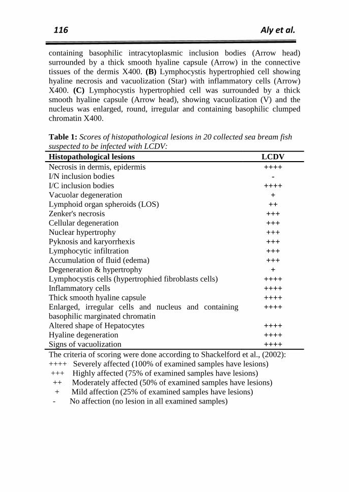
containing basophilic intracytoplasmic inclusion bodies (Arrow head)
surrounded by a thick smooth hyaline capsule (Arrow) in the connective
tissues of the dermis X400. (B) Lymphocystis hypertrophied cell showing
hyaline necrosis and vacuolization (Star) with inflammatory cells (Arrow)
X400. (C) Lymphocystis hypertrophied cell was surrounded by a thick
smooth hyaline capsule (Arrow head), showing vacuolization (V) and the
nucleus was enlarged, round, irregular and containing basophilic clumped
chromatin X400.
Table 1: Scores of histopathological lesions in 20 collected sea bream fish
suspected to be infected with LCDV:
Histopathological lesions LCDV
Necrosis in dermis, epidermis ++++
I/N inclusion bodies -
I/C inclusion bodies ++++
Vacuolar degeneration +
Lymphoid organ spheroids (LOS) ++
Zenker's necrosis +++
Cellular degeneration +++
Nuclear hypertrophy +++
Pyknosis and karyorrhexis +++
Lymphocytic infiltration +++
Accumulation of fluid (edema) +++
Degeneration & hypertrophy +
Lymphocystis cells (hypertrophied fibroblasts cells) ++++
Inflammatory cells ++++ Thick smooth hyaline capsule ++++ Enlarged, irregular cells and nucleus and containing
basophilic marginated chromatin ++++
Altered shape of Hepatocytes ++++ Hyaline degeneration ++++ Signs of vacuolization ++++
The criteria of scoring were done according to Shackelford et al., (2002):
++++ Severely affected (100% of examined samples have lesions)
+++ Highly affected (75% of examined samples have lesions)
++ Moderately affected (50% of examined samples have lesions)
+ Mild affection (25% of examined samples have lesions)
- No affection (no lesion in all examined samples)
SCVMJ, XXIII (2) 2018 117
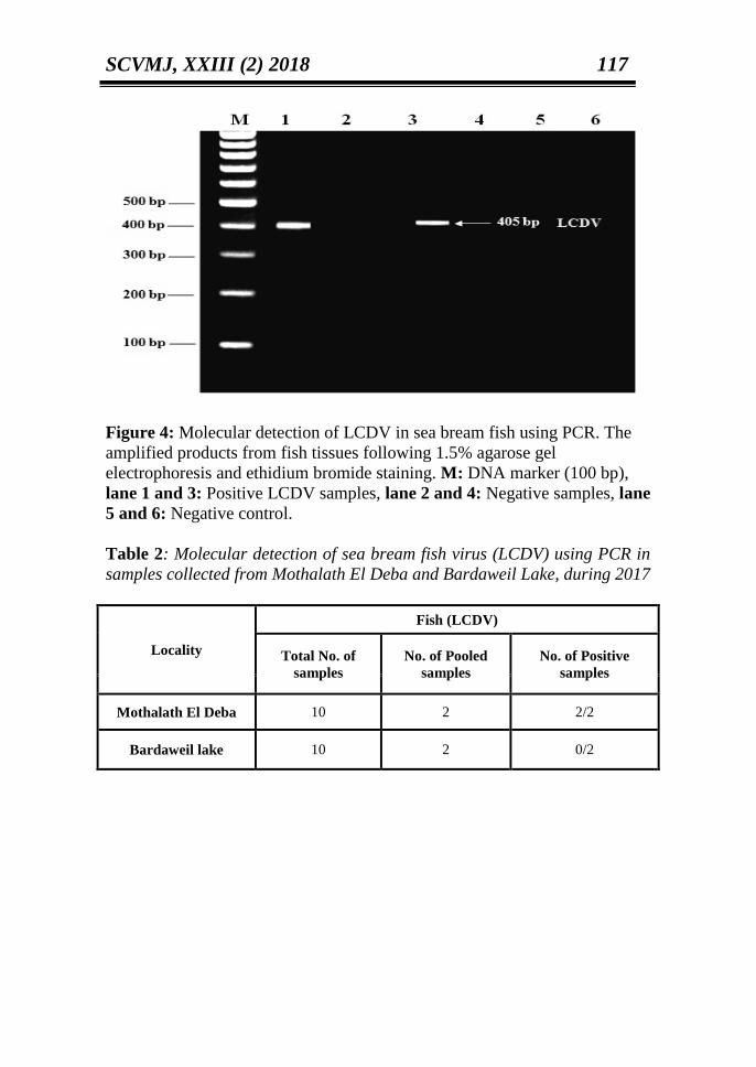
Figure 4: Molecular detection of LCDV in sea bream fish using PCR. The
amplified products from fish tissues following 1.5% agarose gel
electrophoresis and ethidium bromide staining. M: DNA marker (100 bp),
lane 1 and 3: Positive LCDV samples, lane 2 and 4: Negative samples, lane
5 and 6: Negative control.
Table 2: Molecular detection of sea bream fish virus (LCDV) using PCR in
samples collected from Mothalath El Deba and Bardaweil Lake, during 2017
Locality
Fish (LCDV)
Total No. of
samples
No. of Pooled
samples
No. of Positive
samples
Mothalath El Deba 10 2 2/2
Bardaweil lake 10 2 0/2

118 Aly et al.
Figure 5: Phylogenetic tree of LCDV based on a partial nucleotide
sequence. The tree was constructed using the Neighbor-joining method in
MEGA5. The robustness of individual nodes of the tree was assessed using
1000 replications of bootstrap re-sampling of the originally aligned
nucleotide sequences. Bootstrap values ≥70% is shown above the branches.
The virus isolated in this study is marked with solid quadrilateral.
Discussion
Emerging disease epizootics
frequently cause substantial, often
explosive, losses among
populations of shrimp and fish,
resulting in large economic losses in
commercial aquaculture and threats
to valuable stocks of wild aquatic
animals (Walker & Winton, 2010).
A large number of pathogens
threaten the fish aquaculture
industry with a majority of these
being viral in etiology, so viral
illnesses constitute the main
problem faced by penaeid shrimp
and seabream fish farms worldwide
AY823414.1 LCDV from Korea
AY849392.1 LCDV from Sebastes schlegeli
AB213006.1 LCDV strain RF04JinJu
AB213004.1 LCDV strain RF03Yosu
GU939626.2 LCDV 1 isolate yellow perch
GU290550.1 LCDV strain Leetown NFH
HE650105.1 LCDV strain SA1.ETun.2011
EF184306.1 LCDV isolate LCDV-SA-Eilat
EMBOSS_1411_LCDVB
EMBOSS_1536_LCDVM
KJ408272.1 LCDV isolate LCDV-PF
KT438163.1 LCDV strain LCDV-ss
EF375710.1 LCDV from Israel
KT438164.1 LCDV strain LCDV-ss
99
99
94
98
89
75
96
5

SCVMJ, XXIII (2) 2018 119
(Verônyca Coelho-Melo et al.,
2014). In Egypt, few published
studies addressed the marine fish
diseases. Accordingly, it is
important to investigate the current
situation of the emerging viral
diseases among cultured marine fish
in Egypt.
To better understand the
circumstances of some emerging
viral diseases among fish farms in
Egypt, twenty fish were collected
from Mothalath El Deba, Damietta
Province as well as Bardaweil Lake,
North Sinai Province that were
examined for clinical signs of
disease and then investigated for the
presence of viral infection. Under
field conditions, it was possible to
suspect clinically affected seabream
fish. The gross lesion and
histopathological pictures were
recorded in some cases and later
confirmed by molecular detection
and sequencing of the viruses.
LCD outbreaks are frequently
observed in the Mediterranean
gilthead seabream aquaculture.
Generally, viral diseased fish
show low growth rates, which
may be caused by the anemia
generally associated with this
disease as mentioned by Iwamoto
et al. (2002), although it is usually
described as a self-limiting disease,
there are several reports on
mortalities up to 45 % in juvenile
fish, which may be related to
secondary bacterial infections or
with particularly large growth of
lymphocystis, which severely
impaired fish osmoregulation,
breathing or feeding, cannibalism
and/or parasitic infestations
(Colorni & Padrós, 2011; Haddad-
Boubaker et al., 2013; Dezfuli et
al., 2012). In heavily affected fish,
lymphocystis may cover the entire
body, spreading from the gills to
the fins and less frequently, they
have also been described on eyes,
causing exophthalmia, and
internally over the mesenteries,
peritoneum and several internal
organs as mentioned by Xing et al.
(2006). LCDV is considered a
dermotropic virus so diagnosis of
LCD is generally based on typical
skin lesion observation. In the
current study, abundant and
extensive nodules were seen all
over the skin especially in the
pectoral and dorsal regionas well as
caudal fins of cultured sea bream
fish. The pathognomonic signs of
LCD include the appearance of
small pearl-like nodules on the skin
and fins that are usually grouped in
clusters, papillomatous in
appearance, and can cover the entire
body surface of the fish as similarly
reported by Wolf (1988). These
nodules consist of LCDV-infected
hypertrophied dermal fibroblasts
(up to 1 mm in diameter), named
lymphocysts or lymphocystis cells
(Bowden et al., 1995). Histopathological examination of
LCDV-infected fish showed the
presence of LCDV in the skeletal
muscle and gill lamella. The
cytoplasm of lymphocystis cells
were changed, developing
basophilic, intracytoplasmic

120 Aly et al.
inclusion bodies that appeared as
dense vacuolated bodies with
enlarged nucleus and cellular
hypertrophy. In addition, a thick
hyaline capsule surrounding the
hypertrophied fibroblast was
observed in the cytoplasm,
especially in the mature
lymphocystis cells as previously
confirmed by Hossain and Oh
(2011). Histopathological studies
carried out in LC-diseased fish have
been focused on the description of
lymphocystis cells, with few reports
dealing with histological
observations of the internal organs,
except when lymphocysts were also
present (Sheng & Zhan, 2004). In
this study, LC-diseased gilthead
seabream specimens showed
lymphocystis cells only in the
dermis of the caudal fin, with
histological characteristics
resembling those previously
described in this fish species as
recorded by Gonzalez de Canales et
al. (1996). Histopathological
alterations of varied severities were
also observed in other organs,
including necrotic changes in the
liver and kidney, inflammatory
response in the intestine and
intraventricular hemorrhage. On the
other hand, necrotic changes in the
epithelium were the only
histological alterations described so
far in gilthead seabream fishes
affected by LCD (Cano et al.,
2009). In the present study,
hepatocytes showed vacuolization
and increased cytoplasmic
basophilia with some areas of
necrosis.
The second approach of this work
was to molecularly detect the viral
infection in diseased fish. Fish
tissues were analyzed by PCR using
primer set to LCDV. Although
these methods are quite accurate,
they are too expensive to be used
viably as mentioned by Mello et al.
(2011). The availability of sensitive
and specific tests for detection of
pathogens in sea bream fish is
essential for accurate diagnosis of
diseases affecting cultured
population. PCR is a rapid, sensitive
and highly specific detection
method for fish viruses, which can
be a powerful tool to detect
iridovirus infections; LCDV in sea
bream fish as reported by Mao et al.
(1997). A primers set was used to
detect 405 bp amplicons in LCDV
using PCR. The primers was shown
to be specific for Major capsid
protein (MCP) gene of LCDV and
no amplicons were detected using
DNA extracted from sea bream fish
infected with other fish viruses as
indicated by Kvitt et al. (2008). The
PCR has been used to amplify a
portion of LCDV genome using
specific primer as stated by Kvitt et
al. (2008). It was used for definite
identification of LCDV and the
results showed higher sensitivity to
detect the LCDV like those
described before. In this study, 20
samples were collected, pooled
together in 4 tubes and submitted to
DNA extraction using specific
primer to detect viral DNA in the

SCVMJ, XXIII (2) 2018 121
tissue. 2 out of 4 samples were
positive in LCDV from locality
Mothalath El Deba, which showing
that affection is high in this area
than another one.
The PCR has been used to amplify a
portion of LCDV genome using
specific primer of MCP gene. It was
used for definite identification of
LCDV, the results showed higher
sensitivity to detect the LCDV like
those described before as recorded
by Kvitt et al. (2008). In this study,
4 samples were collected, pooled
together and submitted to DNA
extraction and PCR using MCP
gene specific primer to detect viral
DNA in the tissue. 2 out of 4
samples were positive.
The third approach of this work was
to assess genetic characteristics of
viruses via nucleotide sequencing
and phylogenetic analysis. Thus, nt
sequence analysis is needed to
completely identify the viruses of
collected samples as stated by
Mello et al. (2011). The
phylogenetic tree based on
sequence of specific gene for
Lymphocystis disease virus showed
that samples are clustered and
compared with other related
nucleotide sequences on Genbank.
In Egypt, there are no records for
LCDV until now.
In sea bream fish, LCDV of our
samples was related to Korea,
Canada, USA, Tunisia and Israel
with homology 80-89.7%.
Although the reported LCDV
samples showed similar
macroscopic and microscopic
picture with those detected by
Hossain and Oh (2011). However,
low identity was reported between
our nucleotide sequences and those
published sequences derived from
Korea, Canada, USA, Tunisia and
Israel isolates, as the published
sequences recorded that the isolates
from those countries collected from
different species of fishes not from
sea bream fish.
In the Mediterranean Sea, the major
constraints in aquaculture of sea
bream were LCDV as stated by
García-Rosado et al. (2007). Since
the 1980s, LCDV has been reported
in different countries of the
Northern Mediterranean coasts as
recorded by Menezes et al. (1987);
Le Deuff and Renault (1993). In
the Southern coasts, only a partial
MCP sequence was reported in
Tunisia in 2005, but with no
information about its
epidemiological impact or its
geographic origin as mentioned by
Cano et al. (2010). LCDV has been
reported more than 100 different
marine and freshwater fish species,
it seems obvious that, the existence
of differences regarding the viral
genome structure, gene organization
and DNA sequence depending on
the host fish or the geographical
location as indicated by Kitamura
et al. (2005). In fact, although MCP
gene is highly conserved and
contains sufficient variable regions
to allow phylogenetic analysis, the
use of different genes may be more
significant to trace the
epidemiological origin in such a

122 Aly et al.
context. Unfortunately, this analysis
requires availability of relevant
sequences of entire LCDV genome
but currently a few number of
LCDV genome sequences are
available (Haddad-Boubaker et al.,
2013).
Conclusion
The present study revealed
detection of LCDV from sea bream
fish cultured in Egypt that might
constitute a negative impacts on the
national economy.
Gross lesion and histopathology
may aid in diagnosis of LCDV
among fish however, molecular
detection; PCR and sequencing as
well as phylogenetic analysis
confirm the diagnosis.
The active international trade with
lack implementation for strict
regulations regarding fish transfer
between countries may be
responsible for the dissemination of
viral strains, especially in the
absence of a certification as free
stocks.
The current results recommend
further studies for the diagnosis and
development of preventive
measures and control strategies
against LCDV in sea bream fish in
Egypt.
References
Aly, S. (2013): A Review of Fish
Diseases in the Egyptian
Aquaculture Sector. Working
Report.
Borrego, J.J.; Castro, D.;
Balebona, M.C.; Garcia-Rosado,
E. and Lopez-Cortes, L. (2001):
Patologías que Afectan al Cultivo
de la Dorada (Sparus aurata, L.) en
la Comunidad Autónoma Andaluza.
Editada por la Consejería de
Agricultura y Pesca, Junta de
Andalucía, Sevilla, España.
Bowden, R.A.; Oestmann, D.J.;
Lewis, D.H. and Frey, M.S.
(1995): Lymphocystis in Red
Drum. J. Aquat. Anim. Health,
7(3): 231–235. doi:10.1577/1548-
8667(1995)007<0231:LIRD>2.3.C
O;2
Cano, I.; Ferro, P.; Alonso, M.C.;
Sarasquete, C.;Garcia-Rosado,
E., Borrego, J.J. and Castro, D.
(2009): Application of in situ
detection techniques to determine
the systemic condition of
lymphocystis disease virus (LCDV)
infection in cultured gilt-head
seabream, Sparus aurata L. J. Fish
Dis. 32: 143–150. doi:
10.1111/j.1365-2761.2008.00970.x.
Cano, I.; Valverde, E. J.; Lopez-
Jimena, B.; Alonso, M. C.;
Garcia-Rosado, E.; Sarasquete,
C.; Borrego, J. J. and Castro, D.
(2010): A new genotype of
lymphocystivirus isolated from
cultured gilthead seabream, Sparus
aurata L., and Senegalese sole,
Solea senegalensis (Kaup). J. Fish
Dis., 33: 695–700.
Colorni, A. and Padrós, F. (2011): Diseases and health management.
In: Sparidae: Biology and
Aquaculture of Gilthead Sea Bream
and other Species, edited by M.A.
Pavlidis & C.C. Mylonas, pp. 321–
357. Wiley-Blackwell, Oxford,
United Kingdom.

SCVMJ, XXIII (2) 2018 123
Colorni, A. and Padrós, F. (2011): Diseases and health management.
In: Sparidae: Biology and
Aquaculture of Gilthead Sea Bream
and other Species, edited by M.A.
Pavlidis & C.C. Mylonas, pp. 321–
357. Wiley-Blackwell, Oxford,
United Kingdom.
Dezfuli, B.S.; Lui, A.; Giari, L.;
Castaldelli, G.; Mulero, V. and
Noga, E.J. (2012): Infiltration and
activation of acidophilic
granulocytes in skin lesions of
gilthead seabream, Sparus aurata,
naturally infected with
lymphocystis disease virus. Dev.
Comp. Immunol; 36: 174–182. doi:
10.1016/j.dci.2011.06.017.
García-Rosado, E.; Cano, I.;
Martín-Antonio, B.; Labella, A.;
Manchado, M.; Alonso, M. C.;
Castro, D. and Borrego, J. J.
(2007): Co-occurrence of viral and
bacterial pathogens in disease
outbreaks affecting newly cultured
sparid fish. Int. microbiol., 10: 193–
199.
Gonzalez de Canales, M.L.;
Munoz-Cueto, J.A.; Arellano, J.;
García-Garcia, A. and
Sarasquete, C. (1996): Histological
and histochemical characteristics of
the lymphocystis disease in gilthead
sea bream, Sparus aurata, L. from
the South-Atlantic coast of Spain.
European Journal of Histochemistry
40: 143–152.
Haddad-Boubaker, S.;
Bouzgarou, N.; Fakhfakh, E.;
Khayech, M.; Mohamed, S.B.;
Megdich, A. and Chéhida, N.B.
(2013): Detection and genetic
characterization of lymphocystis
disease virus (LCDV) isolated
during disease outbreaks in cultured
gilt-head sea bream Sparus
aurata in Tunisia. Fish Pathol.; 48:
101–104. doi: 10.3147/jsfp.48.101.
Iwamoto, R.; Hasegawa, O.;
LaPatra, S. and Yoshimizu, M.
(2002): Isolation and
characterization of the Japanese
flounder (Paralichthys olivaceus)
lymphocystis disease virus. J.
Aquat. Anim. Health, 14: 114–123.
Jancovich, J.K.; Chinchar, V.G.;
Hyatt, A.; Miyazaki, T.; Williams,
T. and Zhang, Q.Y. (2012): Family Iridoviridae. In: Virus
Taxonomy: Ninth Report of the
International Committee on
Taxonomy of Viruses. Edited by.
A.M.Q. King, M.J. Adams, E.B.
Carstens & E.J. Lefkowitz. pp. 193–
210. Elsevier Academic Press, San
Diego.
Kitamura, S.I.; Jung, S.J.; Kim,
W.S.; Nishizawa, T.; Yoshimizu,
M. and Oh, M.J. (2005): A new
genotype Lymphocystis, LCDV-
RF, from lymphocytes diseased
rockfish. Arch. Virol., 151: 607–
615.
Kvitt, H.; Heinisch, G. and
Diamant, A. (2008): Detection and
phylogeny of Lymphocystivirus in
sea bream Sparus aurata based on
the DNA polymerase gene and
major capsid protein sequences.
Aquaculture, 275: 58–63.
Le Deuff, R.M. and Renault, T.
(1993): Lymphocystis outbreaks in
farmed seabream, Sparus aurata,
first report on French

124 Aly et al.
Mediterranean coast. Bulletin of the
European Association of Fish
Pathologists 13: 130–133.
Mao, J.; Hedrick, R.P. and
Chinchar, V.G. (1997): Molecular
characterization, sequence analysis
and taxonomic position of newly
isolated fish iridoviruses. Virology
229: 212–220.
Mello, M.V.; Aragao, M.E.;
Torres-Franklin, M.L.; Neto,
J.M. and Guedes, M.I. (2011): Purification of infectious
myonecrosis virus (IMNV) in
species of marine shrimp
Litopenaeus vannamei in the State
of Ceara. J Virol Methods, 177(1):
10–14.
doi:10.1/016j.jviromet.2011.05.032
Menezes, J.; Ramos, A. and
Pereira, T.G. (1987): Lymphocystis disease: an outbreak
in Sparus aurata from Ria Formosa,
south coast of Portugal. Aquaculture, 67: 222–225.
Michaelsen, K.F.; Dewey, K.G.;
Perez-Exposito, A.B.; Nurhasan,
M.; Lauritzen, L. and Roos, N.
(2011): Food sources and intake of
n-6 and n-3 fatty acids in low-
income countries with emphasis on
infants, young children (6–24
months), and pregnant and lactating
women. Maternal Child Nutr.
7(Suppl.2): 124–140.
Mosharrof Hossain and Myung-
Joo Oh (2011): Histopathology of
marine and freshwater fish
lymphocystis disease virus
(LCDV). Sains Malaysiana, 40(10):
pp. 1049–1052. ISSN 0126-6039.
Mustapha, A.; Driss, B. and
Mohamed, B. (2014): The main
species of freshwater fish
aquaculture interest in Morocco,
current status and prospects. Int. J.
Fish. Aquat. Stud., 2(1): 216–218.
Paperna, I.; Vilenkin, M. and
Alves de Matos, A.P. (2001): Iridovirus infections in farm-reared
tropical ornamental fish. Dis Aquat
Org 48: 17−25
Paperna, I.; Ilana-Sabnai, H. and
Colorni, A. (1982): An outbreak of
lymphocystis in Sparus aurata L. in
the Gulf of Aqaba, Red Sea. Journal
of Fish Diseases, 5(5): 433–437.
doi:10.1111/j.1365-
2761.1982.tb00500.x
Poulos, B.T. and Lightner, D.V.
(2006): Detection of infectious
myonecrosis virus (IMNV) of
penaeid shrimp by reverse-
transcriptase polymerase chain
reaction (RT-PCR). Dis. Aquat.
Org., 73: 69–72.
Rodger, G.K. and Davies, I.M.
(2000): Summary of mariculture
production in countries
neighbouring the European Union.
J. Appl. Ichthyol., 16(45): 224–229.
Shaalan, M.; El-Mahdy, M.;
Saleh, M. and El-Matbouli, M.
(2018): Aquaculture in Egypt:
Insights on the Current Trends and
Future Perspectives for Sustainable
Development. Reviews in Fisheries
Science & Aquaculture, 26(1): 99–
110. DOI:
10.1080/23308249.2017.1358696
Shackelford, C.; Long, G.; Wolf,
J.; Okerberg, C. and Herbert, R.
(2002): Qualitative and quantitative

SCVMJ, XXIII (2) 2018 125
analysis of nonneoplastic lesions in
toxicology studies. Toxicol Pathol.
30(1): 93–6.
Sheng, X.Z. and Zhan, W.B.
(2004): Occurrence, development
and histochemical characteristics of
lymphocystis in cultured Japanese
flounder (Paralichtys olivaceous).
High Technology Letters 10: 92–96.
Suvarna, S.K.; Layuton, C. and
Bancroft, J.D. (2013): Bancroft’s
theory and practice of histological
techniques. 7th edition, Churchill
Livingstone Press, New York, USA.
Tacon, A.G.J. and Metian, M.
(2013): Fish matters: importance of
aquatic foods in human nutrition
and global food supply. Rev. Fish.
Sci. 21(1): 22–38.
Thilsted, S.H. (2013): Fish
diversity and fish consumption in
Bangladesh. In: Fanzo, J., Hunter,
D., Borelli, T., Mattei, F. (Eds.),
Diversifying Food and Diets: Using
Agricultural Biodiversity to
Improve Nutrition and Health.
Earthscan, London.
Tidona, C.A. and Darai, G.
(1999): Lymphocystis disease virus
(Iridoviridae). In: Granoff A,
Webster RG (eds) Encyclopedia of
Virology, 2nd edn,
pp. 908–911. Academic Press, New
York.
Verônyca Coelho-Melo, M.;
Florindo Guedes, M.I.;
Rodriguez-Málaga, S.; Magalhaes
De Almeida, L.; De Freitas
Moreira, M. and Rodrigues De
Oliveira, T. (2014): Molecular
characterization of Infectious
Myonecrosis Virus (IMNV) isolated
from the shrimp Litopenaeus
vannamei farmed in Ceará state,
Brazil. Latin American Journal of
Aquatic Research, 42(3): 649–652.
Walker, P. J. and Winton, J. R.
(2010): Emerging viral diseases of
fish and shrimp. Vet Res, 41(6): 51.
doi: 10.1051/vetres/2010022
Wang, Q.; Cheng, L.; Liu, J.; Li,
Z.; Xie, S. and De Silva, S. S.
(2015): Freshwater aquaculture in
PR China: Trends and prospects.
Rev. Aquacult., 7(4): 283–302.
Wolf, K. (1988): Lymphocystis
disease. In: Fish Viruses and Fish
Viral Disease. Edited by K. Wolf,
pp. 268–291. Ithaca: Cornell
University Press, New York,
EE.UU.
World Bank, (2006): Aquaculture:
Changing the Face of the Waters:
Meeting the Promise and Challenge
of Sustainable Aquaculture. World
Bank, Washington, DC.
Xing, J.; Sheng, X. and Zhan, W.
(2006): Lymphocystis disease and
diagnostic methods in China.
Aquaculture Asia Magazine,
January-March: 30–33.

126 Aly et al.
الحويصالت الليمفاوية لفيروسالجزيئى الباثولوجى والتوصيف
الدنيس البحري في مصرفى أسماك
3يسري ثابترندا , 2محمد منصور ء, شيما 1على صالح الدين مصيلحي
جامعة قناة السويس -قسم الباثولوجيا بكلية الطب البيطري -1
الزقازيقجامعة -بكلية الطب البيطري فيروساتقسم ال -2
معة جا -ك قسم مكافحة أمراض االستزراع المائي بمعهد االستزراع السمكي وتكنولوجيا األسما -3
قناة السويس
ن هو مرض منتشر جغرافيا يؤثر على أكثر م (LCDV) مرض الحويصالت الليمفاوية الفيروسى
سببه ض الذي يالمر نوًعا مختلفًا من أسماك المياه العذبة والبحرية في جميع أنحاء العالم. يتميز 150
ة الورم التي تحتوي على خاليا متضخم ( بمظهرLCDVفيروس مرض الحويصالت الليمفاوية )
رور ما مع بشكل كبير )الحويصالت الليمفاوية( لألسماك المصابة التي عادة ما تحل نفسها بنفسه
بيض حر األتعد أسماك الدنيس من أهم أنواع األسماك المستزرعة اقتصاديًا في منطقة الب الوقت.
وال رية المصرية غير مفهوم بشكل جيد،( في أسماك الدنيس البحLCDVفيروس مرض )المتوسط.
سي من لذلك كان الهدف الرئي في مصر. LCDVتوجد معلومات متاحة بشأن العدوى المحتملة للـ
في الدنيس البحري المستزرع فى مصر عن طريق فحص LCDVهذا العمل هو تشخيص
والتسلسل األعراض الظاهرية وفحص النسيجي المرضي ومن خالل اختبار الطرق الجزيئية
.جينيةالجزيئي واختبار التتابعات الجينية وتحليل السالالت عن طريق عمل تحليل الشجرة ال
ا من تم جمعهعينة من أسماك الدنيس البحري أورام في الجلد والزعانف 20وأظهرت عينات من
الجلد . وأظهر الفحص النسيجي بعض التغيرات في أنسجةمزارع الدنيس البحري في مصر
أعطى (MCPمع بادئات محددة لبروتين ) PCRنتائج الـ عانف مع نخر واستجابة التهابية. والز
س قات لفيروتشجع النتائج الحالية المزيد من التحقي بواسطة الفصل الكهربائي. 405bpنتيجة عند
ات في المزارع المصرية ويوصي بوضع استراتيجي lymphocystisمرض الحويصالت الليمفاوية
في مصر. LCDV فيروس مرض الحويصالت الليمفاوية والمكافحة ضدالوقاية
: الدنيس، مصر، فيروس مرض الحويصالت الليمفاوية، الفحص المرضيالكلمات االفتتاحية
.لألنسجة، الفحص الجزيئي، تفاعل البلمرة المتسلسل، الشجرة الجينية