pathological and parasitological aspects of the peacock ... · sitando pavões (pavo cristatus...
TRANSCRIPT

Pesq. Vet. Bras. 35(5):466-469, maio 2015DOI: 10.1590/S0100-736X2015000500014
466
RESUMO.- [Aspectos patológicos e parasitológicos da infecção por Tanaisia (Paratanaisia) bragai em pavões (Pavo cristatus).] Os trematódeos da família Eucotylidae, incluindo Tanaisia (Paratanaisia) bragai Santos, 1934, são parasitos de rins e ureteres de várias espécies de aves do-mésticas e silvestres. Tanaisia bragai é considerada uma espécie pouco patogênica, mas que pode determinar com-plicações clínicas como apatia, perda de peso, diarreia e morte, quando em cargas parasitárias elevadas. No presen-
Pathological and parasitological aspects of the peacock (Pavo cristatus) infection by Tanaisia (Paratanaisia) bragai1
Rafael C. Costa2, Natália A. Ambrósio3, Bruno A. Soares3, Pedro S. Bezerra Júnior2, Thales A. Barçante4, Priscilla R. Barrios3 and Joziana M.P. Barçante3*
ABSTRACT.- Costa R.C., Ambrósio N.A., Soares B.A., Bezerra Jr P.S., Barçante T.A., Barrios P.R. & Barçante J.M.P. 2015. Pathological and parasitological aspects of the peacock (Pavo cristatus) infection by Tanaisia (Paratanaisia) bragai. Pesquisa Veterinária Bra-sileira 35(5):466-469. Setor de Medicina Veterinária Preventiva, Departamento de Medicina Veterinária, Universidade Federal de Lavras, Campus Universitário, Cx. Postal 3037, Lavras, MG 37200-000, Brazil. E-mail: [email protected]
Trematodes belonging to the family Eucotylidae, including Tanaisia (Paratanaisia) bra-gai Santos, 1934, are parasites of the kidney and ureter that affect several species of do-mestic and wild birds. Tanaisia bragai is considered a low pathogenic parasite, but high worm burdens may determine clinical complications, including signs of apathy, weight loss, diarrhea and death. This paper describes the first report of infection by T. bragai in pea-cocks (Pavo cristatus), which constitutes a new host record and offers data on the lesions associated to this parasitism, although the degree of pathogenicity and parasite load may be considered mild. These birds did not exhibit clinical signs of parasitism. The macros-copic exam revealed discreet yellow spots on the liver. In the histological sections of the kidney, specimens of T. bragai were found in the collecting ducts, which were markedly dilated, with a thickened wall. Other findings included a mild inflammatory reaction in the wall of the ducts (but sometimes absent), flattening of lining epithelial cells and small, mul-tifocal points of calcification around the collecting ducts. The microscopic examination of the parasites revealed trematodes with an elongated body, well-developed sub terminal oral sucker, pharynx present, short esophagus, cecum somewhat undulating or not, with blind end, testes symmetrical, equatorial, irregular in shape or slightly lobed, vitelline fiel-ds extending in both pre-ovarian and post ovarian fields, uterus very long, intercecal or sometimes overlapping the cecum and containing large quantities of eggs. The present fin-dings suggest the need for further diagnostic studies on the prevalence of this trematode in peacocks as well as pathologic studies for the determination of the potential pathogenicity of this parasite in this species of bird. Moreover, infected peacocks could serve as carriers of T. bragai to be transferred to other bird species, thereby contributing to the dispersion of the parasite.INDEX TERMS: Tanaisia (Paratanaisia) bragai, trematode, kidney, exotic bird, peacock, Pavo cristatus, pathology.
1 Received on October 7, 2014.Accepted for publication on February 27, 2015.
2 Setor de Patologia Veterinária, Departamento de Medicina Veteriná-ria, Universidade Federal de Lavras (UFLA), Cx. Postal 3037, Lavras, MG 37200-000, Brasil.
3 Setor de Medicina Veterinária Preventiva, Departamento de Medicina Veterinária, UFLA, Cx Postal 3037, Lavras, MG 37200-000, Brasil. *Corres-ponding author: [email protected]
4 Pontifícia Universidade Católica de Minas Gerais. Curso de Medicina Veterinária, Av. Cletus Francis Cox, Poços de Caldas, MG 37701-355, Brasil.

Pesq. Vet. Bras. 35(5):466-469, maio 2015
467Pathological and parasitological aspects of the peacock (Pavo cristatus) infection by Tanaisia (Paratanaisia) bragai
te trabalho, descreve-se o primeiro relato de T. bragai para-sitando pavões (Pavo cristatus Linnaeus, 1758), incluindo os achados parasitológicos e patológicos associados à in-fecção. A ausência de sinais clínicos evidencia uma infecção moderada, na qual, ao exame macroscópico dos órgãos in-ternos, verificou-se a presença de pequenas manchas ama-reladas no fígado. Ao exame histopatológico, foram verifi-cados espécimes de T. bragai nos ductos coletores dos rins, que se apresentavam marcadamente dilatados e com adel-gaçamento das paredes. Verificou-se também moderada re-ação inflamatória (por vezes ausente) na parede dos ductos e pequenos pontos de calcificação ao redor dos mesmos. No interior do órgão foram recuperados parasitos de corpo alongado, com ventosa oral subterminal e bem desenvolvi-da, ventosa acetabular, faringe, esôfago curto, cecos intes-tinais em fundo cego, testículos simétricos e equatoriais e irregulares, glândulas vitelínicas presentes nas regiões pré e pós-ovarianas, útero longo e contendo grande quantidade de ovos. O presente achado sugere a necessidade de novos estudos diagnósticos a fim de determinar a prevalência e a importância deste parasito para pavões. Além disso, os pa-vões demonstraram ser hospedeiros definitivos de T. bra-gai, podendo servir de fonte de infecção para o ambiente, contribuindo para a dispersão do parasito.TERMOS DE INDEXAÇÃO: Tanaisia (Paratanaisia) bragai, trema-tode, rim, ave exótica, pavão, Pavo cristatus, patologia.
INTRODUCTIONTanaisia (Paratanaisia) bragai (Freitas, 1959) is a digenetic trematode found in the kidney collecting ducts of different species of wild and domesticated birds (Brandolini et al. 1997, Kanev et al. 2002). This species has been identified in Central and South America, Asia and Oceania. In Brazil, T. bragai has been found in the following birds: chicken (Gallus gallus domesticus), pigeon (Columba livia), phea-sant (Phasianus colchicus), helmeted Guineafowl (Numida meleagris), turkey (Meleagris gallopavo), white-eared pa-rakeet (Pyrrhura leucotis),red-winged timamou (Rhyncho-tus rufescens),blue-winged macaw (Propyrrhura maraca-na) and ruddy ground dove (Columbina talpacoti) (Keller & Araujo 1992, Menezes et al. 2001, Mapeli et al. 2003, Pinto et al. 2004,Gomes et al. 2005, Luppi et al. 2007).Birds be-come infested by ingesting gastropods containing metacer-cariae. Infection is generally subclinical, with more evident signs in birds with a high parasite burden (Greve 1986). Al-though T. bragai is considered to be a low pathogenic para-site, infection may determine clinical signs, such as apathy, weight loss and diarrhea, sometimes followed by death. The main necropsy findings are an increase in kidney volu-me, polycystic kidney of abnormal shape, a friable surface and yellowish brown coloration (Portugal et al. 1972, Mena et al. 1986). Microscopically, adults and eggs are found in the renal parenchyma, with dilation of the collecting ducts affecting kidney morphology (Portugal et al. 1972, Mene-zes et al. 2001, Mapeli et al. 2003, Pinto et al. 2004, Gomes et al. 2005). Cases of glomerulonephritis leading to chronic kidney failure and death have been described in species of Psittacidae (Luppi et al. 2007). The few studies on his-
topathological lesions indicate that the hosts and lesions associated with this parasite are not yet well established (Gomes et al. 2005).
This article reports the first record of T. bragai parasi-tizing peacocks (Pavo cristatus) and describes the macros-copic and microscopic findings associated with parasitism.
MATERIALS AND METHODSA male and a female peacock (Pavo cristatus Linnaeus, 1758) about 14 years old were sent to the Avian and Swine Disease Laboratory of the Federal University of Lavras, Minas Gerais, Brazil, for necropsy and parasitological and histopathological evaluation. The organs were collected in buffered formalin, em-bedded in paraffin, cut into 5μm and stained with hematoxylin--eosin for histological exam. Tissue fragments were submitted to the anti-Salmonella spp. immunohistochemical technique. All organs were individually removed for macroscopic analysis and parasitological evaluation. For the trematode study, the kidneys were sectioned and placed in 9% saline solution. The organs were then transferred to a BOD chamber for 12h at 37oC. The parasites were collected with the aid of a brush and transferred to PBS1X solution at room temperature. Trematodes were placed on glass slides and slightly compressed with a cover slip, followed by the addition of alcohol-formalin-acetic acid solution for fixation. Sli-des with compressed parasites were kept under refrigeration for 12 hours. The fixed parasites were dehydrated in a series of so-lutions with an increasing concentration of alcohol and stained with carmine hydrochloride. Stained parasites were fixed on sli-des with resin for identification based on Travassos et al. (1969) and Kanev et al. (2002).
RESULTSThe macroscopic exam revealed discreet yellow spots on the liver tissue of both peacocks. The microscopic exam re-vealed an absence of significant findings in the liver. The immunohistochemical analysis for anti-Salmonella spp was negative. The histological sections of the kidney reve-aled parasites in the collecting ducts, which were marke-dly dilated with a thickened wall. Other findings included a mild inflammatory reaction in the wall of the ducts (but sometimes absent), flattening of lining epithelial cells and small, multifocal points of calcification around the collec-ting ducts. (Fig.1A-F). Sections of the liver also revealed a mild inflammatory reaction and multifocal points of calci-fication.
During the parasitological exam, a mean of 33 parasi-tes were retrieved from the kidneys (36 parasites in the left kidney and 30 parasites in the right kidney). The mi-croscopic exam revealed dorsally-ventrally flat trematodes with an elongated body of medium size (Mean: 1.5mm, SD: 0.5mm), well-developed sub terminal oral sucker, pharynx present, short esophagus, cecum with blind end, testes symmetrical, equatorial, irregular in shape or slightly lo-bed, partially overlapping the ceca, pre-ovarian and post ovarian extra-cecalvitelline fields, uterus very long, interce-cal or sometimes overlapping the ceca and containing large quantities of eggs, and spines on the tegument (Fig.2A,B). The morphological findings and location of the parasites allowed the conclusive diagnosis of infection by Tanaisia (Paratanaisia) bragai.

Pesq. Vet. Bras. 35(5):466-469, maio 2015
468 Rafael C. Costa et al.
DISCUSSIONThe trematode Tanaisia (Paratanaisia) bragai is commonly found parasitizing different bird species in Brazil, but has not previously been described in peacocks. The morpho-metric findings of the specimens described in the present study are in agreement descriptions of this species in the literature (Menezes et al. 2001, Pinto et al. 2004).
Menezes et al. (2001) reported the presence of 142 trematodes in a single kidney. In the present study, the low parasite burden found in the infected bird, the ab-sence of significant macroscopic findings and the discre-et microscopic findings suggest that the species has low pathogenicity for peacocks, at least with a low parasite burden.
The microscopic findings may be considered mild in comparison to reports in other species, such as the phea-sant, blue-winged macaw and ruddy ground dove (Gomes et al., 2005, Pinto et al., 2004, Luppi et al., 2007). The fin-
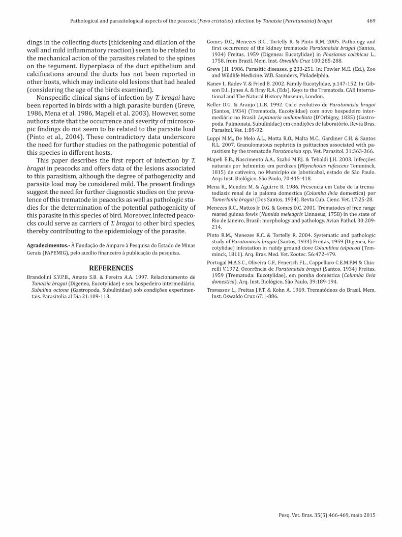
Fig.1. Histological section a pe-acock (Pavo cristatus) kidney showing Tanaisia (Parata-naisia) bragai. (A) Accentua-ted dilation of collecting duct and mild inflammatory reac-tion around duct (arrows), HE, obj.10x. (B) Parasites (P) in interior of collecting duct. Observe the accentu-ated dilation of collecting duct, with intense peripheral inflammatory reaction (ar-rows) and associated with calcifications (C), HE, obj.4x. (C) Detail of metaplasia of the wall of the collecting duct with presence of parasite (P), Thickening of epithelial cells extending to the center of the duct (arrow) and mild peripheral inflammatory re-action (I). HE, obj.40x. (D) Collecting duct with parasite, cut reveals internal struc-tures of parasite: ceca (C), testicles (T) and pre-vitelline spaces (arrows), with meta-plasia of duct epithelial cells. HE, obj.10x. (E) Cross-sec-tion showing specimen of T. bragai in markedly dilated collecting duct, cut reveals oral sucker of parasite adhe-red to tubular epithelium. HE, obj.10x. (F) Specimen of T. bragai in markedly dilated collection duct, cut reveals oral sucker of parasite (ar-row) adhered to tubular epi-thelium. HE, obj.20x.
Fig.2. Adult specimen of Tanaisia (Paratanaisia) bragai retrieved from kidney of naturally infected peacock. (A) Fixed specimen showing intestinal ceca (c), uterus filled with eggs (ut), oral sucker (os) and acetabulum (a). (B) Tegument with spines.
Pesq. Vet. Bras. 35(5):466-469, maio 2015
469Pathological and parasitological aspects of the peacock (Pavo cristatus) infection by Tanaisia (Paratanaisia) bragai
dings in the collecting ducts (thickening and dilation of the wall and mild inflammatory reaction) seem to be related to the mechanical action of the parasites related to the spines on the tegument. Hyperplasia of the duct epithelium and calcifications around the ducts has not been reported in other hosts, which may indicate old lesions that had healed (considering the age of the birds examined).
Nonspecific clinical signs of infection by T. bragai have been reported in birds with a high parasite burden (Greve, 1986, Mena et al. 1986, Mapeli et al. 2003). However, some authors state that the occurrence and severity of microsco-pic findings do not seem to be related to the parasite load (Pinto et al., 2004). These contradictory data underscore the need for further studies on the pathogenic potential of this species in different hosts.
This paper describes the first report of infection by T. bragai in peacocks and offers data of the lesions associated to this parasitism, although the degree of pathogenicity and parasite load may be considered mild. The present findings suggest the need for further diagnostic studies on the preva-lence of this trematode in peacocks as well as pathologic stu-dies for the determination of the potential pathogenicity of this parasite in this species of bird. Moreover, infected peaco-cks could serve as carriers of T. bragai to other bird species, thereby contributing to the epidemiology of the parasite.
Agradecimentos.- À Fundação de Amparo à Pesquisa do Estado de Minas Gerais (FAPEMIG), pelo auxílio financeiro à publicação da pesquisa.
REFERENCESBrandolini S.V.P.B., Amato S.B. & Pereira A.A. 1997. Relacionamento de
Tanaisia bragai (Digenea, Eucotylidae) e seu hospedeiro intermediário, Subulina octona (Gastropoda, Subulinidae) sob condições experimen-tais. Parasitolia al Día 21:109-113.
Gomes D.C., Menezes R.C., Tortelly R. & Pinto R.M. 2005. Pathology and first occurrence of the kidney trematode Paratanaisia bragai (Santos, 1934) Freitas, 1959 (Digenea: Eucotylidae) in Phasianus colchicus L., 1758, from Brazil. Mem. Inst. Oswaldo Cruz 100:285-288.
Greve J.H. 1986. Parasitic diseases, p.233-251. In: Fowler M.E. (Ed.), Zoo and Wildlife Medicine. W.B. Saunders, Philadelphia.
Kanev I., Radev V. & Fried B. 2002. Family Eucotylidae, p.147-152. In: Gib-son D.I., Jones A. & Bray R.A. (Eds), Keys to the Trematoda. CAB Interna-tional and The Natural History Museum, London.
Keller D.G. & Araujo J.L.B. 1992. Ciclo evolutivo de Paratanaisia bragai (Santos, 1934) (Trematoda, Eucotylidae) com novo hospedeiro inter-mediário no Brasil: Leptinaria unilamellata (D’Orbigny, 1835) (Gastro-poda, Pulmonata, Subulinidae) em condições de laboratório. Revta Bras. Parasitol. Vet. 1:89-92.
Luppi M.M., De Melo A.L., Motta R.O., Malta M.C., Gardiner C.H. & Santos R.L. 2007. Granulomatous nephritis in psittacines associated with pa-rasitism by the trematode Paratanaisia spp. Vet. Parasitol. 31:363-366.
Mapeli E.B., Nascimento A.A., Szabó M.P.J. & Tebaldi J.H. 2003. Infecções naturais por helmintos em perdizes (Rhynchotus rufescens Temminck, 1815) de cativeiro, no Município de Jaboticabal, estado de São Paulo. Arqs Inst. Biológico, São Paulo, 70:415-418.
Mena R., Mendez M. & Aguirre R. 1986. Presencia em Cuba de la trema-todiasis renal de la paloma domestica (Columba livia domestica) por Tamerlania bragai (Dos Santos, 1934). Revta Cub. Cienc. Vet. 17:25-28.
Menezes R.C., Mattos Jr D.G. & Gomes D.C. 2001. Trematodes of free range reared guinea fowls (Numida meleagris Linnaeus, 1758) in the state of Rio de Janeiro, Brazil: morphology and pathology. Avian Pathol. 30:209-214.
Pinto R.M., Menezes R.C. & Tortelly R. 2004. Systematic and pathologic study of Paratanaisia bragai (Santos, 1934) Freitas, 1959 (Digenea, Eu-cotylidae) infestation in ruddy ground dove Columbina talpacoti (Tem-minck, 1811). Arq. Bras. Med. Vet. Zootec. 56:472-479.
Portugal M.A.S.C., Oliveira G.F., Fenerich F.L., Cappellaro C.E.M.P.M & Chia-relli V.1972. Ocorrência de Paratanaisia bragai (Santos, 1934) Freitas, 1959 (Trematoda: Eucotylidae), em pomba doméstica (Columba livia domestica). Arq. Inst. Biológico, São Paulo, 39:189-194.
Travassos L., Freitas J.F.T. & Kohn A. 1969. Trematódeos do Brasil. Mem. Inst. Oswaldo Cruz 67:1-886.