pathophysiology of attention deficit/ hyperactivity disorder · in patients with adhd is...
TRANSCRIPT

43
PATHOPHYSIOLOGY OF ATTENTION-DEFICIT/HYPERACTIVITY DISORDER
STEPHEN V. FARONEJOSEPH BIEDERMAN
Attention-deficit/hyperactivity disorder (ADHD) is a child-hood-onset, clinically heterogeneous disorder of inatten-tion, hyperactivity, and impulsivity. Its impact on societyis enormous in terms of its financial cost, stress to families,adverse academic and vocational outcomes, and negativeeffects on self-esteem (1). Children with ADHD are easilyrecognized in clinics, in schools, and in the home. Theirinattention leads to daydreaming, distractibility, and diffi-culties in sustaining effort on a single task for a prolongedperiod. Their impulsivity makes them accident prone, cre-ates problems with peers, and disrupts classrooms. Theirhyperactivity, often manifest as fidgeting and excessive talk-ing, is poorly tolerated in schools and is frustrating to par-ents, who can easily lose them in crowds and cannot getthem to sleep at a reasonable hour. In their teenage years,symptoms of hyperactivity and impulsivity diminish, butin most cases the symptoms and impairments of ADHDpersist. The teen with ADHD is at high risk of low self-esteem, poor peer relationships, conflict with parents, delin-quency, smoking, and substance abuse (1).
The validity of diagnosing ADHD in adults has been asource of much controversy (2). Some investigators arguethat most cases of ADHD remit by adulthood (3), a viewthat questions the validity of the diagnosis in adulthood.Others argue that the diagnosis of ADHD in adults is bothreliable and valid (2). These investigators point to longitudi-nal studies of children with ADHD, studies of clinicallyreferred adults, family-genetic studies, and psychopharma-cologic studies. Longitudinal studies have found that asmany as two thirds of children with ADHD have impairingADHD symptoms as adults. Studies of clinically referred
Stephen V. Farone: Pediatric Psychopharmacology Unit, Child Psychia-try Service, Massachusetts General Hospital; Harvard Medical School; Massa-chusetts Mental Health Center; Harvard Institute of Psychiatric Epidemiologyand Genetics, Boston, Massachusetts.
Joseph Biederman: Pediatric Psychopharmacology Unit, Child Psychia-try Service, Massachusetts General Hospital; Harvard Medical School, Boston,Massachusetts.
adults with retrospectively defined childhood-onset ADHDshow them to have a pattern of psychosocial disability, psy-chiatric comorbidity, neuropsychological dysfunction, fa-milial illness, and school failure that resemble the wellknown features of children with ADHD.
Throughout the life cycle, a key clinical feature observedin patients with ADHD is comorbidity with conduct, de-pressive, bipolar, and anxiety disorders (4,5). Although spu-rious comorbidity can result from referral and screeningartifacts (5), these artifacts cannot explain the high levels ofpsychiatric comorbidity observed for ADHD (4). Notably,epidemiologic investigators find comorbidity in unselectedgeneral population samples (6,7), a finding that cannot becaused by the biases that inhere in clinical samples. More-over, as we discuss later, family studies of comorbidity dis-pute the notion that artifacts cause comorbidity; instead,they assign a causal role to etiologic relationships amongdisorders.
NEUROPSYCHOPHARMACOLOGY
Pharmacotherapy
Any pathophysiologic theory about ADHD must addressthe large pharmacotherapy literature about the disorder.The mainline treatments for ADHD are the stimulant med-ications methylphenidate, pemoline, and dextroampheta-mine. These compounds are safe and effective for treatingADHD in children, adolescents, and adults (8,9). In addi-tion, to improving ADHD’s core symptoms of inattentive-ness, hyperactivity, and impulsivity, stimulants also improveassociated behaviors, including on-task behavior, academicperformance, and social functioning in the home and atschool. In adults, occupational and marital dysfunction tendto improve with stimulant treatment. There is little evidenceof a differential response to methylphenidate, pemoline, anddextroamphetamine. The average response rate for each is70%.
Stimulants enhance social skills at home and in school.

Neuropsychopharmacology: The Fifth Generation of Progress578
They also improve maternal-child and sibling interactions.Children with ADHD who are treated with stimulants haveincreased abilities to perceive peer communications and sit-uational cues and to modulate the intensity of their behav-ior. They also show improved communication, greaterresponsiveness, and fewer negative interactions. Neuro-psychological studies show that stimulants improve vigi-lance, cognitive impulsivity, reaction time, short-termmemory, and learning of verbal and nonverbal material inchildren with ADHD.
Although stimulants are the mainstay of anti-ADHDpharmacotherapy, tricyclic antidepressants (TCAs) also areeffective anti-ADHD agents. TCAs include secondary andtertiary amines with a wide range of receptor actions, effi-cacy, and side effects. Secondary amines are more selective(noradrenergic) with fewer side effects. Most studies ofTCAs have found either a moderate or robust response rateof ADHD symptoms (8–10). These studies show anti-ADHD efficacy for imipramine, desipramine, amitriptyline,nortriptyline, and clomipramine. Both short- and long-termstudies show that TCAs produce moderate to strong effectson ADHD symptoms. In contrast, neurocognitive symp-toms are do not respond well to TCA treatment. Becauseof rare reports of sudden death among TCA-treated chil-dren, these drugs are not a first-line treatment for ADHDand are only used after carefully weighing the risks andbenefits of treating or not treating a child who does notrespond to other agents.
Other noradrenergic agents help to control ADHDsymptoms. Bupropion hydrochloride, which has both dopa-minergic and noradrenergic effects, is effective for ADHDin children (11,12)as well as in adults (13). Although theyare rarely used because of their potential for hypertensivecrisis, several studies suggested that monoamine oxidase in-hibitors may be effective in juvenile and adult ADHD (14).The experimental noradrenergic compound tomoxetineshowed efficacy in a controlled study of adults with ADHD(15) and in an open study of children with ADHD (16).
In contrast to the beneficial effects of stimulants andTCAs, there is only weak evidence that either �2-noradren-ergic agonists or serotonin reuptake inhibitors effectivelycombat ADHD (17). A controlled clinical trial showed thattransdermal nicotine improved ADHD symptoms andneuropsychological functioning in adults with ADHD (18).Consistent with this finding, a controlled study found theexperimental compound ABT-418 to treat adult ADHDeffectively (19). ABT-418 is a potent and selective agonistfor �4�2-subtype central nervous system neuronal nicotinicreceptors.
Catecholamine Hypothesis
As the foregoing review shows, effective medications forADHD act in noradrenergic and dopaminergic systems.Stimulants block the reuptake of dopamine and norepi-
nephrine into the presynaptic neuron and increase the re-lease of these monoamines into the extraneuronal space(20). Solanto suggested that stimulants may also activatepresynaptic inhibitory autoreceptors and may lead to re-duced dopaminergic and noradrenergic activity (21). Themaximal therapeutic effects of stimulants occur during theabsorption phase of the kinetic curve, within 2 hours afteringestion. The absorption phase parallels the acute releaseof neurotransmitters into synaptic clefts, a finding providingsupport for the hypothesis that alteration of monoaminergictransmission in critical brain regions may be the basis forstimulant action in ADHD (22). A plausible model for theeffects of stimulants in ADHD is that, through dopami-nergic or noradrenergic pathways, these drugs increase theinhibitory influences of frontal cortical activity on subcorti-cal structures (22).
Human studies of the catecholamine hypothesis ofADHD that focused on catecholamine metabolites and en-zymes in serum and cerebrospinal fluid produced conflict-ing results (23,24). Perhaps the best summary of this litera-ture is that aberrations in no single neurotransmitter systemcan account for the available data. Of course, because studiesof neurotransmitter systems rely on peripheral measures,which may not reflect brain concentrations, we cannot ex-pect such studies to be completely informative. Neverthe-less, although such studies do not provide a clear profile ofneurotransmitter dysfunction in ADHD, on balance, theyare consistent with the idea that catecholaminergic dysregu-lation plays a role in the origin of at least some cases ofADHD.
The catecholamine hypothesis of ADHD finds furthersupport from animal studies. One approach has been theuse of 6-hydroxydopamine to create lesions in dopaminepathways in developing rats. Because these lesions createdhyperactivity, they were thought to provide an animalmodel of ADHD (25). Disruption of catecholaminergictransmission with chronic low-dose N-methyl-4-phenyl-1,2,3,6-tetrahydropyridine (MPTP), a neurotoxin, createsan animal model of ADHD in monkeys. In this latter work,MPTP administration to monkeys caused cognitive impair-ments on tasks thought to require efficient frontal-striatalneural networks. These cognitive impairments mirroredthose seen in monkeys with frontal lesions (26,27). Likechildren with ADHD, MPTP-treated monkeys show atten-tional deficits and task impersistence. Methylphenidate andthe dopamine D2 receptor agonist LY-171555 reversed thebehavioral deficits but not the cognitive dysfunction (28,29).
Several investigators used the spontaneously hypertensiverat (SHR) as an animal model of ADHD because of theanimal’s locomotor hyperactivity and impaired discrimina-tive performance. Studies using the SHR have implicateddopaminergic and noradrenergic systems. For example, thedopamine D2 receptor agonist, quinpirole, caused signifi-cantly greater inhibition of dopamine release from caudate-
Chapter 43: Pathophysiology of ADHD 579
putamen but not from nucleus accumbens or prefrontalcortex slices in SHR compared with control mice (30). Inanother study, dopamine release secondary to electricalstimulation was significantly lower in caudate-putamen andprefrontal cortex slices of SHR compared with control mice.These findings were attributed to increased autoreceptor-mediated inhibition of dopamine release in caudate-puta-men slices but not in the prefrontal cortex. Another studyshowed that the altered presynaptic regulation of dopaminein SHR led to the down-regulation of the dopamine system(31). The authors hypothesized that this may have occurredearly in development as a compensatory response to abnor-mally high dopamine concentrations.
Other SHR studies implicated an interaction betweenthe noradrenergic and dopaminergic system in the nucleusaccumbens, but they ruled out the idea that a dysfunctionallocus ceruleus and A2 nucleus impairs dopaminergic trans-mission in the nucleus accumbens through �2-adreno-ceptor–mediated inhibition of dopamine release (32). Papaet al. used molecular imaging techniques to assess the neuralsubstrates of ADHD-like behaviors in the SHR rat (33).Their data showed the corticostriatopallidal system to me-diate these behaviors. King et al. showed that exposure toexcess androgen levels early in development led to decreasedcatecholamine innervation in frontal cortex and enhancedexpression of ADHD-like behaviors (34). Carey et al. usedquantitative receptor autoradiography and computer-as-sisted image analysis to show a higher density of low-affinityD1 and D5 dopamine receptors in the caudate-putamen,the nucleus accumbens, and the olfactory tubercle of SHR(35). Stimulant treatment normalized these receptors by de-creasing the number of binding sites and increasing affinityto the control level.
In contrast to the large body of evidence implicatingdopaminergic and noradrenergic systems in ADHD, evi-dence implicating serotonergic systems is mixed. Althoughthe tertiary amines (imipramine and amitriptyline) are moreselective for the serotonin transporter than the norepineph-rine transporter (36), the secondary amines (desipramine,nortriptyline, and protriptyline) are more selective for thenorepinephrine transporter (36). Moreover, measures of se-rotonin metabolism appear minimally related to the clinicalefficacy of the stimulants (22), a finding consistent with thelack of efficacy of serotonergic drugs for treating ADHD.This suggests that the anti-ADHD efficacy of the TCAsstems from their actions on catecholamine reuptake, partic-ularly that of norepinephrine.
Despite these equivocal findings, work by Gainetdinovet al. suggests that we cannot rule out a role for serotonergicsystems in the pathophysiology of ADHD (37). These au-thors studied knockout mice lacking the gene encoding thedopamine transporter (DAT). These mice have elevated do-paminergic tone, are hyperactive, and show decreased loco-motion in response to stimulants. Gainetdinov et al. showed
that the effects of stimulants were mediated by serotonergicneurotransmission (37).
The anti-ADHD efficacy of nicotine and ABT-418 sug-gests that nicotinic dysregulation may also play a role in thepathophysiology of ADHD. Patients with ADHD are morelikely to smoke and have an earlier age of onset of smokingthan persons who do not have ADHD (38–40). In addition,maternal smoking during pregnancy appears to increase therisk of ADHD in the children (41), and in utero exposureto nicotine in animals confers a heightened risk of anADHD-like syndrome in the newborn (42,43). That nico-tine dysregulation could play an important role in the path-ophysiology of ADHD is not surprising considering thatnicotinic activation enhances dopaminergic neurotransmis-sion (44,45).
BRAIN ABNORMALITIES
Satterfield and Dawson were among the first to propose thatADHD symptoms were caused by frontolimbic dysfunction(46). These investigators suggested that weak frontal corticalinhibitory control over limbic functions could lead toADHD. A review of the neurologic literature showing simi-larities in disinhibited behavior between adult patients withfrontal lobe damage and children with ADHD providedfurther evidence that the frontal lobes could be involved inthe pathophysiology of the disorder (47). Two sources ofdata have tested the frontolimbic hypothesis of ADHD:neuropsychological studies and neuroimaging studies.
Neuropsychological Studies
Neuropsychological tests indirectly assess brain functioningby assessing features of human perception, cognition, orbehavior that have been clinically or experimentally linkedto specific brain functions (48). Although limited in theirability to localize brain dysfunction, these tests have severaladvantages. Many of these tests have been standardized onlarge populations, thus making it straightforward to definedeviant performance. Because of the extensive use of thesetests in brain-damaged populations, performance on manyof these tests can lead to hypotheses, albeit weak, about thelocus of brain dysfunction. Being noninvasive and inexpen-sive, neuropsychological tests are frequently used to generatehypotheses about brain dysfunction.
Given that inattention is a one of the defining clinicalfeatures of ADHD, many neuropsychological studies of thedisorder have assessed the attention of children withADHD. The most commonly used measure of attention isthe continuous performance test, which requires subjects tosustain their attention to subtle sensory signals, to avoidbeing distracted by irrelevant stimuli, and to maintain alert-ness for the duration of the session. Most of these studies

Neuropsychopharmacology: The Fifth Generation of Progress580
find children with ADHD to be impaired on this measure(1).
Children with ADHD also perform poorly on tasks re-quiring inhibition of motor responses, organization of cog-nitive information, planning, complex problem solving, andthe learning and recall of verbal material (49). Examples oftests that measure these functions are the Stroop Test, theWisconsin Card Sorting Test, the Rey-Osterrieth Test, theFreedom from Distractibility factor from Wechsler’s Testsof Intelligence, and the California Verbal Learning Test.
Some studies suggest that the impairments found in chil-dren with ADHD cannot be accounted for by psychiatriccomorbidity (50). Moreover, having a family history ofADHD may predict a greater degree of neuropsychologicalimpairment. This latter finding suggests that familialADHD and neuropsychological impairment identify amore biologically based type of ADHD. In contrast, nonfa-milial cases of ADHD with lesser neuropsychological im-pairments may have other etiologic factors. Children withADHD do not appear to be impaired on simple motorspeed, verbal fluency, or visual spatial accuracy, findingsthat suggest that observed neuropsychological impairmentsare caused by specific, not generalized, deficits (51).
Notably, neuropsychological studies have consistentlyfound adults with ADHD to be impaired on measures ofvigilance using the continuous performance test (52,53).These studies have also shown adults with ADHD to beimpaired in other functions known to affect children withADHD. These include the following: perceptual-motorspeed as assessed by the digit symbol/coding tests (54,55);working memory as assessed by digit span tests (53,56);verbal learning, especially semantic clustering (52,56); andresponse inhibition as assessed by the Stroop Color-WordTest (57,58). Because neuropsychological tests are free ofthe potential biases of self-reported symptoms, the findingthat the neurocognitive profiles of adults with ADHD aresimilar to those of children with ADHD suggests that thediagnosis of ADHD is valid as applied in adulthood.
Our description of neuropsychological dysfunction inADHD describes trends that have emerged in the researchliterature, not findings that have been consistently repli-cated. Although there are inconsistencies among studies, itis notable that the pattern of deficits that has emerged issimilar to what has been found among adults with frontallobe damage. Thus, the neuropsychological data tend tosupport the hypothesis that the frontal cortex or regionsprojecting to the frontal cortex are dysfunctional in at leastsome children with ADHD.
Because neuropsychological tests provide indirect mea-sures of brain function, we must be cautious in using themto make inferences about the locus of brain impairment inADHD. Yet because many of these tests have been standard-ized on normative populations and administered extensivelyto brain-damaged populations, observed deficits tests can
stimulate hypotheses about the role of specific brain regionsin the pathophysiology of ADHD.
With this considerations in mind, we view the patternof neuropsychological impairment in children with ADHDas consistent with Satterfield and Dawson’s (46) idea thatsymptoms of ADHD derive from abnormalities of prefron-tal cortex or its neural connections to subcortical structures.This inference derives from the clinical and behavioral fea-tures that have been linked to regions of the prefrontal cor-tex (59). Notably, orbital frontal lesions predict social disin-hibition and impulsivity, and dorsolateral lesions affectorganizational abilities, planning, working memory, and at-tention. Studies of children with ADHD find impairmentin all these neuropsychological domains. Thus, the neuro-psychological test data—along with the clinical features ofthe disorder—implicate both orbitofrontal and dorsolateralprefrontal dysfunction in ADHD. In contrast, the mesialprefrontal region, where lesions predict dysfluency and theslowing of spontaneous behavior, is not implicated inADHD.
Given the complexity of prefrontal circuitry (60), alongwith the limitations of neuropsychological inference, wecannot endorse a simple lesion model of ADHD. The ‘‘pre-frontal’’ abnormalities in ADHD may result from abnor-malities of prefrontal cortex, but they may also reflect thedysfunction of brain areas with projections to prefrontalcortex. Given the known role of subcortical networks asmodulators of prefrontal functioning, the term frontosubcor-tical seems appropriate for ADHD. This term denotes abehavioral or cognitive dysfunction that looks ‘‘frontal’’ butmay be influenced by subcortical projections.
The neuropsychological findings in ADHD provide afertile resource for speculations about the role of subcorticalstructures. For example, the cingulate cortex influences mo-tivational aspects of attention and in response selection andinhibition. The brainstem reticular activating system regu-lates attentional tone and reticular thalamic nuclei filter in-terference. Working memory deficits implicate a distributednetwork including anterior hippocampus, ventral anteriorand dorsolateral thalamus, anterior cingulate, parietal cor-tex, and dorsolateral prefrontal cortex. Moreover, the atten-tional problems of children with ADHD may implicate awider distribution of neural networks. A system mainly in-volving right prefrontal and parietal cortex is activated dur-ing sustained and directed attention across sensory modali-ties. The inferior parietal lobule and superior temporalsulcus are polymodal sensory convergence areas that providea representation of extrapersonal space and play an impor-tant role in focusing on and selecting a target stimulus.
Neuroimaging Studies
Fortunately, hypotheses based on neuropsychological infer-ence can be tested with neuroimaging paradigms. Becauseneuroimaging studies provide direct assessments of brain

Chapter 43: Pathophysiology of ADHD 581
TABLE 43.1. STRUCTURAL NEUROIMAGING STUDIES OF ADHD
Study Diagnosis Method Findings
Shaywitz et al. (199) ADD CT No abnormalities foundNasrallah et al. (200) HYP CT Sulcal widening, cerebellar atrophyLou et al. (201) ADD CT Slight frontal cortex atrophyHynd et al. (202) ADD/H MRI Smaller frontal cortex
Loss of normal asymmetry in frontal cortexHynd et al. (203) ADHD MRI Smaller corpus callosumAylward et al. (204) ADHD MRI Smaller left globus pallidusSinger et al. (205) ADHD+TS MRI Smaller left globus pallidusBaumgardner (206) ADHD MRI Small corpus callosumSemrud-Clikeman et al. (207) ADHD MRI Small corpus callosumCastellanos et al. (208) ADHD MRI Smaller right prefrontal cortex, right caudate, and globus pallidusMostofsky et al. (209) ADHD MRI Smaller inferior posterior vermis of cerebellumNopoulos et al. (70) ADHD MRI Neural migration anomalies and excess cerebrospinal fluid in the
posterior fossa but no differences in cavum septi pellucidiOvermeyer et al. (210) ADHD MRI No corpus callosum abnormalitiesMataro et al. (211) ADHD MRI Larger right caudate nucleusKayl et al. (212) ADHDa MRI Increased severity of attention problems was associated with small
total callosal areasBerquin et al. (213) ADHD MRI Smaller inferior posterior vermis of cerebellumCasey et al. (214) ADHD MRI Poor response inhibition associated with right sided abnormalities
prefrontal cortex, caudate, and globus pallidus, but not putamenFilipek et al. (215) ADHD MRI Smaller left caudate, right frontal cortex, and bilateral peribasal
ganglia and parietal-occipital regions
ADD, DSM-III attention-deficit disorder; ADD/H, DSM-III ADD with hyperactivity; ADHD, DSM-III-R attention-deficit hyperactivity disorder; CT,computed tomography; HYP, DSM-II hyperkinesis; MRI, magnetic resonance imaging; TS, Tourette syndrome.aIn this study, ADHD was secondary to neurofibromatosis.
structure and function, they are ideal for testing hypothesesabout the locus of brain dysfunction. Table 43.1 reviews18 structural neuroimaging studies of children, adolescents,and adults with ADHD that used computed tomographyor magnetic resonance imaging. Among these studies, themost consistent findings implicated frontal cortex, usuallylimited to the right side, cerebellum, globus pallidus, cau-date, and corpus callosum. Several other regions were lessconsistently implicated. Consistent with these findings, theI/LnJ mouse strain shows total callosal agenesis along withbehavioral features that resemble ADHD (61). These miceshow learning impairments, impulsiveness, and hyperactiv-ity. Metabolic mapping studies suggest that their behavioraldeficits are associated with lower 2-deoxyglucose uptake inthe left striatum and the frontal and parietal cortex (61).
Table 43.2 reviews 14 functional neuroimaging studiesof ADHDusing regional cerebral blood flow, positron emis-sion tomography, single photon emission tomography,functional magnetic resonance imaging, or electroencephal-ography. The most consistent findings were hypoactivity offrontal cortex and subcortical structures, usually on the rightside. Because Ernst et al. found significant brain dysfunctionfor girls, but not boys, with ADHD (62), and Baving et al.found gender differences in lateralization (63), future stud-ies will need to assess gender differences and to determinehow they may be related to the male predominance of thedisorder.
Ernst et al. noted that findings of frontal hypoactivity arestronger in adult ADHD compared with adolescent ADHD(64). They offered two explanations for this finding. First,the adolescent samples studied may have been more hetero-geneous than the adult samples. Although all the adults hadpersistent ADHD, some of the adolescent cases may haveremitted by adulthood. Thus, frontal dopaminergic hypoac-tivity may be associated with persistent ADHD only. Alter-natively, Ernst et al. speculated that, because of brain matu-ration, the locus of ADHD’s dopamine abnormality mayshift from the midbrain in childhood to the prefrontal cor-tex in adults.
Anterior cingulate cortex, lying on the medial surfaceof the frontal lobe, has strong connections to dorsolateralprefrontal cortex. Bush et al. used a Stroop task to compareanterior cingulate cortex activation in adults with ADHDand those who did not have ADHD (65). In contrast tocontrols, the adults with ADHD failed to activate the ante-rior cingulate cortex. Notably, in the prior study by Zamet-kin et al. (66), cingulate cortex was one of only four (of60) regions evaluated that still showed regional hypoactivityafter global normalization.
The neurochemical basis of brain dysfunction in ADHDwas studied by Dougherty et al. (67). They measured DATdensity by single photon emission computed tomographywith the radiopharmaceutical iodine 123–labeled altropane.Their findings were consistent with the catecholamine hy-

Neuropsychopharmacology: The Fifth Generation of Progress582
TABLE 43.2. FUNCTIONAL NEUROIMAGING STUDIES OF ADHD
Study Diagnosis Method Findings
Lou et al. (201) ADD rCBF Hypoperfusion in frontal cortex and caudate, hyperperfusion in occipital cortexLou et al. (216) ADHD rCBF Hypoperfusion in right striatal region, hyperperfusion in occipital cortex, left
sensorimotor, and primary auditory regionsLou et al. (217) ADHD rCBF Hypoperfusion in striatal and posterior periventricular regions; hyperperfusion in
occipital cortex, left sensorimoter, and primary auditory regionsZametkin et al. (66) ADHD PET Lower glucose metabolism in premotor and superior prefrontal cortex, right
thalamus, right caudate, right hippocampus, and right cingulateErnst et al. (62) ADHD PET ADHD girls (but not boys) show lower glucose metabolism in right prefrontal
cortex, right temporal cortex, right and left posterior putamen, and middlecingulate
Amen et al. (218) ADHD SPECT Decreased perfusion in prefrontal cortexRubia et al. (219) ADHD fMRI Lower activation in right mesial prefrontal cortex, right inferior prefrontal cortex,
and left caudateBaving et al. (63) ADHD EEG Boys show a less right-lateralized frontal activation pattern; girls show a more
right-lateralized frontal activation pattern than healthy control girlsSchweitzer et al. (220) ADHD rCBF Task-related changes in rCBF in non-ADHD men without ADHD were prominent
in frontal and temporal regions; changes in ADHD men were more widespread,suggesting the use of compensatory mental and neural strategies
Silberstein et al. (221) ADHD EEG Increased speed of prefrontal processing in non-ADHD children, ADHD followingpriming stimulus, and a deficit in such processes in ADHD children
Vaidya et al. (222) ADHD fMRI ADHD is characterized by atypical frontal-striatal function, and methylphenidateaffects striatal activation differently in ADHD than in healthy children
Ernst et al. (223) ADHD PET More accumulation of [18F]DOPA in the right midbrain correlated with symptomseverity
Bush et al. (65) ADHD fMRI ADHD adults show weak activation of anterior cingulate cognitive divisionduring counting Stroop task
Dougherty et al. (67) ADHD SPECT Dopamine transporter density in striatum greater in ADHD adults
ADD, DSM-III attention-deficit disorder; ADHD, DSM-III-R attention-deficit hyperactivity disorder; EEG, Electroencephalogram; fMRI, functionalmagnetic resonance imaging; PET, position emission tomography, rCBF, regional cerebral blood flow; SPECT, photon emission computedtomography.
pothesis of ADHD in showing the DAT to be elevated byabout 70% in adults with ADHD.
The functional studies are consistent with the structuralstudies in implicating frontosubcortical system in the patho-physiology of ADHD. Taken together, the brain imagingstudies fit well with the idea that dysfunction in frontosub-cortical pathways occurs in ADHD. They are also consistentwith the report of a father and son, both having methyl-phenidate-responsive ADHD secondary to frontal lobe epi-lepsy (68). Notably, the frontosubcortical systems that con-trol attention and motor behavior are rich incatecholamines, which have been implicated in ADHD bythe mechanism of action of stimulants.
In a novel approach to assessing brain regions implicatedin ADHD, Herskovits et al. used magnetic resonance imag-ing to assess the spatial distribution of lesions in childrenwho developed ADHD after closed-head injuries (69).Compared with head-injured children who did not developADHD, the children with ADHD had more lesions in theright putamen and a trend for more lesions in the rightcaudate nucleus and right globus pallidus.
Very little is known about when ADHD-related brainabnormalities emerge. To address this issue, Nopoulos et
al. assayed four brain abnormalities believed to occur beforebirth: neural migration anomalies, corpus callosum agenesisor partial agenesis, enlarged cavum septi pellucidi, and mal-formations of the posterior fossa (70). Neural migrationanomalies and malformations of the posterior fossa weremore common among patients with ADHD compared withcontrol subjects. Both these abnormalities were rare. How-ever, given that several other studies showed partial agenesisof the corpus callosum or anomalies of the cerebellar vermis(also formed before birth), it seems reasonable to concludethat at least some children with ADHD have a very earlyonset of brain abnormalities.
GENETICS
Family Studies
Figure 43.1A shows rates of hyperactivity among the sib-lings of hyperactive probands (71–75). Figure 43.1B showsan elevated prevalence of ADHD among mothers and fa-thers of children with ADHD that provides further supportfor the familiality of the disorder and evidence that the adultdiagnosis is valid. These studies leave no doubt that ADHD

Chapter 43: Pathophysiology of ADHD 583
A
B
C
FIGURE 43.1. ADHD in relatives of ADHD and controls children.A: ADHD in siblings. B: ADHD in fathers. C: ADHD in mothers.
is familial. Moreover, studies of more distant relatives areconsistent with this idea as well (76).
Family studies of ADHD suggest that its psychiatriccomorbidities may help to clarify its genetic heterogeneity.The Harvard/Massachusetts General Hospital (Boston)ADHD family project studied two independent samples ofchildren with attention-deficit disorder (ADD) as definedby the DSM-III (74) and ADHD as defined by the DSM-III-R (77). These data show that (a) ADHD and majordepression share common familial vulnerabilities (78,79),(b) children with ADHD who have conduct (80,81) andbipolar (82,83) disorders may comprise a distinct familialsubtype of ADHD, and (c) ADHD is familially indepen-dent of anxiety disorders (84) and learning disabilities (85).Thus, stratification by conduct and bipolar disorders maycleave the universe of children with ADHD intomore famil-ially homogeneous subgroups. In contrast, major depressionmay be a nonspecific manifestation of different ADHDsubforms. In a sample of 132 ADHD sib-pair families,Smalley et al. reported further evidence that ADHD withconduct disorder is a distinct subtype (86). These investiga-tors also examined comorbidity with learning disability, butthese data produced equivocal results.
Faraone et al. proposed that stable or persistent ADHDmay be a useful subtype of ADHD for genetic studies (87).These investigators reasoned that cases that remit beforeadolescence could have a smaller genetic component to theirdisorder than persistent cases. Evidence supporting this hy-pothesis derives from several studies. In a prospective follow-up study, Biederman et al. showed that by midadolescence,85% of boys with ADHD continued to have ADHD; 15%remitted (88). The prevalence of ADHD among parentswas 16.3% for the persistent ADHD probands and 10.8%for the remitted ADHD probands. For sibs, the respectiveprevalences were 24.4% and 4.6%. Thus, these data suggestthat children with persistent ADHD have a more familialform of ADHD than those whose ADHD remits by adoles-cence.
Consistent with this finding, Biederman et al. showedthat children of parents with clinically referred, childhood-onset, ADHD were at high risk of meeting diagnostic crite-ria for ADHD: 84% of the adults with ADHD who hadchildren had at least one child with ADHD, and 52% hadtwo or more children with ADHD (89). The 57% rate ofADHD among children of adults with ADHD was muchhigher than the more modest 15% risk for ADHD in sib-lings of referred children with this disorder. These findingswere consistent with a prior study by Manshadi et al. (72).They studied the siblings of 22 alcoholic adult psychiatricpatients who met DSM-III criteria for ADD, residual type.The authors compared these patients with 20 patientsmatched for age and comorbid psychiatric diagnoses. Forty-one percent of the siblings of the adult ADD probands werediagnosed with ADHD compared with 0% of the non-ADHD comparison siblings.
In another retrospective study, Biederman et al. com-pared adolescents with ADHD having retrospectively re-ported childhood-onset ADHD with children with ADHD(90). These investigators found that the relatives of adoles-cent probands had higher rates of ADHD compared withthe relatives of child probands. Thus, a prospective study ofchildren and retrospective studies of adolescents and adultssuggested that, when ADHD persists into adolescence andadulthood, it is highly familial. This idea is consistent withone of Ernst’s explanations for the finding that frontal dopa-minergic hypoactivity is stronger in adult ADHD comparedwith adolescent ADHD; that is, frontal dopaminergic hypo-activity may be associated with persistent ADHD.
Twin and Adoption Studies
Although family studies provide much useful information,they cannot disentangle genetic from environmental sourcesof transmission. To do so, we must turn to twin and adop-tion studies. There are two types of twins: identical ormonozygotic twins share 100% of their genes in common.In contrast, fraternal or dizygotic twins are no more geneti-cally alike than siblings and therefore share only 50% of
Neuropsychopharmacology: The Fifth Generation of Progress584
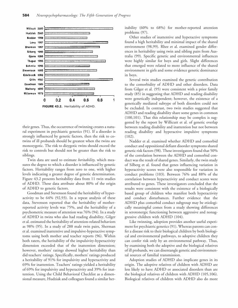
FIGURE 43.2. Heritability of ADHD.
their genes. Thus, the occurrence of twinning creates a natu-ral experiment in psychiatric genetics (91). If a disorder isstrongly influenced by genetic factors, then the risk to co-twins of ill probands should be greatest when the twins aremonozygotic. The risk to dizygotic twins should exceed therisk to controls but should not be greater than the risk tosiblings.
Twin data are used to estimate heritability, which mea-sures the degree to which a disorder is influenced by geneticfactors. Heritability ranges from zero to one, with higherlevels indicating a greater degree of genetic determination.Figure 43.2 presents heritability data from 11 twin studiesof ADHD. These data attribute about 80% of the originof ADHD to genetic factors.
Goodman and Stevenson found the heritability of hyper-activity to be 64% (92,93). In a repeat analysis of thesedata, Stevenson reported that the heritability of mother-reported activity levels was 75%, and the heritability of apsychometric measure of attention was 76% (94). In a studyof ADHD in twins who also had reading disability, Gilgeret al. estimated the heritability of attention-related behaviorsas 98% (95). In a study of 288 male twin pairs, Shermanet al. examined inattentive and impulsive-hyperactive symp-toms using both mother and teacher reports (96). Withinboth raters, the heritability of the impulsivity-hyperactivitydimension exceeded that of the inattention dimension;however, mothers’ ratings showed higher heritability thandid teachers’ ratings. Specifically, mothers’ ratings produceda heritability of 91% for impulsivity and hyperactivity and69% for inattention. Teachers’ ratings yielded a heritabilityof 69% for impulsivity and hyperactivity and 39% for inat-tention. Using the Child Behavioral Checklist as a dimen-sional measure, Hudziak and colleagues found a similar her-
itability (60% to 68%) for mother-reported attentionproblems (97).
Other studies of inattentive and hyperactive symptomsfound a high heritability and minimal impact of the sharedenvironment (98,99). Rhee et al. examined gender differ-ences in heritability using twin and sibling pairs from Aus-tralia (99). Specific genetic and environmental influenceswere highly similar for boys and girls. Slight differencesthat emerged were related to more influence of the sharedenvironment in girls and some evidence genetic dominancein boys.
Several twin studies examined the genetic contributionto the comorbidity of ADHD and other disorders. Datafrom Gilger et al. (95) were consistent with a prior familystudy (85) in suggesting that ADHD and reading disabilitywere genetically independent; however, the existence of agenetically mediated subtype of both disorders could notbe excluded. In contrast, two twin studies suggested thatADHD and reading disability share some genes in common(100,101). That this relationship may be complex is sug-gested by the report by Willicutt et al. of genetic overlapbetween reading disability and inattention but not betweenreading disability and hyperactive impulsive symptoms(102).
Nadder et al. examined whether ADHD and comorbidconduct and oppositional defiant disorder symptoms sharedgenetic risk factors (98). These investigators found that 50%of the correlation between the ADHD and comorbid con-duct was the result of shared genes. Similarly, the twin studyof Silberg et al. found that genes influencing variation inhyperactivity scores were also responsible for variation inconduct problems (103). Between 76% and 88% of thecorrelation between hyperactivity and conduct scores wereattributed to genes. These investigators concluded that theresults were consistent with the existence of a biologicallybased group of children who manifest both hyperactivityand conduct disturbances. Further evidence that theADHD plus comorbid conduct subgroup may be etiologi-cally meaningful comes from a study showing differencesin serotonergic functioning between aggressive and nonag-gressive children with ADHD (104).
Like twinning, adoption provides another useful experi-ment for psychiatric genetics (91).Whereas parents can con-fer a disease risk to their biological children by both biologi-cal and environmental pathways, to adoptive children theycan confer risk only by an environmental pathway. Thus,by examining both the adoptive and the biological relativesof ill probands, we can disentangle genetic and environmen-tal sources of familial transmission.
Adoption studies of ADHD also implicate genes in itsorigin. The adoptive relatives of children with ADHD areless likely to have ADHD or associated disorders than arethe biological relatives of children with ADHD (105,106).Biological relatives of children with ADHD also do more
Chapter 43: Pathophysiology of ADHD 585
poorly on standardized measures of attention than do adop-tive relatives of children with ADHD (107).
Segregation Analysis Studies
Segregation analysis provides evidence of genetic transmis-sion by demonstrating that the pattern of illness in familiesis consistent with known genetic mechanisms. An early ap-proach to this was reported by Morrison and Stewart, whoconcluded that polygenic inheritance was a likely mode oftransmission for ADHD (108). Contrasting data were pre-sented by Deutsch et al. (109). They found preliminaryevidence for a single dominant gene regulating the transmis-sion of ADHD and minor physical anomalies in 48 families.Similarly, Faraone et al. reported that the familial distribu-tion of ADHD was consistent with the effects of a singlemajor gene (75). Similar results were since reported in atwin study by Eaves et al. (110) and in a pedigree study byHess et al. (111). Consistent findings also emerged fromSouth America (112). Based on a sample of families fromColombia, the only models of inheritance that could notbe rejected were those of dominant and codominant majorgene effects. Finally, when families of ADHD probandswere ascertained by the father’s diagnosis of substance abuse,Maher et al. found that a sex-dependent mendelian codomi-nant model was the best explanation for the pattern of trans-mission of ADHD (113).
Although the segregation analyses of ADHD suggest thata single gene of major effect is involved in the origin ofADHD, the differences in fit among genetic models wasmodest. This was especially true for the comparison of mul-tifactorial and single gene inheritance. Several interpreta-tions of these results are possible. If ADHD had more thanone genetic cause, then the evidence of any single mode oftransmission could be relatively weak. Alternatively, ADHDmay be caused by several interacting genes of modest effect.This latter idea is consistent with ADHD’s high populationprevalence (2% to 7% for ADHD) and high concordancein monozygotic twins but modest recurrence risks in first-degree relatives.
The studies by Deutsch et al. and Faraone et al. predictedthat only about 40% of children carrying the putativeADHD gene would develop ADHD. This finding and otherfeatures of the genetic epidemiology of ADHD suggest thatsuch a gene likely interacts with other genes and environ-mental factors to produce ADHD. Moreover, the segrega-tion studies indicated that about 2% of people without theADHD gene would develop ADHD, a finding suggestingthat nongenetic forms of ADHD may exist.
Chromosomal Anomalies and MolecularGenetic Studies
Anomalies in the number or gross structure of chromosomesusually lead to very early-onset disorders having severe clini-
cal manifestations (e.g., mental retardation, gross physicalanomalies). No systematic studies of gross chromosomalanomalies in ADHD have been conducted, but there areseveral reports that such anomalies cause hyperactivity andinattention. Examples include the fragile X syndrome, du-plication of the Y chromosome in boys, and loss of an Xchromosome in girls. These associations are intriguing butrare. Thus, they can account for only a very small proportionof cases of ADHD.
Molecular genetic studies use the methods of linkage andassociation to search for aberrant genes that cause disease.Such studies of ADHD are relatively new and far fromdefinitive. Hauser et al. demonstrated that a rare familialform of ADHD is associated with generalized resistance tothyroid hormone, a disease caused by mutations in the thy-roid receptor-� gene (114). The thyroid receptor-� genecannot, however, account for many cases of ADHD becausethe prevalence of generalized resistance to thyroid hormoneis very low among patients with ADHD (1 in 2,500) (115),and, among pedigrees with generalized resistance to thyroidhormone, the association between ADHD and the thyroidreceptor-� gene has not been consistently found (116).
Several research teams have examined candidate genesin dopamine pathways because, as discussed earlier, animalmodels, theoretic considerations, and the effectiveness ofstimulant treatment implicate dopaminergic dysfunction inthe pathophysiology of this disorder. Several groups havereported an association between ADHD and dopamine D4receptor gene (DRD4) gene (117–123). Notably, eachstudy showed the 7-repeat allele of DRD4 to be associatedwith ADHD despite the use of different diagnostic systems(DSM-IIIR and DSM-IV) and measures of ADHD (ratingscales and structured interviews). However, like many find-ings in psychiatric genetics (91), these positive findings areoffset by some negative studies (124–128).
The positive DRD4 findings could be caused by anothergene in linkage disequilibrium with DRD4 or another var-iant within DRD4. However, because the DRD4 7-repeatallele mediates a blunted response to dopamine, it is a bio-logically reasonable risk factor for ADHD (129). The 7-repeat allele has also been implicated in novelty seeking, apersonality trait related to ADHD (130,131). Moreover,both norepinephrine and dopamine are potent agonists ofDRD4 (132).
When the D4 gene is disabled in a knockout mousemodel, dopamine synthesis increases in the dorsal striatum,and the mice show locomotor supersensitivity to ethanol,cocaine, and methamphetamine. (133). D4 knockout micealso show reduced novelty-related exploration (134), a find-ing consistent with human data suggesting a role for D4 innovelty-seeking behaviors.
Cook et al. reported an association between ADHD andthe 480-bp allele of the DAT gene using a family-basedassociation study (135). This finding was replicated by Gillet al. (136), Daly et al. (126), and Waldman et al. (137), but

Neuropsychopharmacology: The Fifth Generation of Progress586
not in other studies (124,138). In the study by Waldman etal. (137), hyperactive-impulsive symptoms but not inatten-tive symptoms were related to the number of DAT high-risk alleles. Further support for a link between the DATgene and ADHD comes from a study that relates this geneto poor methylphenidate response in children with ADHD(139) and from the neuroimaging study (Table 43.2) show-ing that DAT activity in the striatum is elevated by 70%in adults with ADHD (67).
In mice, eliminating DAT gene function leads to severalfeatures suggestive of ADHD: hyperactivity, deficits in in-hibitory behavior, and a paradoxical response to stimulants(i.e., stimulants reduce hyperactivity) (37,140). Studies ofthis knockout mouse model show the potential complexitiesof gene–disease associations. The loss of the DAT gene hasmany biological effects: increased extracellular dopamine, adoubling of the rate of dopamine synthesis (141), decreaseddopamine and tyrosine hydoxylase in striatum (142), anda nearly complete loss of functioning of dopamine autore-ceptors (143). Because ADHD is believed to be a hypodopa-minergic disorder, the decreased striatal dopamine may bemost relevant to the disorder.
Gainetdinov et al. showed that enhancement of seroto-nergic transmission mediates the mouse’s paradoxical re-sponse to stimulants (37). These researchers attributed thisto the effects of stimulants on the serotonin transporter. Tocomplicate matters further, Bezard et al. showed that DATknockout mice did not experience MPTP-induced dopami-nergic cell death (144), and another study found a gradienteffect such that mice with zero, one, and two functionalDAT genes showed increasing susceptibility to MPTP(145). These latter findings suggest that individual differ-ences in theDAT gene may mediate susceptibility to neuro-toxins having an affinity for the DAT.
A population-based association study has also implicatedthe A1 allele of the dopamine D2 receptor gene in ADHD(146). Absence of the D2 gene in mice leads to significantlyreduced spontaneous movements, a finding suggesting thatD2 plays a role in the regulation of activity levels (147,148). Mice without D2 genes also show decreased striatalDAT functioning (149), a finding that illustrates the poten-tial effects of gene–gene interaction on simple phenotypessuch as locomotion in mice. In addition, Calabresi et al.used the D2 knockout mouse to study the role of the D2receptor in striatal synaptic plasticity (150). In these mice,these researchers found abnormal synaptic plasticity at corti-costriatal synapses and long-term changes in synaptic effi-cacy in the striatum.
The only human study of the D3 receptor gene foundno evidence of an association with ADHD (151). However,homozygous mice lacking D3 receptors displayed increasedlocomotor activity, and heterozygous mice showed less pro-nounced hyperactivity. These results led Accili et al. to con-clude that D3 receptors play an inhibitory role in the controlof certain behaviors (152).
Four human studies of ADHD have examined the cate-chol-O-methyltransferase (COMT) gene, the product ofwhich is involved in the breakdown of dopamine and norep-inephrine. Although one study found that ADHD was asso-ciated with the Val allele (153), others have found no associ-ation between the COMT polymorphism and ADHD inIrish (154), Turkish (155), and Canadian (156) samples.Despite the negative finding, the positive finding is intrigu-ing because the Val allele leads to high COMT activity andan increased breakdown of catecholamines.
Another study found an association with the DXS7 locusof the X chromosome, a marker for monoamine oxidasethat encode enzymes that metabolize dopamine and otherneurotransmitters (157). Finally, Comings and colleaguesfound associations and additive effects of polymorphismsat three noradrenergic genes (the adrenergic �2A, adrenergic�2C, and dopamine-�-hydroxylas) on ADHD symptoms ina sample of patients with Tourette syndrome (158), butthey found no association between the tyrosine hydroxylasegene and ADHD in this sample (159).
Some investigators have used the coloboma mouse modelto investigate the genetics of ADHD. These mice have thecoloboma mutation, a hemizygous, 2-centimorgan deletionof a segment on chromosome 2q. The mutation leads tospontaneous hyperactivity (which is reversed by stimulants),delays in achieving complex neonatal motor abilities, defi-cits in hippocampal physiology that may contribute tolearning deficiencies, and deficits in Ca2�-dependent dopa-mine release in dorsal striatum (160).
The coloboma deletion region includes the gene encod-ing SNAP-25, a neuron-specific protein implicated in exo-cytotic neurotransmitter release. Hess et al. suggested thatinterference with SNAP-25 maymediate the mouse’s hyper-activity (161). As predicted by this hypothesis, when theseinvestigators bred a SNAP-25 transgene into colobomamice, the animals’ hyperactivity was reduced. Moreover,other work suggested that reduced SNAP-25 expressionleads to striatal dopamine and serotonin deficiencies, whichmay be involved in hyperactivity (162).
Hess et al. tested the idea that the human homologueof the mouse coloboma gene could be responsible forADHD by completing linkage studies of families withADHD by using markers on human chromosome 20p11-p12, which is syntenic to the coloboma deletion region(111). These investigators used five families for which segre-gation analysis suggested that ADHD was the result of asex-influenced, single gene. However, no significant linkagewas detected between ADHD and markers on chromosome20p11-p12.
ENVIRONMENTAL RISK FACTORS
Although genetic studies of ADHD unequivocally showthat genes are risk factors for the disorder, they also show

Chapter 43: Pathophysiology of ADHD 587
that the environment has a strong influence on the emer-gence of the disorder. This conclusion follows from studiesof identical twins, which show that when one twin hasADHD, the probability of the other, genetically identical,twin’s having ADHD is only about 60%. This less thanperfect identical twin concordance implicates environmen-tal risk factors. The nature of these risk factors has emergedfrom studies assessing features of the biological and psycho-social environment that may increase the risk of ADHD.
Biological Adversity
The idea that certain foods could cause ADHD receivedmuch attention in the popular press after claims were madethat ADHD could be cured by eliminating food additivesfrom the diet. The Feingold diet for ADHD was popular-ized by the media and was accepted by many parents of illchildren. Systematic studies, however, showed the diet wasnot effective and concluded that food additives do not causeADHD (163). Another popular theory posited that exces-sive sugar intake would lead to ADHD symptoms. Althoughsome positive studies supported this idea, the bulk of sys-tematic, controlled research did not (164).
In contrast to the mostly negative studies of dietary fac-tors, some toxins have been implicated in the origin of atleast some cases of ADHD. Several groups have shown thatlead contamination leads to distractibility, hyperactivity,restlessness, and lower intellectual functioning (165). How-ever, many children with ADHD do not show lead contami-nation, and many children with high lead exposure do notdevelop ADHD. Thus, lead exposure cannot account forthe bulk of cases of ADHD.
The literature examining the association of ADHD withpregnancy and delivery complications (PDCs) presents con-flicting results; it tends to support the idea that PDCs canpredispose children to ADHD (166–168), although someinvestigators do not (169). The PDCs implicated in ADHDfrequently lead to hypoxia and tend to involve chronic expo-sures to the fetus, such as toxemia, rather than acute, trau-matic events, such as delivery complications.
For example, Conners reported that mothers of childrenwith ADHD had high rates of toxemia during pregnancy(166). Hartsough and Lambert described eight PDCs asso-ciated with ADHD: maternal illness, toxemia, eclampsia,older maternal age, parity of child, fetal postmaturity, dura-tion of labor, and fetal distress during labor or birth (170).Nichols and Chen found that hyperactivity was significantlyassociated with low birth weight (171), and Chandola etal. reported antepartum hemorrhage, maternal age, lengthof labor, sex, and 1-minute Apgar scores to be significantprenatal and perinatal risk factors for subsequent referralfor hyperactivity (172).
Sprich-Buckminster et al. showed that the associationbetween ADHD and PDCs was strongest for children withADHD who had psychiatric comorbidity (168). PDCs were
also elevated among children with ADHD who had no fam-ily history of ADHD. These investigators concluded thatPDCs may be more common among those children withADHD having a weaker genetic predisposition, but thishypothesis was not confirmed in another study by the samegroup (167). The latter study found that children withADHD and a history of PDCs showed more school failureand psychometric evidence of cognitive impairment thanother children with ADHD. In addition to confirming theetiologic role of medical complications, this study showedthat psychosocial stress during pregnancy predicted subse-quent ADHD and poor cognitive performance in children.Notably, catecholamines are secreted in response to stress,and mouse studies showed that catecholamine administra-tion produces uterine vasoconstriction and fetal hypoxia(173).
One extensively studied risk factor has been maternalsmoking during pregnancy. By exposing the fetus to nico-tine, maternal smoking can damage the brain at criticaltimes in the developmental process. The smoking motheris at increased risk of antepartum hemorrhage, low maternalweight, and abruptio placentae (173). Her fetus is at risk oflow birth weight (173,174), and because smoking increasescarboxyhemoglobin levels in both maternal and fetal blood,it places the fetus at risk of hypoxia (175). Consistent withthese effects, maternal smoking during pregnancy predictsbehavioral and cognitive impairment in children andADHD (41,176).
Animal studies in pregnant mice and rats have shown apositive association between chronic exposure to nicotineand hyperactivity in offspring (42). Neonatal nicotine expo-sure prevents the development of low-affinity nicotine re-ceptors (177), and chronic exposure results in tolerance tothe drug and an increase in brain nicotinic receptors(178–181). Because nicotinic receptors modulate dopami-nergic activity and dopaminergic dysregulation may be in-volved in the pathophysiology of ADHD, it is theoreticallycompelling to consider maternal smoking as a risk factorfor ADHD.
Little is known about the potential role of in utero expo-sure to viral infections. Because maternal viral infectionscan affect the fetus and can have an adverse impact on thedeveloping brain, viral infections could be associated withlater psychopathology. Because viral infections occur morecommonly in winter than in other seasons, season-of-birthdata have been used to implicate in utero viral infectionfor several disorders including schizophrenia (182), autism(183), and dyslexia (184)
Although Mick et al. found no evidence of a strong sea-sonal pattern of birth in children with ADHD (185), theydid find statistically significant peaks for September birthsin children with ADHD who had comorbid learning dis-abilities and in children with ADHD who had no additionalpsychiatric comorbidity. Thus, it is possible that winter in-fections during the first trimester of pregnancy may account

Neuropsychopharmacology: The Fifth Generation of Progress588
for some subtypes of ADHD. Mick et al. found no evidencefavoring the idea that putative viral exposure led to a nonfa-milial form of ADHD. In contrast, they found a weak trendtoward an increase in winter births for children with ADHDwho have a positive family history of ADHD. If replicated,this finding suggests that a seasonally mediated infection atbirth may be an environmental ‘‘trigger’’ for the geneticpredisposition to the disorder.
Psychosocial Adversity
The delineation of psychosocial features in the child’s envi-ronment associated with more impaired outcome in chil-dren with ADHD has potentially important clinical, scien-tific, and public health implications. Such efforts can help toidentify etiologic risk factors associated with more impairedoutcome in ADHD and can characterize early predictors ofpersistence and morbidity of this disorder. Moreover, find-ing environmental risk factors for ADHD could help todesign improved preventive and therapeutic interventionprograms.
The classic studies by Rutter et al. of the Isle of Wightand the inner borough of London provide a compellingexample of how psychosocial risk factors influence childpsychopathology (186). Compelling examples of how psy-chosocial risk factors affect child psychopathology, thesestudies examined the prevalence of mental disorders in chil-dren living in two very different geographic areas. This re-search revealed six risk factors within the family environ-ment that correlated significantly with childhood mentaldisturbances: (a) severe marital discord, (b) low social class,(c) large family size, (d) paternal criminality, (e) maternalmental disorder, and (f) foster placement. This work foundthat it was the aggregate of adversity factors, rather thanthe presence of any single one, that impaired development.Other studies also found that as the number of adverseconditions accumulated, the risk of impaired outcome inthe child increased proportionally (187). Biederman et al.found a positive association between Rutter’s index of adver-sity and ADHD, measures of ADHD-associated psycho-pathology, impaired cognition, and psychosocial dysfunc-tion (188).
Other cross-sectional and longitudinal studies have iden-tified variables such as marital distress, family dysfunction,and low social class as risk factors for psychopathology anddysfunction in children. For example, the Ontario ChildHealth Study in Canada showed that family dysfunctionand low income predicted persistence and onset of one ormore psychiatric disorders over a 4-year follow-up period(189). Other work implicated low maternal education, lowsocial class, and single parenthood as important adversityfactors for ADHD (171,190). These studies suggested thatthe mothers of children with ADHD had more negativecommunication patterns, more conflict with their children,and a greater intensity of anger than did control mothers.
Biederman et al. showed that long-term conflict, de-creased family cohesion, and exposure to parental psycho-pathology, particularly maternal psychopathology, weremore common in ADHD-affected families compared withcontrol families (191). The differences between childrenwith ADHD and control children could not be accountedfor by either socioeconomic status or parental history ofmajor psychopathology. Moreover, increased levels of fam-ily-environment adversity predicted impaired psychosocialfunctioning. Measures indexing long-term family conflictshowed a more pernicious impact on the exposed child thanthose indexing exposure to parental psychopathology. In-deed, marital discord in families has consistently predicteddisruptive behaviors in boys (192). This research shows thatthe extent of discord and overt conflict, regardless ofwhether the parents are separated, predicts the child’s risksof psychopathology and dysfunction (193).
Thus, dysfunctional family environments appear to be anonspecific risk factor for psychiatric disorders and psycho-logical distress. Reid and Crisafulli reported a metaanalysisof the impact of marital discord on the psychological adjust-ment of children and found that parental conflict signifi-cantly predicted a variety of child behavior problems (194).The Ontario Child Health Study provided a prospectiveexample of the impact of parental conflict on children’smental health: family dysfunction (and low income) pre-dicted persistence and onset of one or more psychiatric dis-orders over a 4-year period (189).
Low maternal warmth and high maternal malaise andcriticism were previously associated with ADHD in children(195), and an epidemiologic study examining family attri-butes in children who had undergone stressful experiencesfound that children’s perceptions of mothers, but not fa-thers, differentiated stress-resilient and stress-affected chil-dren (196).
An extensive literature documents maternal depressionas a risk factor for psychological maladjustment and psychi-atric disorder in children (197). This is consistent with theknown familial link between ADHD and depression (79).Some investigators have suggested that depressed mood maylead mothers to perceive their children as more deviant thanwarranted by the child’s behavior. Richters, however, re-viewed 22 studies of this issue and concluded that, owingto methodologic problems with research in the area, therewas no empiric foundation for this claim (198).
Other data revealed a link between maternal depressionand child functioning that was independent of the mother’sperceptions. These data suggested that depressed mothersaccurately perceive symptomatic behavior but react to it ina negative manner that worsens the condition of the child.This conclusion was echoed by Gelfand and Teti (197).Their comprehensive review of relevant literature foundmany studies to document the assertion that depressedmothers have attitudes of insensitivity, disengagement, dis-approval, and hostility toward their children. They also

Chapter 43: Pathophysiology of ADHD 589
found maternal depression to be associated with undesirableparenting practices such as intrusiveness, unresponsiveness,and inept discipline. In addition, their review supported theidea that depressed mothers had negative perceptions oftheir children.
Other work shows that ADHD in children predictsdepression in mothers, but maternal depression provides noadditional information for predicting ADHD in siblingsof ADHD probands. This finding suggests that maternaldepression is a heterogeneous disorder. It may be that somemothers have a disorder that is genetically linked to ADHD,whereas others may experience depression resulting fromthe stress of raising a child with ADHD (and perhaps livingwith an ADHD-affected or antisocial husband). Further-more, it is possible that maternal depression exacerbatesfamily conflict and poor parenting, both of which couldexacerbate ADHD symptoms.
Notably, although many studies provide strong evidenceof the importance of psychosocial adversity for ADHD,these factors tend to emerge as universal predictors of chil-dren’s adaptive functioning and emotional health, not pre-dictors that are specific to ADHD. Thus, they can be con-ceptualized as nonspecific triggers of an underlyingpredisposition or as modifiers of the course of illness.
SUMMARY AND CONCLUSIONS
It is not yet possible to describe the origin and pathophysiol-ogy of ADHD completely. Nevertheless, converging evi-dence from the studies reviewed in this chapter supportsseveral empiric generalizations, which should be useful inguiding future research and theory.
Catecholamine Hypothesis
Much research supports the idea that catecholaminergic sys-tems mediate the onset and expression of ADHD symp-toms. The key data supporting this idea are as follows: (a)anti-ADHD medications have noradrenergic and dopami-nergic effects; (b) lesion studies in mouse and monkeymodels implicate dopaminergic pathways; (c) the SHR ratshows deficits in catecholaminergic systems; (d) D2, D3,and D4 knockout mice studies show that these genes regu-late locomotor activity; and (e) human studies implicate theDRD4 and DAT genes in the origin of ADHD.
Although the role of catecholamine systems cannot bedisputed, future work must also consider other neurotrans-mitter systems that exert upstream effects on catechola-mines. Two prime candidates are nicotinic and serotonergicsystems. Nicotinic agonists help to control the symptomsof ADHD, and nicotinic activation enhances dopaminergicneurotransmission. Serotonergic drugs have not been shownto be effective anti-ADHD agents, but knockout mice stud-ies suggest that the paradoxical effects of stimulants on hy-
peractivity are mediated by serotonergic neurotransmission.Moreover, SNAP-25, which has been implicated in studiesof the coloboma mouse, leads to striatal dopamine and sero-tonin deficiencies. These data call for further studies of sero-tonergic and nicotinic systems.
Brain Systems
Several types of study provide information about the locusof ADHD’s pathophysiology in the brain: neuropsychologi-cal studies, neuroimaging studies, and animal models.Taken together, these studies support the idea that ADHDarises from the dysregulation of frontal cortex, subcorticalstructures, and networks connecting them. This idea fitswith the pharmacotherapy of ADHD because a plausiblemodel for the effects of stimulants is that, through dopami-nergic or noradrenergic pathways, these drugs increase theinhibitory influences of frontal cortical activity on subcorti-cal structures.
Additional data supporting frontal-subcortical involve-ment in ADHD are as follows: (a) neuropsychological stud-ies implicate orbitofrontal and dorsolateral prefrontal cortexor regions projecting to these regions; (b) the monkey modelof ADHD implicates frontal-striatal neural networks; (c)studies of the SHR rat implicate caudate, putamen, nucleusaccumbens, and frontal cortex; patients with frontal lobedamage show ADHD-like behaviors; (d) structural neu-roimaging implicates frontal cortex, usually limited to theright side, cerebellum, globus pallidus, caudate, and corpuscallosum; (e) the I/LnJ mouse strain shows total callosalagenesis along with behavioral features that resembleADHD; (f) functional neuroimaging finds hypoactivity offrontal cortex, anterior cingulate cortex, and subcorticalstructures, usually on the right side; (g) ADHD secondaryto brain injury shows lesions in right putamen, right caudatenucleus, and right globus pallidus; (h) disabling the D4gene in mice leads to increased dopamine synthesis in dorsalstriatum; (i) mice without D2 genes also show decreasedstriatal DAT functioning, abnormal synaptic plasticity atcorticostriatal synapses, and long-term changes in synapticefficacy in the striatum; and (j) the coloboma mouse showsdeficient dopamine release in dorsal striatum.
Etiologic Factors
In a word, the origin of ADHD is complex. Although rarecases may have a single cause such as lead exposure, general-ized resistance to thyroid hormone, head injury, and frontallobe epilepsy, most cases of ADHD are probably caused bya complex combination of risk factors.
From the many twin studies of ADHD, we know forcertain that genes mediate susceptibility to ADHD. Molec-ular genetic studies suggest that two of these genes may bethe DRD4 gene and the DAT gene. To confirm these find-ings, we need much more work because, even if the positive

Neuropsychopharmacology: The Fifth Generation of Progress590
studies are correct, they may implicate neighboring genesinstead of those targeted by the studies. It seems unlikelythat a single ‘‘ADHD gene’’ causes ADHD with certainty.Instead, it seems likely that several genes act together toform the genetic substrate of the disorder.
When the ADHD-related variants of these genes are dis-covered, they will probably be ‘‘normal’’ variants and willmost certainly not have the devastating effects seen inknockout mouse models. For example, suppose future workconfirms that the 7-repeat allele is a risk factor for ADHD.We would consider this a normal variant because about20% of people who do not have ADHD carry this versionof the DRD4 gene. Most of these people do not developADHD despite the blunted dopaminergic transmission as-sociated with that allele, and many patients with ADHDdo not carry the allele. Thus, the 7-repeat allele cannot bea necessary or sufficient cause of the disorder. Instead, itacts in concert with other genes and environmental riskfactors to bring forth ADHD.
Like genetic studies, studies of environmental risk factorssuggest that most of these risks exert small but significantinfluences on the origin of ADHD. For example, most chil-dren with a history of PDCs do not develop ADHD, andmost children with ADHD do not have a history of ADHD.Nevertheless, research suggests that such complications aremore common among children with ADHD.
These considerations lead us to conclude that the originof ADHD is multifactorial. A simple multifactorial modelposits ADHD to arise a pool of genetic and environmentalvariables—each of small effect—that act in concert to pro-duce vulnerability to ADHD. If a person’s cumulative vul-nerability exceeds a certain threshold, he or she will manifestthe signs and symptoms of ADHD. According to the multi-factorial model, no single factor is a necessary or sufficientcause for ADHD, and each of the etiologic factors is inter-changeable (i.e., it does not matter which factors one has;only the total number is important). Whether risk factorscombine in an additive or interactive manner is unknown.
The mouse models of ADHD we described provide ex-amples of multifactorial causation in a simple system. Onemodel showed that individual differences in the DAT genecould directly produce a hypodopaminergic state; thesestudies showed that dopamine transporter variants differ intheir affinity for neurotoxins. Thus, dopamine transporterabnormalities could interact with environmental toxins toproduce hyperactivity. Another line of work shows that cate-cholamines are secreted in response to stress, and catechol-amine administration produces fetal hypoxia. Human stud-ies implicate both stress during pregnancy and fetal hypoxiaas risk factors for ADHD.
These simple examples suggest that unraveling the com-plexities of multifactorial causation will be a difficult taskfor ADHD researchers. However, because technological de-velopments in neuroscience andmolecular genetics are mov-ing at a rapid pace, the next decade of work should provide
us with more accurate assessments of the brain along witha complete sequence of the human genome. These advancesshould set the stage for breakthroughs in our understandingof the neurobiology of ADHD and in our ability to treataffected persons.
DISCLAIMERS
Dr. Biederman receives research support from Shire Labora-tories, Gliatec, Cephalon, Novartis Pharmaceuticals, and EliLilly & Company. In addition, he serves on speaking bu-reaus for SmithKline Beecham, Eli Lilly & Company, andPfizer Pharmaceuticals.
REFERENCES
1. Barkley RA. Attention deficit hyperactivity disorder: a handbookfor diagnosis and treatment. New York: Guilford, 1998.
2. Spencer T, Biederman J, Wilens T, et al. Is attention deficithyperactivity disorder in adults a valid disorder? Harvard RevPsychiatry 1994;1:326–335.
3. Hill J, Schoener E. Age-dependent decline of attention deficithyperactivity disorder. Am J Psychiatry 1996;153:1143–1146.
4. Biederman J, Newcorn J, Sprich S. Comorbidity of attentiondeficit hyperactivity disorder with conduct, depressive, anxiety,and other disorders. Am J Psychiatry 1991;148:564–577.
5. Caron C, Rutter M. Comorbidity in child psychopathology:concepts, issues and research strategies. J Child Psychol Psychiatry1991;32:1063–1080.
6. Bird HR, Canino G, Rubio-Stipec M, et al. Estimates of theprevalence of childhood maladjustment in a community surveyin Puerto Rico: the use of combined measures. Arch Gen Psychia-try 1988;45:1120–1126.
7. Anderson JC, Williams S, McGee R, et al. DSM-III disordersin preadolescent children: prevalence in a large sample from thegeneral population. Arch Gen Psychiatry 1987;44:69–76.
8. Spencer TJ, Biederman J, Wilens T, et al. Pharmacotherapy ofattention deficit hyperactivity disorder across the lifecycle: aliterature review. J Am Acad Child Adolesc Psychiatry 1996;35:409–432.
9. Wilens T, Biederman J, Spencer T, et al. Pharmacotherapy ofadult attention deficit/hyperactivity disorder: a review. J ClinPsychopharmacol 1995;15:270–279.
10. Prince J, Wilens T, Biederman J, et al. A controlled study ofnortriptyline in children and adolescents with attention deficithyperactivity disorder. In: Scientific proceedings of the annualmeeting of the American Academy of Child and AdolescentPsychiatrists XV, Chicago, 1999.
11. Casat CD, Pleasants DZ, Van Wyck Fleet J. A double-blindtrial of bupropion in children with attention deficit disorder.Psychopharmacol Bull 1987;23:120–122.
12. Casat CD, Pleasants DZ, Schroeder DH, et al. Bupropion inchildren with attention deficit disorder. Psychopharmacol Bull1989;25:198–201.
13. Wender PH, Reimherr FW. Bupropion treatment of attention-deficit hyperactivity disorder in adults. Am J Psychiatry 1990;147:1018–1020.
14. Ernst M, Liebenauer LL, Jons PH, et al. Selegiline in adultswith attention deficit hyperactivity disorder: clinical efficacy andsafety. Psychopharmacol Bull 1996;32:327–334.
15. Spencer T, Wilens TE, Biederman J. A double-blind, crossovercomparison of tomoxetine and placebo in adults with ADHD.

Chapter 43: Pathophysiology of ADHD 591
In: Scientific proceedings of the annual meeting of the AmericanAcademy of Child and Adolescent Psychiatrists XII, New Orle-ans, 1995.
16. Spencer T, Biederman J, Wilens T, et al. An open, dose rangingstudy of tomoxetine in children with ADHD. In: ScientificProceedings of the annual meeting of the American Academyof Child and Adolescent Psychiatry XV, Chicago, 1999.
17. Biederman J, Spencer T, Wilens T. Psychopharmacology inchildren and adolescents. In: Wiener J, ed. Textbook of childand adolescent psychiatry.Washington, DC: American Psychiat-ric Press, 1997:779–813.
18. Levin ED, Conners CK, Sparrow E, et al. Nicotine effects onadults with attention-deficit/hyperactivity disorder. Psychophar-macology 1996;123:55–63.
19. Wilens TE, Biederman J, Spencer TJ, et al. A pilot controlledclinical trial of ABT-418, a cholinergic agonist, in the treatmentof adults with attention deficit hyperactivity disorder. Am JPsychiatry 1999;156:1931–1937.
20. Elia J, Borcherding BG, Potter WZ, et al. Stimulant drug treat-ment of hyperactivity: biochemical correlates. Clin PharmacolTher 1990;48:57–66.
21. Solanto M. Neuropsychopharmacological mechanisms of stim-ulant drug action in attention-deficit hyperactivity disorder: areview and integration. Behav Brain Res 1998;94:127–152.
22. Zametkin AJ, Rapoport JL. Noradrenergic hypothesis of atten-tion deficit disorder with hyperactivity: a critical review. In:Meltzer HY, ed. Psychopharmacology: the third generation ofprogress. New York: Raven, 1987:837–842.
23. Zametkin AJ, Rapoport JL.Neurobiology of attention deficitdisorder with hyperactivity: where have we come in 50 years?J Am Acad Child Adolesc Psychiatry 1987;26:676–686.
24. Pliszka S, McCracken J, Maas J. Catecholamines in attention-deficity hyperactivity disorder: current perspectives. J Am AcadChild Adolesc Psychiatry 1996;35:264–272.
25. Shaywitz SE, Cohen DJ, Shaywitz BA. The biochemical basisof minimal brain dysfunction. J Pediatr 1978;92:179–187.
26. Schneider JS, Roeltgen DP. Delayed matching-to-sample, ob-ject retrieval, and discrimination reversal deficits in chronic lowdose MPTP-treated monkeys. Brain Res 1993;615:351–354.
27. Schneider JS, Kovelowski CJD. Chronic exposure to low dosesof MPTP. I. Cognitive deficits in motor asymptomatic mon-keys. Brain Res 1990;519:122–128.
28. Schneider JS, Sun ZQ, Roeltgen DP. Effects of dopamine ago-nists on delayed response performance in chronic low-doseMPTP-treated monkeys. Pharmacol Biochem Behav 1994;48:235–240.
29. Roeltgen DP, Schneider JS. Task persistence and learning abilityin normal and chronic low dose MPTP-treated monkeys. BehavBrain Res 1994;60:115–124.
30. Russell V, de Villiers A, Sagvolden T, et al. Altered dopami-nergic function in the prefrontal cortex, nucleus accumbensand caudate-putamen of an animal model of attention-deficithyperactivity disorder: the spontaneously hypertensive rat. BrainRes 1995;676:343–351.
31. Russell VA. The nucleus accumbens motor-limbic interface ofthe spontaneously hypertensive rat as studied in vitro by thesuperfusion slice technique. Neurosci Biobehav Rev 2000;24:133–136.
32. de Villiers AS, Russell VA, Sagvolden T, et al. Alpha 2-adreno-ceptor mediated inhibition of [3H]dopamine release from nu-cleus accumbens slices and monoamine levels in a rat model forattention-deficit hyperactivity disorder. Neurochem Res 1995;20:427–433.
33. Papa M, Berger DF, Sagvolden T, et al. A quantitative cyto-chrome oxidase mapping study, cross-regional and neurobehav-ioural correlations in the anterior forebrain of an animal model
of attention deficit hyperactivity disorder. Behav Brain Res 1998;94:197–211.
34. King JA, Barkley RA, Delville Y, et al. Early androgen treatmentdecreases cognitive function and catecholamine innervation inan animal model of ADHD. Behav Brain Res 2000;107:35–43.
35. Carey MP,Diewald LM, Esposito FJ, et al. Differential distribu-tion, affinity and plasticity of dopamine D-1 and D-2 receptorsin the target sites of the mesolimbic system in an animal modelof ADHD. Behav Brain Res 1998;94:173–185.
36. Tatsumi M, Groshan K, Bakely R, et al. Pharmacological profileof antidepressants and related compounds at human mono-amine transporters. Eur J Pharmacol 1997;340:249–258.
37. Gainetdinov RR, Wetsel WC, Jones SR, et al. Role of serotoninin the paradoxical calming effect of psychostimulants on hyper-activity. Science 1999;283:397–402.
38. Milberger S, Biederman J, Faraone S, et al. Further evidence ofan association between attention-deficit/hyperactivity disorderand cigarette smoking: findings from a high-risk sample of sib-lings. Am J Addict 1997;6:205–217.
39. Milberger S, Biederman J, Faraone SV, et al. Attention deficithyperactivity disorder is associated with early initiation of ciga-rette smoking in children and adolescents. J Am Acad ChildAdolesc Psychiatry 1997;36:37–44.
40. Riggs PD, Mikulich SK, Whitmore EA, et al. Relationship ofADHD, depression, and non-tobacco substance use disorders tonicotine dependence in substance-dependent delinquents. DrugAlcohol Depend 1999;54:195–205.
41. Milberger S, Biederman J, Faraone S, et al. Is maternal smokingduring pregnancy a risk factor for attention deficit hyperactivitydisorder in children? Am J Psychiatry 1996;153:1138–1142.
42. Johns JM, Louis TM, Becker RF, et al. Behavioral effects ofprenatal exposure to nicotine in guinea pigs.Neurobehav ToxicolTeratol 1982;4:365–369.
43. Fung YK, Lau YS. Effects of prenatal nicotine exposure on ratstriatal dopaminergic and nicotinic systems. Pharmacol BiochemBehav 1989;33:1–6.
44. Westfall TC, Grant H, Perry H. Release of dopamine and 5-hydroxytryptamine from rat striatal slices following activationof nicotinic cholinergic receptors. Gen Pharmacol 1983;14:321–325.
45. Mereu G, Yoon K, Gessa G, et al. Preferential stimulation ofventral tegmental area dopaminergic neurons by nicotine. EurJ Pharmacol 1987;141:395-399.
46. Satterfield JH, Dawson ME. Electrodermal correlates of hyper-activity in children. Psychophysiology 1971;8:191–197.
47. Mattes JA. The role of frontal lobe dysfunction in childhoodhyperkinesis. Comp Psychiatry 1980;21:358–369.
48. Weiss JL, Seidman LJ. The clinical use of psychological andneuropsychological tests. In: Nicholi A, ed. The new Harvardguide to psychiatry. Cambridge, MA: Harvard University Press,1988:46–69.
49. Barkley RA, Grodzinsky G, DuPaul GJ. Frontal lobe functionsin attention deficit disorder with and without hyperactivity: areview and research report. J Abnorm Child Psychol 1992;20:163–188.
50. Seidman LJ, Biederman J, Faraone SV, et al. Effects of familyhistory and comorbidity on the neuropsychological performanceof children with ADHD: preliminary findings. J Am Acad ChildAdolesc Psychiatry 1995;34:1015–1024.
51. Seidman LJ, Biederman J, Faraone SV, et al. Toward defininga neuropsychology of attention deficit-hyperactivity disorder:performance of children and adolescents from a large clinicallyreferred sample. J Consult Clin Psychol 1997;65:150–160.
52. Seidman LJ, Biederman J, Weber W, et al. Neuropsychologicalfunction in adults with attention-deficit hyperactivity disorder.Biol Psychiatry 1998;44:260–268.

Neuropsychopharmacology: The Fifth Generation of Progress592
53. Barkley R, Murphy K, Kwasnik D. Psychological adjustmentand adaptive impairments in young adults with ADHD. J AttenDisord 1996;1:41–54.
54. Buchsbaum MS, Haier RJ, Sostek AJ, et al. Attention dysfunc-tion and psychopathology in college men. Arch Gen Psychiatry1985;42:354–360.
55. Gualtieri CT, Ondrusek MG, Finley C. Attention deficit disor-ders in adults. Clin Neuropharmacol 1985;8:343–356.
56. Holdnack JA, Moberg PJ, Arnold SE, et al. Speed of processingand verbal learning deficits in adults diagnosed with attentiondeficit disorder. Neuropsychiatry Neuropsychol Behav Neurol1995;8:282–292.
57. Lovejoy DW, Ball JD, Keats M, et al. Neuropsychological per-formance of adults with attention deficit hyperactivity disorder(ADHD): diagnostic classification estimates for measures offrontal lobe/executive functioning. J Int Neuropsychol Soc 1999;5:222–233.
58. Taylor CJ, Miller DC. Neuropsychological assessment of atten-tion in ADHD adults. J Atten Disord 1997;2:77–88.
59. Fuster J. The prefrontal cortex. New York: Raven, 1989.60. Cummings JL. Frontal-subcortical circuits and human behavior.
Arch Neurol 1993;50:873–880.61. Magara F, Ricceri L, Wolfer DP, et al. The acallosal mouse
strain I/LnJ: a putative model of ADHD? Neurosci BiobehavRev 2000;24:45–50.
62. Ernst M, Liebenauer L, King A, et al. Reduced brain metabolismin hyperactive girls. J Am Acad Child Adolesc Psychiatry 1994;33:858–868.
63. Baving L, Laucht M, Schmidt MH. Atypical frontal brain acti-vation in ADHD: preschool and elementary school boys andgirls. J Am Acad Child Adolesc Psychiatry 1999;38:1363–1371.
64. Ernst M, Zametkin A, Matochik J, et al. DOPA decarboxylaseactivity in attention deficit hyperactivity disorder adults: a [fluo-rine-18]fluorodopa positron emission tomographic study. JNeurosci 1998;18:5901–5907.
65. Bush G, Frazier JA, Rauch SL, et al. Anterior cingulate cortexdysfunction in attention deficit/hyperactivity disorder revealedby fMRI and the counting stroop. Biol Psychiatry 1999;45:1542–1552.
66. Zametkin AJ, Nordahl TE, Gross M, et al. Cerebral glucosemetabolism in adults with hyperactivity of childhood onset. NEngl J Med 1990;323:1361–1366.
67. Dougherty DD, Bonab AA, Spencer TJ, et al. Dopamine trans-porter density is elevated in patients with attention deficit hyper-activity disorder. Lancet 1999;354:2132–2133.
68. Powell AL, Yudd A, Zee P, et al. Attention deficit hyperactivitydisorder associated with orbitofrontal epilepsy in a father anda son. Neuropsychiatry Neuropsychol Behav Neurol 1997;10:151–154.
69. Herskovits EH, Megalooikonomou V, Davatzikos C, et al. Isthe spatial distribution of brain lesions associated with closed-head injury predictive of subsequent development of attention-deficit/hyperactivity disorder? Analysis with brain-image data-base. Radiology 1999;213:389–394.
70. Nopoulos P, Berg S, Castellenos FX, et al. Developmental brainanomalies in children with attention-deficit hyperactivity disor-der. J Child Neurol 2000;15:102–108.
71. Welner Z, Welner A, Stewart M, et al. A controlled study ofsiblings of hyperactive children. J Nerv Ment Dis 1977;165:110–117.
72. Manshadi M, Lippmann S, O’Daniel R, et al. Alcohol abuse andattention deficit disorder. J Clin Psychiatry 1983;44:379–380.
73. Pauls DL, Shaywitz SE, Kramer PL, et al. Demonstration ofvertical transmission of attention deficit disorder. Ann Neurol1983;14:363.
74. Biederman J, Faraone SV, Keenan K, et al. Family-genetic and
psychosocial risk factors in DSM-III attention deficit disorder.J Am Acad Child Adolesc Psychiatry 1990;29:526–533.
75. Faraone S, Biederman J, Chen WJ, et al. Segregation analysisof attention deficit hyperactivity disorder: evidence for singlegene transmission. Psychiatr Genet 1992;2:257–275.
76. Faraone SV, Tsuang MT. Methods in psychiatric genetics. In:Tohen M, Tsuang MT, Zahner GEP, eds. Textbook in psychiat-ric epidemiology. New York: John Wiley, 1995:81–134.
77. Biederman J, Faraone SV, Keenan K, et al. Further evidencefor family-genetic risk factors in attention deficit hyperactivitydisorder: patterns of comorbidity in probands and relatives psy-chiatrically and pediatrically referred samples. Arch Gen Psychia-try 1992;49:728–738.
78. Biederman J, Faraone SV, Keenan K, et al. Evidence of familialassociation between attention deficit disorder and major affec-tive disorders. Arch Gen Psychiatry 1991;48:633–642.
79. Faraone SV, Biederman J. Do attention deficit hyperactivitydisorder and major depression share familial risk factors? J NervMent Diss 1997;185:533–541.
80. Faraone S, Biederman J, Monuteaux MC. Attention deficit dis-order and conduct disorder in girls: evidence for a familial sub-type. Biol Psychiatry 2000;48:21–29.
81. Faraone S, Biederman J, Garcia Jetton J, et al. Attention deficitdisorder and conduct disorder: longitudinal evidence for a famil-ial subtype. Psychol Med 1997;27:291–300.
82. Faraone SV, Biederman J, Mennin D, et al. Bipolar and antiso-cial disorders among relatives of ADHD children: parsing famil-ial subtypes of illness. Am J Med Genet 1998;81:108–116.
83. Faraone SV, Biederman J, Mennin D, et al. Attention-deficithyperactivity disorder with bipolar disorder: a familial subtype?J Am Acad Child Adolesc Psychiatry 1997;36:1378–1387;discus-sion 1387–1390.
84. Biederman J, Faraone SV, Keenan K, et al. Familial associationbetween attention deficit disorder and anxiety disorders. Am JPsychiatry 1991;148:251–256.
85. Faraone S, Biederman J, Krifcher Lehman B, et al. Evidencefor the independent familial transmission of attention deficithyperactivity disorder and learning disabilities: results from afamily genetic study. Am J Psychiatry 1993;150:891–895.
86. Smalley SL, McCracken J, McGough J. Refining the ADHDphenotype using affective sibling pair families. Am J Med Genet2001;105:31–33.
87. Faraone SV, Biederman J, Monuteaux MC. Toward guidelinesfor pedigree selection in genetic studies of attention deficit hy-peractivity disorder. Genet Epidemiol 2000;18:1–16.
88. Biederman J, Faraone SV, Milberger S, et al. Predictors of persis-tence and remission of ADHD: results from a four-year prospec-tive follow-up study of ADHD children. J Am Acad Child Ad-olesc Psychiatry 1996;35:343–351.
89. Biederman J, Faraone SV, Mick E, et al. High risk for attentiondeficit hyperactivity disorder among children of parents withchildhood onset of the disorder: a pilot study. Am J Psychiatry1995;152:431–435.
90. Biederman J, Faraone SV, Taylor A, et al. Diagnostic continuitybetween child and adolescent ADHD: findings from a longitu-dinal clinical sample. J Am Acad Child Adolesc Psychiatry 1998;37:305–313.
91. Faraone SV, Tsuang D, Tsuang MT. Genetics and mental disor-ders: a guide for students, clinicians, and researchers. New York:Guilford, 1999.
92. Goodman R, Stevenson J. A twin study of hyperactivity. II.The aetiological role of genes, family relationships and perinataladversity. J Child Psychol Psychiatry 1989;30:691–709.
93. Goodman R, Stevenson J. A twin study of hyperactivity. I. Anexamination of hyperactivity scores and categories derived from

Chapter 43: Pathophysiology of ADHD 593
Rutter teacher and parent questionnaires. J Child Psychol Psy-chiatry 1989;30:671–689.
94. Stevenson J. Evidence for a genetic etiology in hyperactivity inchildren. Behav Genet 1992;22:337–344.
95. Gilger JW, Pennington BF, DeFries C. A twin study of theetiology of comorbidity: attention deficit hyperactivity disorderand dyslexia. J Am Acad Child Adolesc Psychiatry 1992;31:343–348.
96. Sherman D, Iacono W, McGueM. Attention deficit hyperactiv-ity disorder dimensions: a twin study of inattention and impul-sivity hyperactivity. J Am Acad Child Adolesc Psychiatry 1997;36:745–753.
97. Hudziak JJ, Rudiger LP, Neale MC, et al. A twin study ofinattentive, aggressive, and anxious/depressed behaviors. J AmAcad Child Adolesc Psychiatry in press.
98. Nadder TS, Silberg JL, Eaves LJ, et al. Genetic effects onADHD symptomatology in 7- to 13-year-old twins: results froma telephone survey. Behav Genet 1998;28:83–99.
99. Rhee SH, Waldman ID, Hay DA, et al. Sex differences in ge-netic and environmental influences on DSM-III-R attention-deficit/hyperactivity disorder. J Abnorm Psychol 1999;108:24–41.
100. Light JG, Pennington BF, Gilger J, et al. Reading disability andhyperactivity disorder: evidence for a common genetic etiology.Dev Neuropsychol 1995;11:323–335.
101. Stevenson J, Pennington BF, Gilger JW, et al. Hyperactivityand spelling disability: testing for shared genetic aetiology. JChild Psychol Psychiatry 1993;34:1137–1152.
102. Willicutt EG, Pennington BF, DeFries JC. A twin study of theetiology of comorbidity between reading disability and atten-tion-deficit/hyperactivity disorder. Am J Med Genet 2000;96:293–301.
103. Silberg J, Rutter M, Meyer J, et al. Genetic and environmentalinfluences on the covariation between hyperactivity and conductdisturbance in juvenile twins. J Child Psychol Psychiatry 1996;37:803–816.
104. Halperin J, Sharma V, Siever L, et al. Serotonergic function inaggressive and nonaggressive boys with attention deficit hyper-activity disorder. Am J Psychiatry 1994;151:243–248.
105. Cantwell DP. Genetics of hyperactivity. J Child Psychol Psychia-try 1975;16:261–264.
106. Morrison JR, Stewart MA. The psychiatric status of the legalfamilies of adopted hyperactive children. Arch Gen Psychiatry1973;28:888–891.
107. Alberts-Corush J, Firestone P, Goodman JT. Attention andimpulsivity characteristics of the biological and adoptive parentsof hyperactive and normal control children. Am J Orthopsychia-try 1986;56:413–423.
108. Morrison JR, Stewart MA. Bilateral inheritance as evidence forpolygenicity in the hyperactive child syndrome. J Nerv MentDis 1974;158:226–228.
109. Deutsch CK, Matthysse S, Swanson JM, et al. Genetic latentstructure analysis of dysmorphology in attention deficit disor-der. J Am Acad Child Adolesc Psychiatry 1990;29:189–194.
110. Eaves L, Silberg J, Hewitt J, et al. Genes, personality, and psy-chopathology: a latent class analysis of liability to symptoms ofattention-deficit hyperactivity disorder in twins. In: Plomin R,McLearn G, eds. Nature, Nurture and Psychology. Washington,DC: American Psychological Association, 1993:285–306.
111. Hess EJ, Rogan PK, Domoto M, et al. Absence of linkage ofapparently single gene mediated ADHD with the human syn-tenic region of the mouse mutant coloboma. Am J Med Genet1995;60:573–579.
112. Lopera F, Palacio LG, Jimenez I, et al. Discrimination betweengenetic factors in attention deficit. Rev Neurol 1999;28:660–664.
113. Maher BS, Marazita ML, Moss HB. Segregation analysis ofattention deficit hyperactivity disorder. Am J Med Gen 1999;88:71–78.
114. Hauser P, Zametkin A, Martinez P, et al. Attention deficit-hyperactivity disorder in people with generalized resistance tothyroid hormone. N Engl J Med 1993;328:997–1001.
115. Weiss R, Stein M, Trommer B, et al. Attention-deficit hyperac-tivity disorder and thyroid function. J Pediatr 1993;123:539–545.
116. Weiss RE, Stein MA, Duck SC, et al. Low intelligence butnot attention deficit hyperactivity disorder is associated withresistance to thyroid hormone caused by mutation R316H inthe thyroid hormone receptor B gene. J Clin Endocrinol Metab1994;78:1525–1528.
117. Barr CL, Wigg KG, Bloom S, et al. Further evidence fromhaplotype analysis for linkage of the dopamine D4 receptorgene and attention-deficit hyperactivity disorder. Am J MedGenet 2000;96:262–267.
118. Comings DE, Gonzalez N, Wu S, et al. Studies of the 48 bprepeat polymorphism of the DRD4 gene in impulsive, compul-sive, addictive behaviors: Tourette syndrome, ADHD, patho-logical gambling, and substance abuse. Am J Med Genet 1999;88:358–368.
119. Faraone SV, Biederman J, Weiffenbach B, et al. Dopamine D4gene 7-repeat allele and attention deficit hyperactivity disorder.Am J Psychiatry 1999;156:768–770.
120. LaHoste GJ, Swanson JM,Wigal SB, et al. Dopamine D4 recep-tor gene polymorphism is associated with attention deficit hy-peractivity disorder. Mol Psychiatry 1996;1:121–124.
121. Rowe DC, Stever C, Giedinghagen LN, et al. Dopamine DRD4receptor polymorphism and attention deficit hyperactivity dis-order [see Comments]. Mol Psychiatry 1998;3:419–426.
122. Smalley SL, Bailey JN, Palmer CG, et al. Evidence that thedopamineD4 receptor is a susceptibility gene in attention deficithyperactivity disorder [see Comments]. Mol Psychiatry 1998;3:427–430.
123. Swanson JM, Sunohara GA, Kennedy JL, et al. Association ofthe dopamine receptor D4 (DRD4) gene with a refined pheno-type of attention deficit hyperactivity disorder (ADHD): a fam-ily-based approach. Mol Psychiatry 1998;3:38–41.
124. Asherson P, Virdee V, Curran S, et al. Association of DSM-IVattention deficit hyperactivity disorder and monoamine path-way genes. Am J Med Genet 1998;81:548.
125. Castellanos FX, Lau E, Tayebi N, et al. Lack of an associationbetween a dopamine-4 receptor polymorphism and attention-deficit/hyperactivity disorder: genetic and brain morphometricanalyses [see Comments]. Mol Psychiatry 1998;3:431–434.
126. Daly G, Hawi Z, Fitzgerald M, et al. Attention deficit hyperac-tivity disorder: association with the dopamine transporter(DAT1) but not with the dopamine D4 receptor (DRD4). AmJ Med Genet 1998;81:501.
127. Eisenberg J, Zohar A, Mei-Tal G, et al. A halotype relative riskstudy of the dopamine D4 receptor (DRD4) exon III repeatpolymorphism and attention deficit hyperactivity disorder(ADHD). Am J Med Genet 2000;96:258–261.
128. Hawi Z, McCarron M, Kirley A, et al. No association of dopa-mine DRD4 receptor (DRD4) gene polymorphism in attentiondeficit hyperactivity disorder (ADHD) in the Irish population.Am J Med Genet 2000;96:268–272.
129. Asghari V, Sanyal S, Buchwaldt S, et al. Modulation of intracel-lular cyclic AMP levels by different human dopamine D4 recep-tor variants. J Neurochem 1995;65:1157–1165.
130. Ebstein RP, Novick O, Umansky R, et al. Dopamine D4 recep-tor (D4DR) exon III polymorphism associated with the humanpersonality trait of novelty seeking. Nat Genet 1996;12:78–80.
131. Benjamin J, Patterson C, Greenberg BD, et al. Population and

Neuropsychopharmacology: The Fifth Generation of Progress594
familial association between the D4 dopamine receptor geneand measures of novelty seeking. Nat Genet 1996;12:81–84.
132. Lanau F, Zenner M, Civelli O, et al. Epinephrine and norepi-nephrine act as potent agonists at the recombinant human dopa-mine D4 receptor. J Neurochem 1997;68:804–812.
133. Rubinstein M, Phillips TJ, Bunzow JR, et al. Mice lacking dopa-mine D4 receptors are supersensitive to ethanol, cocaine, andmethamphetamine. Cell 1997;90:991–1001.
134. Dulawa SC, Grandy DK, Low MJ, et al. Dopamine D4 recep-tor-knock-out mice exhibit reduced exploration of novel stimuli.J Neurosci 1999;19:9550–9556.
135. Cook EH, Stein MA, Krasowski MD, et al. Association of atten-tion deficit disorder and the dopamine transporter gene. Am JMed Genet 1995;56:993–998.
136. Gill M, Daly G, Heron S, et al. Confirmation of assocationbetween attention deficit hyperactivity disorder and a dopaminetransporter polymorphism. Mol Psychiatry 1997;2:311–313.
137. Waldman ID, Rowe DC, Abramowitz A, et al. Association andlinkage of the dopamine transporter gene and attention-deficithyperactivity disorder in children: heterogeneity owing to diag-nostic subtype and severity. Am J Med Genet 1998;63:1767–1776.
138. Poulton K, Holmes J, Hever T, et al. A molecular genetic studyof hyperkinetic disorder/attention deficit hyperactivity disorder.Am J Med Genet 1998;81:458.
139. Winsberg BG, Comings DE. Association of the dopamine trans-porter gene (DAT1) with poor methylphenidate response [seeComments]. J Am Acad Child Adolesc Psychiatry 1999;38:1474–1477.
140. Giros B, Jaber M, Jones SR, et al. Hyperlocomotion and indif-ference to cocaine and amphetamine in mice lacking the dopa-mine transporter. Nature 1996;379:606–612.
141. Gainetdinov RR, Jones SR, Fumagalli F, et al. Re-evaluationof the role of the dopamine transporter in dopamine systemhomeostasis. Brain Res Brain Res Rev 1998;26:148–153.
142. Jaber M, Dumartin B, Sagne C, et al. Differential regulationof tyrosine hydroxylase in the basal ganglia of mice lacking thedopamine transporter. Eur J Neurosci 1999;11:3499–3511.
143. Jones SR, Gainetdinov RR, Hu XT, et al. Loss of autoreceptorfunctions in mice lacking the dopamine transporter. Natl Neu-rosci 1999;2:649–655.
144. Bezard E, Gross CE, Fournier MC, et al. Absence of MPTP-induced neuronal death in mice lacking the dopamine trans-porter. Exp Neurol 1999;155:268–273.
145. Gainetdinov RR, Fumagalli F, Jones SR, et al. Dopamine trans-porter is required for in vivo MPTP neurotoxicity: evidencefrom mice lacking the transporter. J Neurochem 1997;69:1322–1325.
146. Comings DE, Comings BG, Muhleman D, et al. The dopamineD2 receptor locus as a modifying gene in neuropsychiatric disor-ders. JAMA 1991;266:1793–1800.
147. Baik JH, Picetti R, Saiardi A, et al. Parkinsonian-like locomotorimpairment in mice lacking dopamine D2 receptors. Nature1995;377:424–428.
148. Kelly MA, Rubinstein M, Phillips TJ, et al. Locomotor activityin D2 dopamine receptor-deficient mice is determined by genedosage, genetic background, and developmental adaptations. JNeurosci 1998;18:3470–3479.
149. Dickinson SD, Sabeti J, Larson GA, et al. Dopamine D2 recep-tor-deficient mice exhibit decreased dopamine transporter func-tion but no changes in dopamine release in dorsal striatum. JNeurochem 1999;72:148–156.
150. Calabresi P, Saiardi A, Pisani A, et al. Abnormal synaptic plastic-ity in the striatum of mice lacking dopamine D2 receptors. JNeurosci 1997;17:4536–4544.
151. Barr CL, Wigg KG, Wu J, et al. Linkage study of two polymor-
phisms at the dopamine D3 receptor gene and attention-deficithyperactivity disorder. Am J Med Genet 2000;96:114–117.
152. Accili D, Fishburn CS, Drago J, et al. A targeted mutation ofthe D3 dopamine receptor gene is associated with hyperactivityin mice. Proc Natl Acad Sci USA 1996;93:1945–1949.
153. Eisenberg J, Mei-Tal G, Steinberg A, et al. Haplotype relativerisk study of catechol-O-methyltransferase (COMT) and atten-tion deficit hyperactivity disorder (ADHD): association of thehigh-enzyme activity Val allele with ADHD impulsive-hyperac-tive phenotype. Am J Med Genet 1999;88:497–502.
154. Hawi Z, Millar N, Daly G, et al. No association between cate-chol-O-methyltransferase (COMT) gene polymorphism and at-tention deficit hyperactivity disorder (ADHD) in an Irish sam-ple. Am J Med Genet 2000;96:282–284.
155. Tahir E, Curran S, Yazgan Y, et al. No association between lowand high activity catecholamine-methyl-transferase (COMT)and attention deficit hyperactivity disorder (ADHD) in a sampleof Turkish children. Am J Med Genet 2000;96:285–288.
156. Barr CL, Wigg K, Malone M, et al. Linkage study of catechol-O-methyltransferase and attention-deficit hyperactivity disor-der. Am J Med Genet 1999;88:710–713.
157. Jiang S, Xin R, Wu X, et al. Association between attentiondeficit disorder and the DXS7 locus. Am J Med Genet 2000;96:289–292.
158. Comings D, Gade-Andavolu R, Gonzalez N, et al. Additiveeffect of three noradenergic genes (ADRA2A, ADRA2C, DBH)on attention-defecit hyperactivity disorder and learning disabili-ties an Tourette syndrome subjects. Clin Genet 1999;55:160–172.
159. Comings D, Gade R, Muhleman D, et al. No association of atyrosine hydroxylase gene tetranucleotide repeat polymorphismin autism, Tourette syndrome, or ADHD. Biol Psychiatry 1995;37:484–486.
160. Wilson MC. Coloboma mouse mutant as an animal model ofhyperkinesis and attention deficit hyperactivity disorder. Neu-rosci Biobehav Rev 2000;24:51–57.
161. Hess EJ, Jinnah HA, Kozak CA, et al. Spontaneous locomotorhyperactivity in a mouse mutant with a deletion including theSnap gene on chromosome 2. J Neurosci 1992;12:2865–2874.
162. Raber J, Mehta PP, Kreifeldt M, et al. Coloboma hyperactivemutant mice exhibit regional and transmitter-specific deficitsin neurotransmission. J Neurochem 1997;68:176–186.
163. Conners CK. Food additives and hyperactive children.New York:Plenum, 1980.
164. Wolraich M, Wilson D, White W. The effect of sugar on behav-ior or cognition in children. JAMA 1995;274:1617–1621.
165. Needleman HL. The neuropsychiatric implications of low levelexposure to lead. Psychol Med 1982;12:461–463.
166. Conners CK. Controlled trial of methylphenidate in preschoolchildren with minimal brain dysfunction. Int J Ment Health1975;4:61–74.
167. Milberger S, Biederman J, Faraone S, et al. Pregnancy deliveryand infancy complications and ADHD: issues of gene-environ-ment interactions. Biol Psychiatry 1997;41:65–75.
168. Sprich-Buckminster S, Biederman J, Milberger S, et al. Areperinatal complications relevant to the manifestation of ADD?Issues of comorbidity and familiality. J Am Acad Child AdolescPsychiatry 1993;32:1032–1037.
169. Schmidt MH, Esser G, Allehoff W, et al. Evaluating the signifi-cance of minimal brain dysfunction: results of an epidemiologicstudy. J Child Psychol Psychiatry 1987;28:803–821.
170. Hartsough CS, Lambert NM. Medical factors in hyperactiveand normal children: prenatal, developmental, and health his-tory findings. Am J Orthopsychiatry 1985;55:191–201.
171. Nichols PL, Chen TC. Minimal brain dysfunction: a prospectivestudy. Hillsdale, NJ: Lawrence Erlbaum Associates, 1981.
Chapter 43: Pathophysiology of ADHD 595
172. Chandola C, Robling M, Peters T, et al. Pre-and perinatal fac-tors and the risk of subsequent referral for hyperactivity. ChildPsychol Psychiatry 1992;33:1077–1090.
173. Kline J, Stein Z, Susser M. Conception to birth: epidemiology ofprenatal development.New York: Oxford University Press, 1989.
174. Landesman-Dwyer S, Emmanuel I. Smoking during pregnancy.Teratology 1979;19:119.
175. Fielding JE. Smoking: health effects and control. N Engl J Med1985;313:491–498.
176. Denson R, Nanson J, McWatters J. Hyperkinesis and maternalsmoking. Can Psychiatr Assoc J 1975;20:183–187.
177. Eriksson P, Ankarberg E, Fredriksson A. Exposure to nicotineduring a defined period in neonatal life induces permanentchanges in brain nicotinic receptors and in behaviour of adultmice. Brain Res 2000;853:41–48.
178. Hagino N, Lee J. Effect of maternal nicotine on the develop-ment of sites for [3H] nicotine binding in the fetal brain. Int JDev Neurosci 1985;3:567–571.
179. Marks MJ, Pauly JR, Gross SD, et al. Nicotine binding andnicotinic receptor subunit RNA after chronic nicotine treat-ment. J Neurosci 1992;12:2765–2784.
180. Marks MJ, Grady SR, Collins AC. Downregulation of nicotinicreceptor function after chronic nicotine infusion. J PharmacolExp Ther 1993;266:1268–1276.
181. Slotkin TA, Lappi SE, Seidler FJ. Impact of fetal nicotine expo-sure on development of rat brain regions: critical sensitive pe-riods or effects of withdrawal? Brain Res Bull 1993;31:319–328.
182. Bradbury T, Miller GA. Season of birth in schizophrenia: areview of evidence, methodology, and etiology. Psychol Bull1985;98:569–594.
183. Mouridsen SE, Nielsen S, Rich B, et al. Season of birth ininfantile autism and other types of childhood psychoses. ChildPsychiatry Hum Dev 1994;25:31–43.
184. Livingston R, Adam B, Bracha H. Season of birth and neurode-velopmental disorders: summer birth is associated with dyslexia.J Am Acad Child Adolesc Psychiatry 1993;32:612–616.
185. Mick E, Biederman J, Faraone SV. Is season of birth a riskfactor for attention deficit hyperactivity disorder? J Am AcadChild Adolesc Psychiatry 1996;35:1470–1476.
186. Rutter M, Cox A, Tupling C, et al. Attainment and adjustmentin two geographical areas. I. The prevalence of psychiatric disor-ders. Br J Psychiatry 1975;126:493–509.
187. Blanz B, Schmidt MH, Esser G. Familial adversities and childpsychiatric disorders. J Child Psychol Psychiatr Disord 1991;32:939–950.
188. Biederman J, Milberger S, Faraone SV, et al. Family-environ-ment risk factors for attention deficit hyperactivity disorder: atest of Rutter’s indicators of adversity. Arch Gen Psychiatry 1995;52:464–470.
189. Offord DR, Boyle MH, Racine YA, et al. Outcome, prognosisand risk in a longitudinal follow-up study. J Am Acad ChildAdolesc Psychiatry 1992;31:916–923.
190. Palfrey JS, Levine MD, Walker DK, et al. The emergence ofattention deficits in early childhood: a prospective study. DevBehav Pediatr 1985;6:339–348.
191. Biederman J, Milberger S, Faraone SV, et al. Impact of exposureto parental psychopathology and conflict on adaptive function-ing and comorbidity in children with attention deficit hyperac-tivity disorder. J Am Acad Child Adolesc Psychiatry 1995;34:1495–1503.
192. Institute of Medicine. Research on children and adolescents withmental, behavioral and developmental disorders. Washington,DC: National Academy Press, 1989.
193. Hetherington EM, Cox M, Cox R. Effects of divorce on parentsand children. In: Lamb M, ed. Non-traditional families. Hills-dale, NJ: Lawrence Erlbaum Associates, 1982:223–285.
194. Reid WJ, Crisafulli A. Marital discord and child behavior prob-lems: a meta-analysis. J Abnorm Child Psychol 1990;18:105–117.
195. Barkley RA, Fischer M, Edlebrock C, et al. The adolescentoutcome of hyperactive children diagnosed by research criteria.III. Mother-child interactions, family conflicts and maternalpsychopathology. J Child Psychol Psychiatry 1991;32:233–255.
196. Wyman PA, Cowen EL, Work WC, et al. Interviews with chil-dren who experienced major life stress: family and child attri-butes that predict resilient outcomes. J Am Acad Child AdolescPsychiatry 1992;31:904–911.
197. Gelfand DM, Teti DM. The effects of maternal depression onchildren. Clin Psychol Rev 1990;10:329–353.
198. Richters JE. Depressed mothers as informants about their chil-dren: a critical review of the evidence for distortion. Psychol Bull1992;112:485–499.
199. Shaywitz BA, Shaywitz SE, Byrne T, et al. Attention deficitdisorder: quantitative analysis of CT. Neurology 1983;33:1500–1503.
200. Nasrallah HA, Loney J, Olson SC, et al. Cortical atrophy inyoung adults with a history of hyperactivity in childhood. Psy-chiatry Res 1986;17:241–246.
201. Lou H, Henriksen L, Bruhn P. Focal cerebral hypoperfusionin children with dysphasia and/or attention defeicit disorder.Arch Neurol 1984;41:825–829.
202. Hynd GW, Semrud-Clikeman MS, Lorys AR, et al. Brain mor-phology in developmental dyslexia and attention deficit/hyper-activity. Arch Neurol 1990;47:919–926.
203. Hynd GW, Semrud-Clikeman M, Lorys AR, et al. Corpus callo-sum morphology in attention-deficit hyperactivity disorder:morphometric analysis of MRI. J Learn Disabil 1991;24:141–146.
204. Aylward EH, Reiss AL, Reader MJ, et al. Basal ganglia volumesin children with attention-deficit hyperactivity disorder. J ChildNeurol 1996;11:112–115.
205. Singer HS, Reiss AL, Brown JE. Volumetric MRI changes inbasal ganglia of children with Tourette’s syndrome. Neurology1993;43:950–956.
206. Baumgardner TL, Singer HS, Denckla MB, et al. Corpus callo-summorphology in children with Tourette syndrome and atten-tion deficit hyperactivity disorder. Neurology 1996;47:1–6.
207. Semrud-Clikeman MS, Filipek PA, Biederman J, et al. Atten-tion-deficit hyperactivity disorder: magnetic resonance imagingmorphometric analysis of the corpus callosum. J Am Acad ChildAdolesc Psychiatry 1994;33:875–881.
208. Castellanos F, Giedd J, Marsh W, et al. Quantitative brainmagnetic resonance imaging in attention deficit hyperactivitydisorder. Arch Gen Psychiatry 1996;53(July):607–616.
209. Mostofsky S, Reiss AL, Lockhart P, et al. Evaluation of cerebellarsize in attention-deficit hyperactivity disorder. J Child Neurol1998;13:434–439.
210. Overmeyer S, Simmons A, Santosh J, et al. Corpus callosummay be similar in children with ADHD and siblings of childrenwith ADHD. Dev Med Child Neurol 2000;42:8–13.
211. Mataro M, Garcia-Sanchez C, Junque C, et al. Magnetic reso-nance imaging measurement of the caudate nucleus in adoles-cents with attention-deficit hyperactivity disorder and its rela-tionship with neuropsychological and behavioral measures. ArchNeurol 1997;54:963–968.
212. Kayl AE, Moore BD 3rd, Slopis JM, et al. Quantitative mor-phology of the corpus callosum in children with neurofibro-matosis and attention-deficit hyperactivity disorder. J ChildNeurol 2000;15:90–96.
213. Berquin PC, Giedd JN, Jacobsen LK, et al. Cerebellum in atten-tion-deficit hyperactivity disorder: a morphometric MRI study.Neurology 1998;50:1087–1093.

Neuropsychopharmacology: The Fifth Generation of Progress596
214. Casey B, Castellanos X, Giedd J, et al. Implication of rightfrontostriatal circuitry in response inhibition and attention-defi-cit/hyperactivity disorder. J Am Acad Child Adolesc Psychiatry1997;36:374–383.
215. Filipek PA, Semrud-ClikemanM, Steingrad R, et al. VolumetricMRI analysis: comparing subjects having attention-deficit hy-peractivity disorder with normal controls. Neurology 1997;48:589–601.
216. Lou HC, Henriksen L, Bruhn P, et al. Striatal dysfunction inattention deficit and hyperkinetic disorder. Arch Neurol 1989;46:48–52.
217. Lou H, Henriksen L, Bruhn P. Focal cerebral dysfunction indevelopmental learning disabilities. Lancet 1990;335:8–11.
218. Amen D, Carmichael B. High-resolution brain SPECT imagingin ADHD. Ann Clin Psychiatry 1997;9:81–86.
219. Rubia K, Overmeyer S, Taylor E, et al. Hypofrontality in atten-
Neuropsychopharmacology: The Fifth Generation of Progress. Edited by Kenneth L. Davis, Dennis Charney, Joseph T. Coyle, andCharles Nemeroff. American College of Neuropsychopharmacology � 2002.
tion deficit hyperactivity disorder during higher-order motorcontrol: a study with functional MRI. Am J Psychiatry 1999;156:891–896.
220. Schweitzer JB, Faber TL, Grafton ST, et al. Alterations in thefunctional anatomy of working memory in adult attention defi-cit hyperactivity disorder. Am J Psychiatry 2000;157:278–280.
221. Silberstein RB, Farrow M, Levy F, et al. Functional brain electri-cal activity mapping in boys with attention-deficit/hyperactivitydisorder. Arch Gen Psychiatry 1998;55:1105–1112.
222. Vaidya C, Austin G, Kirkorian G, et al. Selective effects ofmethylphenidate in attention deficit hyperactivity disorder: afunctional magnetic resonance study. Proc Natl Acad Sci USA1998;95:14494–14499.
223. Ernst M, Zametkin AJ, Matochik JA, et al. High midbrain[18F]DOPA accumulation in children with attention deficit hy-peractivity disorder. Am J Psychiatry 1999;156:1209–1215.