pathophysiology of hantavirus pulmonary syndrome in … · pathophysiology of hantavirus pulmonary...
TRANSCRIPT

Pathophysiology of hantavirus pulmonary syndrome inrhesus macaquesDavid Safronetza,1, Joseph Prescotta,1, Friederike Feldmannb, Elaine Haddocka, Rebecca Rosenkeb, Atsushi Okumurac,Douglas Briningb, Eric Dahlstromd, Stephen F. Porcellad, Hideki Ebiharaa, Dana P. Scottb, Brian Hjellee,and Heinz Feldmanna,f,2
aLaboratory of Virology, bRocky Mountain Veterinary Branch, and dGenomics Unit, Rocky Mountain Laboratory Research Technologies Section, RockyMountain Laboratories, Division of Intramural Research, National Institute of Allergy and Infectious Diseases, National Institutes of Health, Hamilton,MT 59840; cDepartment of Microbiology, University of Washington, Seattle, WA 98195; eCenter for Infectious Diseases and Immunity, Department ofPathology, University of New Mexico Health Sciences Center, Albuquerque, NM 87131; and fDepartment of Medical Microbiology, University of Manitoba,Winnipeg, MB, Canada R3E 0J9
Edited* by Michael B. A. Oldstone, The Scripps Research Institute, La Jolla, CA, and approved March 28, 2014 (received for review February 6, 2014)
The pathophysiology of hantavirus pulmonary syndrome (HPS)remains unclear because of a lack of surrogate disease modelswith which to perform pathogenesis studies. Nonhuman primates(NHP) are considered the gold standard model for studying theunderlying immune activation/suppression associated with immu-nopathogenic viruses such as hantaviruses; however, to date anNHP model for HPS has not been described. Here we show thatrhesus macaques infected with Sin Nombre virus (SNV), the primaryetiological agent of HPS in North America, propagated in deer micedevelop HPS, which is characterized by thrombocytopenia,leukocytosis, and rapid onset of respiratory distress caused bysevere interstitial pneumonia. Despite establishing a systemicinfection, SNV differentially activated host responses exclusivelyin the pulmonary endothelium, potentially the mechanism leadingto acute severe respiratory distress. This study presents a uniquechronological characterization of SNV infection and provides mecha-nistic data into the pathophysiology of HPS in a closely relatedsurrogate animal model. We anticipate this model will advanceour understanding of HPS pathogenesis and will greatly facilitateresearch toward the development of effective therapeutics and vac-cines against hantaviral diseases.
New World hantaviruses | emerging pathogen | disease modeling
Hantavirus pulmonary syndrome (HPS), otherwise referred toas “hantavirus cardiopulmonary syndrome,” is an acute re-
spiratory disorder that was described initially in 1993 during anoutbreak in the Four Corners region of the United States (1).The etiological agent was identified quickly as a novel hantavirusand subsequently was named “Sin Nombre virus” (SNV) (2).Over the last two decades several species of hantaviruses havebeen documented throughout the Americas, many of which areetiological agents of HPS (3). More than 2,000 cases of HPShave been reported with mortality rates ranging from 30 to 50%(4). Although HPS often occurs as isolated and sporadic incidents,outbreaks have been documented, most recently in YosemiteNational Park, which resulted in nine cases of HPS with threedeaths and an international public health response (5–8). TheYosemite outbreak highlights the importance of developing ef-fective medical counter measures against HPS; none are currentlyavailable.The genus Hantavirus (family Bunyaviridae) comprises a unique
group of viruses that are maintained in nature and transmitted tohumans from specific small-mammal reservoirs (4). In addition toHPS, pathogenic hantaviruses also cause hemorrhagic fever withrenal syndrome (HFRS), which can result from infection with OldWorld hantaviruses. Hantavirus infections are associated with vas-cular leakage that is believed to be primarily immune mediated,although the target organs for each syndrome differ; HPS pri-marily affects the lung, whereas HFRS targets the kidneys. Ourunderstanding of the pathogenesis of these viruses is limited, inlarge part because of a lack of animal models (4, 9, 10). Currentlythe only model of lethal hantavirus disease is the Syrian hamster,
which, after infection with Andes virus, develops severe diseasethat faithfully recapitulates the cardiopulmonary phase of HPSin humans (11–13). Nonhuman primates (NHPs) often are viewedas the gold-standard model for the study of emerging viralpathogens, especially those that are immunopathogenic, becausethese animals are believed to recapitulate most accurately theunderlying deleterious host immune responses associated withdisease progression in humans. To date, attempts at developingan NHP model for HPS or HFRS have largely been unsuccessful(14, 15). The lone NHP model for hantavirus infection is a mildHFRS-like disease that follows infection of cynomolgus macaqueswith Puumala virus propagated in bank voles (16). Cynomolgusmonkeys develop mild signs of disease including lethargy, pro-teinuria, and microhematuria, indicative of acute nephropathy(17, 18). Here we have developed an NHP model for HPS basedon infection of rhesus macaques with SNV passaged in deermice. This model provided the first, to our knowledge, in-depthpathogenesis study in the nearest surrogate host and identifiedvirus replication, systemic hematologic abnormalities, and lung-specific proinflammatory responses as hallmarks of HPS path-ogenesis. This model will advance the development of interventionstrategies combating infections with HPS-causing hantaviruses.
Significance
Hantavirus pulmonary syndrome (HPS) is a rare but often fataldisease caused by infection with New World hantaviruses.A limitation to understanding the pathogenesis of HPS and de-veloping medical countermeasures against this disease is a lackof experimental disease models. In this study we describe thecharacterization of a novel nonhuman primate model of HPS.After infection with deer mouse-only–passaged Sin Nombre virus,macaques developed severe respiratory disease indicative of HPS.Viremia and hematological abnormalities were the earliestmarkers of ensuing disease, and the hyperpermeability asso-ciated with the onset of respiratory distress coincided withdysregulation of host responses exclusively in the pulmonaryendothelium. This model will help advance our understandingof HPS and preclinical development of therapeutic strategies.
Author contributions: D.S., J.P., H.E., and H.F. designed research; D.S., J.P., F.F., E.H., R.R.,A.O., D.B., E.D., D.P.S., and H.F. performed research; E.D., S.F.P., D.P.S., and B.H. contrib-uted new reagents/analytic tools; D.S., J.P., A.O., E.D., S.F.P., D.P.S., B.H., and H.F. analyzeddata; and D.S., J.P., and H.F. wrote the paper.
The authors declare no conflict of interest.
*This Direct Submission article had a prearranged editor.
Data deposition: The sequences reported in this paper have been deposited in the GenBankdatabase (accession nos. KF537001–KF537006).1D.S. and J.P. contributed equally to this work.2To whom correspondence should be addressed. E-mail: [email protected].
This article contains supporting information online at www.pnas.org/lookup/suppl/doi:10.1073/pnas.1401998111/-/DCSupplemental.
7114–7119 | PNAS | May 13, 2014 | vol. 111 | no. 19 www.pnas.org/cgi/doi/10.1073/pnas.1401998111

ResultsDisease Progression. Seventeen rhesus macaques (Macaca mulatta),all negative for anti-hantavirus antibodies, were inoculated withclarified lung homogenates prepared from deer mice infected witha deer mouse-only–passaged SNV (19) (DM-SNV, n = 10), naive(uninfected) deer mouse lung homogenates (n = 3), or purifiedSNV derived from propagation in Vero cells (VA-SNV, n = 4) viamultiple routes using a protocol optimized for infection with re-spiratory viruses (20). Signs of infection were not apparent until6 d postinoculation (dpi), at which point one animal infected withthe VA-SNV (NHP no. VA-SNV 1) demonstrated mild, transientsigns of illness (slightly elevated respiration rate) which resolvedwithin 48 h. None of the remaining three animals in the VA-SNVgroup (VA-SNV 2, VA-SNV 3, VA-SNV 4) and none of themock-infected animals (Mock 1, Mock 2, Mock 3) demonstratedany signs of infection throughout the entire study. In contrast,seven NHPs (DM-SNV 1, DM-SNV 3, DM-SNV 5, DM-SNV 6,DM-SNV 7, DM-SNV 8, and DM-SNV 10) infected with DM-SNVdeveloped severe respiratory disease indicative of HPS (Table S1).Pulmonary manifestations were noted first around 14–16 dpi andinitially presented as coughing and abnormal breathing patterns(rapid and shallow abdominal breathing) with occasional chestcrackles apparent upon physical examination. Within 24–72 h ofonset, respiratory disease progressed rapidly to acute severe re-spiratory distress. At the time of euthanasia, animals were hypoxic,as suggested by pale pink or more commonly bluish mucusmembranes and had elevated temperature (average, 39.8 °C;range, 37.3–40.5 °C). The average time to severe HPS in NHPswas 18 d (range, 15–22 d), which is strikingly similar to the incu-bation period in humans (21, 22). Six animals that developed HPShad detectable anti-hantavirus IgG antibodies in terminal serasamples with ELISA titers ranging from 400 to ≥12,800 (TableS1). A single macaque (DM-SNV 3, euthanized at 18 dpi) re-mained seronegative, and two other animals (DM-SNV 1 and10, euthanized at 15 and 16 dpi, respectively) were serologicallyequivocal with titers of 100. Of the three animals inoculatedwith DM-SNV that did not develop HPS, one did not seroconvert(DM-SNV 4), and the other two had anti-hantavirus IgG titers of400 (DM-SNV 2) and ≥12,800 (DM-SNV 9). All four animalsinfected with VA-SNV were seropositive with titers of 3,200(VA-SNV 2) or ≥12,800 (VA-SNV 1, VA-SNV 3, VA-SNV 4).The development of HPS in NHPs after inoculation with DM-SNV was statistically significant compared with animals inoculatedwith VA-SNV (70% versus 0%, P = 0.0350 by Fisher’s exact test).The development of respiratory disease was monitored by
digital radiographic imaging. Beginning at 6 or 9 dpi (approxi-mately 10 d before respiratory distress), small areas of increaseddensity indicating interstitial infiltrates were noted in the rightlower lobe of the majority of infected NHPs (Fig. 1A). Thepredilection for the early involvement of the right lung likelyreflects the anatomy of the respiratory tract favoring infection inthe right side through intratracheal installation and has beenobserved previously in other NHP models of respiratory disease(23). Over the next 6 d, the affected areas spread to include rightand left lower and middle lobes. At the time when respiratorysigns were apparent (∼24–72 h before the animals were eutha-nized), severe interstitial infiltrates were observed throughout thelungs, as is consistent with severe pulmonary edema and pneumo-nia. In addition, pleural fissure lines suggestive of pleural effusionas well as cardiomegaly, particularly right ventricle enlargement,were noted in radiographs taken within 48 h of euthanasia (Fig.1B). Combined, the radiographs suggest edema from cardiac fail-ure, although echocardiography would be necessary to characterizecardiac function and differentiate between pericardial effusion anddilated cardiomyopathy. In accordance with the radiographicalobservations, infected animals developed tachypnea over thecourse of the study with an average rate of 72 breaths per minuterecorded at the time of euthanasia compared with 36 breaths perminute prechallenge. Nasal, oral, and rectal swabs collected fromall animals were uniformly negative for viral RNA, suggesting
that even at the peak of disease SNV is unlikely to transmit viaclose contact.
Histopathological Analysis. Gross pathological abnormalities weremost prominent in the thoracic cavity of animals that developedHPS. Confirming the radiographical observations, severe pleuraland pericardial effusion was noted in most animals, with abun-dant straw-colored fluid present (Fig. 1C). Moderate edema alsowas present in the mediastinum of several NHPs, and frothy fluidwas observed in the trachea and bronchi. The mediastinal andbronchial lymph nodes were generally swollen and edematous.The most striking gross pathological changes were in the lungs,which were diffusely dense and wet with multifocal to coalescingareas of hyperemia. Mild hepatomegaly was noted in a minority(n = 2) of NHPs necropsied. No other irregularities were noted.Histologically, the most prominent changes were noted in the
lungs of NHPs that developed HPS. Consistent with the humancondition (24), HPS in NHPs was characterized by moderate tosevere interstitial pneumonia (Fig. 2). In most instances the
Fig. 1. Radiographical examinations and gross pathology of macaques thatdeveloped HPS. Rhesus macaques infected with DM-SNV developed severerespiratory distress indicative of HPS. (A) Radiographic imaging. Chest X-rayswere taken at regular intervals during the course of infection. Shown areserial images from a single representative animal demonstrating a rapidprogression from small areas of increased density suggestive of light in-terstitial infiltrates to diffuse bilateral consolidation indicating severe pul-monary edema or pneumonia. (B) Right ventricle enlargement. Severalanimals that developed HPS demonstrated an expanded right ventriclesuggestive of acute cardiac failure. (C) Gross pathology. At the time of eu-thanasia the majority of macaques that developed HPS presented withfrothy tracheal exudates and red-tinged pleural effusions. The lungs wereedematous, firm, and failed to collapse. There was multifocal consolidationand dark red discoloration that was most prevalent on the dorsal surfaces ofthe lung. These findings are consistent with interstitial pneumonia.
Safronetz et al. PNAS | May 13, 2014 | vol. 111 | no. 19 | 7115
MICRO
BIOLO
GY

changes were multifocal to coalescing and were characterized bythickening of the alveolar septae with edema, fibrin, macrophages,and fewer neutrophils. Multifocal type II pneumocyte hyperplasiaalso was noted in these animals. The remaining tissues analyzeddemonstrated no discernable pathological changes, with the ex-ception of liver samples from two animals that, in addition to thehistological changes noted in lung samples, developed multifocalhepatic coagulative necrosis with acute inflammation (Fig. S1B).This lesion is consistent with acute viral hepatitis and is mostlikely a result of SNV infection. Although liver pathology is notcommonly associated with SNV infection of humans, similarabnormalities have been noted in the hamster model of HPS(11, 25). No histological abnormalities were noted in any tissuesamples collected from the mock-infected animals or in tissuesamples collected from NHPs inoculated with VA-SNV.Despite the localized histological changes, SNV RNA was
readily detectable by real-time RT-PCR in all tissue samplescollected from animals that developed HPS, confirming thatDM-SNV achieved widespread and systemic infection (Fig. S1A).Consistent with the quantitative RT-PCR (qRT-PCR) analysis,NHPs infected with DM-SNV demonstrated diffuse, abundantviral RNA by in situ hybridization (ISH) in endothelial cells in allorgans analyzed, including lung, liver, spleen, kidney, and heart(Fig. 2B and Fig. S1B). Similar results were obtained for viralantigen by immunohistochemistry (IHC) with hantaviral nucle-ocapsid protein (N) present in the endothelial cells of theseorgans. Confirming the tropism for pulmonary endothelial cells,N antigen was observed predominantly in CD31+ cells (Fig. S2).In contrast, VA-SNV infection either was focal, primarily inbronchial lymph nodes, lung, and spleen samples, or clearedrapidly from animals, with low levels of viral RNA detected byqRT-PCR. ISH demonstrated drastically reduced numbers ofSNV RNA-positive cells in lung samples (Fig. 2C), and no viralRNA was detected in other organs analyzed. Likewise, viral an-tigen was not detected in any organs from these animals analyzedby IHC. SNV was isolated from bronchial lymph nodes, lung, andspleen samples from animals infected with DM-SNV but notfrom animals receiving VA-SNV.
Viremia, Hematology, Serum Biochemistry, and Coagulation Parameters.Viremia, as measured by the detection of viral RNA, was theearliest indicator of the ensuing disease and was noted in allanimals that demonstrated signs of illness, including the loneNHP (VA-SNV 1) that experienced mild, transient signs of in-fection following inoculation with VA-SNV. In animals that de-veloped HPS, SNV RNA was detected first by RT-PCR in whole-blood samples and sera between 12 and 16 dpi and precededrespiratory distress and death by 4–10 d (average of 5.2 d). Peaktiters were between 104 and 105 S-segment copies/mL of wholeblood and were observed generally in samples collected 2 or 3 dbefore euthanasia. Hematology revealed thrombocytopenia inNHPs that developed HPS (Table S2). In addition, these animalsdemonstrated leukocytosis, which was caused predominantly byneutrophilia, eosinophilia, and monocytosis. The remaining he-matological parameters evaluated did not demonstrate any dis-cernible changes (Table S2). Throughout the course of theexperiment, clinically relevant changes were not observed inthe majority of biochemical mediators or coagulation parametersmonitored. Increased activated partial thromboplastin time andglucose and aspartate aminotransferase concentrations and de-creased alkaline phosphatase concentration were noted in severalNHPs; however, these changes were observed only perimortemand therefore are not likely to have contributed to the devel-opment of HPS. D-dimers were not detected in any of the plasmasamples collected.
Immunological Responses to Infection. To examine the activation ofthe immune response, polychromatic flow cytometry was per-formed on peripheral blood mononuclear cells (PBMCs) iso-lated at 1, 12, and 20 dpi or at the time when animals thatdeveloped HPS were euthanized, from a single mock-inoculatedanimal (Mock 1), and from eight NPHs inoculated with DM-SNV (DM-SNV 3–DM-SNV 10). Although the proportion ofCD4+/CD8+ T cells remained constant in the healthy group, thisratio decreased markedly in animals that developed HPS, espe-cially between 12 dpi and the terminal time point, indicatinga proportionally high abundance of cytotoxic T lymphocytes latein infection (Fig. 3A). To characterize the phenotype of theCD8+ responses, we examined their expression of effector andmemory markers (CD28 and CD95). The two animals that didnot seroconvert (Mock 1 and DM-SNV 4) had the highest per-centage of naive CD8+ T cells, whereas infected NHPs generallyhad higher percentages of memory cells (often effector memorycells, but in some cases there was an increase in central memorycells) (Fig. 3B). CD69 is one of the most sensitive and earliestmarkers of activation, and its presence indicates recently acti-vated antigen-specific lymphocytes (26). The proportion ofCD8+ T cells expressing CD69 was elevated slightly at 12 dpiand was elevated significantly at the terminal time point indiseased NHPs as compared with healthy animals (Fig. 3C).Similarly, the percent of cells expressing Ki67, a reliable measureof cell proliferation (27), was significantly elevated uniquely inthe DM-SNV–inoculated NHPs that developed HPS, but only atthe terminal time point (Fig. 3D). To assess the effector potentialof total T cells (CD3+), cells were stained for granzyme B ex-pression. Only T cells in the diseased group at the terminal stageshowed an increase in the proportion of cells expressing thiscytolytic factor (Fig. 3E).Increased cytokine expression is thought to contribute to the
capillary leakage observed in HPS. Analysis of serum concen-trations of immunological mediators revealed increases in pro-inflammatory Th1 and Th2 cytokines including IFN-γ, IL-1β, IL-6,IL-18, and IL-13, the anti-inflammatory IL-1 receptor antagonistIL-15, and GM-CSF in NHPs that developed HPS, although onlyat the time when respiratory signs of disease were apparent(Fig. 4). Serum concentrations of VEGF remained static through-out the course of the experiment. To determine whether circu-lating lymphocytes secreted proinflammatory cytokines, theproportion of cells producing TNF or IFN-γ in response tononspecific stimulation was assessed. Both TNF and IFN-γ were
Fig. 2. Histopathology of lungs from control and infected macaques. Rhe-sus macaques were inoculated with clarified lung homogenates from naivedeer mice (A) or with DM-SNV (B) or VA-SNV (C). Shown are lung samplescollected from representative animals in each group necropsied between18 and 21 dpi and stained with H&E or tested for the presence of virus byIHC (anti-N antigen) or ISH (anti-N RNA). Consistent with the clinical pre-sentation, lung samples from macaques infected with DM-SNV demon-strated multifocal to coalescing interstitial pneumonia characterized bythickening of the alveolar septae with edema, fibrin (arrowhead; Inset isstained for fibrin with phosphotungstic acid-hematoxylin), macrophages,and fewer neutrophils. Diffuse, abundant viral antigen and RNA were de-tected in the pulmonary endothelium in these animals. In contrast, matchedsamples from animals inoculated with VA-SNV demonstrated no histologicalabnormalities, with drastically reduced detection of viral RNA by ISH and nodetectable viral antigen by IHC.
7116 | www.pnas.org/cgi/doi/10.1073/pnas.1401998111 Safronetz et al.

increased slightly at 12 dpi but increased significantly at theterminal stage of disease as compared with NHPs that did notdevelop disease (Fig. 3 F and G). Neutrophilia, with a left-shift,often is associated with HPS and is used as a supportive di-agnostic tool in human diagnoses (28). PBMCs were stained forCD14, and the relative abundance of neutrophils in these sam-ples was determined using previously reported gating strategies(29). Only NHPs with terminal HPS displayed neutrophilia, andthese cells were increased drastically (Fig. 3H). These neutrophilsdid not express significant levels of proliferation (Ki67) and acti-vation (CD69) markers, suggesting an immature phenotype, likelyin transit from the bone marrow to infected tissues. In all cases,the activation of the immune response was limited primarily to theterminal stage of disease and was activated only slightly at 12 dpi,a time only days before the onset of disease in these animals.Despite a systemic infection of endothelial cells in all organs
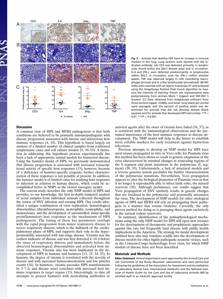
analyzed, host responses were elevated primarily in lung samples(Fig. 5), coinciding with the pathological abnormalities noted inthese animals and the clinical presentation of HPS. CD3 antigenwas increased significantly in lung specimens collected fromNHPs that developed HPS and was detected primarily in lym-phocytes found within bronchiolar-associated lymphoid tissue(BALT) and in pulmonary circulation (Fig. 5 A and B). IL-4
and IFN-γ also were detected in lymphocytes of BALT andpulmonary circulation, and medium-sized lymphocytes werefound within the alveolar spaces. Both antigens were increaseddramatically (2.5- and sixfold, respectively) in diseased animals(Fig. 5C). TNF was detected in alveolar macrophages and fewerlymphocytes and was increased nearly threefold in animals thatdeveloped HPS. Heart, kidney, spleen, and liver samples from asubset of NHPs were evaluated similarly, and no distinguishabledifferences were observed between animals that developed HPSand control animals.
Genetic Analysis of SNV. To determine the genetic differencesbetween the seemingly attenuated VA-SNV and the pathogenicDM-SNV, viruses were sequenced from cell culture supernatantor directly out of sera from an NHP that developed HPS usingnext-generation sequencing (NGS) technologies. Samples frommultiple NHPs that developed HPS were processed, but onlysera from a single animal yielded RNA of sufficient quality/quantityfor NGS. Interestingly, the viruses that were sequenced differedby only two nucleotides, both of which were in the polymerase-coding region. Specifically, Vero propagation resulted in nucle-otide changes at positions 166 (A to G), which resulted in anamino acid change (Lys to Glu), and 5364 (G to A), which wassilent (GenBank accession IDs KF537001–KF537006).
Fig. 3. Animals that develop HPS mount a robust immune response at thetime of disease. PBMCs isolated from macaques at 1, 12, and 20 dpi (forsurvivors) or at the time of euthanasia were stained with panels of anti-bodies to distinguish cell populations. (A) The ratio of CD4+ and CD8+ T cells(CD3+ cells) decreased in animals that developed HPS. (B) Memory cells weredetermined by their CD28 and CD95 expression on CD8+ T cells (CD3+) at theterminal stages of disease (or at 20 dpi for survivors). Animals that de-veloped HPS had an increase in memory (effector and/or memory) cell per-centages. (C–H) Cells were stained with the indicated antibodies (y axis), andpercentages of cells expressing markers are shown. (E–G) PBMCs werestimulated with phorbol12-myristate13-acetate and ionomycin for 6 h, withthe addition of brefeldin A for the last 4.5 h, and were stained with theindicated antibodies. Cytokine expression was increased in PBMCs fromanimals that developed HPS. *P < 0.05, **P < 0.01, ***P < 0.0.001.
Fig. 4. Animals that develop HPS have increased concentrations of immunemediators in serum. Concentrations of cytokines and chemokines were de-termined in serum samples collected from macaques at 1, 12, and 20 dpi (forsurvivors) or at the time of euthanasia using a multiplex NHP cytokine de-tection kit. At the time of euthanasia, animals that developed HPS hadsignificantly increased levels of several immune mediators, including proin-flammatory Th1 and Th2 cytokines (IFN-γ, IL-1β, IL-6, IL-18, and IL-13); theanti-inflammatory IL1 receptor antagonist (IL-1ra); and IL-15 and GM-CSF.*P < 0.05, **P < 0.01, ***P < 0.0.001.
Safronetz et al. PNAS | May 13, 2014 | vol. 111 | no. 19 | 7117
MICRO
BIOLO
GY
DiscussionA common view of HPS and HFRS pathogenesis is that bothconditions are believed to be primarily immunopathogenic withdisease progression associated with intense and deleterious hostimmune responses (4, 10). This hypothesis is based largely onanalysis of a limited number of clinical samples from confirmedsymptomatic cases and cell culture models (9, 30–33). A limita-tion in addressing this hypothesis process experimentally hasbeen a lack of appropriate animal models for hantaviral disease.Using the hamster model of HPS, we previously demonstratedthat disease progression is associated with increased transcrip-tional activity of specific host responses (13); however, becauseof a deficiency of hamster-specific reagents, further character-ization of these responses is not possible at present. In addition,the hamster model is of limited value for studying host responsesto infection in relation to human disease, which could be ac-complished better in NHPs as the closest surrogate model.The current study describes the only NHP model of HPS and
provides, to our knowledge, the first in-depth temporal analysisof serial samples from individual animals collected throughoutthe course of SNV infection and ensuing HPS. Our results iden-tified a unique combination of virus replication, hematologicalabnormalities (thrombocytopenia, neutrophilia, eosinophilia, andmonocytosis), and the development of uncontrolled, tissue-specificproinflammatory host responses as the mechanisms of HPSpathogenesis. The timing of these key parameters overlapswith the rapid progression from minor respiratory disease tosevere respiratory distress, which is the hallmark of the cardio-pulmonary phase of HPS, and supports their role in the hyper-permeability associated with late-stage disease. Viremia was theearliest indicator of disease and was first detected 4–10 d beforethe onset of respiratory distress and immediately before theobserved hematological abnormalities and activated host im-mune responses. Viremia also has been implicated in the de-velopment of HPS in humans and hamsters (13, 25, 34, 35). Inhumans, the degree of viremia is correlated with the severity ofdisease and with increased hemoconcentration and low plateletcounts (34). In hamsters, viremia precedes HPS manifestationsby 5–7 d, and disease onset correlates with increased host im-mune responses in target organs (13). Interestingly, to date allattempts to protect hamsters with neutralizing antibodies or
antiviral agents after the onset of viremia have failed (36, 37), asis consistent with the immunological observations and the pre-sumed importance of the host immune responses in disease de-velopment. The NHP model will help in the future to establishmore reliable markers for early treatment against hantavirusinfections.Previous attempts to develop an NHP model for HPS have
used viruses propagated in vitro in Vero cells; for Puumala virusthis method has been shown to result in genetic adaptation of thevirus characterized by minimal changes in noncoding regions ofthe S segment and point mutations in the polymerase (L seg-ment) (38, 39). Unfortunately, a lack of structural models anda reverse genetics system precludes the further characterizationof the polymerase mutations. Nevertheless, Vero propagationappears to alter the biological properties of Puumala virus in thatit no longer is reproducibly able to infect bank voles, the naturalreservoir (38). Although preliminary, our results suggest thatVero propagation of SNV similarly results in genetic changesthat are localized in the polymerase and potentially attenuatesthe virus. The development of NHP models for other etiologicalagents of HPS and HFRS will rely on propagating these patho-gens in a manner that retains virulence. Currently, the onlyproven method for doing so is passaging these agents exclusivelyin the natural rodent reservoirs.In summary, identification of the pathophysiological mecha-
nisms using the only NHP model for HPS will open new avenuesfor the development of therapeutic and prophylactic approachesagainst this rare but frequently fatal disease with public healthimplications in the Americas. The strategy for model developmentoutlined here also may translate readily into the development ofdisease models for other highly pathogenic zoonotic viruses, suchas the Crimean-Congo hemorrhagic fever virus, for which NHPmodels of disease have not been described.
Materials and MethodsEthics Statement.Animal experiments were approved by the Animal Care andUse Committee of the Rocky Mountain Laboratories and were performedfollowing the guidelines of the Association for Assessment and Accreditationof Laboratory Animal Care, International (AAALAC) and the National Insti-tutes of Health Guide for the Care and Use of Laboratory Animals (40) bycertified staff in an AAALAC approved facility.
Fig. 5. Animals that develop HPS have an increase in immunemarkers in the lung. Lung sections were stained with the in-dicated antibody. (A) CD3 was detected primarily in lympho-cytes found within the BALT (boxed area) and in circulation.IL-4 and IFN-γ were detected predominantly in lymphocyteswithin BALT, in circulation, and—for IFN-γ—within alveolarspaces. TNF was observed largely in cells resembling macro-phages (arrows) and in a few lymphocytes (arrowhead). (B) IHCslides were scanned with an Aperio ScanScope XT and analyzedusing the ImageScope Positive Pixel Count algorithm to mea-sure the intensity of staining. Shown are representative datapostprocessing from animals Mock 1 (Upper) and DM-SNV 5(Lower). (C) Data obtained from ImageScope software fromthree sections (upper, middle, and lower lung lobes) per animalwere averaged, and the percent of positive pixels was de-termined for animals that did not develop disease (blacksquares) and for animals that developed HPS (red circles). **P <0.01, ***P < 0.0.001.
7118 | www.pnas.org/cgi/doi/10.1073/pnas.1401998111 Safronetz et al.

Animal Infection. Seventeen rhesus macaques (Macaca mulatta, 3.3–6.7 kg)were exposed to lung homogenates (10% wt/vol) from naive (uninfected)deer mice (n = 3) or from deer mice infected with DM-SNV (n = 10), or VA-SNV(n = 4) using an established protocol of simultaneous installation (20). Thechallenge dose was 6 × 10⁶ focus-forming units (FFU) for VA-SNV or theequivalent to 6 × 10⁶ FFU for DM-SNV. Briefly, the DM-SNV inoculum wasstandardized to the VA-SNV based on real-time qRT-PCR analysis of S ge-nomic segment copies. Mock-infected animals received a similar amount ofnaive lung homogenates. For detailed information on inoculum prepara-tion, clinical scoring, and sample collection, see SI Materials and Methods.
Hematology, Serum Biochemistry, and Coagulation Parameters. Hematologywas performed on EDTA blood with the HemaVet 950FS+ hematology an-alyzer (Drew Scientific). Serum biochemistries were measured using a PiccoloBlood Analyzer (Abaxis). Coagulation parameters were measured in plasmaon a STart4 instrument (Diagnostica Stago). For detailed information, see SIMaterials and Methods.
Virus Detection and Isolation. Clinical specimens were screened for thepresence of SNV RNA as previously described (19). A subset of RT-PCR–positivetissues were homogenized and used to infect triplicate monolayers ofVero cells. Cells were incubated for 14 d and scored for cytopathic effect.
Histopathology, IHC, and in Situ ISH. Tissues were stained with H&E or phos-photungstic acid-hematoxylin using standard methods. Viral antigen andRNA was detected by IHC or ISH as previously described (41–43). CD3, IL-4,TNF, and INF-γ were stained in selected tissue specimens using standard IHCmethodologies. Sections were scanned with an Aperio ScanScope XT (Aperio
Technologies, Inc.) and quantified using the ImageScope Positive Pixel Countalgorithm (version 9.1). For detailed information, see SI Materials and Methods.
Serum Cytokines and Chemokines. Cytokines and chemokines were measuredin serum samples using a NHP Cytokine kit (Millipore). Data were analyzedusing a two-way ANOVA with a Bonferroni posttest.
Polychromatic Flow Cytometric Analysis of PBMCs. PBMCs were stained forCD3, CD8α, Ki67, CD69, TNF, IFN-γ, Granzyme B, CD28, CD95, CD14, or CD4.Polychromatic analysis was performed using a LSR II flow cytometer equip-ped with FACS DiVA software (BD), and data were analyzed using FlowJosoftware (TreeStar). For detailed information, see SI Materials and Methods.
Serology. Sera collected from NHPs prechallenge and at the time of necropsywere tested for the presence of hantavirus-specific IgG antibodies by stan-dard ELISA methodologies and using a bacterially expressed, recombinantSNV N protein as antigen, essentially as previously described (44).
NGS.Genomic sequences of VA-SNV andDM-SNVwere determined using NGStechnology. For detailed information, see SI Materials and Methods.
ACKNOWLEDGMENTS. We thank Edward Schreckendgust, Rocky Rivera,Sandy Skorupa, Kathleen Meuchel, Jayne Faris, Amanda Weidow, RachaelLaCasse, Kimberly Meade-White, Tina Thomas, Dan Long, Shelly Robertson,Aaron Carmody, Sarah L. Anzick, Stacy M. Ricklefs, and Dan Bruno for scien-tific discussions and technical assistance and Anita Mora and Heather Murphyfor preparing the graphics. This study was financially supported by the Divisionof Intramural Research, National Institute of Allergy and Infectious Diseases,National Institutes of Health.
1. Duchin JS, et al.; The Hantavirus Study Group (1994) Hantavirus pulmonary syndrome:A clinical description of 17 patients with a newly recognized disease. N Engl J Med330(14):949–955.
2. Nichol ST, et al. (1993) Genetic identification of a hantavirus associated with anoutbreak of acute respiratory illness. Science 262(5135):914–917.
3. Macneil A, Nichol ST, Spiropoulou CF (2011) Hantavirus pulmonary syndrome. VirusRes 162(1-2):138–147.
4. Jonsson CB, Figueiredo LT, Vapalahti O (2010) A global perspective on hantavirusecology, epidemiology, and disease. Clin Microbiol Rev 23(2):412–441.
5. Centers for Disease Control and Prevention (CDC) (2012) Hantavirus pulmonary syn-drome in visitors to a national park—Yosemite Valley, California, 2012. MMWR MorbMortal Wkly Rep 61(46):952.
6. Webster D, et al. (2007) Cluster of cases of hantavirus pulmonary syndrome in Alberta,Canada. Am J Trop Med Hyg 77(5):914–918.
7. MacNeil A, Ksiazek TG, Rollin PE (2011) Hantavirus pulmonary syndrome, UnitedStates, 1993-2009. Emerg Infect Dis 17(7):1195–1201.
8. Roehr B (2012) US officials warn 39 countries about risk of hantavirus among trav-ellers to Yosemite. BMJ 345:e6054.
9. Borges AA, et al. (2006) Hantavirus cardiopulmonary syndrome: Immune responseand pathogenesis. Microbes Infect 8(8):2324–2330.
10. Mackow ER, Gavrilovskaya IN (2009) Hantavirus regulation of endothelial cell func-tions. Thromb Haemost 102(6):1030–1041.
11. Hooper JW, Larsen T, Custer DM, Schmaljohn CS (2001) A lethal disease model forhantavirus pulmonary syndrome. Virology 289(1):6–14.
12. Safronetz D, Ebihara H, Feldmann H, Hooper JW (2012) The Syrian hamster model ofhantavirus pulmonary syndrome. Antiviral Res 95(3):282–292.
13. Safronetz D, et al. (2011) Pathogenesis and host response in Syrian hamsters fol-lowing intranasal infection with Andes virus. PLoS Pathog 7(12):e1002426.
14. Yanagihara R, et al. (1988) Experimental hantavirus infection in nonhuman primates.Arch Virol 101(1-2):125–130.
15. McElroy AK, Bray M, Reed DS, Schmaljohn CS (2002) Andes virus infection of cyn-omolgus macaques. J Infect Dis 186(12):1706–1712.
16. Groen J, et al. (1995) A macaque model for hantavirus infection. J Infect Dis 172(1):38–44.
17. Klingström J, Plyusnin A, Vaheri A, Lundkvist A (2002) Wild-type Puumala hantavirusinfection induces cytokines, C-reactive protein, creatinine, and nitric oxide in cyn-omolgus macaques. J Virol 76(1):444–449.
18. Sironen T, et al. (2008) Pathology of Puumala hantavirus infection in macaques. PLoSONE 3(8):e3035.
19. Botten J, et al. (2000) Experimental infection model for Sin Nombre hantavirus in thedeer mouse (Peromyscus maniculatus). Proc Natl Acad Sci USA 97(19):10578–10583.
20. Kobasa D, et al. (2007) Aberrant innate immune response in lethal infection ofmacaques with the 1918 influenza virus. Nature 445(7125):319–323.
21. Young JC, et al. (2000) The incubation period of hantavirus pulmonary syndrome. AmJ Trop Med Hyg 62(6):714–717.
22. Vial PA, et al. (2006) Incubation period of hantavirus cardiopulmonary syndrome.Emerg Infect Dis 12(8):1271–1273.
23. Safronetz D, et al. (2011) Pandemic swine-origin H1N1 influenza A virus isolates showheterogeneous virulence in macaques. J Virol 85(3):1214–1223.
24. Zaki SR, et al. (1995) Hantavirus pulmonary syndrome. Pathogenesis of an emerginginfectious disease. Am J Pathol 146(3):552–579.
25. Wahl-Jensen V, et al. (2007) Temporal analysis of Andes virus and Sin Nombre virusinfections of Syrian hamsters. J Virol 81(14):7449–7462.
26. Testi R, D’Ambrosio D, De Maria R, Santoni A (1994) The CD69 receptor: A multi-purpose cell-surface trigger for hematopoietic cells. Immunol Today 15(10):479–483.
27. Shedlock DJ, et al. (2010) Ki-67 staining for determination of rhesus macaque T cellproliferative responses ex vivo. Cytometry A 77(3):275–284.
28. Mertz GJ, et al. (2006) Diagnosis and treatment of new world hantavirus infections.Curr Opin Infect Dis 19(5):437–442.
29. Lafont BA, Gloeckler L, D’Hautcourt JL, Gut JP, Aubertin AM (2000) One-round de-termination of seven leukocyte subsets in rhesus macaque blood by flow cytometry.Cytometry 41(3):193–202.
30. Borges AA, et al. (2008) Role of mixed Th1 and Th2 serum cytokines on pathogenesisand prognosis of hantavirus pulmonary syndrome.Microbes Infect 10(10-11):1150–1157.
31. Geimonen E, et al. (2002) Pathogenic and nonpathogenic hantaviruses differentiallyregulate endothelial cell responses. Proc Natl Acad Sci USA 99(21):13837–13842.
32. Mori M, et al. (1999) High levels of cytokine-producing cells in the lung tissues ofpatients with fatal hantavirus pulmonary syndrome. J Infect Dis 179(2):295–302.
33. Spiropoulou CF, Albariño CG, Ksiazek TG, Rollin PE (2007) Andes and Prospect Hillhantaviruses differ in early induction of interferon although both can downregulateinterferon signaling. J Virol 81(6):2769–2776.
34. Terajima M, et al. (1999) High levels of viremia in patients with the Hantavirus pul-monary syndrome. J Infect Dis 180(6):2030–2034.
35. Xiao R, et al. (2006) Sin Nombre viral RNA load in patients with hantavirus cardio-pulmonary syndrome. J Infect Dis 194(10):1403–1409.
36. Safronetz D, Haddock E, Feldmann F, Ebihara H, Feldmann H (2011) In vitro and invivo activity of ribavirin against Andes virus infection. PLoS ONE 6(8):e23560.
37. Custer DM, Thompson E, Schmaljohn CS, Ksiazek TG, Hooper JW (2003) Active andpassive vaccination against hantavirus pulmonary syndrome with Andes virus M ge-nome segment-based DNA vaccine. J Virol 77(18):9894–9905.
38. Lundkvist A, et al. (1997) Cell culture adaptation of Puumala hantavirus changes theinfectivity for its natural reservoir, Clethrionomys glareolus, and leads to accumula-tion of mutants with altered genomic RNA S segment. J Virol 71(12):9515–9523.
39. Nemirov K, Lundkvist A, Vaheri A, Plyusnin A (2003) Adaptation of Puumala hanta-virus to cell culture is associated with point mutations in the coding region of theL segment and in the noncoding regions of the S segment. J Virol 77(16):8793–8800.
40. Committee on Care and Use of Laboratory Animals (1985) Guide for the Care and Useof Laboratory Animals (Natl Inst Health, Bethesda), DHHS Publ No (NIH) 85-23.
41. Medina RA, Mirowsky-Garcia K, Hutt J, Hjelle B (2007) Ribavirin, human convalescentplasma and anti-beta3 integrin antibody inhibit infection by Sin Nombre virus in thedeer mouse model. J Gen Virol 88(Pt 2):493–505.
42. Safronetz D, et al. (2013) Hamster-adapted Sin Nombre virus causes disseminatedinfection and efficiently replicates in pulmonary endothelial cells without signs ofdisease. J Virol 87(8):4778–4782.
43. Wang F, et al. (2012) RNAscope: A novel in situ RNA analysis platform for formalin-fixed, paraffin-embedded tissues. J Mol Diagn 14(1):22–29.
44. Feldmann H, et al. (1993) Utilization of autopsy RNA for the synthesis of the nucle-ocapsid antigen of a newly recognized virus associated with hantavirus pulmonarysyndrome. Virus Res 30(3):351–367.
Safronetz et al. PNAS | May 13, 2014 | vol. 111 | no. 19 | 7119
MICRO
BIOLO
GY