pathophysiology small airways disease of small... · pathophysiology of small airways disease...
TRANSCRIPT

Pathophysiology of small airways disease
Antoine Magnang
Nantes UniversityService de Pneumologie
l f l d’ ll lPlate‐forme Transversale d’AllergologieUMR 915, équipe avenir Bronchial diseases and allergies
antoine magnan@inserm [email protected]

DisclosureDisclosure
• Investigator in clinical trials:
GSK, Boehringer, Novartis
• Consultancy advisory boards• Consultancy, advisory boards:
Novartis, MSD, Astra‐Zeneca
ALK, Teva
• Symposia:
ll è h l h hALK, Stallergènes, Novartis, Schering‐Plough, MSD, Chiesi

Small airways: what are we dealing with ?
Diameter
Divisions
1 Trachea
10 mm
3 mm
234
BronchiB
ronc
hi
3 mm 4
5 Bronchioles
B
1,5 mm 8
nchi
oles
16
17 - 19
Terminal Bronchioles
Respiratory
Bro
bule 9
20 - 22
23
Respiratory Bronchioles
Alveolar ducts
Al l i
Lob
Aci
nus
23 Alveolar airspaces
50 to 100 cm²

Small airways• Small but complete
– Epithelium– Smooth muscle
• Difficult to explore: – Lung tissue from cases of fatal asthma– Surgical specimens
Transbronchial biopsies Mauad T, J Allergy Clin Immunol – Transbronchial biopsies– Broncho‐alveolar lavages
gy2007;120:997‐1009

Inflammation in asthmatic small airwaysInflammation in asthmatic small airways
Mauad T, J Allergy Clin Immunol 2007;120:997‐1009
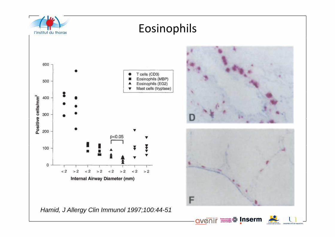
Eosinophils
Hamid, J Allergy Clin Immunol 1997;100:44-51

T cells
CD 45 stainingO id h b hiOutside the bronchi
Haley KJ, AJRCCM 1998, 158: 565

Th2 cells
Minshall, J Allergy Clin Immunol 1998;101:386-90

Mast cellsMast cells
Mast cells
FcRI + Mast cells
Andersson, J Allergy Clin Immunol 2011;127:905-12
FcRI + Mast cells

Mast cellsCentral airways Alveolar parenchyma
Mast cells
Andersson, J Allergy Clin Immunol 2011;127:905-12

Inflammatory cells present in asthmatic small airways
• EosinophilsEosinophils– More in fatal asthma compared with patients died from other
cause– Greater proportion of activated Eosinophils in distal airways
Hamid, J Allergy Clin Immunol 1997;100:44‐51
• T cells• T cells– More IL5 m RNA expression in distal airways
Minshall, J Allergy Clin Immunol 1998;101:386‐90
• Mast cells– Extend to alveolar parenchyma in uncontrolled patients– Correlated with FcRI staining
Andersson, J Allergy Clin Immunol 2011;127:905‐12Den Otter, Clin Exp Allergy 2010, 40: 1473‐1481p gy

Remodeling in small airways
• Mucus plugging• Epithelial detachment• Epithelial detachment• Subepithelial fibrosis• Smooth muscle hypertrophy• Smooth muscle hypertrophy
Contoli Allergy 2010; 65: 141‐151Contoli, Allergy 2010; 65: 141 151
• Decreased elastic fiber content
d d lMauad Am J Respir Crit Care Med Vol 170. pp 857–862, 2004

Remodeling in small airways
Collagen 1
Dolhnikoff M et al, JACI 2009

T b hi l bi i i l hTransbronchial biopsies in nocturnal asthma• Alveolar eosinophils increased at night and correlated p g
with lung functionKraft M Am J Respir Crit Care Med 1996, 154: 1505
l l ll l d i h i hil d l• Alveolar CD4+ cells correlated with eosinophils and lung function
Kraft M Am J Respir Crit Care Med 1999 159: 228–234Kraft M Am J Respir Crit Care Med 1999, 159: 228 234

Small airways disease in severe asthma
Wenzel S, AJRCCM 1997
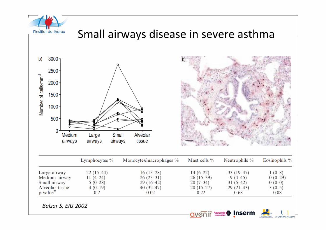
Small airways disease in severe asthmaSmall airways disease in severe asthma
Balzar S, ERJ 2002

Small airways disease in severe asthmay
Balzar, Am J Respir Crit Care Med 2005: 171: 431–439

S ll i di i hSmall airways disease in severe asthma• Studies of BAL, surgical and transbronchial biopsiesStudies of A , surgical and transbronchial biopsies• Small airways inflammation characterized by the presence
of neutrophils and mast cellsp
Balzar, Am J Respir Crit Care Med 2005: 171: 431–439

Small airways involvement in asthma: a distinct phenotype ?distinct phenotype ?
• Asthma : heterogeneous disease with various clinical
expression, inflammatory and functional patterns
– Asthma phenotypes described for better care– Asthma phenotypes described for better care
• Atopic / non atopic
• Early onset / late onset• Early onset / late onset
• Eosinophilic / neutrophilic
• Obese / non obeseObese / non obese
• …
– Asthma phenotypes described from response to biotherapiesAsthma phenotypes described from response to biotherapies
• Anti‐IgE
• Anti‐ IL‐5Anti IL 5
• …

Do small airways involvement define h ?
• NO: Small airways studies relate to known asthma
a new phenotype ?NO: Small airways studies relate to known asthma phenotypes:– Neutrophilic severe asthma– Asthma with remodeling– …
• BUT:• BUT: – Small airways inflammation most variable from a patient to
another – Irregular response to extrafine formulations of inhaled steroids
or/and LABA
A t f di ti t ll i thAssessment of a distinct « small airways asthma » supposes prospective large studies of distal samples…

Conclusions• Small airways (< 2 mm) disease represent a variable but
frequently major part of asthma pathophysiology with:frequently major part of asthma pathophysiology with:– Inflammatory cell infiltrate:
• Eosinophils (more activated in small vs large airways)• T cells (with high IL‐5 production)• Mast cells (higher proportion in small vs large airways, and severe asthma))
• Neutrophils (especially in severe asthma)
– Remodeling:S b i h li l fib i• Subepithelial fibrosis
• Smooth muscle hypertrophy• Mucus plugging
• Pathophysiology of small airways disease– is still difficult to document – could define a distinct phenotype of asthma