patient-derived hipsc neurons with heterozygous cntnap2 ... · doherty, j. l. & owen, m. j....
TRANSCRIPT

BRIEF COMMUNICATION OPEN
Patient-derived hiPSC neurons with heterozygous CNTNAP2deletions display altered neuronal gene expression and networkactivityErin Flaherty1,2, Rania M. Deranieh3, Elena Artimovich3, Inkyu S. Lee1,2, Arthur J. Siegel4, Deborah L. Levy5, Michael W. Nestor3 andKristen J. Brennand 1,2,6
Variants in CNTNAP2, a member of the neurexin family of genes that function as cell adhesion molecules, have been associated withmultiple neuropsychiatric conditions such as schizophrenia, autism spectrum disorder and intellectual disability; animal studiesindicate a role for CNTNAP2 in axon guidance, dendritic arborization and synaptogenesis. We previously reprogrammed fibroblastsfrom a family trio consisting of two carriers of heterozygous intragenic CNTNAP2 deletions into human induced pluripotent stemcells (hiPSCs) and described decreased migration in the neural progenitor cells (NPCs) differentiated from the affected CNTNAP2carrier in this trio. Here, we report the effect of this heterozygous intragenic deletion in CNTNAP2 on global gene expression andneuronal activity in the same cohort. Our findings suggest that heterozygous CNTNAP2 deletions affect genes involved in neuronaldevelopment and neuronal activity; however, these data reflect only one family trio and therefore more deletion carriers, with avariety of genetic backgrounds, will be needed to understand the molecular mechanisms underlying CNTNAP2 deletions.
npj Schizophrenia (2017) 3:35 ; doi:10.1038/s41537-017-0033-5
SHORT REPORTThe shared genetic architecture underlying neuropsychiatricdisorders implicates common molecular mechanisms.1 For exam-ple, while homozygous null mutations in CNTNAP2 lead to corticaldysplasia-focal epilepsy syndrome,2, 3 heterozygous intragenicdeletions are associated with schizophrenia, intellectual disability,language deficits, seizures, and autism traits.4 Critically, CNTNAP2variants are not completely penetrant.2, 5 Animal studies indicate arole for CNTNAP2 in axon guidance, dendritic arborization, andsynaptogenesis.6–8
We obtained fibroblast samples from a family trio with twocarriers of heterozygous intragenic CNTNAP2 deletions, oneaffected and one unaffected, and an unaffected non-carriercontrol (Table 1). The CNTNAP2 carriers display discordant clinicalphenotypes; the daughter (DL7078) presented with schizo-affective disorder (depressed subtype) while the father (DL8735)was neurotypical.9 We previously used sendai viral vectors toreprogram fibroblasts from this trio into hiPSCs that were thendifferentiated via dual-SMAD induction into NPCs and neurons.We characterized decreased migration in NPCs and allele-biasedexpression of the mutant CNTNAP2 transcript by qPCR in neuronsfrom the affected CNTNAP2 carrier in this trio.9 Here, we report theeffect of this heterozygous intragenic deletion in CNTNAP2 onglobal gene expression and neuronal activity in this same cohort.CNTNAP2 is highly expressed in Ngn2-induced neurons, a
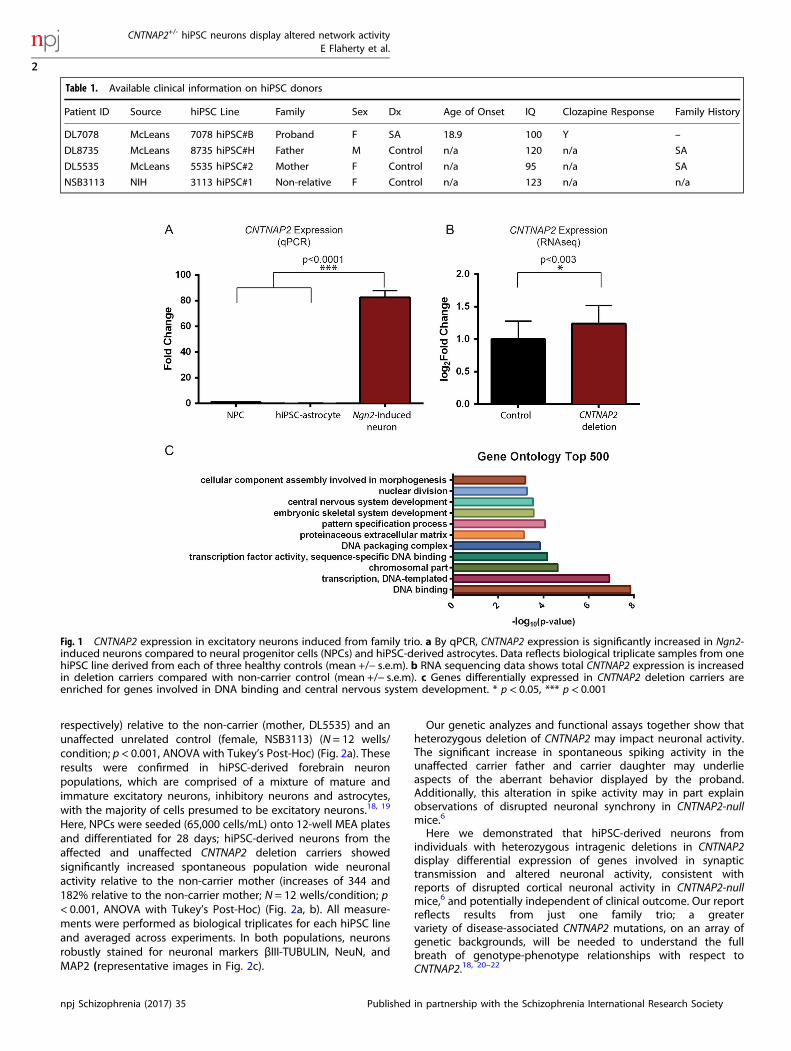
population of nearly pure excitatory neurons,10 relative to hiPSC-derived NPCs11 and hiPSC-astrocytes12 (qPCR FC = 82.5, p < 0.0001,ANOVA with Tukey’s Post-Hoc) (Fig. 1a). RNA was harvested after21 days of Ngn2-induction. The New York Genome Center
prepared RNAseq libraries using the Kapa Total 350 bp kit,followed by 2 × 125 bp Illumina RNA sequencing to a read depthof 40 M reads per sample on the HiSeq 2500.We queried the expression of CNTNAP2 in Ngn2-induced
neurons from each member of this family trio, hypothesizing thatheterozygous intragenic deletions may affect the expression ofCNTNAP2. Surprisingly, overall CNTNAP2 expression was increasedin the CNTNAP2 deletion carriers compared with the non-carriermother (log2FC = 1.24, padj = 0.003) (Fig. 1b).Differential expression analysis was performed using DESeq213
and the top 500 differentially expressed genes were used toperform gene ontology using DAVID14, 15 (SI Table 1). The mostsignificant subset of genes mapped to terms relating to DNAbinding and central nervous system (CNS) development (FC = 1.8,p < 0.00001 and FC = 1.9, p = 0.0003) (Fig. 1c; SI Table 2). Within thegene subset involved in CNS development, there are someinteresting candidate genes such as CNTN6 and CNTN4, which areinvolved in regulating cell surface interactions during nervoussystem development and are also thought to be important insynaptogenesis (SI Table 3).Given the differences in gene expression of critical neuronal
and synaptic genes, we applied an Axion multi-electrode array(MEA) (see similar applications to Amyotrophic Lateral Sclerosis16
and Parkinson’s disease17) to record population-wide neuronalactivity under conditions similar to those used in our RNAseqanalyzes. 21-day-old Ngn2-induced neurons from both theaffected (daughter, DL7078) and unaffected (father, DL8735)CNTNAP2 deletion carriers showed significantly increased sponta-neous network level activity (an increase of 210 and 253%,
Received: 23 December 2016 Revised: 4 August 2017 Accepted: 11 August 2017
1Departments of Neuroscience, Icahn School of Medicine at Mount Sinai, New York, NY 10029, USA; 2Friedman Brain Institute, Icahn School of Medicine at Mount Sinai, New York,NY 10029, USA; 3Hussman Institute for Autism, 801W. Baltimore St., Baltimore, MD 21201, USA; 4Internal Medicine Department, McLean Hospital, Belmont, MA 02478, USA;5Psychology Research Laboratory, McLean Hospital, Belmont, MA 02478, USA and 6Departments of Psychiatry, Icahn School of Medicine at Mount Sinai, New York, NY 10029, USACorrespondence: Michael W. Nestor ([email protected]) or Kristen J. Brennand ([email protected])
www.nature.com/npjschz
Published in partnership with the Schizophrenia International Research Society
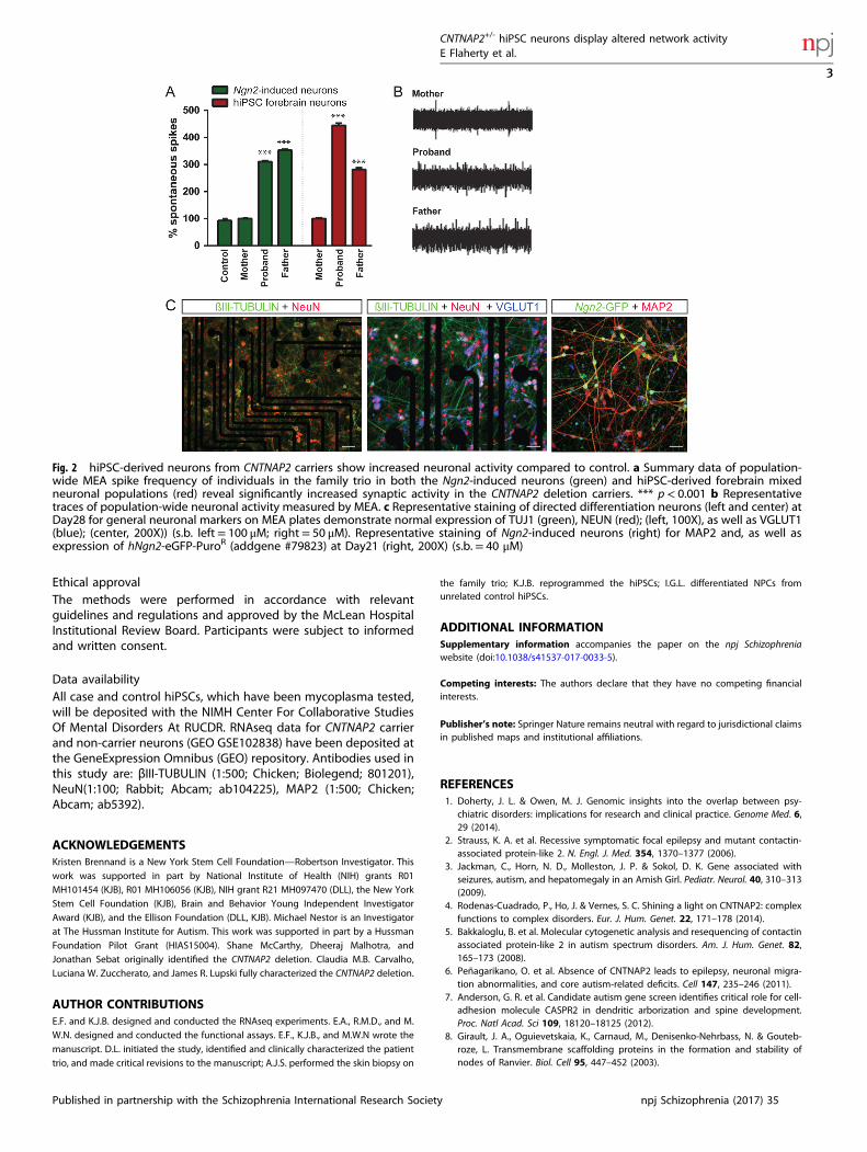
respectively) relative to the non-carrier (mother, DL5535) and anunaffected unrelated control (female, NSB3113) (N = 12 wells/condition; p < 0.001, ANOVA with Tukey’s Post-Hoc) (Fig. 2a). Theseresults were confirmed in hiPSC-derived forebrain neuronpopulations, which are comprised of a mixture of mature andimmature excitatory neurons, inhibitory neurons and astrocytes,with the majority of cells presumed to be excitatory neurons.18, 19
Here, NPCs were seeded (65,000 cells/mL) onto 12-well MEA platesand differentiated for 28 days; hiPSC-derived neurons from theaffected and unaffected CNTNAP2 deletion carriers showedsignificantly increased spontaneous population wide neuronalactivity relative to the non-carrier mother (increases of 344 and182% relative to the non-carrier mother; N = 12 wells/condition; p< 0.001, ANOVA with Tukey’s Post-Hoc) (Fig. 2a, b). All measure-ments were performed as biological triplicates for each hiPSC lineand averaged across experiments. In both populations, neuronsrobustly stained for neuronal markers βIII-TUBULIN, NeuN, andMAP2 (representative images in Fig. 2c).
Our genetic analyzes and functional assays together show thatheterozygous deletion of CNTNAP2 may impact neuronal activity.The significant increase in spontaneous spiking activity in theunaffected carrier father and carrier daughter may underlieaspects of the aberrant behavior displayed by the proband.Additionally, this alteration in spike activity may in part explainobservations of disrupted neuronal synchrony in CNTNAP2-nullmice.6
Here we demonstrated that hiPSC-derived neurons fromindividuals with heterozygous intragenic deletions in CNTNAP2display differential expression of genes involved in synaptictransmission and altered neuronal activity, consistent withreports of disrupted cortical neuronal activity in CNTNAP2-nullmice,6 and potentially independent of clinical outcome. Our reportreflects results from just one family trio; a greatervariety of disease-associated CNTNAP2 mutations, on an array ofgenetic backgrounds, will be needed to understand the fullbreath of genotype-phenotype relationships with respect toCNTNAP2.18, 20–22
Table 1. Available clinical information on hiPSC donors
Patient ID Source hiPSC Line Family Sex Dx Age of Onset IQ Clozapine Response Family History
DL7078 McLeans 7078 hiPSC#B Proband F SA 18.9 100 Y –
DL8735 McLeans 8735 hiPSC#H Father M Control n/a 120 n/a SA
DL5535 McLeans 5535 hiPSC#2 Mother F Control n/a 95 n/a SA
NSB3113 NIH 3113 hiPSC#1 Non-relative F Control n/a 123 n/a n/a
Fig. 1 CNTNAP2 expression in excitatory neurons induced from family trio. a By qPCR, CNTNAP2 expression is significantly increased in Ngn2-induced neurons compared to neural progenitor cells (NPCs) and hiPSC-derived astrocytes. Data reflects biological triplicate samples from onehiPSC line derived from each of three healthy controls (mean +/− s.e.m). b RNA sequencing data shows total CNTNAP2 expression is increasedin deletion carriers compared with non-carrier control (mean +/− s.e.m). c Genes differentially expressed in CNTNAP2 deletion carriers areenriched for genes involved in DNA binding and central nervous system development. * p< 0.05, *** p< 0.001
CNTNAP2+/- hiPSC neurons display altered network activityE Flaherty et al.
2
npj Schizophrenia (2017) 35 Published in partnership with the Schizophrenia International Research Society
1234567890
Ethical approvalThe methods were performed in accordance with relevantguidelines and regulations and approved by the McLean HospitalInstitutional Review Board. Participants were subject to informedand written consent.
Data availabilityAll case and control hiPSCs, which have been mycoplasma tested,will be deposited with the NIMH Center For Collaborative StudiesOf Mental Disorders At RUCDR. RNAseq data for CNTNAP2 carrierand non-carrier neurons (GEO GSE102838) have been deposited atthe GeneExpression Omnibus (GEO) repository. Antibodies used inthis study are: βIII-TUBULIN (1:500; Chicken; Biolegend; 801201),NeuN(1:100; Rabbit; Abcam; ab104225), MAP2 (1:500; Chicken;Abcam; ab5392).
ACKNOWLEDGEMENTSKristen Brennand is a New York Stem Cell Foundation—Robertson Investigator. Thiswork was supported in part by National Institute of Health (NIH) grants R01MH101454 (KJB), R01 MH106056 (KJB), NIH grant R21 MH097470 (DLL), the New YorkStem Cell Foundation (KJB), Brain and Behavior Young Independent InvestigatorAward (KJB), and the Ellison Foundation (DLL, KJB). Michael Nestor is an Investigatorat The Hussman Institute for Autism. This work was supported in part by a HussmanFoundation Pilot Grant (HIAS15004). Shane McCarthy, Dheeraj Malhotra, andJonathan Sebat originally identified the CNTNAP2 deletion. Claudia M.B. Carvalho,Luciana W. Zuccherato, and James R. Lupski fully characterized the CNTNAP2 deletion.
AUTHOR CONTRIBUTIONSE.F. and K.J.B. designed and conducted the RNAseq experiments. E.A., R.M.D., and M.W.N. designed and conducted the functional assays. E.F., K.J.B., and M.W.N wrote themanuscript. D.L. initiated the study, identified and clinically characterized the patienttrio, and made critical revisions to the manuscript; A.J.S. performed the skin biopsy on
the family trio; K.J.B. reprogrammed the hiPSCs; I.G.L. differentiated NPCs fromunrelated control hiPSCs.
ADDITIONAL INFORMATIONSupplementary information accompanies the paper on the npj Schizophreniawebsite (doi:10.1038/s41537-017-0033-5).
Competing interests: The authors declare that they have no competing financialinterests.
Publisher’s note: Springer Nature remains neutral with regard to jurisdictional claimsin published maps and institutional affiliations.
REFERENCES1. Doherty, J. L. & Owen, M. J. Genomic insights into the overlap between psy-
chiatric disorders: implications for research and clinical practice. Genome Med. 6,29 (2014).
2. Strauss, K. A. et al. Recessive symptomatic focal epilepsy and mutant contactin-associated protein-like 2. N. Engl. J. Med. 354, 1370–1377 (2006).
3. Jackman, C., Horn, N. D., Molleston, J. P. & Sokol, D. K. Gene associated withseizures, autism, and hepatomegaly in an Amish Girl. Pediatr. Neurol. 40, 310–313(2009).
4. Rodenas-Cuadrado, P., Ho, J. & Vernes, S. C. Shining a light on CNTNAP2: complexfunctions to complex disorders. Eur. J. Hum. Genet. 22, 171–178 (2014).
5. Bakkaloglu, B. et al. Molecular cytogenetic analysis and resequencing of contactinassociated protein-like 2 in autism spectrum disorders. Am. J. Hum. Genet. 82,165–173 (2008).
6. Peñagarikano, O. et al. Absence of CNTNAP2 leads to epilepsy, neuronal migra-tion abnormalities, and core autism-related deficits. Cell 147, 235–246 (2011).
7. Anderson, G. R. et al. Candidate autism gene screen identifies critical role for cell-adhesion molecule CASPR2 in dendritic arborization and spine development.Proc. Natl Acad. Sci 109, 18120–18125 (2012).
8. Girault, J. A., Oguievetskaia, K., Carnaud, M., Denisenko-Nehrbass, N. & Gouteb-roze, L. Transmembrane scaffolding proteins in the formation and stability ofnodes of Ranvier. Biol. Cell 95, 447–452 (2003).
Fig. 2 hiPSC-derived neurons from CNTNAP2 carriers show increased neuronal activity compared to control. a Summary data of population-wide MEA spike frequency of individuals in the family trio in both the Ngn2-induced neurons (green) and hiPSC-derived forebrain mixedneuronal populations (red) reveal significantly increased synaptic activity in the CNTNAP2 deletion carriers. *** p< 0.001 b Representativetraces of population-wide neuronal activity measured by MEA. c Representative staining of directed differentiation neurons (left and center) atDay28 for general neuronal markers on MEA plates demonstrate normal expression of TUJ1 (green), NEUN (red); (left, 100X), as well as VGLUT1(blue); (center, 200X)) (s.b. left = 100 μM; right= 50 μM). Representative staining of Ngn2-induced neurons (right) for MAP2 and, as well asexpression of hNgn2-eGFP-PuroR (addgene #79823) at Day21 (right, 200X) (s.b.= 40 μM)
CNTNAP2+/- hiPSC neurons display altered network activityE Flaherty et al.
3
Published in partnership with the Schizophrenia International Research Society npj Schizophrenia (2017) 35

9. Lee, I. S. et al. Characterization of molecular and cellular phenotypes associatedwith a heterozygous CNTNAP2 deletion using patient-derived hiPSC neural cells.npj Schizophr. 1, 15019 (2015).
10. Ho, S. M. et al. Rapid Ngn2-induction of excitatory neurons from hiPSC-derivedneural progenitor cells. Methods 101, 113–124 (2016).
11. Topol, A., Tran, N. N. & Brennand, K. J. A guide to generating and using hiPSCderived NPCs for the study of neurological diseases. J. Vis. Exp. JoVE 96, e52495(2015).
12. TCW, J. et al. An efficient platform for astrocyte differentiation from humaninduced pluripotent stem cells. Stem Cell Rep. doi:10.1016/j.stemcr.2017.06.018.(2017).
13. Love, M. I., Huber, W. & Anders, S. Moderated estimation of foldchange and dispersion for RNA-seq data with DESeq2. Genome Biol. 15, 550(2014).
14. Huang, D. W., Lempicki, Ra & Sherman, B. T. Systematic and integrative analysis oflarge gene lists using DAVID bioinformatics resources. Nat. Protoc. 4, 44–57(2009).
15. Huang, D. W., Sherman, B. T. & Lempicki, R. A. Bioinformatics enrichment tools:Paths toward the comprehensive functional analysis of large gene lists. NucleicAcids Res. 37, 1–13 (2009).
16. Wainger, B. J. et al. Intrinsic membrane hyperexcitability of amyotrophic lateralsclerosis patient-derived motor neurons. Cell Rep. 7, 1–11 (2014).
17. Woodard, C. M. et al. iPSC-derived dopamine neurons reveal differences betweenmonozygotic twins discordant for parkinson’s disease. Cell Rep. 9, 1173–1182(2014).
18. Brennand, K. J. et al. Modelling schizophrenia using human induced pluripotentstem cells. Nature 473, 221–225 (2011).
19. Brennand, K. et al. Phenotypic differences in hiPSC NPCs derived from patientswith schizophrenia. Mol. Psychiatry 20, 1–8 (2014).
20. Brennand, K. J., Landek-Salgado, M. A. & Sawa, A. Modeling heterogeneouspatients with a clinical diagnosis of Schizophrenia with induced pluripotent stemcells. Biol. Psychiatry 75, 936–944 (2014).
21. Shi, Y., Kirwan, P., Smith, J., Robinson, H. P. C. & Livesey, F. J. Human cerebralcortex development from pluripotent stem cells to functional excitatory synap-ses. Nat. Neurosci. 15, 477–486 (2012).
22. Prè, D. et al. A time course analysis of the electrophysiological properties ofneurons differentiated from human induced Pluripotent stem cells (iPSCs). PLoSONE 9, e103418 (2014).
Open Access This article is licensed under a Creative CommonsAttribution 4.0 International License, which permits use, sharing,
adaptation, distribution and reproduction in anymedium or format, as long as you giveappropriate credit to the original author(s) and the source, provide a link to the CreativeCommons license, and indicate if changes were made. The images or other third partymaterial in this article are included in the article’s Creative Commons license, unlessindicated otherwise in a credit line to the material. If material is not included in thearticle’s Creative Commons license and your intended use is not permitted by statutoryregulation or exceeds the permitted use, you will need to obtain permission directlyfrom the copyright holder. To view a copy of this license, visit http://creativecommons.org/licenses/by/4.0/.
© The Author(s) 2017
CNTNAP2+/- hiPSC neurons display altered network activityE Flaherty et al.
4
npj Schizophrenia (2017) 35 Published in partnership with the Schizophrenia International Research Society